MINI-FOCUS: STRUCTURAL State-of-the-Art Paper Effect of Valve Design on the Stent Internal Diameter of a Bioprosthetic Valve A Concept of True Internal Diameter and Its Implications for the Valve-in-Valve Procedure Vinayak N. Bapat, MD, Rizwan Attia, MD, Martyn Thomas, MD London, United Kingdom The goal of this study was to provide a measurement of the true internal diameter (ID) of various surgical heart valves (SHV) to facilitate the valve-in-valve (VIV) procedure. During a VIV procedure, it is important to choose the right of the transcatheter heart valve (THV). Most users use the stent ID of an SHV to select the appropriate THV size. Echocardiography and computed tomography measurements are not yet standardized for measuring the ID of a variety of SHVs. Hence, we measured the true ID of SHV to assess the effect of valve design on the stent ID. Thirteen types of stented and 3 types of stentless valves were evaluated. True ID measurements were obtained using calipers and Hegar dilators. These were compared with the stent ID measurements. Fluoroscopy was used to confirm the impact of SHV designs on the true ID. Caliper measurements were found to be inaccurate and are hence not recommended. Hegar dilator measurements revealed a trend of reduction in stent ID. Porcine valves were most affected by their design, with reduction in the stent ID by at least 2 mm; pericardial valves with leaflets sutured inside the stent had the stent ID reduced by at least 1 mm, and SHV with leaflets sutured outside the stent had no effect on stent ID. In the majority of SHV designs, there is a reduction in the stent ID as a result of leaflet tissue. This is important in borderline sizes to avoid problems associated with oversizing and also to confirm suitability for the VIV procedure in the smaller label sizes of SHV. (J Am Coll Cardiol Intv 2014;7:115–27) ª 2014 by the American College of Cardiology Foundation Transcatheter aortic valve implantation (TAVI) has established itself as an accepted therapy for inop- erable and high-risk patients with calcific aortic stenosis (1). Clinical need has led to the use of this technology in treating degenerated bioprosthetic surgical heart valves (SHV) to avoid redo open heart surgery (2). Multiple reports of valve-in-valve (VIV) procedures have appeared in the literature over the last 2 years, with substantial experience being acquired in treating a degenerated SHV in the aortic and mitral positions, and lesser experience in treating degenerated SHV in pul- monary and tricuspid positions (2–11). This therapy area continues to grow rapidly because VIV treatment appears promising when compared with a redo open heart operation, due to its less invasive nature. One of the important de- terminants of immediate and long-term success of this novel treatment is choosing the right size of the transcatheter heart valve (THV) for a given SHV type and size. In a native aortic valve, measurements are performed at the level of the aortic annulus to determine the size of the THV (12). When per- forming a VIV procedure, the majority of current users use the stent internal diameter (ID) of an SHV to select the appropriate THV size (2–5). However, the SHV design may have an impact on this measurement because of the leaflet tissue mounted within the stent frame. We evaluated the effect of valve design on stent ID and discuss the From the Department of Cardiothoracic Surgery and Cardiology, Guy’s and St. Thomas’ Hospital, London, United Kingdom. Mr. Bapat is a consultant to Edwards Lifesciences, Medtronic Inc., St. Jude Medical, and Symetis. Dr. Thomas is a consultant to Edwards Lifesciences, St. Jude Medical, and Mitralign. Dr. Attia has reported that he has no relation- ships relevant to the contents of this paper to disclose. Manuscript received July 16, 2013; revised manuscript received October 15, 2013, accepted October 24, 2013. JACC: CARDIOVASCULAR INTERVENTIONS VOL. 7, NO. 2, 2014 ª 2014 BY THE AMERICAN COLLEGE OF CARDIOLOGY FOUNDATION ISSN 1936-8798/$36.00 PUBLISHED BY ELSEVIER INC. http://dx.doi.org/10.1016/j.jcin.2013.10.012

Welcome message from author
This document is posted to help you gain knowledge. Please leave a comment to let me know what you think about it! Share it to your friends and learn new things together.
Transcript

J A C C : C A R D I O V A S C U L A R I N T E R V E N T I O N S V O L . 7 , N O . 2 , 2 0 1 4
ª 2 0 1 4 B Y T H E A M E R I C A N C O L L E G E O F C A R D I O L O G Y F O U N D A T I O N I S S N 1 9 3 6 - 8 7 9 8 / $ 3 6 . 0 0
P U B L I S H E D B Y E L S E V I E R I N C . h t t p : / / d x . d o i . o r g / 1 0 . 1 0 1 6 / j . j c i n . 2 0 1 3 . 1 0 . 0 1 2
MINI-FOCUS: STRUCTURALState-of-the-Art Paper
Effect of Valve Design on the Stent InternalDiameter of a Bioprosthetic Valve
A Concept of True Internal Diameter and Its Implications forthe Valve-in-Valve Procedure
Vinayak N. Bapat, MD, Rizwan Attia, MD, Martyn Thomas, MD
London, United Kingdom
The goal of this study was to provide a measurement of the true internal diameter (ID) of various surgicalheart valves (SHV) to facilitate the valve-in-valve (VIV) procedure. During a VIV procedure, it is important tochoose the right of the transcatheter heart valve (THV). Most users use the stent ID of an SHV to selectthe appropriate THV size. Echocardiography and computed tomography measurements are not yetstandardized for measuring the ID of a variety of SHVs. Hence, we measured the true ID of SHV to assess theeffect of valve design on the stent ID. Thirteen types of stented and 3 types of stentless valves wereevaluated. True ID measurements were obtained using calipers and Hegar dilators. These were comparedwith the stent ID measurements. Fluoroscopy was used to confirm the impact of SHV designs on the trueID. Caliper measurements were found to be inaccurate and are hence not recommended. Hegar dilatormeasurements revealed a trend of reduction in stent ID. Porcine valves were most affected by their design,with reduction in the stent ID by at least 2 mm; pericardial valves with leaflets sutured inside the stent hadthe stent ID reduced by at least 1 mm, and SHV with leaflets sutured outside the stent had no effect onstent ID. In the majority of SHV designs, there is a reduction in the stent ID as a result of leaflet tissue. This isimportant in borderline sizes to avoid problems associated with oversizing and also to confirm suitability forthe VIV procedure in the smaller label sizes of SHV. (J Am Coll Cardiol Intv 2014;7:115–27) ª 2014 by theAmerican College of Cardiology Foundation
Transcatheter aortic valve implantation (TAVI) hasestablished itself as an accepted therapy for inop-erable and high-risk patients with calcific aorticstenosis (1). Clinical need has led to the use of thistechnology in treating degenerated bioprostheticsurgical heart valves (SHV) to avoid redo openheart surgery (2). Multiple reports of valve-in-valve(VIV) procedures have appeared in the literatureover the last 2 years, with substantial experiencebeing acquired in treating a degenerated SHVin the aortic and mitral positions, and lesser
From the Department of Cardiothoracic Surgery and Cardiology, Guy’s
and St. Thomas’ Hospital, London, United Kingdom. Mr. Bapat is a
consultant to Edwards Lifesciences, Medtronic Inc., St. Jude Medical,
and Symetis. Dr. Thomas is a consultant to Edwards Lifesciences, St. Jude
Medical, and Mitralign. Dr. Attia has reported that he has no relation-
ships relevant to the contents of this paper to disclose.
Manuscript received July 16, 2013; revised manuscript received October
15, 2013, accepted October 24, 2013.
experience in treating degenerated SHV in pul-monary and tricuspid positions (2–11).
This therapy area continues to grow rapidlybecause VIV treatment appears promising whencompared with a redo open heart operation, due toits less invasive nature. One of the important de-terminants of immediate and long-term success ofthis novel treatment is choosing the right size of thetranscatheter heart valve (THV) for a given SHVtype and size. In a native aortic valve, measurementsare performed at the level of the aortic annulus todetermine the size of the THV (12). When per-forming a VIV procedure, the majority of currentusers use the stent internal diameter (ID) of anSHV to select the appropriate THV size (2–5).However, the SHV design may have an impact onthis measurement because of the leaflet tissuemounted within the stent frame. We evaluated theeffect of valve design on stent ID and discuss the

Bapat et al. J A C C : C A R D I O V A S C U L A R I N T E R V E N T I O N S , V O L . 7 , N O . 2 , 2 0 1 4
True ID of a Surgical Heart Valve F E B R U A R Y 2 0 1 4 : 1 1 5 – 2 7
116
concept of the true ID of an SHV, which is the relevant IDfor the VIV procedure. We also briefly discuss sizing con-siderations for a VIV procedure in the aortic versus the mitralposition.
Methods
Thirteen types of stented and 3 types of stentless aortic SHVof all sizes were obtained from various manufacturers(Table 1, Fig. 1). The 3 types of stentless aortic SHVstudied were those implanted as aortic roots (Fig. 2).Stentless valves, which are implanted in a subcoronary po-sition within a native aortic root, were excluded because theyessentially behave like native aortic valves and take up thedimensions of the root in which they are implanted (Fig. 2).Stent ID. We have previously published the stent ID of allSHV along with other dimensions (13). The industrystandard when reporting the stent ID is the ID of the stentframe when covered with fabric or pericardium but withoutthe valve leaflets (Fig. 3).
Abbreviationsand Acronyms
CE = Carpentier-Edwards
CT = computed tomography
ID = internal diameter
SHV = surgical heart valve(s)
TAVI = transcatheter aortic
valve implantation
THV = transcatheter heart
valve(s)
True ID. True ID was defined asthe ID of the inflow of the SHV.Caliper measurements. An at-tempt was made to obtain the IDmeasurements with the use of aVernier caliper (Fig. 4). The caliperwas introduced through the inflowof the SHV, and measurementswere obtained with minimal dis-tortion. At least 2 measurementswere obtained for each SHV, and 3operators independently measuredall sizes of each type of SHV.
Hegar dilator measurements. We used Hegar dilators withincrements of 0.5 mm to measure the true ID. The Hegardilator has a conical tip; thus, it can be easily introducedwithin an SHV (Fig. 5) from the valve inflow. The mainbody of the dilator is a perfect circle in cross section and,hence, gives an exact diameter. Measurements were per-formed using incremental sizes of Hegar dilators, and thelargest size of the dilator that could be placed within an SHVwas noted for each SHV (Fig. 5). Measurements were per-formed by 3 operators independently.Confirmation of hypotheses using fluoroscopy. SHV areessentially of 3 types depending upon the leaflet material andplacement of the leaflets: 1) porcine valve with leaflets su-tured inside of the stent; 2) pericardial valve with bovinepericardial leaflets sutured inside of the stent; and 3) peri-cardial valve with bovine pericardial leaflets sutured outsideof the stent. To assess the effects of valve design on the stentID, 1 SHV of each type with a radio-opaque stent framewas chosen. These were the Carpentier-Edwards (CE)standard, Perimount (Edwards Lifesciences, Irvine, Califor-nia), and Trifecta (St. Jude Medical, St. Paul, Minnesota),
VIV = valve-in-valve
respectively. A CoreValve THV (Medtronic, Minneapolis,Minnesota) was implanted within each of these, and thedegree of separation between the radio-opaque stent frameand the CoreValve stent frame (inflow portion) wasobserved; this essentially is the difference between the stentID and the true ID.
Results
True ID measurement with calipers. Because the SHV can beeasily distorted with the lateral push of the caliper arms,there was a large variability in these measurements (from 0.5to 2 mm) (Fig. 4). This was particularly true for SHV de-signs such as the CE porcine valve, which has an asymmetricleaflet structure (Fig. 4) and lacks a complete polymer/metalring at the base of its frame. This was also observed with thestentless valves because they lack a rigid base. An attemptwas made to obtain measurements without distorting thevalve, but the interoperator variability was still large; thus, wedid not use this method for measurement of the true ID.True ID measurement with Hegar dilators. STENTEDAORTICSHV.
The true ID was smaller than the stent ID in the majority ofSHV (Table 2). There was no interoperator variability foundin the Hegar dilator measurements except for the CEporcine valve, where 0.5-mm variability was noticed only inthe larger sizes for the reasons mentioned in the precedingtext. In these cases, we have taken the largest measurementfor analysis. We found the following trends in the reductionof the true ID depending on the design of the SHV:
1. Porcine SHV: The porcine valve leaflets are alwayssutured inside of the stent frame, and the true ID is atleast 2 mm less than the stent ID (Fig. 6A). Examples:Hancock II (Medtronic), Mosaic (Medtronic), Aspire(Vascutek, Inchinnan, United Kingdom), CE porcinestandard, CE porcine S.A.V. (Edwards Lifesciences),Epic/Biocor (St. Jude Medical), and Epic/Biocor Su-pra (St. Jude Medical).
2. Pericardial SHV with leaflets sutured inside of thestent: The effect of the pericardial leaflets was less thanthat of the porcine leaflets, and the difference betweentrue ID and stent ID was 1 mm (Fig. 6B). Examples:Perimount (Edwards Lifesciences), Perimount 2700(Edwards Lifesciences),Magna/Magna Ease (EdwardsLifesciences), and Soprano (Sorin, Milan, Italy).
3. Pericardial SHV with leaflets sutured outside of thestent: Because the leaflets were sutured outside of thestent, the stent ID and the true ID were similar(Fig. 6C). Examples: Mitroflow (Sorin) and Trifecta.
STENTLESS AORTIC SHV. Although there is variability be-tween various manufacturers, the true ID was always smallerthan the labeled size (which corresponds to the root diameter)(Table 2).

Table 1. SHV Characteristics
Manufacturer Valve Model Referred to asLeafletTissue
Relationship of Leafletsto the Stent Frame
Stented SHV
Edwards Lifesciences (Irving, California) Carpentier-Edwards Perimount 2700 P2700 Pericardium Inside
Carpentier-Edwards Perimount Perimount Pericardium Inside
Carpentier-Edwards Perimount Magna and Magna Ease Magna Pericardium Inside
Carpentier-Edwards aortic porcine bioprosthesis CE porcine Porcine Inside
Carpentier-Edwards SupraAnnular (S.A.V.) aorticporcine bioprosthesis
CE S.A.V. Porcine Inside
Medtronic (Minneapolis, Minnesota) Mosaic tissue valve Mosaic Porcine Inside
Hancock II tissue valve Hancock II Porcine Inside
St. Jude Medical (St. Paul, Minnesota) Epic (Biocor) valve Epic Porcine Inside
Epic Supra (Biocor Supra) valve Epic Supra Porcine Inside
Trifecta valve Trifecta Pericardium Outside
Sorin (Milan, Italy) Mitroflow Mitroflow Pericardium Outside
Soprano Armonia Soprano Pericardium Inside
Vascutek (Inchinnan, United Kingdom) Aspire Aspire Porcine Inside
Stentless SHVs
Edwards Lifesciences Prima root Prima Porcine root Inside
Medtronic Freestyle root Freestyle Porcine root Inside
St. Jude Medical Toronto SPV root Toronto SPV Porcine root Inside
CE ¼ Carpentier-Edwards; S.A.V. ¼ SupraAnnular porcine valve; SHV ¼ surgical heart valve.
J A C C : C A R D I O V A S C U L A R I N T E R V E N T I O N S , V O L . 7 , N O . 2 , 2 0 1 4 Bapat et al.
F E B R U A R Y 2 0 1 4 : 1 1 5 – 2 7 True ID of a Surgical Heart Valve
117
Fluoroscopy. Fluoroscopy confirmed the aforementionedfindings (i.e., the effect of leaflet mounting on the reductionof stent ID). The degree of separation of a fully deployedCoreValve THV was greater in the CE porcine standardvalve when compared with the Perimount SHV. In Trifecta,the stent frame and CoreValve inflow were in approxima-tion, confirming the Hegar dilator findings (Fig. 7).
Discussion
TAVI is now well established as a treatment modality foraortic stenosis. A large body of experience is accumulatingwith the VIV procedure using Sapien and Sapien XT valves(Edwards Lifesciences) and CoreValve and Evolut valves(Medtronic) (2–11). Because both of the valves were designedfor the aortic position, the largest experience within the VIVfield has been in the treatment of degenerated aortic SHV(2–8). Because of favorable early results, the indication hasslowly progressed to treating degenerated SHV in the mitral,tricuspid, and pulmonary positions (9–11). However, recentdata reported in the global registry have raised doubts aboutthe utility of VIV in treating smaller label sizes of a variety ofSHV (14). This is due to the higher residual gradientsobserved in SHV with label sizes �21 mm leading to“patient–prosthesis mismatch.” Mismatch is well known tobe associated with a smaller reduction in gradients and lesssatisfactory benefits in terms of left ventricular systolic anddiastolic function and left ventricular mass regression. The
global registry also highlighted a 15.3% incidence of malpo-sition and a 3.5% incidence of coronary obstruction (14).
Part of the problem is a limited understanding of thevarious designs of the SHVs implanted in the last 2 decadesand their impact on the VIV procedure. One must take intoaccount the design of an SHV, the design of a THV, andtheir interaction to achieve optimal results. Two importantdeterminants of immediate and long-term success of thisnovel treatment are choosing the right THV size andsecuring the THV in an optimal position within the SHV.With limited sizes of THV available, one must choose acorrectly sized THV to match an SHV. Undersizing willlead to a large paravalvular leak and/or embolization,whereas oversizing will lead to incomplete expansion of theTHV, which can contribute to improper functioning and/orhigher residual gradients (14).
In a native aortic valve, the ID measurements are per-formed at the level of the aortic annulus to determine thesize of the THV (1,12). These are obtained either bytransthoracic echocardiography, transesophageal echocardi-ography, computed tomography (CT) scan, or balloon sizingduring the procedure (12,15). The THV size is then chosenwith a degree of oversizing to achieve secure fixation. Themajority of TAVI users have extrapolated the same principlewhen performing a VIV procedure (2–11). It is well knownthat for a given native aortic annulus diameter, there is acertain amount of variability in measurements obtained byvarious modalities, and hence, the ID of an SHV measuredby these modalities may not be the same and may reflect

Figure 1. Images of Various SHV With Fluoroscopic Images for Stented Valves
All stentless valves and a few stented valves are not radio-opaque. (A to M) The fluoroscopic image is shown on the right. (A) Perimount 2700. (B) Perimount.(C) Magna. (D) CE porcine. (E) CE S.A.V. Continued on the next page
Bapat et al. J A C C : C A R D I O V A S C U L A R I N T E R V E N T I O N S , V O L . 7 , N O . 2 , 2 0 1 4
True ID of a Surgical Heart Valve F E B R U A R Y 2 0 1 4 : 1 1 5 – 2 7
118
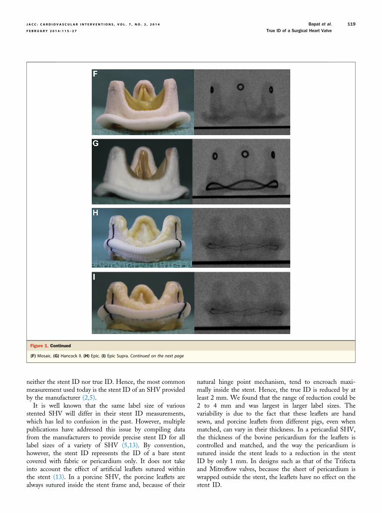
Figure 1. Continued
(F) Mosaic. (G) Hancock II. (H) Epic. (I) Epic Supra. Continued on the next page
J A C C : C A R D I O V A S C U L A R I N T E R V E N T I O N S , V O L . 7 , N O . 2 , 2 0 1 4 Bapat et al.
F E B R U A R Y 2 0 1 4 : 1 1 5 – 2 7 True ID of a Surgical Heart Valve
119
neither the stent ID nor true ID. Hence, the most commonmeasurement used today is the stent ID of an SHV providedby the manufacturer (2,5).
It is well known that the same label size of variousstented SHV will differ in their stent ID measurements,which has led to confusion in the past. However, multiplepublications have addressed this issue by compiling datafrom the manufacturers to provide precise stent ID for alllabel sizes of a variety of SHV (5,13). By convention,however, the stent ID represents the ID of a bare stentcovered with fabric or pericardium only. It does not takeinto account the effect of artificial leaflets sutured withinthe stent (13). In a porcine SHV, the porcine leaflets arealways sutured inside the stent frame and, because of their
natural hinge point mechanism, tend to encroach maxi-mally inside the stent. Hence, the true ID is reduced by atleast 2 mm. We found that the range of reduction could be2 to 4 mm and was largest in larger label sizes. Thevariability is due to the fact that these leaflets are handsewn, and porcine leaflets from different pigs, even whenmatched, can vary in their thickness. In a pericardial SHV,the thickness of the bovine pericardium for the leaflets iscontrolled and matched, and the way the pericardium issutured inside the stent leads to a reduction in the stentID by only 1 mm. In designs such as that of the Trifectaand Mitroflow valves, because the sheet of pericardium iswrapped outside the stent, the leaflets have no effect on thestent ID.

Figure 1. Continued
(J) Trifecta. (K) Mitroflow. (L) Soprano. (M) Aspire. Continued on the next page
Bapat et al. J A C C : C A R D I O V A S C U L A R I N T E R V E N T I O N S , V O L . 7 , N O . 2 , 2 0 1 4
True ID of a Surgical Heart Valve F E B R U A R Y 2 0 1 4 : 1 1 5 – 2 7
120
The implications of these findings are 4-fold:
1. When treating borderline sizes, especially with porcineSHV, use of the true ID instead of the stent ID shouldlead to choosing a smaller THV size than initiallyplanned. For example, label size 25 of Hancock II hasa stent ID of 22.5 mm and, hence, would point towardthe use of Sapien/Sapien XT size 26. However, thetrue ID is 20.5 mm, and hence, the correct size ofSapien/Sapien XT would be 23. A similar example forCoreValve usage concerns label size 27 of the Epic,where the stent ID (25 mm) would direct one to the
use of a size 29 CoreValve, but the true ID (22.5 mm)points to the use of a size 26 CoreValve.
2. When treating labeled SHV sizes �21 mm, althoughthe label size and/or stent ID appear adequate, a lookat the true ID may reveal them to be unsuitable forVIV treatment. For example, Hancock II size 21 has atrue ID of 16.5 mm, and hence, if treated with eitherSapien size 23 or Evolut size 23, will result in a sizemismatch and high residual gradients. This is also truefor pericardial valves with a small true ID such asPerimount size 19 (true ID 17 mm). This finding also
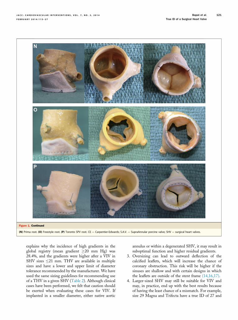
Figure 1. Continued
(N) Prima root. (O) Freestyle root. (P) Toronto SPV root. CE ¼ Carpentier-Edwards; S.A.V. ¼ SupraAnnular porcine valve; SHV ¼ surgical heart valves.
J A C C : C A R D I O V A S C U L A R I N T E R V E N T I O N S , V O L . 7 , N O . 2 , 2 0 1 4 Bapat et al.
F E B R U A R Y 2 0 1 4 : 1 1 5 – 2 7 True ID of a Surgical Heart Valve
121
explains why the incidence of high gradients in theglobal registry (mean gradient �20 mm Hg) was28.4%, and the gradients were higher after a VIV inSHV sizes �21 mm. THV are available in multiplesizes and have a lower and upper limit of diametertolerance recommended by the manufacturer. We haveused the same sizing guidelines for recommending useof a THV in a given SHV (Table 2). Although clinicalcases have been performed, we felt that caution shouldbe exerted when evaluating these cases for VIV. Ifimplanted in a smaller diameter, either native aortic
annulus or within a degenerated SHV, it may result insuboptimal function and higher residual gradients.
3. Oversizing can lead to outward deflection of thecalcified leaflets, which will increase the chance ofcoronary obstruction. This risk will be higher if thesinuses are shallow and with certain designs in whichthe leaflets are outside of the stent frame (14,16,17).
4. Larger-sized SHV may still be suitable for VIV andmay, in practice, end up with the best results becauseof having the least chance of a mismatch. For example,size 29 Magna and Trifecta have a true ID of 27 and
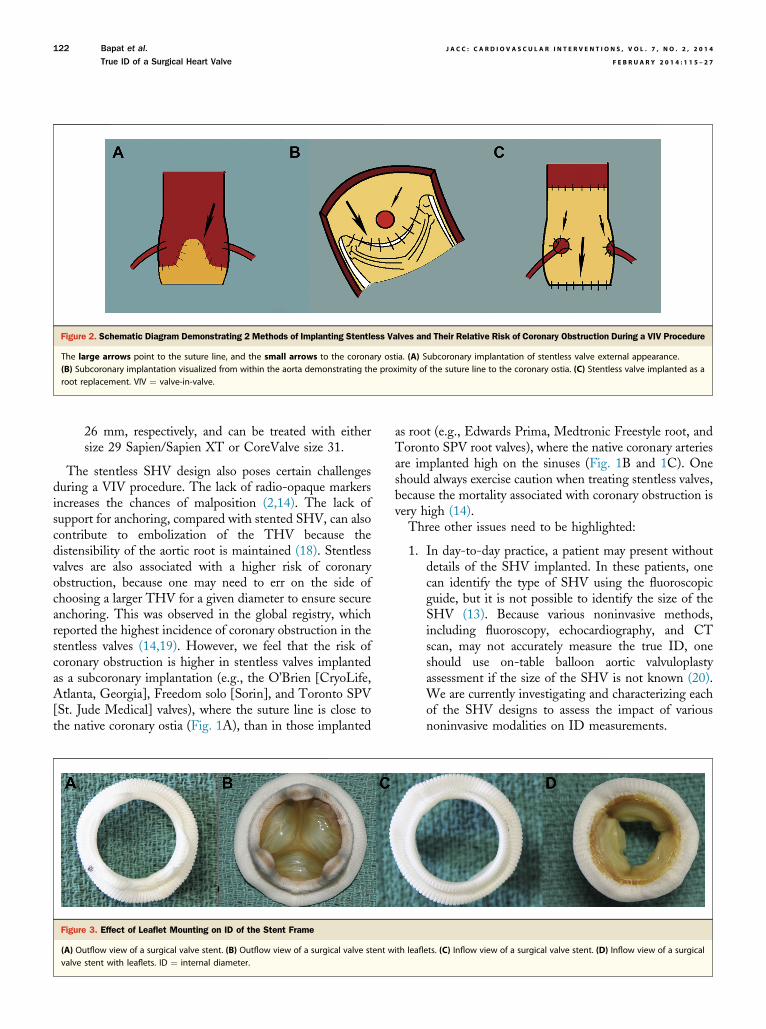
Figure 2. Schematic Diagram Demonstrating 2 Methods of Implanting Stentless Valves and Their Relative Risk of Coronary Obstruction During a VIV Procedure
The large arrows point to the suture line, and the small arrows to the coronary ostia. (A) Subcoronary implantation of stentless valve external appearance.(B) Subcoronary implantation visualized from within the aorta demonstrating the proximity of the suture line to the coronary ostia. (C) Stentless valve implanted as aroot replacement. VIV ¼ valve-in-valve.
Bapat et al. J A C C : C A R D I O V A S C U L A R I N T E R V E N T I O N S , V O L . 7 , N O . 2 , 2 0 1 4
True ID of a Surgical Heart Valve F E B R U A R Y 2 0 1 4 : 1 1 5 – 2 7
122
26 mm, respectively, and can be treated with eithersize 29 Sapien/Sapien XT or CoreValve size 31.
The stentless SHV design also poses certain challengesduring a VIV procedure. The lack of radio-opaque markersincreases the chances of malposition (2,14). The lack ofsupport for anchoring, compared with stented SHV, can alsocontribute to embolization of the THV because thedistensibility of the aortic root is maintained (18). Stentlessvalves are also associated with a higher risk of coronaryobstruction, because one may need to err on the side ofchoosing a larger THV for a given diameter to ensure secureanchoring. This was observed in the global registry, whichreported the highest incidence of coronary obstruction in thestentless valves (14,19). However, we feel that the risk ofcoronary obstruction is higher in stentless valves implantedas a subcoronary implantation (e.g., the O’Brien [CryoLife,Atlanta, Georgia], Freedom solo [Sorin], and Toronto SPV[St. Jude Medical] valves), where the suture line is close tothe native coronary ostia (Fig. 1A), than in those implanted
Figure 3. Effect of Leaflet Mounting on ID of the Stent Frame
(A) Outflow view of a surgical valve stent. (B) Outflow view of a surgical valve stent wvalve stent with leaflets. ID ¼ internal diameter.
as root (e.g., Edwards Prima, Medtronic Freestyle root, andToronto SPV root valves), where the native coronary arteriesare implanted high on the sinuses (Fig. 1B and 1C). Oneshould always exercise caution when treating stentless valves,because the mortality associated with coronary obstruction isvery high (14).
Three other issues need to be highlighted:
1. In day-to-day practice, a patient may present withoutdetails of the SHV implanted. In these patients, onecan identify the type of SHV using the fluoroscopicguide, but it is not possible to identify the size of theSHV (13). Because various noninvasive methods,including fluoroscopy, echocardiography, and CTscan, may not accurately measure the true ID, oneshould use on-table balloon aortic valvuloplastyassessment if the size of the SHV is not known (20).We are currently investigating and characterizing eachof the SHV designs to assess the impact of variousnoninvasive modalities on ID measurements.
ith leaflets. (C) Inflow view of a surgical valve stent. (D) Inflow view of a surgical
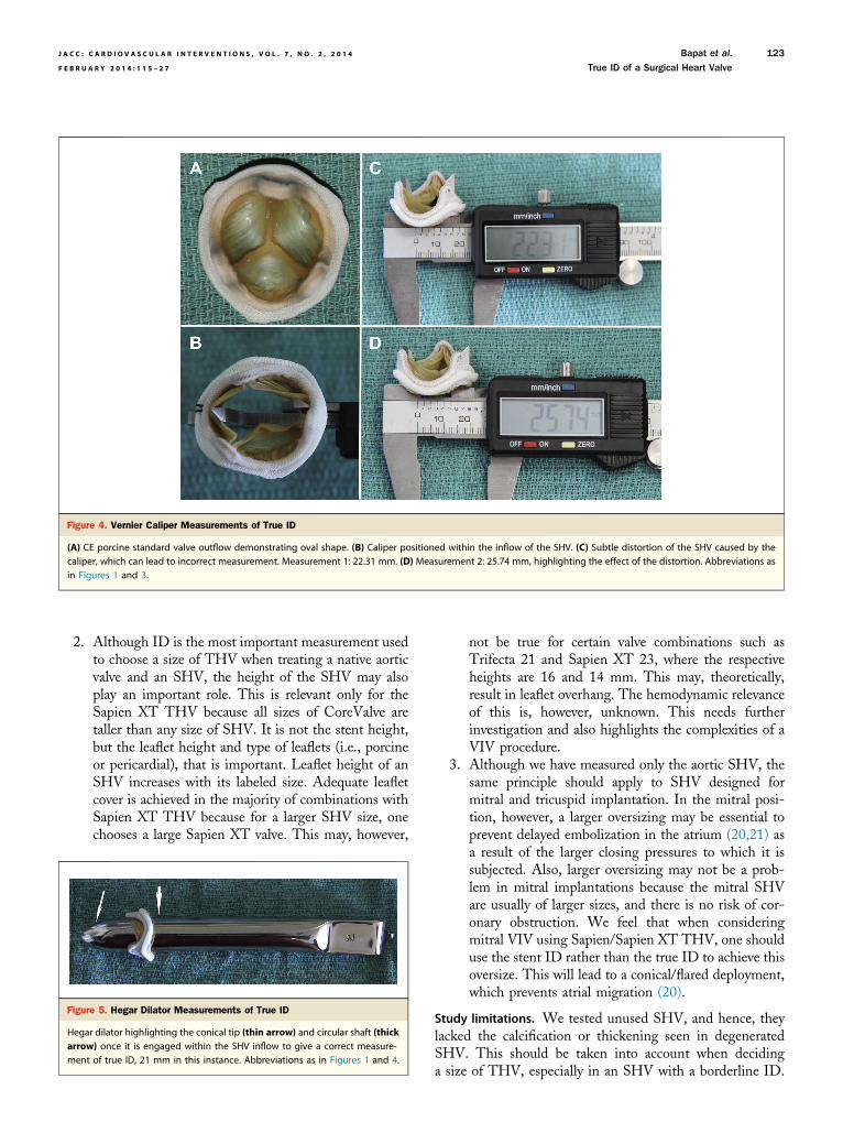
Figure 4. Vernier Caliper Measurements of True ID
(A) CE porcine standard valve outflow demonstrating oval shape. (B) Caliper positioned within the inflow of the SHV. (C) Subtle distortion of the SHV caused by thecaliper, which can lead to incorrect measurement. Measurement 1: 22.31 mm. (D) Measurement 2: 25.74 mm, highlighting the effect of the distortion. Abbreviations asin Figures 1 and 3.
F
Ham
J A C C : C A R D I O V A S C U L A R I N T E R V E N T I O N S , V O L . 7 , N O . 2 , 2 0 1 4 Bapat et al.
F E B R U A R Y 2 0 1 4 : 1 1 5 – 2 7 True ID of a Surgical Heart Valve
123
2. Although ID is the most important measurement usedto choose a size of THV when treating a native aorticvalve and an SHV, the height of the SHV may alsoplay an important role. This is relevant only for theSapien XT THV because all sizes of CoreValve aretaller than any size of SHV. It is not the stent height,but the leaflet height and type of leaflets (i.e., porcineor pericardial), that is important. Leaflet height of anSHV increases with its labeled size. Adequate leafletcover is achieved in the majority of combinations withSapien XT THV because for a larger SHV size, onechooses a large Sapien XT valve. This may, however,
igure 5. Hegar Dilator Measurements of True ID
egar dilator highlighting the conical tip (thin arrow) and circular shaft (thickrrow) once it is engaged within the SHV inflow to give a correct measure-ent of true ID, 21 mm in this instance. Abbreviations as in Figures 1 and 4.
not be true for certain valve combinations such asTrifecta 21 and Sapien XT 23, where the respectiveheights are 16 and 14 mm. This may, theoretically,result in leaflet overhang. The hemodynamic relevanceof this is, however, unknown. This needs furtherinvestigation and also highlights the complexities of aVIV procedure.
3. Although we have measured only the aortic SHV, thesame principle should apply to SHV designed formitral and tricuspid implantation. In the mitral posi-tion, however, a larger oversizing may be essential toprevent delayed embolization in the atrium (20,21) asa result of the larger closing pressures to which it issubjected. Also, larger oversizing may not be a prob-lem in mitral implantations because the mitral SHVare usually of larger sizes, and there is no risk of cor-onary obstruction. We feel that when consideringmitral VIV using Sapien/Sapien XT THV, one shoulduse the stent ID rather than the true ID to achieve thisoversize. This will lead to a conical/flared deployment,which prevents atrial migration (20).
Study limitations. We tested unused SHV, and hence, theylacked the calcification or thickening seen in degeneratedSHV. This should be taken into account when decidinga size of THV, especially in an SHV with a borderline ID.
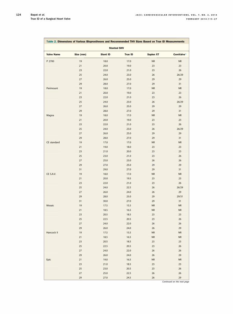
Table 2. Dimensions of Various Bioprostheses and Recommended THV Sizes Based on True ID Measurements
Stented SHV
Valve Name Size (mm) Stent ID True ID Sapien XT CoreValve*
P 2700 19 18.0 17.0 NR NR
21 20.0 19.0 23 23
23 22.0 21.0 23 26
25 24.0 23.0 26 26/29
27 26.0 25.0 29 29
29 28.0 27.0 29 31
Perimount 19 18.0 17.0 NR NR
21 20.0 19.0 23 23
23 22.0 21.0 23 26
25 24.0 23.0 26 26/29
27 26.0 25.0 29 29
29 28.0 27.0 29 31
Magna 19 18.0 17.0 NR NR
21 20.0 19.0 23 23
23 22.0 21.0 23 26
25 24.0 23.0 26 26/29
27 26.0 25.0 29 29
29 28.0 27.0 29 31
CE standard 19 17.0 17.0 NR NR
21 19.0 18.0 23 23
23 21.0 20.0 23 23
25 23.0 21.0 23 26
27 25.0 23.0 26 26
29 27.0 25.0 29 29
31 29.0 27.0 29 31
CE S.A.V. 19 18.0 17.0 NR NR
21 20.0 19.5 23 23
23 22.0 21.0 23 26
25 24.0 22.5 26 26/29
27 26.0 24.0 26 29
29 28.0 25.0 29 29/31
31 30.0 27.0 29 31
Mosaic 19 17.5 15.5 NR NR
21 18.5 16.5 NR NR
23 20.5 18.5 23 23
25 22.5 20.5 23 26
27 24.0 22.0 26 26
29 26.0 24.0 26 29
Hancock II 19 17.5 15.5 NR NR
21 18.5 16.5 NR NR
23 20.5 18.5 23 23
25 22.5 20.5 23 26
27 24.0 22.0 26 26
29 26.0 24.0 26 29
Epic 21 19.0 16.5 NR NR
23 21.0 18.5 23 23
25 23.0 20.5 23 26
27 25.0 22.5 26 26
29 27.0 24.5 26 29
Continued on the next page
Bapat et al. J A C C : C A R D I O V A S C U L A R I N T E R V E N T I O N S , V O L . 7 , N O . 2 , 2 0 1 4
True ID of a Surgical Heart Valve F E B R U A R Y 2 0 1 4 : 1 1 5 – 2 7
124

Table 2. Continued
Stented SHV
Valve Name Size (mm) Stent ID True ID Sapien XT CoreValve*
Epic Supra 19 19.0 16.5 NR NR
21 21.0 18.5 23 23
23 23.0 20.5 23 26
25 25.0 22.5 26 26
27 27.0 24.5 26 29
Trifecta 19 17.0 16.0 NR NR
21 19.0 18.0 23 23
23 21.0 20.5 23 26
25 23.0 22.0 26 26
27 25.0 24.0 26 29
29 27.0 26.0 29 31
Mitroflow 19 15.4 15.5 NR NR
21 17.3 17.0 NR NR
23 19.0 19.0 23 23
25 21.0 21.0 23 26
27 22.9 23.0 26 26
29 24.7 24.5 26 29
Soprano 18 17.8 18.0 23 23
20 19.8 20.0 23 23
22 21.7 22.0 26 26
24 23.7 23.5 26 29
26 25.6 25.5 29 29
28 27.6 27.5 29 31
Aspire 20 18.2 16.5 NR NR
21 19.2 17.5 NR NR
23 21.0 19.0 23 23
25 23.0 20.0 23 23
27 25.0 22.0 26 26
Stentless SHV
Valve Name Size (mm)Tissue Annulus
Diameter True ID Sapien XT CoreValve
Prima 19 19 17.5 NR NR
21 21 19.5 23 23
23 23 21.5 23/26 26
25 25 23.5 26 26/29
27 27 25.5 29 29
29 29 27.5 29 31
Freestyle 19 19 16 NR NR
21 21 18 23 23
23 23 20 23 23
25 25 22 26 26
27 27 25 29 29
Toronto SPV 19 19 17 NR NR
21 21 19 23 23
23 23 21 23 26
25 25 23 26 26/29
27 27 25 29 29
29 29 27 29 31
When a valve is not recommended (NR), this recommendation is based on sizing guidance of the currently available THV valves for the native
aortic annulus diameter. It may be still feasible to perform a VIV, but this may result in suboptimal results such as higher residual gradients and/or
leaflet malfunction. *The 23 size in the CoreValve column represents the Evolut THV.
ID ¼ internal diameter; THV ¼ transcatheter heart valve; other abbreviations as in Table 1.
J A C C : C A R D I O V A S C U L A R I N T E R V E N T I O N S , V O L . 7 , N O . 2 , 2 0 1 4 Bapat et al.
F E B R U A R Y 2 0 1 4 : 1 1 5 – 2 7 True ID of a Surgical Heart Valve
125

Figure 6. Effect of Leaflet Type and Mounting on the Stent ID
(A) Porcine valves: true internal diameter (ID) is at least 2 mm less than the stent ID. (B) Pericardial valves with leaflets sutured inside the stent frame: true ID is at least1 mm less than the stent ID. (C) Pericardial valves with leaflets sutured outside the stent frame: true ID is the same as the stent ID.
Bapat et al. J A C C : C A R D I O V A S C U L A R I N T E R V E N T I O N S , V O L . 7 , N O . 2 , 2 0 1 4
True ID of a Surgical Heart Valve F E B R U A R Y 2 0 1 4 : 1 1 5 – 2 7
126
We have observed heavy calcification in SHV implanted intricuspid and pulmonary positions, and because the right-sided pressures are low, there could be an argument for usinga smaller THV rather than larger in a borderline case.Another factor that can affect the true ID in a clinical sce-nario is pannus. However, both calcification and pannus canbe easily evaluated by echocardiography and CT scan; atworst, the ID is going to be less than the true ID, but nevermore.
Conclusions
Design features of SHV may lead to a reduction in the stentID in the majority of SHV. It is important to identify theSHV design so as to take this reduction into consideration
Figure 7. Fluoroscopic Confirmation of the Effect of Leaflet Type and Mounting o
The outer thin arrows point to the fluoroscopic stent frame of the SHV, and the innerCoreValve (size 26) implanted within CE Porcine standard SHV (size 25) demonstrateframe of the CE and CoreValve bioprostheses. (B) CoreValve (size 26) implanted withradio-opaque stent frame of the Perimount and CoreValve. (C) CoreValve (size 26) imopaque stent frame of the Trifecta and CoreValve. Abbreviations as in Figures 1 and
when choosing the appropriate THV during a VIV proce-dure. The true ID chart in this paper provides realistic IDmeasurements for VIV treatment in the aortic position. Oneshould use the true ID for selecting an appropriate THVdevice in the aortic position, but use the stent ID to choosethe device for a mitral VIV procedure. If the SHV size is notknown, balloon aortic valvuloplasty may be the best way toconfirm the true ID, because ID measurements obtained byother modalities may be inaccurate.
Acknowledgments
The authors thank Miss Maya Guthrie and Mrs. ShalinaSunni for their help in obtaining fluoroscopic imagesrequired for the paper, and Mrs. Urmi Bapat for editorialassistance.
n the Stent ID
thick arrows point to the inflow portion of the stent frame of the CoreValve. (A)d greater degree of separation (at least 2 mm) between the radio-opaque stentin Perimount (size 25), demonstrated 1-mm degree of separation between theplanted within Trifecta (size 25) demonstrated no separation between the radio-3.
J A C C : C A R D I O V A S C U L A R I N T E R V E N T I O N S , V O L . 7 , N O . 2 , 2 0 1 4 Bapat et al.
F E B R U A R Y 2 0 1 4 : 1 1 5 – 2 7 True ID of a Surgical Heart Valve
127
Reprint requests and correspondence: Mr. Vinayak (Vinnie) N.Bapat, 6th Floor East Wing, Department of CardiothoracicSurgery, St. Thomas’Hospital, Westminster Bridge Road, LondonSE1 7EH, United Kingdom. E-mail: [email protected].
REFERENCES
1. Généreux P, Head SJ, Wood DA, et al. Transcatheter aortic valveimplantation: 10-year anniversary part II: clinical implications. EurHeart J 2012;33:2399–402.
2. Bapat V, Attia R, Redwood S, et al. Use of transcatheter heart valves fora valve-in-valve implantation in patients with degenerated aortic bio-prosthesis: technical considerations and results. J Thorac CardiovascSurg 2012;144:1372–9.
3. Gurvitch R, Cheung A, Ye J, et al. Transcatheter valve-in-valve im-plantation for failed surgical bioprosthetic valves. J Am Coll Cardiol2011;58:2196–209.
4. Khawaja MZ, Haworth P, Ghuran A, et al. Transcatheter aortic valveimplantation for stenosed and regurgitant aortic valve bioprosthesis:CoreValve for failed bioprosthetic aortic valve replacements. J Am CollCardiol 2010;55:97–101.
5. Piazza N, Bleiziffer S, Brockmann G, et al. Transcatheter aortic valveimplantation for failing surgical aortic bioprosthetic valve: from conceptto clinical application and evaluation (part 2). J Am Coll Cardiol Intv2011;4:733–42.
6. Latib A, Ielasi A, Montorfano M, et al. Transcatheter valve-in-valveimplantation with the Edwards SAPIEN in patients with bioprostheticheart valve failure: the Milan experience. EuroIntervention 2012;7:1275–84.
7. Gotzmann M, Mugge A, Bojara W. Transcatheter aortic valve im-plantation for treatment of patients with degenerated aortic bio-prostheses: valve-in-valve technique. Catheter Cardiovasc Interv 2010;26:1000–6.
8. Pasic M, Unbehaun A, Dreysse S, et al. Transapical aortic valve im-plantation after previous aortic valve replacement: clinical proof of the“valve-in-valve” concept. J Thorac Cardiovasc Surg 2011;142:270–7.
9. Seiffert M, Franzen O, Conradi L, et al. Series of transcatheter valve-in-valve implantations in high-risk patients with degenerated bio-prostheses in aortic and mitral position. Catheter Cardiovasc Interv2010;76:608–15.
10. Cheung A, Soon JL, Webb JG, Ye J. Transatrial transcatheter tricuspidvalve-in-valve technique. J Card Surg 2012;27:196–8.
11. Faza N, Kenny D, Kavinsky C, Amin Z, Heitschmidt M, Hijazi Z.Single center comparative outcomes of the Edwards SAPIEN andMedtronic Melody transcatheter heart valves in the pulmonary position.Catheter Cardiovasc Interv 2013;82:E535–41.
12. Ng AC, Delgado V, van der Kley F, et al. Comparison of aortic rootdimensions and geometries before and after transcatheter aortic valveimplantation by 2-and 3-dimmensional transesophageal echocardiog-raphy and multislice computed tomography. Circ Cardiovasc Imaging2010;3:94–102.
13. Bapat V, Chadalavada S, Tehrani H, Attia R, Thomas M. A guide tofluoroscopic identification and design of bioprosthetic valves: a refer-ence for valve-in-valve procedure. Catheter Cardiovasc Interv 2013;81:853–61.
14. Dvir D, Webb J, Brecker S, et al. Transcatheter aortic valve replacementfor degenerative bioprosthetic surgical valves: results from the globalvalve-in-valve registry. Circulation 2012;126:2335–44.
15. Babaliaros VC, Jungadhwalla Z, Lerakis S, et al. Use of balloon aorticvalvuloplasty to size the aortic annulus before implantation of a balloon-expandable heart valve. J Am Coll Cardiol Intv 2010;3:114–8.
16. Gurvitch R, Cheung A, Bedogni F, Webb JG. Coronary obstructionfollowing transcatheter aortic valve-in-valve implantation for failedsurgical bioprosthesis. Catheter Cardiovasc Interv 2011;77:439–44.
17. Gonzalo BC, Jorge SF, Nicolas VG. Acute coronary artery occlusionscomplicating a valve-in-valve procedure. Heart 2013;99:591–2.
18. Funder JA, Frost MW, Klaaborg KE, et al. Aortic root distensibilityafter subcoronary stentless valve implantation. J Heart Valve Dis 2012;21:181–8.
19. Chakravarty T, Jilahawi H, Nakamura M, et al. Pre-emptive positioningof a coronary stent in the left-anterior descending artery for a left mainprotection: a prerequisite for transcatheter aortic valve-in-valve im-plantation for failing stentless bioprosthesis? Catheter Cardiovasc Interv2013;82:E630–6.
20. Bapat V, Khaliel F, Ihleberg L. Delayed migration of Sapien valvefollowing a transcatheter mitral valve-in-valve implantation. CatheterCardiovasc Interv 2014;83:E150–4.
21. Cheung A, Webb JG, Barbanti M, et al. 5-year experience withtranscatheter transapical mitral valve-in-valve implantation for bio-prosthetic valve dysfunction. J Am Coll Cardiol 2013;61:1759–66.
Key Words: heart valve - valve replacement -
valve-in-valve.
Related Documents