Journal of Alloys and Compounds 523 (2012) 66–71 Contents lists available at SciVerse ScienceDirect Journal of Alloys and Compounds j our na l ho me p ag e: www.elsevier.com/locate/jallcom Effect of sintering temperature on structural, magnetic properties of lithium chromium ferrite R.P. Patil a,∗ , P.P. Hankare a,∗ , K.M. Garadkar a , R. Sasikala b a Department of Chemistry, Shivaji University, Kolhapur 416004, India b Chemistry Division, Bhabha Atomic Research Center, Mumbai 400085, India a r t i c l e i n f o Article history: Received 27 October 2010 Received in revised form 24 December 2011 Accepted 4 January 2012 Available online 12 January 2012 Keywords: Ferrites Sol–gel synthesis X-ray diffraction Raman spectra TEM a b s t r a c t Nanocrystalline mixed lithium–chromium ferrites were prepared by using sol–gel method. The samples were sintered at different temperatures in air. Their structural and magnetic properties were studied by X-ray diffraction, scanning electron microscopy, transmission electron microscopy, Raman scattering, FT-IR and magnetic hysteresis loop tracer techniques. The X-ray diffraction patterns reveal that all the samples consist of nanocrystalline single cubic phase structure. The morphological studies of synthesized nanocrystalline samples were obtained from the scanning electron microscopy and transmission electron microscopy techniques. Raman scattering and FT-IR studies indicate that the spinel phase formation takes place at higher sintering temperature. The magnetic studies show that, the ferrimagnetic behavior increases with sintering temperature. © 2012 Elsevier B.V. All rights reserved. 1. Introduction Ferrites are magnetic semiconductors which cannot be replaced by any other magnetic material, because of their stable, relatively inexpensive, ease formation. These materials have interesting structural, electrical and magnetic properties, which make them in widespread applications such as electronics and telecommuni- cation industries [1]. These properties are tunable with chemical composition, method of preparation, sintering temperature, sin- tering time, etc. [2]. Among various ferrites, lithium and metal substituted lithium ferrites have been attractive structural and magnetic features that render them as a potential material for various technological appli- cations such as, lithium batteries, recording heads, transformer cores, and noise filters [3–11]. The structural and magnetic prop- erties of these ferrite systems with respect to different sintering temperature conditions have been reported by many workers [12,13]. In this communication, we report the effects on structural and magnetic properties of the Li 0.5 Fe 1.5 Cr 1.0 O 4 samples with sin- tering temperature (T s ) from 673 to 973 K. ∗ Corresponding authors. Tel.: +91 231 2609381. E-mail addresses: raj rbm [email protected] (R.P. Patil), p [email protected] (P.P. Hankare). 2. Experimental details Analytical grade chromium nitrate [Cr(NO3)3·9H2O], iron nitrate [Fe((NO3)2·9H2O], lithium nitrate [LiNO3] and citric acid [C6H8O7·H2O] were used to prepare Li0.5Fe1.5Cr1.0O4 by a sol–gel method. Metal nitrates and citric acid were dissolved in deionized water with 1:1 molar ratio. The pH of the solution was adjusted to 9.0–9.5 using ammonia solution. Then, the solution was heated at 353 K, which results gel formation. When ignited, the dried gel burnt in a self propagating combustion manner until it was converted into a floppy loose powder. This powder was then sintered at different temperatures such as, 673, 773, 873 and 973 K for 8 h. The sintered powders were granulated and using 2% polyvinyl alcohol as a binder were uniaxially pressed at a pressure of 8 ton/cm 2 to form pellet specimens. The structural studies of the sintered samples were obtained by X-ray diffrac- tion technique using a Philips PW-1710 X-ray diffractometer with Cu K radiation ( = 1.54056 ˚ A). Raman spectra and FT-IR studies were used for interpretation of the vibrational modes in mixed metal oxides. Microstructural analyses of the samples were carried out using transmission electron microscope (TEM-Model Philips 200 CX) operating at a voltage of 120 kV and also using scanning electron microscope (JEOL-JSM 6360 Microscope). The high field hysteresis loop tracer (Magenta) was used to measure the saturation magnetization, coercivity and remanance magneti- zation of all the samples as function of sintering temperature. 3. Results and discussion 3.1. X-ray diffraction studies X-ray powder diffraction pattern of the Li 0.5 Fe 1.5 Cr 1.0 O 4 sam- ples as prepared and sintered at different temperatures are shown in Fig. 1. X-ray diffraction data shows that, the cubic phase is observed at 973 K. The diffraction pattern of as prepared sample 0925-8388/$ – see front matter © 2012 Elsevier B.V. All rights reserved. doi:10.1016/j.jallcom.2012.01.025

Welcome message from author
This document is posted to help you gain knowledge. Please leave a comment to let me know what you think about it! Share it to your friends and learn new things together.
Transcript
Ec
Ra
b
a
ARR2AA
KFSXRT
1
bisicct
frccet[at
p
0d
Journal of Alloys and Compounds 523 (2012) 66– 71
Contents lists available at SciVerse ScienceDirect
Journal of Alloys and Compounds
j our na l ho me p ag e: www.elsev ier .com/ locate / ja l l com
ffect of sintering temperature on structural, magnetic properties of lithiumhromium ferrite
.P. Patil a,∗, P.P. Hankarea,∗, K.M. Garadkara, R. Sasikalab
Department of Chemistry, Shivaji University, Kolhapur 416004, IndiaChemistry Division, Bhabha Atomic Research Center, Mumbai 400085, India
r t i c l e i n f o
rticle history:eceived 27 October 2010eceived in revised form4 December 2011ccepted 4 January 2012vailable online 12 January 2012
a b s t r a c t
Nanocrystalline mixed lithium–chromium ferrites were prepared by using sol–gel method. The sampleswere sintered at different temperatures in air. Their structural and magnetic properties were studiedby X-ray diffraction, scanning electron microscopy, transmission electron microscopy, Raman scattering,FT-IR and magnetic hysteresis loop tracer techniques. The X-ray diffraction patterns reveal that all thesamples consist of nanocrystalline single cubic phase structure. The morphological studies of synthesized
eywords:erritesol–gel synthesis-ray diffractionaman spectra
nanocrystalline samples were obtained from the scanning electron microscopy and transmission electronmicroscopy techniques. Raman scattering and FT-IR studies indicate that the spinel phase formationtakes place at higher sintering temperature. The magnetic studies show that, the ferrimagnetic behaviorincreases with sintering temperature.
© 2012 Elsevier B.V. All rights reserved.
EM
. Introduction
Ferrites are magnetic semiconductors which cannot be replacedy any other magnetic material, because of their stable, relatively
nexpensive, ease formation. These materials have interestingtructural, electrical and magnetic properties, which make themn widespread applications such as electronics and telecommuni-ation industries [1]. These properties are tunable with chemicalomposition, method of preparation, sintering temperature, sin-ering time, etc. [2].
Among various ferrites, lithium and metal substituted lithiumerrites have been attractive structural and magnetic features thatender them as a potential material for various technological appli-ations such as, lithium batteries, recording heads, transformerores, and noise filters [3–11]. The structural and magnetic prop-rties of these ferrite systems with respect to different sinteringemperature conditions have been reported by many workers12,13]. In this communication, we report the effects on structuralnd magnetic properties of the Li0.5Fe1.5Cr1.0O4 samples with sin-
ering temperature (Ts) from 673 to 973 K.∗ Corresponding authors. Tel.: +91 231 2609381.E-mail addresses: raj rbm [email protected] (R.P. Patil),
[email protected] (P.P. Hankare).
925-8388/$ – see front matter © 2012 Elsevier B.V. All rights reserved.oi:10.1016/j.jallcom.2012.01.025
2. Experimental details
Analytical grade chromium nitrate [Cr(NO3)3·9H2O], iron nitrate[Fe((NO3)2·9H2O], lithium nitrate [LiNO3] and citric acid [C6H8O7·H2O] wereused to prepare Li0.5Fe1.5Cr1.0O4 by a sol–gel method. Metal nitrates and citric acidwere dissolved in deionized water with 1:1 molar ratio. The pH of the solutionwas adjusted to 9.0–9.5 using ammonia solution. Then, the solution was heatedat 353 K, which results gel formation. When ignited, the dried gel burnt in a selfpropagating combustion manner until it was converted into a floppy loose powder.This powder was then sintered at different temperatures such as, 673, 773, 873and 973 K for 8 h. The sintered powders were granulated and using 2% polyvinylalcohol as a binder were uniaxially pressed at a pressure of 8 ton/cm2 to form pelletspecimens.
The structural studies of the sintered samples were obtained by X-ray diffrac-tion technique using a Philips PW-1710 X-ray diffractometer with Cu K� radiation(� = 1.54056 A). Raman spectra and FT-IR studies were used for interpretation of thevibrational modes in mixed metal oxides. Microstructural analyses of the sampleswere carried out using transmission electron microscope (TEM-Model Philips 200CX) operating at a voltage of 120 kV and also using scanning electron microscope(JEOL-JSM 6360 Microscope). The high field hysteresis loop tracer (Magenta) wasused to measure the saturation magnetization, coercivity and remanance magneti-zation of all the samples as function of sintering temperature.
3. Results and discussion
3.1. X-ray diffraction studies
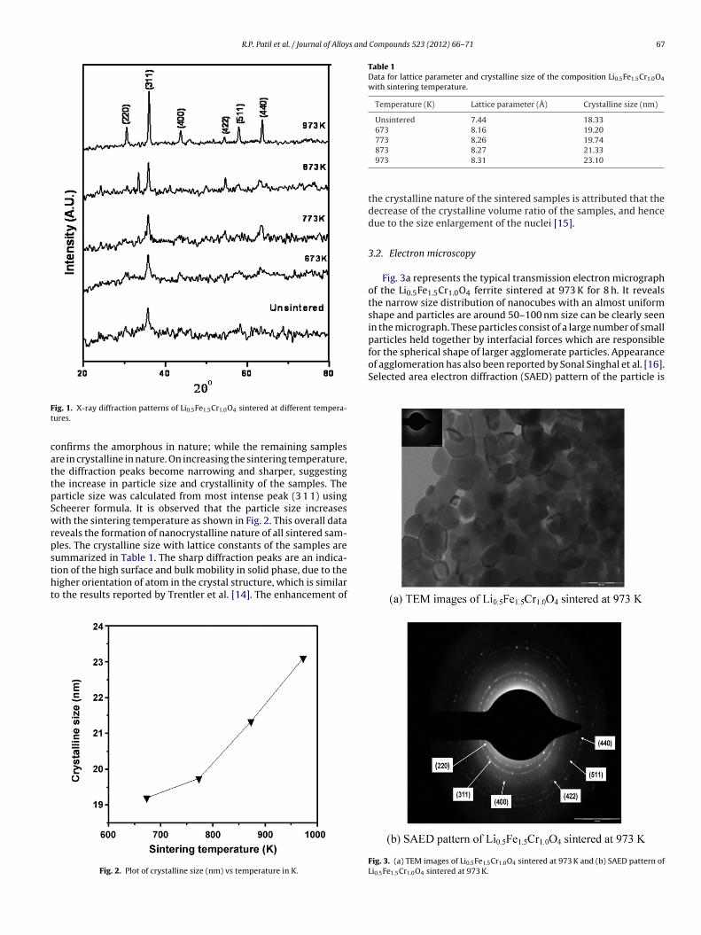
X-ray powder diffraction pattern of the Li0.5Fe1.5Cr1.0O4 sam-ples as prepared and sintered at different temperatures are shownin Fig. 1. X-ray diffraction data shows that, the cubic phase isobserved at 973 K. The diffraction pattern of as prepared sample
R.P. Patil et al. / Journal of Alloys and Compounds 523 (2012) 66– 71 67
Ft
cattpSwrpstht
Table 1Data for lattice parameter and crystalline size of the composition Li0.5Fe1.5Cr1.0O4
with sintering temperature.
Temperature (K) Lattice parameter (Å) Crystalline size (nm)
Unsintered 7.44 18.33673 8.16 19.20773 8.26 19.74
for the spherical shape of larger agglomerate particles. Appearanceof agglomeration has also been reported by Sonal Singhal et al. [16].Selected area electron diffraction (SAED) pattern of the particle is
ig. 1. X-ray diffraction patterns of Li0.5Fe1.5Cr1.0O4 sintered at different tempera-ures.
onfirms the amorphous in nature; while the remaining samplesre in crystalline in nature. On increasing the sintering temperature,he diffraction peaks become narrowing and sharper, suggestinghe increase in particle size and crystallinity of the samples. Thearticle size was calculated from most intense peak (3 1 1) usingcheerer formula. It is observed that the particle size increasesith the sintering temperature as shown in Fig. 2. This overall data
eveals the formation of nanocrystalline nature of all sintered sam-les. The crystalline size with lattice constants of the samples areummarized in Table 1. The sharp diffraction peaks are an indica-
ion of the high surface and bulk mobility in solid phase, due to theigher orientation of atom in the crystal structure, which is similaro the results reported by Trentler et al. [14]. The enhancement ofFig. 2. Plot of crystalline size (nm) vs temperature in K.
873 8.27 21.33973 8.31 23.10
the crystalline nature of the sintered samples is attributed that thedecrease of the crystalline volume ratio of the samples, and hencedue to the size enlargement of the nuclei [15].
3.2. Electron microscopy
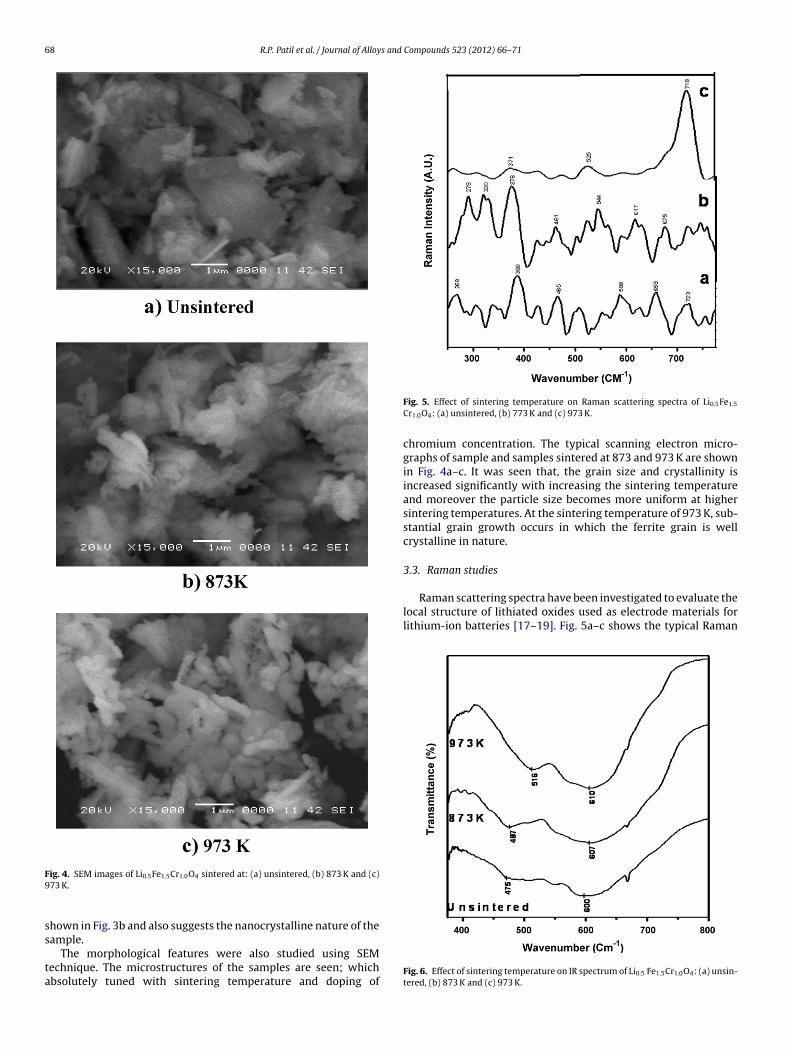
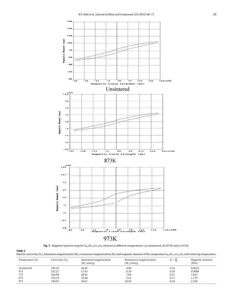
Fig. 3a represents the typical transmission electron micrographof the Li0.5Fe1.5Cr1.0O4 ferrite sintered at 973 K for 8 h. It revealsthe narrow size distribution of nanocubes with an almost uniformshape and particles are around 50–100 nm size can be clearly seenin the micrograph. These particles consist of a large number of smallparticles held together by interfacial forces which are responsible
Fig. 3. (a) TEM images of Li0.5Fe1.5Cr1.0O4 sintered at 973 K and (b) SAED pattern ofLi0.5Fe1.5Cr1.0O4 sintered at 973 K.
68 R.P. Patil et al. / Journal of Alloys and Compounds 523 (2012) 66– 71
F9
ss
ta
Raman scattering spectra have been investigated to evaluate thelocal structure of lithiated oxides used as electrode materials forlithium-ion batteries [17–19]. Fig. 5a–c shows the typical Raman
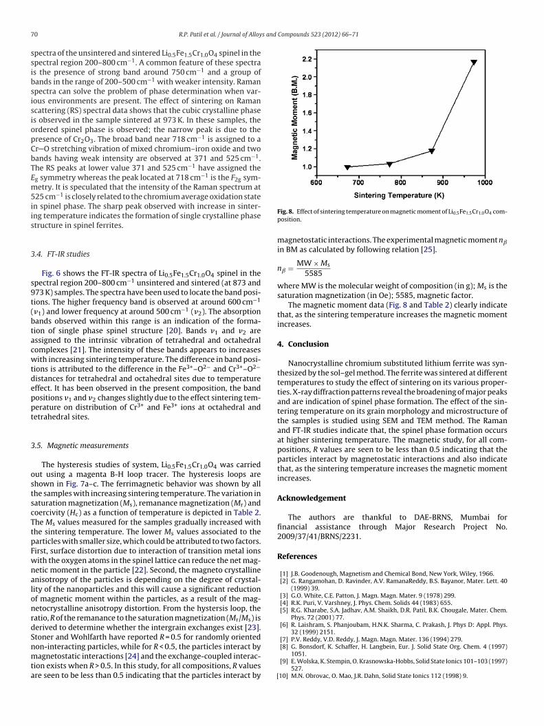
ig. 4. SEM images of Li0.5Fe1.5Cr1.0O4 sintered at: (a) unsintered, (b) 873 K and (c)73 K.
hown in Fig. 3b and also suggests the nanocrystalline nature of theample.
The morphological features were also studied using SEMechnique. The microstructures of the samples are seen; whichbsolutely tuned with sintering temperature and doping of
Fig. 5. Effect of sintering temperature on Raman scattering spectra of Li0.5Fe1.5
Cr1.0O4: (a) unsintered, (b) 773 K and (c) 973 K.
chromium concentration. The typical scanning electron micro-graphs of sample and samples sintered at 873 and 973 K are shownin Fig. 4a–c. It was seen that, the grain size and crystallinity isincreased significantly with increasing the sintering temperatureand moreover the particle size becomes more uniform at highersintering temperatures. At the sintering temperature of 973 K, sub-stantial grain growth occurs in which the ferrite grain is wellcrystalline in nature.
3.3. Raman studies
Fig. 6. Effect of sintering temperature on IR spectrum of Li0.5 Fe1.5Cr1.0O4: (a) unsin-tered, (b) 873 K and (c) 973 K.
R.P. Patil et al. / Journal of Alloys and Compounds 523 (2012) 66– 71 69
Fig. 7. Magnetic hystersis loop for Li0.5Fe1.5Cr1.0O4 sintered at different temperatures: (a) unsintered, (b) 873 K and (c) 973 K.
Table 2Data for coercivity (Hc), Saturation magnetization (Ms), remanence magnetization (Mr) and magnetic moment of the composition Li0.5Fe1.5Cr1.0O4 with sintering temperature.
Temperature (K) Coercivity (Hc) Saturation magnetization(Ms) emu/g
Remanence magnetization(Mr) emu/g
R = MrMs
Magnetic moment(B.M.)
Unsintered 196.43 26.45 4.90 0.18 0.9625673 227.27 27.45 8.36 0.30 0.9988773 324.68 28.41 7.89 0.27 1.033873 330.10 32.40 5.61 0.17 1.179973 349.03 59.61 20.29 0.34 2.169
7 s and Compounds 523 (2012) 66– 71
ssibsisiopCbTEm5iis
3
s9t(btacwtdeppt
3
ostscTtpFwnalonrdSnmta
0 R.P. Patil et al. / Journal of Alloy
pectra of the unsintered and sintered Li0.5Fe1.5Cr1.0O4 spinel in thepectral region 200–800 cm−1. A common feature of these spectras the presence of strong band around 750 cm−1 and a group ofands in the range of 200–500 cm−1 with weaker intensity. Ramanpectra can solve the problem of phase determination when var-ous environments are present. The effect of sintering on Ramancattering (RS) spectral data shows that the cubic crystalline phases observed in the sample sintered at 973 K. In these samples, therdered spinel phase is observed; the narrow peak is due to theresence of Cr2O3. The broad band near 718 cm−1 is assigned to ar O stretching vibration of mixed chromium–iron oxide and twoands having weak intensity are observed at 371 and 525 cm−1.he RS peaks at lower value 371 and 525 cm−1 have assigned theg symmetry whereas the peak located at 718 cm−1 is the F2g sym-etry. It is speculated that the intensity of the Raman spectrum at
25 cm−1 is closely related to the chromium average oxidation staten spinel phase. The sharp peak observed with increase in sinter-ng temperature indicates the formation of single crystalline phasetructure in spinel ferrites.
.4. FT-IR studies
Fig. 6 shows the FT-IR spectra of Li0.5Fe1.5Cr1.0O4 spinel in thepectral region 200–800 cm−1 unsintered and sintered (at 873 and73 K) samples. The spectra have been used to locate the band posi-ions. The higher frequency band is observed at around 600 cm−1
�1) and lower frequency at around 500 cm−1 (�2). The absorptionands observed within this range is an indication of the forma-ion of single phase spinel structure [20]. Bands �1 and �2 aressigned to the intrinsic vibration of tetrahedral and octahedralomplexes [21]. The intensity of these bands appears to increasesith increasing sintering temperature. The difference in band posi-
ions is attributed to the difference in the Fe3+–O2− and Cr3+–O2−
istances for tetrahedral and octahedral sites due to temperatureffect. It has been observed in the present composition, the bandositions �1 and �2 changes slightly due to the effect sintering tem-erature on distribution of Cr3+ and Fe3+ ions at octahedral andetrahedral sites.
.5. Magnetic measurements
The hysteresis studies of system, Li0.5Fe1.5Cr1.0O4 was carriedut using a magenta B-H loop tracer. The hysteresis loops arehown in Fig. 7a–c. The ferrimagnetic behavior was shown by allhe samples with increasing sintering temperature. The variation inaturation magnetization (Ms), remanance magnetization (Mr) andoercivity (Hc) as a function of temperature is depicted in Table 2.he Ms values measured for the samples gradually increased withhe sintering temperature. The lower Ms values associated to thearticles with smaller size, which could be attributed to two factors.irst, surface distortion due to interaction of transition metal ionsith the oxygen atoms in the spinel lattice can reduce the net mag-etic moment in the particle [22]. Second, the magneto crystallinenisotropy of the particles is depending on the degree of crystal-ity of the nanoparticles and this will cause a significant reductionf magnetic moment within the particles, as a result of the mag-etocrystalline anisotropy distortion. From the hystersis loop, theatio, R of the remanance to the saturation magnetization (Mr/Ms) iserived to determine whether the intergrain exchanges exist [23].toner and Wohlfarth have reported R = 0.5 for randomly oriented
on-interacting particles, while for R < 0.5, the particles interact byagnetostatic interactions [24] and the exchange-coupled interac-ion exists when R > 0.5. In this study, for all compositions, R valuesre seen to be less than 0.5 indicating that the particles interact by [
Fig. 8. Effect of sintering temperature on magnetic moment of Li0.5Fe1.5Cr1.0O4 com-position.
magnetostatic interactions. The experimental magnetic moment nˇ
in BM as calculated by following relation [25].
nˇ = MW × Ms
5585
where MW is the molecular weight of composition (in g); Ms is thesaturation magnetization (in Oe); 5585, magnetic factor.
The magnetic moment data (Fig. 8 and Table 2) clearly indicatethat, as the sintering temperature increases the magnetic momentincreases.
4. Conclusion
Nanocrystalline chromium substituted lithium ferrite was syn-thesized by the sol–gel method. The ferrite was sintered at differenttemperatures to study the effect of sintering on its various proper-ties. X-ray diffraction patterns reveal the broadening of major peaksand are indication of spinel phase formation. The effect of the sin-tering temperature on its grain morphology and microstructure ofthe samples is studied using SEM and TEM method. The Ramanand FT-IR studies indicate that, the spinel phase formation occursat higher sintering temperature. The magnetic study, for all com-positions, R values are seen to be less than 0.5 indicating that theparticles interact by magnetostatic interactions and also indicatethat, as the sintering temperature increases the magnetic momentincreases.
Acknowledgement
The authors are thankful to DAE-BRNS, Mumbai forfinancial assistance through Major Research Project No.2009/37/41/BRNS/2231.
References
[1] J.B. Goodenough, Magnetism and Chemical Bond, New York, Wiley, 1966.[2] G. Rangamohan, D. Ravinder, A.V. RamanaReddy, B.S. Bayanor, Mater. Lett. 40
(1999) 39.[3] G.O. White, C.E. Patton, J. Magn. Magn. Mater. 9 (1978) 299.[4] R.K. Puri, V. Varshney, J. Phys. Chem. Solids 44 (1983) 655.[5] R.G. Kharabe, S.A. Jadhav, A.M. Shaikh, D.R. Patil, B.K. Chougale, Mater. Chem.
Phys. 72 (2001) 77.[6] R. Laishram, S. Phanjoubam, H.N.K. Sharma, C. Prakash, J. Phys D: Appl. Phys.
32 (1999) 2151.[7] P.V. Reddy, V.D. Reddy, J. Magn. Magn. Mater. 136 (1994) 279.
[8] G. Bonsdorf, K. Schaffer, H. Langbein, Eur. J. Solid State Org. Chem. 4 (1997)1051.[9] E. Wolska, K. Stempin, O. Krasnowska-Hobbs, Solid State Ionics 101–103 (1997)
527.10] M.N. Obrovac, O. Mao, J.R. Dahn, Solid State Ionics 112 (1998) 9.

s and
[[
[[
[
[
[[[
[
[[
[
Publishers, USA, 2003.
R.P. Patil et al. / Journal of Alloy
11] Y. Sakurai, H. Arai, J. Yamaki, Solid State Ionics 113 (1998) 29.12] P.P. Hankare, S.D. Jadhav, U.B. Sankpal, S.S. Chavan, K.J. Waghmare, B.K.
Chougule, J. Alloys Compd. 475 (2009) 926.13] A. Beitollahi, M. Hoor, J. Mater. Sci. Mater. Electron. 14 (2003) 477.14] T.J. Trentler, K.M. Hickman, S.C. Geol, A.M. Viano, P.C. Gibbons, W.E. Bahro,
Science 270 (1995) 1791.15] Y.P. Sui, X.F. Huang, Z.Y. Ma, W. Li, F. Qiao, K. Chen, K.J. Chen, J. Phys. Condens.
Matter 15 (2003) 5793.
16] Sonal Singhal, S.K. Barthwal, Kailash Chandra, Indian J. Pure Appl. Phys. 45(2007) 821.17] E. Wolska, K. Stempin, O. Krasnowska-Hobbs, Sol. Stat. Ionics 101 (1997) 527.18] M.N. Obrovac, O. Mao, J.R. Dahn, Sol. Stat. Ionics 112 (1998) 9.19] Y. Sakurai, H. Arai, J. Yamaki, Sol. Stat. Ionics 113 (1998) 29.
[
[
Compounds 523 (2012) 66– 71 71
20] B.P. Ladgaonkar, C.B. Kolekar, A.S. Vaingankar, Bull. Mater. Sci. 25 (4) (2002)351.
21] R.D. Waldron, Phys. Rev. 99 (1955) 1727.22] Rajendran, R.C. Puller, A.K. Bhattacharya, D. Das, S.N. Chintalapudi, C.K. Majum-
dar, J. Magn. Magn. Mater, 232 (2001) 71.23] Wang, Y. Liu, Z. Zang, Handbook of Nanophase and Nanostuctured Materi-
als (Vol. III: Materials Systems and Applications I), Kluwer Academic/Plenun
24] Stoner, E.P. Wohlfarth, Phil. Trans. R. Soc. Lond. A Math. Phys. Sci. A 240 (1948)599.
25] S. Jan, Book of magnetic properties of materials, Intra-University Electron.Series 13 (1971) 89.
Related Documents