inherently complex and integrative nature, as well as the difficulty in dissociating the effects of normal aging from those manifested as a consequence of age-associated dis- ease conditions [5]. Although many disciplines ranging from physiology and genetics to epidemiology and demog- raphy have developed a large number of theories that attempt to explain why we age [6], definitive mechanisms to explain the process across species and systems still remain unknown. Nevertheless, one of the prevalent theories revolves around free radicals, which are molecules containing unpaired, highly reactive electrons. The “Free Radical Theory” postulates that damage to cellular macromole- cules via free radical production in aerobic organisms is a major determinant of life span [7]. Once generated, free radicals can oxidize proteins and accumulation of oxidized proteins in many tissues is considered a hallmark of aging [8]. Other targets for free radicals’ action are lipids, which peroxidation yield a large number of compounds like 4-hydroxynonenal (4-HNE), resulting from the peroxida- tion of omega-6-conjugated fatty acids such as arachidonic and linoleic acids [9]. Occurrence of this process explains why the levels of arachidonic acid are decreased in the hippocampus of aged rats with impaired ability to sustain long-term potentiation. Oxidative damage to lipids can Effect of natural exogenous antioxidants on aging and on neurodegenerative diseases C. Guerra-Araiza 1 , A. L. Álvarez-Mejía 2,3,4 , S. Sánchez-Torres 2,3,5 , E. Farfan-García 1,5 , R. Mondragón-Lozano 3,4,6 , R. Pinto-Almazán 1 & H. Salgado-Ceballos 2,3 1 Unidad de Investigación Médica en Farmacología, Centro Médico Nacional Siglo XXI, IMSS, México City, México, 2 Unidad de Investigación Médica en Enfermedades Neurológicas, Centro Médico Nacional Siglo XXI, IMSS, México City, México, 3 Proyecto Camina A.C., México City, México, 4 Universidad Autónoma Metropolitana, Iztapalapa, México City, México, 5 Instituto Politécnico Nacional, Escuela Superior de Medicina, México City, México, and 6 Instituto Nacional de Neurología y Neurocirugía, México City, México Abstract Aging and neurodegenerative diseases share oxidative stress cell damage and depletion of endogenous antioxidants as mechanisms of injury, phenomena that are occurring at different rates in each process. Nevertheless, as the central nervous system (CNS) consists largely of lipids and has a poor catalase activity, a low amount of superoxide dismutase and is rich in iron, its cellular components are damaged easily by overproduction of free radicals in any of these physiological or pathological conditions. Thus, antioxidants are needed to prevent the forma- tion and to oppose the free radicals damage to DNA, lipids, proteins, and other biomolecules. Due to endogenous antioxidant defenses are inadequate to prevent damage completely, different efforts have been undertaken in order to increase the use of natural antioxidants and to develop antioxidants that might ameliorate neural injury by oxidative stress. In this context, natural antioxidants like flavonoids (quercetin, curcumin, luteolin and catechins), magnolol and honokiol are showing to be the efficient inhibitors of the oxidative process and seem to be a better therapeutic option than the traditional ones (vitamins C and E, and β-carotene) in various models of aging and injury in vitro and in vivo conditions. Thus, the goal of the present review is to discuss the molecular basis, mechanisms of action, functions, and targets of flavonoids, magnolol, honokiol and traditional antioxidants with the aim of obtaining better results when they are prescribed on aging and neurodegenerative diseases. Keywords: Alzheimer’s disease, oxidative stress, Parkinson’s disease, flavonoid, quercetin, magnolol, honokiol, neuronal injury Introduction Alteration in cellular redox levels is a common mechanism of neuronal injury in aging and neurodegenerative dis- eases (Figure 1). Lipid peroxidation process that results from this alteration involves high production of reactive oxygen species, which are generated in excess in patholo- gies, such as Alzheimer’s disease (AD) and Parkinson’s disease (PD) [1,2]. On aging and neurodegenerative diseases, oxygen, nutri- ents, and ATP are available to the nerve cells in a limited way during long periods of time [3]. So, aging is character- ized as a progressive decline in biological functions with time, which results in a decreased resistance to multiple forms of stress, as well as in an increased susceptibility to numerous diseases. In other words, aging is the result of the accumulation of diverse deleterious changes in cells and tissues that increase the risk of disease and death [4], while neurodegenerative diseases are the result of similar pro- cesses but in a faster or more severe manner. Aging To date, aging remains as one of the most poorly under- stood of all biological phenomena, due in large part to its Correspondence: Salgado-Ceballos Hermelinda, Calle 25 No. 52- 2 Col. Pro-hogar, c.p. 02600, México D.F., México. Phone: (52 55 55780240), (52 55 57190741). E-mail: [email protected] (Received date: 4 October 2012; Accepted 10 April 2013; Published Online: 10 May 2013) Free Radical Research, June–July 2013; 47(6–7): 451–462 © 2013 Informa UK, Ltd. ISSN 1071-5762 print/ISSN 1029-2470 online DOI: 10.3109/10715762.2013.795649 REVIEW ARTICLE Free Radic Res Downloaded from informahealthcare.com by EBSCO on 01/25/14 For personal use only.

Welcome message from author
This document is posted to help you gain knowledge. Please leave a comment to let me know what you think about it! Share it to your friends and learn new things together.
Transcript
inherently complex and integrative nature, as well as the diffi culty in dissociating the eff ects of normal aging from those manifested as a consequence of age-associated dis-ease conditions [5]. Although many disciplines ranging from physiology and genetics to epidemiology and demog-raphy have developed a large number of theories that attempt to explain why we age [6], defi nitive mechanisms to explain the process across species and systems still remain unknown.
Nevertheless, one of the prevalent theories revolves around free radicals, which are molecules containing unpaired, highly reactive electrons. The “ Free Radical Theory ” postulates that damage to cellular macromole-cules via free radical production in aerobic organisms is a major determinant of life span [7]. Once generated, free radicals can oxidize proteins and accumulation of oxidized proteins in many tissues is considered a hallmark of aging [8]. Other targets for free radicals ’ action are lipids, which peroxidation yield a large number of compounds like 4-hydroxynonenal (4-HNE), resulting from the peroxida-tion of omega-6-conjugated fatty acids such as arachidonic and linoleic acids [9]. Occurrence of this process explains why the levels of arachidonic acid are decreased in the hippocampus of aged rats with impaired ability to sustain long-term potentiation. Oxidative damage to lipids can
Eff ect of natural exogenous antioxidants on aging and on neurodegenerative diseases C. Guerra-Araiza 1 , A. L. Á lvarez-Mej í a 2,3,4 , S. S á nchez-Torres 2,3,5 , E. Farfan-Garc í a 1,5 , R. Mondrag ó n-Lozano 3,4,6 , R. Pinto-Almaz á n 1 & H. Salgado-Ceballos 2,3
1 Unidad de Investigaci ó n M é dica en Farmacolog í a, Centro M é dico Nacional Siglo XXI, IMSS, M é xico City, M é xico, 2 Unidad de Investigaci ó n M é dica en Enfermedades Neurol ó gicas, Centro M é dico Nacional Siglo XXI, IMSS, M é xico City, M é xico, 3 Proyecto Camina A.C., M é xico City, M é xico, 4 Universidad Aut ó noma Metropolitana, Iztapalapa, M é xico City, M é xico, 5 Instituto Polit é cnico Nacional, Escuela Superior de Medicina, M é xico City, M é xico, and 6 Instituto Nacional de Neurolog í a y Neurocirug í a, M é xico City, M é xico
Abstract Aging and neurodegenerative diseases share oxidative stress cell damage and depletion of endogenous antioxidants as mechanisms of injury, phenomena that are occurring at diff erent rates in each process. Nevertheless, as the central nervous system (CNS) consists largely of lipids and has a poor catalase activity, a low amount of superoxide dismutase and is rich in iron, its cellular components are damaged easily by overproduction of free radicals in any of these physiological or pathological conditions. Thus, antioxidants are needed to prevent the forma-tion and to oppose the free radicals damage to DNA, lipids, proteins, and other biomolecules. Due to endogenous antioxidant defenses are inadequate to prevent damage completely, diff erent eff orts have been undertaken in order to increase the use of natural antioxidants and to develop antioxidants that might ameliorate neural injury by oxidative stress. In this context, natural antioxidants like fl avonoids (quercetin, curcumin, luteolin and catechins), magnolol and honokiol are showing to be the effi cient inhibitors of the oxidative process and seem to be a better therapeutic option than the traditional ones (vitamins C and E, and β -carotene) in various models of aging and injury in vitro and in vivo conditions. Thus, the goal of the present review is to discuss the molecular basis, mechanisms of action, functions, and targets of fl avonoids, magnolol, honokiol and traditional antioxidants with the aim of obtaining better results when they are prescribed on aging and neurodegenerative diseases.
Keywords: Alzheimer ’ s disease , oxidative stress , Parkinson ’ s disease , fl avonoid , quercetin , magnolol , honokiol , neuronal injury
Introduction
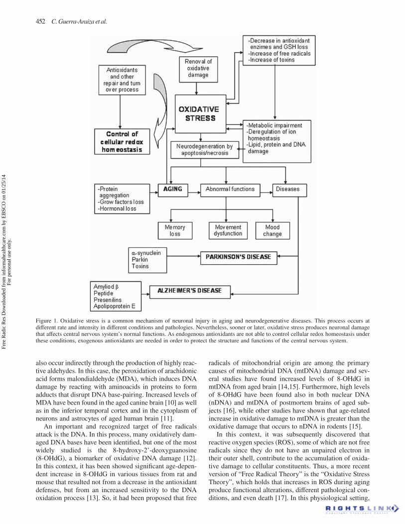
Alteration in cellular redox levels is a common mechanism of neuronal injury in aging and neurodegenerative dis-eases (Figure 1). Lipid peroxidation process that results from this alteration involves high production of reactive oxygen species, which are generated in excess in patholo-gies, such as Alzheimer ’ s disease (AD) and Parkinson ’ s disease (PD) [1,2].
On aging and neurodegenerative diseases, oxygen, nutri-ents, and ATP are available to the nerve cells in a limited way during long periods of time [3]. So, aging is character-ized as a progressive decline in biological functions with time, which results in a decreased resistance to multiple forms of stress, as well as in an increased susceptibility to numerous diseases. In other words, aging is the result of the accumulation of diverse deleterious changes in cells and tissues that increase the risk of disease and death [4], while neurodegenerative diseases are the result of similar pro-cesses but in a faster or more severe manner.
Aging
To date, aging remains as one of the most poorly under-stood of all biological phenomena, due in large part to its
Correspondence: Salgado-Ceballos Hermelinda, Calle 25 No. 52- 2 Col. Pro-hogar, c.p. 02600, M é xico D.F., M é xico. Phone: � (52 55 55780240), � (52 55 57190741). E-mail: [email protected]
(Received date: 4 October 2012 ; Accepted 10 April 2013; Published Online: 10 May 2013 )
Free Radical Research, June–July 2013; 47(6–7): 451–462© 2013 Informa UK, Ltd.ISSN 1071-5762 print/ISSN 1029-2470 onlineDOI: 10.3109/10715762.2013.795649
REVIEW ARTICLE
Free
Rad
ic R
es D
ownl
oade
d fr
om in
form
ahea
lthca
re.c
om b
y E
BSC
O o
n 01
/25/
14Fo
r pe
rson
al u
se o
nly.
452 C. Guerra-Araiza et al.
Figure 1. Oxidative stress is a common mechanism of neuronal injury in aging and neurodegenerative diseases. This process occurs at diff erent rate and intensity in diff erent conditions and pathologies. Nevertheless, sooner or later, oxidative stress produces neuronal damage that aff ects central nervous system ’ s normal functions. As endogenous antioxidants are not able to control cellular redox homeostasis under these conditions, exogenous antioxidants are needed in order to protect the structure and functions of the central nervous system.
also occur indirectly through the production of highly reac-tive aldehydes. In this case, the peroxidation of arachidonic acid forms malondialdehyde (MDA), which induces DNA damage by reacting with aminoacids in proteins to form adducts that disrupt DNA base-pairing. Increased levels of MDA have been found in the aged canine brain [10] as well as in the inferior temporal cortex and in the cytoplasm of neurons and astrocytes of aged human brain [11].
An important and recognized target of free radicals attack is the DNA. In this process, many oxidatively dam-aged DNA bases have been identifi ed, but one of the most widely studied is the 8-hydroxy-2 ’ -deoxyguanosine (8-OHdG), a biomarker of oxidative DNA damage [12]. In this context, it has been showed signifi cant age-depen-dent increase in 8-OHdG in various tissues from rat and mouse that resulted not from a decrease in the antioxidant defenses, but from an increased sensitivity to the DNA oxidation process [13]. So, it had been proposed that free
radicals of mitochondrial origin are among the primary causes of mitochondrial DNA (mtDNA) damage and sev-eral studies have found increased levels of 8-OHdG in mtDNA from aged brain [14,15]. Furthermore, high levels of 8-OHdG have been found also in both nuclear DNA (nDNA) and mtDNA of postmortem brains of aged sub-jects [16], while other studies have shown that age-related increase in oxidative damage to mtDNA is greater than the oxidative damage that occurs to nDNA in rodents [15].
In this context, it was subsequently discovered that reactive oxygen species (ROS), some of which are not free radicals since they do not have an unpaired electron in their outer shell, contribute to the accumulation of oxida-tive damage to cellular constituents. Thus, a more recent version of “ Free Radical Theory ” is the “ Oxidative Stress Theory ” , which holds that increases in ROS during aging produce functional alterations, diff erent pathological con-ditions, and even death [17]. In this physiological setting,
Free
Rad
ic R
es D
ownl
oade
d fr
om in
form
ahea
lthca
re.c
om b
y E
BSC
O o
n 01
/25/
14Fo
r pe
rson
al u
se o
nly.
Antioxidants on aging and neurodegeneration 453
oxidative stress can be defi ned as an excessive bioavail-ability of ROS, which its net result is an imbalance between production and destruction of ROS [18,19] exacerbated by the high content of unsaturated fatty acids that are more liable to peroxidation, the high content of pro-oxidant iron ions, and the low reserve of antioxidant defenses of the central nervous system [12] that directly aff ect the neu-ronal survival.
According to all these information, numerous studies have associated aging with a signifi cant accumulation of diff erent markers of oxidative stress [1,8,10], showing the importance of this biochemical process on natural aging and disease.
Neurodegenerative diseases
Progressive degeneration and/or death of nerve cells result in neurodegenerative diseases, which causes problems with movement and/or mental functioning. Oxidative stress is involved in the pathology of the neurodegenera-tive diseases since it can cause neuronal injury, cell death, and necrosis [20]. In the brain, some stimuli such as glu-tamate and infl ammation seem to be the major source of the oxidative stress by means of diff erent mechanisms [20 – 22]. Glutamate-induced oxidative stress results in the depletion of glutathione and the accumulation of ROS lev-els by suppressing the cystine uptake through the cystine/glautamate antiporter [21], while in neuroinfl ammatory pathological conditions, nitric oxide (NO), reactive NO spe-cies and ROS can initiate and in turn activate transcriptional factor NF-kB leading to expression of proinfl ammatory genes such as inducible nitric oxide synthase (iNOS) and cyclooxygenase 2 (COX-2), whose excessive production may contribute to the progress of neurodegeneration [23].
Aging brain and oxidative stress
Although brain aging should be associated or explained by a decrease in neuronal numbers by ROS destruction, some studies had shown few brain regions with signifi cant neuronal losses [24], where aging could be associated just with a small decline in cognitive and memory functions [25] and the substrate must be functional instead of ana-tomical, probably at the level of synaptic activity where ROS are also implied but in a diff erent manner. One way or another, disruptions of the homeostasis from the ner-vous system by ROS can manifest themselves in diff erent ways, including alterations in synaptic transmission such hyperexcitability, impaired neuronal plasticity, and even cell death. The point is that the generation of ROS results in secondary injury to the brain microvasculature, and interference with cerebral blood fl ow autoregulation [26] produces high activation of nuclear transcriptional factors such as NFkB and AP-1, responsible for the stimulation of a broad range of extracellular signaling molecules involved in infl ammation, tissue remodeling, oncogenesis, and apoptosis [27].
Neurodegenerative brain diseases and oxidative stress
Increased levels of lipid peroxidation and particularly 4-HNE adduct products have been demonstrated in the brain of human subjects with neurodegenerative diseases [28,29].
Moreover, in the brain tissues of patients with AD, the carbonyl derivative of protein oxidation is increased in the cytoplasm and nuclei of neurons and glial cells. Further-more, the same type of oxidative damage that is present in the brain of humans with AD is showed in the trans-genic animals overexpressing the amyloid beta peptide (A β ), and this damage is directly correlated with the pres-ence of deposits of such protein. It has also been identifi ed an increase in lipid peroxidation in transgenic mice with mutations in A β precursor protein (APP) and it has been proved that this increase precedes the A β level rise and amyloid plaque formation [30].
Additionally, it is known that in the early stages of exposure to ROS, neurotransmitter release is increased in several systems including cholinergic, but this release diminishes later [31]. It has been described that in patients with AD, there are changes in cholinergic neurotransmis-sion and alterations caused by oxidative stress in the hip-pocampus and prefrontal cortex [32] producing dysfunction (complex IV uncoupling of the respiratory chain enzyme pyruvate dehydrogenase decreased and hyperphosphory-lation of Tau protein); changes in the proteasome prote-olytic activity (enzyme altered ubiquitin ligase); and changes in the generation of ROS, specifi cally on the role of enzymes such as superoxide dismutase (SOD) [33].
Since oxidative stress increases the expression and activity of COX2 and α -synuclein, oxidative stress also causes abnormal deposits of proteins such as A β whose accumulation is associated with apoptotic cell death [34]. COX2 enhances the generation of peptide A β through the alteration of the activity of γ -secretase. Moreover, oxida-tive stress increases the levels of 4-HNE, the major prod-uct of lipid peroxidation, which is co-localized with abnormal aggregates of protein Tau and is associated with the physiopathology of AD. In both, PD and Huntington ’ s disease, accumulation of intracellular toxic proteins has also been described. Toxicity of this product of lipid per-oxidation exerts via several mechanisms, such as alteration in the membrane fl uidity and macromolecular intercom-munication [35].
Analysis of biochemical markers for oxidative damage in tissue samples from patients or in postmortem brains provided direct evidence of oxidative stress in PD. Increased levels of lipid peroxidation were found in the substantia nigra , as suggested by decreased levels of poly-unsaturated fatty acids (substrates for lipid peroxidation) and by increased levels of MDA and 4-HNE [36].
The nucleic acid oxidation product 8-OHdG is also elevated in the neurons aff ected in the disease, compared to surrounding brain regions in the PD ’ s brain, as well as in comparison with the substantia nigra of age-matched controls [37]. Lipid and DNA oxidation was found to be
Free
Rad
ic R
es D
ownl
oade
d fr
om in
form
ahea
lthca
re.c
om b
y E
BSC
O o
n 01
/25/
14Fo
r pe
rson
al u
se o
nly.
454 C. Guerra-Araiza et al.
systemically elevated in patients with PD [38]. Oxidative and nitrative posttranslational modifi cations have been identifi ed on proteins that may aff ect the disease progres-sion [39], as well as increased levels of oxidized protein carbonyls and nitration of tyrosine residues in the substan-tia nigra of patients with PD [40]. Furthermore, there is considerable evidence for the involvement of mitochon-drial dysfunction in PD, because mutations or polymor-phisms in certain genes increase the risk of PD and they are linked to mitochondrial function [41].
Cognitive dysfunctions on aging
Loss of cognitive abilities during aging is a complex pro-cess that starts to become evident during middle age in humans (35 – 65 years) and in rats (12 – 24 months) even in the absence of specifi c neurodegenerative diseases [42]. The aged human brain exhibits a number of alterations in structure and function, leading to a decline in cognitive and motor abilities. Two hallmarks of the aged human brain which have proven amenable to nutritional interven-tion are oxidative stress and polyunsaturated fatty acid (PUFA) deregulation [43]. Although cells routinely pro-duce ROS as a byproduct of normal respiration, cellular production surpasses endogenous antioxidant defenses in the aged human brain [44], leading to an increase in oxida-tive damage to proteins, DNA, and lipids [45] as it was explained before. During loss of cognitive abilities, lipid peroxidation is particularly toxic to neurons because it alters cell membrane properties as well as the function of membrane-bound receptors, ion channels, and signaling molecules [46].
Most fatty acids enriched in neuronal membranes are essential fatty acids, so called because they cannot be syn-thesized in vivo and must be obtained from the diet. In animals, the aged brain exhibits lower concentrations of PUFAs in neuronal membranes than that of young animals [47] and less phospholipid-bound fatty acid is found in the aged brain, particularly in the cortex and hippocampus [48]. These alterations contribute to a decrease in mem-brane fl uidity, which is exacerbated in the hippocampus, cortex, cerebellum, and striatum of aged rats [49]. In addi-tion, with less membrane-bound fatty acids available for cleavage, less free fatty acid is liberated from cellular stores to participate in cell signaling cascades. The point is that age-related decline in neuronal membrane fatty acid composition, therefore, contributes not only to membrane fl uidity [50] but also to alterations in neuronal structure and reduced synaptic plasticity [51].
It is known that acute cognitive impairments are common in elderly humans and often occur in association with peripheral infection [52,53] because during a periph-eral infection, cells associated with the peripheral innate immune system, such as macrophages and mono-cytes, produce infl ammatory cytokines like interleukin (IL)-1b, IL-6, and tumor necrosis factor- α (TNF- α ) that conveys information to the brain, where microglial cells react and produce infl ammatory cytokines too. In chronic
neurodegenerative diseases as well as in aging, microglial cells are activated and produce excessive amounts of infl ammatory cytokines that can generate severe behav-ioral defi cits and promote neurotoxicity in the brain [54]. This phenomenon has been demonstrated in the brains of healthy aged mice where MHC class II, a marker for acti-vated microglia, was shown to be increased [55], while HLA-DR, a MHC class II marker, was found to be increased on hippocampal microglia of aged humans [56] as well as in patients with infl ammatory disease or injury [57]. Moreover, on intraperitoneal administration of lipopolysaccharide (LPS), an exaggerated infl ammatory cytokine response in the aged mice is observed [55,58], and more severe anorexia, depressive behaviors, and defi cits in hippocampal-dependent learning and memory are observed in the aged mice than in the younger ones [55,58,59].
Cognitive dysfunctions in neurodegenerative diseases
In patients with neurodegenerative diseases, an associa-tion between cognitive impairments and peripheral infec-tions (57, 60, 61) has also been described. In this matter, in a model of chronic neurodegenerative disease using the murine ME7 experimental model of prion disease, stimu-lation of the peripheral innate immune system with LPS induced an aggressive infl ammatory cytokine response by brain microglial cells, excessive sickness behavior, and accelerated progression of the disease [60,61] because the neurodegenerative diseases provide a sensitizing stimulus for microglia, while subsequent signals from peripheral immune stimulation provide a secondary triggering stimu-lus [57]. So, combination of these two stimuli results in an overall response of a greater magnitude than the sum of the responses to individual stimuli alone [62]. This interaction explains, at least partially, why infection is a risk factor for relapse in multiple sclerosis patients and for dementia in patients with AD [63,64].
On neurodegenerative disease and aging, the infl amma-tory cytokine IL-6 plays a role in modulating cognition. The severity of dementia in AD has been linked to high levels of IL-6 in plaques [65] and IL-6 levels in plasma were cor-related with cognitive impairments in humans [66]. In transgenic mice whose astrocytes overexpressed IL-6, defi -cits in avoidance learning and a reduction in dendritic com-plexity in the hippocampus havebeen showed [67,68].
Motor function defi cits in aging
On the other hand, it has been also postulated that accu-mulation of oxidized proteins or lipid peroxidation in aging may be involved in the age-related functional neu-ronal loss contributing to de fi cits in motor performance [69,70]. These alterations in motor function may specifi -cally include decreases in balance, muscle strength, and coordination. Both, aged rats and mice, show decrements in performance on several tasks requiring coordinated
Free
Rad
ic R
es D
ownl
oade
d fr
om in
form
ahea
lthca
re.c
om b
y E
BSC
O o
n 01
/25/
14Fo
r pe
rson
al u
se o
nly.
Antioxidants on aging and neurodegeneration 455
control of motor and re fl exive responses, such as suspen-sion time on a horizontal wire or inclined wire mesh screen, and the length of time it takes for the animal to traverse a wooden rod or plank [71 – 75]. Age-related de fi cits in motor performance are thought to be the result of alterations in the striatal dopamine system since the striatum, a brain area which mediates several aspects of motor behavior [76], shows marked neurodegenerative changes on aging [69].
Motor function defi cits in pd
PD is a neurodegenerative disorder of the brain character-ized by degeneration or dysfunction of the dopaminergic nigrostriatal system of the basal ganglia and its connec-tion. It begins most often between 45 and 65 years of age, and the number of people with PD in the most populous nations is estimated to be between 4.1 and 4.6 million [77]. Clinically, it presents mainly with resting tremor, muscular rigidity, bradykinesia, postural instability, and autonomic abnormalities. The pathological hallmarks include depigmentation of the substantia nigra and the locus ceruleus with the presence of Lewy bodies [78]. The exact cause of the disease and the nature of its progression are still not fully understood. However, environmental fac-tors, such as exposure to pesticides and herbicides, genetic predisposition and age, have all been identifi ed as the etiological factors [79]. Recently, growing evidences suggest a crucial link between the oxidative stress and the development of many neurodegenerative diseases, including PD [80,81], since postmortem studies in brains from patients with PD indicate that a wide range of molecules, including lipids, proteins, and DNA, undergo oxidative damage [82]. The common use of highly reac-tive neurotoxins, such as 6-hydroxydopamine (6-OHDA) to induce parkinsonism in animal models, further supports the possible involvement of oxidative stress in the etiopathogenesis of PD.
Endogenous and exogenous antioxidants
Defense mechanisms to protect against ROS-induced cell damage are crucial in human health. In this framework, the main endogenous antioxidants in humans have included glutathione peroxidase, catalase, and SOD. These anti-oxidants are primary enzymes, which metabolize toxic oxidative intermediates and require micronutrient as cofactors such as selenium, iron, copper, zinc, and man-ganese for optimum catalytic activity and eff ective anti-oxidant defense [83,84]. Glutathione peroxidase, catalase, and SOD are involved in direct elimination of ROS like hydroxyl radical, superoxide radical, and hydrogen perox-ide, whereas glutathione reductase, glucose-6-phosphate dehydrogenase, and cytosolic glutathione S-transferase are secondary enzymes which help in the detoxifi cation of ROS by decreasing peroxide levels or maintaining a steady supply of metabolic intermediates, like glutathione and
NADPH, essential for optimum functioning of the pri-mary antioxidant enzymes [85].
As a result, if the antioxidant levels are inadequate, reactions will only be delayed until the antioxidants have been consumed [86]. So, on aging as well as in the case of several neurodegenerative diseases, there is a proved decline in the normal antioxidant defense mechanisms, which increases the vulnerability of the brain to the del-eterious eff ects of oxidative damage [87], and the anti-oxidant enzymes like SOD, catalase, glutathione peroxidase and glutathione reductase display reduced activities in the brain [88].
Nevertheless, others have been supported that endog-enous levels of antioxidant enzymes in brain tissues do not decrease during aging [89]. Furthermore, studies on mam-mals, in which levels of antioxidants are experimentally increased, have shown that maximum longevity is not aff ected. However, experiments on Drosophila melano-gaster have shown that overexpression of mimetic SOD (MnSOD) increased life span while overexpression of CuZn-SOD had only minor incremental eff ects on it [90]. This outcome has also been observed in mice [91]. Although several studies have shown that elevated CuZN-SOD induced protection against oxidative stress, other reports did not fi nd a correlation between such pro-tection and increased life span as homozygous transgenic mice with a two- to fi vefold increase in CuZN-SOD that showed only a small increase [92].
Even so, when levels of endogenous antioxidants such as reduced glutathione (GSH), decrease with age, the sus-ceptibility of vascular endothelial cells and fi broblasts to oxidative stress associated infl ammatory stimuli in vitro increased [27]. This correlates with the idea that the sig-nifi cant accumulation of diff erent markers of oxidative stress resulted not from a decrease in the antioxidant defenses, but from an increased sensitivity to oxidation pro-cess [13]. Moreover, it has been postulated that there may not be partial benefi ts if inadequate exogenous antioxidant dose is given. In this context, the earlier the antioxidants are started in a branching-free radical chain reaction, the lower the dose of antioxidants will be needed [86].
As antioxidants are needed to prevent the formation and to oppose the actions of ROS and reactive nitrogen species (RNS), which are generated in vivo and cause damage to DNA, lipids, proteins, and other biomolecules because endogenous antioxidant defenses (SOD, hydrogen perox-ide-removing enzymes and metal binding proteins) are inadequate to prevent damage completely, diet-derived antioxidants are important in maintaining health.
So, diff erent eff orts have been undertaken in order to increase the use of natural antioxidants and to develop anti-oxidants that might ameliorate neural injury associated with ROS. The most widely studied dietary antioxidants are vita-min C, vitamin E, and beta-carotene. Vitamin C is consid-ered to be the most important water-soluble antioxidant in extracellular fl uids, as it is capable of neutralizing ROS in the aqueous phase before lipid peroxidation is initiated, while vitamin E is a major lipid-soluble antioxidant and is the most eff ective chain-breaking antioxidant within the
Free
Rad
ic R
es D
ownl
oade
d fr
om in
form
ahea
lthca
re.c
om b
y E
BSC
O o
n 01
/25/
14Fo
r pe
rson
al u
se o
nly.
456 C. Guerra-Araiza et al.
cell membrane where it protects fatty acids from lipid per-oxidation. Beta-carotene and other carotenoids also pro-vide antioxidant protection to lipid-rich tissues but in a less eff ective manner. Fruits and vegetables are the major sources of vitamin C and carotenoids, while whole grains, that is, cereals and high-quality vegetable oils are the major sources of vitamin E [93].
Beyond the traditional vitamins, a number of other dietary antioxidants collectively known as phytonutrients or phytochemicals exist, which are being increasingly appreciated for their antioxidant activity and may have important roles as dietary components via cytoprotective actions in many organs [94,95].
In this context, an important group of natural antioxi-dants are the fl avonoids whose role in protecting human health is well known [96]. They are found in leaves, fl ow-ers, fruits (especially citrus fruits), apples, onions, seeds, nuts, grains, spices, olive oil, diff erent medicinal plants, and beverages such as red wine, green tea, and beer [97,98] which contain thousands of phytochemicals, including polyphenolic compounds that express anti-infl ammatory, anti-carcinogenic, antibacterial, vasodilator, immune-stim-ulating anti-allergic and antiviral compounds [99,100], free radical scavenging properties and neuroprotective activity from oxidative injury by their ability to modulate intracel-lular signals (Figure 2) promoting cellular survival [101].
One of the mechanisms of the antioxidant activity of fl avonoids is characterized by direct scavenging or quench-ing of oxygen free radicals or excited oxygen species [102]. Flavonoids, ubiquitously included as glucosylated derivatives in vegetables, fruits, and herbs have been char-acterized as neuroprotectants and used with the aim to prevent degenerative diseases as well as in the treatment of various neurological disorders. Among fl avonoids, quercetin (3,5,7,3 ′ ,4 ′ -pentahydroxyfl avone), a biofl a-vonoid typically present in plants as glycone conjugates that include rutin and thujin or as carbohydrate conjugates, is one of the best studied. It is regarded as one of the most potent antioxidants among all fl avonoids and is capable of scavenging ROS and binding transition metal ions (e.g., iron and copper); hence, it protects the body from oxida-tive damage [103,104].
Quercetin has many benefi cial eff ects on human health, including cardiovascular protection, anticancer and antivi-ral activity, cataract prevention, and anti-infl ammatory eff ects [105] due to its ability to chelate Fe 2 � and to scav-enge free radicals [106]. Furthermore, quercetin is also a potent regulator of the cell apoptotic death program asso-ciated with the modulation of several signaling molecules such as protein and lipid kinases [107,108].
Thus, diff erent researchers support the administration of ROS scavengers and polyphenols in monotherapy, or as part of an antioxidant cocktail formulation for the treat-ment of diverse diseases [109]. Moreover, research on the useful eff ects of fl avonoids in the treatment of aging and brain dysfunctions (like cognition) has been suggested successfully in animals [110] and demonstrated benefi cial eff ects on age-related cognitive decline in humans since an inverse relationship between fl avonoid intake and
cognitive decline in older persons has been reported, and fl avonoid intake has been associated with better cognitive performance at baseline as well as with better evolution of performance over time [111] and that the relative risk of dementia is inversely related to the mean fl avonoid intake in subjects above 65 years of age [112].
Recent evidence suggests that ingestion of fl avonoids is associated with improved cognitive function and a lower risk of neurodegenerative disease, but the benefi cial eff ects produced by fl avonoids may be conferred not only by the promotion of synaptic plasticity and neurogenesis, but also by the suppressed neuronal cell damage secondary to oxidative stress and by the activation of primary transcrip-tion factors that regulate infl ammatory genes such as NF-kB and AP-1 as well as MAPK pathways (involved in regulating infl ammatory gene expression) in activated microglia, which results in an attenuation of the produc-tion of infl ammatory molecules or in an anti-infl ammatory eff ect [62,113,114].
Anti-infl ammatory eff ect of fl avonoids could aff ect the antioxidative ones positively since the regulation of acti-vating NF-kB and AP-1 transcription factors as well as MAPK pathways are sensitive to redox. Diff erent benefi -cial eff ects of fl avonoid-rich foods on age-related cogni-tive and motor dysfunctions have been shown with blueberries [115], strawberries, concord grapes [87], and polyphenols from red wine [88]. Initial studies showed that long-term (age of 6 – 15 months in F344 rats) feeding with a diet supplemented with strawberries, spinach, or vitamin E prevented age-related decrements in cognitive function as assessed by the Morris water maze [116]. A subsequent experiment showed that dietary supplementa-tion with spinach, strawberries, or blueberries could actu-ally reverse already established defi cits in behavioral and cognitive functions in aged (19 months) F344 rats [115].
In the past decade, the antioxidant activity of fl avonoids has been given much attention, since many fl avonoids such as quercetin, luteolin, and catechins may be better antioxidants than the antioxidant nutrients such as vitamin C, vitamin E, and β -carotene [117].
Antioxidant eff ectiveness of vitamin C, an antioxidant with a proven ability to reduce the risk of neurodegenera-tive disorders such as AD, was evaluated and compared with quercetin properties in an in vitro model of hydroxy peroxide-induced neurodegeneration in PC12 cells. Thus, cells were preincubated for 2 h with quercetin or vitamin C before hydroxy peroxide treatment where quercetin decreased oxidative stress-induced neuronal cell mem-brane damage more than vitamin C and improved cell viability [118]. Previously, it was shown that vegetables had antioxidant quality comparable with that of pure fl a-vonoids and were superior to vitamin antioxidants [119] as it was demonstrated recently. Due to the fact that quer-cetin had stronger antioxidative activity than vitamin C, it could be a better therapeutic option for AD and other pathologies from central nervous system [118].
As neuronal loss in AD is preceded by the extracellular accumulation of A β , quercetin can increase the resistance of neurons against oxidative stress and excitotoxicity by
Free
Rad
ic R
es D
ownl
oade
d fr
om in
form
ahea
lthca
re.c
om b
y E
BSC
O o
n 01
/25/
14Fo
r pe
rson
al u
se o
nly.
Antioxidants on aging and neurodegeneration 457
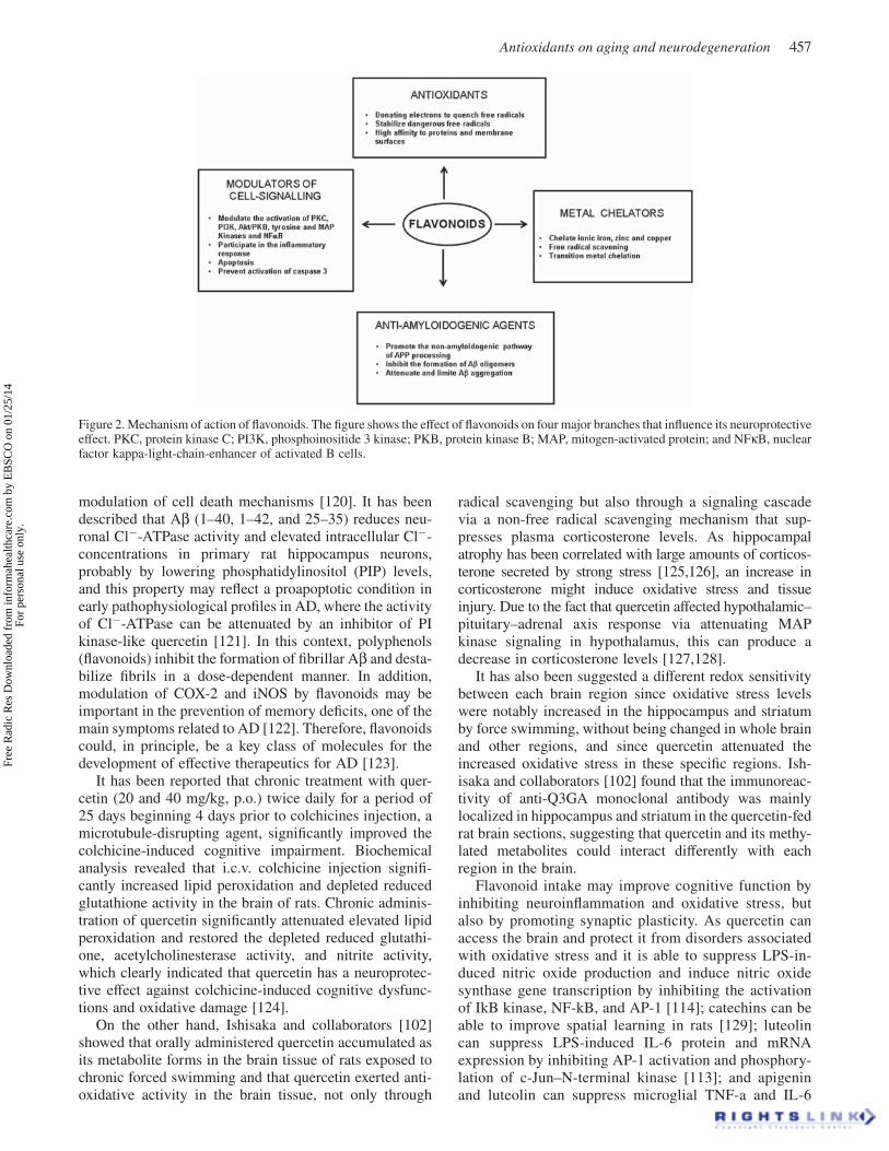
Figure 2. Mechanism of action of fl avonoids. The fi gure shows the eff ect of fl avonoids on four major branches that infl uence its neuroprotective eff ect. PKC, protein kinase C; PI3K, phosphoinositide 3 kinase; PKB, protein kinase B; MAP, mitogen-activated protein; and NF κ B, nuclear factor kappa-light-chain-enhancer of activated B cells.
modulation of cell death mechanisms [120]. It has been described that A β (1 – 40, 1 – 42, and 25 – 35) reduces neu-ronal Cl � -ATPase activity and elevated intracellular Cl � -concentrations in primary rat hippocampus neurons, probably by lowering phosphatidylinositol (PIP) levels, and this property may refl ect a proapoptotic condition in early pathophysiological profi les in AD, where the activity of Cl � -ATPase can be attenuated by an inhibitor of PI kinase-like quercetin [121]. In this context, polyphenols (fl avonoids) inhibit the formation of fi brillar A β and desta-bilize fi brils in a dose-dependent manner. In addition, modulation of COX-2 and iNOS by fl avonoids may be important in the prevention of memory defi cits, one of the main symptoms related to AD [122]. Therefore, fl avonoids could, in principle, be a key class of molecules for the development of eff ective therapeutics for AD [123].
It has been reported that chronic treatment with quer-cetin (20 and 40 mg/kg, p.o.) twice daily for a period of 25 days beginning 4 days prior to colchicines injection, a microtubule-disrupting agent, signifi cantly improved the colchicine-induced cognitive impairment. Biochemical analysis revealed that i.c.v. colchicine injection signifi -cantly increased lipid peroxidation and depleted reduced glutathione activity in the brain of rats. Chronic adminis-tration of quercetin signifi cantly attenuated elevated lipid peroxidation and restored the depleted reduced glutathi-one, acetylcholinesterase activity, and nitrite activity, which clearly indicated that quercetin has a neuroprotec-tive eff ect against colchicine-induced cognitive dysfunc-tions and oxidative damage [124].
On the other hand, Ishisaka and collaborators [102] showed that orally administered quercetin accumulated as its metabolite forms in the brain tissue of rats exposed to chronic forced swimming and that quercetin exerted anti-oxidative activity in the brain tissue, not only through
radical scavenging but also through a signaling cascade via a non-free radical scavenging mechanism that sup-presses plasma corticosterone levels. As hippocampal atrophy has been correlated with large amounts of corticos-terone secreted by strong stress [125,126], an increase in corticosterone might induce oxidative stress and tissue injury. Due to the fact that quercetin aff ected hypothalamic – pituitary – adrenal axis response via attenuating MAP kinase signaling in hypothalamus, this can produce a decrease in corticosterone levels [127,128].
It has also been suggested a diff erent redox sensitivity between each brain region since oxidative stress levels were notably increased in the hippocampus and striatum by force swimming, without being changed in whole brain and other regions, and since quercetin attenuated the increased oxidative stress in these specifi c regions. Ish-isaka and collaborators [102] found that the immunoreac-tivity of anti-Q3GA monoclonal antibody was mainly localized in hippocampus and striatum in the quercetin-fed rat brain sections, suggesting that quercetin and its methy-lated metabolites could interact diff erently with each region in the brain.
Flavonoid intake may improve cognitive function by inhibiting neuroinfl ammation and oxidative stress, but also by promoting synaptic plasticity. As quercetin can access the brain and protect it from disorders associated with oxidative stress and it is able to suppress LPS-in-duced nitric oxide production and induce nitric oxide synthase gene transcription by inhibiting the activation of IkB kinase, NF-kB, and AP-1 [114]; catechins can be able to improve spatial learning in rats [129]; luteolin can suppress LPS-induced IL-6 protein and mRNA expression by inhibiting AP-1 activation and phosphory-lation of c-Jun – N-terminal kinase [113]; and apigenin and luteolin can suppress microglial TNF-a and IL-6
Free
Rad
ic R
es D
ownl
oade
d fr
om in
form
ahea
lthca
re.c
om b
y E
BSC
O o
n 01
/25/
14Fo
r pe
rson
al u
se o
nly.
458 C. Guerra-Araiza et al.
production stimulated by interferon-gamma [130], fl a-vonoids could be an important therapeutic option for the prevention and treatment of aging and neurodegenerative diseases.
Endogenous and exogenous antioxidants in neurodegenerative disorders
As oxidative stress has been strongly implicated in PD, diet-derived phenolic compounds such as curcumin, naringenin, quercetin, and fi setin had been tested as neu-roprotectors in the 6-OHDA-induced model of PD, where a signifi cant loss of tyrosine hydroxylase-positive cells in the substantia nigra as well as a decreased of dopamine content in the striata is produced. Rats pre-treated with curcumin or naringenin showed a clear pro-tection of tyrosine hydroxylase-positive cells in substantia nigra and dopamine levels in the striata. However, pretreatment with either quercetin or fi setin had any eff ects on tyrosine hydroxylase-positive cells or dopamine levels [131].
However, another study with 6-OHDA-induced model of PD in rats showed that quercetin treatment (30 mg/kg body weight) over 14 consecutive days markedly increased the striatal dopamine and antioxidant enzyme levels com-pared with similar measurements in the group treated with 6-OHDA alone. A signifi cant decrease in protein carbonyl content in the striatum was also observed when compared with that of rats that did not receive quercetin and a sig-nifi cant increase in neuronal survivability was also found with quercetin treatment in rats administered 6-OHDA [132]. So, in this rodent model of PD, treatment with quer-cetin defended against the oxidative stress in the striatum and reduced the dopaminergic neuronal loss.
Moreover, in another study, the neuroprotective eff ect of quercetin against oxidative neuronal injuries induced in primary cultured rat cortical cells was evaluated, and under in vitro conditions, quercetin was shown to inhibit xanthine oxidase activity and eff ectively protected the neurons from oxidative injury [133].
In the 1-methyl-4-phenyl-1, 2, 3, 6-tetrahydropyridine (MPTP)-induced mouse model of PD, results show that quercetin treatment markedly improves the motor balance and coordination of MPTP-treated mice. Signifi cant increases were observed in the activities of glutathione peroxidase, SOD, Na � /K � -ATPase, AchE and the content of dopamine in animals from groups treated with querce-tin in comparison with those without treatment. Signifi -cant reduction in 4-HNE immunoreactivity in striatum of brains was observed also in the quercetin-treated groups. Taken together, these results suggested that quercetin has shown antiparkinsonian properties in this model [134].
Although polyphenols possess anti-infl ammatory and antioxidant activities that may contribute, via the diet, to the prevention of chronic diseases such as AD [135], it seems that not every neurodegenerative conditions can be benefi ciated the same way. So, diff erent doses, targets, and underlying mechanisms had been investigated.
In this area, other important antioxidants such as mag-nolol and honokiol that can be obtained from a medici-nal herb Magnolia dealbata Zucc (Magnoliaceae), an endemic species from Mexico used in traditional Mexi-can medicine as a tranquilizer and as an antiepileptic treatment [136], has been tested. In oriental medicine, herbs from the same species are used for the treatment of anxiety and neurosis [137]. Magnolol and honokiol, obtained from Magnolia dealbata Zucc, Magnolia offi -cinalis , or Magnolia obovata, have neuroprotective activity [138] since these compounds prevent age-related learning and memory impairment by preserving cholinergic neurons in the forebrain [139]. They also increase the NMDA-induced seizure thresholds [140] and have important anti-hypoxic, anti-infl ammatory [141], and antioxidative actions [138]. Moreover, the activation of GABA
A receptor by the treatment of
honokiol plays an important role in a variety of central nervous system-related activities [142–143] as its neurotrophic actions since honokiol can aff ect neurite extension as well as neuronal survival in the primary cultures of rat cortical neurons [144].
Conclusion
Under conditions such as as aging and neurodegenerative diseases, endogenous antioxidant defenses seem to be inadequate to prevent free radicals damage to DNA, lipids, proteins, and other biomolecules. So, the development and the use of diff erent exogenous antioxidants that might ameliorate neural injury by oxidative stress had been increased during the last years. In this context, natural antioxidants like fl avonoids (quercetin, curcumin, luteolin, and catechins), as well as magnolol and honokiol, are shown to be effi cient inhibitors of the oxidative process, and they have been considered as a better therapeutic option than the traditional antioxidants like vitamin C, vitamin E, and β -carotene. Nevertheless, as fl avonoids, magnolol, honokiol, and traditional antioxidants have some diff erent targets and mechanisms of actions as well as diff erent functions, if they will be used in combination, better results could be obtained on diff erent processes and pathologies where the oxidative stress is a clue key as in aging and neurodegenerative diseases.
Acknowledgment
We acknowledge support from FIS/IMSS projects No. FIS/IMSS/PROT/G11-2/1013 and FIS/IMSS/PROT552 as well as FP-2003/025 and 2005/1/I/061. Mondrag ó n-Lozano R.; Rodolfo Pinto received fi nancial support from CONACYT; and Farf á n-Garc í a E., Á lvarez-Mej í a A.L., and S á nchez-Torres S. received fi nancial support from CONACYT and from IMSS.
We thank M.S. Julia Segura-Uribe from Hospital Infantil Federico G ó mez, SSA, for the revision of the manuscript in English.
Free
Rad
ic R
es D
ownl
oade
d fr
om in
form
ahea
lthca
re.c
om b
y E
BSC
O o
n 01
/25/
14Fo
r pe
rson
al u
se o
nly.
Antioxidants on aging and neurodegeneration 459
Declaration of interest
The authors report no declarations of interest. The authors alone are responsible for the content and writing of the paper.
References
Butterfi eld DA , Sultana R . Methionine-35 of a [1] β (1 – 42): importance for oxidative stress in Alzheimer disease . J Amino Acids 2011 ; 2011 : 198430. Epub 2011 Jun 4 . Mythri RB , Venkateshappa C , Harish G , Mahadevan A , [2] Muthane UB , Yasha TC , et al . Evaluation of markers of oxi-dative stress, antioxidant function and astrocytic proliferation in the striatum and frontal cortex of Parkinson ’ s disease brains . Neurochem Res 2011 ; 36 : 1452 – 1463 . Farooqui AA , Horrocks LA . Lipid peroxides in the free [3] radical pathophysiology of brain diseases . Cell Mol Neuro-biol 1998 ; 18 : 599 – 608 . Harman D . Aging: overview . Ann N Y Acad Sci 2001 ; 928 :[4] 1 – 21 . Kregel KC , Zhang HJ . An integrated view of oxidative stress [5] in aging: basic mechanisms, functional eff ects, and patho-logical considerations . Am J Physiol Regul Integr Comp Physiol 2007 ; 1 : 18 – 36 . Medvedev ZA . An attempt at a rational classifi cation of theo-[6] ries of ageing . Biol Rev Camb Philos Soc 1990 ; 65 : 375 – 398 . Harman D . Aging: a theory based on free radical and radia-[7] tion chemistry . J Gerontol 1956 ; 11 : 298 – 300 . Stadtman ER . Protein oxidation and aging . Science [8] 1992 ; 257 : 1220 – 1224 . Esterbauer H , Schaur RJ , Zollner H . Chemistry and biochem-[9] istry of 4-hydroxynonenal, malonaldehyde and related alde-hydes . Free Radic Biol Med 1991 ; 1181 – 128 . Head E , Liu J , Hagen TM , Muggenburg BA , Milgram NW , [10] Ames BN , Cotman CW . Oxidative damage increases with age in a canine model of human brain aging . J Neurochem 2002 ; 82 : 375 – 381 . Dei R , Takeda A , Niwa H , Li M , Nakagomi Y , Watanabe M , [11] et al . Lipid peroxidation and advanced glycation end prod-ucts in the brain in normal aging and in Alzheimer ’ s disease . Acta Neuropathologica 2002 ; 104 : 113 – 122 . Floyd RA , Hensley K . Oxidative stress in brain aging. [12] Implications for therapeutics of neurodegenerative diseases . Neurobiol Aging 2002 ; 23 : 795 – 807 . Hamilton ML , Van Remmen H , Drake JA , Yang H , Guo ZM , [13] Kewitt K , et al . Does oxidative damage to DNA increase with age? Proc Natl Acad Sci U S A 2001 ; 98 : 10469 – 10474 . Richter C , Park JW , Ames BN . Normal oxidative damage to [14] mitochondrial and nuclear DNA is extensive . Proc Natl Acad Sci U S Am 1988 ; 85 : 6465 – 6467 . Agarwal S , Sohal RS . Aging and protein oxidative damage . [15] Mechanisms of Ageingand Dev 1994 ; 75 : 11 – 19 . Mecocci P , MacGarvey U , Kaufman AE , Koontz D , [16] Shoff ner JM , Wallace DC , Beal MF . Oxidative damage to mitochondrial DNA shows marked age-dependent increases in human brain . Ann Neurol 1993 ; 34 : 609 – 616 . Hagen TM . Oxidative stress, redox imbalance, and the aging [17] process . Antioxid Redox Signal 2003 ; 5 : 503 – 506 . Ashok BT , Ali R . The aging paradox: free radical theory of [18] aging . Exp Gerontol 1999 ; 34 : 293 – 303 . Balaban RS , Nemoto S , Finkel T . Mitochondria, oxidants, [19] aging . Cell 2005 ; 120 : 483 – 495 . Love S . Oxidative stress in brain ischemia . Brain Pathol [20] 1999 ; 9 : 119 – 131 . Davis JB , Mahe P . Protein kinase C activation inhibits gluta-[21] mate-induced cytotoxicity in a neuronal cell line . Brain Res 1994 ; 652 : 169 – 173 .
McGeer PL , McGeer EG . The infl ammatory response [22] system of brain: implications for therapy of Alzheimer and other neurodegenerative diseases . Brain Res Rev 1995 ; 21 : 195 – 218 . Chabrier PE , Demerle-Pallardy C , Auguet M . Nitric oxide [23] synthases: targets for therapeutic strategies in neurological diseases . Cell Mol Life Sci 1999 ; 55 : 1029 – 1035 . Hof PR , Morrison JH . The aging brain: morphomolecular [24] senescence of cortical circuits . Trends Neuroscie 2004 ; 27 : 607 – 613 . Rosenzweig ES , Barnes CA . Impact of aging on hippocam-[25] pal function: plasticity, network dynamics, and cognition . Prog Neurobiol 2003 ; 69 : 143 – 179 . Wei EP , Kontos HA , Dietrich WD , Povlishock JT , Ellis EF . [26] Inhibition of free radical scanvengers and by ciclooxigenase inhibitors of pial arteriolar abnormalities from concussive head injury in cats . Circ Res 1981 ; 48 : 95 – 103 . Hu HL , Forsey RJ , Blades TJ , Barratt ME , Parmar P , Powell [27] JR . Antioxidants may contribute in the fi ght against ageing: an in vitro model . Mech Ageing Dev 2001 ; 121 : 217 – 230 . Jenner P . Oxidative stress in Parkinson ’ s disease . Ann Neurol [28] 2003 ; 53 : S26 – S36; discussion S36 – S38 . Singh M , Dang TN , Arseneault M , Ramassamy C . Role of [29] by-products of lipid oxidation in Alzheimer ’ s disease brain: a focus on acrolein . J Alzheimer ’ s Dis 2010 ; 21 : 741 – 756 . Rodrigues R , Smith MA , Wang X , Perry G , Lee HG , Zhu X , [30] Petersen RB . Molecular neuropathogenesis of Alzheimer ’ s disease: an interaction model stressing the central role of oxidative stress . Future Neurol 2012 ; 7 : 287 – 305 . Halliwell B . Proteasomal dysfunction: a common feature of [31] neurodegenerative diseases? Implications for the environ-mental origins of neurodegeneration . Antioxid Redox Signal 2006 ; 8 : 2007 – 2019 . Dumas JA , Newhouse PA . The cholinergic hypothesis of [32] cognitive aging revisited again: cholinergic functional com-pensation . Pharmacol Biochem Behav 2011 ; 99 : 254 – 261 . Reddy PH , Tripathi R , Troung Q , Tirumala K , Reddy TP , [33] Anekonda V , et al . Abnormal mitochondrial dynamics and synaptic degeneration as early events in Alzheimer ’ s disease: implications to mitochondria-targeted antioxidant therapeu-tics . Biochim Biophys Acta 2012 ; 1822 : 639 – 649 . Xiang Z , Ho L , Yemul S , Zhao Z , Qing W , Pompl P , et al . [34] Cyclooxygenase-2 promotes amyloid plaque deposition in a mouse model of Alzheimer ’ s disease neuropathology . Gene Expr 2002 ; 10 : 271 – 278 . Sonnen JA , Breitner JC , Lovell MA , Markesbery WR , Quinn [35] JF , Montine TJ . Free radical-mediated damage to brain in Alzheimer ’ s disease and its transgenic mouse models . Free Radic Biol Med 2008 ; 45 : 219 – 30 . Perfeito R , Cunha-Oliveira T , Rego AC . Revisiting oxidative [36] stress and mitochondrial dysfunction in the pathogenesis of Parkinson disease – resemblance to the eff ect of amphetamine drugs of abuse . Free Radic Biol Med 2012 ; 53 : 1791 – 806 . Abe T , Isobe C , Murata T , Sato C , Tohgi H . Alteration of [37] 8-hydroxyguanosine concentrations in the cerebrospinal fl uid and serum from patients with Parkinson ’ s disease . Neurosci Lett 2003 ; 336 : 105 – 108 . Seet RC , Lee CY , Lim EC , Tan JJ , Quek AM , Chong WL , [38] et al . Oxidative damage in Parkinson disease: measurement using accurate biomarkers . Free Radic Biol Med 2010 ; 48 : 560 – 566 . Danielson SR , Andersen JK . Oxidative and nitrative protein [39] modifi cations in Parkinson ’ s disease . Free Radic Biol Med 2008 ; 44 : 1787 – 1794 . Nakamura T , Cho DH , Lipton SA . Redox regulation of [40] protein misfolding, mitochondrial dysfunction, synaptic damage, and cell death in neurodegenerative diseases . Exp Neurol 2012 ; 238 : 12 – 21 . Navarro A , Boveris A . Brain mitochondrialdysfunction and [41] oxidative damage in Parkinson ’ s disease . J Bioenerg Biomembr 2009 ; 41 : 517 – 521 .
Free
Rad
ic R
es D
ownl
oade
d fr
om in
form
ahea
lthca
re.c
om b
y E
BSC
O o
n 01
/25/
14Fo
r pe
rson
al u
se o
nly.
460 C. Guerra-Araiza et al.
Kluger A , Gianutsos JG , Golomb J , Ferris SH , George AE , [42] Franssen E , Reisberg B . Patterns of motor impairment in normal aging, mild cognitive decline, and early Alzheimer ’ s disease . J Gerontol 1997 ; 52 : P28 – 39 . Van Dyk K , Sano M . The impact of nutrition on cognition [43] in the elderly . Neurochem Res 2007 ; 32 : 893 – 904 . Andersen JK . Oxidative stress in neurodegeneration: cause [44] or consequence? Nat Med 2004 ; 10 : S18 – S25 . Ames BN , Shigena MK , Hagen TM . Oxidants, antioxidants [45] and anticarcinogens: oxygen radicals and degenerative dis-ease . Proc Natl Acad Sci U S Am 1993 ; 90 : 7915 – 7922 . Sultana R , Perluigi M , Butterfi eld DA . Protein oxidation and [46] lipid peroxidation in brain of subjects with Alzheimer ’ s disease: insights into mechanism of neurodegeneration from redox pro-teomics . Antioxid Redox Signal 2006 ; 8 : 2021 – 2037 . Ulmann L , Mimouni V , Roux S , Porsolt R , Poisson JP . Brain [47] and hippocampus fatty acid composition in phospholipid classes of aged-relative cognitive defi cit rats . Prostaglandins Leukot Essent Fatty Acids 2001 ; 64 : 189 – 195 . Lopez GH , Ilincheta de Boschero MG , Castagnet PI , [48] Giusto NM . Age-associated changes in the content and fatty acid composition of brain glycerophospholipids . Comp Bio-chem Physiol B Biochem Mol Biol 1995 ; 112 : 331 – 343 . Yehuda S , Rabinovitz S , Carasso RL , Mostofsky DI . The role [49] of polyunsaturated fatty acids in restoring the aging neuronal membrane . Neurobiol Aging 2002 ; 23 : 843 – 853 . Youdim A , Martin A , Joseph JA . Essential fatty acids and [50] the brain: possible health implications . Int J Dev Neurosci 2000 ; 18 : 383 – 399 . Mora F , Segovia G , del Arco A . Aging, plasticity and envi-[51] ronmental enrichment: structural changes and neurotransmit-ter dynamics in several areas of the brain . Brain Res Rev 2007 ; 55 : 78 – 88 . Woff ord JL , Loehr LR , Schwartz E . Acute cognitive impair-[52] ment in elderly ED patients: etiologies and outcomes . Am J Emerg Med 1996 ; 14 : 649 – 653 . Chiovenda P , Vincentelli GM , Alegiani F . Cognitive impair-[53] ment in elderly ED patients: need for multidimensional assessment for better management after discharge . Am J Emerg Med 2002 ; 20 : 332 – 335 . Dantzer R , O ’ Connor JC , Freund GG , Johnson RW , [54] Kelley KW . From infl ammation to sickness and depression: when the immune system subjugates the brain . Nat Rev Neu-rosci 2008 ; 9 : 46 – 56 . Godbout JP , Chen J , Abraham J , Richwine AF , Berg BM , [55] Kelley KW , Johnson RW . Exaggerated neuroinfl ammation and sickness behavior in aged mice following activation of the peripheral innate immune system . FASEB J 2005 ; 19 : 1329 – 1331 . Streit WJ , Sparks DL . Activation of microglia in the brains [56] of humans with heart disease and hypercholesterolemic rab-bits . J Mol Med 1997 ; 75 : 130 – 138 . Perry VH , Cunningham C , Holmes C . Systemic infections [57] and infl ammation aff ect chronic neurodegeneration . Nat Rev Immunol 2007 ; 7 : 161 – 167 . Chen J , Buchanan JB , Sparkman NL , Godbout JP , [58] Freund GG , Johnson RW . Neuroinfl ammation and disruption in working memory in aged mice after acute stimulation of the peripheral innate immune system . Brain Behav Immun 2008 ; 22 : 301 – 311 . Godbout JP , Moreau M , Lestage J , Chen J , Sparkman NL , [59] O ’ Connor J , et al . Aging exacerbates depressive-like behavior in mice in response to activation of the peripheral innate immune system . Neuropsychopharmacology 2008 ; 33 : 2341 – 2351 . Combrinck MI , Perry VH , Cunningham C . Peripheral infec-[60] tion evokes exaggerated sickness behaviour in pre-clinical murine prion disease . Neuroscience 2002 ; 112 : 7 – 11 . Cunningham C , Wilcockson DC , Campion S , Lunnon K , [61] Perry VH . Central and systemic endotoxin challenges exac-erbate the local infl ammatory response and increase neuronal
death during chronic neurodegeneration . J Neurosci 2005 ; 25 : 9275 – 9284 . Jang S , Johnson RW . Can consuming fl avonoids restore [62] old microglia to their youthful state? Nutr Rev 2010 ; 68 : 719 – 728 . Sibley WA , Bamford CR , Clark K . Clinical viral infections [63] and multiple sclerosis . Lancet 1985 ; 1 : 1313 – 1315 . Holmes C , El-Okl M , Williams AL , Cunningham C , [64] Wilcockson D , Perry VH . Systemic infection, interleukin 1beta, and cognitive decline in Alzheimer ’ s disease . J Neurol Neurosurg Psychiatry 2003 ; 74 : 788 – 789 . Huell M , Strauss S , Volk B , Berger M , Bauer J . Interleukin-6 [65] is present in early stages of plaque formation and is restricted to the brains of Alzheimer ’ s disease patients . Acta Neu-ropathol 1995 ; 89 : 544 – 551 . Weaver JD , Huang MH , Albert M , Harris T , Rowe JW , [66] Seeman TE . Interleukin-6 and risk of cognitive decline: Mac-Arthur studies of successful aging . Neurology 2002 ; 59 : 371 – 378 . Campbell IL . Structural and functional impact of the trans-[67] genic expression of cytokines in the CNS . Ann N Y Acad Sci 1998 ; 840 : 83 – 96 . Heyser CJ , Masliah E , Samimi A , Campbell IL , Gold LH . [68] Progressive decline in avoidance learning paralleled by infl ammatory neurodegeneration in transgenic mice express-ing interleukin 6 in the brain . Proc Natl Acad Sci U S A 1997 ; 94 : 1500 – 1505 . Joseph JA . The putative role of free radicals in the loss of [69] neuronal functioning in senescence . Integ Physiol Behav Sci 1992 ; 27 : 216 – 227 . Forster MJ , Dubey A , Dawson KM , Stutts WA , Lal H , Sohal [70] RS . Age-related losses of cognitive function and motor skills in mice are associated with oxidative protein damage in the brain . Proc Natl Acad Sci U S Am 1996 ; 93 : 4765 – 4769 . Dean RL , Scozzafava J , Goas JA , Regan B , Beer B , Bartus [71] RT . Age-related diff erences in behavior across the life span of the C57BL/6J mouse . Exp Aging Res 1981 ; 7 : 427 – 451 . Joseph JA , Bartus RT , Clody D , Morgan D , Finch C , [72] Beer B , et al . Psychomotor performance in the senescent rodent: reduction of defi cits via striatal dopamine receptor up-regulation . Neurobiol Aging 1983 ; 4 : 313 – 319 . Joseph JA , Lippa AS . Reduction of motor behavioral defi cits [73] in senescent animals via chronic prolactin administration-II . Non-stereotypic behaviors. Neurobiol Aging 1986 ; 7 : 37 – 40 . Ingram DK , Joseph JA , Spangler EL , Roberts D , Hengemihle [74] J , Fanelli RJ . Chronic nimodipine treatment in aged rats: analysis of motor and cognitive eff ects and muscarinic-in-duced striatal dopamine release . Neurobiol Aging 1994 ; 15 : 55 – 61 . Shukitt-Hale B , Mouzakis G , Joseph JA . Psychomotor and [75] spatial memory performance in aging male Fischer 344 rats . Exp Gerontol 1998 ; 33 : 615 – 624 . Joseph JA , Roth GS . Altered striatal dopaminergic and [76] cholinergic reciprocal inhibitory control and motor behavio-ral decrements in senescence . Ann N Y Acad Sci 1988 ; 521 : 110 – 122 . Jankovic J . Pathophysiology and clinical assessment of [77] parkinsonian symptoms and signs . In: Pahwa R , Lyons K , Koller WC (ed.) . Handbook of Parkinson ’ s Disease , 3rd ed . London: Informa Health Care , 2003 . pp. 71 – 98 . Fearnley M , Lees AJ . Ageing and Parkinson ’ s disease: [78] substantia nigra regional selectivity . Brain 1991 ; 114 : 2283 – 2301 . Chinta SJ , Andersen JK . Redox imbalance in Parkinson ’ s [79] disease . Biochim Biophys Acta 2008 ; 1780 : 1362 – 1367 . Benjamin W . Book review: iron and Parkinson ’ s disease . [80] Neuroscientist 2002 ; 8 : 22 – 32 . Jenner P . Oxidative stress in Parkinson ’ s disease . Ann Neurol [81] 2003 ; 53 : S26 – S38 .
Free
Rad
ic R
es D
ownl
oade
d fr
om in
form
ahea
lthca
re.c
om b
y E
BSC
O o
n 01
/25/
14Fo
r pe
rson
al u
se o
nly.
Antioxidants on aging and neurodegeneration 461
Alam ZI , Daniel SE , Lees AJ , Marsden DC , Jenner P , [82] Halliwell B . A generalized increase in protein carbonyls in the brain in Parkinson ’ s but not incidental Lewy body dis-ease . J Neurochem 1997 ; 69 : 1326 – 1329 . Halliwell B . Drug antioxidant eff ects. A basis for drug selec-[83] tion? . Drugs 1991 ; 42 : 569 – 605 . Halliwell B . Role of free radicals in the neurodegenerative [84] diseases: therapeutic implications for antioxidant treatment . Drugs Aging 2001 ; 18 : 685 – 716 . Singh I , Gulati S , Orak JK , Singh AK . Expression of anti-[85] oxidant enzymes in rat kidney during ischemia-reperfusion injury . Mol Cell Biochem 1993 ; 125 : 97 – 104 . Demopoulos HB , Flamm ES , Seligman ML , Pietronigro DD , [86] Tomasula J , DeCrescito V . Further studies on free-radical pathology in the major central nervous system disorders: eff ect of very high doses of methylprednisolone on the func-tional outcome, morphology and chemistry of experimental spinal cord impact injury . Can J Physiol Pharmacol 1982 ; 60 : 1415 – 1424 . Finkel T , Holbrook NJ . Oxidants, oxidative stress and the [87] biology of ageing . Nature 2000 ; 408 : 239 – 247 . Toescu EC . Normal brain ageing: models and mechanisms . [88] Philoso Trans R Soc B Biol Sci 2005 ; 360 : 2347 – 2354 . Barja G . Free radicals and aging . Trends Neurosci 2004 ;[89] 27 : 595 – 600 . Orr WC , Mockett RJ , Benes JJ , Sohal RS . Eff ects of overex-[90] pression of copper-zinc and manganese superoxide dis-mutases, catalase, and thioredoxin reductase genes on longevity in Drosophila melanogaster . J Biol Chem 2003 ; 278 : 26418 – 26422 . Li Y , Huang TT , Carlson EJ , Melov S , Ursell PC , Olson JL , [91] et al . Dilated cardiomyopathy and neonatal lethality in mutant mice lacking manganese superoxide dismutase . Nat Genet 1995 ; 11 : 376 – 381 . Dudas SP , Arking R . A coordinate upregulation of antioxi-[92] dant gene activities is associated with the delayed onset of senescence in a long-lived strain of Drosophila . J Gerontol A Biol Sci Med Sci 1995 ; 50 : B117 – 127 . Sinha R , Block G , Taylor PR . Determinants of plasma ascor-[93] bic acid in a healthy male population . Cancer Epidemiol Biomarkers Prev 1992 ; 1 : 297 – 302 . Youdim KA , Joseph JA . A possible emerging role of phyto-[94] chemicals in improving age-related neurological dysfunc-tions: a multiplicity of eff ects . Free Radic Biol Med 2001 ; 30 : 583 – 594 . Paganga G , Miller N , Rice-Evans CA . The polyphenolic con-[95] tent of fruit and vegetables and their antioxidant activities. What does a serving constitute? . Free Radic Res 1999 ; 30 : 153 – 162 . Havsteen BH . The biochemistry and medical signifi cance of [96] the fl avonoids . Pharmacol Ther 2002 ; 96) : 67 – 202 . Miean KH , Mohamed S . Flavonoid (myricetin, quercetin, [97] kaempferol, luteolin, and apigenin) content of edible tropical plants . J Agric Food Chem 2001 ; 49 : 3106 – 3112 . Pietta PG . Flavonoids as antioxidants . J Nat Prod 2000 ; [98] 63 : 1035 – 1042 . Brown JP . A review of the genetic eff ects of naturally occur-[99] ring fl avonoids, anthraquinones and related compounds . Mutat Res 1980 ; 75 : 243 – 277 . Duarte J , P é rez-Vizca í no F , Zarzuelo A , Jim é nez J , Tamargo [100] J . Vasodilator eff ects of quercetin in isolated rat vascular smooth muscle . Eur J Pharmacol 1993 ; 239 : 1 – 7 . Mercer LD , Kelly BL , Horne MK , Beart PM . Dietary [101] polyphenols protect dopamine neurons from oxidative insults and apoptosis: investigations in primary rat mesencephalic cultures . Biochem Pharmacol 2005 ; 69 : 339 – 345 . Ishisaka A , Ichikawa S , Sakakibara H , Piskula MK , [102] Nakamura T , Kato Y , et al . Accumulation of orally adminis-tered quercetin in brain tissue and its antioxidative eff ects in rats . Free Radic Biol Med 2011 ; 51 : 1329 – 1336 .
Nijveldt RJ , van Nood E , van Hoorn DEC , Boelens PG , [103] van Norren K , van Leeuwen PAM . Flavonoids: a review of probable mechanisms of action and potential applications . Am J Clin Nutr 2001 ; 74 : 418 – 425 . Georgetti SR , Casagrande R , Di Mambro VM , Azzolini [104] AECS , Fonseca MJV . Evaluation of the antioxidant activity of diff erent fl avonoids by the chemiluminescence method . AAPS Pharmac Sci 2003 ; 5 : E20 . Rice-Evans CPL . Flavonoids in Health and Diseases . Boca [105] Raton, Fla.: CRC Press ; 2003 . pp. 329 – 395 . Dajas F , Rivera F , Blasina F , Arredondo F , Echeverry C , [106] Lafon L , et al . Cell culture protection and in vivo neuroprotec-tive capacity of fl avonoids . Neurotox Res 2003 ; 5 : 425 – 432 . Chow JM , Shen SC , Huan SK , Lin HY , Chen YC . Quercetin, [107] but not rutin and quercitrin, prevention of H
2 O
2 -induced
apoptosis via anti-oxidant activity and heme oxygenase 1 gene expression in macrophages . Biochem Pharmacol 2005 ; 69 : 1839 – 1851 . Williams RJ , Spencer JP , Rice-Evans C . Flavonoids: antioxi-[108] dants or signalling molecules? Free Radic Biol Med 2004 ; 36 : 838 – 849 . Slikker W , Youdim MB , Palmer GC , Hall E , Williams C , [109] Trembley B . The future of neuroprotection . Ann N Y Acad Sci 1999 ; 890 : 135 – 172 . Singh A , Naidu PS , Kulkarni SK . Reversal of aging and [110] chronic ethanol-induced cognitive dysfunction by quercetin a biofl avonoid . Free Radic Res 2003 ; 37 : 1245 – 1252 . Letenneur L , Proust-Lima C , Le Gouge A , Dartigues JF , [111] Barberger-Gateau P . Flavonoid intake and cognitive decline over a 10-year period . Am J Epidemiol. 2007 ; 165 : 1364 – 1371 . Commenges D , Scotet V , Renaud S , Jacqmin-Gadda H , [112] Barberger-Gateau P , Dartigues JF . Intake of fl avonoids and risk of dementia . Eur J Epidemiol. 2000 ; 16 : 357 – 363 . Jang S , Kelley KW , Johnson RW . Luteolin reduces IL-6 [113] production in microglia by inhibiting JNK phosphorylation and activation of AP-1 . Proc Natl Acad Sci U S A 2008 ; 105 : 7534 – 7539 . Chen JC , Ho FM , Pei-Dawn Lee C , Chen CP , Jeng KC , et al . [114] Inhibition of iNOS gene expression by quercetin is mediated by the inhibition of IkappaB kinase, nuclear factor-kappa B and STAT1, and depends on heme oxygenase-1 induction in mouse BV-2 microglia . Eur J Pharmacol 2005 ; 521 : 9 – 20 . Joseph JA , Shukitt-Hale B , Denisova NA , Bielinski D , [115] Martin A , McEwen JJ , Bickford PC . Reversals of age-related declines in neuronal signal transduction, cognitive, and motor behavioral defi cits with blueberry, spinach, or strawberry dietary supplementation . J Neurosci 1999 ; 19 : 8114 – 8121 . Joseph JA , Shukitt-Hale B , Denisova NA , Prior RL , Cao G , [116] Martin A , et al . Long-term dietary strawberry, spinach, or vitamin E supplementation retards the onset of age-related neuronal signal-transduction and cognitive behavioral defi -cits . J Neurosci 1998 ; 18 : 8047 – 8055 . Rice-Evans CA , Miller NJ , Bolwell PG , Bramley PM , [117] Pridham JB . The relative antioxidant activities of plant derived polyphenolic fl avonoids . Free Radic Res 1995 ; 22 : 375 – 383 . Heo HJ , Lee CY . Protective eff ects of quercetin and vitamin [118] C against oxidative stress-induced neurodegeneration . J Agric Food Chem. 2004 ; 52 : 7514 – 7517 . Vinson JA , Hao Y , Su X , Zubik L . Phenol Antioxidant Quan-[119] tity and Quality in Foods Vegetables . J. Agric. Food Chem 1998 ; 46 : 3630 – 3634 . Bate C , Salmona M , Williams A . Ginkgolide B inhibits the [120] neurotoxicity of prions or amyloid-beta1 – 42 . J Neuroinfl am-mation 2004 ; 1 : 4 . Yagyu K , Kitagawa K , Wu B , Zhang NY , Irie T , Hattori N , [121] Inagaki C . Protective eff ects of estradiol against amyloid beta protein-induced inhibition of neuronal Cl � -ATPase activity . Neuropharmacology 2002 ; 43 : 1297 – 1304 .
Free
Rad
ic R
es D
ownl
oade
d fr
om in
form
ahea
lthca
re.c
om b
y E
BSC
O o
n 01
/25/
14Fo
r pe
rson
al u
se o
nly.
462 C. Guerra-Araiza et al.
Schroeter H , Spencer JP , Rice-Evans C , Williams RJ . [122] Flavonoids protect neurons from oxidized lowdensity- lipo-protein-induced apoptosis involving c-Jun N-terminal kinase (JNK), c-Jun and caspase-3 . Biochem J. 2001 ; 358 : 547 – 557 . Patil CS , Singh VP , Satyanarayan PS , Jain NK , Singh A , [123] Kulkarni SK . Protective eff ect of fl avonoids against aging- and lipopolysaccharide-induced cognitive impairment in mice . Pharmacology 2003 ; 69 : 59 – 67 . Kumar A , Sehgal N , Kumar P , Padi SS , Naidu PS . Protective [124] eff ect of quercetin against ICV colchicine-induced cognitive dysfunctions and oxidative damage in rats . Phytother Res. 2008 ; 22 : 1563 – 1569 . Galea L , McEwen BS , Tanapat P , Deak T , Spencer RL , [125] Dhabhar FS . Sex diff erences in dendritic atrophy of CA3 pyramidal neurons in response to chronic restraint stress . Neuroscience 1997 ; 81 : 689 – 697 . Sousa N , Cerqueira JJ , Almeida OF . Corticosteroid receptors [126] and neuroplasticity . Brain Res Rev 2008 ; 57 : 561 – 570 . Williamson G , Barron D , Shimoi K , Terao J . In vitro bio-[127] logical properties of fl avonoid conjugates found in vivo . Free Radic Res 2005 ; 39 : 457 – 469 . Kawabata K , Kawai Y , Terao J . Suppressive eff ect of [128] quercetin on acute stress induced hypothalamic – pituitary – adrenal axis response in Wistar rats . J Nutr Biochem 2010 ; 21 : 374 – 380 . Haque AM , Hashimoto M , Katakura M , Tanabe Y , Hara Y , [129] Shido O . Long-term administration of green tea catechins improves spatial cognition learning ability in rats . J Nutr 2006 ; 136 : 1043 – 1047 . Rezai-Zadeh K , Ehrhart J , Bai Y , Sanberg PR , Bickford P , [130] Tan J , Shytle RD . Apigenin and luteolin modulate microglial activation via inhibition of STAT1- induced CD40 expres-sion . J Neuroinfl ammation 2008 ; 5 : 41 . Zbarsky V , Datla KP , Parkar S , Rai DK , Aruoma OI , [131] Dexter DT . Neuroprotective properties of the natural phe-nolic antioxidants curcumin and naringenin but not quercetin and fi setin in a 6-OHDA model of Parkinson ’ s disease . Free Radic Res 2005 ; 39 : 1119 – 1125 . Haleagrahara N , Siew CJ , Mitra NK , Kumari M . [132] Neuroprotective eff ect of biofl avonoid quercetin in 6-hydrox-ydopamine-induced oxidative stress biomarkers in the rat striatum . Neurosci Lett 2011 ; 500 : 139 – 143 . Dok-Go H , Lee KH , Kim HJ , Lee EH , Lee J , Song YS , [133] et al . Neuroprotective eff ects of antioxidative fl avonoids,
quercetin, ( � )-dihydroquercetin and quercetin 3- methyl ether, isolated from Opuntia fi cus-indica var . saboten. Brain Res . 2003 ; 965 : 130 – 136 . Lv C , Hong T , Yang Z , Zhang Y , Wang L , Dong M , et al . Eff ect [134] of Quercetin in the 1-Methyl-4-phenyl-1, 2, 3, 6-tetrahydropy-ridine-Induced Mouse Model of Parkinson ’ s Disease . Evid Based Complement Alternat Med: eCAM 2012 ; 928643 . Singh M , Areseneault M , Sanderson T , Murthy V , [135] Ramassamy C . Challenges for research on polyphenols from foods in Alzheimer ’ s disease . J Agric Food Chem 2008 ; 56 : 4855 – 4873 . Mart í nez AL , Dom í nguez F , Orozco S , Ch á vez M , Salgado [136] H , Gonz á lez M , et al . Neuropharmacological eff ects of an ethanol extract of the Magnolia dealbata Zucc. leaves in mice . J Ethnopharmacol 2006 ; 106 : 250 – 255 . Watanabe K , Watanabe H , Goto Y , Yamaguchi M , Yamamoto [137] N , Hagino K . Pharmacological properties of magnolol and honokiol extracted from Magnolia offi cinalis: central depres-sant eff ects . Planta Med 1983 ; 49 : 103 – 108 . Lin YR , Chen HH , Ko CH , Chan MH . Neuroprotective activ-[138] ity of honokiol and magnolol in cerebellar granule cell dam-age . Eur J Pharmacol. 2006 ; 537 : 64 – 69 . Matsui N , Takahashi K , Takeichi M , Kuroshita T , Noguchi [139] K , Yamazaki K , et al . Magnolol and honokiol prevent learn-ing and memory impairment and cholinergic defi cit in SAMP8 mice . Brain Res. 2009 ; 1305 : 108 – 117 . Lin YR , Chen HH , Ko CH , Chan MH . Diff erential inhibitory [140] eff ects of honokiol and magnolol on excitatory amino acid-evoked cation signals and NMDA-induced seizures . Neurop-harmacology 2005 ; 49 : 542 – 550 . Hoi CP , Ho YP , Baum L , Chow AH . Neuroprotective eff ect [141] of honokiol and magnolol, compounds from Magnolia offi c-inalis, on beta-amyloid-induced toxicity in PC12 cells . Phy-tother Res 2010 ; 24 : 1538 – 1542 . Fried LE , Arbiser JL . Honokiol, a multifunctional antiang-[142] iogenic and antitumor agent . Antioxid Redox Signal 2009 ; 11 : 1139 – 1148 . Ai J , Wang X , Nielsen M . Honokiol and magnolol selectively [143] interact with GABAA receptor subtypes in vitro . Pharmacol-ogy 2001 ; 63 : 34 – 41 . Fukuyama Y , Nakade K , Minoshima Y , Yokoyama R , Zhai [144] H , Mitsumoto Y . Neurotrophic activity of honokiol on the cultures of fetal rat cortical neurons . Bioorg Med Chem Lett 2002 ; 12 : 1163 – 1166 .
Free
Rad
ic R
es D
ownl
oade
d fr
om in
form
ahea
lthca
re.c
om b
y E
BSC
O o
n 01
/25/
14Fo
r pe
rson
al u
se o
nly.
Related Documents