34 Various factors affecting the results of meniscal allograft transplantation have been reported. It has been shown that the midterm and long-term results are significantly affected by the initial condition of the articular cartilage and ligamentous stability. 21 Cadaveric studies have demonstrated that the surgical technique influences the biomechanical situation. Anatomical and secure fixation of both the anterior and posterior horn of the allograft is nec- essary to restore contact mechanics close to normal. 1,3,14 Biomechanics are also influenced by the transplant mate- rial itself. Neither a lyophilized nor a deep-frozen meniscus reaches the tensile strength of normal meniscal tissue; however, the deep-frozen meniscus has higher strength than does the lyophilized transplant. 11 Effect of Lateral Meniscal Allograft Sizing on Contact Mechanics of the Lateral Tibial Plateau An Experimental Study in Human Cadaveric Knee Joints Michael Dienst,* † MD, Patrick E. Greis, ‡ MD, Benjamin J. Ellis, § Kent N. Bachus, ll PhD, and Robert T. Burks, ‡ MD From the † Department of Orthopaedic Surgery, University Hospital, Homburg/Saar, Germany, ‡ Department of Orthopaedic Surgery, University Hospital, Salt Lake City, Utah, § Scientific Computing and Imaging Institute, Musculoskeletal Research Laboratories, University of Utah, Salt Lake City, Utah, and ll Orthopaedic Research Laboratory, University of Utah, Salt Lake City, Utah Background: A mismatch of the original lateral meniscus and a lateral meniscus allograft by inaccurate preoperative radi- ographic sizing can have significant consequences on ultimate function. Hypothesis: The size of a lateral meniscal allograft affects the contact mechanics of the femoral condyle on the tibial plateau. Study Design: Controlled laboratory study. Methods: Four right and 2 left knees were tested as intact joints, after meniscectomy, and after replantation with the original menisci and 16 right or 9 left human, fresh-frozen lateral meniscal allografts, respectively. The allografts were allocated into 7 groups according to their outer and inner anteroposterior and mediolateral diameters. Biomechanical testing was performed as compressive loadings with constrained motions in extension and 30° of flexion. Measurements were done with Fuji pressure- sensitive films for contact parameters of the direct femorotibial and meniscotibial contact. Results: Oversized lateral meniscal allografts led to greater forces across the articular cartilage, whereas undersized allografts resulted in normal forces across the articular cartilage but greater forces across the meniscus. Two undersized transplants failed. Most of the contact parameters of allografts 10% smaller or larger than the original menisci were in the range of the intact knees. The knees after meniscectomy showed greater forces of the direct femorotibial contact areas than did the intact knees and the knees with the replanted original menisci. The contact mechanics of the knees with the replanted original menisci were close to normal. Conclusion: The size of a lateral meniscal allograft has a significant effect on the contact mechanics of the tibial plateau. Clinical Relevance: Preoperative radiographic sizing needs to be performed precisely to identify a suitable lateral meniscal allograft. A mismatch may be the reason for failure of the allograft or subsequent development of degenerative changes. A mis- match on graft selection of less than 10% of the size of the original meniscus may be acceptable. Keywords: knee; meniscal allograft; transplantation; meniscal sizing *Address correspondence to Michael Dienst, MD, Department of Orthopaedic Surgery, University Hospital, 66 421 Homburg/Saar, Germany (e-mail: [email protected]). No potential conflict of interest declared. The American Journal of Sports Medicine, Vol. 35, No. 1 DOI: 10.1177/0363546506291404 © 2007 American Orthopaedic Society for Sports Medicine

Welcome message from author
This document is posted to help you gain knowledge. Please leave a comment to let me know what you think about it! Share it to your friends and learn new things together.
Transcript
34
Various factors affecting the results of meniscal allografttransplantation have been reported. It has been shownthat the midterm and long-term results are significantly
affected by the initial condition of the articular cartilageand ligamentous stability.21 Cadaveric studies havedemonstrated that the surgical technique influences thebiomechanical situation. Anatomical and secure fixation ofboth the anterior and posterior horn of the allograft is nec-essary to restore contact mechanics close to normal.1,3,14
Biomechanics are also influenced by the transplant mate-rial itself. Neither a lyophilized nor a deep-frozen meniscusreaches the tensile strength of normal meniscal tissue;however, the deep-frozen meniscus has higher strengththan does the lyophilized transplant.11
Effect of Lateral Meniscal AllograftSizing on Contact Mechanics ofthe Lateral Tibial PlateauAn Experimental Study in Human Cadaveric Knee Joints
Michael Dienst,*† MD, Patrick E. Greis,‡ MD, Benjamin J. Ellis,§ Kent N. Bachus,ll PhD,and Robert T. Burks,‡ MDFrom the †Department of Orthopaedic Surgery, University Hospital, Homburg/Saar, Germany,‡Department of Orthopaedic Surgery, University Hospital, Salt Lake City, Utah, §ScientificComputing and Imaging Institute, Musculoskeletal Research Laboratories, University of Utah, SaltLake City, Utah, and llOrthopaedic Research Laboratory, University of Utah, Salt Lake City, Utah
Background: A mismatch of the original lateral meniscus and a lateral meniscus allograft by inaccurate preoperative radi-ographic sizing can have significant consequences on ultimate function.
Hypothesis: The size of a lateral meniscal allograft affects the contact mechanics of the femoral condyle on the tibial plateau.
Study Design: Controlled laboratory study.
Methods: Four right and 2 left knees were tested as intact joints, after meniscectomy, and after replantation with the originalmenisci and 16 right or 9 left human, fresh-frozen lateral meniscal allografts, respectively. The allografts were allocated into 7groups according to their outer and inner anteroposterior and mediolateral diameters. Biomechanical testing was performed ascompressive loadings with constrained motions in extension and 30° of flexion. Measurements were done with Fuji pressure-sensitive films for contact parameters of the direct femorotibial and meniscotibial contact.
Results: Oversized lateral meniscal allografts led to greater forces across the articular cartilage, whereas undersized allograftsresulted in normal forces across the articular cartilage but greater forces across the meniscus. Two undersized transplants failed. Mostof the contact parameters of allografts 10% smaller or larger than the original menisci were in the range of the intact knees. The kneesafter meniscectomy showed greater forces of the direct femorotibial contact areas than did the intact knees and the knees with thereplanted original menisci. The contact mechanics of the knees with the replanted original menisci were close to normal.
Conclusion: The size of a lateral meniscal allograft has a significant effect on the contact mechanics of the tibial plateau.
Clinical Relevance: Preoperative radiographic sizing needs to be performed precisely to identify a suitable lateral meniscalallograft. A mismatch may be the reason for failure of the allograft or subsequent development of degenerative changes. A mis-match on graft selection of less than 10% of the size of the original meniscus may be acceptable.
Keywords: knee; meniscal allograft; transplantation; meniscal sizing
*Address correspondence to Michael Dienst, MD, Department ofOrthopaedic Surgery, University Hospital, 66 421 Homburg/Saar,Germany (e-mail: [email protected]).
No potential conflict of interest declared.
The American Journal of Sports Medicine, Vol. 35, No. 1DOI: 10.1177/0363546506291404© 2007 American Orthopaedic Society for Sports Medicine
Vol. 35, No. 1, 2007 Meniscal Allograft Sizing and Contact Mechanics 35
Accurate sizing and positioning of a meniscal allografthave been reported as other important factors for a suc-cessful outcome of meniscal transplantation.4,5 However,reliability of preoperative radiographs or MRI for sizing ofthe meniscal allograft is variable.14,15,19 Using even lessstringent criteria for accuracy, radiography or MRI was79% or 83% reliable, respectively.19 Sekaran et al18 haveshown that placement of the posterior horn tunnel in anonanatomical location caused a significant alteration ofthe contact pressure distribution of the knee. Thus, betterunderstanding of the tolerance for meniscus size and posi-tioning mismatch is necessary.
Multiple surgical techniques have been used for medialand lateral meniscal transplantation. For lateral meniscalallograft transplantation, many surgeons use a lateralmeniscal allograft with a single bone bridge attached toboth the anterior and posterior horns.22 Fixation is donevia a trough or a keyhole created in the lateral tibialplateau using transosseous suture fixation or press-fit fix-ation, respectively. A unique feature of this technique maybe the fixed size of the allograft. In contrast to a singlebone plug or suture-only fixation into separate bone tun-nels, the allograft size cannot be adjusted by pulling thehorns of the allograft into the tunnels. Thus, accurate pre-operative sizing of the lateral meniscal transplant appearscrucial to achieve a transplant of equal size to the originallateral meniscus (OM). A mismatch of the allograft andOM size may be the reason for early failure of transplan-tation, such as a rupture of the allograft, or the cause ofprogressive deterioration of the joint. Most authors reportmore than half of their transplant failures within the first4 years after transplantation.12,16,20
It is unknown whether the size of the meniscal allograftaffects the contact variables of the tibial plateau. Theobjective of this study was to determine whether the con-tact variables with a lateral meniscal transplant of a sizesimilar to the OM were different from those with trans-plants of smaller or larger menisci.
MATERIALS AND METHODS
Cadaveric Knees and Meniscal Allografts
Thirteen human, fresh-frozen cadaveric knees were usedfor testing in this study. Anteroposterior and lateral radi-ographs were obtained of each knee for assessment ofsigns of osteoarthritis. After the specimens were thawedovernight at room temperature, arthroscopy of the cadav-eric knees was performed for identification of meniscaltears and cartilage degeneration. Five knee joints had tobe excluded from the study because of meniscal tears of themedial or lateral meniscus or radiographic or arthroscopicsigns of osteoarthritis (cartilage degeneration worse thanIIA13). Two knee joints had to be excluded during testingbecause of a fracture of the femoral metaphysis and a pro-gressive insufficiency of the ACL. Six cadaveric knees(4 right and 2 left) from 5 women and 1 man (mean age,66 years; range, 49-79 years) were included in this study.
Sixteen right and 9 left human, fresh-frozen lateralmeniscal allografts were obtained from a tissue bank(Cryolife Inc, Marietta, Ga). Age and gender of the trans-plants were not known. Macroscopic inspection and palpa-tion of the transplants showed intact menisci without anysigns of degenerative changes, and each was still attachedto a complete lateral tibial plateau.
Preparation of the CadavericKnees and Meniscal Allografts
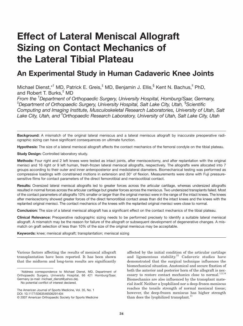
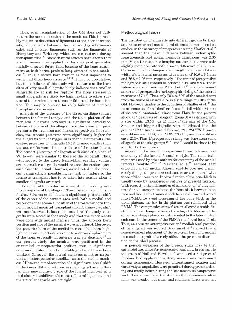
Each knee was prepared by transecting the femur and thetibia about 25 cm from the joint line. Both the femur and thetibia were potted in hollow, cylindrical tubes with low melt.The lateral compartment was accessed via a lateral para-patellar arthrotomy. The subsequent lateral femoral condyleosteotomy was planned as a vertical cut starting directly lat-eral to the femoral origin of the ACL aiming to the lateraltransition of the femoral diaphysis and metaphysis.10 Two8-mm tunnels were drilled perpendicular to the plannedosteotomy plane. The tunnels were filled with polymethylmethacrylate (PMMA), which was threaded for introductionof 6-mm threaded rods. The threaded tunnels of the condylefragment were overdrilled to 7 mm for compression of thecondyle during relocation (Figure 1). The defect of the sawblade was compensated for with a Teflon film.
For orientation of the pressure-sensitive film for lateranalysis of the films, 2 bone tunnels were drilled from thetibial metaphysis to the posterolateral and anteromediallateral tibial plateau. Two guide wires were introduced andadvanced underneath the surface of the articular cartilage.
After the intact knee was tested, the OM was removed.For later anteroposterior and mediolateral orientation of
Figure 1. Relocated osteotomy of the lateral femoralcondyle. The defect of the saw blade is compensated forwith a Teflon film.
36 Dienst et al The American Journal of Sports Medicine
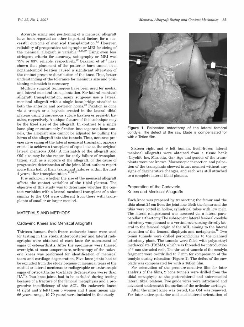
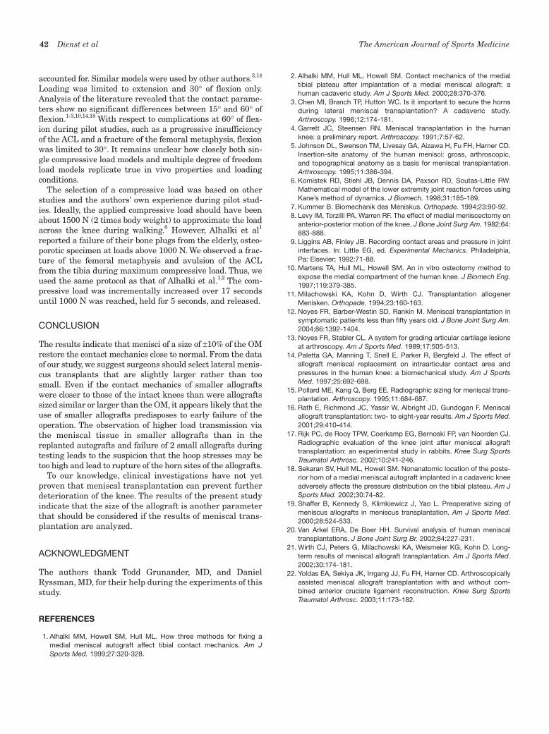
the lateral meniscus allograft, the tip of the lateral emi-nence was marked. The lateral condyle was dissembledand the meniscocapsular junction dissected. Using smallosteotomes, we removed the meniscus, with both hornsadherent to a small bone bridge. Exactly half of the tibialeminence was removed with the bone bridge, so that thelateral half with the mark was left attached to the lateraltibial plateau. The tibial defect was further depressed andfilled with PMMA. A plastic block of a size slightly biggerthan the bony meniscal bridge was used to mold a box intothe cement (Figure 2). The block was flush with the lateralhalf of the tibial eminence to avoid fixation of the trans-plant in a nonanatomical medial position. During themolding process, the anteroposterior position of the lateraleminence was marked on the plastic block. At the sameanteroposterior position, a thread was cut into the bottomof the box for a 4.5-mm cortical screw (Figure 2).
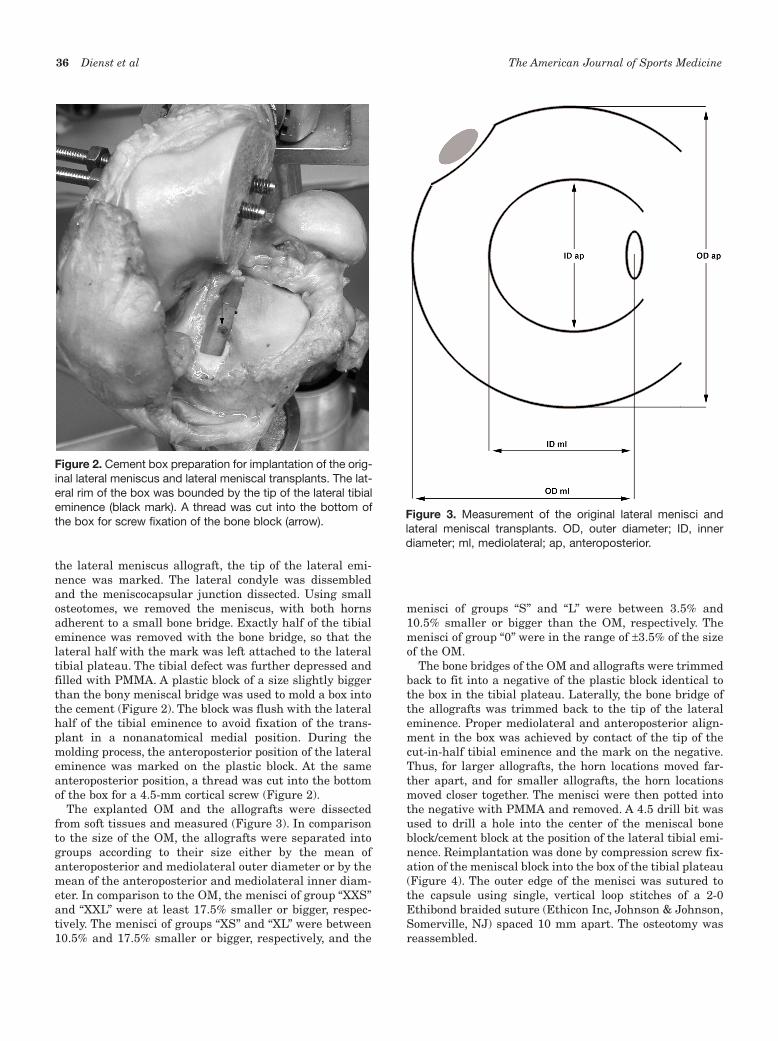
The explanted OM and the allografts were dissectedfrom soft tissues and measured (Figure 3). In comparisonto the size of the OM, the allografts were separated intogroups according to their size either by the mean ofanteroposterior and mediolateral outer diameter or by themean of the anteroposterior and mediolateral inner diam-eter. In comparison to the OM, the menisci of group “XXS”and “XXL” were at least 17.5% smaller or bigger, respec-tively. The menisci of groups “XS” and “XL” were between10.5% and 17.5% smaller or bigger, respectively, and the
menisci of groups “S” and “L” were between 3.5% and10.5% smaller or bigger than the OM, respectively. Themenisci of group “0” were in the range of ±3.5% of the sizeof the OM.
The bone bridges of the OM and allografts were trimmedback to fit into a negative of the plastic block identical tothe box in the tibial plateau. Laterally, the bone bridge ofthe allografts was trimmed back to the tip of the lateraleminence. Proper mediolateral and anteroposterior align-ment in the box was achieved by contact of the tip of thecut-in-half tibial eminence and the mark on the negative.Thus, for larger allografts, the horn locations moved far-ther apart, and for smaller allografts, the horn locationsmoved closer together. The menisci were then potted intothe negative with PMMA and removed. A 4.5 drill bit wasused to drill a hole into the center of the meniscal boneblock/cement block at the position of the lateral tibial emi-nence. Reimplantation was done by compression screw fix-ation of the meniscal block into the box of the tibial plateau(Figure 4). The outer edge of the menisci was sutured tothe capsule using single, vertical loop stitches of a 2-0Ethibond braided suture (Ethicon Inc, Johnson & Johnson,Somerville, NJ) spaced 10 mm apart. The osteotomy wasreassembled.
Figure 2. Cement box preparation for implantation of the orig-inal lateral meniscus and lateral meniscal transplants. The lat-eral rim of the box was bounded by the tip of the lateral tibialeminence (black mark). A thread was cut into the bottom ofthe box for screw fixation of the bone block (arrow).
Figure 3. Measurement of the original lateral menisci andlateral meniscal transplants. OD, outer diameter; ID, innerdiameter; ml, mediolateral; ap, anteroposterior.

Vol. 35, No. 1, 2007 Meniscal Allograft Sizing and Contact Mechanics 37
Testing
Biomechanical testing was performed with a universaltesting machine (858 Bionix Test System, MTS Systems
Corp, Eden Prairie, Minn). The femoral cylinder of eachknee joint was mounted into custom fixtures that allowedthe knee to be fixed at 0° and 30° of flexion. Rotation wasfixed with the patella facing parallel to the transepicondy-lar line. Depth of the cylinder was set so that the distanceto the upper center of rotation was the same as the dis-tance to the distal center of rotation measured from thejoint line of the knee. The tibial fixation rig allowed flexion,rotation, and varus-valgus angulation.
Each knee joint was tested with the joint intact (INT),with the replanted OM, after meniscectomy (NM), andwith each of the right or left meniscal allografts. Each kneewas removed from the testing machine only between con-ditions INT and OM. For testing of the OM and the allo-grafts, the knee was kept in the machine; the OM andallografts were removed and replanted after dissemblingand reassembling of the osteotomy only. Each conditionwas tested in full extension and 30° of flexion only (Figure5). Analysis of the literature revealed that the contactparameters show no significant differences between 15°and 60° of flexion.1-3,10,14,18
Preconditioning was performed by 3 loadings of the speci-men with the testing protocol. For the first 2 preconditioning
Figure 4. Fixation of the meniscal transplant in the cementbox with a compression screw. Proper anteroposterior andmediolateral orientation was achieved by contact of the tip ofthe lateral tibial eminence (black mark).
Figure 5. Knee specimen mounted into the testing machine(30° of flexion).
Figure 6. Graphical superimposing of the film scans on a dig-ital picture of the tibial plateau using the marker tunnels ofthe plateau and corresponding dots on the films.
38 Dienst et al The American Journal of Sports Medicine
cycles, rotation and varus-valgus angulation of the tibialrig were unlocked. At the last preconditioning cycle, bothdirections were locked during the maximum load of 1000N. Contact area and pressure were measured usinga 2-layered, pressure-sensitive film (Super-Low RangePressurex Indicating Film, SPI, East Hanover, NJ).9
Custom-sized, 0.25-mm-thick polyethylene film packetswere prepared from a template of the lateral tibial plateau.A braided suture was incorporated to the medial margin ofthe film packet for introduction of the films via a smallperforation of the posterior capsule. Under slight distrac-tion of the knees, the film packets were pulled under themenisci before the anterior horn sutures were closed.Loading of the knee joints was increased over a linearramp to 1000 N for 17 seconds, held over a plateau for5 seconds, and released.1,2 At the plateau phase, the two1.5-mm pins were shortly advanced to stain the pressure-sensitive film for later orientation. Each compressive load-ing was performed 5 times. Two films with higher noise orscratches were excluded; thus, 3 films for each conditionand position were used for further evaluation.
Data Analysis
A calibration curve for the film was generated.1,9 The dye-receiving layers of the knee-exposed film packets werescanned simultaneously with a high-resolution scanner. Adigital picture of the tibial articular surface of each kneejoint was taken. For the following graphical preparationand analysis, Adobe Photoshop software (Adobe SystemsInc, San Jose, Calif) was used. The digital pictures of thetibial surfaces were rotated until the line connecting theposterior edges of the medial and lateral articular surfaceswas oriented horizontally. After thresholding of the scans,the single scans of the film packets were copied in the pic-tures of the tibial surfaces, sized, and oriented using theholes of the 2 small 1.5-mm wires of the tibial surface andthe corresponding dots on the pressure-sensitive films(Figure 6). Each scan was placed on a new layer, superim-posing the image of the plateau.
From each layer, the same rectangular area was copied intoa new file. The upper limit of that area was defined by thehorizontal, posterior articular line (x-axis, y = 0); the laterallimit was defined by the lateral margin of the articular sur-face (y-axis, x = 0). Thus, an identical coordination systemwas defined for each film.The limits of the cartilage of the lat-eral tibial plateaus were marked and transferred to each fileof the single scans. The following parameters were graphi-cally marked with a graphic table board: the direct contactarea between the articular cartilage surfaces of the lateralfemoral condyle and the lateral tibial plateau (AFC), the meanpressure of the direct contact area (PFC), the peak pressure ofan area of 0.3 cm² (PFC0.3cm²), the peak pressure of an area of0.1 cm² (PFC0.1cm²), the mean pressure of the area of the artic-ular surface of the lateral tibial plateau minus AFC (PMen), andthe x and y locations of the maximum pressure (Figure 7).
Descriptive statistical analysis was performed usingbox diagrams for illustration of the differences betweenthe groups INT, OM, and NM and the groups of different
allograft sizes. For statistical analysis of differences betweenthe groups, a t test for unpaired samples was used (signifi-cant at P < .05). Linear regression analysis (Pearson) wasperformed for evaluation of a linear dependency of the sizeof the meniscal transplant and the contact parameters (sig-nificant at P < .05).
RESULTS
Comparison Between INT, OM, and NM
In extension and at 30° of flexion, the mean pressure PFCand the peak pressures PFC0.3cm² and PFC0.1cm² for the kneesNM were greater than for the INT and the knees with thereplanted OM (Figures 8-11). These differences were signif-icant in extension and bigger for the peak pressures. Forboth extension and flexion, there were only slightly higherpressures for PFC, PFC0.3cm², and PFC0.1cm² in OM than in INTwithout a significant difference. The pressure PMen was sig-nificantly smaller in NM and OM knees than in INT knees.
For both extension and flexion, the contact area AFC forthe INT was smaller than for the knees with the replantedOM, and AFC for the knees with the replanted OM wassmaller than for the knees NM (Figures 12 and 13). Thesedifferences were not significant. After meniscectomy, thecenter of the contact area was shifted laterally 1.1 mm and1.9 mm in extension and flexion, respectively, and shiftedanteriorly 0.9 mm in extension and posteriorly 4 mm inflexion. These shifts were not significant.
Comparison Between the Different Allograft Sizes
A significant increase of PFC, PFC0.3cm², and PFC0.1cm² wasseen with increasing size of the allografts in extension(Figures 8 and 10). On the other hand, PMen significantlydecreased. PFC, PFC0.3cm², and PFC0.1cm² in allografts of thegroup “XXL” were significantly higher than in INT. On theother side, PMen was significantly smaller in allografts ofthe group “XXL” than in INT. Some of the measured pres-sures for the allografts of sizes smaller than “S” were sig-nificantly smaller for PFC, PFC0.3cm², and PFC0.1cm² and biggerfor PMen than in the knee NM. Most of the measured pres-sures of the groups “S,” “0,” and “L” were in the range of theINT knees and knees after replantation of the OM. In flex-ion, the differences were less significant (Figures 9 and11). The tendency was similar to the results at extension;a significant difference in comparison to the INT kneeswas found for PMen in “XXL” only.
For both extension and flexion, the contact area AFCincreased with larger sizes of the allografts (Figures 12 and13). Linear regression analysis revealed a significant increasefor outer diameter in flexion and for inner diameter in exten-sion. In extension and flexion, the center of the contact areawas shifted laterally with increasing sizes of the allografts.There was a significant linear correlation in flexion.
During testing, 2 lateral meniscal allografts in the group“XXS” failed. One rupture occurred during flexion at theposterior horn, and the other allograft failed at the ante-rior horn during extension.

Vol. 35, No. 1, 2007 Meniscal Allograft Sizing and Contact Mechanics 39
DISCUSSION
Interpretation of Results
The purpose of this study was to determine whether thecontact variables with the lateral meniscal allografts ofdifferent sizes were different from the contact variableswith the lateral meniscal autograft or lateral meniscalallografts of similar sizes to the OM. The key findings werethat (1) the replanted autografts did restore the contactmechanics only close to normal, (2) there was a significantcorrelation between the size of the allograft and the
Figure 7. Measurement parameters on the pressure-sensitivefilms. AFC, femorotibial contact area; PFC, mean femorotibialcontact pressure; PFC0.3cm² and PFC0.1cm², peak pressures of thefemorotibial contact; x and y, location of the maximum pres-sure (arrows); PMEN, mean meniscotibial pressure.
0
0.5
1
1.5
2
2.5
3
INT OM NM XXS XS S 0 L XL XXL
[MPa]
* * **
*
*
**
*
+
+
+
+ ++ +
++
++
+
x
x
FK FK 0.3 cm2 FK 0.1 cm2 Men
Figure 8. Contact pressures for the intact knees (INT), kneeswith the replanted original meniscus (OM), and knee aftermeniscectomy (NM) and for the different allograft groups afterallocation from the mean of their outer diameters in extension.*pNM < .05. +pINT < .05. xpINT < .05. Linear regression analysis:pFFK = .032. pFFK0.3cm² = .022. pFFK0.1cm² = .033. pFMen = .003. Seetext for complete description of abbreviations.
0
0.5
1
1.5
2
2.5
3
INT OM NM XXS XS S 0 L XL XXL
[MPa]
++
++
FK FK 0.3 cm2 FK 0.1 cm2 Men
Figure 9. Contact pressures for the intact knees (INT), kneeswith the replanted original meniscus (OM), and knee aftermeniscectomy (NM) and for the different allograft groups afterallocation from the mean of their outer diameters in flexion.+pINT < .05. See text for complete description of abbreviations.
0
0.5
1
1.5
2
2.5
3
INT OM NM XXS XS S 0 L XL XXL
[MPa]
***
*
*
*
*
*
*
*
*
+
+
+++
++
+x
x
FK FK 0.3 cm2 FK 0.1 cm2 Men
Figure 10. Contact pressures for the intact knees (INT), kneeswith the replanted original meniscus (OM), and knee aftermeniscectomy (NM) and for the different allograft groups afterallocation from the mean of their inner diameters in extension.*pNM < .05. +pINT < .05. xpINT < .05. Linear regression analysis:pFFK0.3cm² = .044; pFFK0.1cm² = .033; pFMen = .039. See text forcomplete description of abbreviations.
Michael
Durchstreichen
Michael
Eingefügter Text
P
Michael
Durchstreichen
Michael
Eingefügter Text
P
Michael
Durchstreichen
Michael
Eingefügter Text
P
Michael
Durchstreichen
Michael
Eingefügter Text
P
Michael
Durchstreichen
Michael
Eingefügter Text
P
Michael
Durchstreichen
Michael
Eingefügter Text
P

40 Dienst et al The American Journal of Sports Medicine
contact mechanics of the tibial plateau, (3) allografts sized17.5% bigger than the OM showed significantly highercontact pressures than did the OM, and (4) allografts sized10.5% smaller than the OM showed increased forces in themeniscus and thus may be predisposed to early failure.
Analysis of the contact mechanics showed only aslightly nonsignificant increase of the direct femorotibialcontact pressures of the knees with the autografts (OM)compared with the INT. The differences were more obvi-ous in extension. Here, the mean contact pressure (PFC)increased by a mean of 0.15 MPa. The knees with thereplanted autografts showed significantly smaller or atrend for smaller contact pressures than did the kneesNM in extension and flexion, respectively. These findingsare similar to the results of other studies. Chen et al3
showed that both autografts with a bony bridge and
autografts with both horns secured gave similar results tothose for the INT. Alhalki et al1 found that the contactpressures of autografts replanted with bone plug fixationwere close to normal; however, the peak pressure was sig-nificantly higher than in the intact knees. These findingswere also more prominent in extension.
The results from this study showed that the directcontact area between the cartilage of the lateral femoralcondyle and the lateral tibial plateau (AFC) was not signif-icantly different for the INT, the knees with the replantedautografts, and the knees NM for both extension and flex-ion. There was even a trend for larger contact areas for thereplanted OM and the knees NM. This is in contrast toother studies. Chen et al3 and Alhalki et al1 found a signif-icant decrease of the contact area of up to 50% for theknees NM compared with the INT knees. This must berelated to a different area selection. In the present study,only the direct contact area between the cartilage of thefemoral condyle and the tibial plateau was traced. The lessintense and irregular staining of the contact between themeniscus and the tibial plateau was not included. Thus, ifthe meniscus were removed or an oversized allograftwere used, the contact area should be bigger. This was thecase in the present study: the contact areas increased withthe size of the meniscal allografts.
The film analysis in the present study included a newparameter (PMen): the area of the tibial plateau minus thedirect femorotibial cartilage contact area. The mean pres-sure of this area was defined as the simplified pressuretransmitted via the meniscal tissue. The film analysisrevealed a significant pressure reduction for the kneeswith the replanted autografts and the knees NM for bothextension and flexion. This was consistent with the resultsfor the meniscal allografts. There was a significant nega-tive correlation of the pressure transmitted via the menis-cal tissue and the size of the meniscal allograft for bothextension and flexion. Thus, the bigger the allograft, theless the load transmission via the meniscal tissue.Conversely, the load transmission via the meniscal tissueincreased with smaller size of the meniscal transplants.
Figure 13. Contact areas AFC in flexion and extension for theintact knees (INT), knees with the replanted original meniscus(OM), and knee after meniscectomy (NM) and the differentallograft groups after allocation from the mean of their innerdiameters. Linear regression analysis: pExtension = .045.
0
0.5
1
1.5
2
2.5
3
3.5
INT OM NM XXS XS S 0 L XL XXL
[MPa]
+ +**+ +
FK FK 0.3 cm2 FK 0.1 cm2 Men
Figure 11. Contact pressures for the intact knees (INT),knees with the replanted original meniscus (OM), and kneeafter meniscectomy (NM) and for the different allograftgroups after allocation from the mean of their inner diametersin flexion. *pNM < .05. +pINT < .05. xpINT < .05. See text for com-plete description of abbreviations.
Figure 12. Contact areas AFC in flexion and extension for theintact knees (INT), knees with the replanted original meniscus(OM), and knee after meniscectomy (NM) and for the differentallograft groups after allocation from the mean of their outerdiameters. *pNM < .05. Linear regression analysis: pFlexion = .008.
Vol. 35, No. 1, 2007 Meniscal Allograft Sizing and Contact Mechanics 41
Thus, even reimplantation of the OM does not fullyrestore the normal function of the meniscus. This is proba-bly related to dissection of ligaments at the horn insertionsite, of ligaments between the menisci (Lig intermenis-cale), and of other ligaments such as the ligaments ofHumphrey and Wrisberg, which are not restored duringtransplantation.17 Biomechanical studies have shown thata compressive force applied to the knee joint generatesradially directed forces that, because of the bone attach-ment at both horns, produce hoop stresses in the menis-cus.3,7 Thus, a secure horn fixation is most important towithstand these hoop stresses.1,3,14 It may be speculative,but the 2 failures of this study with ruptures at the hornsites of very small allografts likely indicate that smallerallografts are at risk for rupture. The hoop stresses insmall allografts are likely too high, leading to either rup-ture of the meniscal horn tissue or failure of the horn fixa-tion. This may be a cause for early failures of meniscaltransplantation in vivo.
Analysis of the pressure of the direct cartilage contactbetween the femoral condyle and the tibial plateau of themeniscal allografts revealed a significant correlationbetween the size of the allograft and the mean and peakpressures for extension and flexion, respectively. In exten-sion, the contact pressures were significantly higher forthe allografts of much bigger sizes than the autografts. Thecontact pressures of allografts 10.5% or more smaller thanthe autografts were similar to those of the intact knees.The contact pressures of allograft with sizes of a mean of7% to –7% were similar to those of the autograft. Thus,with respect to the direct femorotibial cartilage contactareas, smaller allografts would restore the contact pres-sure closer to normal. However, as indicated in the previ-ous paragraphs, a possible higher risk for failure of themeniscus transplant has to be taken into consideration ifsmaller allografts are used.
The center of the contact area was shifted laterally withincreasing size of the allograft. This was significant only inflexion. Sekaran et al18 found a significant posterior shiftof the center of the contact area with both a medial andposterior nonanatomical position of the posterior horn tun-nel in medial meniscal transplantation. A transverse shiftwas not observed. It has to be considered that only auto-grafts were tested in that study and that the experimentswere done with medial menisci. Thus, the anterior hornposition and size of the menisci were not altered. Moreover,the posterior horn of the medial meniscus has been high-lighted as an important restraint to anterior displacementof the tibia, especially in anterior cruciate deficiency.8 Inthe present study, the menisci were positioned in theanatomical anteroposterior position; thus, a significantanterior or posterior shift in a stable joint would have beenunlikely. Moreover, the lateral meniscus is not as impor-tant an anteroposterior stabilizer as is the medial menis-cus.8 However, our observation of a significant lateral shiftin the knees NM and with increasing allograft size in flex-ion only may indicate a role of the lateral meniscus as amediolateral stabilizer when the collateral ligaments andthe articular capsule are not tight.
Methodological Issues
The distribution of allografts into different groups by theiranteroposterior and mediolateral dimensions was based onstudies on the accuracy of preoperative sizing. Shaffer et al19
showed that the mean difference between radiographicmeasurements and actual meniscus dimensions was 2.35mm. Magnetic resonance imaging measurements were onlyslightly more accurate with a mean difference of 2.25 mm.Considering an anteroposterior length and mediolateralwidth of the lateral meniscus with a mean of 36.6 ± 6.1 mmand 26.8 ± 2.96 mm, respectively,19 the error of preoperativeradiographic sizing would be between 6.4% and 8.8%. Thesevalues were confirmed by Pollard et al,15 who determinedan error of preoperative radiographic sizing of the lateralmeniscus of 7.4%. Thus, only 70% to 80% of the menisci sentfrom the tissue bank would be in a size range of ±10% of theOM. However, similar to the definition of Shaffer et al,19 themeasurements of an “ideal” graft should fall within ±1 mmof the actual anatomical dimensions. Thus, for the presentstudy, an “ideally sized” allograft (group 0) was defined witha size within ±3.5% (ca ±1 mm) of the size of the OM.Smaller and bigger allografts were distributed into thegroups “L”/”S” (mean size difference, 7%), “XS”/”XL” (meansize difference, 14%), and “XXS”/”XXL” (mean size differ-ence, 21%). Thus, if preoperative sizing was done accurately,allografts of the size groups S, 0, and L would be those to besent by the tissue bank.
Access to the lateral compartment was achieved viaosteotomy of the lateral femoral condyle. The same tech-nique was used by other authors for osteotomy of the medialfemoral condyle.2,10,18,19 Martens et al10 showed thatosteotomy of the medial femoral condyle does not signifi-cantly change the pressure and contact area compared withthose of the intact knee. In vivo, fixation of the bone block isusually done by transosseous sutures or press-fit fixation.With respect to the information of Alhalki et al1 of plug fail-ures due to osteoporotic bone, the bone block between bothmeniscal horns was trimmed back to a small rim and pottedinto PMMA. To avoid loosening of the bone block in thetibial plateau, the box in the plateau was reinforced withPMMA. The compressive screw fixation allowed a stable fix-ation and fast change between the allografts. Moreover, thescrew was always placed directly medial to the lateral tibialeminence in the center of the PMMA-reinforced bone block.Thus, an accurate anteroposterior and mediolateral positionof the allograft was secured. Sekaran et al18 showed that anonanatomical placement of the posterior horn of a medialmeniscal autograft adversely affects the pressure distribu-tion on the tibial plateau.
A possible weakness of the present study may be thatour model accounted for compressive load only. In contrast tothe group of Hull and Howell,1,2,18 who used a 6 degrees offreedom load application system, motion was constrainedduring compression. However, unconstrained rotation andvarus-valgus angulation were permitted during precondition-ing and finally locked during the last maximum compressiveload. Thus, smearing of the stain on the pressure-sensitivefilms was avoided, but shear and rotational forces were not
42 Dienst et al The American Journal of Sports Medicine
accounted for. Similar models were used by other authors.3,14
Loading was limited to extension and 30° of flexion only.Analysis of the literature revealed that the contact parame-ters show no significant differences between 15° and 60° offlexion.1-3,10,14,18 With respect to complications at 60° of flex-ion during pilot studies, such as a progressive insufficiencyof the ACL and a fracture of the femoral metaphysis, flexionwas limited to 30°. It remains unclear how closely both sin-gle compressive load models and multiple degree of freedomload models replicate true in vivo properties and loadingconditions.
The selection of a compressive load was based on otherstudies and the authors’ own experience during pilot stud-ies. Ideally, the applied compressive load should have beenabout 1500 N (2 times body weight) to approximate the loadacross the knee during walking.6 However, Alhalki et al1
reported a failure of their bone plugs from the elderly, osteo-porotic specimen at loads above 1000 N. We observed a frac-ture of the femoral metaphysis and avulsion of the ACLfrom the tibia during maximum compressive load. Thus, weused the same protocol as that of Alhalki et al.1,2 The com-pressive load was incrementally increased over 17 secondsuntil 1000 N was reached, held for 5 seconds, and released.
CONCLUSION
The results indicate that menisci of a size of ±10% of the OMrestore the contact mechanics close to normal. From the dataof our study, we suggest surgeons should select lateral menis-cus transplants that are slightly larger rather than toosmall. Even if the contact mechanics of smaller allograftswere closer to those of the intact knees than were allograftssized similar or larger than the OM, it appears likely that theuse of smaller allografts predisposes to early failure of theoperation. The observation of higher load transmission viathe meniscal tissue in smaller allografts than in thereplanted autografts and failure of 2 small allografts duringtesting leads to the suspicion that the hoop stresses may betoo high and lead to rupture of the horn sites of the allografts.
To our knowledge, clinical investigations have not yetproven that meniscal transplantation can prevent furtherdeterioration of the knee. The results of the present studyindicate that the size of the allograft is another parameterthat should be considered if the results of meniscal trans-plantation are analyzed.
ACKNOWLEDGMENT
The authors thank Todd Grunander, MD, and DanielRyssman, MD, for their help during the experiments of thisstudy.
REFERENCES
1. Alhalki MM, Howell SM, Hull ML. How three methods for fixing amedial meniscal autograft affect tibial contact mechanics. Am JSports Med. 1999;27:320-328.
2. Alhalki MM, Hull ML, Howell SM. Contact mechanics of the medialtibial plateau after implantation of a medial meniscal allograft: ahuman cadaveric study. Am J Sports Med. 2000;28:370-376.
3. Chen MI, Branch TP, Hutton WC. Is it important to secure the hornsduring lateral meniscal transplantation? A cadaveric study.Arthroscopy. 1996;12:174-181.
4. Garrett JC, Steensen RN. Meniscal transplantation in the humanknee: a preliminary report. Arthroscopy. 1991;7:57-62.
5. Johnson DL, Swenson TM, Livesay GA, Aizawa H, Fu FH, Harner CD.Insertion-site anatomy of the human menisci: gross, arthroscopic,and topographical anatomy as a basis for meniscal transplantation.Arthroscopy. 1995;11:386-394.
6. Komistek RD, Stiehl JB, Dennis DA, Paxson RD, Soutas-Little RW.Mathematical model of the lower extremity joint reaction forces usingKane’s method of dynamics. J Biomech. 1998;31:185-189.
7. Kummer B. Biomechanik des Meniskus. Orthopade. 1994;23:90-92.8. Levy IM, Torzilli PA, Warren RF. The effect of medial meniscectomy on
anterior-posterior motion of the knee. J Bone Joint Surg Am. 1982;64:883-888.
9. Liggins AB, Finley JB. Recording contact areas and pressure in jointinterfaces. In: Little EG, ed. Experimental Mechanics. Philadelphia,Pa: Elsevier; 1992:71-88.
10. Martens TA, Hull ML, Howell SM. An in vitro osteotomy method toexpose the medial compartment of the human knee. J Biomech Eng.1997;119:379-385.
11. Milachowski KA, Kohn D, Wirth CJ. Transplantation allogenerMenisken. Orthopade. 1994;23:160-163.
12. Noyes FR, Barber-Westin SD, Rankin M. Meniscal transplantation insymptomatic patients less than fifty years old. J Bone Joint Surg Am.2004;86:1392-1404.
13. Noyes FR, Stabler CL. A system for grading articular cartilage lesionsat arthroscopy. Am J Sports Med. 1989;17:505-513.
14. Paletta GA, Manning T, Snell E, Parker R, Bergfeld J. The effect ofallograft meniscal replacement on intraarticular contact area andpressures in the human knee: a biomechanical study. Am J SportsMed. 1997;25:692-698.
15. Pollard ME, Kang Q, Berg EE. Radiographic sizing for meniscal trans-plantation. Arthroscopy. 1995;11:684-687.
16. Rath E, Richmond JC, Yassir W, Albright JD, Gundogan F. Meniscalallograft transplantation: two- to eight-year results. Am J Sports Med.2001;29:410-414.
17. Rijk PC, de Rooy TPW, Coerkamp EG, Bernoski FP, van Noorden CJ.Radiographic evaluation of the knee joint after meniscal allografttransplantation: an experimental study in rabbits. Knee Surg SportsTraumatol Arthrosc. 2002;10:241-246.
18. Sekaran SV, Hull ML, Howell SM. Nonanatomic location of the poste-rior horn of a medial meniscal autograft implanted in a cadaveric kneeadversely affects the pressure distribution on the tibial plateau. Am JSports Med. 2002;30:74-82.
19. Shaffer B, Kennedy S, Klimkiewicz J, Yao L. Preoperative sizing ofmeniscus allografts in meniscus transplantation. Am J Sports Med.2000;28:524-533.
20. Van Arkel ERA, De Boer HH. Survival analysis of human meniscaltransplantations. J Bone Joint Surg Br. 2002;84:227-231.
21. Wirth CJ, Peters G, Milachowski KA, Weismeier KG, Kohn D. Long-term results of meniscal allograft transplantation. Am J Sports Med.2002;30:174-181.
22. Yoldas EA, Sekiya JK, Irrgang JJ, Fu FH, Harner CD. Arthroscopicallyassisted meniscal allograft transplantation with and without com-bined anterior cruciate ligament reconstruction. Knee Surg SportsTraumatol Arthrosc. 2003;11:173-182.
Related Documents