Research Article Effect of Hyaluronan on Developmental Competence and Quality of Oocytes and Obtained Blastocysts from In Vitro Maturation of Bovine Oocytes Jolanta Opiela, 1 Joanna Romanek, 1 Daniel LipiNski, 2,3 and ZdzisBaw Smordg 1 1 Department of Biotechnology of Animal Reproduction, National Research Institute of Animal Production, Krakowska 1 Street, 32-083 Balice, Krak´ ow, Poland 2 Department of Biochemistry and Biotechnology, Pozna´ n University of Life Sciences, Dojazd 11 Street, 60-632 Pozna´ n, Poland 3 Institute of Human Genetics, Polish Academy of Sciences, Strzeszy´ nska 32, 60-479 Pozna´ n, Poland Correspondence should be addressed to Jolanta Opiela; [email protected] Received 19 November 2013; Revised 31 December 2013; Accepted 3 January 2014; Published 13 February 2014 Academic Editor: Irma Virant-Klun Copyright © 2014 Jolanta Opiela et al. is is an open access article distributed under the Creative Commons Attribution License, which permits unrestricted use, distribution, and reproduction in any medium, provided the original work is properly cited. e objective of the present study was to evaluate the effect of hyaluronan (HA) during IVM on meiotic maturation, embryonic development, and the quality of oocytes, granulosa cells (GC), and obtained blastocysts. COCs were matured in vitro in control medium and medium with additional 0.035% or 0.07% of exogenous HA. e meiotic maturity did not differ between the analysed groups. e best rate and the highest quality of obtained blastocysts were observed when 0.07% HA was used. A highly significant difference ( < 0.001) was noted in the mean number of apoptotic nuclei per blastocyst and in the DCI between the 0.07% HA and the control blastocysts ( < 0.01). Our results suggest that addition of 0.035% HA and 0.07% HA to oocyte maturation media does not affect oocyte nuclear maturation and DNA fragmentation. However, the addition of 0.07% HA during IVM decreases the level of blastocysts DNA fragmentation. Finally, our results suggest that it may be risky to increase the HA concentration during IVM above 0.07% as we found significantly higher Bax mRNA expression levels in GC cultured with 0.07% HA. e final concentration of HA being supplemented to oocyte maturation media is critical for the success of the IVP procedure. 1. Introduction e large discrepancy between the number of oocytes under- going in vitro fertilisation and the number of embryos devel- oping to the blastocyst stage (approximately 30–40%) calls for further research to improve the oocyte maturation medium in order to mimic the in vivo maturation conditions. A few studies have focused their attention on the supplementation of in vitro embryo culture media with hyaluronan (HA) due to its biochemical properties [1–3]. Important biological func- tions are not properly regulated during in vitro culture due to the two-dimensional nature of the culture. Hyaluronan forms a concentration dependent, three-dimensional, jelly-like net- work. A three-dimensional network improves the interac- tion between cellular receptors and the molecules in the external environment by improving the binding of molecules such as polypeptide growth factors that play an important role during early embryonic development [4]. Hyaluronan is a major component of the cumulus-oocyte complex (COCs) and is synthesised and secreted by granulosa cells under the stimulation of LH and FSH [5]. e insufficient interac- tions of hyaluronan and its receptor CD44 during in vitro maturation may decrease the capacity of fertilization and development of oocytes matured in vitro [6]. e resumption of meiosis in mares, pigs, and rats is associated with decreased levels of connexin 43 (Cx43) [7]. Yokoo et al. [8] revealed that the interaction of HA with the CD44 receptor in granulosa cells during cumulus expansion lowers the levels of Cx43. As a result of this process, the transport of cAMP between cells and the oocyte is blocked. A decrease in the concentration of cAMP in oocytes is a signal for the activation of MPF, which is responsible for the resumption of meiosis (GVBD) [8]. In addition, HA participates in the process of sperm capacitation [9], determines the proper course of fertilisation, and affects Hindawi Publishing Corporation BioMed Research International Volume 2014, Article ID 519189, 8 pages http://dx.doi.org/10.1155/2014/519189

Welcome message from author
This document is posted to help you gain knowledge. Please leave a comment to let me know what you think about it! Share it to your friends and learn new things together.
Transcript
Research ArticleEffect of Hyaluronan on Developmental Competence andQuality of Oocytes and Obtained Blastocysts from In VitroMaturation of Bovine Oocytes
Jolanta Opiela,1 Joanna Romanek,1 Daniel LipiNski,2,3 and ZdzisBaw Smordg1
1 Department of Biotechnology of Animal Reproduction, National Research Institute of Animal Production, Krakowska 1 Street,32-083 Balice, Krakow, Poland
2Department of Biochemistry and Biotechnology, Poznan University of Life Sciences, Dojazd 11 Street, 60-632 Poznan, Poland3 Institute of Human Genetics, Polish Academy of Sciences, Strzeszynska 32, 60-479 Poznan, Poland
Correspondence should be addressed to Jolanta Opiela; [email protected]
Received 19 November 2013; Revised 31 December 2013; Accepted 3 January 2014; Published 13 February 2014
Academic Editor: Irma Virant-Klun
Copyright © 2014 Jolanta Opiela et al. This is an open access article distributed under the Creative Commons Attribution License,which permits unrestricted use, distribution, and reproduction in any medium, provided the original work is properly cited.
The objective of the present study was to evaluate the effect of hyaluronan (HA) during IVM on meiotic maturation, embryonicdevelopment, and the quality of oocytes, granulosa cells (GC), and obtained blastocysts. COCs were matured in vitro in controlmedium andmedium with additional 0.035% or 0.07% of exogenous HA.Themeiotic maturity did not differ between the analysedgroups. The best rate and the highest quality of obtained blastocysts were observed when 0.07% HA was used. A highly significantdifference (𝑃 < 0.001) was noted in the mean number of apoptotic nuclei per blastocyst and in the DCI between the 0.07%HA andthe control blastocysts (𝑃 < 0.01). Our results suggest that addition of 0.035% HA and 0.07% HA to oocyte maturation media doesnot affect oocyte nuclear maturation and DNA fragmentation. However, the addition of 0.07% HA during IVM decreases the levelof blastocysts DNA fragmentation. Finally, our results suggest that it may be risky to increase the HA concentration during IVMabove 0.07% as we found significantly higher BaxmRNA expression levels in GC cultured with 0.07% HA.The final concentrationof HA being supplemented to oocyte maturation media is critical for the success of the IVP procedure.
1. Introduction
The large discrepancy between the number of oocytes under-going in vitro fertilisation and the number of embryos devel-oping to the blastocyst stage (approximately 30–40%) calls forfurther research to improve the oocyte maturation mediumin order to mimic the in vivo maturation conditions. A fewstudies have focused their attention on the supplementationof in vitro embryo culturemediawith hyaluronan (HA) due toits biochemical properties [1–3]. Important biological func-tions are not properly regulated during in vitro culture due tothe two-dimensional nature of the culture. Hyaluronan formsa concentration dependent, three-dimensional, jelly-like net-work. A three-dimensional network improves the interac-tion between cellular receptors and the molecules in theexternal environment by improving the binding of moleculessuch as polypeptide growth factors that play an important
role during early embryonic development [4]. Hyaluronan isa major component of the cumulus-oocyte complex (COCs)and is synthesised and secreted by granulosa cells underthe stimulation of LH and FSH [5]. The insufficient interac-tions of hyaluronan and its receptor CD44 during in vitromaturation may decrease the capacity of fertilization anddevelopment of oocytes matured in vitro [6].The resumptionofmeiosis inmares, pigs, and rats is associatedwith decreasedlevels of connexin 43 (Cx43) [7]. Yokoo et al. [8] revealed thatthe interaction of HA with the CD44 receptor in granulosacells during cumulus expansion lowers the levels of Cx43. Asa result of this process, the transport of cAMP between cellsand the oocyte is blocked. A decrease in the concentration ofcAMP in oocytes is a signal for the activation of MPF, whichis responsible for the resumption of meiosis (GVBD) [8]. Inaddition,HAparticipates in the process of spermcapacitation[9], determines the proper course of fertilisation, and affects
Hindawi Publishing CorporationBioMed Research InternationalVolume 2014, Article ID 519189, 8 pageshttp://dx.doi.org/10.1155/2014/519189

2 BioMed Research International
the growth potential of early embryos [10]. Many cellularprocesses are regulated by the interaction betweenHA and itssurface receptors, such as CD44, RHAMM (receptor for HAmediated motility), and ICAM-1 (intercellular adhesionmolecule 1) [11, 12]. These processes include the maintenanceof tissue homeostasis [13], cell adhesion and migration [14,15], cell proliferation and differentiation, and the inhibitionof apoptosis and immune response [16, 17].
The objective of the present study was to evaluate the HAsupplementation effect on meiotic maturation at two con-centrations within in vitromaturationmedium. Additionally,embryonic development after in vitro fertilisation as wellas the quality of bovine oocytes, granulosa cells that served ascoculture during IVM, and obtained blastocysts will beevaluated. The results of our previous experiments [18] haveindicated that the supplementation of IVM with HA didnot increase the nuclear maturity of oocytes. Moreover, ourprevious results have indicated that the 0.035% and 0.07%HA concentrations present opposing effects on IVM. Themeiotic maturity of the 0.07% HA treated oocytes was thelowest [18], inclining us to apply them in this experiment.Therefore, in the present experiment, we TUNEL analysedoocytes in two experimental groups. Moreover, blastocystsobtained after fertilisation of HA treated COCs provided dataon their developmental competence.
2. Materials and Methods
Immature oocytes were isolated from ovaries obtained fromheifers and cows from the slaughterhouse, which were deliv-ered in a warm flask to the laboratory within 2-3 hours afterslaughter.
2.1. Chemicals and Supplies. The reagents used in the pre-sented experiments were purchased from Sigma-Aldrich,Poland, unless otherwise indicated.
2.2. Experimental Design. The research was performed intwo directions. First, we evaluated the effect of two concen-trations of HA supplementation on meiotic maturation andembryonic development after in vitro fertilisation. Second,weevaluated the possible negative impact of HA on the oocytes,the blastocysts, and granulosa cells. TUNEL staining and theestimation of apoptotic genes transcripts expression by real-time PCR were employed as quality markers.
2.3. Recovery and Selection of Bovine Oocytes. The detaileddescription of procedure is provided in Opiela et al. [18].Cumulus-oocyte complexes (COCs) were retrieved fromovaries of slaughtered cows by aspiration of follicular fluidof ovarian follicles. For this purpose, a syringe and needlewere used. Follicular fluid was collected into tubes (Corning,12mL) and left for 15–30 minutes at room temperature tosediment. The supernatant was then collected, and theremaining sediment was transferred into a holding medium(TCM 199—Earle’s salt with 25mM of HEPES containing10% foetal calf serum). Next, COCs were recovered understereomicroscope. Before placing COCs into the in vitro
maturation, they were washed three times for selection.Degenerated oocytes and those without compact cumuluscells were discarded.
2.4. In Vitro Maturation in Coculture with Granulosa Cells.For the culture of COCs the commercial medium TCM-199buffered with sodium bicarbonate and supplemented withL-glutamine was used. Selected COCs were cultured for 22hours in 2mL of medium supplemented with 20% estrus cowserum (ECS, heat inactivated-prepared in our laboratory) andan additional 3 to 5 × 10⁶ granulosa cells/mL (GC). Between30 and 40 COCs were used per culture dish. Granulosacells were obtained from follicles with a diameter of 4–6mmwithout atretic changes and containing oocytes with normalmorphological appearance.Theobtained granulosa cells weresuspended in 7mL of in vitro maturation medium withoutexogenous HA. After carefully mixing the medium with GC,the suspension was divided between 3 culture dishes, for2mL each. In vitro maturation was performed with differentconcentrations of 1% high molecular weight HA (CromaPharma GmbH, Austria), with 75 𝜇L/2mL medium (a finalconcentration of HA 0.035%) and 150𝜇L/2mL medium (afinal concentration of 0.07%). As a control, we used COCsmatured only in maturation medium and cocultured withgranulosa cells [18].This approachwas provided the samewayto experimental conditions in all of the groups, as the estruscow serum may contain HA and HA is also synthesised andsecreted by granulosa cells in undefined and variable amount.Moreover, the same ESC was used in all IVM media duringthe whole experiment and each repetition of IVM/IVF/IVCwas performed in three simultaneous experimental groups.Oocytes matured for 22 h at 38.5∘C under 5% CO
2in air.
2.5. Effect of HA on Meiotic Maturity and ChromatinFragmentation in Oocytes
2.5.1. TUNEL. The detailed description of procedure is pro-vided in Opiela et al. [18]. TUNEL analysis was performedusing a Deadend Fluorometric TUNEL System (Promega,Poland).
Fixed denuded oocytes were first incubated with 0.2%Triton X-100 solution for 5 minutes, then a reaction mixtureconsisting of equilibration buffer, the mixture of nucleotidesand TdT enzyme for 1 h at maximum humidity, and lastly asolution of 2 × SSC for 15mins. After the performed incuba-tions, the oocytes were washed three times in PBS/PVP for atleast 5 minutes. Finally, the oocytes were placed in a drop ofVECTASHIELD + DAPI solution. Oocytes that were treatedwith UV light for 30mins immediately after maturation wereused as a positive control. As a negative control, oocytesplaced in the reaction mixture lacking the rTdT enzyme wereused. Oocytes were analysed under a fluorescent microscope.To visualise the stages of meiosis and DNA fragmentation ofoocytes, a filter of>460 nmwas used for the blue fluorescenceof DAPI stained cells and a filter with a wavelength of 520 ±20 nm was used for the green fluorescence of apoptoticcells, which incorporated into the nucleus with a fluorescein-conjugated dUTP.
BioMed Research International 3
2.5.2. Estimation of Meiotic Maturity. We have applied sim-ilar criteria to those of Warzych et al. [19]. In cases ofstrong fluorescent/DAPI signals from the nuclei and thepolar body, meiotic stage classification (GV, MI, AI, TI, MII)was performed. A detailed description of assessment criteriais also available in Opiela et al. [18]. Briefly, the germinalvesicle (GV) was characterised by varying degrees of chro-matin condensation, metaphase of first meiotic division (MI)was characterised by chromosomes arranged as a groupof separated bivalents, and metaphase second (MII) wascharacterised by the presence of haploid set of chromosomesand chromosomes forming the first polar body (PB1).Meioticmaturity was counted as the ratio of oocytes in metaphase IIstage to the total number of analysed oocytes.
2.5.3. Estimation of Oocytes Apoptotic Index DCI. TUNELpositive oocytes indicated fragmented DNA by a strongfluorescent/FITC signal from the nuclei or polar body. As acontrol for the experiment, the COCs were subjected to theUV (30mins. at room temperature in theM199medium con-taining HEPES supplemented with 5% FCS). This generated“damage” to the DNA, which would show a positive TUNELsignal and indicate that theTUNEL reactionworks.Thedeathcell index (DCI) was expressed as a ratio of apoptotic oocytesexhibiting a strong fluorescent/FITC signal to all analysedoocytes from each group [18, 19].
2.6. Effect of HA on the Developmental Competence ofBovine Oocytes
2.6.1. In Vitro Fertilisation. IVF was performed according toour standard protocol that was previously described [20, 21].Briefly, after 22–24 hrs of maturation, the selected oocyteswere fertilised using frozen semen from single bull. Themotile sperm were selected by centrifugation (300 g at roomtemp.) on a discontinuous (1mL 45% over 1mL 90%) Percoll(Pharmacia, Sweden). After washing, the sperm were sus-pended in the fertilisation medium containing 10𝜇g/mL ofheparin and amixture of penicillamine (20𝜇M), hypotaurine(10 𝜇M), and epinephrine (1 𝜇M) at a concentration of 1-2 ×10⁶ spermatozoa/mL of medium. After washing and partialdeprivation of the expanded cumulus, themature COCsweretransferred in groups of 10 into 50 𝜇L of fertilisationmedium.Gametes were incubated together for 18 to 21 h at 38.5∘Cunder 5% CO
2in air [20, 21].
2.6.2. In Vitro Embryo Culture and VERO Cell Preparation.IVC and VERO cell preparation was performed according toour previously described standard protocol [20, 21]. Briefly,at 24 h after fertilisation, the presumptive zygotes were trans-ferred into 50 𝜇L drops of B
2medium (C.C.D., Paris, France)
under mineral oil. The number of cleaved embryos wasrecorded on Day 2 following fertilisation, and approximately20 embryos were placed in coculture with Vero cells in 50 𝜇Ldrops of B
2medium supplemented with 2.5% FCS, under
mineral oil. Medium in culture drops was partially changed(20𝜇L) at intervals of 48 h. Embryos were maintained in
coculture for 7 to 8 days. At the end of the culture period,the total blastocyst rate and the hatching rate were recorded.
Vero cells were obtained frozen from ECAGC, Salisbury,UK. Cells were seeded at a concentration of 1 × 10⁶ cells in5mL of medium per flask (for passages) and 1 × 10² cells in50 𝜇L ofmediumper drop (for coculturewith the small groupof embryos).Themedium for coculture, B
2medium enriched
with 2.5% FCS, was changed before the embryos were addedand then partially changed every 48 h. The in vitro embryoculture was performed at 38.5∘C under 5% CO
2in air.
2.7. Effect of HA on Blastocysts Quality Measured by TUNEL.TUNEL analysis was performed using a Deadend Fluoro-metric TUNEL System, (Promega, Poland) according to theprotocol described in Warzych et al. [19]. The analysis wasperformed using a fluorescent microscope (Nikon EclipseE600).
2.7.1. Estimation of Blastocysts Apoptotic Index, DCI. Day8 blastocysts (middle, late, expanding, and hatched) weresubjected to TUNEL analysis. The number of all blastomerenuclei and the number of all apoptotic nuclei were recordedfor each embryo. To assess the DCI for the single blastocystthe sum of all apoptotic nuclei detected in the analysed bla-stocyst was divided by the sum of all nuclei detected in anal-ysed blastocyst and multiplied by 100.
2.8. Effect of HA on Bax and Bcl-2 Transcript Levels inGranulosa Cells Used as Coculture during IVM
2.8.1. RNA Isolation, RT, and Quantitative PCR (qPCR).After IVM, the remaining culture medium of the threeexperimental groups with cocultured granulosa cells wastransferred into Eppendorf tubes for centrifugation (300×g).The remaining pellet of cells was washed in PBS (withoutMg2+ and Ca2+) three times. After washing, the cells wereresuspended in 10 𝜇L of PBS, snap frozen in liquid nitrogen,and stored at −80∘C until use. The samples from the threeexperimental groups had the same number of cells as duringthe granulose-cells coculture setup. The cells obtained fromthe follicles were equally divided into three culture dishes, thecontrol, HA at 0.035%, andHA at 0.07%.This experiment wasrepeated three times with the same COCs number for eachrepetition (40–50COCs).
Total RNA was isolated as previously described [22].The relative expression levels of glyceraldehyde-3-phosphatedehydrogenase (GAPDH)were used to normalise themarkergene expression in each sample. A One-Step Brilliant IISYBR Green QRT-PCR Master Mix Kit (Stratagene, La Jolla,CA, USA) was used to perform relative quantification ofgene expression. Each PCR reaction (total volume of 25𝜇L)consisted of total RNA (2 ng/𝜇L), 1× of SYBR Green QRT-PCR master mix (contains an optimised RT-PCR buffer,2.5 nM of MgCl2, nucleotides (GAUC), SureStart Taq DNApolymerase, SYBR Green and stabilisers), 200 nM each of theforward and reverse primer [21], and 1× of RT/RNase blockenzymemixture.Thermal cycling conditions were as follows:30mins at 50∘C (for the first-strand synthesis); 10mins
4 BioMed Research International
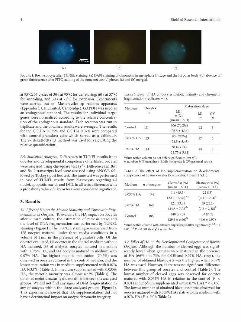
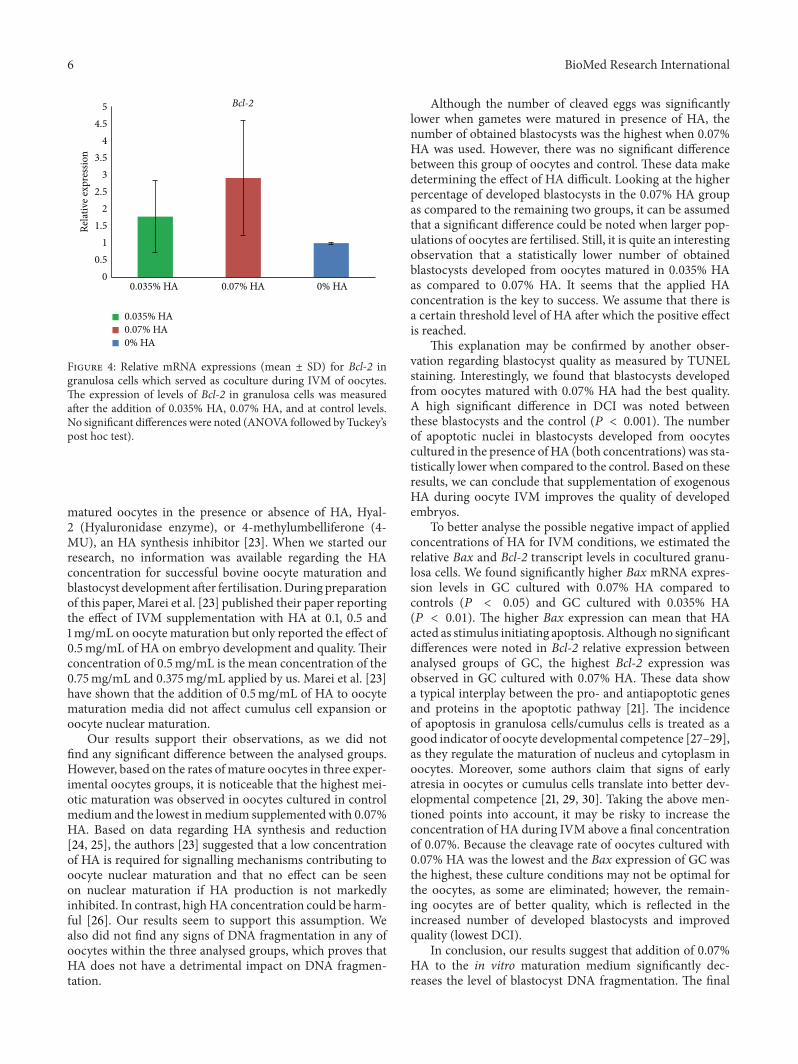
(a) (b) (c)
Figure 1: Bovine oocyte after TUNEL staining. (a) DAPI staining of chromatin in metaphase II stage and the 1st polar body; (b) absence ofgreen fluorescence after FITC staining of the same oocyte; (c) photos (a) and (b) merged.
at 95∘C; 35 cycles of 30 s at 95∘C for denaturing; 60 s at 57∘Cfor annealing; and 30 s at 72∘C for extension. Experimentswere carried out on Mastercycler ep realplex apparatus(Eppendorf, UK Limited, Cambridge). GAPDH was used asan endogenous standard. The results for individual targetgenes were normalised according to the relative concentra-tion of the endogenous standard. Each reaction was run intriplicate and the obtained results were averaged. The resultsfor the GC HA 0.035% and GC HA 0.07% were comparedwith control granulosa cells which served as a calibrator.The 2–[delta][delta]Ct method was used for calculating therelative quantification.
2.9. Statistical Analysis. Differences in TUNEL results fromoocytes and developmental competence of fertilised oocyteswere assessed using chi-square test (𝜒2). Differences in Baxand Bcl-2 transcripts level were assessed using ANOVA fol-lowed by Tuckey’s post hoc test.The same test was performedin case of TUNEL results from blastocysts regarding allnuclei, apoptotic nuclei, and DCI. In all tests differences witha probability value of 0.05 or less were considered significant.
3. Results
3.1. Effect of HA on the Meiotic Maturity and Chromatin Frag-mentation of Oocytes. To evaluate the HA impact on oocytesafter in vitro culture, the estimation of meiosis stage andthe level of DNA fragmentation was performed by TUNELstaining (Figure 1). The TUNEL staining was analysed from428 oocytes matured under three media conditions in avolume of 2mL in the presence of granulosa cells. Of theoocytes evaluated, 151 oocytes in the control mediumwithoutHA matured, 133 of analysed oocytes matured in mediumwith 0.035% HA, and 144 oocytes matured in medium with0.07% HA. The highest meiotic maturation (70.2%) wasobserved in oocytes cultured in the control medium, and thelowest maturation was in medium supplemented with 0.07%HA (63.1%) (Table 1). In medium supplemented with 0.035%HA, the meiotic maturity was almost 67.7% (Table 1). Theobtainedmeioticmaturity did not differ between the analysedgroups. We did not find any signs of DNA fragmentation inany of oocytes within the three analysed groups (Figure 1).This experiment showed that HA supplementation did nothave a detrimental impact on oocyte chromatin integrity.
Table 1: Effect of HA on oocytes meiotic maturity and chromatinfragmentation (replicates = 4).
Medium Oocytes𝑛
Maturation stageMII𝑛 (%)
(mean ± S.D)
MI𝑛
GV𝑛
Control 151 106 (70.2%) 42 3(26.5 ± 4.36)
0.035% HA 133 90 (67.7%) 37 6(22.5 ± 5.45)
0.07% HA 144 91 (63.1%) 48 5(22.75 ± 5.91)
Values within column do not differ significantly (test 𝜒2).𝑛: number; MII: metaphase II; MI: metaphase I; GV: germinal vesicle.
Table 2: The effect of HA supplementation on developmentalcompetence of bovine oocytes (5 replicates) (mean ± S.D.).
Medium 𝑛 of oocytes Cleaved 𝑛 (%)(mean ± S.D.)
Blastocyst 𝑛 (%)(mean ± S.D.)
0.035% HA 174 114 (65.5) 22 (13)(22.8 ± 5.26)
A,a(4.4 ± 3.04)
a
0.07% HA 169 124 (73.4) 39 (23.1)(24.8 ± 7.05)
b(7.8 ± 2.38)
b
Control 186 148 (79.5) 33 (17.7)(29.6 ± 6.66)
B(6.6 ± 4.97)
Values within column with different superscripts differ significantly: a,b𝑃 <0.05; A,B𝑃 < 0.001 (test 𝜒2). 𝑛: number.
3.2. Effect of HA on the Developmental Competence of BovineOocytes. Although the number of cleaved eggs was signif-icantly lower when gametes were matured in the presenceof HA (66% and 73% for 0.035 and 0.07% HA, resp.), thenumber of obtained blastocysts was the highest when 0.07%HA was used. However, there was no significant differencebetween this group of oocytes and control (Table 2). Thelowest number of cleaved eggs was observed for oocytesmatured with 0.035% HA in relation to the control (𝑃 <0.001) andmedium supplementedwith 0.07%HA (𝑃 < 0.05).The lowest number of obtained blastocysts was observed foroocytesmaturedwith 0.035%HA relative to themediumwith0.07% HA (𝑃 < 0.05; Table 2).
BioMed Research International 5
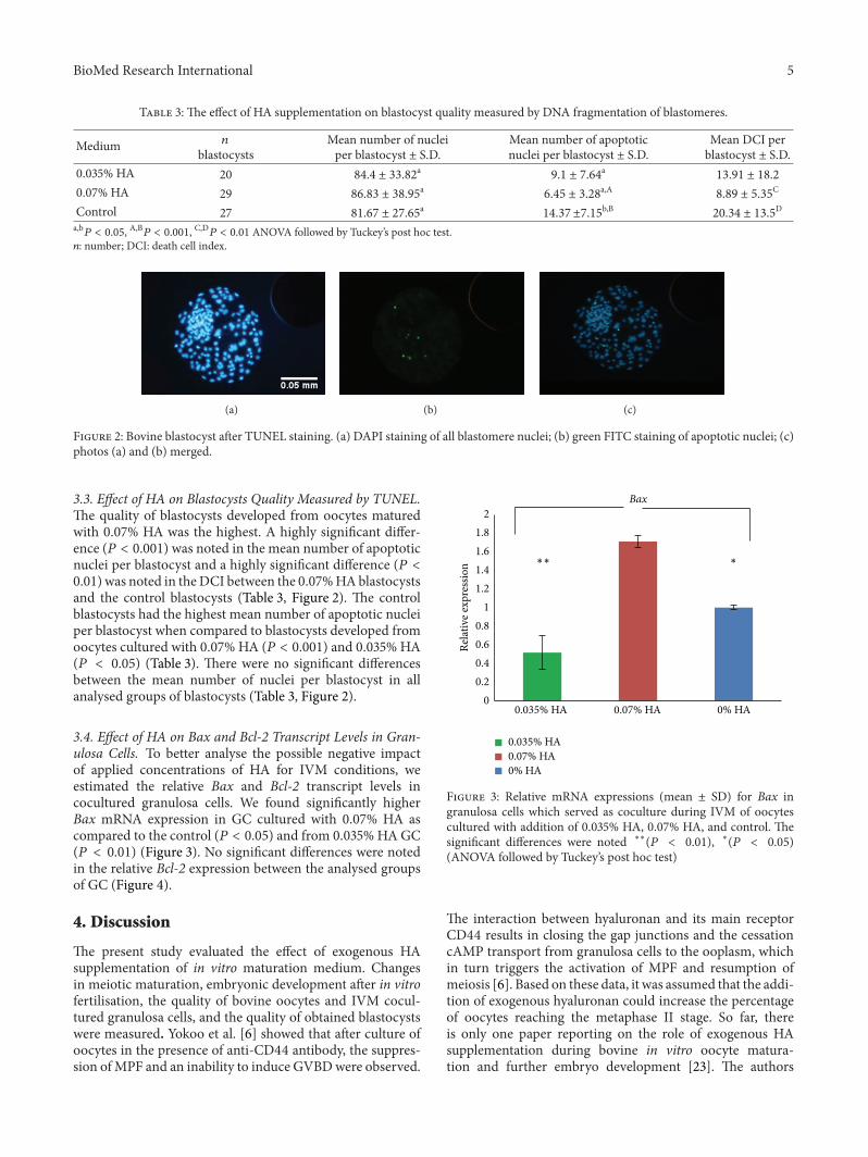
Table 3: The effect of HA supplementation on blastocyst quality measured by DNA fragmentation of blastomeres.
Medium 𝑛
blastocystsMean number of nucleiper blastocyst ± S.D.
Mean number of apoptoticnuclei per blastocyst ± S.D.
Mean DCI perblastocyst ± S.D.
0.035% HA 20 84.4 ± 33.82a
9.1 ± 7.64a
13.91 ± 18.2
0.07% HA 29 86.83 ± 38.95a
6.45 ± 3.28a,A
8.89 ± 5.35C
Control 27 81.67 ± 27.65a
14.37 ±7.15b,B
20.34 ± 13.5D
a,b𝑃 < 0.05, A,B𝑃 < 0.001, C,D𝑃 < 0.01 ANOVA followed by Tuckey’s post hoc test.𝑛: number; DCI: death cell index.
(a) (b) (c)
Figure 2: Bovine blastocyst after TUNEL staining. (a) DAPI staining of all blastomere nuclei; (b) green FITC staining of apoptotic nuclei; (c)photos (a) and (b) merged.
3.3. Effect of HA on Blastocysts Quality Measured by TUNEL.The quality of blastocysts developed from oocytes maturedwith 0.07% HA was the highest. A highly significant differ-ence (𝑃 < 0.001) was noted in the mean number of apoptoticnuclei per blastocyst and a highly significant difference (𝑃 <0.01) was noted in theDCI between the 0.07%HAblastocystsand the control blastocysts (Table 3, Figure 2). The controlblastocysts had the highest mean number of apoptotic nucleiper blastocyst when compared to blastocysts developed fromoocytes cultured with 0.07% HA (𝑃 < 0.001) and 0.035% HA(𝑃 < 0.05) (Table 3). There were no significant differencesbetween the mean number of nuclei per blastocyst in allanalysed groups of blastocysts (Table 3, Figure 2).
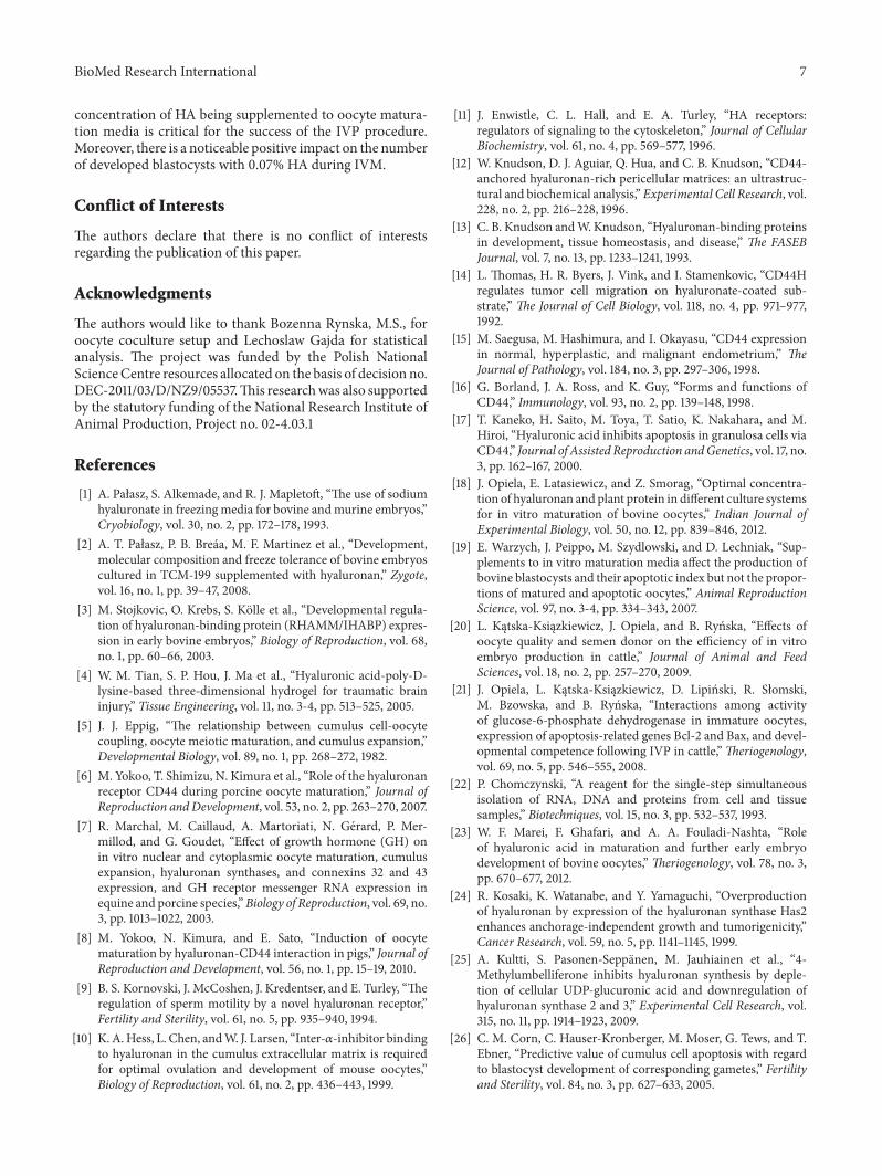
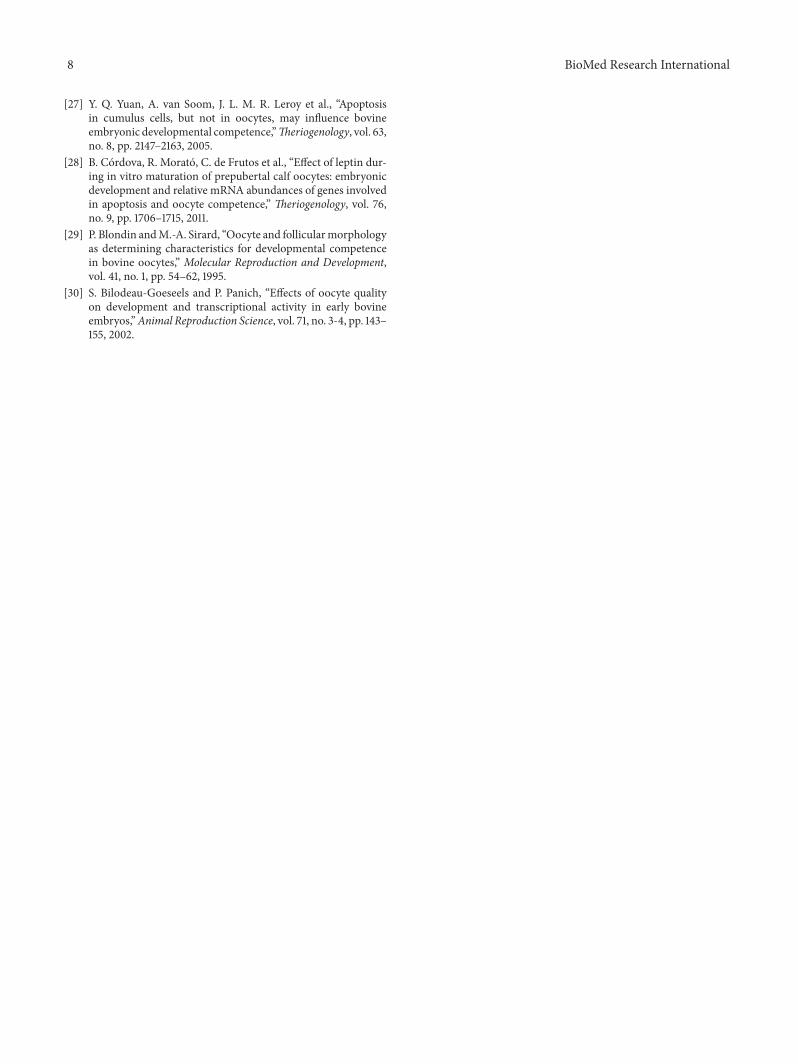
3.4. Effect of HA on Bax and Bcl-2 Transcript Levels in Gran-ulosa Cells. To better analyse the possible negative impactof applied concentrations of HA for IVM conditions, weestimated the relative Bax and Bcl-2 transcript levels incocultured granulosa cells. We found significantly higherBax mRNA expression in GC cultured with 0.07% HA ascompared to the control (𝑃 < 0.05) and from 0.035% HAGC(𝑃 < 0.01) (Figure 3). No significant differences were notedin the relative Bcl-2 expression between the analysed groupsof GC (Figure 4).
4. Discussion
The present study evaluated the effect of exogenous HAsupplementation of in vitro maturation medium. Changesin meiotic maturation, embryonic development after in vitrofertilisation, the quality of bovine oocytes and IVM cocul-tured granulosa cells, and the quality of obtained blastocystswere measured. Yokoo et al. [6] showed that after culture ofoocytes in the presence of anti-CD44 antibody, the suppres-sion ofMPF and an inability to induce GVBDwere observed.
00.20.40.60.8
11.21.41.61.8
2
0.035% HA 0.07% HA 0% HA
Relat
ive e
xpre
ssio
n
Bax
0.035% HA0.07% HA0% HA
∗∗ ∗
Figure 3: Relative mRNA expressions (mean ± SD) for Bax ingranulosa cells which served as coculture during IVM of oocytescultured with addition of 0.035% HA, 0.07% HA, and control. Thesignificant differences were noted ∗∗(𝑃 < 0.01), ∗(𝑃 < 0.05)(ANOVA followed by Tuckey’s post hoc test)
The interaction between hyaluronan and its main receptorCD44 results in closing the gap junctions and the cessationcAMP transport from granulosa cells to the ooplasm, whichin turn triggers the activation of MPF and resumption ofmeiosis [6]. Based on these data, it was assumed that the addi-tion of exogenous hyaluronan could increase the percentageof oocytes reaching the metaphase II stage. So far, thereis only one paper reporting on the role of exogenous HAsupplementation during bovine in vitro oocyte matura-tion and further embryo development [23]. The authors
6 BioMed Research International
00.5
11.5
22.5
33.5
44.5
5
0.035% HA 0.07% HA 0% HA
Relat
ive e
xpre
ssio
n
Bcl-2
0.035% HA0.07% HA0% HA
Figure 4: Relative mRNA expressions (mean ± SD) for Bcl-2 ingranulosa cells which served as coculture during IVM of oocytes.The expression of levels of Bcl-2 in granulosa cells was measuredafter the addition of 0.035% HA, 0.07% HA, and at control levels.No significant differences were noted (ANOVA followed by Tuckey’spost hoc test).
matured oocytes in the presence or absence of HA, Hyal-2 (Hyaluronidase enzyme), or 4-methylumbelliferone (4-MU), an HA synthesis inhibitor [23]. When we started ourresearch, no information was available regarding the HAconcentration for successful bovine oocyte maturation andblastocyst development after fertilisation.During preparationof this paper, Marei et al. [23] published their paper reportingthe effect of IVM supplementation with HA at 0.1, 0.5 and1mg/mL on oocyte maturation but only reported the effect of0.5mg/mL of HA on embryo development and quality. Theirconcentration of 0.5mg/mL is the mean concentration of the0.75mg/mL and 0.375mg/mL applied by us. Marei et al. [23]have shown that the addition of 0.5mg/mL of HA to oocytematuration media did not affect cumulus cell expansion oroocyte nuclear maturation.
Our results support their observations, as we did notfind any significant difference between the analysed groups.However, based on the rates ofmature oocytes in three exper-imental oocytes groups, it is noticeable that the highest mei-otic maturation was observed in oocytes cultured in controlmedium and the lowest inmedium supplemented with 0.07%HA. Based on data regarding HA synthesis and reduction[24, 25], the authors [23] suggested that a low concentrationof HA is required for signalling mechanisms contributing tooocyte nuclear maturation and that no effect can be seenon nuclear maturation if HA production is not markedlyinhibited. In contrast, highHA concentration could be harm-ful [26]. Our results seem to support this assumption. Wealso did not find any signs of DNA fragmentation in any ofoocytes within the three analysed groups, which proves thatHA does not have a detrimental impact on DNA fragmen-tation.
Although the number of cleaved eggs was significantlylower when gametes were matured in presence of HA, thenumber of obtained blastocysts was the highest when 0.07%HA was used. However, there was no significant differencebetween this group of oocytes and control. These data makedetermining the effect of HA difficult. Looking at the higherpercentage of developed blastocysts in the 0.07% HA groupas compared to the remaining two groups, it can be assumedthat a significant difference could be noted when larger pop-ulations of oocytes are fertilised. Still, it is quite an interestingobservation that a statistically lower number of obtainedblastocysts developed from oocytes matured in 0.035% HAas compared to 0.07% HA. It seems that the applied HAconcentration is the key to success. We assume that there isa certain threshold level of HA after which the positive effectis reached.
This explanation may be confirmed by another obser-vation regarding blastocyst quality as measured by TUNELstaining. Interestingly, we found that blastocysts developedfrom oocytes matured with 0.07% HA had the best quality.A high significant difference in DCI was noted betweenthese blastocysts and the control (𝑃 < 0.001). The numberof apoptotic nuclei in blastocysts developed from oocytescultured in the presence ofHA (both concentrations) was sta-tistically lower when compared to the control. Based on theseresults, we can conclude that supplementation of exogenousHA during oocyte IVM improves the quality of developedembryos.
To better analyse the possible negative impact of appliedconcentrations of HA for IVM conditions, we estimated therelative Bax and Bcl-2 transcript levels in cocultured granu-losa cells. We found significantly higher Bax mRNA expres-sion levels in GC cultured with 0.07% HA compared tocontrols (𝑃 < 0.05) and GC cultured with 0.035% HA(𝑃 < 0.01). The higher Bax expression can mean that HAacted as stimulus initiating apoptosis. Althoughno significantdifferences were noted in Bcl-2 relative expression betweenanalysed groups of GC, the highest Bcl-2 expression wasobserved in GC cultured with 0.07% HA. These data showa typical interplay between the pro- and antiapoptotic genesand proteins in the apoptotic pathway [21]. The incidenceof apoptosis in granulosa cells/cumulus cells is treated as agood indicator of oocyte developmental competence [27–29],as they regulate the maturation of nucleus and cytoplasm inoocytes. Moreover, some authors claim that signs of earlyatresia in oocytes or cumulus cells translate into better dev-elopmental competence [21, 29, 30]. Taking the above men-tioned points into account, it may be risky to increase theconcentration of HA during IVM above a final concentrationof 0.07%. Because the cleavage rate of oocytes cultured with0.07% HA was the lowest and the Bax expression of GC wasthe highest, these culture conditions may not be optimal forthe oocytes, as some are eliminated; however, the remain-ing oocytes are of better quality, which is reflected in theincreased number of developed blastocysts and improvedquality (lowest DCI).
In conclusion, our results suggest that addition of 0.07%HA to the in vitro maturation medium significantly dec-reases the level of blastocyst DNA fragmentation. The final
BioMed Research International 7
concentration of HA being supplemented to oocyte matura-tion media is critical for the success of the IVP procedure.Moreover, there is a noticeable positive impact on the numberof developed blastocysts with 0.07% HA during IVM.
Conflict of Interests
The authors declare that there is no conflict of interestsregarding the publication of this paper.
Acknowledgments
The authors would like to thank Bozenna Rynska, M.S., foroocyte coculture setup and Lechoslaw Gajda for statisticalanalysis. The project was funded by the Polish NationalScienceCentre resources allocated on the basis of decision no.DEC-2011/03/D/NZ9/05537.This researchwas also supportedby the statutory funding of the National Research Institute ofAnimal Production, Project no. 02-4.03.1
References
[1] A. Pałasz, S. Alkemade, and R. J. Mapletoft, “The use of sodiumhyaluronate in freezingmedia for bovine andmurine embryos,”Cryobiology, vol. 30, no. 2, pp. 172–178, 1993.
[2] A. T. Pałasz, P. B. Breaa, M. F. Martinez et al., “Development,molecular composition and freeze tolerance of bovine embryoscultured in TCM-199 supplemented with hyaluronan,” Zygote,vol. 16, no. 1, pp. 39–47, 2008.
[3] M. Stojkovic, O. Krebs, S. Kolle et al., “Developmental regula-tion of hyaluronan-binding protein (RHAMM/IHABP) expres-sion in early bovine embryos,” Biology of Reproduction, vol. 68,no. 1, pp. 60–66, 2003.
[4] W. M. Tian, S. P. Hou, J. Ma et al., “Hyaluronic acid-poly-D-lysine-based three-dimensional hydrogel for traumatic braininjury,” Tissue Engineering, vol. 11, no. 3-4, pp. 513–525, 2005.
[5] J. J. Eppig, “The relationship between cumulus cell-oocytecoupling, oocyte meiotic maturation, and cumulus expansion,”Developmental Biology, vol. 89, no. 1, pp. 268–272, 1982.
[6] M. Yokoo, T. Shimizu, N. Kimura et al., “Role of the hyaluronanreceptor CD44 during porcine oocyte maturation,” Journal ofReproduction andDevelopment, vol. 53, no. 2, pp. 263–270, 2007.
[7] R. Marchal, M. Caillaud, A. Martoriati, N. Gerard, P. Mer-millod, and G. Goudet, “Effect of growth hormone (GH) onin vitro nuclear and cytoplasmic oocyte maturation, cumulusexpansion, hyaluronan synthases, and connexins 32 and 43expression, and GH receptor messenger RNA expression inequine and porcine species,”Biology of Reproduction, vol. 69, no.3, pp. 1013–1022, 2003.
[8] M. Yokoo, N. Kimura, and E. Sato, “Induction of oocytematuration by hyaluronan-CD44 interaction in pigs,” Journal ofReproduction and Development, vol. 56, no. 1, pp. 15–19, 2010.
[9] B. S. Kornovski, J. McCoshen, J. Kredentser, and E. Turley, “Theregulation of sperm motility by a novel hyaluronan receptor,”Fertility and Sterility, vol. 61, no. 5, pp. 935–940, 1994.
[10] K. A.Hess, L. Chen, andW. J. Larsen, “Inter-𝛼-inhibitor bindingto hyaluronan in the cumulus extracellular matrix is requiredfor optimal ovulation and development of mouse oocytes,”Biology of Reproduction, vol. 61, no. 2, pp. 436–443, 1999.
[11] J. Enwistle, C. L. Hall, and E. A. Turley, “HA receptors:regulators of signaling to the cytoskeleton,” Journal of CellularBiochemistry, vol. 61, no. 4, pp. 569–577, 1996.
[12] W. Knudson, D. J. Aguiar, Q. Hua, and C. B. Knudson, “CD44-anchored hyaluronan-rich pericellular matrices: an ultrastruc-tural and biochemical analysis,” Experimental Cell Research, vol.228, no. 2, pp. 216–228, 1996.
[13] C. B. Knudson andW. Knudson, “Hyaluronan-binding proteinsin development, tissue homeostasis, and disease,” The FASEBJournal, vol. 7, no. 13, pp. 1233–1241, 1993.
[14] L. Thomas, H. R. Byers, J. Vink, and I. Stamenkovic, “CD44Hregulates tumor cell migration on hyaluronate-coated sub-strate,” The Journal of Cell Biology, vol. 118, no. 4, pp. 971–977,1992.
[15] M. Saegusa, M. Hashimura, and I. Okayasu, “CD44 expressionin normal, hyperplastic, and malignant endometrium,” TheJournal of Pathology, vol. 184, no. 3, pp. 297–306, 1998.
[16] G. Borland, J. A. Ross, and K. Guy, “Forms and functions ofCD44,” Immunology, vol. 93, no. 2, pp. 139–148, 1998.
[17] T. Kaneko, H. Saito, M. Toya, T. Satio, K. Nakahara, and M.Hiroi, “Hyaluronic acid inhibits apoptosis in granulosa cells viaCD44,” Journal of Assisted Reproduction andGenetics, vol. 17, no.3, pp. 162–167, 2000.
[18] J. Opiela, E. Latasiewicz, and Z. Smorag, “Optimal concentra-tion of hyaluronan and plant protein in different culture systemsfor in vitro maturation of bovine oocytes,” Indian Journal ofExperimental Biology, vol. 50, no. 12, pp. 839–846, 2012.
[19] E. Warzych, J. Peippo, M. Szydlowski, and D. Lechniak, “Sup-plements to in vitro maturation media affect the production ofbovine blastocysts and their apoptotic index but not the propor-tions of matured and apoptotic oocytes,” Animal ReproductionScience, vol. 97, no. 3-4, pp. 334–343, 2007.
[20] L. Kątska-Ksiązkiewicz, J. Opiela, and B. Rynska, “Effects ofoocyte quality and semen donor on the efficiency of in vitroembryo production in cattle,” Journal of Animal and FeedSciences, vol. 18, no. 2, pp. 257–270, 2009.
[21] J. Opiela, L. Kątska-Ksiązkiewicz, D. Lipinski, R. Słomski,M. Bzowska, and B. Rynska, “Interactions among activityof glucose-6-phosphate dehydrogenase in immature oocytes,expression of apoptosis-related genes Bcl-2 and Bax, and devel-opmental competence following IVP in cattle,”Theriogenology,vol. 69, no. 5, pp. 546–555, 2008.
[22] P. Chomczynski, “A reagent for the single-step simultaneousisolation of RNA, DNA and proteins from cell and tissuesamples,” Biotechniques, vol. 15, no. 3, pp. 532–537, 1993.
[23] W. F. Marei, F. Ghafari, and A. A. Fouladi-Nashta, “Roleof hyaluronic acid in maturation and further early embryodevelopment of bovine oocytes,” Theriogenology, vol. 78, no. 3,pp. 670–677, 2012.
[24] R. Kosaki, K. Watanabe, and Y. Yamaguchi, “Overproductionof hyaluronan by expression of the hyaluronan synthase Has2enhances anchorage-independent growth and tumorigenicity,”Cancer Research, vol. 59, no. 5, pp. 1141–1145, 1999.
[25] A. Kultti, S. Pasonen-Seppanen, M. Jauhiainen et al., “4-Methylumbelliferone inhibits hyaluronan synthesis by deple-tion of cellular UDP-glucuronic acid and downregulation ofhyaluronan synthase 2 and 3,” Experimental Cell Research, vol.315, no. 11, pp. 1914–1923, 2009.
[26] C. M. Corn, C. Hauser-Kronberger, M. Moser, G. Tews, and T.Ebner, “Predictive value of cumulus cell apoptosis with regardto blastocyst development of corresponding gametes,” Fertilityand Sterility, vol. 84, no. 3, pp. 627–633, 2005.
8 BioMed Research International
[27] Y. Q. Yuan, A. van Soom, J. L. M. R. Leroy et al., “Apoptosisin cumulus cells, but not in oocytes, may influence bovineembryonic developmental competence,”Theriogenology, vol. 63,no. 8, pp. 2147–2163, 2005.
[28] B. Cordova, R. Morato, C. de Frutos et al., “Effect of leptin dur-ing in vitro maturation of prepubertal calf oocytes: embryonicdevelopment and relative mRNA abundances of genes involvedin apoptosis and oocyte competence,” Theriogenology, vol. 76,no. 9, pp. 1706–1715, 2011.
[29] P. Blondin andM.-A. Sirard, “Oocyte and follicularmorphologyas determining characteristics for developmental competencein bovine oocytes,” Molecular Reproduction and Development,vol. 41, no. 1, pp. 54–62, 1995.
[30] S. Bilodeau-Goeseels and P. Panich, “Effects of oocyte qualityon development and transcriptional activity in early bovineembryos,”Animal Reproduction Science, vol. 71, no. 3-4, pp. 143–155, 2002.

Submit your manuscripts athttp://www.hindawi.com
Stem CellsInternational
Hindawi Publishing Corporationhttp://www.hindawi.com Volume 2014
Hindawi Publishing Corporationhttp://www.hindawi.com Volume 2014
MEDIATORSINFLAMMATION
of
Hindawi Publishing Corporationhttp://www.hindawi.com Volume 2014
Behavioural Neurology
EndocrinologyInternational Journal of
Hindawi Publishing Corporationhttp://www.hindawi.com Volume 2014
Hindawi Publishing Corporationhttp://www.hindawi.com Volume 2014
Disease Markers
Hindawi Publishing Corporationhttp://www.hindawi.com Volume 2014
BioMed Research International
OncologyJournal of
Hindawi Publishing Corporationhttp://www.hindawi.com Volume 2014
Hindawi Publishing Corporationhttp://www.hindawi.com Volume 2014
Oxidative Medicine and Cellular Longevity
Hindawi Publishing Corporationhttp://www.hindawi.com Volume 2014
PPAR Research
The Scientific World JournalHindawi Publishing Corporation http://www.hindawi.com Volume 2014
Immunology ResearchHindawi Publishing Corporationhttp://www.hindawi.com Volume 2014
Journal of
ObesityJournal of
Hindawi Publishing Corporationhttp://www.hindawi.com Volume 2014
Hindawi Publishing Corporationhttp://www.hindawi.com Volume 2014
Computational and Mathematical Methods in Medicine
OphthalmologyJournal of
Hindawi Publishing Corporationhttp://www.hindawi.com Volume 2014
Diabetes ResearchJournal of
Hindawi Publishing Corporationhttp://www.hindawi.com Volume 2014
Hindawi Publishing Corporationhttp://www.hindawi.com Volume 2014
Research and TreatmentAIDS
Hindawi Publishing Corporationhttp://www.hindawi.com Volume 2014
Gastroenterology Research and Practice
Hindawi Publishing Corporationhttp://www.hindawi.com Volume 2014
Parkinson’s Disease
Evidence-Based Complementary and Alternative Medicine
Volume 2014Hindawi Publishing Corporationhttp://www.hindawi.com
Related Documents