_____________________________________________________________________________________________________ *Corresponding author: E-mail: [email protected]; Microbiology Research Journal International 27(6): 1-14, 2019; Article no.MRJI.49121 ISSN: 2456-7043 (Past name: British Microbiology Research Journal, Past ISSN: 2231-0886, NLM ID: 101608140) Effect of Fermentation on the Nutrient and Anti- nutrient Contents of African Bush Mango (Irvingia gabonensis) Seeds Ayeni Oluwanifemi Helen 1* and Ojokoh Anthony Okhonlaye 2 1 Department of Microbiology, Federal University of Technology, Akure, Nigeria. 2 Department of Biotechnology, Federal University of Technology, Akure, Nigeria. Authors’ contributions This work was carried out in collaboration between both authors. Author AOH designed the study, performed the statistical analysis, wrote the protocol, wrote the first draft of the manuscript and managed literature searches. Author OAO managed the analyses of the study and literature searches. Both authors read and approved the final manuscript. Article Information DOI: 10.9734/MRJI/2019/v27i630118 Editor(s): (1) Dr. Ng Zhi Xiang, Department of Biomedical Sciences, Faculty of Medicine, MAHSA University, Malaysia. Reviewers: (1) M. M. V. Baig, Yeshwant Mahavidyalaya, India. (2) R. Prabha, Dairy Science College, Karnataka Veterinary, Animal and Fisheries Sciences University, India. Complete Peer review History: http://www.sdiarticle3.com/review-history/49121 Received 13 March 2019 Accepted 29 May 2019 Published 08 June 2019 ABSTRACT Aim: Effect of fermentation on nutrient and anti-nutrient contents of defatted and un-defatted African bush mango seeds. Study Design: Ground African bush mango seeds used in this study were divided into two portions; A, and B. Portion A was defatted while portion B was not defatted; both portions were fermented. Place and Duration of Study: Department of Microbiology and Chemistry Department, Federal University of Technology Akure, Ondo State between November 2017 and July 2018. Methodology: Microbial analysis was carried out using pour plate technique. The temperature, pH and total titratable acidity were monitored throughout the fermenting period. Proximate, mineral and anti-nutrient contents of the samples were carried out using standard methods. Results: Seventeen microorganisms comprising 11 bacteria and 6 molds were isolated and identified as; Staphylococcus aureus, Bacillus subtilis, B. cereus, S. epidermis, B. licheniformis, Micrococcus luteus, Proteus vulgaris, Enterococcus faecalis, Lactobacillus fermentum, L. plantarum, Original Research Article

Welcome message from author
This document is posted to help you gain knowledge. Please leave a comment to let me know what you think about it! Share it to your friends and learn new things together.
Transcript
_____________________________________________________________________________________________________ *Corresponding author: E-mail: [email protected];
Microbiology Research Journal International 27(6): 1-14, 2019; Article no.MRJI.49121 ISSN: 2456-7043 (Past name: British Microbiology Research Journal, Past ISSN: 2231-0886, NLM ID: 101608140)
Effect of Fermentation on the Nutrient and Anti-nutrient Contents of African Bush Mango
(Irvingia gabonensis) Seeds
Ayeni Oluwanifemi Helen1* and Ojokoh Anthony Okhonlaye2
1Department of Microbiology, Federal University of Technology, Akure, Nigeria. 2Department of Biotechnology, Federal University of Technology, Akure, Nigeria.
Authors’ contributions
This work was carried out in collaboration between both authors. Author AOH designed the study,
performed the statistical analysis, wrote the protocol, wrote the first draft of the manuscript and managed literature searches. Author OAO managed the analyses of the study and literature
searches. Both authors read and approved the final manuscript.
Article Information
DOI: 10.9734/MRJI/2019/v27i630118
Editor(s):
(1) Dr. Ng Zhi Xiang, Department of Biomedical Sciences, Faculty of Medicine, MAHSA University, Malaysia.
Reviewers:
(1) M. M. V. Baig, Yeshwant Mahavidyalaya, India.
(2) R. Prabha, Dairy Science College, Karnataka Veterinary, Animal and Fisheries Sciences University, India.
Complete Peer review History: http://www.sdiarticle3.com/review-history/49121
Received 13 March 2019 Accepted 29 May 2019
Published 08 June 2019
ABSTRACT Aim: Effect of fermentation on nutrient and anti-nutrient contents of defatted and un-defatted African bush mango seeds. Study Design: Ground African bush mango seeds used in this study were divided into two portions; A, and B. Portion A was defatted while portion B was not defatted; both portions were fermented. Place and Duration of Study: Department of Microbiology and Chemistry Department, Federal University of Technology Akure, Ondo State between November 2017 and July 2018. Methodology: Microbial analysis was carried out using pour plate technique. The temperature, pH and total titratable acidity were monitored throughout the fermenting period. Proximate, mineral and anti-nutrient contents of the samples were carried out using standard methods. Results: Seventeen microorganisms comprising 11 bacteria and 6 molds were isolated and identified as; Staphylococcus aureus, Bacillus subtilis, B. cereus, S. epidermis, B. licheniformis, Micrococcus luteus, Proteus vulgaris, Enterococcus faecalis, Lactobacillus fermentum, L. plantarum,
Original Research Article

Helen and Okhonlaye; MRJI, 27(6): 1-14, 2019; Article no.MRJI.49121
2
L. brevis, Aspergillus clavatus, A. flavus, A. niger, Rhizopus stolonifer, Penicillium chrysogenum and A. fumigatus. The pH and TTA values reduced and increased respectively while the temperature varied significantly as the fermentation day increases. The non-defatted fermented sample showed increase in protein (10.34-12.09%), moisture (6.98-7.84%) and carbohydrate contents (24.98-29.20%); while there was a reduction in the ash (3.91-2.93%), fibre (1.55-1.30%) and fat (52.24-46.64%) contents. The defatted fermented sample showed an increase in the protein content (17.39-26.44%) while there was a reduction in the moisture (26.60-26.46%), carbohydrate (41.02-38.96%) ash (4.07-3.01%), fat (9.44-4.02%) and fibre contents (1.48-1.11%). The mineral composition of the fermented samples increased significantly when compared to the raw samples. The anti-nutrient content of the samples decreased significantly with fermentation. Conclusion: This study revealed that African bush mango seeds can be defatted and fermented to produce food of enhanced nutritional value.
Keywords: Fermentation; bush mango; African bush mango; proximate; anti-nutrient.
1. INTRODUCTION Trees and shrubs with medicinal and nutritional potentials proliferate in Nigeria and several of these plants have fruit and seeds which have been identified to be of nutritional relevance [1]. Mostly in developing countries, seeds are prominent features in the peasant dietary and in countries where the diet is plant based, oilseeds are becoming valuable sources of nutrient for man [2]. Attention has therefore been focused on under-utilized local seeds for possible development and use [1]. Irvingia gabonensis (Aubry-Lecomte ex O'Rorke) Baill is an economic food tree of West and Central Africa which belongs to the genus Irvingia within the family Irvingiaceae [3,4]. The genus Irvingia comprises of seven specie out of which only Irvingia gabonensis and Irvingia excelsa (wombulu) which are frequently mistaken for each other are the only varieties identified in Nigeria and are subject of several transaction and some physiochemical studies [1,5,6]. The term African bush mango refers to these two economically most important Irvingia species that occur in the humid lowland forests of West and Central Africa and can be differentiated in that their flesh can either be sweet and edible (Irvingia gabonensis) or bitter and inedible (Irvingia excelsa) [7]. African bush mango bears edible mango-like fruit which is made up of the fleshy part and the nut, which consists of a hard shell and the kernel/seed. Its seeds have an outer brown testa (hull) and two white cotyledons which are especially valued for being rich in fat and protein [3,8]. The seed has nutritive, medicinal and industrial benefits and are richer in lipids than other oil seeds and legumes [9]. They also serve as source of human food and constitute important
part of the diet in Nigeria as they are good source of vitamins and minerals. The ground seeds are used as thickening agents in soups and the oil can be processed into soap, cosmetics or pharmaceuticals [3]. It has been reported that ethno-medicinal treatments utilize other parts of the tree, like the bark, kernels, leaves, or roots for a variety of ailments [8].
Fermentation is one of the oldest biotechnologies used in the enhancement of the nutrient content and preservation of food through the biosynthesis of vitamins, essential amino acids and proteins, fibre digestibility and degrading anti-nutritional factors [5]. Fermented foods constitute an important part of the world’s diet and are estimated to provide about 20-40% of human food supply. Chemical compounds, which are end products of fermentation process are not only enjoyed and tasty to a large number of people of different ethnic groups, it has also been noted that no single group or category of foods or food products are as important as fermented foods and have been relative to man’s nutritional well-being throughout the world [1,10]. In order to maximize the nutritional benefits of African bush mango seeds and owing to the fact that not enough research has been carried out on the defatted seeds, it became necessary to determine the effect of fermentation on both the defatted and un-defatted seeds. The objective of this research is to determine the effect of fermentation on the nutrient and anti-nutrient contents of African bush mango seeds.
2. MATERIALS AND METHODS
2.1 Collection of Samples
African bush mango seeds used for this study were obtained from “Oja-oba” a local market in Akure, Ondo State, Nigeria.
Helen and Okhonlaye; MRJI, 27(6): 1-14, 2019; Article no.MRJI.49121
3
2.2 Processing of African Bush Mango Seeds
The seeds were sorted by removal of stones and other foreign materials. They were surface disinfected by dipping in 70% ethyl alcohol for 60seconds, rinsed in several changes of sterile distilled water and then grinded using mortar and pestle.
2.2.1 Defatting of the sample
A portion of the ground seed was defatted using the soxhlet extraction method as described by [11] All the glass apparatus used were rinsed with the solvent which is n-hexane after appropriate cleaning. The apparatus was set up by placing the distillation flask filled with n-hexane up to three quarters on the heat source. The thimble containing ground African bush mango seeds was loaded into the main chamber of the soxhlet extractor which was placed on the distillation flask and a condenser was placed on top. The solvent is heated to reflux and the evaporated solvent passes through the side tube of the extractor and condenses in the condenser fitted at the top of the extractor. The condensed hot solvent runs into the thimble and soaks the sample extracting its constituent. The chamber holding the thimble becomes full and the solvent siphons down to the flask. This process was repeated till extraction is complete usually between 5-7 hours
2.3 Fermentation of Samples
The submerged state fermentation was employed for the fermentation of the African Bush Mango seeds in different ratios due to the nature of the samples for 96 hours. The un-defatted seeds were soaked in sterile distilled water in ratio 1:5 while the defatted sample in the ratio 1:20.
2.4 Microbiological Analysis of the Samples
Bacteria and fungi were evaluated using nutrient agar (NA) and potato dextrose agar (PDA) respectively while De Man Rogosa and Sharpe agar was used to isolate lactic acid bacteria. Techniques were enumerated by using appropriate serial dilution and pour plate techniques. The bacterial culture was incubated at 37°C for 18 to 24 hours, fungal plates were inverted and incubated at 24°C for 48 to 72 hours. De Man Rogosa and Sharpe agar plates were incubated at 32°C for 18- 24 hours anaerobically. Bacteria isolates were
characterized based on biochemical and morphological observations according to the method of [12]. The results were compared with Bergey’s Manual of Determinative Bacteriology [13]. Fungi isolates were identified according to [14].
2.5 Determination of pH, Total Titratable Acidity (TTA) and Temperature
The pH, temperature and TTA of the samples were monitored throughout the fermentation period. The pH was ascertained using the pH meter metrom E520 which was calibrated using buffer solution of pH 4.0, 7.0 and 9.0. 1 g of the sample was homogenized in 10 ml 0f distilled water and the pH glass electrode was inserted for 2 minutes ensuring that the glass electrode did not touch the bottom of the bottle. The resultant value was read on the meter scale and then recorded in triplicate. The temperature was determined using a mercury in-bulb thermometer which was dipped into the fermenting sample for about 3 minutes under sterile condition, it was then withdrawn and the temperature was read and recorded in triplicate. TTA was estimated according to the official methods of analysis [11]. 2 g of each sample was weighed into 20 ml of distilled water in different beakers, 2 drops of phenolphthalein was added as an indicator and then 150 ml of the aliquots were titrated against 0.1 N NaOH.
2.6 Determination of Proximate Composition
The samples were analysed daily for Moisture, Ash, Fat, Protein, Crude fiber and Carbohydrate according to the method described by [11]. Moisture content was determined by drying to constant weight at 105°C in an oven, ash by ignition at 55°C in a muffle furnace, fat content by soxhlet extraction with hexane, nitrogen by micro-Kjedahl and the percentage nitrogen was converted to crude protein by multiplying by 6.25, crude fibre by acid/alkali digestion methods and carbohydrate determined by difference. The proximate composition was expressed in percentage (%).
2.7 Mineral Determination
The mineral composition of the samples throughout the fermentation period was carried out on the product obtained by dry-ashing the sample in a muffle furnace at 550°C. The ashed samples were cooled in the desiccator, dissolved in 10 ml of 10% HCL and was made up to 50 ml

Helen and Okhonlaye; MRJI, 27(6): 1-14, 2019; Article no.MRJI.49121
4
with deionized water in a volumetric flask. Sodium and potassium were determined using a flame photometer (photometer (model 405, corning UK) while calcium (Ca), zinc (Zn), iron (Fe) and magnesium (Mg) were determined by atomic absorption spectrophotometer (AAS) [11]. The minerals were expressed in mg/g.
2.8 Anti-Nutrient Determination Phytate and tannin was determined using the method of [11], oxalate content was by the titrimetric method as modified by [15] while saponin was determined by the spectrophotometric method as described by [16]. The anti-nutrients were expressed in mg/g.
2.9 Statistical Analysis All analyses were performed in triplicates. The data obtained were subjected to one-way analysis of variance (ANOVA) while differences in mean were determined using Duncan’s New Multiple Range Test (DMRT). All data analyses were done with SPSS 23.0 version.
3. RESULTS AND DISCUSSION 3.1 Microbial Growth during Fermentation
of African Bush Mango Seeds Seventeen (17) microorganisms were isolated from African bush mango seeds which were identified as shown on Tables 4 and 5. Eleven (11) bacteria: Bacillus subtilis Bacillus cereus, Staphylococcus epidermis, Bacillus licheniformis, Micrococcus luteus, Proteus vulgaris, Enterococcus faecalis, Lactobacillus fermentum, Lactobacillus plantarum, Lactobacillus brevis and Staphylococcus aureus. Six fungi: Aspergillus clavatus, Aspergillus flavus, Aspergillus niger, Penicillium chrysogenum, Rhizopus stolonifer and Aspergillus fumigatus. Many factors contribute to the presence of microorganisms in foods, the endogenous presence and cross contaminations are the factors most pointed out as being the sources. However, the diverse kind and number of microorganisms on any food depends on various factors of which the pH, moisture and nutrient composition of the food are major factors [17]. In this study, a total of seventeen microorganisms were isolated from African bush mango seeds. These organisms have been found to be responsible for the fermentation of some legumes as reported by [5,18]. Bacillus species is the predominant
bacteria flora isolated from the samples and this could be as a result of their ability to survive in slightly acidic and alkaline environment. Moreover, they are known to have better competitive ability compared to other bacteria species present in the same environment [19]. Aspergillus and Rhizopus species were isolated from fermenting mango peel reported by [20] as also isolated from the African bush mango seeds in this study. The presence of Staphylococcus specie, Enterococcus faecalis and Proteus vulgaris could have been as a result of contamination during handling and processing, this is in line with the work of [21] who reported that the presence of Staphylococcus sp. during the fermentation of popcorn and groundnut composite flour. 3.2 Changes in Bacteria Population
during Fermentation of African Bush Mango Seeds
Fig. 1 shows the changes in the bacteria population of the samples during fermentation for 96hours. The total bacterial count for both samples (Un-defatted and defatted) increased at 24 hours and 48 hours then decreased at 72 hours and 96 hours. For sample A (Un-defatted sample) the bacteria population increased with time till 48 hours with values 7.00 × 10
5 cfu/ml,
12.02 × 10⁵ cfu/ml and 15.97 × 10
5 cfu/ml while
at 72 hours and 96 hours of the fermentation it decreased to 9.01× 10
5 cfu/ml and 6.01× 10
5
cfu/ml respectively. The bacteria population for sample B (Defatted sample) also increased with time till 48 hours with values 4.00 × 10
5 cfu/ml,
9.02 × 10⁵ cfu/ml, 14.02 × 10⁵ cfu/ml, while a decrease was recorded at 72hours and 96 hours with values 6.97 × 10⁵ cfu/ml, 4.00 × 10⁵ cfu/ml. The decrease observed in bacteria load after 48hours of fermentation may be as a result of nutrient depletion and some bioactive substances which may have produced an inhibitory effect on other organisms present in the medium. This is in line with the report of [19] who reported a decrease in bacteria load after 48hours liquid fermentation of Kersting’s groundnut.
3.3 Changes in Lactic Acid Bacteria
Population during Fermentation of African Bush Mango Seeds
Fig. 2 shows the total lactic acid bacterial count for the African bush mango seeds during fermentation for 96 hours. There was no Lactic

Helen and Okhonlaye; MRJI, 27(6): 1-14, 2019; Article no.MRJI.49121
5
Table 1. Biochemical characteristics of bacteria isolated during fermentation of African bush mango seeds
S/N
Gra
m sta
inin
g
Sh
ap
e
Sp
ore
Ind
ole
Cit
rate
Sta
rch
hyd
roly
sis
Co
ag
ula
se
test
Cata
las
e
H2s
Gas
Nit
rate
red
ucti
on
Mo
tility
te
st
Arr
an
gem
en
t
Oxid
ase
Vo
ges p
rosk
aeu
r M
eth
yl
red
Ure
ase
Lacto
se
Su
cro
se
Fru
cto
se
Glu
co
se
Man
nit
ol
Malt
ose
Dextr
os
e
Pro
bab
le
mic
roo
rgan
ism
1. + Cocci - - + + + + - - + - Cluster - + + + + + + + + + + Staphylococcus aureus 2. + Rod + - + + - + - - + + Singly - + - - - + + + + + + Bacillus subtilis 3. + Rod + - + + - + - - + + Chains - + - - - + + + - + + Bacillus cereus 4. + Cocci - - + + - + - - + - Cluster - + + + + + + + - + + Staphylococcus epidermis 5. + Rod + - + + - + - - + + Singly - + + + + + + + + + Bacillus licheniformis 6. + Cocci - - + - - + - - - - Cluster + - + + - - - - - - + Micrococcus luteus 7. - Rod - + + - - + + + + + Singly - - + + - + + - + + Proteus vulgaris 8. - Cocci - - - - - - - + - Cluster - + - - + + + + + + + Enterococcus faecalis 9. + Rod - - - + - - - + - - Singly - - - - + - + + - + + Lactobacillus fermentum 10. + Rod - - + + - - - + - - Singly - - - - + + + + + - + Lactobacillus plantarum 11. + Cocci - - + - + - + + - - Singly - - + + + + - - - + - Lactobacillus brevis
Keys: + : Positive reaction; - : Negative reaction; H2S: Hydrogen Sulphide gas
Table 2. Characteristics of fungi isolated during fermentation of African bush mango seeds
Cultural characteristics Morphological description Probable fungi Blue-green colonies which appear to be generally coarse and smooth-walled Uniseriate conidia, large club shaped vessicle Aspergillus clavatus Yellow-green colonies, rough walled stipes Radiate conidia which later split to form loose columns, mature vesicles bearing
phialides over their entire surface and conspicuously echinulate conidia Aspergillus flavus
Colonies growth spread rapidly with fluffy and velvety in texture with aerial mycelia white at first, frequently developing dark-brown to black conidia heads
Dark brown conidia, conidiophores are long globose, vesicles that are completely covered with biserate phialides which are borne on brown metulae
Aspergillus niger
Blue-green colonies with yellow pigments Brush-shaped conidiophores, subglobulus conidia, smooth stide and flask-shaped philiade
Penicillium chrysogenum
White cotton-like fluffy mycelium Non-septate hyphae, coenocytic twin sporangiosphores Rhizopus stolonifer Suede-like blue-green colonies that is smooth walled Uniserate and columnar conidial heads with the phalides limited to the upper two thirds
of the vessicle and curving to be roughly parallel to each other Aspergillus fumigatus

Helen and Okhonlaye; MRJI, 27(6): 1-14, 2019; Article no.MRJI.49121
6
acid bacteria growth at the initial hour for both sample however, the growth thereafter increased with increase in fermentation time. For sample A (un-defatted sample) the lactic acid bacteria population increased from 24 hours till 96 hours with values 3.02 × 10
5 cfu/ml, 5.97 × 10
5 cfu/ml,
13.97 × 10⁵ cfu/ml and 16.02 × 10
5 cfu/ml
respectively. The lactic acid bacteria population for sample B (defatted sample) also increased from 24 hours till 96 hours with values 2.00 × 10⁵ cfu/ml, 5.02 × 10
5 cfu/ml, 11.97 × 10
5 cfu/ml and
14.97 × 10⁵ cfu/ml respectively.
3.4 Changes in Fungi Population during Fermentation of African Bush Mango Seeds
Fig. 3 shows the total fungal mean count for the African bush mango seeds during fermentation for 96 hours. There was no fungal growth at the initial hour for both sample however, the growth thereafter increased with increase in fermentation time. For sample A, 3.02 × 10
5
cfu/ml was observed at 24hours, 4.97 × 10⁵
cfu/ml at 48 hours, 9.02 × 10⁵ cfu/ml at 72 hours and 10.97 × 10
5 cfu/ml at 96 hours. For sample
B, 2.97 × 10⁵ cfu/ml, 4.02 × 10⁵ cfu/ml, 6.97 × 10
5 cfu/ml and 8.97 × 10
5 cfu/ml was observed
from 24hours to 96hours respectively. The significant increase observed in the fungal load during fermentation may be due to the ability of fungi to thrive in lower pH and water activity even more than bacteria [19].
3.5 Bacteria Occurrence during Fermentation of African Bush Mango Seeds
Results of the bacteria isolated during fermentation of African bush mango seeds are shown on Table 3. Staphylococcus aureus was isolated from sample A at 24 and 48 hours while it was isolated from sample B at 24, 48 and 72 hours. Bacillus subtilis was isolated from sample A throughout the fermentation period while it was isolated from sample B at 0, 24 and 48 hours. Proteus vulgaris, Bacillus licheniformis and Enterococcus faecalis were isolated from sample A at 48 and 72 hours, and at 0, 24 and 48 hours respectively while Bacillus cereus, Micrococcus luteus and Lactobacillus brevis were isolated from sample B at 0, 24 and 48 hours, 24, 48 and 72 hours, 48, 72 and 96 hours respectively. Staphylococcus epidermis was isolated from sample A at 0, 24 and 72 hours, from sample B at 0, 24 and 96 hours. Lactobacillus plantarum and Lactobacillus fermentum were the dominant
microorganisms isolated from samples A and B at 24, 48, 72 and 96 hours.
3.6 Fungi Occurrence during Fermentation of African Bush Mango Seeds
Results of the fungi isolated during fermentation of African bush mango seeds are shown on Table 4. Aspergillus niger was isolated from sample A at 48, 72 and 96 hours while Aspergillus clavatus was isolated from sample B at 24, 48 and 72 hours. Aspergillus flavus, Pennicillum chrysogenum, Rhizopus stolonifer and Aspergillus fumigatus were the most dominant microorganism in both samples at 48, 72 and 96 hours
3.7 Changes in pH, Total Titratable
Acidity and Temperature during Fermentation of African Bush Mango Seeds
The pH variations during the fermentation of African bush mango seeds are shown in Fig. 1. Sample A (Un-defatted sample) decreased from 6.10 ± 0.01 to 5.22 ±0.01 while Sample B (Defatted sample) decreased from 5.80 ± 0.01 to 5.12 ± 0.01. Variations in titratable acidity (TTA) during fermentation of African bush mango seeds are represented in Fig. 2. Sample A had TTA of 3.8 ± 0.01 at 0 hour; this increased slightly to 3.85 ± 0.01 and 3.88 ± 0.02 at 24 hours and 48 hours, 4.02 ± 0.01 at 72 hours and finally to 4.08 ± 0.01 at 96 hours. TTA for Sample B increased slightly from 3.75 ± 0.02 at 0 hour to 3.78 ± 0.01 at 24 hours, increased to 3.82 ± 0.01 at 48 hours, 3.95 ± 0.02 at 72 hours and finally to 4.0 ± 0.01 at 96 hours. Fig. 3 shows the variation of temperature during the fermentation of African bush mango seeds. The temperature for sample A at 0 and 24 hours is 32 ± 0.01 and 32 ± 0.02 respectively. This increased to 34 ± 0.01 at 48 hours, decreased to 28 ± 0.01 at 72 hours and finally increased to 30 ± 0.02 at 96 hours. Sample B had a temperature of 32 ± 0.02 and 32 ± 0.01 at 0 and 24 hours respectively. An increase of 34 ± 0.02 was recorded at 48 hours and a decrease of 29 ± 0.01 at 72 and 96 hours. The reduction in pH observed in this study could be attributed to the production of acids by the fermenting microorganisms and the observed increase in titratable acidity could be due to the dominance of the fermenting medium by lactic acid bacteria which degrade carbohydrates resulting in acidification. This observation is in

Helen and Okhonlaye; MRJI, 27(6): 1-14, 2019; Article no.MRJI.49121
7
agreement with earlier studies by many researchers [22,23]. Temperature of both samples was observed to fluctuate. This
fluctuation may be due to the presence of different microorganisms during fermentation process.
Table 3. Bacterial succession during fermentation of african bush mango seeds Organisms UDS (h) DS (h) 0 24 48 72 96 0 24 48 72 96 Staphylococcus aureus - + + - - - + + + - Bacillus subtilis + + + + + + + + - - Bacillus cereus - - - - - + + + - - Staphylococcus epidermis + + - + - + + - - - Bacillus licheniformis + + + - - - - - - - Micrococcus luteus - - - - - - + + + - Proteus vulgaris - - + + - - - - - - Enterococcus faecalis + + + - - - - - - - Lactobacillus fermentum - + + + + - + + + + Lactobacillus plantarum - + + + + - + + + + Lactobacillus brevis - - - - - - - + + +
Keys: +: Present; -: Absent; h: hours
Table 4. Fungal succession during fermentation of African bush mango seeds
Organisms UDS (h) DS (h) 0 24 48 72 96 0 24 48 72 96 Aspergillus clavatus - - - - - - + + + - Aspergillus flavus - + + + + - - + + + Aspergillus niger - - + + + - - - - - Pennicillum chrysogenum - + + + + - + + + + Rhizopus stolonifera - + + + + - + + + + Aspergillus fumigatus - + + + + - - + + +
Keys:+: Present -: Absent h: hours
Fig. 1. pH variation during the fermentation of African bush mango seeds Keys: A- Un-defatted African bush mago seeds B- Defatted African bush mango seeds
4.6
4.8
5
5.2
5.4
5.6
5.8
6
6.2
C 24 48 72 96
pH
Fermentation period (hours)Error bars: +/- 2 SE
A
B

Fig. 2. Total titratable acidity variation during the fermentation of Keys: A- Un-defatted African bush mago seeds
Fig. 3. Temperature (°C) variation during fermentation of African bush mango seeds
Keys: A- Un-defatted African
3.5
3.6
3.7
3.8
3.9
4
4.1
4.2
Control 24 hours
TTA
(%
)
4.6
4.8
5
5.2
5.4
5.6
5.8
6
6.2
C 24
Tem
per
atu
re (◦C
)
Helen and Okhonlaye; MRJI, 27(6): 1-14, 2019; Article no.
8
titratable acidity variation during the fermentation of african bush mango seeds
defatted African bush mago seeds B- Defatted African bush mango seeds
Fig. 3. Temperature (°C) variation during fermentation of African bush mango seedsdefatted African bush mago seeds B- Defatted African bush mango seeds
24 hours 48 hours 72 hours 96 hours
Fermentation period (hours)Error bars: +/- 2 SE
24 48 72 96
Fermentation period (hours)Error bars: +/- 2 SE
; Article no.MRJI.49121
bush mango seeds African bush mango seeds
Fig. 3. Temperature (°C) variation during fermentation of African bush mango seeds Defatted African bush mango seeds
A
B
A
B

Helen and Okhonlaye; MRJI, 27(6): 1-14, 2019; Article no.MRJI.49121
9
3.8 Changes in Proximate Composition during Fermentation of African Bush Mango Seeds
Fig. 4 shows the proximate composition of the samples throughout the fermentation period. There was a significant increase in the moisture content of sample A (UDS) from 6.98±0.01% to 7.84±0.02% at 96 hours while there was a slight decrease in that of sample B (DS) from 26.60±0.02% to 26.46±0.01% at 96 hours. There was a significant decrease in the ash, fat and fibre content of both samples at the end of the fermentation period. Ash content for sample A reduced from 3.91±0.04% to 2.93±0.03% while there was a reduction in that of sample B from 4.07±0.12% to 3.01±0.07%. The fat content for
sample A reduced from 52.24±0.04% to 46.64±0.02% while a reduction of 9.44±0.02% to 4.02±0.05% was recorded for sample B. Fibre content for sample A reduced from 1.45±0.03% to 1.30±0.01% while a significant reduction of 1.48±0.04% to 1.11±0.02% was recorded for sample B. A significant increase was recorded in the protein content for both samples at the end of the fermentation period. The protein content for sample A increased from 10.34 ± 0.08 to 12.09±0.04 while for sample B increased from 17.39±0.03% to 26.44±0.12%. There was a significant increase from 24.98±0.04% to 29.20±0.03% in the carbohydrate content of sample A while there was a significant decrease of 41.02±0.02% to 38.96±0.12% in that of sample B (Fig. 4).
Fig. 4. Proximate composition of African bush mango seeds Keys: A- Un-defatted African bush mago seeds B- Defatted African bush mango seeds
Helen and Okhonlaye; MRJI, 27(6): 1-14, 2019; Article no.MRJI.49121
10
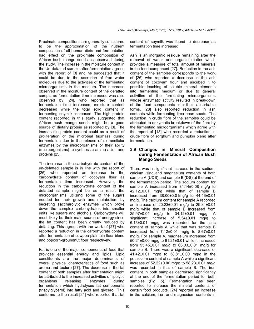
Proximate compositions are generally considered to be the approximation of the nutrient composition of all human diets and fermentation had effect on the proximate composition of African bush mango seeds as observed during the study. The increase in the moisture content in the Un-defatted sample after fermentation agrees with the report of [3] and he suggested that it could be due to the secretion of free water molecules due to the activities of the fermenting microorganisms in the medium. The decrease observed in the moisture content of the defatted sample as fermentation time increased was also observed by [24], who reported that as fermentation time increased, moisture content decreased while the total solid content in fermenting soymilk increased. The high protein content recorded in this study suggested that African bush mango seeds might be a good source of dietary protein as reported by [3]. The increase in protein content could as a result of proliferation of the microbial biomass during fermentation due to the release of extracellular enzymes by the microorganisms or their ability (microorganisms) to synthesize amino acids and proteins [25].
The increase in the carbohydrate content of the un-defatted sample is in line with the report of [26] who reported an increase in the carbohydrate content of cocoyam flour as fermentation time increased. However, the reduction in the carbohydrate content of the defatted sample might be as a result the microorganisms utilizing some of the sugars needed for their growth and metabolism by secreting saccharolytic enzymes which broke down the complex carbohydrates into smaller units like sugars and alcohols. Carbohydrate will most likely be their main source of energy since the fat content has been greatly reduced by defatting. This agrees with the work of [27] who reported a reduction in the carbohydrate content after fermentation of cowpea-plaintain flour blend and popcorn-groundnut flour respectively.
Fat is one of the major components of food that provides essential energy and lipids. Lipid constituents are the major determinants of overall physical characteristics of food such as aroma and texture [27]. The decrease in the fat content of both samples after fermentation might be attributed to the increased activities of lipolytic organisms releasing enzymes during fermentation which hydrolyses fat components (triacylglycerol) into fatty acid and glycerol. This conforms to the result [24] who reported that fat
content of soymilk was found to decrease as fermentation time increased. Ash is an inorganic residue remaining after the removal of water and organic matter which provides a measure of total amount of minerals in the food component [27]. Reduction in the ash content of the samples corresponds to the work of [26] who reported a decrease in the ash content of cocoyam flour and ascribed it to possible leaching of soluble mineral elements into fermenting medium or due to general activities of the fermenting microorganisms whose enzymatic activity resulted in breakdown of the food components into their absorbable forms. [28] also reported reduction in ash contents while fermenting lima bean seeds. The reduction in crude fibre of the samples could be attributed to enzymatic breakdown of the fibre by the fermenting microorganisms which agree with the report of [18] who recorded a reduction in crude fibre of sorghum and pumpkin blend after fermentation.
3.9 Changes in Mineral Composition during Fermentation of African Bush Mango Seeds
There was a significant increase in the sodium, calcium, zinc and magnesium contents of both sample A (UDS) and sample B (DS) at the end of the fermentation period. The sodium content for sample A increased from 34.14±0.08 mg/g to 42.12±0.01 mg/g while that of sample B increased from 38.00±0.01mg/g to 44.46±0.01 mg/g. The calcium content for sample A recorded an increase of 20.23±0.01 mg/g to 29.34±0.01 mg/g while that of sample B increased from 25.97±0.04 mg/g to 34.12±0.01 mg/g. A significant increase of 5.34±0.01 mg/g to 6.13±0.01 mg/g was recorded for the zinc content of sample A while that was sample B increased from 7.12±0.01 mg/g to 8.67±0.01 mg/g. For sample A, magnesium increased from 50.21±0.00 mg/g to 61.21±0.01 while it increased from 55.45±0.01 mg/g to 66.33±0.01 mg/g for sample B. There was a significant decrease of 41.42±0.01 mg/g to 38.81±0.00 mg/g in the potassium content of sample A while a significant increase of 52.22±0.00 mg/g to 58.23±0.01 mg/g was recorded in that of sample B. The iron content in both samples decreased significantly at the end of the fermentation period for both samples (Fig. 5). Fermentation has been reported to increase the mineral contents of certain food products. [24] reported an increase in the calcium, iron and magnesium contents in

Helen and Okhonlaye; MRJI, 27(6): 1-14, 2019; Article no.MRJI.49121
11
soymilk with increase in natural fermentation. [5] also reported an increase in magnesium, calcium, sodium and phosphorus of African bush mango seeds after fermentation. The significant decrease in the potassium content of the non-defatted sample, iron content of the non-defatted and defatted samples after fermentation has been reported in various reports and can be attributed to their utilization by some fermenting microorganisms for their growth and metabolism. It was noted that fermented sample was rich in some essential minerals which perform various functions in the body [3,24]
3.10 Anti-nutritional Composition of African Bush Mango Seeds
The anti-nutrient content of the samples decreased significantly with increase in fermentation time. The highest phytate content
(mg/g) was recorded in sample A (un-defatted African bush mango seeds) at the start-up of the fermentation with a value of 30.46+ 0.02 mg/g while the least phytate content was recorded in sample B (defatted African bush mango seeds) at 96hours of fermentation with a value of 13.68+ 0.04 mg/g. Tannin content recorded the highest in sample B at the start-up of the fermentation with a value of 4.05+ 0.02 mg/g and lowest at 96 hours with value 0.55+ 0.01 mg/g. At the initial, sample B has the highest oxalate value of 5.76+ 0.00 mg/g and it also has the lowest oxalate value of 1.54+ 0.03 mg/g at 96 hours. Saponin content recorded the highest value of 33.46+ 0.02 mg/g in sample A at the initial while sample B recorded the lowest value of 2.16± 0.01 mg/g at 96 hours. (Fig. 6). The reduction observed in the anti-nutrient content of African bush mango seeds after fermentation had been reported in many fermented legumes [29,30]. A wide
Fig. 5. Mineral content of African bush mango seeds
Keys: A- Un-defatted African bush mago seeds B- Defatted African bush mango seeds

Helen and Okhonlaye; MRJI, 27(6): 1-14, 2019; Article no.MRJI.49121
12
Fig. 6. Anti-nutrient content of African bush mango seeds Keys: A- Un-defatted African bush mago seeds B- Defatted African bush mango seeds
range of microflora has been known to possess phytase activity [31]. The decrease in phytate content could be attributed to the activity of the endogenous phytase enzyme from the sample and inherent microorganisms which are able to secrete the hydrolytic enzyme (phytase) capable of degrading the phytic acid in the fermented African bush mango seeds. Some lactic acid bacteria and fungi such have been known to secrete phytases which could degrade phytate to considerable levels. The significant reductions in the anti-nutrient contents of the sample are welcome development because the minerals and other nutrients bound to them become more readily available [28]. The decrease in tannin could be attributed to presence of microorganisms capable of secreting the enzyme tannase which could degrade tannin content to considerable levels. Reduction in the tannin content of African oil bean seed was observed by [32].
4. CONCLUSION This study on the effect of fermentation on the nutrient and anti-nutrient content of African bush
mango seeds revealed that there was improvement in the protein, minerals, nutritional quality of samples after fermentation compared with the raw samples. Fermentation reduced most of the anti-nutrients significantly. The defatted sample recorded a lower microbial load during fermentation and has higher nutritional quality than the un-defatted sample. Therefore, the defatted fermented sample showed the most desirable nutritional qualities which suggest its relevance in human diet for improved nutritional benefits.
COMPETING INTERESTS
Authors have declared that no competing interests exist.
REFERENCES
1. Etong D, Mustapha O, Taleat A. Physicochemical properties and fatty acid composition of Dikanut (Irvingia Gabonensis) seed oil. Research Journal of Chemical Sciences. 2014;4(12):70-74.
2. Bamidele OP, Ojedokun OS, Fasogbon BM. Physico- chemical properties of

Helen and Okhonlaye; MRJI, 27(6): 1-14, 2019; Article no.MRJI.49121
13
instant ogbono (Irvingia gabonensis) mix powder. Food Science & Nutrition. 2015; 3(4):313-318.
3. Adegbehingbe KT, Adeleke BS, Soji F. Solid substrate fermentation of African bush mango seeds (Irvingia gabonensis) seed cotyledons. African Journal of Microbiology Research. 2017;3(1):1-9.
4. Kengni E, Mbofung C, Tchoundjeu Z, Tchouanguep F. Sensory evaluation of tropical bush mango (Irvingia gabonensis) fruits. Pakistan Journal of Nutrition. 2017; 16(8):562-570.
5. Ekundayo FO, Oladipupo OA, Ekundayo EA. Studies on the effects of microbial fermentation on bush mango (Irvingia gabonensis) seed cotyledons. African Journal of Microbiology Research. 2013; 7(34):4363-4367.
6. Festus C, Ibor MN. The effect of defatting on the sliminess and shelf-life of Ugiri Cotyledons. Archives of Applied Science Research. 2014;6(6):1-6.
7. Asaah EK, Tchoundjeu Z, Atangana AR. Cultivation and conservation status of Irvingia wombolu in humid lowland forest of Cameroon. Food Agriculture and Environ-ment. 2003;1(3-4):251-256.
8. Etta HE, Olisaeke CC, Iboh CI. Effect of Irvingia gabonensis (Aubry-Lecomte ex O’Rorke) Seeds on the Liver and Gonads of Male Albino Rats. Journal of Biology, Agriculture and Healthcare. 2014;4(1): 2224-3208.
9. Kuyooro SE, Abam EO, Agbede EB. Hypolipidemic effects of Irvingia gabonensis - supplemented diets in male albino rats. Biochemistry & Analytical Biochemistry. 2017;6(2):1-5.
10. Idowu M, Omoniyi S, Henshaw F, Olayiwola O. Sensory acceptability of partially defatted dikanut (Irvingia gabonensis) flour in ogbono soup. J. Culin. Sci. Tech. 2013;11:346-355.
11. AOAC. Official methods of analysis of the Association of Official Analaytical Chemists international (19th editio n). Gathersburg, Maryland, U.S.A. 2012;59-72.
12. Fawole M, Oso B. Characterization of bacteria: Laboratory manual of Microbiology: 4
th edition, Ibadan, Nigeria:
Spectrum books Limited. 2004;24-33. 13. Cowan ST, Steel KJ. Bergey’s manual of
determinative microogranisms. 4th edition. Cambridge University Press.1990;58.
14. Barnett JA, Payne RW, Yarrow O. Yeast characteristics and identification 3rd
Edition, Cambridge University Press, London. 2000;1376-1378
15. Akindahunsi AA, Salawu SO. Phyto-chemical screening and nutrient anti-nutrient composition of selected tropical green leafy vegetable. Afr. J. Biotech. 2005;4:6.
16. Brinner JH. Direct spectrophometer determination of saponin. Animal Chemistry. 1994;34:1314-1326
17. Moral U, Nagar P, Maan S, Kaur K. A growth of different types of microorganism, intrinsic and extrinsic factors of microorganism and their affects in food: A review. International Journal of Current Microbiology and Applied Sciences. 2017; 6(1):290-298.
18. Ojokoh AO, Ojokoh E. Effect of fermentation on proximate composition and microbiological changes of sorghum and pumpkin blend. British Microbiology Research Journal. 2015; 10(6):1-14.
19. Abiola C, Oyetayo VO. Isolation and bio-chemical characterization of micro-organisms associated with the fermenta-tion of kersting's groundnut (Macrotyloma geocarpum). Research Journal of Micro-biology. 2016;11(2-3):47-55.
20. Ojokoh AO. Effect of fermentation on the chemical composition of mango (Mangifera indica R) peels. African Journal of Biotechnology. 2007;6(16):1979-1981.
21. Ojokoh AO, Abiola AB, Lawal RT. Changes in nutrient and antinutrient composition of popcorn and groundnut composite flour subjected to solid substrate fermentation. African Journal of Agricultural Research. 2012;7(23):3439-3445.
22. Hassan GF, Adebolu TT, Onifade AK. Effect of fermentation on mineral and anti-nutritional composition of cocoyam (Colocasia esculenta Linn). Sky J. Food Sci. 2015;4(4):42-49.
23. Ojokoh AO, Daramola MK, Ochukwa AD. Studies on the microbiological, proximate composition, and anti-nutritional content of fermented groundnut and plantain blends. 2014;15(2):251-258.
24. Obadina AO, Akionla OJ, Shittu TA, Bakare HA. Effect of natural fermentation on the chemical and nutritional com-position of fermented soymilk nono. Nigerian Food Journal. 2013;31(2):91-97.
25. Ekpe OO, Umoh IB, Eka OU. Effect of a typical rural processing method on the

Helen and Okhonlaye; MRJI, 27(6): 1-14, 2019; Article no.MRJI.49121
14
proximate composition and amino acid profile of bush mango seeds Iirvingia gabonensis). African J. Food. Agric. Nut. Dev. 2007;7(1):1-12.
26. Igbabul BD, Amove J, Twadue I. Effect of fermentation on the proximate com-position, antinutritional factors and functional properties of cocoyam (Colocasia esculenta) flour. African Journal of Food Science and Technology 2014; 5(3):67-74.
27. Ojokoh AO, Fagbemi AO. Effects of fermentation and extrusion on the proxymate and organoleptic properties of cowpea-plantain flour blends. British Microbiology Research Journal. 2016; 13(4):1-13.
28. Adegbehingbe KT, Adetuyi FC, Akinyosoye FA. Effect of fermentation on nutrient and antinutrient contents of ground-cooked lima bean (Phaseolus lunatus) seeds using Bacillus subtilis and
Bacillus pumilus. Brit. Microbiol. Res. J. 2014;4(11):1285-1298.
29. Effiong OO, Umoren UE. Effects of multiprocessing techniques on the chemical composition of horse eye beans (Mucuna urens). Asian J. Anim. Sci. 2011; 5(5):340-248.
30. Olagunju AI, Ifesan BO. Changes in nutrient and antinutritional contents of sesame seeds during fermentation. J. Microbiol. Biotechnol. Food Sci. 2013;2(6): 2407-2410.
31. Ojokoh AO. Changes in nutrient and antinutrient contents of sweet potato (Ipomea batatas) peels subjected to solid substrate fermentation. Bioscience Biotech. Research Asia 2005;3(1):17-20.
32. Enujiugha VN, Akanbi CT. Compositional changes in African oil bean (Pentaclethra macrophylla Benth) seeds during thermal processing. Pak. J. Nutr. 2005;4(1):27- 31.
_________________________________________________________________________________ © 2019 Helen and Okhonlaye; This is an Open Access article distributed under the terms of the Creative Commons Attribution License (http://creativecommons.org/licenses/by/4.0), which permits unrestricted use, distribution, and reproduction in any medium, provided the original work is properly cited.
Peer-review history: The peer review history for this paper can be accessed here:
http://www.sdiarticle3.com/review-history/49121
Related Documents