Pathology – Research and Practice 207 (2011) 300–305 Contents lists available at ScienceDirect Pathology – Research and Practice journal homepage: www.elsevier.de/prp Animal and In Vitro Models in Human Diseases Effect of estrogen therapy, soy isoflavones, and the combination therapy on the submandibular gland of ovariectomized rats Vinicius Diniz Chaves Carvalho a , Vanessa Ávila Sarmento Silveira b , Renata Falchete do Prado c,∗ , Yasmin Rodarte Carvalho a a Department of Bioscience and Oral Diagnosis, São José dos Campos School of Dentistry, State University of São Paulo, São Paulo, Brazil b Laboratory of Anatomy, Faculty of Pindamonhangaba (FAPI), Pindamonhangaba, São Paulo, Brazil c Department of Physiotherapy, Cruzeiro Superior School, São Paulo, Brazil article info Article history: Received 16 March 2010 Received in revised form 3 January 2011 Accepted 7 January 2011 Keywords: Salivary glands Ovariectomy Estrogen therapy Isoflavones abstract The objective of this study was to evaluate the effect of estrogen deficiency, estrogen therapy, and soy isoflavones on the salivary glands in female rats. Ninety-six animals were ovariectomized, and 24 were sham-operated. Among the ovariectomized rats, 24 received 17-estradiol; 24 received isoflavone extract; 24 received a combination therapy of both; and 24 received water as placebo. The submandibular glands were histomorphometrically analyzed. As a result, the ANOVA test revealed that the hormonal deficiency affected the acini and the ducts of ovariectomized rats, reducing their percentage compared to the sham group. All treatments caused an increase in ducts and acini compared to the placebo group. It was concluded that the estrogen deficiency may be related to salivary gland function due to a reduction in the quantity of salivary acini and ducts secondary to ovariectomy. The estrogen therapy, soy isoflavone therapy, and the combination of both are effective in reducing the effects of ovariectomy on the salivary glands. © 2011 Elsevier GmbH. All rights reserved. Introduction Several aspects of menopause are universal: hot flashes, excessive transpiration, and vaginal atrophy, caused by estrogen deficiency and manifested in most women after menopause [5]. The systemic aspects of menopause are well documented and include oral manifestations, such as changes in salivary secretion, gingivitis, bleeding, and altered taste sensation [19]. The ovarian hormones act on the oral mucosa by specific recep- tors, which select, recognize, and bind the hormone to the cell cytoplasm or nucleus. Estrogen receptors (ER) have been identi- fied on the gingival tissues using immunohistochemical methods [25]. The deficiency of sexual hormones (estrogen) possibly causes changes in the oral mucosa at the tissue level, because these hormones influence the proliferation, differentiation, and kera- tinization of the gingival epithelium and stimulate the proliferation of fibroblasts [32,38]. The influence of ovarian hormones on the oral mucosa has been studied, and some authors believe that their defi- ∗ Corresponding author at: Av. Engenheiro Francisco José Longo, 777, Jardim São Dimas, São José dos Campos, São Paulo, CEP 12245-000, Brazil. Tel.: +55 12 39479035; fax: +55 12 39479010. E-mail address: [email protected] (R.F. do Prado). ciency is related to the burning mouth syndrome (BMS), also called oral discomfort [18,40]. The BMS is a clinical condition characterized by dry mouth with a burning sensation [5,18]. Gin- gival atrophy and ulceration of the oral mucosa may also be observed [18]. Some studies demonstrate a higher prevalence of BMS among women, especially after menopause. Even though the BMS probably has a multifactorial origin, the hormonal aspect has been implicated in this association [18,40]. After an extensive literature review, Maltsman-Tseikhin et al. [26] advocated the individualized mul- tidisciplinary treatment for each patient with the syndrome due to the wide variety of possible related causes. Even though the salivary glands are not classical targets of female sexual hormones, the influence of the hormones on these glands has been analyzed [12]. However, little is known about this relation. Streckfus et al. [34] compared the salivary flow rates of three groups of premenopausal, perimenopausal, and postmenopausal women and concluded that there was reduction in the salivary flow of submandibular and sublingual glands with the increase in age and reduction in the levels of estrogen production. Eliasson et al. [16] observed increase rates of salivary flow and buffering capcity as a result of estrogen therapy (ET). Meanwhile the capacity to prevent the bacterial aggregation was reduced in the group submitted to estrogen therapy. The authors concluded 0344-0338/$ – see front matter © 2011 Elsevier GmbH. All rights reserved. doi:10.1016/j.prp.2011.01.002

Welcome message from author
This document is posted to help you gain knowledge. Please leave a comment to let me know what you think about it! Share it to your friends and learn new things together.
Transcript
A
Es
VYa
b
c
a
ARRA
KSOEI
I
edsob
tcfi[chto
h
DT
0d
Pathology – Research and Practice 207 (2011) 300–305
Contents lists available at ScienceDirect
Pathology – Research and Practice
journa l homepage: www.e lsev ier .de /prp
nimal and In Vitro Models in Human Diseases
ffect of estrogen therapy, soy isoflavones, and the combination therapy on theubmandibular gland of ovariectomized rats
inicius Diniz Chaves Carvalhoa, Vanessa Ávila Sarmento Silveirab, Renata Falchete do Pradoc,∗,asmin Rodarte Carvalhoa
Department of Bioscience and Oral Diagnosis, São José dos Campos School of Dentistry, State University of São Paulo, São Paulo, BrazilLaboratory of Anatomy, Faculty of Pindamonhangaba (FAPI), Pindamonhangaba, São Paulo, BrazilDepartment of Physiotherapy, Cruzeiro Superior School, São Paulo, Brazil
r t i c l e i n f o
rticle history:eceived 16 March 2010eceived in revised form 3 January 2011ccepted 7 January 2011
a b s t r a c t
The objective of this study was to evaluate the effect of estrogen deficiency, estrogen therapy, andsoy isoflavones on the salivary glands in female rats. Ninety-six animals were ovariectomized, and 24were sham-operated. Among the ovariectomized rats, 24 received 17�-estradiol; 24 received isoflavoneextract; 24 received a combination therapy of both; and 24 received water as placebo. The submandibular
eywords:alivary glandsvariectomystrogen therapysoflavones
glands were histomorphometrically analyzed. As a result, the ANOVA test revealed that the hormonaldeficiency affected the acini and the ducts of ovariectomized rats, reducing their percentage comparedto the sham group. All treatments caused an increase in ducts and acini compared to the placebo group. Itwas concluded that the estrogen deficiency may be related to salivary gland function due to a reductionin the quantity of salivary acini and ducts secondary to ovariectomy. The estrogen therapy, soy isoflavonetherapy, and the combination of both are effective in reducing the effects of ovariectomy on the salivary
glands.ntroduction
Several aspects of menopause are universal: hot flashes,xcessive transpiration, and vaginal atrophy, caused by estrogeneficiency and manifested in most women after menopause [5]. Theystemic aspects of menopause are well documented and includeral manifestations, such as changes in salivary secretion, gingivitis,leeding, and altered taste sensation [19].
The ovarian hormones act on the oral mucosa by specific recep-ors, which select, recognize, and bind the hormone to the cellytoplasm or nucleus. Estrogen receptors (ER) have been identi-ed on the gingival tissues using immunohistochemical methods25]. The deficiency of sexual hormones (estrogen) possibly causeshanges in the oral mucosa at the tissue level, because theseormones influence the proliferation, differentiation, and kera-
inization of the gingival epithelium and stimulate the proliferationf fibroblasts [32,38].The influence of ovarian hormones on the oral mucosaas been studied, and some authors believe that their defi-
∗ Corresponding author at: Av. Engenheiro Francisco José Longo, 777, Jardim Sãoimas, São José dos Campos, São Paulo, CEP 12245-000, Brazil.el.: +55 12 39479035; fax: +55 12 39479010.
E-mail address: [email protected] (R.F. do Prado).
344-0338/$ – see front matter © 2011 Elsevier GmbH. All rights reserved.oi:10.1016/j.prp.2011.01.002
© 2011 Elsevier GmbH. All rights reserved.
ciency is related to the burning mouth syndrome (BMS), alsocalled oral discomfort [18,40]. The BMS is a clinical conditioncharacterized by dry mouth with a burning sensation [5,18]. Gin-gival atrophy and ulceration of the oral mucosa may also beobserved [18].
Some studies demonstrate a higher prevalence of BMS amongwomen, especially after menopause. Even though the BMS probablyhas a multifactorial origin, the hormonal aspect has been implicatedin this association [18,40]. After an extensive literature review,Maltsman-Tseikhin et al. [26] advocated the individualized mul-tidisciplinary treatment for each patient with the syndrome due tothe wide variety of possible related causes.
Even though the salivary glands are not classical targets offemale sexual hormones, the influence of the hormones on theseglands has been analyzed [12]. However, little is known about thisrelation.
Streckfus et al. [34] compared the salivary flow rates of threegroups of premenopausal, perimenopausal, and postmenopausalwomen and concluded that there was reduction in the salivary flowof submandibular and sublingual glands with the increase in age
and reduction in the levels of estrogen production.Eliasson et al. [16] observed increase rates of salivary flow andbuffering capcity as a result of estrogen therapy (ET). Meanwhilethe capacity to prevent the bacterial aggregation was reduced inthe group submitted to estrogen therapy. The authors concluded
esearch and Practice 207 (2011) 300–305 301
tt
aBim(eeoowdisf
cegd
s[l[bgdco
M
cwCimi
tsw
g(eeww
PB
immLe
i
Table 1Quantification of the isoflavones according to Herbarium Botanical Laboratory Ltda.
Type of isoflavones ofthe extract 40%
Molecular mass
Glycitin 446.41 g/molGlycitein 284.30 g/mol
V.D.C. Carvalho et al. / Pathology – R
hat the ET affects some characteristics of human saliva produc-ion.
The negative effects of estrogen therapy encouraged theccomplishment of studies searching for alternative options [11].reitman et al. [8] suggested that diets rich in foods containing
soflavones and calcium may contribute to maintaining the boneass in postmenopausal women. Isoflavones are phytoestrogens
non-steroidal compounds found in different vegetables) presentspecially in soy, which may act as agonists or antagonists ofstrogens [1]. Wood et al. [42] demonstrated the protective rolef soy isoflavones in the diet against breast cancer. Genistein isne of the soy isoflavones most widely investigated and presentsell-demonstrated estrogenic properties [28]. Blake et al. [7] con-ucted a cell culture study and observed that genistein presents an
nhibitory effect on the proliferation of cells obtained from humanubmandibular gland adenocarcinoma; thus, it may be beneficialor patients with this neoplasia.
Knowledge of the mechanisms through which estrogen defi-iency influences the integrity and function of the salivary glandpithelium and how the treatment may be beneficial to the affectedland is fundamental for advances in the treatment of menopausalisorders, especially the burning mouth syndrome.
Some papers have previously reported the effects of estrogen onalivary glands, yet focusing on the salivary function and enzymes4,22], on granular ducts [17,31], or on the effect of submaxil-ary gland excision on ovarian androgen and estrogen production35]. de Rijk et al. [13] showed ductal hyperplasia in rats inducedy a novel synthetic steroid with combined estrogenic and pro-estagenic properties. However, no studies in the literature haveetermined the influence of estrogens, soy isoflavones, and theirombination therapy on the salivary glands of rats, which was thebjective of this study.
aterials and methods
Our study was conducted on 120 female rats (Rattus norvegi-us, variation albinus, Wistar) aged 90 days, with an approximateeight of 300 g, supplied by the Animal Laboratory of São José dosampos School of Dentistry – UNESP. The study was performed
n accordance with the Ethical Principles for Experiments on Ani-als and was approved by the Institutional Review Board of this
nstitution.At study onset, 96 rats were submitted to bilateral removal of
he ovaries, constituting the OVX group, while 24 animals wereham-operated, forming the SHAM group. All surgical proceduresere conducted under general anesthesia.
According to the treatment provided, animals in the OVXroup were subdivided into the following groups: isoflavone groupOVX-I), which received 15 mg/kg/day of 40% isoflavone extract;strogen group (OVX-E), which received 1 mg/kg/day of 17�-stradiol valerate, and the combination therapy group (OVX-A),hich received 1 mg/kg/day of 17�-estradiol valerate associatedith 15 mg/kg/day of 40% isoflavone extract.
The 17�-estradiol valerate was a commercially pill ofrimogyna® (Bayer Schering Phama, Santo Amaro, São Paulo,razil) grinded and suspended in distilled water.
The isoflavone extract was obtained from Herbarium Botan-cal Laboratory Ltda. It is an extract from the grain of Glycine
ax (L.) Merril, Leguminosae. The 40% isoflavone extract is com-
ercially available at Isoflavine® (Herbarium Botanical Laboratorytda, Colombo, Paraná, Brazil). The excipient is oil soybean. Thextract was suspended in distilled water.
The laboratory quantified the principal following types ofsoflavones (Table 1).
Genistin 432.38 g/molGenistein 270.23 g/molDaidzin 416.38 g/molDaidzein 254.24 g/mol
Finally, the vehicle ovariectomized group (OVX-veh) receivedonly the vehicle (water). The 24 animals in the sham group receiveda placebo treatment (water). The treatment was administeredorally, initiated daily on the day of ovariectomy. The animals werekilled after 3, 5, and 8 weeks (n = 8) to evaluate the influence of thetreatment period on the salivary gland. The submandibular glandswere removed and fixed in 10% formalin solution and embeddedin paraffin blocks that were sectioned and conventionally stainedwith PAS (periodic acid-Schiff) and hematoxylin and eosin, andthen histomorphometric analysis was performed.
Histological analysis
The sections were analyzed by light microscopy for qualitativeevaluation of the different groups.
Histomorphometric analysis
For the histomorphometric analysis, five semi-serial sections,with intervals of 100 mm, were stained with hematoxylin andeosin, and four random fields of each section were photographedat an original magnification of 400×. An integration grid with 70points symmetrically distributed over the total area of the pho-tograph was used, adding up to 1400 points counted per rat forthe point-counting planimetry. Points coinciding with serous orseromucous acini and striated ducts were individually counted.
The volume density of acinar and ductal cells was evaluated inpercentage, using the stereology principles established by Weibelet al. [41].
Statistical analysis
Data were submitted to descriptive and inferential analysisaccording to two approaches. The first considered the effects of thefactors ovariectomy (presence or absence of ovarian hormones) andperiod of sacrifice. The analysis of variance (two-way ANOVA) andthe Tukey test for multiple comparisons (5%) were applied to thedata of the SHAM and OVX groups.
The second approach considered the effect of the factors treat-ment (vehicle, estrogen, isoflavones, and combination therapy) andthe period of sacrifice. As in the first approach, these data wereassessed by analysis of variance (two-way ANOVA) and the Tukeytest for multiple comparisons (5%).
Results
Histological analysis
In general, the histological pattern of sections stained with HEwas similar for the study groups, without differences between the
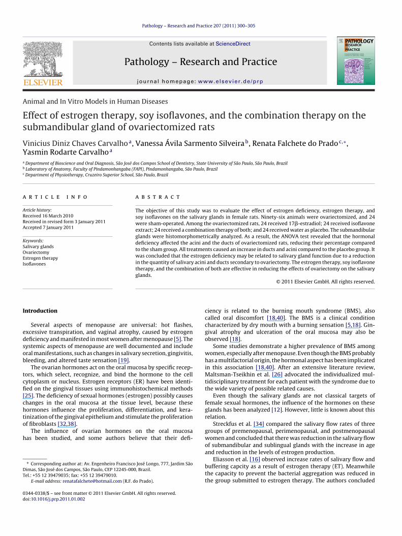
study periods. The PAS staining reduces the quantity of morpho-logical details of glands, yet the histological pattern of labeling byPAS, evidencing the glycogen, was also similar between groupsand periods. The submandibular gland exhibited predominantlyserous acini and a lower quantity of seromucous acini (Fig. 1). Mixed
302 V.D.C. Carvalho et al. / Pathology – Researc
Fig. 1. Architectural pattern of submandibular gland in the OVX-veh (ovariec-tm
apttlmgf(tamtmsaswtais
FOh
omized group that received placebo) at eight weeks after ovariectomy. Originalagnification 200×, hematoxylin–eosin.
cini were also observed, which are easily recognized because theyresent a serous demilune that caps the ends of mucus-secretingubules. The intercalary ducts were not frequent in this gland, andhe striated ducts were lined by cylindrical cells with centrallyocated nuclei and cytoplasm intensely stained by eosin, which
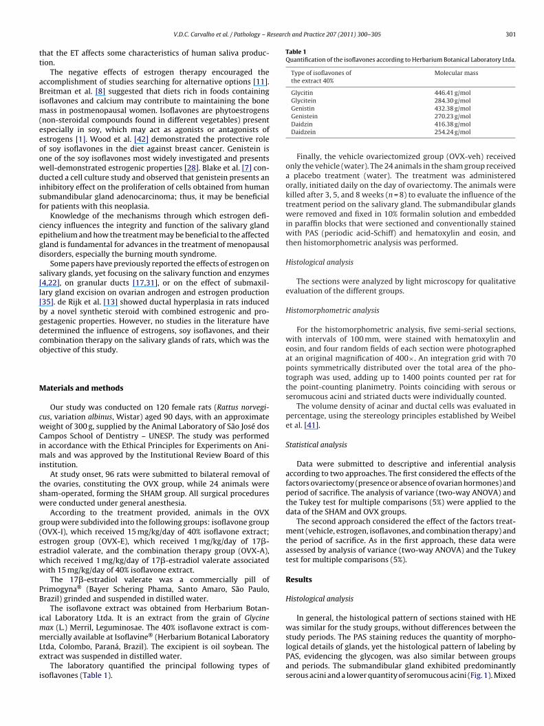
ade them easily recognizable. There was also a large quantity ofranular ducts, with conical cells, basal nuclei, and multiple bire-ringent eosinophilic round granules filling the entire cytoplasmFig. 2). On this gland, the PAS labeled the glycogen especially inhe granular ducts. The aforementioned granules were PAS-positivend were stained in magenta. Connective septa separated the sub-andibular from the sublingual gland. These septa also separated
he gland lobes, being composed of normal connective tissue, pri-arily containing fibroblasts and collagen fibers, yet other cells
uch as macrophages, fat cells, mast cells, and plasma cells werelso occasionally observed, as well as blood vessels and nerves. Theublingual gland was located over the submandibular gland andas nearly exclusively mucous; this gland was not analyzed by his-
omorphometry. In both glands, the excretory ducts were presentnd were observed as true stratified epithelium. Nuclei of myoep-thelial cells were observed on sections below the basal portion of
ecretory cells.ig. 2. Detail of granular and striated ducts of submandibular gland in theVX-veh group at eight weeks after ovariectomy. Original magnification 1000×,ematoxylin–eosin.
h and Practice 207 (2011) 300–305
Histomorphometric analysis of serous or seromucous acini
It was observed that the percentage of acini was differentbetween SHAM and OVX-veh groups. The ANOVA test revealed theestrogen deficiency as a significant effect (p = 0.001). The Tukeytest for multiple comparisons demonstrated that the OVX-vehgroup exhibited a lower percentage of acini than the SHAM group(Fig. 3).
Analysis of data of ovariectomized groups by the ANOVArevealed that the period of killing (p = 0.001), treatment (p = 0.001),and the interaction of both (p = 0.007) were significant effects.
The Tukey test for multiple comparisons applied to the ovariec-tomized groups revealed that the OVX-veh group, at three weeks,presented the lower percentage of acini. At the five- and eight-week periods, the isoflavone group was the best therapy, exhibitinga higher percentage of acini, yet without statistically significantdifference compared to the other treatments (Fig. 3).
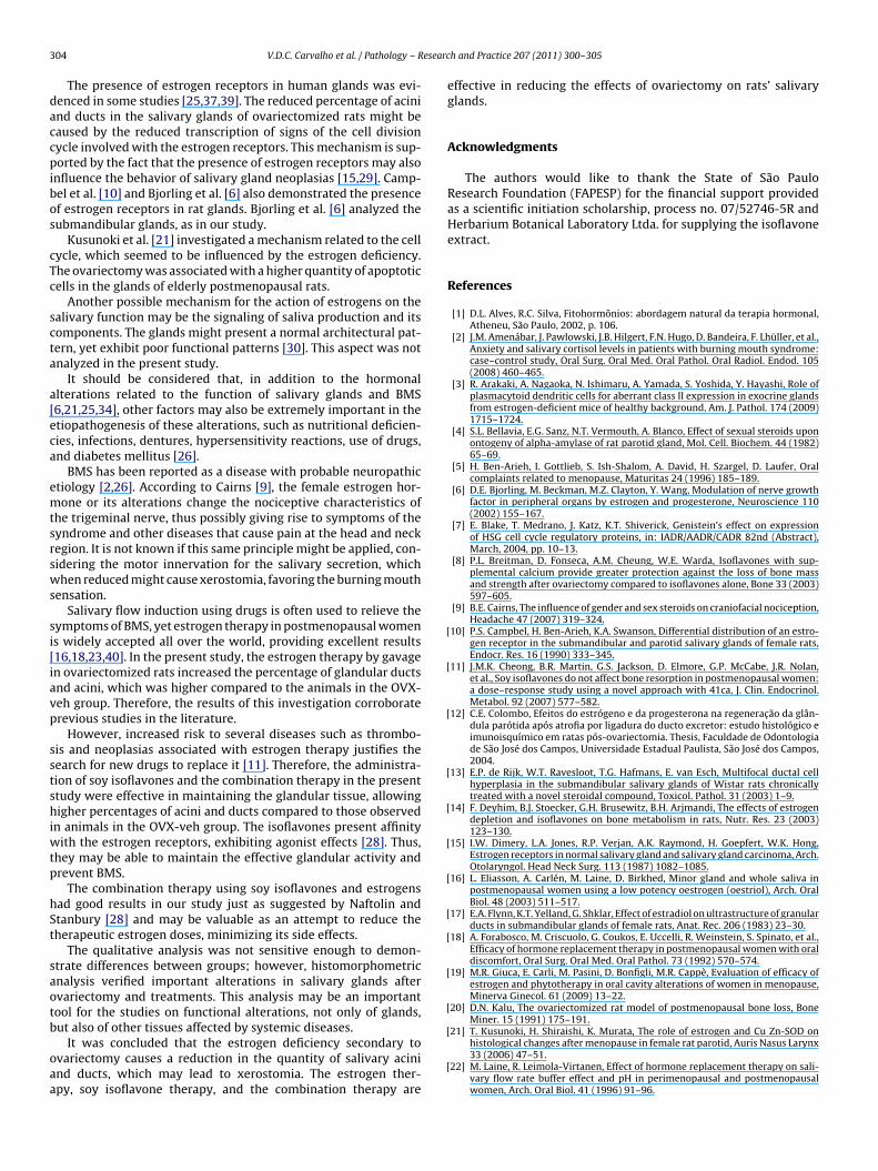
Striated ducts
Analysis of data of ovariectomized groups by the ANOVArevealed that the period of killing (p = 0.001), estrogen deficiency(p = 0.002), and the interaction of both (p = 0.001) were significanteffects.
Application of the Tukey test revealed that the percentage ofducts was increased with time and was higher in the SHAM groupcompared to the OVX-veh group, except for the three-week period(Fig. 4).
Assessment of data of the ovariectomized group by the ANOVAtest revealed a significant effect for the treatment (p = 0.000). TheTukey test showed that the means of the OVX-veh group were lowerat all periods. The estrogen and the combined therapy exhibited ahigher mean at the initial period and a reduction thereafter. Theisoflavone therapy exhibited a lower mean at the initial period anda slight increase thereafter (Fig. 4).
Discussion
This study evaluated the effects of estrogen deficiency onthe salivary glands of ovariectomized rats, compared to sham-operated rats. The study also aimed to analyze the effects ofestrogen therapy, soy isoflavone therapy, and the association ofboth on the glands. The estrogen deficiency affected both theacini and the ducts of ovariectomized rats, reducing their per-centage compared to the sham group. All treatments proposedseemed to effectively reverse the consequences of ovariectomyon the glands, causing an increase in the percentage of ducts andacini compared to the ovariectomized group that received theplacebo.
Ovariectomy induces bone loss due to hormone deficiency, per-mitting the study of bone loss characteristics and sequelae resem-bling those found in postmenopausal women [20,24,27,43,44]. Lossof estrogens or androgens increases the rate of bone remodeling byremoving restraining effects on osteoblastogenesis and osteoclas-togenesis, and also causes a focal imbalance between resorptionand formation [36]. This methodology has an initial rapid phaseof bone loss, a greater loss of cancellous than cortical bone, anddecreased intestinal absorption of calcium. Ovariectomy in ratsshows, as in women, a skeletal response to therapy with estro-gen, tamoxifen, bisphosphonates, parathyroid hormone, calcitonin,
and exercise [20]. However, this methodology has some disadvan-tages like protection against bone loss by obesity and difficulties inmaxillary bone loss possibly due to the mastication type for theserodent animals. Specifically regarding salivary gland, ovariectomyis an inadequate method to study the salivary flow rate, which is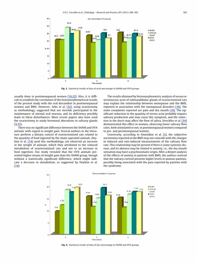
V.D.C. Carvalho et al. / Pathology – Research and Practice 207 (2011) 300–305 303
cini pe
ucowamlt[
atthimfswc[
Fig. 3. Statistical results of data of a
sually done in postmenopausal women [16,22]. Also, it is diffi-ult to establish the correlation of the histomorphometrical resultsf the present study with the oral discomfort in postmenopausalomen and BMS. However, Seko et al. [32], using ovariectomy
s methodology, suggested that sex steroids participated in theaintenance of normal oral mucosa, and its deficiency possibly
eads to these disturbances. More recent papers also have usedhe ovariectomy to study hormonal alterations in salivary glands3,33].
There was no significant difference between the SHAM and OVXnimals with regard to weight gain. Several authors in the litera-ure perform a dietary control of ovariectomized rats related tohe quantity of food ingested by the sham-operated animals. Dey-im et al. [14] used this methodology, yet observed an increase
n the weight of animals, which they attributed to the reducedetabolism of ovariectomized rats and not to an increase in
ood ingestion. Our study revealed that the OVX animals pre-ented higher means of weight gain than the SHAM group, thoughithout a statistically significant difference, which might indi-
ate a decrease in metabolism, as suggested by Deyhim et al.14].
Fig. 4. Statistical results of data of duct pe
rcentage in SHAM and OVX groups.
The results obtained by histomorphometric analysis of serous orseromucous acini of submandibular glands of ovariectomized ratsmay explain the relationship between menopause and the BMS,reported in association with the menopausal disorders [34]. Themain complaints reported are pain and dry mouth [18]. The sig-nificant reduction in the quantity of serous acini probably impairssalivary production and may cause this symptom, and the reduc-tion in the ducts may affect the flow of saliva. Streckfus et al. [34]demonstrated this effect in women, observing lower salivary flowrates, both stimulated or not, in postmenopausal women comparedto pre- and perimenopausal women.
Conversely, according to Amenábar et al. [2], the subjectivexerostomia reported in the BMS may not coincide with the changesin induced and non-induced measurements of the salivary flowrate. This relationship may be present if there is some systemic dis-ease, and its absence may be related to anxiety, i.e., the dry mouth
sensation may have a psychosomatic origin. After a deeper analysisof the effects of anxiety in patients with BMS, the authors noticedthat the salivary cortisol presents higher levels in anxious patients,possibly being associated with the pain reported by patients withthe syndrome.rcentage in SHAM and OVX groups.
3 esearc
daccpibos
cTc
scta
a[eca
emtsrsws
si[iavp
sstshiwtp
hSt
saotb
oaa
[
[
[
[
[
[
[
[
[
[
[
04 V.D.C. Carvalho et al. / Pathology – R
The presence of estrogen receptors in human glands was evi-enced in some studies [25,37,39]. The reduced percentage of acinind ducts in the salivary glands of ovariectomized rats might beaused by the reduced transcription of signs of the cell divisionycle involved with the estrogen receptors. This mechanism is sup-orted by the fact that the presence of estrogen receptors may also
nfluence the behavior of salivary gland neoplasias [15,29]. Camp-el et al. [10] and Bjorling et al. [6] also demonstrated the presencef estrogen receptors in rat glands. Bjorling et al. [6] analyzed theubmandibular glands, as in our study.
Kusunoki et al. [21] investigated a mechanism related to the cellycle, which seemed to be influenced by the estrogen deficiency.he ovariectomy was associated with a higher quantity of apoptoticells in the glands of elderly postmenopausal rats.
Another possible mechanism for the action of estrogens on thealivary function may be the signaling of saliva production and itsomponents. The glands might present a normal architectural pat-ern, yet exhibit poor functional patterns [30]. This aspect was notnalyzed in the present study.
It should be considered that, in addition to the hormonallterations related to the function of salivary glands and BMS6,21,25,34], other factors may also be extremely important in thetiopathogenesis of these alterations, such as nutritional deficien-ies, infections, dentures, hypersensitivity reactions, use of drugs,nd diabetes mellitus [26].
BMS has been reported as a disease with probable neuropathictiology [2,26]. According to Cairns [9], the female estrogen hor-one or its alterations change the nociceptive characteristics of
he trigeminal nerve, thus possibly giving rise to symptoms of theyndrome and other diseases that cause pain at the head and neckegion. It is not known if this same principle might be applied, con-idering the motor innervation for the salivary secretion, whichhen reduced might cause xerostomia, favoring the burning mouth
ensation.Salivary flow induction using drugs is often used to relieve the
ymptoms of BMS, yet estrogen therapy in postmenopausal womens widely accepted all over the world, providing excellent results16,18,23,40]. In the present study, the estrogen therapy by gavagen ovariectomized rats increased the percentage of glandular ductsnd acini, which was higher compared to the animals in the OVX-eh group. Therefore, the results of this investigation corroboraterevious studies in the literature.
However, increased risk to several diseases such as thrombo-is and neoplasias associated with estrogen therapy justifies theearch for new drugs to replace it [11]. Therefore, the administra-ion of soy isoflavones and the combination therapy in the presenttudy were effective in maintaining the glandular tissue, allowingigher percentages of acini and ducts compared to those observed
n animals in the OVX-veh group. The isoflavones present affinityith the estrogen receptors, exhibiting agonist effects [28]. Thus,
hey may be able to maintain the effective glandular activity andrevent BMS.
The combination therapy using soy isoflavones and estrogensad good results in our study just as suggested by Naftolin andtanbury [28] and may be valuable as an attempt to reduce theherapeutic estrogen doses, minimizing its side effects.
The qualitative analysis was not sensitive enough to demon-trate differences between groups; however, histomorphometricnalysis verified important alterations in salivary glands aftervariectomy and treatments. This analysis may be an importantool for the studies on functional alterations, not only of glands,
ut also of other tissues affected by systemic diseases.It was concluded that the estrogen deficiency secondary tovariectomy causes a reduction in the quantity of salivary acinind ducts, which may lead to xerostomia. The estrogen ther-py, soy isoflavone therapy, and the combination therapy are
[
[
h and Practice 207 (2011) 300–305
effective in reducing the effects of ovariectomy on rats’ salivaryglands.
Acknowledgments
The authors would like to thank the State of São PauloResearch Foundation (FAPESP) for the financial support providedas a scientific initiation scholarship, process no. 07/52746-5R andHerbarium Botanical Laboratory Ltda. for supplying the isoflavoneextract.
References
[1] D.L. Alves, R.C. Silva, Fitohormônios: abordagem natural da terapia hormonal,Atheneu, São Paulo, 2002, p. 106.
[2] J.M. Amenábar, J. Pawlowski, J.B. Hilgert, F.N. Hugo, D. Bandeira, F. Lhüller, et al.,Anxiety and salivary cortisol levels in patients with burning mouth syndrome:case–control study, Oral Surg. Oral Med. Oral Pathol. Oral Radiol. Endod. 105(2008) 460–465.
[3] R. Arakaki, A. Nagaoka, N. Ishimaru, A. Yamada, S. Yoshida, Y. Hayashi, Role ofplasmacytoid dendritic cells for aberrant class II expression in exocrine glandsfrom estrogen-deficient mice of healthy background, Am. J. Pathol. 174 (2009)1715–1724.
[4] S.L. Bellavia, E.G. Sanz, N.T. Vermouth, A. Blanco, Effect of sexual steroids uponontogeny of alpha-amylase of rat parotid gland, Mol. Cell. Biochem. 44 (1982)65–69.
[5] H. Ben-Arieh, I. Gottlieb, S. Ish-Shalom, A. David, H. Szargel, D. Laufer, Oralcomplaints related to menopause, Maturitas 24 (1996) 185–189.
[6] D.E. Bjorling, M. Beckman, M.Z. Clayton, Y. Wang, Modulation of nerve growthfactor in peripheral organs by estrogen and progesterone, Neuroscience 110(2002) 155–167.
[7] E. Blake, T. Medrano, J. Katz, K.T. Shiverick, Genistein’s effect on expressionof HSG cell cycle regulatory proteins, in: IADR/AADR/CADR 82nd (Abstract),March, 2004, pp. 10–13.
[8] P.L. Breitman, D. Fonseca, A.M. Cheung, W.E. Warda, Isoflavones with sup-plemental calcium provide greater protection against the loss of bone massand strength after ovariectomy compared to isoflavones alone, Bone 33 (2003)597–605.
[9] B.E. Cairns, The influence of gender and sex steroids on craniofacial nociception,Headache 47 (2007) 319–324.
10] P.S. Campbel, H. Ben-Arieh, K.A. Swanson, Differential distribution of an estro-gen receptor in the submandibular and parotid salivary glands of female rats,Endocr. Res. 16 (1990) 333–345.
11] J.M.K. Cheong, B.R. Martin, G.S. Jackson, D. Elmore, G.P. McCabe, J.R. Nolan,et al., Soy isoflavones do not affect bone resorption in postmenopausal women:a dose–response study using a novel approach with 41ca, J. Clin. Endocrinol.Metabol. 92 (2007) 577–582.
12] C.E. Colombo, Efeitos do estrógeno e da progesterona na regeneracão da glân-dula parótida após atrofia por ligadura do ducto excretor: estudo histológico eimunoisquímico em ratas pós-ovariectomia. Thesis, Faculdade de Odontologiade São José dos Campos, Universidade Estadual Paulista, São José dos Campos,2004.
13] E.P. de Rijk, W.T. Ravesloot, T.G. Hafmans, E. van Esch, Multifocal ductal cellhyperplasia in the submandibular salivary glands of Wistar rats chronicallytreated with a novel steroidal compound, Toxicol. Pathol. 31 (2003) 1–9.
14] F. Deyhim, B.J. Stoecker, G.H. Brusewitz, B.H. Arjmandi, The effects of estrogendepletion and isoflavones on bone metabolism in rats, Nutr. Res. 23 (2003)123–130.
15] I.W. Dimery, L.A. Jones, R.P. Verjan, A.K. Raymond, H. Goepfert, W.K. Hong,Estrogen receptors in normal salivary gland and salivary gland carcinoma, Arch.Otolaryngol. Head Neck Surg. 113 (1987) 1082–1085.
16] L. Eliasson, A. Carlén, M. Laine, D. Birkhed, Minor gland and whole saliva inpostmenopausal women using a low potency oestrogen (oestriol), Arch. OralBiol. 48 (2003) 511–517.
17] E.A. Flynn, K.T. Yelland, G. Shklar, Effect of estradiol on ultrastructure of granularducts in submandibular glands of female rats, Anat. Rec. 206 (1983) 23–30.
18] A. Forabosco, M. Criscuolo, G. Coukos, E. Uccelli, R. Weinstein, S. Spinato, et al.,Efficacy of hormone replacement therapy in postmenopausal women with oraldiscomfort, Oral Surg. Oral Med. Oral Pathol. 73 (1992) 570–574.
19] M.R. Giuca, E. Carli, M. Pasini, D. Bonfigli, M.R. Cappè, Evaluation of efficacy ofestrogen and phytotherapy in oral cavity alterations of women in menopause,Minerva Ginecol. 61 (2009) 13–22.
20] D.N. Kalu, The ovariectomized rat model of postmenopausal bone loss, BoneMiner. 15 (1991) 175–191.
21] T. Kusunoki, H. Shiraishi, K. Murata, The role of estrogen and Cu Zn-SOD onhistological changes after menopause in female rat parotid, Auris Nasus Larynx33 (2006) 47–51.
22] M. Laine, R. Leimola-Virtanen, Effect of hormone replacement therapy on sali-vary flow rate buffer effect and pH in perimenopausal and postmenopausalwomen, Arch. Oral Biol. 41 (1996) 91–96.
esearc
[
[
[
[
[
[
[
[
[
[
[
[
[
[
[
[
[
[
[
[
V.D.C. Carvalho et al. / Pathology – R
23] M. Laine, J. Tenovuo, Effect on peroxidase activity and specific binding of thehormone 17 beta-oestradiol and rat salivary glands, Arch. Oral Biol. 28 (1983)847–852.
24] N.E. Lane, J.M. Thompson, D. Haupt, D.B. Kimmel, G. Modin, J.H. Kinney, Acutechanges in trabecular bone connectivity and osteoclast activity in the ovariec-tomized rat in vivo, J. Bone Miner. Res. 13 (1998) 229–236.
25] R. Leimola-Virtanen, T. Salo, S. Toikkanen, J. Pulkinen, S. Syrjanen, Expression ofestrogen receptor (ER) in oral mucosa and salivary glands, Maturitas 36 (2000)131–137.
26] A. Maltsman-Tseikhin, P. Moricca, D. Niv, Burning mouth syndrome: will betterunderstanding yield better management? Pain Pract. 7 (2007) 151–162.
27] W. Most, L. van der Wee-Pals, A. Ederveen, S. Papapoulos, C. Lowik, Ovariectomyand orchidectomy induce a transient increase in the osteoclastogenic potentialof bone marrow cells in the mouse, Bone 20 (1997) 27–30.
28] F. Naftolin, M.G. Stanbury, Phytoestrogens: are they really estrogen mimics?Fertil. Steril. 77 (2002) 15–17.
29] S. Ozono, M. Onozuka, K. Sato, Y. Ito, Immunohistochemical localization ofestradiol, progesterone, and progesterone receptor in human salivary glandand salivary adenoid cystic carcinomas, Cell Struct. Funct. 17 (1992) 169–175.
30] K.R. Purushotham, P.L. Wang, C. Dolce, T. Zelles, J. Blazsek, M.G. Humphreys-Beher, Effects of surgical ovariectomy on rat salivary gland function, Arch. OralBiol. 38 (1993) 779–784.
31] M.L. Rins de David, A. Finkelberg de Sterin, A. Goldraij, Influence of gonadichormones on the rat submaxillary gland, Arch. Int. Physiol. Biochim. Biophys.
99 (1991) 107–109.32] K. Seko, H. Kagami, K. Senga, K. Ozeki, H. Mizutani, M. Ueda, Effects of ovariec-tomy and estrogen replacement on rat oral mucosa, Maturitas 50 (2005) 44–51.
33] J. Smith, M. Lindsay, R. Rahimian, L. Anderson, The influence of estrogen andprogesterone on parasympathetic vasodilatation in the rat submandibulargland, Auton. Neurosci. 146 (2009) 87–94.
[
[
h and Practice 207 (2011) 300–305 305
34] G.F. Streckfus, U. Baur, L.J. Brown, C. Baca, J. Metter, T. Nick, Effects of estrogenstatus and aging on salivary flow rates in healthy Caucasian women, Gerontol-ogy 44 (1998) 32–39.
35] M. Tanaka, M. Watanabe, T. Kumai, H. Nakura, S. Kobayashi, Effects of submax-illariectomy on ovarian androgen and estrogen production in adult female rats,Biochem. Biophys. Res. Commun. 213 (1995) 550–554.
36] A. Tivesten, S. Movérare-Skrtic, A. Chagin, K. Venken, P. Salmon, D. Vander-schueren, L. Sävendahl, A. Holmäng, C. Ohlsson, Additive protective effects ofestrogen and androgen treatment on trabecular bone in ovariectomized rats, J.Bone Miner. Res. 19 (2004) 1833–1839.
37] H. Välimaa, S. Savolainen, T. Soukka, P. Silvoniemi, S. Mäkelä, H. Kujari, et al.,Estrogen receptor-� is the predominant estrogen receptor subtype in humanoral epithelium and salivary glands, J. Endocrinol. 180 (2004) 55–62.
38] J. Vittek, M.R. Hernandez, E.J. Wenk, S.C. Rappaport, A.L. Southren, Specificestrogen receptors in human gingiva, J. Clin. Endocrinol. Metab. 54 (1982)608–612.
39] B.J. Waddell, P.C. O’Leary, Distribution and metabolism of topically appliedprogesterone in a rat model, J. Steroid Biochem. Mol. Biol. 80 (2002) 449–455.
40] R.W. Waldrop, J. Hailes, H. Burger, P.C. Reade, Oral discomfort at menopause,Oral Surg. Oral Med. Oral Pathol. 67 (1989) 535–540.
41] E.R. Weibel, G.S. Kistler, W.F. Scherle, Practical stereological methods for mor-phometric cytology, J. Cell Biol. 30 (1966) 23–38.
42] C.E. Wood, T.C. Register, A.A. Franke, M.S. Anthony, J.M. Cline, Dietary soyisoflavones inhibit estrogen effects in the postmenopausal breast, Cancer Res.
66 (2006) 1241–1249.43] T.J. Wronski, M. Cintrón, L.M. Dann, Temporal relationship between bone lossand increased bone turnover in ovariectomized rats, Calcif Tissue Int. 43 (1988)179–183.
44] D. Zaffe, C. Paganelli, D. Cocchi, Induction and pharmacological treatment oforal osteopenia in rats, Minerva Stomatol. 48 (1999) 45–62.
Related Documents


![Process for the Preparation of Chromones, Isoflavones and ... · S. K. Yadav 237 and protein tyrosine kinase inhibitor [11]. Search for new methodologies for the synthesis of isoflavones](https://static.cupdf.com/doc/110x72/5c94e0c009d3f2a67b8bb18c/process-for-the-preparation-of-chromones-isoflavones-and-s-k-yadav-237.jpg)