Effect of different surface nanoroughness of titanium dioxide films on the growth of human osteoblast-like MG63 cells Marta Vandrovcova, 1 Jan Hanus, 2 Martin Drabik, 2 Ondrej Kylian, 2 Hynek Biederman, 2 Vera Lisa, 1 Lucie Bacakova 1 1 Department of Growth and Differentiation of Cell Populations, Institute of Physiology, Academy of Sciences of the Czech Republic, Videnska 1083, 142 20 Prague 4, Czech Republic 2 Department of Macromolecular Physics, Faculty of Mathematics and Physics, Charles University, V Holesovickach 2, 182 00 Prague 8, Czech Republic Received 27 April 2011; revised 23 September 2011; accepted 5 December 2011 Published online 5 February 2012 in Wiley Online Library (wileyonlinelibrary.com). DOI: 10.1002/jbm.a.34047 Abstract: Cell behavior depends strongly on the physical and chemical properties of the material surface, for example, its chemistry and topography. The authors have therefore assessed the influence of materials of different chemical com- position (i.e., glass substrates with and without TiO 2 films in anatase form) and different surface roughness (R a ¼ 0, 40, 100, or 170 nm) on the adhesion, proliferation, and osteogenic dif- ferentiation of human osteoblast-like MG63 cells. On day 1 af- ter seeding, the largest cell spreading area was found on flat TiO 2 films (R a ¼ 0 nm). On TiO 2 films with R a ¼ 170 nm, the cell spreading area was larger and the number of initially adhering cells was higher than the values on the correspond- ing uncoated glass. On day 3 after seeding, the cell number was higher on the TiO 2 films (R a ¼ 0 and 40 nm) than on the corresponding glass substrates and the standard polystyrene dishes. On day 7, all TiO 2 films contained higher cell numbers than the corresponding glass substrates, and the cells on the TiO 2 films with R a ¼ 40 and 100 nm also contained a higher concentration of b-actin. These results indicate that TiO 2 coat- ing had a positive influence on the adhesion and subsequent proliferation of MG63 cells. In addition, on all investigated materials, the cell population density achieved on day 7 decreased with increasing surface roughness. The concentra- tion of osteocalcin, measured per mg of protein, was signifi- cantly lower in the cells on rougher TiO 2 films (R a ¼ 100 and 170 nm) than in the cells on the polystyrene dishes. Thus, it can be concluded that the adhesion, growth, and phenotypic maturation of MG63 cells were controlled by the interplay between the material chemistry and surface topography, and were usually better on smoother and TiO 2 -coated surfaces than on rougher and uncoated glass substrates. V C 2012 Wiley Periodicals, Inc. J Biomed Mater Res Part A: 100A: 1016–1032, 2012. Key Words: titanium dioxide, nanoscale surface roughness, material surface chemistry, cell adhesion, cell proliferation, cell differentiation How to cite this article: Vandrovcova M, Hanus J, Drabik M, Kylian O, Biederman H, Lisa V, Bacakova L. 2012. Effect of different surface nanoroughness of titanium dioxide films on the growth of human osteoblast-like MG63 cells. J Biomed Mater Res Part A 2012:100A:1016–1032. INTRODUCTION The materials currently used for surgical implants comprise 316L stainless steel, cobalt–chromium alloys, and titanium and its alloys. Amongst the materials available for implant applications, titanium is considered to be the best for bone implants, and is extensively used in biomedical applications. There are various reasons for this, related to the favorable mechanical properties, the chemical stability, and the high biocompatibility of Ti. First, in comparison with other alloys, titanium-based materials have a relatively low modulus of elasticity, which varies from 110 GPa down to 55 GPa, and thus it approaches the elasticity modulus of bone (30 GPa). This is of paramount importance, since it is well known that for a well-functioning bone implant, the modulus of elasticity had to be as similar as possible to the modulus of bone. 1 Recently-used 316L stainless steel and chromium–cobalt alloys, that is, materials characterized by much higher elas- ticity modulus values (210 and 240 GPa, respectively), have often been reported to cause prosthesis failure through aseptic loosening or fracture of the adjacent bone tissue. A second limitation of metallic bone implants is due to local adverse tissue reactions or allergic reactions elicited by the release of metal ions from the implant. 2 However, in the case of titanium as a reactive metal, the release of ions is prevented by spontaneous formation of an oxide layer on the metal surface in air, in water, or in any other electrolyte. The specific mode of oxide growth on titanium causes no metal ion to reach the material surface. Although the tita- nium oxide film is relatively very thin (about 4 nm), it is in No benefit of any kind will be received either directly or indirectly by the author(s). Correspondence to: M. Vandrovcova; e-mail: [email protected] 1016 V C 2012 WILEY PERIODICALS, INC.

Welcome message from author
This document is posted to help you gain knowledge. Please leave a comment to let me know what you think about it! Share it to your friends and learn new things together.
Transcript
Effect of different surface nanoroughness of titanium dioxide filmson the growth of human osteoblast-like MG63 cells
Marta Vandrovcova,1 Jan Hanus,2 Martin Drabik,2 Ondrej Kylian,2 Hynek Biederman,2
Vera Lisa,1 Lucie Bacakova1
1Department of Growth and Differentiation of Cell Populations, Institute of Physiology, Academy of Sciences of the Czech
Republic, Videnska 1083, 142 20 Prague 4, Czech Republic2Department of Macromolecular Physics, Faculty of Mathematics and Physics, Charles University, V Holesovickach 2,
182 00 Prague 8, Czech Republic
Received 27 April 2011; revised 23 September 2011; accepted 5 December 2011
Published online 5 February 2012 in Wiley Online Library (wileyonlinelibrary.com). DOI: 10.1002/jbm.a.34047
Abstract: Cell behavior depends strongly on the physical and
chemical properties of the material surface, for example, its
chemistry and topography. The authors have therefore
assessed the influence of materials of different chemical com-
position (i.e., glass substrates with and without TiO2 films in
anatase form) and different surface roughness (Ra ¼ 0, 40, 100,
or 170 nm) on the adhesion, proliferation, and osteogenic dif-
ferentiation of human osteoblast-like MG63 cells. On day 1 af-
ter seeding, the largest cell spreading area was found on flat
TiO2 films (Ra ¼ 0 nm). On TiO2 films with Ra ¼ 170 nm, the
cell spreading area was larger and the number of initially
adhering cells was higher than the values on the correspond-
ing uncoated glass. On day 3 after seeding, the cell number
was higher on the TiO2 films (Ra ¼ 0 and 40 nm) than on the
corresponding glass substrates and the standard polystyrene
dishes. On day 7, all TiO2 films contained higher cell numbers
than the corresponding glass substrates, and the cells on the
TiO2 films with Ra ¼ 40 and 100 nm also contained a higher
concentration of b-actin. These results indicate that TiO2 coat-
ing had a positive influence on the adhesion and subsequent
proliferation of MG63 cells. In addition, on all investigated
materials, the cell population density achieved on day 7
decreased with increasing surface roughness. The concentra-
tion of osteocalcin, measured per mg of protein, was signifi-
cantly lower in the cells on rougher TiO2 films (Ra ¼ 100 and
170 nm) than in the cells on the polystyrene dishes. Thus, it
can be concluded that the adhesion, growth, and phenotypic
maturation of MG63 cells were controlled by the interplay
between the material chemistry and surface topography, and
were usually better on smoother and TiO2-coated surfaces
than on rougher and uncoated glass substrates. VC 2012 Wiley
Periodicals, Inc. J Biomed Mater Res Part A: 100A: 1016–1032, 2012.
Key Words: titanium dioxide, nanoscale surface roughness,
material surface chemistry, cell adhesion, cell proliferation,
cell differentiation
How to cite this article: Vandrovcova M, Hanus J, Drabik M, Kylian O, Biederman H, Lisa V, Bacakova L. 2012. Effect of differentsurface nanoroughness of titanium dioxide films on the growth of human osteoblast-like MG63 cells. J Biomed Mater Res Part A2012:100A:1016–1032.
INTRODUCTION
The materials currently used for surgical implants comprise316L stainless steel, cobalt–chromium alloys, and titaniumand its alloys. Amongst the materials available for implantapplications, titanium is considered to be the best for boneimplants, and is extensively used in biomedical applications.There are various reasons for this, related to the favorablemechanical properties, the chemical stability, and the highbiocompatibility of Ti.
First, in comparison with other alloys, titanium-basedmaterials have a relatively low modulus of elasticity, whichvaries from 110 GPa down to 55 GPa, and thus itapproaches the elasticity modulus of bone (30 GPa). This isof paramount importance, since it is well known that for awell-functioning bone implant, the modulus of elasticity had
to be as similar as possible to the modulus of bone.1
Recently-used 316L stainless steel and chromium–cobaltalloys, that is, materials characterized by much higher elas-ticity modulus values (210 and 240 GPa, respectively), haveoften been reported to cause prosthesis failure throughaseptic loosening or fracture of the adjacent bone tissue.
A second limitation of metallic bone implants is due tolocal adverse tissue reactions or allergic reactions elicitedby the release of metal ions from the implant.2 However, inthe case of titanium as a reactive metal, the release of ionsis prevented by spontaneous formation of an oxide layer onthe metal surface in air, in water, or in any other electrolyte.The specific mode of oxide growth on titanium causes nometal ion to reach the material surface. Although the tita-nium oxide film is relatively very thin (about 4 nm), it is in
No benefit of any kind will be received either directly or indirectly by the author(s).
Correspondence to: M. Vandrovcova; e-mail: [email protected]
1016 VC 2012 WILEY PERIODICALS, INC.
a crystalline state or in a glassy state. It acts as a good insu-lator and occludes anionic impurities such as chlorine, fluo-rine, or phosphates. By contrast, conductible materials implyredox processes, which can lead to denaturation ofmacromolecules.3
To exploit the favorable barrier properties of titaniumoxide films, especially titanium dioxide, on the surface ofTi-based materials, these films have often been artificiallydeposited on the surface of a material to enhance its bioac-tivity. TiO2 occurs in several crystal phases, the most knownand most widely used of which are rutile, anatase, brookite,and TiO2 in amorphous form. The most stable form is rutile,which has proved to be a good substrate for the growth andviability of various cell types, for example, cultured humandermal fibroblasts,4 and for adherence and axonal growth ofcultured rat cerebral cortex neurons.5 Due to its bioinert-ness, rutile has been used in combination with bioactivemolecules mediating cell adhesion, such as fibronectin.6
Anatase as a form of TiO2 has been used for construct-ing antimicrobial or self-cleaning surfaces, due to its photo-catalytic effect.7 Anatase has also proved to be the best TiO2
phase for the adhesion, growth, and differentiation of osteo-blasts.8 The favorable cell behavior has been furtherenhanced by ultraviolet light irradiation of the anatase filmprior to cell seeding,9 which has been attributed to the sur-face hydrophilicity of the material.8,9 The attachment andspreading of cells, and consequently their subsequentmigration, proliferation, differentiation, long-term viability,and functioning is mediated by extracellular matrix (ECM)molecules, such as vitronectin, fibronectin, collagen, andlaminin, which are spontaneously adsorbed on the materialsurface from biological fluids (cell culture media or bodyfluids), or are deposited on the material by the cells them-selves. Specific sites in these molecules are recognized andbound by cell adhesion receptors, for example, integrins. Itis believed that on hydrophilic surfaces, the ECM moleculesare adsorbed in an advantageous and flexible geometricalconformation mimicking that in the natural ECM, and arethus better accessible by the cell adhesion receptor than onhydrophobic surfaces, where the adsorbed ECM moleculesare rather rigid and denatured (for a review, see10,11).
However, although the chemical composition of the ma-terial surface is an important factor influencing the cell–ma-terial interaction, it is not the only factor. It has been dem-onstrated that material surface roughness and topographycan also significantly alter cell behavior.10,11 As a conse-quence, topographic modification of the tissue response canbe one of the most important criteria in the production ofbiomaterials.8,12,13 Many studies have compared the effectsof microscale or nanoscale topography on various types ofcells.14–18 Most of these studies have shown that nanoscaletopography seems to be more beneficial for the proliferationof bone cells than microscale topography. It is believed thatnanoscale irregularities on the material surface act syner-getically with the surface hydrophilia in improving the geo-metrical conformation of adsorbed cell adhesion-mediatingECM proteins. In addition, they induce preferential adsorp-tion of vitronectin (due to its relatively small and less com-
plicated molecule in comparison with other ECM proteins),which is recognized mainly by osteoblasts over other celltypes.16,17,19,20
In our study, the authors focus on the responses ofbone-derived cells to substrates of various chemical compo-sition and surface roughness in the nanoscale and submi-cron scale, aiming to correlate the physicochemical surfaceproperties of the substrates with the adhesion, growth, andosteogenic differentiation of human osteoblast-like MG63cells in cultures.
MATERIALS AND METHODS
Preparation and characterization of substrate materialsTo evaluate the effect of nanoroughness of TiO2 surfaces oncell behavior, glass slides of various Ra roughness* valueswere coated with TiO2 films. The topography of the glasssubstrates prior TiO2 deposition was changed by grindingthem with grinding paste followed by polishing to therequired roughness, specifically Ra ¼ 0 nm (flat glass), 40,100, and 170 nm, and measured by a stylus surface profiler(SF 200, Planer Industrial, UK) on a line length of 5 mm. Anadditional examination of the roughened glass slides wasperformed by atomic force microscopy (AFM; Q-Scope 350,Quesant, USA) operated in a tapping mode on surface areasof 50 lm � 50 lm.
The flat and roughened glass slides were subsequentlycoated with 200 nm thick TiO2 films by means of reactiveDC magnetron sputtering from a titanium target in a gasmixture of Ar and O2. The magnetron was operated at 0.3 Ain constant current mode (power �125 W), the working gaspressure during deposition was 2 Pa, the Ar/O2 ratio was2:1, and the total flow rate of the working gas was 6 sccm.The deposition time was 60 min.
The chemical structure of the coatings was determinedby X-ray photoelectron spectroscopy (XPS) (Phobios 100,Specs, Germany) before and after cleaning the samples inethanol and annealing them at 160�C for 2 h (simulation ofsterilization).
X-ray diffraction (Panalytical X’Pert MRD, Netherlands)was performed to determine the crystal form of the depos-ited TiO2 films. XRD measurements were made in parallelbeam geometry with an X-ray mirror. Cu Ka (1.54056 Å)radiation was used at an angle of incidence of 1�.
Crystallization of TiO2 films was further investigated bymeasurements of transmittance by UV–Vis spectrometry(Hitachi U-3300, Japan).
Finally, the wettability of the TiO2 films was expressedin terms of the static contact angle of water measured by asessile droplet method using goniometer of our ownconstruction.
For the cell culture experiments, glass slides of the samesurface roughness but without a TiO2 film, and also the bot-tom of standard polystyrene cell culture dishes were usedas reference materials.
*Ra roughness is defined as the average deviation of the roughness profile from
the mean lineRa ¼ 1n
Pni¼1 yij j.
ORIGINAL ARTICLE
JOURNAL OF BIOMEDICAL MATERIALS RESEARCH A | APR 2012 VOL 100A, ISSUE 4 1017
Cell seeding and culture conditionsFor the cell culture experiments, the samples were sterilizedin a hot-air sterilizer, inserted into 12-well polystyrene cellculture plates (TPP, Switzerland; internal well diameter 21.4mm) and seeded with human osteoblast-like MG63 cells(European Collection of Cell Cultures, Salisbury, UK), sus-pended in Dulbecco’s modified Eagle’s Minimum EssentialMedium (DMEM; Sigma, USA, Cat. No. D5648) with 10% fe-tal bovine serum (Sebak GmbH, Aidenbach, Germany), andgentamicin (40 lg/mL, LEK, Ljubljana, Slovenia). Each wellcontained 30,000 cells (i.e., approximately 8000 cells/cm2)and 3 mL of the medium. The cells were cultured for 1, 3,and 7 days at 37�C in a humidified air atmosphere contain-ing 5% CO2. Three samples were used for each experimen-tal group and time interval, and each experiment describedbelow was repeated thrice.
Evaluation of the cell number and viabilityThree samples for each experimental group were used forthe analyses performed on day 1 after seeding. Two of themwere rinsed with phosphate-buffered saline (PBS; Sigma,USA), fixed with 70% frozen ethanol (room temperature, 20min) and stained with a combination of two fluorescencedyes, that is, Texas Red C2-maleimide, which stains proteinsof the cell membrane and cytoplasm (excitation maximum595 nm, emission maximum 615 nm; Molecular Probes,Invitrogen, USA, Cat. No. T6008; 20 ng/mL of PBS) andHoechst #33342, which stains the cell nuclei (excitationmaximum 346 nm, emission maximum 460 nm; Sigma, USA;5 lg/mL of PBS). Both dyes were applied for 2 h at roomtemperature. The number of cells and their shape on thematerial surface were evaluated on microphotographs takenunder an IX 51 microscope, equipped with a DP 70 DigitalCamera (both from Olympus, Japan, objective 20�).
The remaining one sample in each experimental groupwas rinsed with PBS, and the cell number and viabilitywere determined by the LIVE/DEAD viability/cytotoxicitykit for mammalian cells (Invitrogen, Molecular Probes, USA)according to the manufacturer’s protocol. Briefly, the cellswere incubated for 5–10 min at room temperature in a mix-ture of two of the following probes: calcein AM, a marker ofesterase activity in living cells, emitting green fluorescence,and ethidium homodimer-1, which penetrated into deadcells through their damaged membrane and produced redfluorescence. The live and dead cells were then counted onmicrophotographs taken under an epifluorescence micro-scope (Olympus IX 51, DP 70 Digital Camera, Japan, objec-tive 20�).
On day 3 after seeding, the cells stained by immunofluo-rescence against b1-integrins, talin, vinculin, b-actin (seeparagraph 2.5), were used not only for evaluating the mor-phology and distribution of these molecules but also for cellcounting.
Taken together, on day 1 and 3 after seeding, the cellswere counted from microphotographs taken from threesamples for each experimental group and time interval.From each sample, 12–21 microphotographs (in total 36–63) were taken in randomly selected fields (size approxi-
mately 1.38 � 10�3 cm2) homogeneously distributed on thesurface of the sample. The cells were then counted mechani-cally using Adobe Photoshop software. This experiment wasrepeated thrice.
On day 7 after seeding, when the cells reached highnumbers and grew in multilayers (which hampered cellcounting using microphotographs), they were detachedusing trypsin–EDTA solution (Sigma, USA, Cat. No. T4174)in PBS for 10 min at room temperature, and the cell num-ber was evaluated using a Vi-CELL XR analyzer (BeckmanCoulter, USA). Three samples were used for each experimen-tal group, and 50 measurements were performed on eachsample (i.e., 150 measurements in total). This experimentwas also repeated thrice.
Measurement of the cell adhesion areaCells stained with the membrane dye Texas Red C2-malei-mide and the nuclear dye Hoechst #33342 on day 1 afterseeding were also used for measuring their spreading areaon microphotographs. The size of the area projected on thematerial was measured using Atlas Software (Tescan, Brno,Czech Republic). Cells that developed intercellular contactswere excluded from the evaluation. For each experimentalgroup, three independent samples (containing 90–130 cellsin total) were evaluated.
Immunocytochemical staining of molecules participat-ing in cell–matrix adhesion and cell maturationOn day 3 after seeding, the presence and spatial arrange-ment of the following molecules in MG63 cells wereevaluated:
A. Integrins with b1 chain, that is, an important group ofcell–matrix adhesion receptors on osteoblasts, supportingtheir differentiation and their sensitivity to the materialsurface properties.21–23 The group of b1-integrins com-prises receptors for collagen, that is, an important pro-tein of the bone ECM (integrins a1b1, a2b1, a3b1), andlaminin (a8b1), an important component of cell basallamina. Some b1-integrins also bind vitronectin (avb1)and fibronectin (a5b1).
23
B. Talin, an integrin-associated protein present in focaladhesion plaques, also known as a protein of the ‘‘cellmembrane cytoskeleton’’ and to participate in cell–sub-strate adhesion.
C. Vinculin, another protein of focal adhesion plaques, par-ticipating in cell–substrate adhesion and stabilizing thefocal adhesions.24,25
D. b-actin, an important component of the cell cytoplasmiccytoskeleton, associated with focal adhesion plaques.
E. Osteocalcin, a calcium-binding noncollagenous ECMprotein, an important marker of osteogenic celldifferentiation.
For immunofluorescence staining of these molecules, thecells were rinsed twice in PBS and fixed with 70% ethanol(�20�C, 20 min), pre-treated with 1% bovine serum albu-min in PBS containing 0.05% Triton X-100 (Sigma, St. Louis,MO) for 20 min at room temperature, and then incubated
1018 VANDROVCOVA ET AL. CELLS ON TiO2 FILMS
with monoclonal or polyclonal primary antibodies. Themonoclonal antibodies comprised anti-human integrins b1,anti-human talin (both from Chemicon International, Teme-cula, CA; Cat. No. MAB1981 and MAB3264, respectively),anti-human vinculin and anti-synthetic N-terminal peptideof b-actin (both from Sigma, St. Louis, MO; Cat No. V9131and A5441, respectively). The polyclonal antibody was rab-bit anti-human osteocalcin (peptide 1–49) IgG (PeninsulaLaboratories, Bachem Group, San Carlos, CA; Cat. No.T-4743.0400). All antibodies were diluted in PBS to concen-trations of 1:200 and applied overnight at 4�C. After rinsingwith PBS, the secondary antibodies, represented by goatanti-mouse F(ab0)2 fragment of IgG (for samples stainedwith mouse monoclonal antibodies; dilution 1:1000) or goatanti-rabbit F(ab0)2 fragment of IgG (for samples stainedwith the rabbit polyclonal antibody; dilution 1:5000) wereadded for 1 h at room temperature. Both secondary anti-bodies were conjugated with Alexa FluorVR 488 and pur-chased from Molecular Probes, Eugene, OR. (Cat. No.A11017 and A11070, respectively). After incubation withsecondary antibodies, the cells were rinsed twice in PBS,mounted under microscopic glass coverslips in a Gel/Mountpermanent fluorescence-preserving aqueous mounting me-dium (Biomeda Corporation, Foster City, CA) and evaluatedunder an epifluorescence microscope (IX 51, Olympus, Ja-pan) equipped with a digital camera (DP 70, Olympus,Japan).
Enzyme-linked immunosorbent assayThe concentrations of b1-integrin adhesion molecules, talin,vinculin, and b-actin were measured in homogenates ofMG63 cells after 3-day cultivation. The concentration ofosteocalcin was measured on day 7, because on day 3, theamount of it in the MG63 cells was not detectable. The cells(106 cells/mL), resuspended in distilled and deionizedwater, were kept in a freezer at �70�C overnight. The cellswere then homogenized by ultrasonication for 40 s in a son-icator (UP 100 H, Dr. Hielscher GmbH) and the protein con-tent was measured using a method originally developed byLowry et al.25 and modified by Filova et al.26 and Grausovaet al.27 Aliquots of the cell homogenates corresponding to1–50 lg of protein in 50 lL of water were adsorbed on 96-well microtiter plates (Maxisorp, Nunc) at 4�C overnight. Af-ter washing twice with PBS (100 lL/well), the nonspecificbinding sites were blocked by 0.02% gelatin in PBS (100lL/well, 60 min.) and then treated with 1% Tween (Sigma,Cat. No. P1379, 100 lL/well, 20 min). Primary antibodies,the same as those used for immunofluorescence staining(see ‘‘Immunocytochemical Staining of Molecules Participat-ing in Cell–Matrix Adhesion and Cell Maturation’’), werediluted in PBS to concentrations 1:200 to 1:500 and appliedfor 60 min at room temperature (50 lL/well). As for sec-ondary antibodies, goat anti-mouse F(ab0)2 IgG fragment(Sigma, Cat. No. A3682, dilution 1:1000) was used aftermouse monoclonal primary antibodies, and goat anti-rabbitIgG (Sigma, Cat. No. A9169, dilution 1:5000) was used afterthe rabbit polyclonal antibody. Both secondary antibodieswere conjugated with peroxidase and applied for 45 min
(100 lL/well). This step was followed by double washing inPBS and orthophenylendiamine reaction (Sigma, Cat. No.P1526, concentration 2.76 mM) using 0.05% H2O2 in 0.1 Mphosphate buffer (pg 6.0, dark place, 100 lL/well). Thereaction was stopped after 10–30 min by 2 M H2SO4 (50lL/well), and the absorbance was measured at 490 and690 nm by a Versa Max Microplate Reader (Molecular Devi-ces Corporation, Sunnyvale, CA). The absorbances obtainedfrom cells growing on TiO2 and glass samples wereexpressed as a percentage of the values obtained in the con-trol cultures on standard polystyrene dishes.
Statistical analysisThe quantitative data was presented as mean 6 SEM(standard error of mean). The statistical analyses were per-formed using SigmaStat (Jandel Corporation, USA). The mul-tiple comparison procedures were carried out by theANOVA, Student–Newman–Keuls Method. The value p �0.05 was considered significant.
RESULTS
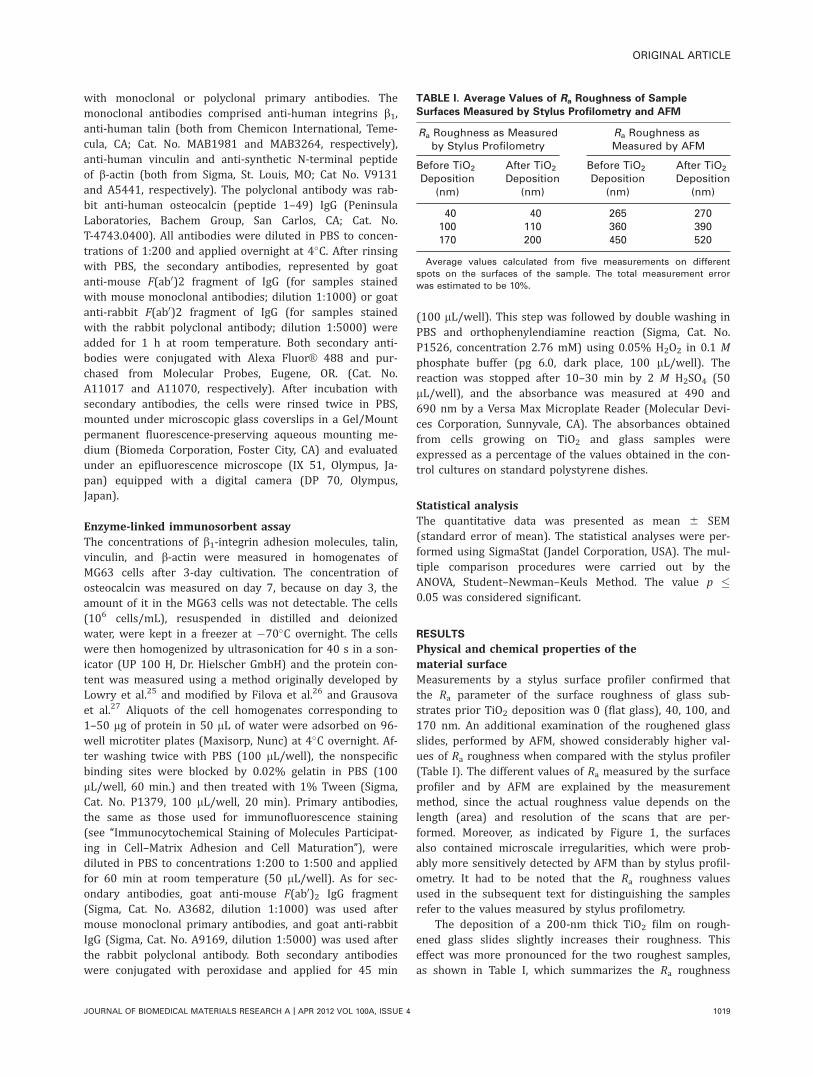
Physical and chemical properties of thematerial surfaceMeasurements by a stylus surface profiler confirmed thatthe Ra parameter of the surface roughness of glass sub-strates prior TiO2 deposition was 0 (flat glass), 40, 100, and170 nm. An additional examination of the roughened glassslides, performed by AFM, showed considerably higher val-ues of Ra roughness when compared with the stylus profiler(Table I). The different values of Ra measured by the surfaceprofiler and by AFM are explained by the measurementmethod, since the actual roughness value depends on thelength (area) and resolution of the scans that are per-formed. Moreover, as indicated by Figure 1, the surfacesalso contained microscale irregularities, which were prob-ably more sensitively detected by AFM than by stylus profil-ometry. It had to be noted that the Ra roughness valuesused in the subsequent text for distinguishing the samplesrefer to the values measured by stylus profilometry.
The deposition of a 200-nm thick TiO2 film on rough-ened glass slides slightly increases their roughness. Thiseffect was more pronounced for the two roughest samples,as shown in Table I, which summarizes the Ra roughness
TABLE I. Average Values of Ra Roughness of Sample
Surfaces Measured by Stylus Profilometry and AFM
Ra Roughness as Measuredby Stylus Profilometry
Ra Roughness asMeasured by AFM
Before TiO2
Deposition(nm)
After TiO2
Deposition(nm)
Before TiO2
Deposition(nm)
After TiO2
Deposition(nm)
40 40 265 270100 110 360 390170 200 450 520
Average values calculated from five measurements on different
spots on the surfaces of the sample. The total measurement error
was estimated to be 10%.
ORIGINAL ARTICLE
JOURNAL OF BIOMEDICAL MATERIALS RESEARCH A | APR 2012 VOL 100A, ISSUE 4 1019
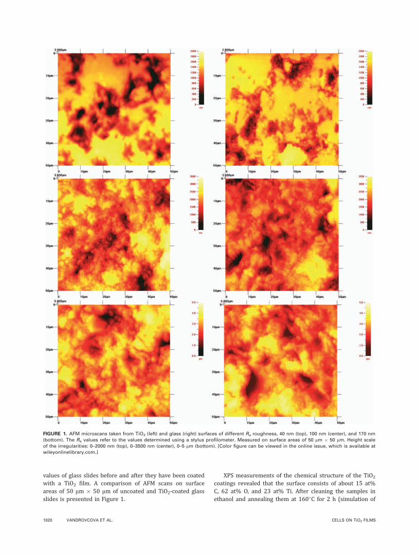
values of glass slides before and after they have been coatedwith a TiO2 film. A comparison of AFM scans on surfaceareas of 50 lm � 50 lm of uncoated and TiO2-coated glassslides is presented in Figure 1.
XPS measurements of the chemical structure of the TiO2
coatings revealed that the surface consists of about 15 at%C, 62 at% O, and 23 at% Ti. After cleaning the samples inethanol and annealing them at 160�C for 2 h (simulation of
FIGURE 1. AFM microscans taken from TiO2 (left) and glass (right) surfaces of different Ra roughness, 40 nm (top), 100 nm (center), and 170 nm
(bottom). The Ra values refer to the values determined using a stylus profilometer. Measured on surface areas of 50 lm � 50 lm. Height scale
of the irregularities: 0–2000 nm (top), 0–3500 nm (center), 0–5 lm (bottom). [Color figure can be viewed in the online issue, which is available at
wileyonlinelibrary.com.]
1020 VANDROVCOVA ET AL. CELLS ON TiO2 FILMS
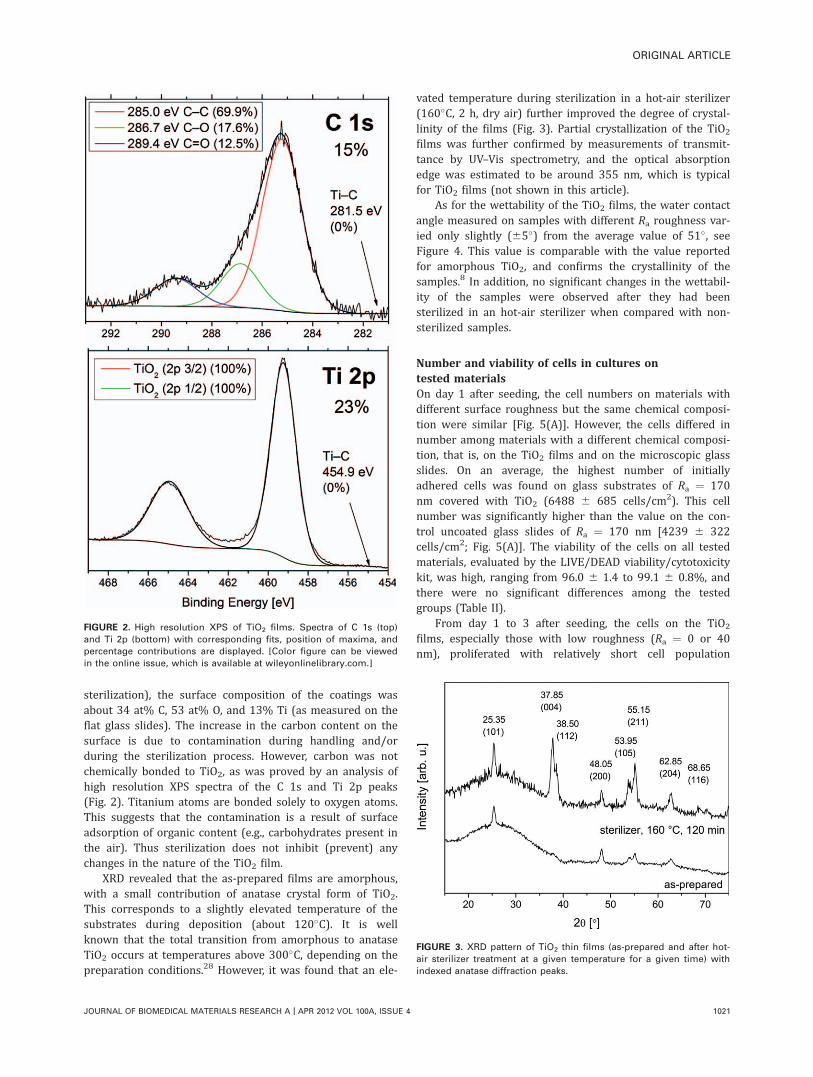
sterilization), the surface composition of the coatings wasabout 34 at% C, 53 at% O, and 13% Ti (as measured on theflat glass slides). The increase in the carbon content on thesurface is due to contamination during handling and/orduring the sterilization process. However, carbon was notchemically bonded to TiO2, as was proved by an analysis ofhigh resolution XPS spectra of the C 1s and Ti 2p peaks(Fig. 2). Titanium atoms are bonded solely to oxygen atoms.This suggests that the contamination is a result of surfaceadsorption of organic content (e.g., carbohydrates present inthe air). Thus sterilization does not inhibit (prevent) anychanges in the nature of the TiO2 film.
XRD revealed that the as-prepared films are amorphous,with a small contribution of anatase crystal form of TiO2.This corresponds to a slightly elevated temperature of thesubstrates during deposition (about 120�C). It is wellknown that the total transition from amorphous to anataseTiO2 occurs at temperatures above 300�C, depending on thepreparation conditions.28 However, it was found that an ele-
vated temperature during sterilization in a hot-air sterilizer(160�C, 2 h, dry air) further improved the degree of crystal-linity of the films (Fig. 3). Partial crystallization of the TiO2
films was further confirmed by measurements of transmit-tance by UV–Vis spectrometry, and the optical absorptionedge was estimated to be around 355 nm, which is typicalfor TiO2 films (not shown in this article).
As for the wettability of the TiO2 films, the water contactangle measured on samples with different Ra roughness var-ied only slightly (65�) from the average value of 51�, seeFigure 4. This value is comparable with the value reportedfor amorphous TiO2, and confirms the crystallinity of thesamples.8 In addition, no significant changes in the wettabil-ity of the samples were observed after they had beensterilized in an hot-air sterilizer when compared with non-sterilized samples.
Number and viability of cells in cultures ontested materialsOn day 1 after seeding, the cell numbers on materials withdifferent surface roughness but the same chemical composi-tion were similar [Fig. 5(A)]. However, the cells differed innumber among materials with a different chemical composi-tion, that is, on the TiO2 films and on the microscopic glassslides. On an average, the highest number of initiallyadhered cells was found on glass substrates of Ra ¼ 170nm covered with TiO2 (6488 6 685 cells/cm2). This cellnumber was significantly higher than the value on the con-trol uncoated glass slides of Ra ¼ 170 nm [4239 6 322cells/cm2; Fig. 5(A)]. The viability of the cells on all testedmaterials, evaluated by the LIVE/DEAD viability/cytotoxicitykit, was high, ranging from 96.0 6 1.4 to 99.1 6 0.8%, andthere were no significant differences among the testedgroups (Table II).
From day 1 to 3 after seeding, the cells on the TiO2
films, especially those with low roughness (Ra ¼ 0 or 40nm), proliferated with relatively short cell population
FIGURE 2. High resolution XPS of TiO2 films. Spectra of C 1s (top)
and Ti 2p (bottom) with corresponding fits, position of maxima, and
percentage contributions are displayed. [Color figure can be viewed
in the online issue, which is available at wileyonlinelibrary.com.]
FIGURE 3. XRD pattern of TiO2 thin films (as-prepared and after hot-
air sterilizer treatment at a given temperature for a given time) with
indexed anatase diffraction peaks.
ORIGINAL ARTICLE
JOURNAL OF BIOMEDICAL MATERIALS RESEARCH A | APR 2012 VOL 100A, ISSUE 4 1021
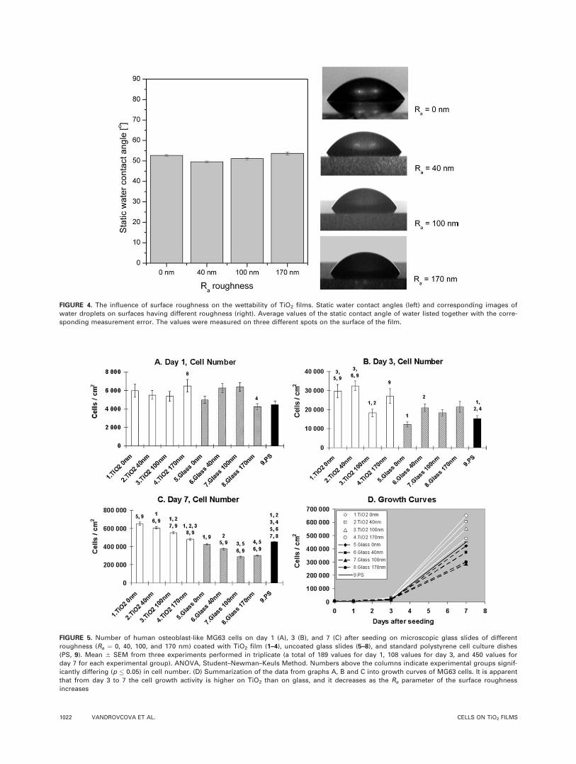
FIGURE 4. The influence of surface roughness on the wettability of TiO2 films. Static water contact angles (left) and corresponding images of
water droplets on surfaces having different roughness (right). Average values of the static contact angle of water listed together with the corre-
sponding measurement error. The values were measured on three different spots on the surface of the film.
FIGURE 5. Number of human osteoblast-like MG63 cells on day 1 (A), 3 (B), and 7 (C) after seeding on microscopic glass slides of different
roughness (Ra ¼ 0, 40, 100, and 170 nm) coated with TiO2 film (1–4), uncoated glass slides (5–8), and standard polystyrene cell culture dishes
(PS, 9). Mean 6 SEM from three experiments performed in triplicate (a total of 189 values for day 1, 108 values for day 3, and 450 values for
day 7 for each experimental group). ANOVA, Student–Newman–Keuls Method. Numbers above the columns indicate experimental groups signif-
icantly differing (p � 0.05) in cell number. (D) Summarization of the data from graphs A, B and C into growth curves of MG63 cells. It is apparent
that from day 3 to 7 the cell growth activity is higher on TiO2 than on glass, and it decreases as the Ra parameter of the surface roughness
increases
1022 VANDROVCOVA ET AL. CELLS ON TiO2 FILMS
doubling times (23.5 6 4.5 and 19.2 6 1.4 h, respectively)compared with the values on the control glass substrateswith corresponding surface roughness (42.7 6 7.0 and 36.36 7.4 h, respectively, Table III). As a result, the cells onTiO2 films with Ra ¼ 0 and 40 nm reached on an averagethe highest population densities on day 3 (29,599 6 3487and 32,451 6 2533 cells/cm2, respectively). These valueswere significantly higher than those on the glass samples ofcorresponding surface roughness (12,132 6 1309 and20,918 6 1963 cells/cm2) and on the control polystyreneculture dishes [15,268 6 1585 cells/cm2; Fig. 5(B)].
In addition, on day 7 after seeding, the cell numbers onthe TiO2-coated samples (from 65,1955 6 16,997 to47,8767 6 11,004 cells/cm2) were significantly higher thanthe values on the uncoated glass substrates of the corre-sponding roughness [42,2797 6 8596 to 30,0762 6 6809cells/cm2; Fig. 5(C)]. On TiO2 films of Ra ¼ 0, 40, and 100nm, the cell numbers were even higher than that on thecontrol cell culture polystyrene dishes (45,1418 6 4815cells/cm2).
Another important finding on day 7 was a clear depend-ence between cell number and surface roughness. On theTiO2 films and uncoated glass slides, the cell numberdecreased significantly with increasing material surfaceroughness [Fig. 5(C)]. On all tested materials, the cell viabil-ity, evaluated during automatic cell counting in the Vi-CELLXR Analyser using the trypan blue exclusion test, rangedfrom 77.7 6 0.8 to 83.8 6 0.7% (Table II). Compared withthe values obtained on day 1, the viability on day 7 was rel-atively low. The reason for this may lie in the differentmethods for evaluating the viability. Whereas on day 1, thecells were stained directly on the material with the LIVE/DEAD viability/cytotoxicity kit, on day 7, the cells were sub-jected to trypsinization and resuspension, which may havedamaged the cells.
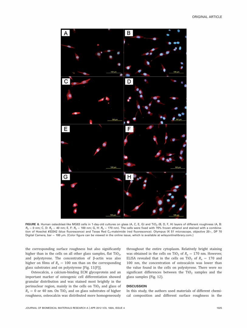
Size of the spreading area of the cells in cultures ontested materialsThe cell spreading area, that is, the cell area projected onthe material surface, was measured on day 1 after seedingin cells stained with Texas Red C2-maleimide and Hoechst33342. It is apparent that all tested materials promoted cellspreading, but this spreading was generally better onthe TiO2-coated substrates than on the uncoated glasssubstrates (Fig. 6). The TiO2 surfaces contained mainly po-lygonal cells, while the cells were rather of elongated spin-dle-like or round shape on the uncoated glass slides. In ac-cordance with this, the spreading areas of the cells on theTiO2 film of Ra ¼ 0 nm were significantly larger (1260 644 lm2) than the values obtained on all remaining materialsamples, including the polystyrene dishes (815 6 26 to1078 6 52 lm2, Fig. 7). On the other TiO2 films (Ra of 40,100, and 170 nm), the cell spreading areas were also signifi-cantly larger (1052 6 37 to 1078 6 52 lm2) than those onglass of Ra ¼ 170 nm (816 6 26 lm2). In addition, the cellspreading area showed a tendency to decrease with increas-ing material surface roughness. On the TiO2 film with Ra ¼0 nm, the cell spreading area was significantly larger thanT
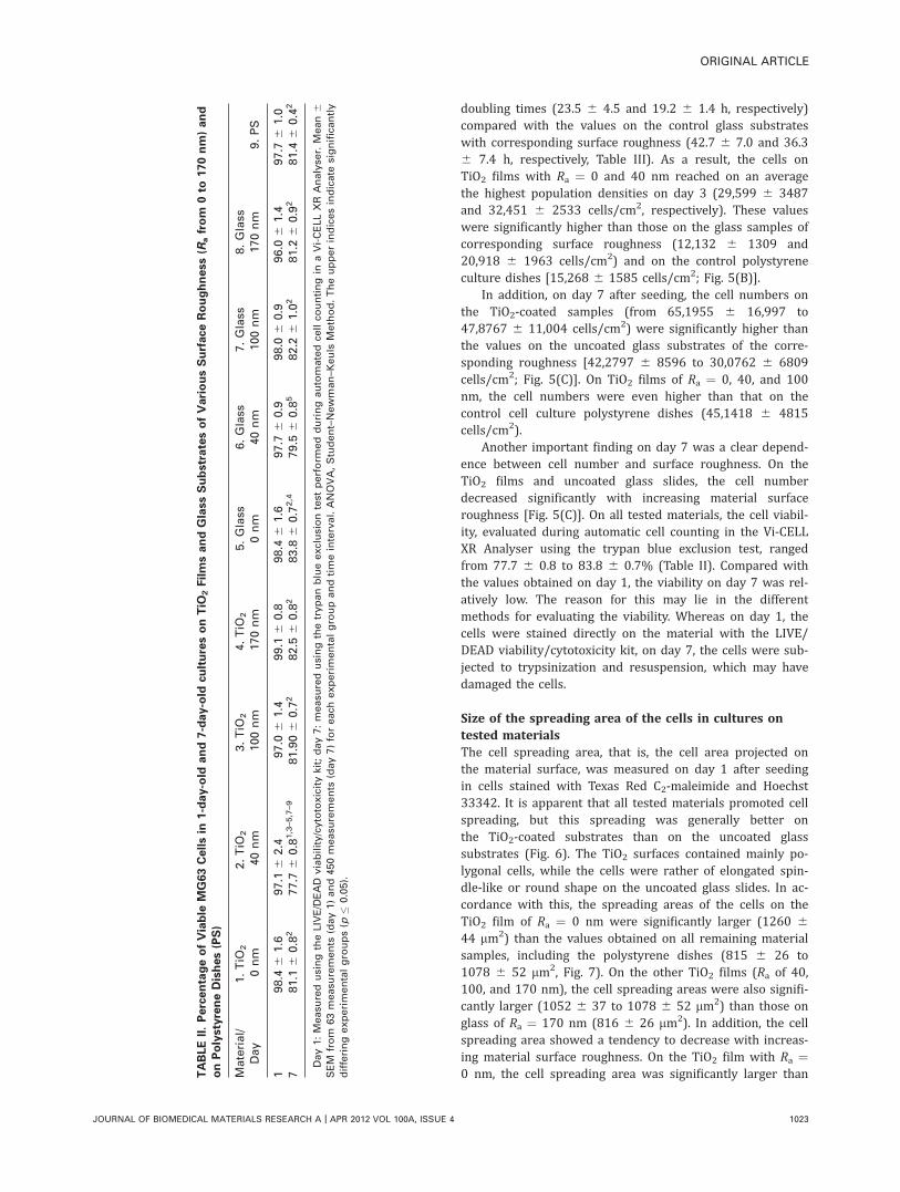
ABLEII.PercentageofViable
MG63Cellsin
1-day-old
and7-day-old
culturesonTiO
2FilmsandGlassSubstratesofVariousSurfaceRoughness(R
afrom
0to
170nm)and
onPolystyreneDishes(PS)
Material/
Day
1.TiO
2
0nm
2.TiO
2
40nm
3.TiO
2
100nm
4.TiO
2
170nm
5.Glass
0nm
6.Glass
40nm
7.Glass
100nm
8.Glass
170nm
9.PS
198.4
61.6
97.1
62.4
97.0
61.4
99.1
60.8
98.4
61.6
97.7
60.9
98.0
60.9
96.0
61.4
97.7
61.0
781.1
60.8
277.7
60.8
1,3–5,7–9
81.906
0.7
282.5
60.8
283.8
60.7
2,4
79.5
60.8
582.2
61.0
281.2
60.9
281.4
60.4
2
Day1:Measu
redusingtheLIVE/DEAD
viability/cytotoxicitykit;day7:measu
redusingthetrypanblueexclusiontest
perform
edduringautomatedce
llco
untingin
aVi-CELLXR
Analyse
r.Mean6
SEM
from
63measu
rements
(day1)and450measu
rements
(day7)foreach
experimentalgroupandtimeinterval.ANOVA,Student–Newman–K
euls
Method.Theupperindicesindicate
significa
ntly
differingexperimentalgroups(p
�0.05).
ORIGINAL ARTICLE
JOURNAL OF BIOMEDICAL MATERIALS RESEARCH A | APR 2012 VOL 100A, ISSUE 4 1023
the areas on the films of Ra ¼ 40, 100, or 170 nm. Similarly,on glass, the cell spreading area was significantly larger onsamples of Ra ¼ 0 nm than Ra ¼ 170 nm (Fig. 7).
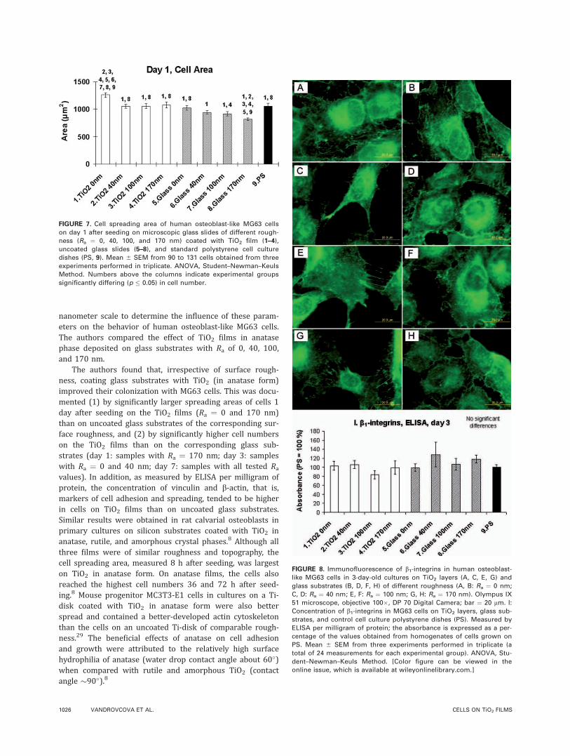
Distribution and concentration of moleculesparticipating in cell adhesion, spreading,and phenotypic maturationImmunofluorescence showed that in the cells on all testedsamples, b1-integrins, that is, receptors for collagen, fibro-nectin, and laminin were most brightly stained in the peri-nuclear localization (Fig. 8). This may be due to the greatestcell thickness in this region and the cytoplasmic localizationof the integrin molecules. Toward the cell periphery, thestaining intensity was lower, but the distribution of integ-rins in dot-like, dash-like, or mesh-like structures was moreapparent. On TiO2-coated surfaces, especially those withnanoscale roughness, the cells often formed protrusions(extensions, filopodia), which were positively stained againstb1-integrins. On nanorough glass surfaces (Ra ¼ 40–170nm), the b1-integrins often created a border on the celledges [Fig. 8(D,F,H)]. Enzyme-linked immunosorbent assay(ELISA) revealed that the concentration of b1-integrins,measured per milligram of protein, was similar in the cellson all tested surfaces, though the highest average value wasobtained in the cells on glass of Ra ¼ 40 nm [Fig. 8(I)].
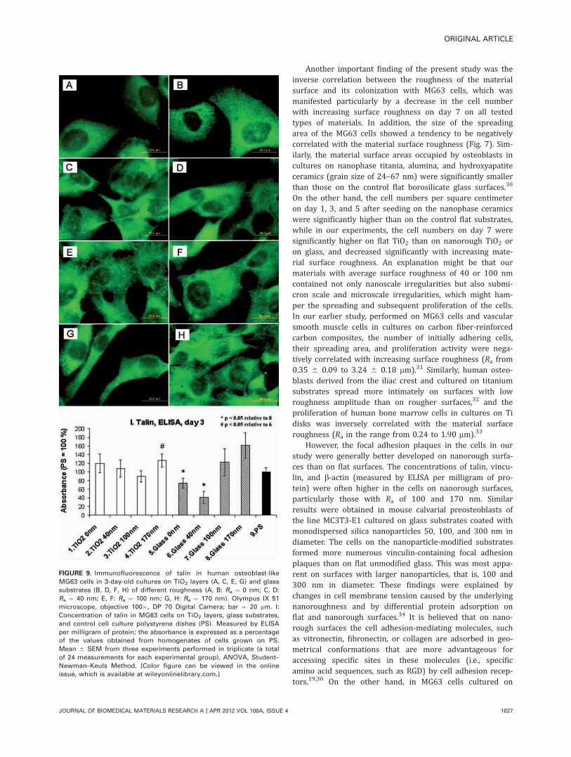
Talin, an integrin-associated protein of focal adhesionsites, showed a similar distribution as av integrins, that is,organized in fine granular and dash-like structures arrangedin parallel and apparent not only at the cell periphery butalso on the entire ventral part of the cell membrane includ-ing the perinuclear region. The dash-like focal adhesion siteswere clearly visible, particularly on the glass substrates ofRa from 40 to 170 nm [Fig. 9(D,F,H)]. On an average, thehighest concentration of talin was achieved in the cells onglass substrates of Ra ¼ 170 nm. This concentration wassignificantly higher than that in cells on glass with Ra of0 and 40 nm. On the TiO2 samples, the highest talin concen-tration was also found in cells on films of Ra ¼ 170 nm[Fig. 9(I)].
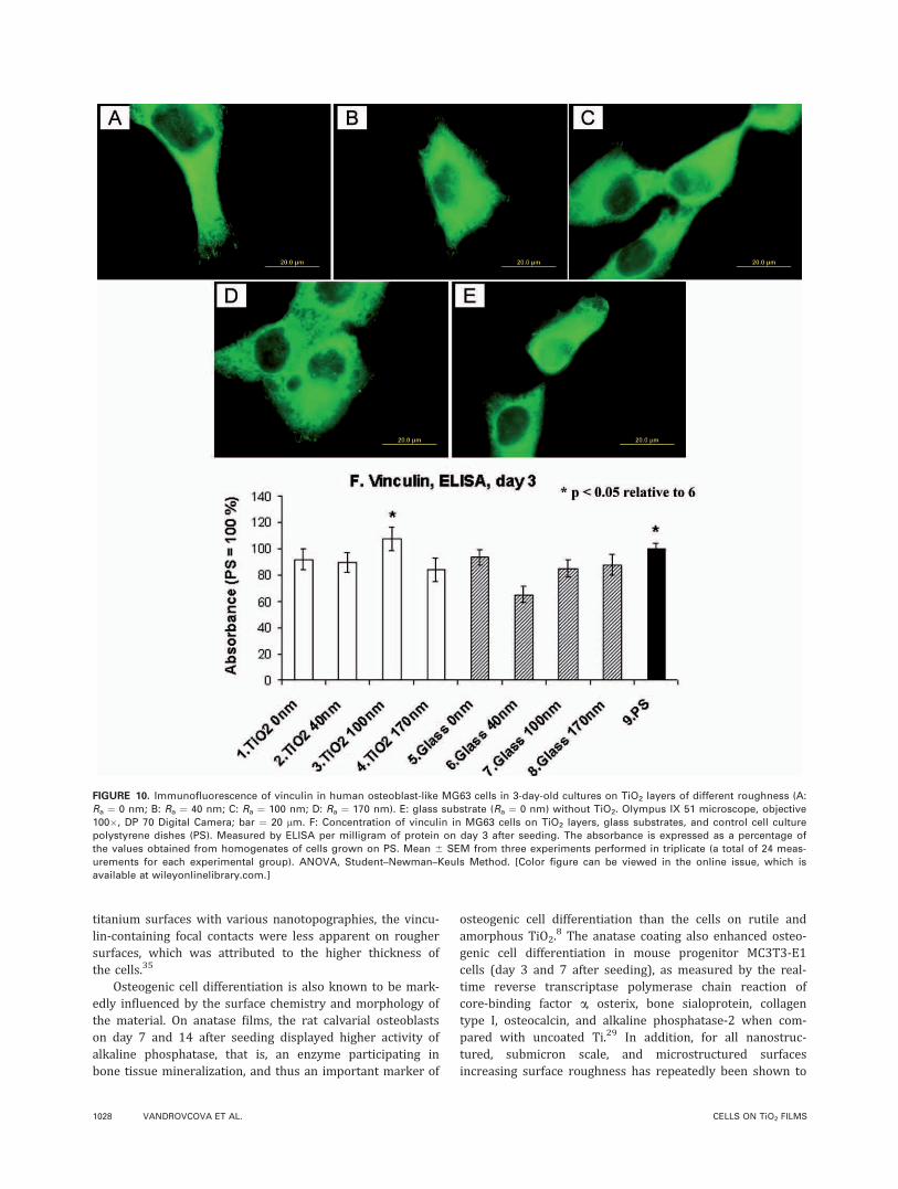
Vinculin, another protein of focal adhesion plaques asso-ciated with integrin and also cell–cell adhesion receptors,formed dot-like or dash-like focal adhesion sites at the celledges, which were well apparent, especially on the TiO2
surfaces. In addition, this protein was homogeneously dis-tributed throughout the cells, often in the form of a fine net-work [Fig. 10(A–E)]. The highest concentration of vinculinwas detected in the cells grown on TiO2 films of Ra ¼ 100nm, and the lowest concentration was in the cells onuncoated glass substrates with Ra ¼ 40 nm, where the vin-culin concentration was significantly lower than in the cellson polystyrene [Fig. 10(F)].
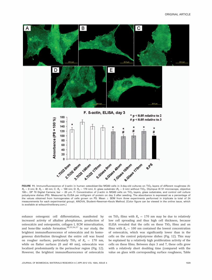
b-actin, an important protein of the cytoplasmic cyto-skeleton associated with focal adhesion sites, formed finefilaments often arranged in parallel and crossing the entireintracellular region in the cells on all tested surfaces [Fig.11(A–E)]. The concentration of b-actin was highest in thecells on TiO2 films of Ra ¼ 40 nm, where it was not onlysignificantly higher than the value on glass substrates withT
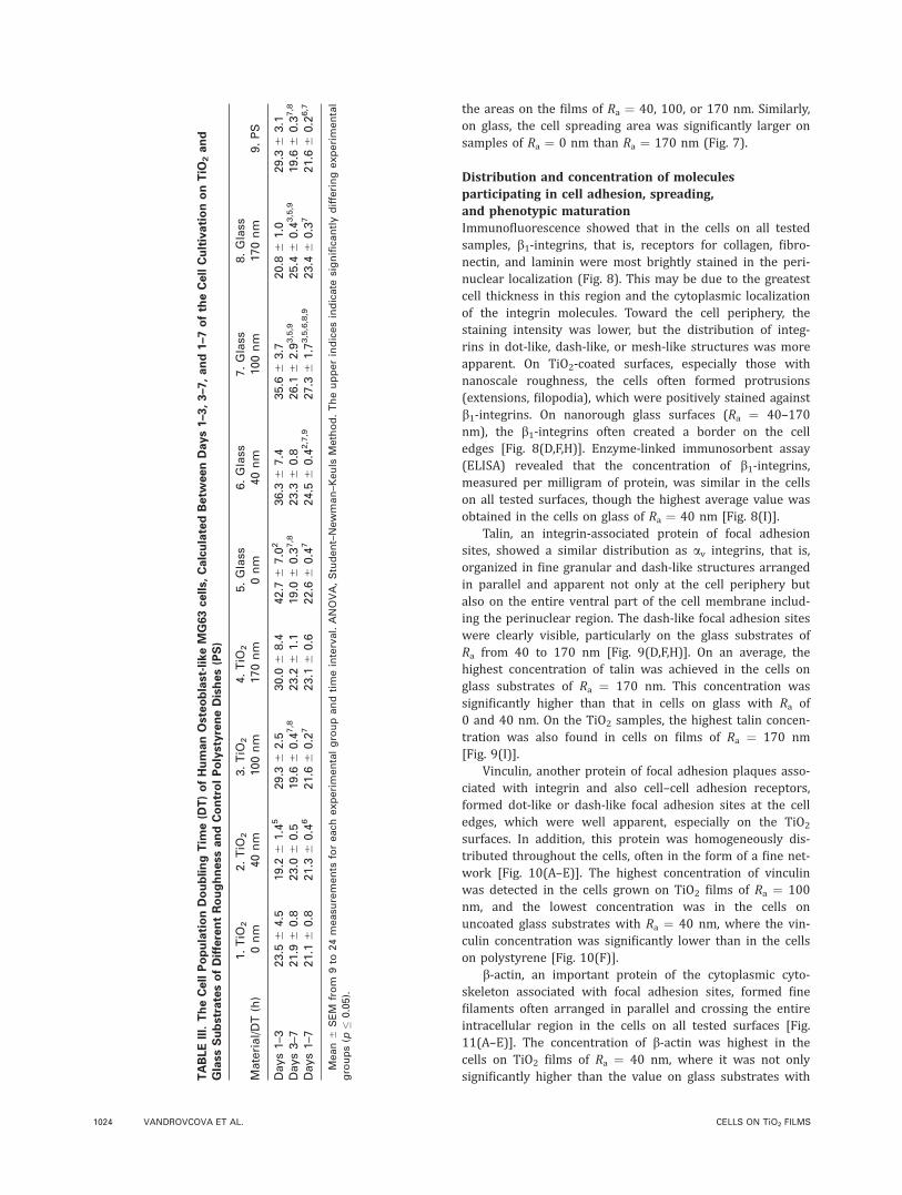
ABLEIII.TheCellPopulationDoublingTim
e(D
T)ofHumanOsteoblast-likeMG63cells,CalculatedBetw
eenDays1–3,3–7,and1–7oftheCellCultivationonTiO
2and
GlassSubstratesofDifferentRoughnessandControlPolystyreneDishes(PS)
Material/DT(h)
1.TiO
2
0nm
2.TiO
2
40nm
3.TiO
2
100nm
4.TiO
2
170nm
5.Glass
0nm
6.Glass
40nm
7.Glass
100nm
8.Glass
170nm
9.PS
Days1–3
23.5
64.5
19.2
61.4
529.3
62.5
30.0
68.4
42.7
67.0
236.3
67.4
35.6
63.7
20.8
61.0
29.3
63.1
Days3–7
21.9
60.8
23.0
60.5
19.6
60.4
7,8
23.2
61.1
19.0
60.3
7,8
23.3
60.8
26.1
62.9
3,5,9
25.4
60.4
3,5,9
19.6
60.3
7,8
Days1–7
21.1
60.8
21.3
60.4
621.6
60.2
723.1
60.6
22.6
60.4
724.5
60.4
2,7,9
27.3
61.7
3,5,6,8,9
23.4
60.3
721.6
60.2
6,7
Mean6
SEM
from
9to
24measu
rements
foreach
experimentalgroupandtimeinterval.ANOVA,Student–Newman–K
euls
Method.Theupperindicesindicate
significa
ntlydifferingexperimental
groups(p
�0.05).
1024 VANDROVCOVA ET AL. CELLS ON TiO2 FILMS
the corresponding surface roughness but also significantlyhigher than in the cells on all other glass samples, flat TiO2,and polystyrene. The concentration of b-actin was alsohigher on films of Ra ¼ 100 nm than on the correspondingglass substrates and on polystyrene [Fig. 11(F)].
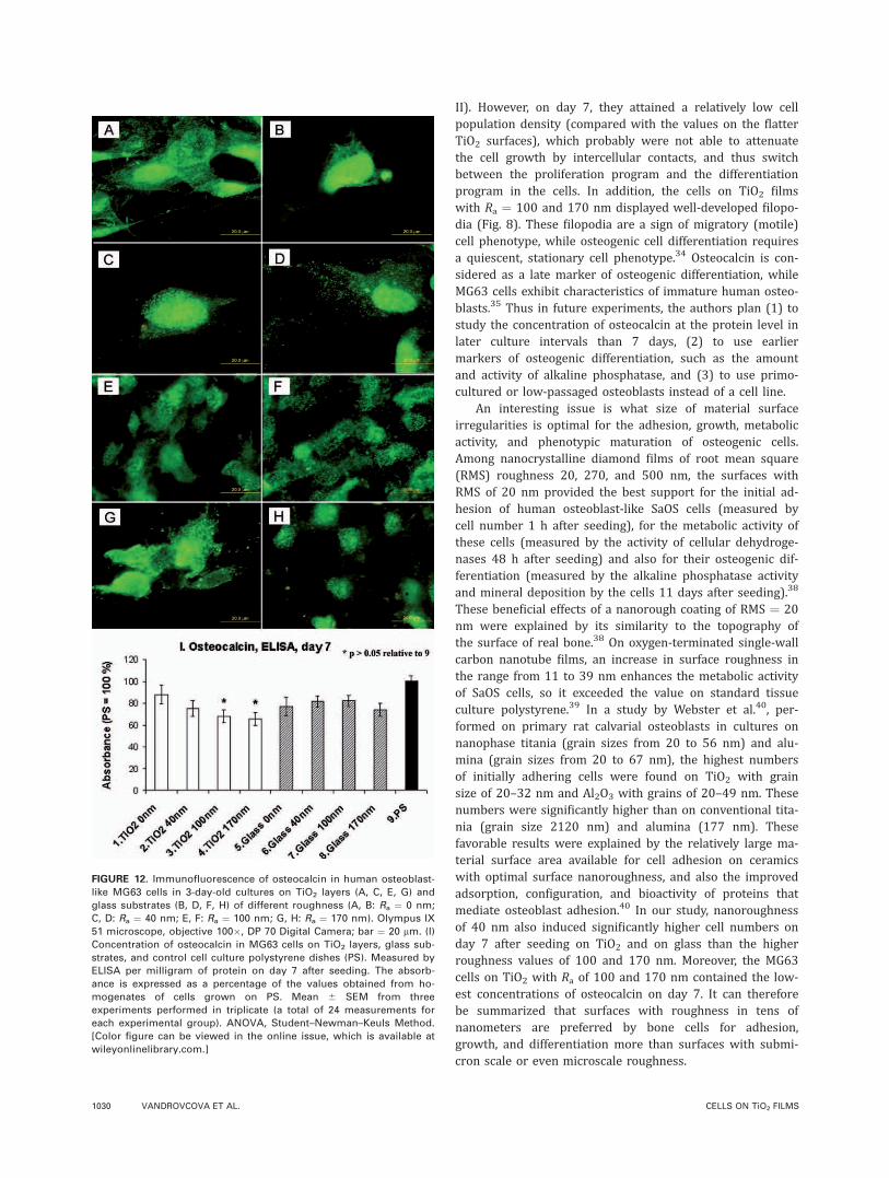
Osteocalcin, a calcium-binding ECM glycoprotein and animportant marker of osteogenic cell differentiation showedgranular distribution and was stained most brightly in theperinuclear region, mainly in the cells on TiO2 and glass ofRa ¼ 0 or 40 nm. On TiO2 and on glass substrates of higherroughness, osteocalcin was distributed more homogeneously
throughout the entire cytoplasm. Relatively bright stainingwas obtained in the cells on TiO2 of Ra ¼ 170 nm. However,ELISA revealed that in the cells on TiO2 of Ra ¼ 170 and100 nm, the concentration of osteocalcin was lower thanthe value found in the cells on polystyrene. There were nosignificant differences between the TiO2 samples and theglass samples (Fig. 12).
DISCUSSION
In this study, the authors used materials of different chemi-cal composition and different surface roughness in the
FIGURE 6. Human osteoblast-like MG63 cells in 1-day-old cultures on glass (A, C, E, G) and TiO2 (B, D, F, H) layers of different roughness (A, B:
Ra ¼ 0 nm; C, D: Ra ¼ 40 nm; E, F: Ra ¼ 100 nm; G, H: Ra ¼ 170 nm). The cells were fixed with 70% frozen ethanol and stained with a combina-
tion of Hoechst #33342 (blue fluorescence) and Texas Red C2-maleimide (red fluorescence). Olympus IX 51 microscope, objective 20�, DP 70
Digital Camera, bar ¼ 100 lm. [Color figure can be viewed in the online issue, which is available at wileyonlinelibrary.com.]
ORIGINAL ARTICLE
JOURNAL OF BIOMEDICAL MATERIALS RESEARCH A | APR 2012 VOL 100A, ISSUE 4 1025
nanometer scale to determine the influence of these param-eters on the behavior of human osteoblast-like MG63 cells.The authors compared the effect of TiO2 films in anatasephase deposited on glass substrates with Ra of 0, 40, 100,and 170 nm.
The authors found that, irrespective of surface rough-ness, coating glass substrates with TiO2 (in anatase form)improved their colonization with MG63 cells. This was docu-mented (1) by significantly larger spreading areas of cells 1day after seeding on the TiO2 films (Ra ¼ 0 and 170 nm)than on uncoated glass substrates of the corresponding sur-face roughness, and (2) by significantly higher cell numberson the TiO2 films than on the corresponding glass sub-strates (day 1: samples with Ra ¼ 170 nm; day 3: sampleswith Ra ¼ 0 and 40 nm; day 7: samples with all tested Ravalues). In addition, as measured by ELISA per milligram ofprotein, the concentration of vinculin and b-actin, that is,markers of cell adhesion and spreading, tended to be higherin cells on TiO2 films than on uncoated glass substrates.Similar results were obtained in rat calvarial osteoblasts inprimary cultures on silicon substrates coated with TiO2 inanatase, rutile, and amorphous crystal phases.8 Although allthree films were of similar roughness and topography, thecell spreading area, measured 8 h after seeding, was largeston TiO2 in anatase form. On anatase films, the cells alsoreached the highest cell numbers 36 and 72 h after seed-ing.8 Mouse progenitor MC3T3-E1 cells in cultures on a Ti-disk coated with TiO2 in anatase form were also betterspread and contained a better-developed actin cytoskeletonthan the cells on an uncoated Ti-disk of comparable rough-ness.29 The beneficial effects of anatase on cell adhesionand growth were attributed to the relatively high surfacehydrophilia of anatase (water drop contact angle about 60�)when compared with rutile and amorphous TiO2 (contactangle �90�).8
FIGURE 7. Cell spreading area of human osteoblast-like MG63 cells
on day 1 after seeding on microscopic glass slides of different rough-
ness (Ra ¼ 0, 40, 100, and 170 nm) coated with TiO2 film (1–4),
uncoated glass slides (5–8), and standard polystyrene cell culture
dishes (PS, 9). Mean 6 SEM from 90 to 131 cells obtained from three
experiments performed in triplicate. ANOVA, Student–Newman–Keuls
Method. Numbers above the columns indicate experimental groups
significantly differing (p � 0.05) in cell number.
FIGURE 8. Immunofluorescence of b1-integrins in human osteoblast-
like MG63 cells in 3-day-old cultures on TiO2 layers (A, C, E, G) and
glass substrates (B, D, F, H) of different roughness (A, B: Ra ¼ 0 nm;
C, D: Ra ¼ 40 nm; E, F: Ra ¼ 100 nm; G, H: Ra ¼ 170 nm). Olympus IX
51 microscope, objective 100�, DP 70 Digital Camera; bar ¼ 20 lm. I:
Concentration of b1-integrins in MG63 cells on TiO2 layers, glass sub-
strates, and control cell culture polystyrene dishes (PS). Measured by
ELISA per milligram of protein; the absorbance is expressed as a per-
centage of the values obtained from homogenates of cells grown on
PS. Mean 6 SEM from three experiments performed in triplicate (a
total of 24 measurements for each experimental group). ANOVA, Stu-
dent–Newman–Keuls Method. [Color figure can be viewed in the
online issue, which is available at wileyonlinelibrary.com.]
1026 VANDROVCOVA ET AL. CELLS ON TiO2 FILMS
Another important finding of the present study was theinverse correlation between the roughness of the materialsurface and its colonization with MG63 cells, which wasmanifested particularly by a decrease in the cell numberwith increasing surface roughness on day 7 on all testedtypes of materials. In addition, the size of the spreadingarea of the MG63 cells showed a tendency to be negativelycorrelated with the material surface roughness (Fig. 7). Sim-ilarly, the material surface areas occupied by osteoblasts incultures on nanophase titania, alumina, and hydroxyapatiteceramics (grain size of 24–67 nm) were significantly smallerthan those on the control flat borosilicate glass surfaces.30
On the other hand, the cell numbers per square centimeteron day 1, 3, and 5 after seeding on the nanophase ceramicswere significantly higher than on the control flat substrates,while in our experiments, the cell numbers on day 7 weresignificantly higher on flat TiO2 than on nanorough TiO2 oron glass, and decreased significantly with increasing mate-rial surface roughness. An explanation might be that ourmaterials with average surface roughness of 40 or 100 nmcontained not only nanoscale irregularities but also submi-cron scale and microscale irregularities, which might ham-per the spreading and subsequent proliferation of the cells.In our earlier study, performed on MG63 cells and vascularsmooth muscle cells in cultures on carbon fiber-reinforcedcarbon composites, the number of initially adhering cells,their spreading area, and proliferation activity were nega-tively correlated with increasing surface roughness (Ra from0.35 6 0.09 to 3.24 6 0.18 lm).31 Similarly, human osteo-blasts derived from the iliac crest and cultured on titaniumsubstrates spread more intimately on surfaces with lowroughness amplitude than on rougher surfaces,32 and theproliferation of human bone marrow cells in cultures on Tidisks was inversely correlated with the material surfaceroughness (Ra in the range from 0.24 to 1.90 lm).33
However, the focal adhesion plaques in the cells in ourstudy were generally better developed on nanorough surfa-ces than on flat surfaces. The concentrations of talin, vincu-lin, and b-actin (measured by ELISA per milligram of pro-tein) were often higher in the cells on nanorough surfaces,particularly those with Ra of 100 and 170 nm. Similarresults were obtained in mouse calvarial preosteoblasts ofthe line MC3T3-E1 cultured on glass substrates coated withmonodispersed silica nanoparticles 50, 100, and 300 nm indiameter. The cells on the nanoparticle-modified substratesformed more numerous vinculin-containing focal adhesionplaques than on flat unmodified glass. This was most appa-rent on surfaces with larger nanoparticles, that is, 100 and300 nm in diameter. These findings were explained bychanges in cell membrane tension caused by the underlyingnanoroughness and by differential protein adsorption onflat and nanorough surfaces.34 It is believed that on nano-rough surfaces the cell adhesion-mediating molecules, suchas vitronectin, fibronectin, or collagen are adsorbed in geo-metrical conformations that are more advantageous foraccessing specific sites in these molecules (i.e., specificamino acid sequences, such as RGD) by cell adhesion recep-tors.19,30 On the other hand, in MG63 cells cultured on
FIGURE 9. Immunofluorescence of talin in human osteoblast-like
MG63 cells in 3-day-old cultures on TiO2 layers (A, C, E, G) and glass
substrates (B, D, F, H) of different roughness (A, B: Ra ¼ 0 nm; C, D:
Ra ¼ 40 nm; E, F: Ra ¼ 100 nm; G, H: Ra ¼ 170 nm). Olympus IX 51
microscope, objective 100�, DP 70 Digital Camera; bar ¼ 20 lm. I:
Concentration of talin in MG63 cells on TiO2 layers, glass substrates,
and control cell culture polystyrene dishes (PS). Measured by ELISA
per milligram of protein; the absorbance is expressed as a percentage
of the values obtained from homogenates of cells grown on PS.
Mean 6 SEM from three experiments performed in triplicate (a total
of 24 measurements for each experimental group). ANOVA, Student–
Newman–Keuls Method. [Color figure can be viewed in the online
issue, which is available at wileyonlinelibrary.com.]
ORIGINAL ARTICLE
JOURNAL OF BIOMEDICAL MATERIALS RESEARCH A | APR 2012 VOL 100A, ISSUE 4 1027
titanium surfaces with various nanotopographies, the vincu-lin-containing focal contacts were less apparent on roughersurfaces, which was attributed to the higher thickness ofthe cells.35
Osteogenic cell differentiation is also known to be mark-edly influenced by the surface chemistry and morphology ofthe material. On anatase films, the rat calvarial osteoblastson day 7 and 14 after seeding displayed higher activity ofalkaline phosphatase, that is, an enzyme participating inbone tissue mineralization, and thus an important marker of
osteogenic cell differentiation than the cells on rutile andamorphous TiO2.
8 The anatase coating also enhanced osteo-genic cell differentiation in mouse progenitor MC3T3-E1cells (day 3 and 7 after seeding), as measured by the real-time reverse transcriptase polymerase chain reaction ofcore-binding factor a, osterix, bone sialoprotein, collagentype I, osteocalcin, and alkaline phosphatase-2 when com-pared with uncoated Ti.29 In addition, for all nanostruc-tured, submicron scale, and microstructured surfacesincreasing surface roughness has repeatedly been shown to
FIGURE 10. Immunofluorescence of vinculin in human osteoblast-like MG63 cells in 3-day-old cultures on TiO2 layers of different roughness (A:
Ra ¼ 0 nm; B: Ra ¼ 40 nm; C: Ra ¼ 100 nm; D: Ra ¼ 170 nm). E: glass substrate (Ra ¼ 0 nm) without TiO2. Olympus IX 51 microscope, objective
100�, DP 70 Digital Camera; bar ¼ 20 lm. F: Concentration of vinculin in MG63 cells on TiO2 layers, glass substrates, and control cell culture
polystyrene dishes (PS). Measured by ELISA per milligram of protein on day 3 after seeding. The absorbance is expressed as a percentage of
the values obtained from homogenates of cells grown on PS. Mean 6 SEM from three experiments performed in triplicate (a total of 24 meas-
urements for each experimental group). ANOVA, Student–Newman–Keuls Method. [Color figure can be viewed in the online issue, which is
available at wileyonlinelibrary.com.]
1028 VANDROVCOVA ET AL. CELLS ON TiO2 FILMS
enhance osteogenic cell differentiation, manifested byincreased activity of alkaline phosphatase, production ofosteocalcin and osteopontin, collagen I, ECM mineralization,and bone-like nodule formation.30,33,36,37 In our study, thebrightest immunofluorescence of osteocalcin and its homo-geneous distribution throughout the entire cell was foundon rougher surfaces, particularly TiO2 of Ra ¼ 170 nm,while on flatter surfaces (0 and 40 nm), osteocalcin waslocalized predominantly in the perinuclear region (Fig. 12).However, the brightest immunofluorescence of osteocalcin
on TiO2 films with Ra ¼ 170 nm may be due to relativelylow cell spreading and thus high cell thickness, becauseELISA revealed that the cells on these TiO2 films and onfilms with Ra ¼ 100 nm contained the lowest concentrationof osteocalcin, which was significantly lower than in thecells on the control polystyrene dishes (Fig. 12). This maybe explained by a relatively high proliferation activity of thecells on these films. Between days 3 and 7, these cells grewwith a relatively short doubling time (compared with thevalue on glass with corresponding surface roughness, Table
FIGURE 11. Immunofluorescence of b-actin in human osteoblast-like MG63 cells in 3-day-old cultures on TiO2 layers of different roughness (A:
Ra ¼ 0 nm; B: Ra ¼ 40 nm; C: Ra ¼ 100 nm; D: Ra ¼ 170 nm). E: glass substrate (Ra ¼ 0 nm) without TiO2. Olympus IX 51 microscope, objective
100�, DP 70 Digital Camera; bar ¼ 20 lm. F: Concentration of b-actin in MG63 cells on TiO2 layers, glass substrates, and control cell culture
polystyrene dishes (PS). Measured by ELISA per milligram of protein on day 3 after seeding. The absorbance is expressed as a percentage of
the values obtained from homogenates of cells grown on PS. Mean 6 SEM from three experiments performed in triplicate (a total of 24
measurements for each experimental group). ANOVA, Student–Newman–Keuls Method. [Color figure can be viewed in the online issue, which
is available at wileyonlinelibrary.com.]
ORIGINAL ARTICLE
JOURNAL OF BIOMEDICAL MATERIALS RESEARCH A | APR 2012 VOL 100A, ISSUE 4 1029
II). However, on day 7, they attained a relatively low cellpopulation density (compared with the values on the flatterTiO2 surfaces), which probably were not able to attenuatethe cell growth by intercellular contacts, and thus switchbetween the proliferation program and the differentiationprogram in the cells. In addition, the cells on TiO2 filmswith Ra ¼ 100 and 170 nm displayed well-developed filopo-dia (Fig. 8). These filopodia are a sign of migratory (motile)cell phenotype, while osteogenic cell differentiation requiresa quiescent, stationary cell phenotype.34 Osteocalcin is con-sidered as a late marker of osteogenic differentiation, whileMG63 cells exhibit characteristics of immature human osteo-blasts.35 Thus in future experiments, the authors plan (1) tostudy the concentration of osteocalcin at the protein level inlater culture intervals than 7 days, (2) to use earliermarkers of osteogenic differentiation, such as the amountand activity of alkaline phosphatase, and (3) to use primo-cultured or low-passaged osteoblasts instead of a cell line.
An interesting issue is what size of material surfaceirregularities is optimal for the adhesion, growth, metabolicactivity, and phenotypic maturation of osteogenic cells.Among nanocrystalline diamond films of root mean square(RMS) roughness 20, 270, and 500 nm, the surfaces withRMS of 20 nm provided the best support for the initial ad-hesion of human osteoblast-like SaOS cells (measured bycell number 1 h after seeding), for the metabolic activity ofthese cells (measured by the activity of cellular dehydroge-nases 48 h after seeding) and also for their osteogenic dif-ferentiation (measured by the alkaline phosphatase activityand mineral deposition by the cells 11 days after seeding).38
These beneficial effects of a nanorough coating of RMS ¼ 20nm were explained by its similarity to the topography ofthe surface of real bone.38 On oxygen-terminated single-wallcarbon nanotube films, an increase in surface roughness inthe range from 11 to 39 nm enhances the metabolic activityof SaOS cells, so it exceeded the value on standard tissueculture polystyrene.39 In a study by Webster et al.40, per-formed on primary rat calvarial osteoblasts in cultures onnanophase titania (grain sizes from 20 to 56 nm) and alu-mina (grain sizes from 20 to 67 nm), the highest numbersof initially adhering cells were found on TiO2 with grainsize of 20–32 nm and Al2O3 with grains of 20–49 nm. Thesenumbers were significantly higher than on conventional tita-nia (grain size 2120 nm) and alumina (177 nm). Thesefavorable results were explained by the relatively large ma-terial surface area available for cell adhesion on ceramicswith optimal surface nanoroughness, and also the improvedadsorption, configuration, and bioactivity of proteins thatmediate osteoblast adhesion.40 In our study, nanoroughnessof 40 nm also induced significantly higher cell numbers onday 7 after seeding on TiO2 and on glass than the higherroughness values of 100 and 170 nm. Moreover, the MG63cells on TiO2 with Ra of 100 and 170 nm contained the low-est concentrations of osteocalcin on day 7. It can thereforebe summarized that surfaces with roughness in tens ofnanometers are preferred by bone cells for adhesion,growth, and differentiation more than surfaces with submi-cron scale or even microscale roughness.
FIGURE 12. Immunofluorescence of osteocalcin in human osteoblast-
like MG63 cells in 3-day-old cultures on TiO2 layers (A, C, E, G) and
glass substrates (B, D, F, H) of different roughness (A, B: Ra ¼ 0 nm;
C, D: Ra ¼ 40 nm; E, F: Ra ¼ 100 nm; G, H: Ra ¼ 170 nm). Olympus IX
51 microscope, objective 100�, DP 70 Digital Camera; bar ¼ 20 lm. (I)
Concentration of osteocalcin in MG63 cells on TiO2 layers, glass sub-
strates, and control cell culture polystyrene dishes (PS). Measured by
ELISA per milligram of protein on day 7 after seeding. The absorb-
ance is expressed as a percentage of the values obtained from ho-
mogenates of cells grown on PS. Mean 6 SEM from three
experiments performed in triplicate (a total of 24 measurements for
each experimental group). ANOVA, Student–Newman–Keuls Method.
[Color figure can be viewed in the online issue, which is available at
wileyonlinelibrary.com.]
1030 VANDROVCOVA ET AL. CELLS ON TiO2 FILMS
It had to be pointed out that not only the size but alsothe shape of the material surface irregularities plays a cru-cial role in the cell–material interaction. For example, po-rous structures of different diameter (0.5 and 2 lm) anddifferent Ra parameter (0.2 and 0.4 lm), created on tita-nium surfaces by anodic oxidation, increased the number ofinitially attached cells to similar values, and induced similarmorphological features in cells, for example, the formationof filopodia, similar shape and distribution of vinculin-con-taining focal adhesion plaques and a similar spatial arrange-ment of the actin cytoskeleton.41 The density of the irregu-larities was also important for the cell–material interaction.For example, increasing the density of particles of the samesize deposited on the material surface decreased the spread-ing and proliferation of rat calvarial osteoblasts42 butincreased the expression of osteocalcin.43
CONCLUSION
Coating glass substrates (roughness parameter Ra of 0, 40,100, or 170 nm) with TiO2 films in anatase form improvedthe initial adhesion of MG63 cells in 1-day-old cultures onsome samples (cell number: Ra ¼ 170 nm; cell spreadingarea: Ra ¼ 0 and 170 nm) and increased the final cell popu-lation densities on day 7 on all samples. The cells on TiO2
films with Ra ¼ 40 and 100 nm also contained a higher con-centration of b-actin than the corresponding glass sub-strates. In addition, the material surface roughness wasinversely correlated with the size of the cell spreading areaand particularly with the cell number on day 7. On theother hand, the concentration of talin was higher in the cellson rougher glass substrates (Ra ¼ 170 nm) than on flattersubstrates (Ra ¼ 0 and 40 nm). The concentration of osteo-calcin on day 7 was similar in the cells on all tested sam-ples except the cells on the TiO2 films with Ra ¼ 100 and170 nm, where these values were significantly lower than inthe cells on the control polystyrene dishes. It can thereforebe concluded that smoother TiO2 films (Ra ¼ 0, 40 nm)seem to be the most appropriate of all the materials testedin this article for forming new bone tissue.
ACKNOWLEDGMENTS
This study was supported by the Academy of Sciences of theCzech Republic (Grant No. KAN101120701). The authors alsothank Lea Nichtova, MSc (Charles University, Faculty of Mathe-matics and Physics, Department of Condensed Matter Physics)for performing XRD measurements and Mrs. Ivana Zajanova(Inst. Physiol., Acad. Sci. CR) for her excellent technical assis-tance in immunofluorescence staining. Mr. Robin Healey(Czech Technical University, Prague) is gratefully acknowl-edged for his language revision of the manuscript.
REFERENCES
1. Geetha M, Singh AK, Asokamani R, Gogia AK. Ti based biomateri-
als, the ultimate choice for orthopaedic implants – A review. Prog
Mater Sci 2009;54:397–425.
2. Long M, Rack HJ. Titanium alloys in total joint replacement – A
materials science perspective. Biomaterials 1998;19:1621–1639.
3. Steinemann SG. Titanium – The material of choice? Periodontol-
ogy 2000 1998;17:7–21.
4. Popescu S, Demetrescu I, Sarantopoulos C, Gleizes AN, Iorda-
chescu D. The biocompatibility of titanium in a buffer solution:
compared effects of a thin film of TiO2 deposited by MOCVD and
of collagen deposited from a gel. J Mater Sci Mater Med 2007;18:
2075–2083.
5. Carballo-Vila M, Moreno-Burriel B, Chinarro E, Jurado JR, Casa~n-
Pastor N, Collazos-Castro JE. Titanium oxide as substrate for neu-
ral cell growth. J Biomed Mater Res A 2009;90:94–105.
6. von Walter M, Ruger M, Ragoss C, Steffens GC, Hollander DA,
Paar O, Maier HR, Jahnen-Dechent W, Bosserhoff AK, Erli HJ.
In vitro behavior of a porous TiO2/perlite composite and its
surface modification with fibronectin. Biomaterials 2005;26:
2813–2826.
7. Chung CJ, Lin HI, Tsou HK, Shi ZY, He JL. An antimicrobial TiO2
coating for reducing hospital-acquired infection. J Biomed Mater
Res B Appl Biomater 2008;85:220–224.
8. He J, Zhou W, Zhou X, Zhong X, Zhang X, Wan P, Zhu B, Chen
W. The anatase phase of nanotopography titania plays an impor-
tant role on osteoblast cell morphology and proliferation. J Mater
Sci Mater Med 2008;19:3465–3472.
9. Sawase T, Jimbo R, Baba K, Shibata Y, Ikeda T, Atsuta M. Photo-
induced hydrophilicity enhances initial cell behavior and early
bone apposition. Clin Oral Implants Res 2008;19:491–496.
10. Bacakova L, Filova E, Rypacek F, Svorcik V, Stary V. Cell adhesion
on artificial materials for tissue engineering. Physiol Res 2004;
53(Suppl. 1):S35–S45.
11. Bacakova L, Svorcik V. Cell colonization control by physical and
chemical modification of materials. In: Kimura D, editor. Cell
Growth Processes: New Research. 2008 Nova Science Publisher,
Inc. Hauppauge, N.Y. p 5–56. ISBN: 978–1-60456–123-6.
12. Clark P. Cell behaviour on micropatterned surfaces. Biosens Bioe-
lectron 1994;9:657–661.
13. Ito Y. Surface micropatterning to regulate cell functions. Biomate-
rials 1999;20:2333–2342.
14. Lincks J, Boyan BD, Blanchard CR, Lohmann CH, Liu Y, Cochran
DL, Dean DD, Schwartz Z. Response of MG63 osteoblast-like cells
to titanium and titanium alloy is dependent on surface roughness
and composition. Biomaterials 1998;19:2219–2232.
15. Zhao G, Raines AL, Wieland M, Schwartz Z, Boyan BD. Require-
ment for both micron- and submicron scale structure for synergis-
tic responses of osteoblasts to substrate surface energy and
topography. Biomaterials 2007;28:2821–2829.
16. Khang D, Lu J, Yao Ch, Haberstroh KM, Webster TJ. The role of
nanometer and sub-micron surface features on vascular and bone
cell adhesion on titanium. Biomaterials 2008;29:970–983.
17. Liu H, Yazici H, Ergun C, Webster TJ, Bermek H. An in vitro evalu-
ation of the Ca/P ratio for the cytocompatibility of nano-to-micron
particulate calcium phosphates for bone regeneration. Acta Bio-
mater 2008;4:1472–1479.
18. Mendonca G, Mendonca DBS, Aragao FJL, Cooper LF. Advancing
dental implant surface technology – From micron- to nanotopog-
raphy. Biomaterials 2008;29:3822–3835.
19. Webster TJ, Ergun C, Doremus CRH, Siegel RW, Bizios R. Specific
proteins mediate enhanced osteoblast adhesion on nanophase
ceramics. J Biomed Mater Res A 2000;51:475–483.
20. Christenson EM, Anseth KS, van den Beucken JJP, Chan CK,
Ercan B, Jansen JA, Laurencin CT, Li W-J, Murugan R, Nair LS,
Ramakrishna S, Tuan RS, Webster TJ, Mikos AG. Nanobiomate-
rial applications in orthopedics. J Orthop Res 2007;25:11–22.
21. Schwartz Z, Bell BF, Wang L, Zhao G, Olivares-Navarrete R, Boyan
BD. Beta-1 integrins mediate substrate dependent effects of
1alpha,25(OH)2D3 on osteoblasts. J Steroid Biochem Mol Biol
2007;103:606–609.
22. Olivares-Navarrete R, Raz P, Zhao G, Chen J, Wieland M, Cochran
DL, Chaudhri RA, Ornoy A, Boyan BD, Schwartz Z. Integrin
alpha2beta1 plays a critical role in osteoblast response to micron-
scale surface structure and surface energy of titanium substrates.
Proc Natl Acad Sci USA 2008;105:15767–15772.
23. Cutler SM, Garcıa AJ. Engineering cell adhesive surfaces that
direct integrin alpha5beta1 binding using a recombinant fragment
of fibronectin. Biomaterials 2003;24:1759–1770.
24. Yamada M, Miyauchi T, Yamamoto A, Iwasa F, Takeuchi M, Anpo
M, Sakurai K, Baba K, Ogawa T. Enhancement of adhesion
ORIGINAL ARTICLE
JOURNAL OF BIOMEDICAL MATERIALS RESEARCH A | APR 2012 VOL 100A, ISSUE 4 1031
strength and cellular stiffness of osteoblasts on mirror-polished ti-
tanium surface by UV-photofunctionalization. Acta Biomater 2010;
6:4578–4588.
25. Lowry OH, Rosebrough NJ, Farr AL, Randall RJ. Protein measure-
ment with the Folin phenol reagent. J Biol Chem 1951;193:
265–275.
26. Filova E, Brynda E, Riedel T, Bacakova L, Chlupac J, Lisa V,
Houska M, Dyr JE. Vascular endothelial cells on two- and three-
dimensional fibrin assemblies for biomaterial coatings. J Biomed
Mater Res A 2009;90:55–69.
27. Grausova L, Bacakova L, Kromka A, Vanecek M, Lisa V. Molecular
markers of adhesion, maturation and immune activation of
human osteoblast-like MG 63 cells on nanocrystalline diamond
films. Diamond Relat Mater 2009;18:258–263.
28. Zhang Q, Gao L, Guo J. Effects of calcination on the photocata-
lytic properties of nanosized TiO2 powders prepared by TiCl4 hy-
drolysis. Appl Catal B Environ 2000;26:207–215.
29. Yang XF, Chen Y, Yang F, He FM, Zhao SF. Enhanced initial adhe-
sion of osteoblast-like cells on an anatase-structured titania sur-
face formed by H2O2/HCl solution and heat treatment. Dent Mater
2009;25:473–480.
30. Webster TJ, Ergun C, Doremus RH, Siegel RW, Bizios R.
Enhanced functions of osteoblasts on nanophase ceramics. Bio-
materials 2000;21:1803–1810.
31. Bacakova L, Stary V, Kofronova O, Lisa V. Polishing and coating
carbon fiber-reinforced carbon composites with a carbon-titanium
layer enhances adhesion and growth of osteoblast-like MG63 cells
and vascular smooth muscle cells in vitro. J Biomed Mater Res
2001;54:567–578.
32. Anselme K, Bigerelle M. Topography effects of pure titanium sub-
strates on human osteoblast long-term adhesion. Acta Biomater
2005;1:211–222.
33. Rosa AL, Beloti MM. Effect of cpTi surface roughness on human
bone marrow cell attachment, proliferation, and differentiation.
Braz Dent J 2003;14:16–21.
34. Lipski AM, Pino CJ, Haselton FR, Chen IW, Shastri VP. The effect
of silica nanoparticle-modified surfaces on cell morphology,
cytoskeletal organization and function. Biomaterials 2008;29:
3836–3846.
35. Zinger O, Anselme K, Denzer A, Habersetzer P, Wieland M, Jean-
fils J, Hardouin P, Landolt D. Time-dependent morphology and
adhesion of osteoblastic cells on titanium model surfaces featur-
ing scale-resolved topography. Biomaterials 2004;25:2695–2711.
36. de Oliveira PT, Nanci A. Nanotexturing of titanium-based surfaces
upregulates expression of bone sialoprotein and osteopontin by
cultured osteogenic cells. Biomaterials 2004;25:403–413.
37. de Oliveira PT, Zalzal SF, Beloti MM, Rosa AL, Nanci A. Enhance-
ment of in vitro osteogenesis on titanium by chemically produced
nanotopography. J Biomed Mater Res A 2006;80:554–564.
38. Kalbacova M, Rezek B, Baresova V, Wolf-Brandstetter C, Kromka
A. Nanoscale topography of nanocrystalline diamonds promotes
differentiation of osteoblasts. Acta Biomater 2009;5:3076–3085.
39. Kalbacova M, Kalbac M, Dunsch L, Kromka A, Vanecek M, Rezek
B, Hempel U, Kmoch S. The effect of SWCNT and nano-diamond
films on human osteoblast cells. Phys Stat Sol B 2007;244:
4356–4359.
40. Webster TJ, Siegel RW, Bizios R. Osteoblast adhesion on nano-
phase ceramics. Biomaterials 1999;20:1221–1227.
41. Zhu X, Chen J, Scheiderler L, Altebaeumer T, Geis-Gerstorfer J,
Kern D. Cellular reactions of osteoblasts to micron- and submi-
cron scale porous structures of titanium surfaces. Cells Tissues
Organs 2004;178:13–22.
42. Kunzler TP, Huwiler Ch, Drobek T, V€or€os J, Spencer ND. System-
atic study of osteoblast response to nanotopography by means of
nanoparticle-density gradient. Biomaterials 2007;28:5000–5006.
43. Rice JM, Hunt JA, Gallagher JA, Hanarp P, Sutherland DS, Gold
J. Quantitative assessment of the response of primary derived
human osteoblast and macrophages to a range of nanotopogra-
phy surfaces in a single culture model in vitro. Biomaterials 2003;
24:4799–4818.
1032 VANDROVCOVA ET AL. CELLS ON TiO2 FILMS
Related Documents