Effect of Cyclosporin A on the Uptake of D 3 -Selective PET Radiotracers in Rat Brain Zhude Tu 1 , Shihong Li 1 , Jinbin Xu 1 , Wenhua Chu 1 , Lynne A. Jones 1 , Robert R. Luedtke 4 , and Robert H. Mach 1,2,3,* 1 Department of Radiology, Washington University School of Medicine, St. Louis, MO 63110 2 Department of Cell Biology & Physiology, Washington University School of Medicine, St. Louis, MO 63110 3 Department of Biochemistry & Molecular Biophysics, Washington University School of Medicine, St. Louis, MO 63110 4 Department of Pharmacology and Neuroscience, University of North Texas Health Science Center, Fort Worth, TX 76107, USA Abstract Introduction—Four benzamide analogs having a high affinity and selectivity for D 3 versus D 2 receptors were radiolabeled with 11 C or 18 F for in vivo evaluation. Methods—Precursors were synthesized and the four D 3 selective benzamide analogs were radiolabeled. The tissue distribution and brain uptake of the four compounds were evaluated in control rats and rats pretreated with cyclosporin A, a modulator of P-glycoprotein and an inhibitor of other ABC efflux transporters that contribute to the blood brain barrier. MicroPET imaging was carried out for [ 11 C]6 in a control and a cyclosporin A pre-treated rat. Results—All four compounds showed low brain uptake in control rats at 5 and 30 min post- injection; despite recently reported rat behavioral studies conducted on analogs 6 (WC-10) and 7 (WC-44). Following administration of cyclosporin A, increased brain uptake was observed with all four PET radiotracers at both 5 and 30 min post-i.v. injection. An increase in brain uptake following modulation/inhibition of the ABC transporters was also observed in the microPET study. Conclusions—These data suggest that D 3 selective conformationally-flexible benzamide analogs which contain a N-2-methoxyphenylpiperazine moiety are substrates for P-glycoprotein or other ABC transporters expressed at the blood-brain barrier, and that PET radiotracers containing this pharmacophore may display low brain uptake in rodents due to the action of these efflux transporters. Keywords D 3 receptors; PET; radiotracer; P-glycoprotein * Address correspondence to: Robert H. Mach, Ph.D., Division of Radiological Sciences, Washington University School of Medicine, Campus Box#: 8225, 510 South Kingshighway, St. Louis, MO 63110, phone: (314) 362-8538, fax: (314) 362-0039, [email protected]. Publisher's Disclaimer: This is a PDF file of an unedited manuscript that has been accepted for publication. As a service to our customers we are providing this early version of the manuscript. The manuscript will undergo copyediting, typesetting, and review of the resulting proof before it is published in its final citable form. Please note that during the production process errors may be discovered which could affect the content, and all legal disclaimers that apply to the journal pertain. NIH Public Access Author Manuscript Nucl Med Biol. Author manuscript; available in PMC 2012 July 1. Published in final edited form as: Nucl Med Biol. 2011 July ; 38(5): 725–739. doi:10.1016/j.nucmedbio.2011.01.002. NIH-PA Author Manuscript NIH-PA Author Manuscript NIH-PA Author Manuscript

Welcome message from author
This document is posted to help you gain knowledge. Please leave a comment to let me know what you think about it! Share it to your friends and learn new things together.
Transcript

Effect of Cyclosporin A on the Uptake of D3-Selective PETRadiotracers in Rat Brain
Zhude Tu1, Shihong Li1, Jinbin Xu1, Wenhua Chu1, Lynne A. Jones1, Robert R. Luedtke4,and Robert H. Mach1,2,3,*
1 Department of Radiology, Washington University School of Medicine, St. Louis, MO 631102 Department of Cell Biology & Physiology, Washington University School of Medicine, St. Louis,MO 631103 Department of Biochemistry & Molecular Biophysics, Washington University School of Medicine,St. Louis, MO 631104 Department of Pharmacology and Neuroscience, University of North Texas Health ScienceCenter, Fort Worth, TX 76107, USA
AbstractIntroduction—Four benzamide analogs having a high affinity and selectivity for D3 versus D2receptors were radiolabeled with 11C or 18F for in vivo evaluation.
Methods—Precursors were synthesized and the four D3 selective benzamide analogs wereradiolabeled. The tissue distribution and brain uptake of the four compounds were evaluated incontrol rats and rats pretreated with cyclosporin A, a modulator of P-glycoprotein and an inhibitorof other ABC efflux transporters that contribute to the blood brain barrier. MicroPET imaging wascarried out for [11C]6 in a control and a cyclosporin A pre-treated rat.
Results—All four compounds showed low brain uptake in control rats at 5 and 30 min post-injection; despite recently reported rat behavioral studies conducted on analogs 6 (WC-10) and 7(WC-44). Following administration of cyclosporin A, increased brain uptake was observed withall four PET radiotracers at both 5 and 30 min post-i.v. injection. An increase in brain uptakefollowing modulation/inhibition of the ABC transporters was also observed in the microPETstudy.
Conclusions—These data suggest that D3 selective conformationally-flexible benzamideanalogs which contain a N-2-methoxyphenylpiperazine moiety are substrates for P-glycoprotein orother ABC transporters expressed at the blood-brain barrier, and that PET radiotracers containingthis pharmacophore may display low brain uptake in rodents due to the action of these effluxtransporters.
KeywordsD3 receptors; PET; radiotracer; P-glycoprotein
*Address correspondence to: Robert H. Mach, Ph.D., Division of Radiological Sciences, Washington University School of Medicine,Campus Box#: 8225, 510 South Kingshighway, St. Louis, MO 63110, phone: (314) 362-8538, fax: (314) 362-0039,[email protected]'s Disclaimer: This is a PDF file of an unedited manuscript that has been accepted for publication. As a service to ourcustomers we are providing this early version of the manuscript. The manuscript will undergo copyediting, typesetting, and review ofthe resulting proof before it is published in its final citable form. Please note that during the production process errors may bediscovered which could affect the content, and all legal disclaimers that apply to the journal pertain.
NIH Public AccessAuthor ManuscriptNucl Med Biol. Author manuscript; available in PMC 2012 July 1.
Published in final edited form as:Nucl Med Biol. 2011 July ; 38(5): 725–739. doi:10.1016/j.nucmedbio.2011.01.002.
NIH
-PA Author Manuscript
NIH
-PA Author Manuscript
NIH
-PA Author Manuscript

IntroductionThe two major classes of dopamine receptors, the D1-like and D2-like receptors, include 5different subtypes. These G-protein receptors differ in their pharmacology and distributionwithin the central nervous system. The D1-like receptor subtypes include D1 (rat D1a) andD5 (rat D1b) receptors while the D2-like receptor class consists of D2, D3, and D4 receptors.Stimulation of D1-like receptors results in an activation of adenylate cyclase while agoniststimulation of D2-like receptors results in an inhibition of adenylyl cyclase activity, anincrease in the release of arachadonic acid, and an increase in phosphatidylinositolhydrolysis [1–3].
It has been recognized that the D3 subtype receptors play an important role in a number ofneurological and neuropsychiatric disorders [4,5]. The high density of D3 receptors in limbicregions suggests that this receptor may play an important role in pathological abnormalitiesassociated with dysregulation of the dopaminergic system [6–9]. D3-selective antagonistsand partial agonists have been proposed as atypical antipsychotics and therapeutics devoidof extrapyramidal effects [10–17] and also as potential therapeutic agents to manage drugdependency [18–21]. Prolonged treatment of 6-hydroxydopamine lesioned rats, a rodentmodel of Parkinson’s Disease, with L-DOPA results in the development of L-DOPA-induced dyskinesia, which may be caused by an upregulation of D3 receptors [22–24]. D3-selective antagonists and partial agonists have been shown to be effective in blocking L-DOPA-induced dyskinesia in 6-hydroxydopamine lesioned rodents, indicating that the D3receptor is an important target in the treatment of Parkinson’s Disease [2,25–28]. Finally,the positive reinforcing effects of psychostimulants such as cocaine and methamphetamineare thought to be primarily mediated by the D3 receptor, and D3-selective partial agonistsand antagonists may be useful in the treatment of substance abuse [27,29–33].
A number of D3 receptor selective ligands described in the literature have served as leadcompounds for radiotracer development. Unfortunately, none of the D3-selective PETradiotracers reported to date (Fig. 1) have shown promise in preliminary brain uptake studiesin rodents or in in vivo imaging studies [34–38]. Over the past decade, our group hasfocused on the development of PET radiotracers for imaging the D3 receptor using thebenzamide analogue 5 as a lead compound [39]. This research has led to the identification ofa series of potential carbon-11 and fluorine-18 labeled PET radiotracers for imaging D3receptors (Fig. 2) [40,41]. The advantages of compounds 6 – 9 (Fig. 2 and Table 1) is that, inaddition to having a high D3 affinity (0.17 – 2.4 nM) and good selectivity for D3 versus D2receptors (selectivity ratios ranging from 23 – 163), these compounds have a calculated logP value within the range that is predictive of its ability to cross the blood-brain barrier andbind to dopamine D3 receptors in vivo. [3H]6 has been validated as a useful tool for in vitrostudies of the dopamine receptor [42,43]. Recent behavioral studies also indicate that theantagonist 6 (also known as WC-10) and the agonist 7 (also known as WC-44) arebiologically active in rat models of prepulse inhibition and L-DOPA-induced dyskinesia[25,26,44].
The goal of this study was to prepare the 11C- and 18F-labeled versions of compounds 6 – 9and to measure their rodent brain uptake as a preliminary step to PET imaging studies innonhuman primates. Initial studies with [11C]6 showed surprisingly low brain uptake in rats,which suggested that the radiotracer was excluded from the CNS by the blood-brain barrier.However, pretreatment of rats with cyclosporin A (CycA), a nonspecific modulator/inhibitorof the ATP-binding cassette (ABC) transporters including P-glycoprotein or P-gp (ABCB1transporter), multi-drug resistant protein (MRP) MRP1 (ABCC1 transporter) and MRP2(ABCC2 transporter) [45], resulted in a dramatic increase in brain uptake of all four D3radiotracers. These data suggest that the conformationally-flexible benzamide compounds as
Tu et al. Page 2
Nucl Med Biol. Author manuscript; available in PMC 2012 July 1.
NIH
-PA Author Manuscript
NIH
-PA Author Manuscript
NIH
-PA Author Manuscript

represented by compounds 6 – 9, are substrates for P-gp or other ABC transportersexpressed at the blood-brain barrier, which may explain the negative data obtained withradiolabeled congeners when rats were used for screening these potential D3 receptorradioligands for their ability cross the blood-brain barrier in vivo.
2. Materials and Methods2.1. Chemistry
2.1.1. General—All analytical grade chemicals and reagents were purchased from Sigma-Aldrich (Milwaukee, WI) and were used without further purification unless otherwise stated.Sandimmune cyclosporine (CycA) (Novartis Pharmaceuticals Corporation, New Jersey) waspurchased through the institutional veterinary pharmacy. Flash column chromatography wasconducted using Scientific Adsorbents, Inc. silica gel, 60 Å, “40 Micron Flash” (32–63microns). When the reactions involved extraction with dichloromethane (CH2Cl2), ethylacetate (EtOAc), or ethyl ether (Et2O), the organic solutions were dried with anhydrousNa2SO4 and concentrated with a rotary evaporator under reduced pressure. Yields were notoptimized. Melting points were determined on a Haake-Buchler or Mel-Temp melting pointapparatus and are uncorrected. 1H NMR spectra were recorded at 300 MHz on a VarianMercury-VX spectrometer. All chemical shift values are reported in ppm (δ). The followingabbreviations were used to describe peak patterns wherever appropriate: b = broad, d =doublet, t = triplet, q = quartet, m=multiplet. Elemental analyses (C, H, N) were determinedby Atlantic Microlab, Inc. Log P values were calculated using the program ACD Log PSuite 7.0 (Advanced Chemistry, Toronto, Ontario, Canada).
The synthesis and in vitro characterization of benzamide analogs 4-(dimethylamino)-N-(4-(4-(2-methoxyphenyl)piperazin-1-yl)butyl)benzamide (6), 4-(2-fluoroethyl)-N-(4-(4-(2-methoxyphenyl)piperazin-1-yl)butyl)benzamide (7), 4-(dimethylamino)-N-(4-(4-(2-(2-fluoroethoxy)phenyl)piperazin-1-yl)butyl)benzamide(8) and (N-(4-(4-(2-(2-fluoroethoxy)phenyl)piperazin-1-yl)butyl)-4-(thiophen-3-yl)benzamide (9) have beenreported previously [40,41]. The synthesis of intermediates 2-(4-(4-(2-methoxyphenyl)piperazin-1-yl)butyl)isoindoline-1,3-dione (11a) and 2-(4-(4-(2-hydroxyphenyl)piperazin-1-yl)butyl)isoindoline-1,3-dione (11b), 4-(4-(2-methoxyphenyl)piperazin-1-yl)butan-1-amine (12a), 2-(4-(4-aminobutyl)piperazin-1-yl)phenol (12b) and of the intermediate/precursor 4-(dimethylamino)-N-(4-(4-(2-hydroxyphenyl)piperazin-1-yl)butyl)benzamide (13b) from 1-(2-methoxyphenyl)piperazine(10a) and 1-(2-hydroxyphenyl)piperazine, (10b), were previously reported by our group[41,42].
4-(2-Hydroxyethyl)-N-(4-(4-(2-methoxyphenyl)piperazin-1-yl)butyl)benzamide (13a):1-Ethyl-3-(3-dimethylaminopropyl)carbodiimide (EDCI) (288 mg, 1.5 mmol) was added toa mixture of 4-(4-(2-methoxyphenyl)piperazin-1-yl)butan-1-amine, 12a (263 mg, 1.0mmol), 4-(2-hydroxyethyl)benzoic acid (249 mg, 1.5 mmol), and hydroxybenzotriazole(HOBt) (203 mg, 1.5 mmol) in dichloromethane (10 mL) at 0 °C. The reaction mixture wasstirred overnight at room temperature until thin-layer chromatography (TLC) indicated thereaction was complete. Ethyl acetate (75 mL) was added into the reaction mixture whichwas then washed with saturated sodium bicarbonate aqueous solution (30 mL), water (30mL), and brine (30 mL). The organic solution was dried over anhydrous sodium sulfate.After concentrating in vacuo, the crude product was purified by silica gel columnchromatography with ether/methanol (10/1, v/v) as the mobile phase to afford 13a (145 mg,35%) as a colorless oil. 1H NMR (300 MHz, CDCl3): δ 1.66 (m, 4H), 2.45 (t, 2H), 2.64 (m,4H), 2.89 (t, 2H), 3.05 (m, 4H), 3.47 (m, 2H), 3.84 (t, 2H), 3.85 (s, 3H), 6.75 (br s, 1H),7.01–6.84 (m, 4H), 7.27 (d, 2H), 7.70 (d, 2H).
Tu et al. Page 3
Nucl Med Biol. Author manuscript; available in PMC 2012 July 1.
NIH
-PA Author Manuscript
NIH
-PA Author Manuscript
NIH
-PA Author Manuscript

4-(4-(4-(2-Methoxyphenyl)piperazin-1-yl)butylcarbamoyl)phenethyl methanesulfonate(14a): Triethylamine (61 mg, 0.6 mmol) was added to a solution of 13a (125 mg, 0.30mmol) and methanesulfonyl chloride (52 mg, 0.45 mmol) in dichloromethane (5 mL) at 0°C. The ice bath was removed and the reaction mixture was stirred at room temperatureovernight until TLC indicated the reaction was complete. Ethyl acetate (50 mL) was addedinto the reaction mixture and the mixture was washed with water (30 mL), brine (30 mL),and dried over anhydrous sodium sulfate. After concentrating in vacuo, the crude productwas purified with by silica gel column chromatography using ether/methanol (10/1, v/v) asthe mobile phase to afford 14a (82 mg, 55%) as a colorless oil. 1H NMR (300 MHz,CDCl3): δ 1.68 (m, 4H), 2.46 (t, 2H), 2.65 (m, 4H), 2.86 (s, 3H), 3.07 (m, 6H), 3.47 (m, 2H),3.85 (s, 3H), 4.40 (t, 2H), 7.00–6.84 (m, 5H), 7.28 (d, 2H), 7.73 (d, 2H).
2-(2-(4-(4-(4-(Dimethylamino)benzamido)butyl)piperazin-1-yl)phenoxy)ethyl acetate(14b): A solution of 13b (342 mg, 0.864 mmol) in acetone (60 mL), 2-bromoethyl acetate(937.5 mg, 5.62 mmol) and K2CO3 (775.56 mg, 5.62 mmol) was stirred at reflux overnight.Volatiles were removed in vacuo and ethyl acetate (100 mL) was added to the residue. Theorganic mixture was washed with water (1 x 50 mL), saturated aqueous sodium carbonate (1x 50 mL) and brine (1 x 50 mL). The organic solution was dried over anhydrous sodiumsulfate, filtered and concentrated in vacuo. The crude product was purified by silica gelcolumn chromatography using ether/methanol (100/10, v/v) as the mobile phase to affordthe intermediate 14b 2-(2-(4-(4-(4-(dimethylamino)benzamido)butyl)piperazin-1-yl)phenoxy)ethyl acetate (253 mg, 60%) as a white solid which was used without furtherpurification. 1H NMR (300MHz, CDCl3): δ 1.50 – 1.70 (m, 4H), 2.07 (s, 3H), 2.40 – 2.50 (t,2H), 2.61 (s, 2H), 3.00 (s, 6H), 3.02 – 3.20 (m, 4H), 3.40 – 3.56 (m, 4H), 4.20 – 4.22 (t, 2H),4.44 – 4.46 (t, 2H), 6.38 (s, 1H), 6.64 – 6.67 (d, 2H), 6.90 – 6.95 (m, 4H), 7.65 – 7.68 (d,2H).
4-(Dimethylamino)-N-(4-(4-(2-(2-hydroxyethoxy)phenyl)piperazin-1-yl)butyl)benzamide (15b): Sodium hydroxide (84 mg, 2.1 mmol) was added into a solution14b (240 mg, 0.51 mmol) in methanol (6 mL) and water (3 mL) and the mixture was stirredat room temperature for 5 h until TLC indicated the reaction was complete. Water (5 mL)was added and the reaction mixture was extracted with dichloromethane (3 x 20 mL). Theorganic solution was dried over anhydrous sodium sulfate, filtered, and concentrated invacuo to give the crude product which was purified by silica gel column chromatographyusing ether/methanol (85/15, v/v) as the mobile phase to afford 15b (197 mg, 90%) as awhite solid. 1H NMR (300 MHz, CDCl3): δ 1.60 – 1.70 (m, 4H), 2.40 – 2.50 (t, 2H), 2.60 –2.80 (s, 4H), 3.00 (s, 6H), 3.09 (s, 4H), 3.45 – 3.49 (t, 4H), 3.67 (t, 2H), 4.15 (t, 2H), 6.40 (s,1H), 6.63 – 6.67 (d, 2H), 6.98 – 7.10 (m, 4H), 7.64 – 7.67 (d, 2H), 8.30 (s, 1H).
N-(4-(4-(2-Hydroxyphenyl)piperazin-1-yl)butyl)-4-(thiophen-3-yl)benzamide (13c): 4-(Thiophen-3-yl)benzoic acid (710 mg, 3.48 mmol), N,N'-dicyclohexylcarbodiimide (DCC)(790 mg, 3.83 mmol) and HOBt (520mg, 3.83mmol) were dissolved in 50 mldichloromethane at room temperature. A solution of 12b (790 mg, 3.16 mmol) indichloromethane (10 ml) was added into the above solution which was stirred overnightuntil TLC indicated the reaction was complete. Dichoromethane (150 mL) was added intothe above reaction mixture and the solution was washed with saturated sodium carbonateaqueous solution (3 x 60 mL). The organic solution was dried over anhydrous sodiumsulfate, filtered and concentrated in vacuo. The residue was purified by silica gel columnchromatography using dichloromethane/methanol (10/1, v/v) as the mobile phase to affordN-(4-(4-(2-hydroxyphenyl)piperazin-1-yl)butyl)-4-(thiophen-3-yl)benzamide, 13c, (536 mg,38.7%) as a white solid. Mp: 177–178 °C. 1H NMR (300MHz, CDCl3): δ 1.55 – 1.75 (m,4H), 2.40 – 2.52 (t, 2H), 2.52 – 2.78 (m, 4H), 2.80 – 2.96 (m, 4H), 3.46 – 3.60 (m, 2H), 6.60
Tu et al. Page 4
Nucl Med Biol. Author manuscript; available in PMC 2012 July 1.
NIH
-PA Author Manuscript
NIH
-PA Author Manuscript
NIH
-PA Author Manuscript
– 6.70 (broad, 1H), 6.77 – 6.86 (t, 1H), 6.90–6.95 (d, 1H), 7.02 – 7.12 (t, 2H), 7.38 – 7.42(m, 2H), 7.50 – 7.56 (t, 1H), 7.62 – 7.72 (m, 2H), 7.76 – 7.86 (m, 2H).
2-(2-(4-(4-(4-(Thiophen-3-yl)benzamido)butyl)-piperazin-1-yl)phenoxy)ethyl acetate(14c): 2-Bromoethyl acetate (1.33 g, 8 mmol) and K2CO3 (1.10 g, 2.95 mmol) were addedto a solution of 13c (0.53 g, 1.22 mmol) in acetone (30 mL) and the mixture was stirred atreflux overnight until TLC indicated the reaction was complete. After concentrating invacuo, ethyl acetate (100 mL) was added to the residue and the solution was washed withwater (50 mL), saturated sodium carbonate aqueous solution (50 mL), and brine (50 mL).The organic solution was dried over anhydrous sodium sulfate, filtered, and concentrated invacuo. The crude product was purified by silica gel column chromatography with ether/methanol (100/10, v/v) as the mobile phase to afford the intermediate 14c, 2-(2-(4-(4-(4-(thiophen-3-yl)benzamido)butyl)-piperazin-1-yl)phenoxy)ethyl acetate, (0.32 g, 50%) aswhite solid which was used without further purification. 1H NMR (300 MHz, CDCl3): δ1.55 – 1.78 (m, 4H), 2.06 (s, 3H), 2.40 – 2.52 (t, 2H), 2.52 – 2.78 (m, 4H), 2.80 – 2.96 (m,2H), 2.96 – 3.10 (m, 2H), 3.40 – 3.60 (m, 2H), 4.14 – 4.24 (t, 2H), 4.40 – 4.52 (t, 2H), 6.62– 6.76 (broad, 1H), 6.68 – 7.10 (m, 4H), 7.38 – 7.42 (m, 2H), 7.50 – 7.56 (t, 1H), 7.62 – 7.73(m, 2H), 7.76 – 7.86 (m, 2H).
N-(4-(4-(2-(2-Hydroxyethoxy)phenyl)piperazin-1-yl)butyl)-4-(thiophen-3-yl)benzamide(15c): Sodium hydroxide (80 mg, 2 mmol) was added into a solution of 14c (320 mg, 0.57mmol) in methanol (5 mL) and water (1 mL) and mixture was stirred at room temperaturefor 5 h until TLC indicated the reaction was complete. Water (5 mL) was added and themixture was extracted with ether (20 mL x 2). The aqueous layer was acidified with aqueous6N HCl to pH = 1. The precipitate was filtered to afford 15c (153 mg, 52%) as a white solidwhich was used without further purification. 1H NMR (300 MHz, CDCl3): δ 1.50 – 1.80 (m,4H), 2.40 – 2.52 (t, 2H), 2.52 – 2.78 (m, 4H), 2.90 – 3.15 (m, 4H), 3.46 – 3.56 (m, 2H), 3.60– 3.70 (t, 2H), 4.12 – 4.22 (t, 2H), 5.47 – 5.50 (broad, 1H), 6.66 – 6.76 (broad, 1H), 6.92 –7.09 (m, 4H), 7.38 – 7.45 (m, 2H), 7.50 – 7.56 (t, 1H), 7.62 – 7.70 (m, 2H), 7.76 – 7.84 (m,2H).
2-(2-(4-(4-(4-(Dimethylamino)benzamido)butyl)piperazin-1-yl)phenoxy)ethylmethanesulfonate (16b): Methanesulfonyl chloride (116 mg, 1.01 mmol) was added to asolution of 15b (150 mg, 0.31 mmol) and triethylamine (160 mg, 0.36 mmol) indichloromethane (20 mL) at 0 °C. The reaction mixture was stirred for 5 h until TLCindicated the reaction was complete. The solution was washed with aqueous sodiumbicarbonate solution (50 mL), dried over anhydrous sodium sulfate, filtered andconcentrated in vacuo. The residue was purified by silica gel column chromatography withether/methanol (10/1, v/v) as the mobile phase to afford 16b (113 mg, 65%) as a colorlesssolid. Mp: 118–120 °C. 1H NMR (300 MHz, CDCl3): δ 1.50 – 1.80 (m, 2H), 1.80 – 1.95 (m,2H), 2.60 – 2.70 (t, 2H), 2.72 – 2.90 (m, 4H), 2.99 (s, 3H), 3.00 (s, 3H), 3.08 (s, 3H), 3.32(br, 4H), 3.48 – 3.51 (t, 2H), 4.26 – 4.28 (t, 2H), 4.58 – 4.62 (t, 2H), 6.65 – 6.68 (d, 2H),6.80 – 6.90 (d, 2H), 6.96 – 7.10 (m, 4H), 7.77 – 7.80 (d, 2H), 8.27 (s, 1H).
2-(2-(4-(4-(4-(Thiophen-3-yl)benzamido)butyl)piperazin-1-yl)phenoxy)ethylmethanesulfonate (16c): Methanesulfonyl chloride (72 mg, 0.62 mmol) was added to asolution of 15c (150 mg, 0.31 mmol) and triethylamine (95 mg, 0.94 mmol) indichloromethane (20 mL) at 0 °C. The reaction mixture was stirred for 8 h at roomtemperature until TLC indicated the reaction was complete. The solution was washed withaqueous sodium bicarbonate (50 mL), dried over anhydrous sodium sulfate, filtered andconcentrated in vacuo. The residue was purified by silica gel column chromatography usingether/methanol (10/1, v/v) as the mobile phase to afford 16c (115 mg, 66%) as a white solid.Mp: 125–127 °C. 1H NMR (300MHz, CDCl3) is δ 1.60–1.80 (m, 4H), 2.42–2.52 (t, 2H),
Tu et al. Page 5
Nucl Med Biol. Author manuscript; available in PMC 2012 July 1.
NIH
-PA Author Manuscript
NIH
-PA Author Manuscript
NIH
-PA Author Manuscript

2.54–2.74 (m, 4H), 2.94–3.15 (m, 7H), 3.46–3.56 (m, 2H), 4.24–4.32 (t, 2H), 4.56–4.66 (t,2H), 6.80–7.02 (m, 5H), 7.38–7.45 (m, 2H), 7.50–7.56 (t, 1H), 7.62–7.70 (m, 2H), 7.77–7.84 (m, 2H).
2.2. Radiochemistry2.2.1. Production of [11C]Methyl Iodide—[11C]CO2 was produced at the WashingtonUniversity School of Medicine Cyclotron Facility using a JSW BC-16/8 cyclotron byirradiating a gas target of 0.2% O2 in N2 for 15–30 min with a 40 μA beam of 16 MeVprotons. [11C]MeI was produced from [11C]CO2 using a GE PETtrace MeI Microlabsynthesis module. Approximately 12 min following the end-of-bombardment (E.O.B.), 800–1000 mCi of [11C]MeI was delivered in the gas phase to the hot cell where theradiosynthesis was accomplished.
2.2.2. Radiochemical Synthesis of [11C]6—The synthesis of [11C]6 (Scheme 2) wasperformed in a gantry system equipped with air-activated valves for remote manipulation ofdelivery and transfers of solutions. The HPLC system consisted of a Rheodyne injectorvalve with 2.0 ml sample loop, a Thermo Separations P200 HPLC binary pump, a SpectraPhysics Spectra 100UV variable detector (237 nm), a Bioscan Flow-Count radioactivitydetector (NaI crystal), a dual-pen chart recorder and a three-way collection valve. [11C]MeIwas bubbled for a period of 5–6 min into a solution of 0.7 – 1.0 mg precursor 13b in DMSO(0.18 ml) containing 2 μl 5N NaOH at room temperature. When the transfer of radioactivitywas complete, the sealed reaction vessel was heated to 85 °C for 5 min. The reaction vesselwas then removed from the oil bath, and 1.8 ml of HPLC mobile phase was added to thereaction vessel. The residue was injected onto a reversed phase Alltech Econosil C-18 semi-preparative HPLC column (250 mm × 10 mm, 10Å) system, UV 237 nm, with a 5 mLinjection loop. The HPLC mobile phase was 35% acetonitrile and 65% 0.1 M ammoniumformate buffer (0.1 M ammonium formate with 1 ml 90% formic acid dissolved into 1 litermilli-Q water, pH 4.0 – 4.5) at a 4 ml/min flow rate. Under these conditions, the radioactive[11C]6 was collected from 15 to 17 min in a 50 mL round bottom flask. After concentrationin vacuo, the residue was diluted with 10% ethanol in saline. The saline solution of [11C]6was filtered through a 0.22-μm pyrogen-free Millipore filter into a 10 mL dose vial. Thetotal synthesis time was 50–55 min. A 100 μl sample was sent to the quality control (QC)laboratory for determination of purity and specific activity. The QC analytical HPLC systemconsisted of an Alltech Econosil reversed phase C18 column (250 × 4.6 mm). The mobilephase was 30% acetonitrile and 70% 0.1M ammonium formate buffer (described above) at1.5 ml/minute flow rate. Under these conditions the radiolabeled product was eluted at 10.5min and was authenticated by co-eluting with nonradioactive standard solution of 6. Theyield was 60% based on the amount of [11C]MeI at the start of the N-alkylation step. Theradiochemical purity was > 99%. The specific activity was > 10 Ci/μmol (decay corrected toE.O.B, N = 15).
2.2.3. Radiochemical Synthesis of 18F-labeled analoguesProcedure A: General Method for making the Substituted Benzoic Acid AmideAnalogues with 18F: 4-(2-[18F]-fluoroethyl)-N-(4-(4-(2-methoxyphenyl)piperazin-1-yl)butyl)benzamide ([18F]7): [18F]Fluoride was produced from 95% enriched [18O] bythe 18O(p, n) 18F nuclear reaction using a JSW BC16/8 cyclotron (The Japan Steel WorksLtd, Tokyo, Japan) or CS-15 cyclotron (The Cyclotron Corp., Berkeley, CA). An anion–exchange column converted into the carbonate ion form was used to separate the enriched[18O]water from the [18F]Fluoride radioactivity.
18F-fluoride (100 – 150 mCi) was added to a 10-mL Pyrex screw cap tube containing 5–6mg of Kryptofix 222 and 1.5 ~ 2.0 mg of K2CO3. Using HPLC grade acetonitrile (3 x 1.0
Tu et al. Page 6
Nucl Med Biol. Author manuscript; available in PMC 2012 July 1.
NIH
-PA Author Manuscript
NIH
-PA Author Manuscript
NIH
-PA Author Manuscript
mL), the water was azeotropically evaporated from this mixture in an oil bath at 110 °Cunder a stream of argon. After all water was removed, a solution of the precursor 14a (1.5 –2.0 mg) in 0.2 mL anhydrous dimethyl sulfoxide (DMSO) was added to a reaction vesselcontaining the 18F/Kryptofix mixture. A 3 mm glass bead was added to the reaction vessel toinsure a more homogeneous heat distribution when the sample was microwave irradiated,and the vessel was firmly capped using a custom designed remotely operated cappingstation. After vortexing, the reaction mixture was irradiated with microwaves for 30–40 secat medium power (60 Watts). Radio-TLC (25% methanol and 75% dichloromethane mobilephase) indicated the incorporation yield was 40–60%.
After cooling the reaction mixture, the radioactivity was loaded onto a normal phase lightweight Alumina N Sep-Pak cartridge (Waters). Using 3 ml of the HPLC mobile phase (10%tetrahydrofuran (THF): 12% acetonitrile: 78% 0.1 M ammonium formate buffer (6.32 mgammonium formate and 1 ml of 90% formic acid dissolved in 1 liter milli-Q water, pH = 4 ~4.5), the radioactivity was eluted and the unreacted [18F]Fluoride was retained on thealumina N Sep-Pak. The eluted radioactivity was loaded on a C-18 Alltech Econosil semi-preparative HPLC column (10 μÅ, 250 × 10 mm). The product was eluted with HPLCmobile phase (above) at 4.0 ml/minute flow rate. The retention time of [18F]7 was ~ 21 minand the retention time for the precursor 14a was 27.8 min. 40 ml of Milli-Q water was addedinto the HPLC collection. The mixture was loaded onto a reverse phase C-18 Sep-Pak Lightcartridge (Waters) to remove the HPLC mobile phase. The trapped radioactive product waseluted with 0.6 mL ethanol into a vial and 5.4 mL 0.9% saline was added to get a 6 mL doseof 10% ethanol/saline solution. The solution was filtered through a 0.22-μm sterile filter. A100 μL aliquot of sample was reserved for quality control. The entire procedure tookapproximately 100 min.
Quality control analysis was performed on an analytical HPLC system that consisted of anAlltech Econosil reversed phase C-18 column (250 × 4.6 mm), UV 254 nm. The mobilephase consisted of 19% tetrahydrofuran and 81% 0.1 M ammonium formate buffer at pH 4.0– 4.5. At a flow rate of 1.4 mL/min, [18F]7 eluted at 10.8 min. The identity of [18F]7 wasconfirmed by co-injection of a nonradioactive standard solution of 7. The radiochemicalpurity was > 99%. The labeling yield was ~30% (decay corrected), and the specific activitywas ~ 2000 Ci/mmol (N =7).
4-(dimethylamino)-N-(4-(4-(2-(2-[18F]fluoroethoxy)phenyl)piperazin-1-yl)butyl)benzamide([18F]8)
Compound [18F]8 was prepared from precursor 16b as described above for [18F]7 with thefollowing modifications. A capped reaction vessel was heated for 10 min at 110 °C insteadof using microwave irradiation. The semi-preparative HPLC column was Phenomenex LunaC-18(2) (10 μÅ, 250 × 10 mm). The mobile phase was 45% methanol and 55% 0.1 Mammonium formate buffer. At a flow rate of 4.0 mL/min, the [18F]8 eluted at ~23 min with aradiochemical purity >99%. The labeling yield was ~20% (decay corrected), and the specificactivity was ~ 2000 Ci/mmol (N = 10). The entire procedure took 2 h. The identity of [18F]8was confirmed by co-injection of a nonradioactive standard solution of 8 on the analyticalHPLC system with a mobile phase of 50% methanol and 50% 0.1 M ammonium formatebuffer. At a flow rate of 1.0 mL/min, the two compounds co-eluted with a retention time of6.7 min.
N-(4-(4-(2-(2-[18F]fluoroethoxy)phenyl)piperazin-1-yl)butyl)-4-(thiophen-3-yl)benzamide([18F]9)
Compound [18F]9 was prepared from precursor 16c as described above for [18F]7 with thefollowing modifications. The capped reaction vessel was heated for 5 min at 85 °C instead
Tu et al. Page 7
Nucl Med Biol. Author manuscript; available in PMC 2012 July 1.
NIH
-PA Author Manuscript
NIH
-PA Author Manuscript
NIH
-PA Author Manuscript

of using microwave irradiation. The semi-preparative HPLC column was Phenomenex LunaC-18(2) (10 μÅ, 250 × 10 mm). The mobile phase was 52% methanol and 48% 0.1 Mammonium formate buffer. At a flow rate of 4.0 mL/min, the [18F]9 eluted at 37 min with aradiochemical purity >98%. The labeling yield was approximately 23% (decay corrected),and the specific activity was ~ 2000 Ci/mmol (N = 15). The entire procedure tookapproximately 2 h. The identity of [18F]9 was confirmed by co-injection of a nonradioactivestandard solution of 9 on the analytical HPLC system with a mobile phase of 75% methanoland 25% 0.1 M ammonium formate buffer. At a flow rate of 1.2 mL/min, the twocompounds co-eluted with a retention time of ~3.44 min.
2.3. In Vitro Assays2.3.1. Dopamine receptor binding assays—Receptor binding studies were conductedusing a filtration binding assay as previously described [40]. For human D2long, D3, andD4.4 dopamine receptors expressed in HEK 293 cells, 50 μL of membrane homogenateswere suspended in 50 mM Tris–HCl/150 mM NaCl/10 mM EDTA buffer, pH = 7.5 andincubated with 50 μL of [125I]-IABN at 37 °C for 60 min, using 20 μM (+)-butaclamol todefine the non-specific binding [46]. The radioligand concentration was equal toapproximately 0.5 times the Kd value and the concentration of the competitive inhibitorranged over 5 orders of magnitude for competition experiments. For each competition curve,two concentrations of inhibitor per decade were used and triplicates were performed.Binding was terminated by the addition of the cold wash buffer (10 mM Tris–HCl/150 mMNaCl, pH = 7.5) and filtration over a glass-fiber filter (Schleicher and Schuell No. 32). APackard Cobra gamma counter was used to measure the radioactivity. The equilibriumdissociation constant and maximum number of binding sites were generated usingunweighted non-linear regression analysis of data modeled according to the equationdescribing mass action binding. The concentration of inhibitor that inhibits 50% of thespecific binding of the radioligand (IC50 value) was determined by using nonlinearregression analysis to analyze the data of competitive inhibition experiments. Competitioncurves were modeled for a single site and the IC50 values were converted to equilibriumdissociation constants (Ki values) using the Cheng and Prusoff correction [47]. Mean Kivalues ± S.E.M. are reported for at least three independent experiments.
2.3.2. Whole cell adenylyl cyclase assay—The accumulation of 3H-cyclic AMP inHEK cells was measured by a modification of the method of Shimizu et al. [48] TransfectedHEK cells were treated with serum-free medium containing 2,8-3H-adenine (ICNPharmaceuticals Inc., Costa Rica, CA) and cells were incubated at 37 °C for 75 min. Cellsand drugs diluted in serum-free media containing 0.1 mM 3-isobutyl-1-methylxanthine(Sigma-Aldrich, Milwaukee, WI) were mixed to give a final volume of 500 μL and cellswere incubated for 20 min at 37 °C. The reaction was stopped by addition of 500 μL of 10%trichloroacetic acid and 1 mM cyclic AMP. After centrifugation, the supernatants werefractionated using Dowex AG1-X8 and neutral alumina to separate the 3H-ATP and the 3H-cyclic AMP. Individual samples were corrected for column recovery by monitoring therecovery of the cyclic AMP using spectrophotometric analysis at OD 259 nm [46,48].
2.4. In Vivo StudiesAll animal experiments were conducted in compliance with the NIH Guidelines for the Careand Use of Research Animals under IACUC approved protocols reviewed by theWashington University Medical School Animal Studies Committee. Mature male SpragueDawley rats were used for all experiments. Cyclosporin A (CycA) is commonly used as anon-selective competitive inhibitor of P-glycoprotein (P-gp) in rodent studies where it canincrease the transport of PET ligands across the blood-brain in a dose-dependent manner[49,50].
Tu et al. Page 8
Nucl Med Biol. Author manuscript; available in PMC 2012 July 1.
NIH
-PA Author Manuscript
NIH
-PA Author Manuscript
NIH
-PA Author Manuscript

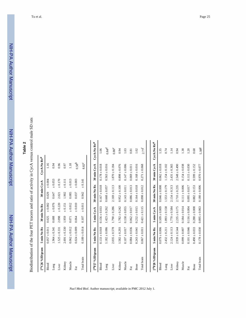
2.4.1 Biodistribution studies—The biodistribution and evaluation of regional brainuptake of the radiolabeled compounds were evaluated in male Sprague Dawley rats (250–450g). A dose of 25 mg/kg CycA (Sandimmune diluted 1:1 with saline) administered i.v. 30min prior to radiotracer injection was used for modulation of the BBB efflux transporters.Groups (n=3–4) of control or CycA treated rats were injected with radiotracers andeuthanized at 5 or 30 min post-injection. Rats were briefly anesthetized with 2–3 %isoflurane/O2 prior to injection with either CycA or radiotracers via the tail vein. [11C]6(250–300 μCi/150 μL) was administered via intravenous tail vein injection. Due to the shorthalf-life of carbon-11 and the number of samples required to evaluate regional brain uptake,no 5 min CycA group was evaluated for [11C]6. For the studies with the three 18F-labeledradiotracers, rats were injected with 20–30 μCi/150 μL. Rats were sacrificed at 5 or 30 minpost injection. Rat brains were rapidly removed, blotted to remove excess blood and thebrain stem, cerebellum, cortex, striatum and hippocampus were separated by grossdissection on a chilled glass plate. The remainder of the brain was collected to determinetotal brain uptake. Samples of clearance and non-target organs including blood, lung, liver,kidney, muscle, fat, and heart were also collected and placed in pre-weighed vials. Bone wastaken from all rats for the 18F-labeled radiotracers tracers. All samples were counted in anautomated well counter with a standard dilution of the injectate. Tissues were weighed andthe percentage of the injected dose per gram of tissue (%ID/g) was calculated. Uptake ofradiotracers in both normal and CycA pre-treated rats was evaluated in blood, lung, liver,kidney, muscle, fat, heart, brain stem, striatum, cortex, hippocampus, cerebellum and totalbrain. Bone uptake was used to estimate metabolic defluorination. The results for all fourtracers are presented as % ID/g in Table 2. The effect of efflux transporter modulation isshown in the table as the ratio of uptake at 30 min in CycA treated vs. normal rats. Theeffect of CycA treatment on regional brain uptake is presented in Fig. 3.
2.4.2. Micro PET imaging of [11C]6 in rats—Brain microPET imaging was performedusing two Siemens microPET scanners (Siemens Preclincial Solutions, Knoxville, TN); amicroPET-Focus-F220 and a microPET-Inveon MultiModality scanner. Imaging studieswere done using 220–225 g male Sprague-Dawley rats under control and CycA treatmentconditions (Fig. 4). Animals were anesthetized using 2% isoflurane/oxygen and a tail veincatheter placed in the lateral tail vein. Gas anesthesia was maintained at ~1.5% isofluraneduring the imaging session; each rat was positioned on the scanner bed at least 30 minutesprior to tracer injection. Body temperature was maintained using a warming lamp. A dose of25 mg/kg CycA (Sandimmune diluted 1:1 with saline) administered i.v. 30 min prior toradiotracer injection was used for modulation of the BBB efflux transporters. Both CycAand the radiotracer were administered using an i.v. catheter placed in the lateral tail vein.The rats were anesthetized with isoflurane and injected with ~250 μCi of [11C]6 via the tailvein. The imaging sessions were carried out as 1.5 h dynamic scan using the MicroPET®Focus 220 and Inveon scanners (Siemens Medical Solutions USA, Inc.). Acquired list modedata were histogrammed into a 3D set of sinograms and binned to the following time frames:12 x 20 sec, 8 x 1 min and 16 x 5 min. Sinogram data was then processed using filter backprojection algorithm with attenuation and scatter corrections. Using 0–90 min summarizedimages (Fig. 4A) as references, regions of interest were manually drawn on the brains of therats with the software Acquisition Sinogram Image Processing (ASIPro) (Siemens MedicalSolutions, Malvern PA) using IDL’s Virtual Machine (ITT Visual Information Solutions,Boulder CO) to obtain the radioactivity uptake (nCi/c.c.) curve over the time course of scan,then the activity was normalized by injected dose as shown in Fig. 4B.
Tu et al. Page 9
Nucl Med Biol. Author manuscript; available in PMC 2012 July 1.
NIH
-PA Author Manuscript
NIH
-PA Author Manuscript
NIH
-PA Author Manuscript

3. Results3.1. Chemistry
The synthesis of the precursors for radiolabeling is shown in Scheme 1. 4-(4-(2-Methoxyphenyl)piperazin-1-yl)butan-1-amine 12a, and its phenol counterpart 12b, weremade via N-alkylation of 10a–b with 2-(4-bromobutyl)isoindoline-1,3-dione to give thecorresponding 2-(4-(4-(2-hydroxyphenyl)-piperazin-1-yl)butyl)isoindoline-1,3-dione, 11a–bin good yield (70 – 80%). Deprotection of 11a–b with the hydrazine in ethanol gave thecorresponding amino analogue 12a–b with high yield (75 – 89%). Condensation of 12a and4-(2-hydroxyethyl)benzoic acid with DCC as coupling agent gave intermediate 13a.Treatment of 13a with methanesulfonyl chloride in dichloromethane afforded compound14a, the precursor for making [18F]7. A similar reaction sequence using 12b and either 4-(dimethylamino)benzoic acid or 4-(thiophen-3-yl)benzoic acid gave the phenol precursors13b–c in about 80% yield [42]. Compound 13b served as the precursor for synthesizing[11C]6. O-alkylation of the phenol group in 13b–c with 2-bromoethyl acetate in the presenceof potassium carbonate in acetone gave compounds 14b–c, which were converted to 15b–cby base hydrolysis using sodium hydroxide in aqueous methanol. Treatment of 15b–c withmethanesulfonyl chloride in chloromethane with triethylamine as the catalyst afforded 16b–c, which served as precursors for the synthesis of [18F]8–9.
3.2. RadiochemistryReaction of the phenol precursor 13b with [11C]MeI in DMSO or DMF containing 5Naqueous NaOH solution (Scheme 2) gave [11C]6 in 40 ~ 60% yield. There was no differencein yield when either DMSO or DMF was used as the reaction solvent. Purification of [11C]6was accomplished using semi-preparative reversed phase HPLC. The entire syntheticprocedure, including production of [11C]MeI, HPLC purification and formulation of theradiotracer for in vivo studies, was complete within 50 – 55 min. [11C]6 was obtained in aspecific activity > 10 Ci/mmol (decay corrected to E.O.B. N =15) with radiochemical andchemical purity > 99%, which is sufficient for in vivo biodistribution and imaging studies.For fluorine–18 labeled analogues [18F]7, [18F]8, and [18F]9, the fluoride displacement ofthe corresponding mesylate precursor was initially achieved using microwave irradiation.The displacement reaction required an irradiation time of only 30 – 40 s for [18F]7.However, when applied to the radiosynthesis of [18F]8 and [18F]9, microwave irradiationwas found to give inconsistent radiolabeling yields. This result may be caused by differentstabilities of precursors 16b–c versus 14a under the microwave irradiation conditions. Wesubsequently investigated a thermal displacement reaction and determined that the optimalcondition for making [18F]8 was heating at 110 °C for 10 min, and heating at 85 °C for 5min for making [18F]9. For [18F]7, purification using a three-component solvent system of10% tetrahydrofuran (THF): 12% acetonitrile: 78% 0.1 M ammonium formate bufferresulted in a clean separation of precursor and UV-positive impurities from [18F]7. A solidphase extraction method was used to separate [18F]7, [18F]8 and [18F]9 from the HPLCsolvent, a commonly used procedure which has several advantages over concentration invacuo. The SPE method is also readily adapted to automated radiosynthesis when needed.
Overall, the procedures used to synthesize [11C]6, [18F]7, [18F]8 and [18F]9 were verystraightforward and produced [11C]6, [18F]7, [18F]8 and [18F]9 in good yield, high specificactivity and high chemical and radiochemical purity for in vivo studies.
3.3 In vitro receptor binding studies and adenylyl cyclase assaysIn vitro binding studies indicate that compounds 6, 7, 8 and 9 each have a subnanomolaraffinity for dopamine D3 receptors, reduced affinity for D2 receptors, and a D3: D2selectivity ratio ranging from 23 – 163. Forskolin-dependent adenylyl cyclase inhibition
Tu et al. Page 10
Nucl Med Biol. Author manuscript; available in PMC 2012 July 1.
NIH
-PA Author Manuscript
NIH
-PA Author Manuscript
NIH
-PA Author Manuscript

assays indicate that 6 is a weak partial agonist/antagonist (21% ± 2.6%), 7 is a full agonist(92.1 ± 5.3%), 8 is a partial agonist (64.5 ± 8.3%), and 9 is a weak partial agonist (34.4 ±1.7%) at D3 dopamine receptors (Table 1) when compared to the full agonist quinpirole.
3.4 Animal Studies 3.4.1 BiodistributionBiodistribution studies were initially conducted with [11C]6 in male Sprague-Dawley rats.Regional brain uptake and tracer accumulation in clearance and non-target organs wereevaluated at 5 and 30 min post-injection. The 5 minute uptake in normal rats (0.189 %ID/gin the whole brain) and 30 min accumulation (0.107 %ID/gram) of [11C]6 in both target andnon-target brain regions was relatively low and 43 – 51% washout was observed within 30min (Table 2 and Fig. 3). The relatively low uptake of [11C]6 in whole brain and D3receptor-rich regions was unexpected since previous studies have shown 6 (i.e., WC-10) tobe pharmacologically active in behavioral studies [26,44].
P-gp has a high affinity for lipophilic molecules of moderate weight and size that containcationic centers and planar aromatic domains [51–53]. CycA, a non-specific competitiveinhibitor of P-gp, reduces P-glycoprotein-dependent drug efflux from the CNS. If uptake ofa radiotracer in the brain is limited by the action of P-gp or other ABC transporters, the ratspretreated with CycA would be expected to show increased uptake of the radiotracer in theCNS [54–60].
To confirm whether the low accumulation of the D3 selective radiotracer [11C]6 in rat brainwas due to the action of the ABC transporters, the in vivo studies were repeated in thepresence and absence of CycA. The 25 mg/kg dose of CycA used for these studies isapproximately the ED50 for blocking P-gp in rat brain [56]. As shown in Table 2 and Fig. 3,pretreatment with CycA caused a large increase in brain uptake of [11C]6. Comparing the 30min uptakes for control rats versus the CycA pretreated rats, the total brain uptake of [11C]6showed a CycA effect: the uptake (%ID/g) of [11C]6 increased 8.82 fold from 0.107 ± 0.001to 0.942 ± 0.141. When compared to the CycA effect on the uptake of [11C]verapamil [49]in rats, our data reveal that CycA pre-treated rats displayed a comparable response in thedelivery of [11C]6, although the brain-to-blood ratio at a single time point is not adequate todescribe the ligand delivery as reported [55]. In the peripheral organs of lung, liver, kidney,muscle and heart, no significant difference in uptake of [11C]6 between the CycA and thecontrol group was observed 30 min post injection. The results of treatment with CycA on theuptake of [11C]6 in brain, the brain-to-blood ratios (Table 2) and uptake in regional brainareas such as striatum, cerebellum, cortex and hippocampus (Fig. 3) is very comparable withthe effect observed for [11C]verapramil [49] and [11C]GR218231 [56], and higher than thatobserved in studies using [11C]TMSX, [11C]MPDX, [11C]flumazenil, [11C]donepezil,[11C]carazolol [49] [18F]fluorocarazolol [50,57] and [18F]MPPF [50,54].
To further investigate the behavior of this class of compounds, the three 18F-labeledradiotracers [18F]7, [18F]8 and [18F]9 were also evaluated in vivo in the presence andabsence of CycA pretreatment and similar results were observed. Because the longer half-life of F-18 allows for more flexibility in study design, the regional brain uptake of thetracers 5 minutes post injection under ABC transporter inhibition/modulation was alsoinvestigated. Whole brain activity levels in normal rats for the three 18F-labeled radiotracers[18F]7, [18F]8 and [18F]9 were also <0.2 %ID/gram 5 min post injection, with very littleadditional washout observed by 30 min post-injection and no evidence of retention in the D3receptor target region of striatum. As can be seen in Table 2, the biodistribution of the fourtracers in normal rats showed rapid clearance from the blood and little accumulation in fat.Initial lung levels of [18F]9 were somewhat higher than the other radiotracers and [18F]8showed a two-fold increase from 5 min to 30 min in bone activity levels that may representmetabolic defluorination. No accumulation was seen in bone for [18F]7 or [18F]9. The
Tu et al. Page 11
Nucl Med Biol. Author manuscript; available in PMC 2012 July 1.
NIH
-PA Author Manuscript
NIH
-PA Author Manuscript
NIH
-PA Author Manuscript
accumulation of all three 18F-labeled radiotracers in the brain increased under ABCtransporter inhibition. That is, all four tracers showed increased retention in the brain 30 minpost injection under conditions of CycA pretreatment (Table 2 and Fig. 3), though minimaleffects were seen in other tissues or organs (Table 2). The reported P values were calculatedusing a two-sample equal variance, two-tailed Students T-test.
3.4.2. Micro PET scans of [11C]6 in rats—MicroPET imaging studies of rat brainuptake under control conditions and following treatment with CycA were also conducted tofurther study the effect of ABC transporter-mediated efflux on the uptake and washoutkinetics of [11C]6 from the rat brain. The two scanners used for this study have comparableresolution: the microPET-Focus-F220 (used for the control rat) has a spatial resolution of1.5 mm and 3.4 % sensitivity at the center of the field of view and the microPET-InveonMultiModality scanner (used for the CycA rat) has 1.5 mm spatial resolution and asensitivity of 10% at the center of the field of view. The radiotracer injections wereperformed within 5 minutes of each other from a single radiosynthesis to eliminate anyvariability due to specific activity. Because of the low density of the D3 receptor, only 36fmol/mg protein in the rat striatum [7], and low tracer uptake in control rats for thebiodistribution study, a region of interest for each rat was drawn over the entire brain. Ascan be seen from the summed images over the 90 min acquisition period shown in Fig. 4A,no tracer accumulation is seen in the brain of the control rat. However, there is an increase inthe accumulation of [11C]6 in the CycA treated rat. The normalized time activity curve inFig. 4A demonstrates comparable initial uptake in the two rats, with a very early peak andrapid washout in the control animal. The CycA treated rat demonstrates slower washoutkinetics with the radioactive peak evident ~ 5 min post injection and an increased retentionof the tracer over the duration of the scan.
4. DiscussionIn recent years, a number of potent and selective dopamine D3 ligands labeled with eithercarbon-11 or fluorine-18 have been reported and evaluated in animal models to determine ifthey possess the pharmacological properties needed to function as an in vivo imaging agent(Fig. 1). Unfortunately, all D3-selective radiotracers evaluated to date have yieldeddisappointing results with respect to their ability to cross the blood-brain barrier andselectively label D3 receptors in vivo. This observation is somewhat perplexing since someof the radiotracers shown in Fig. 1 possess a log P value well within the range needed toenable crossing the blood-brain barrier, with D3 receptor affinity and selectivity whichshould lead to the labeling of D3 receptors in vivo.
Over the past 5 years, our group has synthesized a number of conformationally-flexiblebenzamide analogs based on lead compound 5 and measured their affinity for dopamine D2,D3, and D4 receptors, and their intrinsic activity at D2 and D3 receptors [40,41]. This effortled to the identification of four potential radiotracers for imaging the D3 receptor (Fig. 2)which could be radiolabeled with either carbon-11 or fluorine-18 using conventionalradiolabeling procedures. Initial brain uptake studies of [11C]6, and [18F]7–9 conducted inSprague-Dawley rats revealed low brain uptake 5 min post- injection. This was unexpectedsince the calculated log P values of these analogs (Table 1) indicate that they should be ableto cross the blood-brain and label D3 receptors in vivo. In addition, compounds 6 (WC-10)and 8 (WC-44) were found to be behaviorally active in rat models of L-DOPA-induceddyskinesia [26] and prepulse inhibition [44] in a dose range of 1.0 to 10 mg/kg, whichinitially seemed inconsistent with our initial rodent brain uptake studies using [11C]6 and[18F]8. This unanticipated result suggested that some other mechanism may be operationalat the tracer level which led to a low brain uptake. One possible explanation was theinvolvement of ABC transporter proteins. This hypothesis was suggested by previous
Tu et al. Page 12
Nucl Med Biol. Author manuscript; available in PMC 2012 July 1.
NIH
-PA Author Manuscript
NIH
-PA Author Manuscript
NIH
-PA Author Manuscript
studies demonstrating that the serotonin 5-HT1A PET ligands, [11C](R)-(-)RWAY,[18F]MPPF and [carbonyl-11C]WAY100635, which also contain an N-2-methoxyphenylpiperazine group, are substrates for P-glycoprotein in rats [49,54,59,60]. Although ABCproteins are expressed in many species, some studies have shown differences in ABCtransporter-mediated efflux of PET radiotracers in rodents versus nonhuman primates andhumans [58]. For example, [11C](R)-(-)RWAY is a substrate for P-gp in rodent brain but nota substrate for P-gp in nonhuman primate [61] and human brain [62].
P-gp is a 170 kDa protein with low substrate specificity and widespread tissue distributionbelonging to the ABC transporters which are involved in multidrug resistance [45,53]. P-gpis expressed in the capillary endothelial cells which comprise the blood-brain barrier, andprotect the brain from the accumulation of toxic substances. In order to determine if theradiolabeled benzamides described in this report are substrates for P-gp, the brain uptakestudies in Sprague-Dawley rats were repeated following a 30 min pretreatment with the P-gpinhibitor, cyclosporine A (CycA). In this case, the administration of CycA resulted in a 8.8fold increase in brain uptake of [11C]6 at 30 min post injection. Similarly, CycAadministration resulted in a 6.3-fold, 5.0-fold 6.1-fold increase in brain uptake of [18F]7–9 at5 min, and a 2.7-fold, 5.4-fold and 5.3-fold increase at 30 min for the three 18F-labeledtracers respectively. Furthermore, microPET imaging studies of [11C]6 under control andCycA-treated conditions clearly demonstrate a dramatic increase in brain uptake. This effecton PET imaging agents is often described as P-gp modulation, however, it must be notedthat CycA is also an inhibitor of MDR1 and MDR2. Therefore, our data are consistent withthe hypothesis that [11C]6 and the three 18F-labeled radiotracers [18F]7, [18F]8 and [18F]9are probable substrates of one or more ABC transporter proteins in rat brain since the uptakeof these radiotracers was increased by pre-treatment with CycA.
While this work was being conducted, Mason and colleagues reported that PG 01037, an N-(2,3-dichlorophenyl)piperazine analog structurally similar to compound 5, is a substrate forP-gp expressed in Madin-Darby canine kidney (MDCK)-MDR1 cells, and that efflux wasblocked with verapamil, a known P-gp inhibitor [63]. These data are consistent with thetracer experiments of [11C]6 and [18F]7–9, and may explain why relatively high doses ofcompounds 6 (WC-10), 8 (WC-44) and other structurally-related benzamide analogs areneeded in rodent behavioral assays, despite the fact that these compounds have high (i.e.,nanomolar to subnanomolar) affinity for D3 receptors in vitro. While the low doseadministration of radiolabeled imaging agents [11C]6 and [18F]7–9 likely enabled efficientremoval from the CNS by P-gp, MDR1, and/or MDR2, these transporter proteins werelikely to have been saturated at the higher doses administered to rats in the behavioralpharmacology studies.
In summary, the data described in this paper show that the radiolabeled benzaminde analogs[11C]6 and [18F]7–9 are capable of crossing the rat blood brain barrier provided that CycA isadministered to prevent efflux by ABC transporter proteins. Our data plus the earlier studieswith [11C](R)-(-)RWAY, [18F]MPPF and [carbonyl-11C]WAY100635, which contain eitherthe N-2-methoxyphenylpiperazine or N-2-(2-fluoroethoxy)phenylpiperazine pharmacophore,indicate that these compounds are substrates for one or more ABC transporter proteins inrodent brain. This mechanism may also be responsible for the low brain uptake reported forthe D3 receptor PET radiotracers shown in Figure 1 since most contain the N-phenylpiperazine pharmacophore; these tracers were originally thought to not cross theblood-brain barrier because of the relatively high log P values. MicroPET imaging studies innonhuman primates are currently ongoing to determine if the [11C]6 and [18F] 7–9 arecapable of serving as radiotracers for imaging D3 receptors in vivo with PET in a nonrodentspecies.
Tu et al. Page 13
Nucl Med Biol. Author manuscript; available in PMC 2012 July 1.
NIH
-PA Author Manuscript
NIH
-PA Author Manuscript
NIH
-PA Author Manuscript
AcknowledgmentsThis research was supported by the NIH grant: DA 16181, DA29840, and NS48056. We would like to thank theSmall Animal Imaging Facility for assistance with the microPET imaging study. The microPET-InveonMultiModality scanner was acquired under an NIH-NCRR HEI grant (S10-025097 PI. Richard Laforest).
References1. Sibley DR, Monsma FJ Jr. Molecular biology of dopamine receptors. Trends Pharmacol Sci. 1992;
13:61–9. [PubMed: 1561715]2. Luedtke RR, Mach RH. Progress in developing D3 dopamine receptor ligands as potential
therapeutic agents for neurological and neuropsychiatric disorders. Curr Pharm Des. 2003; 9:643–71. [PubMed: 12570797]
3. Missale C, Nash SR, Robinson SW, Jaber M, Caron MG. Dopamine receptors: from structure tofunction. Physiol Rev. 1998; 78:189–225. [PubMed: 9457173]
4. Reavill C, Taylor SG, Wood MD, Ashmeade T, Austin NE, Avenell KY, et al. Pharmacologicalactions of a novel, high-affinity, and selective human dopamine D3 receptor antagonist, SB-277011-A. J Pharmacol Exp Ther. 2000; 294:1154–65. [PubMed: 10945872]
5. Joyce JN, Gurevich EV. D3 Receptors and the actions of neuroleptics in the ventral striatopallidalsystem of schizophrenics. Annals of the New York Academy of Sciences. 1999; 877:595–613.[PubMed: 10415673]
6. Stanwood GD, Artymyshyn RP, Kung MP, Kung HF, Lucki I, McGonigle P. Quantitativeautoradiographic mapping of rat brain dopamine D3 binding with [125I]7-OH-PIPAT: evidence forthe presence of D3 receptors on dopaminergic and nondopaminergic cell bodies and terminals. JPharmacol Exp Ther. 2000; 295:1223–31. [PubMed: 11082459]
7. Bancroft GN, Morgan KA, Flietstra RJ, Levant B. Binding of [3H]PD 128907, a putatively selectiveligand for the D3 dopamine receptor, in rat brain: a receptor binding and quantitativeautoradiographic study. Neuropsychopharmacology. 1998; 18:305–16. [PubMed: 9509498]
8. Murray AM, Ryoo HL, Gurevich E, Joyce JN. Localization of dopamine D3 receptors tomesolimbic and D2 receptors to mesostriatal regions of human forebrain. Proc Natl Acad Sci U S A.1994; 91:11271–5. [PubMed: 7972046]
9. Levant B. Differential distribution of D3 dopamine receptors in the brains of several mammalianspecies. Brain Res. 1998; 800:269–74. [PubMed: 9685676]
10. Joyce JN. Dopamine D3 receptor as a therapeutic target for antipsychotic and antiparkinsoniandrugs. Pharmacol Ther. 2001; 90:231–59. [PubMed: 11578658]
11. Griffon N, Pilon C, Sautel F, Schwartz JC, Sokoloff P. Two intracellular signaling pathways forthe dopamine D3 receptor: opposite and synergistic interactions with cyclic AMP. J Neurochem.1997; 68:1–9. [PubMed: 8978703]
12. Guigoni C, Aubert I, Li Q, Gurevich VV, Benovic JL, Ferry S, et al. Pathogenesis of levodopa-induced dyskinesia: focus on D1 and D3 dopamine receptors. Parkinsonism Relat Disord. 2005; 11(Suppl 1):S25–9. [PubMed: 15885624]
13. Schwartz JC, Levesque D, Martres MP, Sokoloff P. Dopamine D3 receptor: basic and clinicalaspects. Clin Neuropharmacol. 1993; 16:295–314. [PubMed: 8104095]
14. Sokoloff P, Giros B, Martres MP, Bouthenet ML, Schwartz JC. Molecular cloning andcharacterization of a novel dopamine receptor (D3) as a target for neuroleptics. Nature. 1990;347:146–51. [PubMed: 1975644]
15. Suzuki M, Hurd YL, Sokoloff P, Schwartz JC, Sedvall G. D3 dopamine receptor mRNA is widelyexpressed in the human brain. Brain Res. 1998; 779:58–74. [PubMed: 9473588]
16. Hackling AE, Stark H. Dopamine D3 receptor ligands with antagonist properties. Chembiochem.2002; 3:946–61. [PubMed: 12362359]
17. Millan MJ, Dekeyne A, Rivet JM, Dubuffet T, Lavielle G, Brocco M. S33084, a novel, potent,selective, and competitive antagonist at dopamine D3-receptors: II. Functional and behavioralprofile compared with GR218,231 and L741,626. J Pharmacol Exp Ther. 2000; 293:1063–73.[PubMed: 10869411]
Tu et al. Page 14
Nucl Med Biol. Author manuscript; available in PMC 2012 July 1.
NIH
-PA Author Manuscript
NIH
-PA Author Manuscript
NIH
-PA Author Manuscript
18. Heidbreder CA, Gardner EL, Xi Z-X, Thanos PK, Mugnaini M, Hagan JJ, et al. The role of centraldopamine D3 receptors in drug addiction: a review of pharmacological evidence. Brain ResearchReviews. 2005; 49:77–105. [PubMed: 15960988]
19. Higley AE, Spiller K, Grundt P, Newman AH, Kiefer SW, Xi Z-Z, et al. PG01037, a noveldopamine D3 receptor antagonist, inhibits the effects of methamphetamine in rats. Journal ofPsychopharmacology. published online Feb 8, 2010 before print. 10.1177/0269881109358201
20. Sokoloff P, Le Foll B, Perachon S, Bordet R, Ridray S, Schwartz JC. The dopamine D3 receptorand drug addiction. Neurotox Res. 2001; 3:433–41. [PubMed: 14715457]
21. Newman AH, Grundt P, Nader MA. Dopamine D3 receptor partial agonists and antagonists aspotential drug abuse therapeutic agents. J Med Chem. 2005; 48:3663–79. [PubMed: 15916415]
22. Bordet R, Ridray S, Carboni S, Diaz J, Sokoloff P, Schwartz JC. Induction of dopamine D3receptor expression as a mechanism of behavioral sensitization to levodopa. Proc Natl Acad Sci US A. 1997; 94:3363–7. [PubMed: 9096399]
23. Bordet R, Ridray S, Schwartz JC, Sokoloff P. Involvement of the direct striatonigral pathway inlevodopa-induced sensitization in 6-hydroxydopamine-lesioned rats. Eur J Neurosci. 2000;12:2117–23. [PubMed: 10886351]
24. Visanji NP, Fox SH, Johnston T, Reyes G, Millan MJ, Brotchie JM. Dopamine D3 receptorstimulation underlies the development of L-DOPA-induced dyskinesia in animal models ofParkinson's disease. Neurobiology of Disease. 2009; 35:184–92. [PubMed: 19118628]
25. Kumar R, Riddle L, Griffin SA, Grundt P, Newman AH, Luedtke RR. Evaluation of the D3dopamine receptor selective antagonist PG01037 on L-dopa-dependent abnormal involuntarymovements in rats. Neuropharmacology. 2009; 56:944–55. [PubMed: 19371585]
26. Kumar R, Riddle LR, Griffin SA, Chu W, Vangveravong S, Neisewander J, et al. Evaluation of D2and D3 dopamine receptor selective compounds on L-dopa-dependent abnormal involuntarymovements in rats. Neuropharmacology. 2009; 56:956–69. [PubMed: 19371586]
27. Van Kampen JM, Eckman CB. Dopamine D3 receptor agonist delivery to a model of Parkinson'sdisease restores the nigrostriatal pathway and improves locomotor behavior. J Neurosci. 2006;26:7272–80. [PubMed: 16822985]
28. Ghosh B, Antonio T, Zhen J, Kharkar P, Reith MEA, Dutta AK. Development of (S)-N6-(2-(4-(isoquinolin-1-yl)piperazin-1-yl) ethyl)-N6-propyl-4,5,6,7-tetrahydrobenzo[d]-thiazole-2,6-diamine and its analogue as a D3 receptor preferring agonist: Potent in vivo activity in Parkinson'sdisease animal models. J. Med. Chem. 2010; 53:1023–37.
29. Pilla M, Perachon S, Sautel F, Garrido F, Mann A, Wermuth CG, et al. Selective inhibition ofcocaine-seeking behaviour by a partial dopamine D3 receptor agonist. Nature. 1999; 400:371–5.[PubMed: 10432116]
30. Khaled MATM, Farid Araki K, Li B, Coen KM, Marinelli PW, Varga J, et al. The selectivedopamine D3 receptor antagonist SB 277011-A, but not the partial agonist BP 897, blocks cue-induced reinstatement of nicotine-seeking. Int J Neuropsychopharmacol. 2009:1–10.
31. Garcia-Ladona FJ, Cox BF. BP 897, a selective dopamine D3 receptor ligand with therapeuticpotential for the treatment of cocaine-addiction. CNS Drug Rev. 2003; 9:141–58. [PubMed:12847556]
32. Carr KD, Yamamoto N, Omura M, Cabeza de Vaca S, Krahne L. Effects of the D3 dopaminereceptor antagonist, U99194A, on brain stimulation and d-amphetamine reward, motor activity,and c-fos expression in ad libitum fed and food-restricted rats. Psychopharmacology (Berl). 2002;163:76–84. [PubMed: 12185403]
33. Witkin JM, Levant B, Zapata A, Kaminski R, Gasior M. The dopamine D3/D2 agonist (+)-PD-128,907 [(R-(+)-trans-3,4a,10b-tetrahydro-4-propyl-2H,5H-[1]benzopyrano[4,3-b]-1,4-oxazin-9-ol)] protects against acute and cocaine-kindled seizures in mice: Further evidence for theinvolvement of D3 receptors. Journal of Pharmacology and Experimental Therapeutics. 2008;326:930–8. [PubMed: 18566292]
34. Hocke C, Prante O, Salama I, Hubner H, Lober S, Kuwert T, et al. 18F-Labeled FAUC 346 and BP897 derivatives as subtype-selective potential PET radioligands for the dopamine D3 receptor.ChemMedChem. 2008; 3:788–93. [PubMed: 18306190]
Tu et al. Page 15
Nucl Med Biol. Author manuscript; available in PMC 2012 July 1.
NIH
-PA Author Manuscript
NIH
-PA Author Manuscript
NIH
-PA Author Manuscript

35. Sovago J, Farde L, Halldin C, Langer O, Laszlovszky I, Kiss B, et al. Positron emissiontomographic evaluation of the putative dopamine-D3 receptor ligand, [11C]RGH-1756 in themonkey brain. Neurochem Int. 2004; 45:609–17. [PubMed: 15234102]
36. Kuhnast B, Valette H, Besret L, Demphel S, Coulon C, Ottaviani M, et al. Synthesis andradiolabeling of N-[4-[4-(2-[11C]methoxyphenyl)piperazin-1-yl]butyl]benzo[b]thiophene-2-carboxamide - a potential radiotracer for D3 receptor imaging with PET. Nucl Med Biol. 2006;33:785–95. [PubMed: 16934697]
37. Bennacef I, Salinas CA, Bonasera TA, Gunn RN, Audrain H, Jakobsen S, et al. Dopamine D3receptor antagonists: the quest for a potentially selective PET ligand. Part 3: Radiosynthesis and invivo studies. Bioorg Med Chem Lett. 2009; 19:5056–9. [PubMed: 19635669]
38. Turolla EA, Matarrese M, Belloli S, Moresco RM, Simonelli P, Todde S, et al. 11C-labeling of N-[4-[4-(2,3-dichlorophenyl)piperazin-1-yl]butyl]arylcarboxamide derivatives and evaluation aspotential radioligands for PET imaging of dopamine D3 receptors. J Med Chem. 2005; 48:7018–23. [PubMed: 16250661]
39. Mach RH, Huang Y, Freeman RA, Wu L, Vangveravong S, Luedtke RR. Conformationally-flexible benzamide analogues as dopamine D3 and sigma 2 receptor ligands. Bioorg Med ChemLett. 2004; 14:195–202. [PubMed: 14684327]
40. Chu W, Tu Z, McElveen E, Xu J, Taylor M, Luedtke RR, et al. Synthesis and in vitro binding ofN-phenyl piperazine analogs as potential dopamine D3 receptor ligands. Bioorg Med Chem. 2005;13:77–87. [PubMed: 15582454]
41. Tu Z, Li S, Cui J, Xu J, Taylor M, Ho D, et al. Synthesis and pharmacological evaluation offluorine containing D3 dopamine receptor selective analogues. J Med Chem. under review.
42. Xu J, Chu W, Tu Z, Jones LA, Luedtke RR, Perlmutter JS, et al. [3H]4-(Dimethylamino)-N-[4-(4-(2-methoxyphenyl)piperazin- 1-yl)butyl]benzamide, a selective radioligand for dopamine D3receptors. I. In vitro characterization. Synapse. 2009; 63:717–28. [PubMed: 19425052]
43. Xu J, Hassanzadeh B, Chu W, Tu Z, Jones LA, Luedtke RR, et al. [3H]4-(dimethylamino)-N-(4-(4-(2-methoxyphenyl)piperazin-1-yl) butyl)benzamide: a selective radioligand for dopamine D3receptors. II. Quantitative analysis of dopamine D3 and D2 receptor density ratio in the caudate-putamen. Synapse. 2010; 64:449–59. [PubMed: 20175227]
44. Weber M, Chang WL, Durbin JP, Park PE, Luedtke RR, Mach RH, et al. Using prepulse inhibitionto detect functional D3 receptor antagonism: effects of WC10 and WC44. Pharmacol BiochemBehav. 2009; 93:141–7. [PubMed: 19426754]
45. Loscher W, Potschka H. Blood-brain barrier active efflux transporters: ATP-binding cassette genefamily. NeuroRx. 2005; 2:86–98. [PubMed: 15717060]
46. Luedtke RR, Freeman RA, Boundy VA, Martin MW, Huang Y, Mach RH. Characterization of125I-IABN, a novel azabicyclononane benzamide selective for D2-like dopamine receptors.Synapse. 2000; 38:438–49. [PubMed: 11044891]
47. Cheng Y, Prusoff WH. Relationship between the inhibition constant (K1) and the concentration ofinhibitor which causes 50 per cent inhibition (I50) of an enzymatic reaction. Biochem Pharmacol.1973; 22:3099–108. [PubMed: 4202581]
48. Shimizu H, Daly JW, Creveling CR. A radioisotopic method for measuring the formation ofadenosine 3',5'-cyclic monophosphate in incubated slices of brain. J Neurochem. 1969; 16:1609–19. [PubMed: 4314281]
49. Ishiwata K, Kawamura K, Yanai K, Hendrikse NH. In vivo evaluation of P-glycoproteinmodulation of 8 PET radioligands used clinically. J Nucl Med. 2007; 48:81–7. [PubMed:17204702]
50. Elsinga PH, Hendrikse NH, Bart J, van Waarde A, Vaalburg W. Positron emission tomographystudies on binding of central nervous system drugs and P-glycoprotein function in the rodent brain.Mol Imaging Biol. 2005; 7:37–44. [PubMed: 15912274]
51. van Asperen J, Mayer U, van Tellingen O, Beijnen JH. The functional role of P-glycoprotein in theblood-brain barrier. J Pharm Sci. 1997; 86:881–4. [PubMed: 9269863]
52. Abbott NJ, Romero IA. Transporting therapeutics across the blood-brain barrier. Mol Med Today.1996; 2:106–13. [PubMed: 8796867]
Tu et al. Page 16
Nucl Med Biol. Author manuscript; available in PMC 2012 July 1.
NIH
-PA Author Manuscript
NIH
-PA Author Manuscript
NIH
-PA Author Manuscript

53. Pearce HL, Winter MA, Beck WT. Structural characteristics of compounds that modulate P-glycoprotein-associated multidrug resistance. Adv Enzyme Regul. 1990; 30:357–73. [PubMed:1976291]
54. Passchier J, van Waarde A, Doze P, Elsinga PH, Vaalburg W. Influence of P-glycoprotein on brainuptake of [18F]MPPF in rats. Eur J Pharmacol. 2000; 407:273–80. [PubMed: 11068023]
55. Hendrikse NH, de Vries EG, Franssen EJ, Vaalburg W, van der Graaf WT. In vivo measurement of[11C]verapamil kinetics in human tissues. Eur J Clin Pharmacol. 2001; 56:827–9. [PubMed:11294373]
56. de Vries EF, Kortekaas R, van Waarde A, Dijkstra D, Elsinga PH, Vaalburg W. Synthesis andevaluation of dopamine D3 receptor antagonist 11C-GR218231 as PET tracer for P-glycoprotein. JNucl Med. 2005; 46:1384–92. [PubMed: 16085598]
57. Doze P, Van Waarde A, Elsinga PH, Hendrikse NH, Vaalburg W. Enhanced cerebral uptake ofreceptor ligands by modulation of P-glycoprotein function in the blood-brain barrier. Synapse.2000; 36:66–74. [PubMed: 10700027]
58. Syvänen S, Lindhe Ö, Palner M, Kornum BR, Rahman O, Långström B, et al. Species Differencesin Blood-Brain Barrier Transport of Three Positron Emission Tomography Radioligands withEmphasis on P-Glycoprotein Transport. Drug Metabolism and Disposition. 2009; 37:635–43.[PubMed: 19047468]
59. Liow JS, Lu S, McCarron JA, Hong J, Musachio JL, Pike VW, et al. Effect of a P-glycoproteininhibitor, Cyclosporin A, on the disposition in rodent brain and blood of the 5-HT1A receptorradioligand, [11C](R)-(-)-RWAY. Synapse. 2007; 61:96–105. [PubMed: 17117422]
60. Lacan G, Plenevaux A, Rubins DJ, Way BM, Defraiteur C, Lemaire C, et al. Cyclosporine, a P-glycoprotein modulator, increases [18F]MPPF uptake in rat brain and peripheral tissues: microPETand ex vivo studies. Eur J Nucl Med Mol Imaging. 2008; 35:2256–66. [PubMed: 18604533]
61. Yasuno F, Zoghbi SS, McCarron JA, Hong J, Ichise M, Brown AK, et al. Quantification ofserotonin 5-HT1A receptors in monkey brain with [11C](R)-(-)-RWAY. Synapse. 2006; 60:510–20. [PubMed: 16952161]
62. Zhang XY, Yasuno F, Zoghbi SS, Liow JS, Hong J, McCarron JA, et al. Quantification ofserotonin 5-HT1A receptors in humans with [11C](R)-(-)-RWAY: radiometabolite(s) likelyconfound brain measurements. Synapse. 2007; 61:469–77. [PubMed: 17415792]
63. Mason CW, Hassan HE, Kim KP, Cao J, Eddington ND, Newman AH, et al. Characterization ofthe transport, metabolism, and pharmacokinetics of the dopamine D3 receptor-selective fluorenyl-and 2-pyridylphenyl amides developed for treatment of psychostimulant abuse. J Pharmacol ExpTher. 2010; 333:854–64. [PubMed: 20228156]
Tu et al. Page 17
Nucl Med Biol. Author manuscript; available in PMC 2012 July 1.
NIH
-PA Author Manuscript
NIH
-PA Author Manuscript
NIH
-PA Author Manuscript

Fig. 1.Structures of dopamine D3 receptor PET imaging ligands reported in the literature.
Tu et al. Page 18
Nucl Med Biol. Author manuscript; available in PMC 2012 July 1.
NIH
-PA Author Manuscript
NIH
-PA Author Manuscript
NIH
-PA Author Manuscript

Fig. 2.Structures of lead compound 5 and the N-phenylpiperazine analogs described in this paper.
Tu et al. Page 19
Nucl Med Biol. Author manuscript; available in PMC 2012 July 1.
NIH
-PA Author Manuscript
NIH
-PA Author Manuscript
NIH
-PA Author Manuscript

Fig. 3.Regional rat brain uptake of the four PET tracers in the absence and presence of CycAmodulation of the BBB efflux transporters.
Tu et al. Page 20
Nucl Med Biol. Author manuscript; available in PMC 2012 July 1.
NIH
-PA Author Manuscript
NIH
-PA Author Manuscript
NIH
-PA Author Manuscript

Fig. 4.MicroPET imaging studies of [11C]6 in the absence and presence of CycA modulation of theBBB efflux transporters.
Tu et al. Page 21
Nucl Med Biol. Author manuscript; available in PMC 2012 July 1.
NIH
-PA Author Manuscript
NIH
-PA Author Manuscript
NIH
-PA Author Manuscript

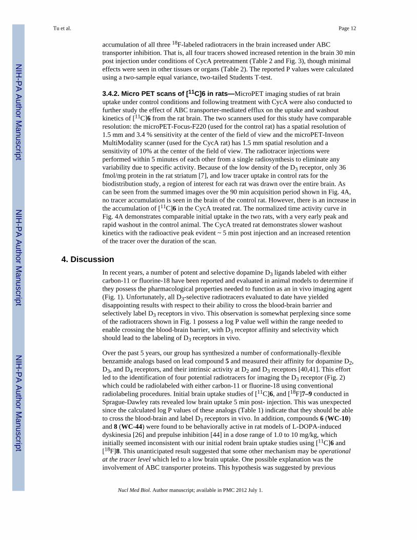
Scheme 1. Reagents(a) N-(4-Bromobutyl)phthalimide, triethylamine, CH2Cl2; (b) hydrazine, ethanol; (c)ArCOOH, DCC/EDCI, HOBt; (d) 2-Bromoethyl acetate, K2CO3, acetone; (e) NaOH,CH3OH/H2O; (f) Methanesulfonyl chloride, triethylamine, CH2Cl2.
Tu et al. Page 22
Nucl Med Biol. Author manuscript; available in PMC 2012 July 1.
NIH
-PA Author Manuscript
NIH
-PA Author Manuscript
NIH
-PA Author Manuscript
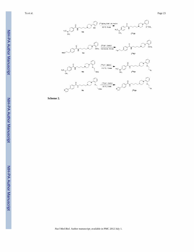
Scheme 2.
Tu et al. Page 23
Nucl Med Biol. Author manuscript; available in PMC 2012 July 1.
NIH
-PA Author Manuscript
NIH
-PA Author Manuscript
NIH
-PA Author Manuscript

NIH
-PA Author Manuscript
NIH
-PA Author Manuscript
NIH
-PA Author Manuscript
Tu et al. Page 24
Tabl
e 1
In v
itro
Bin
ding
Dat
a
Com
poun
dD
2D
3D
4D
2:D
3L
og P
%IA
D2
%IA
D3
6 (W
C-1
0)34
.4 ±
3.3
0.8
± 0.
189
6 ±
193
433.
0934
± 3
19 ±
2
7 (W
C-4
4)54
.7 ±
4.4
2.4
± 0.
480
4 ±
3323
2.94
35 ±
196
± 4
815
.1 ±
1.7
0.65
± 0
.288
6 ±
101
233.
7566
.3 ±
164
.5 ±
8
927
.7 ±
5.4
0.17
± 0
.01
246
± 13
.316
34.
6729
.3 ±
7.3
34.4
± 1
.7
Nucl Med Biol. Author manuscript; available in PMC 2012 July 1.
NIH
-PA Author Manuscript
NIH
-PA Author Manuscript
NIH
-PA Author Manuscript
Tu et al. Page 25
Tabl
e 2
Bio
dist
ribut
ion
of th
e fo
ur P
ET tr
acer
s and
ratio
of a
ctiv
ity in
Cyc
A v
ersu
s con
trol m
ale
SD ra
ts
[11C
]6 %
ID/g
ram
5 m
in N
o R
x30
min
No
Rx
30 m
in C
ycA
Cyc
A:N
o R
xa
Blo
od0.
057
± 0.
011
0.02
5±
0.00
20.
029
± 0.
004
1.16
Lung
1.96
4 ±
0.34
10.
688
± 0.
076
0.64
7±
0.05
30.
94
Live
r1.
525
± 0.
331
2.09
8±
0.22
02.
021
± 0.
170
0.96
Kid
ney
2.69
1 ±
0.33
01.
959
± 0.
151
1.90
2±
0.11
10.
97
Mus
cle
0.06
2 ±
0.01
20.
071
± 0.
012
0.08
5±
0.01
31.
18
Fat
0.02
4 ±
0.00
90.
064
± 0.
010
0.03
7±
0.00
30.
58b
Tota
l bra
in0.
189
± 0.
014
0.10
7±
0.01
10.
942
± 0.
141
8.82
c
[18F]
7 %
ID/g
ram
5 m
in N
o R
x5
min
Cyc
A30
min
No
Rx
30 m
in C
ycA
Cyc
A:N
o R
xa
Blo
od0.
133
± 0.
018
0.15
6 ±
0.03
30.
167
± 0.
018
0.17
8 ±
0.01
81.
06
Lung
1.18
2 ±
0.08
61.
423
± 0.
262
0.66
8 ±
0.05
70.
563
± 0.
016
0.84
d
Live
r2.
035
± 0.
578
1.79
0 ±
0.28
62.
169
± 0.
113
1.87
0 ±
0.18
40.
86e
Kid
ney
1.58
2 ±
0.29
31.
782
± 0.
242
0.95
2 ±
0.10
00.
898
± 0.
076
0.94
Mus
cle
0.10
7 ±
0.01
70.
158
± 0.
055
0.14
3 ±
0.01
10.
147
± 0.
006
1.03
Fat
0.08
4 ±
0.03
60.
062
± 0.
017
0.08
6 ±
0.01
30.
069
± 0.
011
0.81
Bon
e0.
243
± 0.
041
0.23
2 ±
0.01
50.
164
± 0.
018
0.16
8 ±
0.01
61.
02
Tota
l bra
in0.
067
± 0.
011
0.42
1 ±
0.11
50.
098
± 0.
012
0.27
1 ±
0.06
82.
77f
[18F]
8 %
ID/g
ram
5 m
in N
o R
x5
min
Cyc
A30
min
No
Rx
30 m
in C
ycA
Cyc
A:N
o R
xa
Blo
od0.
079
± 0.
006
0.10
9 ±
0.00
90.
046
± 0.
008
0.06
2 ±
0.01
81.
35
Lung
2.43
2 ±
0.21
11.
893
± 0.
158
1.55
3 ±
0.17
91.
154
± 0.
102
0.74
Live
r2.
124
± 0.
113
1.77
0 ±
0.58
42.
134
± 0.
313
2.43
1 ±
0.36
51.
14
Kid
ney
2.93
9 ±
0.54
42.
235
± 0.
175
2.71
0 ±
0.23
52.
548
± 0.
490
0.94
Mus
cle
0.09
9 ±
0.00
70.
184
± 0.
014
0.11
1 ±
0.00
60.
154
± 0.
038
1.38
Fat
0.10
1 ±
0.04
60.
116
± 0.
004
0.09
5 ±
0.01
70.
115
± 0.
030
1.20
Bon
e0.
490
± 0.
033
0.28
8 ±
0.00
80.
882
± 0.
153
0.59
9 ±
0.15
20.
68
Tota
l bra
in0.
178
± 0.
030
0.89
5 ±
0.06
30.
180
± 0.
006
0.97
0 ±
0.07
75.
38g
Nucl Med Biol. Author manuscript; available in PMC 2012 July 1.
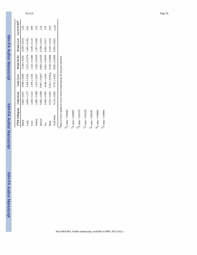
NIH
-PA Author Manuscript
NIH
-PA Author Manuscript
NIH
-PA Author Manuscript
Tu et al. Page 26
[18F]
9 %
ID/g
ram
5 m
in N
o R
x5
min
Cyc
A30
min
No
Rx
30 m
in C
ycA
Cyc
A:N
o R
xa
Blo
od0.
082
± 0.
027
0.09
8 ±
0.00
90.
104
± 0.
010
0.10
9 ±
0.01
91.
05
Lung
4.02
6 ±
1.17
73.
356
± 0.
261
1.31
3 ±
0.35
741.
063
± 0.
182
0.81
Live
r1.
430
± 0.
221
1.47
8 ±
0.33
61.
162
± 0.
1400
1.03
9 ±
0.12
40.
89
Kid
ney
2.48
5 ±
0.60
81.
913
± 0.
037
1.36
3 ±
0.03
591.
397
± 0.
260
1.03
Mus
cle
0.08
3 ±
0.00
90.
097
± 0.
0417
0.08
3 ±
0.01
390.
108
± 0.
014
1.24
Fat
0.02
8 ±
0.00
30.
046
± 0.
028
0.09
1 ±
0.00
290.
093
± 0.
017
1.02
Bon
e0.
253
± 0.
001
0.16
2 ±
0.03
0 g
0.22
0 ±
0.04
940.
143
± 0.
016
0.65
Tota
l bra
in0.
118
± 0.
001
0.72
5 ±
0.02
20.
095
± 0.
0208
0.50
4 ±
0.02
35.
33h
a Rat
io o
f tra
cer u
ptak
e in
Cyc
A v
ersu
s con
trol g
roup
30
min
pos
t inj
ectio
n
b P va
lue
= 0.
0068
2
c P va
lue
= 0.
0000
7
d P va
lue
= 0.
0123
2
e P va
lue
= 0.
0323
0
f P va
lue
= 0.
0024
6
g P va
lue
< 0.
0000
1
h P va
lue
< 0.
0000
1
Nucl Med Biol. Author manuscript; available in PMC 2012 July 1.
Related Documents