Effect of botulinum toxin type A injection into human masseter muscle on stimulated parotid saliva flow rate Jeong-Seung Kwon The Graduate School Yonsei University Department of Dental Science

Welcome message from author
This document is posted to help you gain knowledge. Please leave a comment to let me know what you think about it! Share it to your friends and learn new things together.
Transcript

Effect of botulinum toxin type A injection
into human masseter muscle
on stimulated parotid saliva flow rate
Jeong-Seung Kwon
The Graduate School
Yonsei University
Department of Dental Science
Effect of botulinum toxin type A injection
into human masseter muscle
on stimulated parotid saliva flow rate
A Dissertation Thesis
Submitted to the Department of Dental Science,
the Graduate School of Yonsei University
in partial fulfillment of the
requirements for the degree of
Doctor of Philosophy of Dental Science
Jeong-Seung Kwon
June 2009
This certifies that the doctoral dissertation
of Jeong-Seung Kwon is approved.
Thesis Supervisor : Jong-Hoon Choi
Thesis Committee Member : Seong Taek Kim
Thesis Committee Member : Hyung-Joon Ahn
Thesis Committee Member : Kyoung-Jin Shin
Thesis Committee Member : Chong-Youl Kim
The Graduate School
Yonsei University
June 2009
감사의 글
또 하나의 마침입니다. 하나의 과정을 마칠 때마다 뿌듯함과 함께 아쉬움이
넘치지만 현재보다는 미래에 더 나은 모습을 보일 수 있을 것이라는 믿음과
다짐으로 위안을 삼으려 합니다.
박학다식함과 학문에 대한 끊임 없는 열정으로 길을 열어 주신 김종열
교수님, 구강내과학의 매력과 따뜻함을 느끼게 해 주시고 항상 세심하게 지도해
주시고 배려해 주시는 최종훈 교수님, 항상 날카롭고 현명한 지적으로 많은 것을
느끼게 해 주시는 신경진 교수님, 연구에 대한 아이디어를 제공해 주시고 이끌어
주신 김성택 교수님, 많은 조언과 격려로 지지해 주시고 도와주시는 안형준
교수님의 지도가 있었기에 한 없이 부족한 모습이지만 현재의 제가 있다고
생각합니다. 이 지면을 빌어 다시 한 번 감사드립니다. 항상 궃은 일을 도와 주는
전영미 선생과 전공의 후배들에게도 고마움을 전합니다.
무엇보다도 항상 든든한 후원자로서 지켜봐 주시고 지원해 주시는 부모님께
무한한 사랑과 감사를 드립니다. 또한 따뜻하게 지켜봐 주시고 격려해 주시는
장인어른, 장모님께도 감사드리고, 항상 바쁜 일정으로 더 많은 시간을 함께 하지
못 했지만 믿음과 격려로 힘이 되어 준 사랑하는 아내 은이와 사랑스러운 딸
효린에게 미안함과 고마움을 전합니다. 저를 지지해 주신 많은 분들과 이 기쁨을
함께 나누고 싶습니다.
2009 년 7 월
저자
TABLE OF CONTENTS
LIST OF FIGURES ······························································································· ⅱ
LIST OF TABLE ··································································································· ⅱ
ABSTRACT (ENGLISH) ··················································································· ⅲ
Ⅰ. INTRODUCTION ···························································································· 1
Ⅱ. MATERIALS AND METHODS ··································································· 5
1. Botulinum toxin injection ························································································ 5
2. Collection of parotid saliva ······················································································ 6
3. Electromyographic data ··························································································· 8
4. Statistical analysis ···································································································· 8
Ⅲ. RESULTS ··········································································································· 9
Ⅳ. DISCUSSION ·································································································· 11
REFERENCES ········································································································ 17
ABSTRACT (KOREAN) ······················································································· 27
i
LIST OF FIGURES
Figure 1. Injection point of BTX-A in the hypertrophic masseter
muscles ································································································ 6
Figure 2. The modified Curby cup ······················································ 7
Figure 3. Positioning the modified Curby cup over the orifice of the
Stensen’s duct ······················································································ 8
Figure 4. Electromyographic activity of the masseter muscles and
stimulated parotid saliva flow during 18 weeks after the BTX-A
injection into the masseter muscles ··················································· 10
LIST OF TABLE
Table 1. Stimulated parotid saliva flow rate during 18 weeks ············ 9
ii

Abstract
Effect of botulinum toxin type A injection into human
masseter muscle on stimulated parotid saliva flow rate
Jeong-Seung Kwon
Department of Dental Science, The Graduate School, Yonsei university
Botulinum toxin type A(BTX-A) injection into the masseter muscles have been used to
treat masseteric hypertrophy. No serious side effects of BTX-A have been reported to date,
however sometimes patients complain of xerostomia after a BTX-A injection into the masseter
muscle. The aim of this study was to evaluate the effect of BTX-A into the masseter muscle
for the treatment of masseteric hypertrophy on the parotid saliva flow. 32 volunteers enrolled
in this study. A total of 25 units of BTX-A was injected into each side bilaterally at two points
at the center of the lower 1/3 of the masseter muscle. The parotid saliva was collected from the
parotid gland over a period of 10 min to determine the flow rate for 18 weeks after injection.
The flow rate was calculated by dividing the amount in milliliters by the collection time in
minutes. There were no significant changes in the stimulated parotid saliva flow at 4, 8, 12,
18-weeks compared with the baseline. Within this limited study, it can be concluded that
BTX-A injection into the masseter muscle does not cause any significant difference in saliva
production of the parotid gland.
Key words : Botulinum toxin type A (BTX-A); masseter muscle; stimulated parotid saliva.
iii
Effect of botulinum toxin type A injection into human
masseter muscle on stimulated parotid saliva flow rate
Jeong-Seung Kwon, D.D.S., M.S.D.
Department of Dental Science, The Graduate School, Yonsei university
(Directed by Prof. Jong-Hoon Choi, D.D.S., M.S.D., Ph.D.)
Ⅰ. Introduction
Botulism is a rare, but serious paralytic illness that can also be caused by accidental or
intentional exposure to botulinum toxins and is often caused by consumption of foods such as
home-canned foods or smoked blood-sausages contaminated with botulinum toxin. Botulism
manifest essentially the same distinct clinical syndrome of symmetrical cranial nerve palsies
that may be followed by descending, symmetric flaccid paralysis of voluntary muscles, which
may progress to respiratory compromise and death. It can also cause disruptions in the
autonomic nervous system that are experienced as a dry mouth and throat, postural
hypotension, and constipation (Sobel 2005).
The first accurate and complete description of the clinical symptoms of food-borne
botulism was published between 1817 and 1822 by the German physician Justinus Kerner
(Erbguth, and Naumann 1999). It was suggested that a fatty acid in sausages was the cause of
botulism, and this led to the term botulism (botulus being the Latin word for sausage). Van
1
Ermengen related botulism to a bacterial toxin in 1897 (Münchau, and Bhatia 2000). In 1926,
Sommer, Sommer, and Meyer reported the purification of botulinum toxin (Sommer, Sommer,
and Meyer 1926).
Botulinum toxin is produced by anaerobic bacillus Clostridium botulinum,, Clostridium
barati or Clostridium butirycum and, very small amounts of botulinum toxin can cause
botulism (Münchau, and Bhatia 2000). Clostridium botulinum produces eight
immunologically distinct toxins, which are designated by the letters A, B, C 1, C 2, D, E, F and
G (Hauschild 1990). All of the toxins are large, single polypeptides of similar structure that
block cholinergic nerve fibers to muscles and to exocrine gland by blocking acetylcholine
secretion in the presynaptic efferent nerve terminal, causing neuromuscular blockade,
resulting in paresis and atrophy of muscle and reduction of secretion, respectively (Burgen et
al. 1949 ; Dressler, and Adib Saberi 2005 ; Sugiyama 1980). They consist of a heavy chain
and a light chain joined by a disulphide bond (Dolly 1997). Heavy chain is responsible for
receptor binding and internalization, whereas the light chain is responsible for inhibition of
exocytosis of acetylcholine (Bandyopadhyay et al. 1987). Botulinum toxin bind to distinct
membrane receptors on cholinergic neurons that trigger endocytosis (Black, and Dolly 1986).
The plasma membrane invaginates or folds around the entire toxin-receptor complex, forming
a toxin-containing vesicle within the nerve terminal (Dolly et al. 1984 ; Black, and Dolly
1986). Once internalized, the vesicles release the light chain that cleaves intracellular proteins
essential for exocytosis and acetylcholine release (Bittner, DasGupta, and Holz 1989 ; Dolly
2003).
The German physician Justinus Kerner developed the idea of a possible therapeutic use
of botulinum toxin (Erbguth, and Naumann 1999). The ophthalmologist Alan Scott reported
that Botulinum toxin type A (BTX-A) induced transient weakness of extraocular muscles in
monkeys (Scott, Rosenbaum, and Collins 1973), and in 1981, he used it to correct strabismus
in humans (Scott 1981). In addition, the Canadian Opthalmologist Jean Carruthers found out
2

the reduction of the glabellar wrinkles after BTX-A injection for the treatment of
blepharospasm in 1987 (Carruthers, and Stubbs 1987), and used it cosmetically in the
treatment of glabellar frown lines and other facial wrinkles (Carruthers, and Carruthers 1998).
In dentistry, the use of BTX-A in treating bilateral masseteric hypertrophy was first
introduced in 1994 (Moore, and Wood 1994; Smyth 1994). In 1999, it was reported that BTX-
A injection to the masseter and temporalis muscles in patients with temporomandibular
disorders produced significant improvements in pain, function, mouth opening, and tenderness
to palpation (Freund, Schwartz, and Symington 1999). Besides, there are some reports
associated with bruxism (Pidcock, Wise, and Christensen 2002) and dystonia (Tan, and
Jankovic 1999).
Masseteric hypertrophy is recognized as an asymptomatic enlargement of one or both
masseter muscles. This phenomenon was first described by Legg in 1880 (Legg 1880). The
etiology of this condition remains obscure (Rispoli et al. 2008), but Gurney suggested that
masseteric hypertrophy is commonly associated with abnormal habits such as bruxism and
clenching (Gurney 1947). The conventional treatment of masseteric hypertrophy consists of
surgical reduction such as a masseteric resection (Beckers 1977). The postoperative
complications and the patients’ reluctance to undergo surgery have led to the need for
reversible and conservative treatments. Many conservative treatments including occlusal
adjustment, splint therapy, relaxation therapy, spasmolytics, tranquillisers and antidepressants
have been advocated in the past. However, these are almost always unsuccessful. From 1990s,
BTX-A injections to the masseter muscles were commonly used to treat masseteric
hypertrophy.
No serious side effects of BTX-A have been reported to date, and the side effects that
have occurred have been minor such as local bruising, a painful injection site, or the unwanted
spread of action to the adjacent muscles such as facial muscle weakness. Sometimes, patients
complain of xerostomia after a BTX-A injection into the masseter muscle. However, there are
3

no reports of the relationship between the parotid saliva flow and a BTX-A injection into the
masseter muscle.
The aim of this study was to evaluate the effect of BTX-A into the masseter muscle for
the treatment of masseteric hypertrophy on the parotid saliva flow.
4

Ⅱ. Materials and Methods
This study was performed in accordance with the 1975 Declaration of Helsinki. The
study population consisted of volunteers recruited from dental students and staff at the College
of Dentistry, Yonsei University, Seoul, Korea in 2004 who had complained of a bulky
masseter muscle. After screening by a digital palpation, panoramic view, and posteroanterior
view, volunteers who did not have bony protuberance of mandibular angle but had masseteric
hypertrophy were enrolled in this study. Before admission to the study, the nature and the
established use of BTX-A as well as its potential side effects were fully explained, and a
signed informed consent was obtained from each volunteer. The volunteers were also free to
withdraw from the treatment at any time.
After screening for TMJ and orofacial pain examination, a total of 32 volunteers, aged 22
to 35 years (mean age 26.1 years, 14 males and 18 females) were enrolled in this study. The
exclusion criteria for this study included pregnancy, a history of drug allergy or any other
serious medical illnesses. All the subjects were healthy. None were taking any prescription or
non-prescription medication.
1. Botulinum toxin injection
The BTX-A (BTXA®, Lanzhou Institute of Biological Products, Lanzhou, China) was
supplied as a freeze-dried powder of 100U, and was reconstituted with 2ml of sterile saline to
a concentration of 5U/0.1ml. There constituted drug was used immediately. A total of 25U of
BTX-A was injected into each side bilaterally using a 1ml-syringe with a 29-gauge, and a 1/2-
inch needle. It was injected into two points at the center of the lower 1/3 of the masseter
muscle, which were located 1cm from each other (Figure 1). The clinical effect of BTX-A was
5

evaluated by electromyography (EMG) and clinical photographs 4, 8, 12, 18-weeks after
injection. Subjects were also interviewed about adverse reactions.
Figure 1. Injection point of BTX-A in the hypertrophic
masseter muscles.
2. Collection of parotid saliva
The subjects were asked not to drink alcohol or perform hard physical exercise the day
before. In addition, they were asked not to eat, drink, smoke, brush their teeth, or perform oral
hygiene for a minimum of 1 hour before saliva collection. All the samples were taken at the
same time of the day (between 5 and 7p.m.).
6
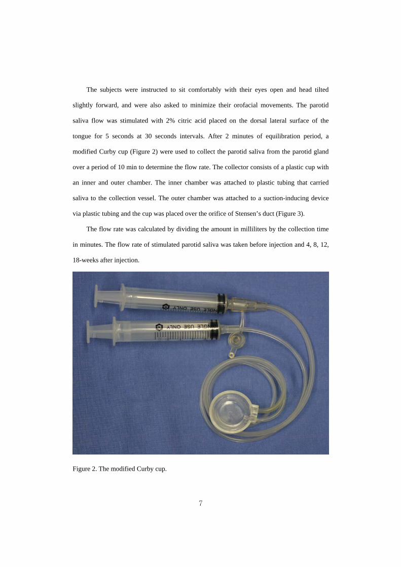
The subjects were instructed to sit comfortably with their eyes open and head tilted
slightly forward, and were also asked to minimize their orofacial movements. The parotid
saliva flow was stimulated with 2% citric acid placed on the dorsal lateral surface of the
tongue for 5 seconds at 30 seconds intervals. After 2 minutes of equilibration period, a
modified Curby cup (Figure 2) were used to collect the parotid saliva from the parotid gland
over a period of 10 min to determine the flow rate. The collector consists of a plastic cup with
an inner and outer chamber. The inner chamber was attached to plastic tubing that carried
saliva to the collection vessel. The outer chamber was attached to a suction-inducing device
via plastic tubing and the cup was placed over the orifice of Stensen’s duct (Figure 3).
The flow rate was calculated by dividing the amount in milliliters by the collection time
in minutes. The flow rate of stimulated parotid saliva was taken before injection and 4, 8, 12,
18-weeks after injection.
Figure 2. The modified Curby cup.
7
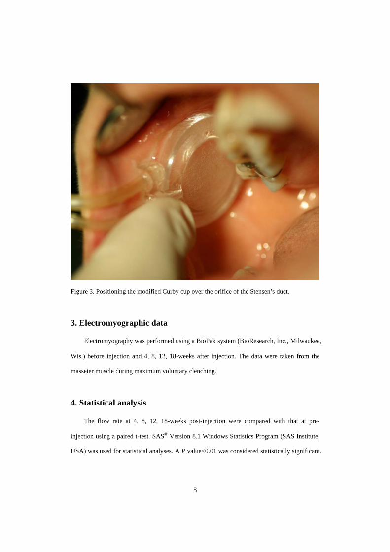
Figure 3. Positioning the modified Curby cup over the orifice of the Stensen’s duct.
3. Electromyographic data
Electromyography was performed using a BioPak system (BioResearch, Inc., Milwaukee,
Wis.) before injection and 4, 8, 12, 18-weeks after injection. The data were taken from the
masseter muscle during maximum voluntary clenching.
4. Statistical analysis
The flow rate at 4, 8, 12, 18-weeks post-injection were compared with that at pre-
injection using a paired t-test. SAS® Version 8.1 Windows Statistics Program (SAS Institute,
USA) was used for statistical analyses. A P value<0.01 was considered statistically significant.
8
Ⅲ. Results
All subjects reported significant clinical improvement in 12 weeks after injection. There
were no major local or systemic complications associated with BTX-A injection. However,
several mild side effects were observed, all of which were temporary and localized. The
muscle weakness, headache, swelling, and pain in the area of the injection was reported.
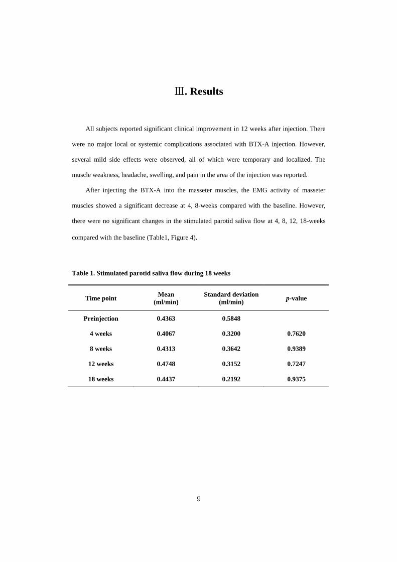
After injecting the BTX-A into the masseter muscles, the EMG activity of masseter
muscles showed a significant decrease at 4, 8-weeks compared with the baseline. However,
there were no significant changes in the stimulated parotid saliva flow at 4, 8, 12, 18-weeks
compared with the baseline (Table1, Figure 4).
Table 1. Stimulated parotid saliva flow during 18 weeks
Time point Mean (ml/min)
Standard deviation(ml/min) p-value
Preinjection 0.4363 0.5848
4 weeks 0.4067 0.3200 0.7620
8 weeks 0.4313 0.3642 0.9389
12 weeks 0.4748 0.3152 0.7247
18 weeks 0.4437 0.2192 0.9375
9
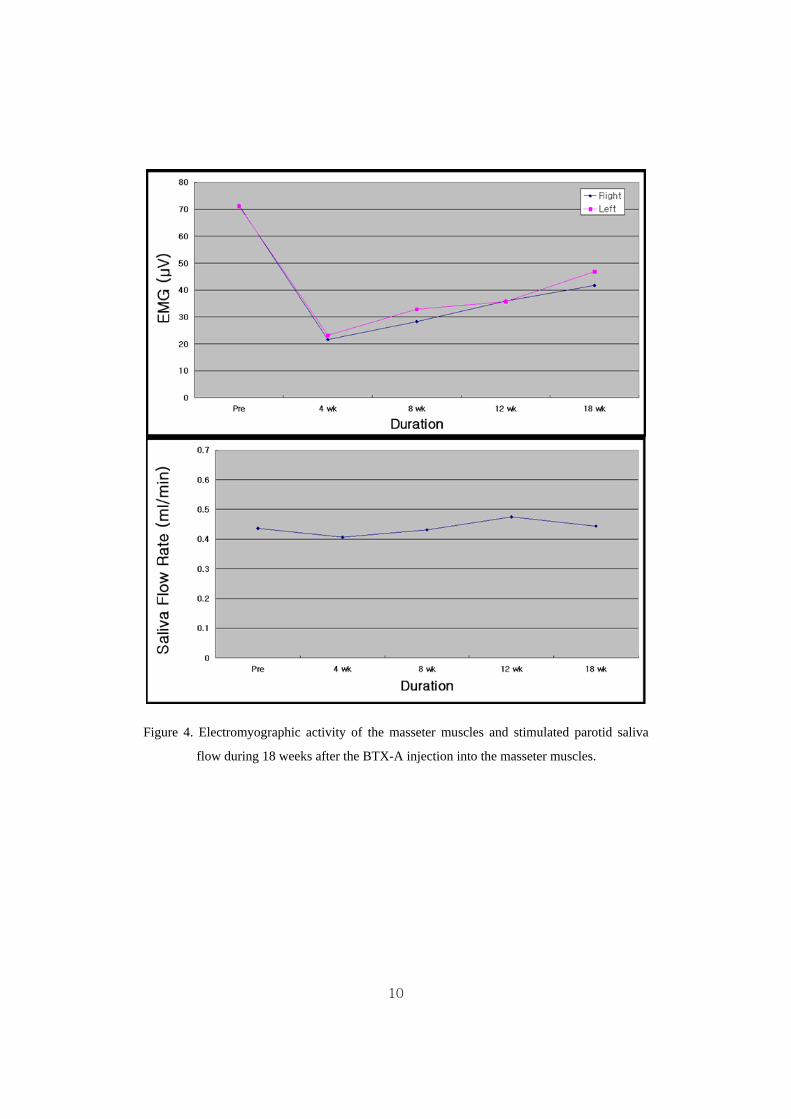
Figure 4. Electromyographic activity of the masseter muscles and stimulated parotid saliva
flow during 18 weeks after the BTX-A injection into the masseter muscles.
10
Ⅳ. Discussion
Botulinum toxin inhibits transmission of alpha motor neurons at the neuromuscular
junction and gamma motor neurons in muscle spindles. So, it can reduce muscular overactivity,
such as dystonia, and may alter reflex overactivity of striated muscles (Priori et al. 1995). It
also inhibits release of acetylcholine in all parasympathetic and cholinergic postganglionic
sympathetic neurons, using it as a treatment for overactive smooth muscles or abnormal
activity of glands (Münchau, and Bhatia 2000). So, the BTX-A injection is known to be
beneficial in many conditions such as overactivity of the muscles and the hypersecretion of
glands (Münchau and Bhatia 2000).
To date, the United States Food and Drug Administration (FDA) has approved the use of
BTX-A for the treatment of strabismus, blepharospasm, seventh cranial nerve disorders
(hemifacial spasm), cervical dystonia, glabellar wrinkles for cosmetic uses and hyperhidrosis
(Lew 2002). Besides the FDA-approved uses, BTX-A has a wide variety of clinical
applications (Thant, and Tan 2003).
Off-label clinical uses include idiopathic dystonia such as laryngeal dystonias (Teive et al.
2001), temporomandibular disorders (Freund, Schwartz, and Symington 2000), myofasical
pain (Acquadro, and Borodic 1994 ; Cheshire, Abashian, and Mann 1994), bruxism (Pidcock,
Wise, and Christensen 2002), myoclonus (Awaad et al. 1999), Tourette syndrome (Kwak,
Hanna, and Jankovic 2000), simple tics (Marras et al. 2001), and writer’s cramp (Behari 1999).
Spastic disorders, including those linked to stroke (Bhakta et al. 2000), cerebral palsy
(Kirschner et al. 2001), and Parkinson’s disease (Giladi et al. 2001) have been shown to
respond well to BTX-A treatment. BTX-A has been also shown to be useful in reducing pain
associated with migraine (Binder et al. 2000 ; Silberstein et al. 2000), tension-type headache
(Porta 2000), and low back pain (Foster et al. 2001). It has been shown to be effective in
11

treating many other conditions due to excessive cholinergic activity including hyperhidrosis
(Heckmann et al. 1999), achalasia (Pasricha et al. 1995), and chronic anal fissure (Fernandez
Lopez et al. 1999).
Among 8 serotypes, serotype A and B is commercially available for clinical use.
Botulinum toxin type A products include BOTOX®, Dysport® (Ipsen Limited, Slough, UK),
Xeomin® (Merz Pharmaceuticals, Frankfurt, Germany), Neuronox® (Medy-Tox,
Chungcheongbuk-do, Korea), and BTXA®, also referred to as Chinatoxin® (Lanzhou Institute
of Biological Products, Lanzhou, China). Botulinum toxin type B(BTX-B) products include
Myobloc®, also known as Neurobloc® (Solstice Neurosciences, South San Francisco, CA,
USA) (Lowe 2007). Serotype A is commonly used for clinical use. It was reported that BTX-
B showed a shorter duration of action than BTX-A and was associated with twice as many
reports of dry mouth and dysphagia as BTX-A (Comella et al. 2005).
The toxin is commonly used to treat masseteric hypertrophy because the paresis induced
by BTX-A injection caused muscle atrophy, which is a decrease in the diameter of the target
muscle (Kim et al. 2003). Recently, there have been some reports showing that a botulinum
toxin type A injection into the masseter muscle can be used as an alternative noninvasive
treatment for masseteric hypertrophy. These studies have revealed atrophy of the hypertrophic
muscles after a BTX-A injection using clinical photographs, ultrasound, electromyography,
and computed tomography.
There are several deficiencies in the interpretation of the effects of botulinum toxin
injection by clinical photographs, but atrophy of the hypertrophic muscles was noted (Moore,
and Wood 1994 ; Smyth 1994). It was also reported that in clinical photographs, marked
atrophy of masseter muscles occurred in seven patients with hypertrophic masseter muscles
over the course of 3 to 8 weeks and this atrophy remained constant over a follow-up period of
up to 25 months (von Lindern et al. 2001).
12
Electromyographic activity has been used as one of the most common diagnostic
measurements in dentistry since the report by Moyer (Moyer 1949). Since a reduction in the
electromyographic potential was reported 4 weeks after injection (Smyth 1994), there are
many reports in which the effects of botulinum toxin was evaluated by measuring of
electromyographic activity (Freund, Schwartz, and Symington 2000 ; Ahn and Kim 2007 ).
It was also reported that the effects of BTX-A on masseteric hypertrophy was evaluated
using ultrasound or computed tomography. To et al. evaluated the effects of BTX-A on
masseteric hypertrophy using ultrasound and electromyography after BTX-A injection into
both masseter muscles. All five patients in their study showed good responses, with a
maximum effect of a 31 percent reduction in muscle bulk 3 months after treatment (To et al.
2001). Computed tomography is more reliable and accurate than ultrasound in evaluating the
outcome of BTX-A injection. So, Kim et al. evaluated the effects of BTX-A on masseteric
hypertrophy using computed tomography and reported that nine subjects showed a mean
reduction of approximately 22 percent in masseteric muscle volume (Kim et al. 2003). Park,
Ahn, and Jung also reported serial measurements of the thickness of the masseter muscle using
ultrasound and computed tomography before the injection and at 1 and 3 months thereafter
(Park, Ahn, and Jung 2003).
Doses are quated in mouse units which is the amount of toxin that kills 50% of a group of
18-20g female Swiss-Webster mice. A recent study comparing Botox® and Dysport® found
that a unit of Botox® is three times as potent as a unit of Dysport® (Odergren et al. 1998). The
dosages used for masseter muscle were generally 25 to 30 units of BTX-A (Mandel, and
Tharakan 1999 ; von Lindern et al. 2001 ; To et al. 2001 ; Kim et al. 2003). It was reported
that the difference of effects between 25 units and 35 units of BTX-A on masseteric atrophy is
not statistically significant (Kim et al. 2007). It was also reported that the size of the
denervation field is determined by the dose and volume (Borodic et al 1994). In addition,
Shaari and Sanders reported that the dose was a stronger predictor of the area of paralysis than
13
the volume (Shaari, and Sanders 1993). To achieve the maximum dose response and to
minimize the side effects, clinicians should use the most effective dose at the smallest volume.
High doses and frequent injections of botulinum toxin have been associated with neutralizing
antibody formation (Atassi, and Oshima 1999). Because botulinum toxin is an antigen, use of
high cumulative doses of botulinum toxin can get treatment ineffective. Thus, to minimize
antibody resistance, a clinician should use the smallest possible effective dose, with treatment
intervals of at least 3 months, and avoid booster injections (Dauer et al. 1998 ; Greene, Fahn,
and Diamond 1994).
In general, maximum clinical effects of BTX-A occur 1 to 2 weeks after the injection in
muscular spasms or wrinkles, because the action of muscular paralysis by BTX-A reaches a
peak 1 to 2 weeks after the injection (Sloop et al. 1996). However, the maximum clinical
effect of BTX-A for masseteric hypertrophy appear to require 3 months bacause this effect is
muscular atrophy secondary to muscular paralysis by BTX-A (Kim et al. 2003).
Muscle function recovers after roughly 120 days through terminal sprouting of motor
axons and formation of new motor end plates (Borodic et al. 1994 ; Duchen 1971). The
presynaptic end-plate region expands and collateral axonal sprouts develop, eventually
reinnervating the neuromuscular junction (Alderson et al. 1991).
The therapeutic use of botulinum toxin is generally safe and well tolerated. In more than
25 years of human use, there has been no reported death from overdose of BTX-A when used
for cosmetic purposes (Coté et al. 2005). The lethal dose of BTX-A causing death in 50
percent of humans has been estimated to be 40 U/kg, or 2800 U in the 70 kg patient
(Matarasso, and Deva 2002). BTX-A’s long-term safety has been demonstrated with the
majority of reported adverse events considered mild to moderate in severity (Naumann et al.
2006). Common local reactions to BTX-A therapy include pain, edema, erythema, ecchymosis,
headache, and short-term hyperesthesia (Naumann et al. 2006). Patients may also report
systemic reactions after BTX-A treatment, and these may include complaints of nausea,
14
fatigue, malaise, flu-like symptoms, rash, and a metallic taste (Naumann et al. 2006 ; Tugnoli
et al. 2002). Allergic reactions to BTX-A are rare, but has been reported (Lu, and Lippitz
2009).
There have been some side effects after injecting BTX-A into the hypertrophic masseter
muscles. The side effects such as change in bite force, speech disturbance, muscle pain, facial
asymmetry, and prominent zygoma have been reported (Kim et al. 2003 ; Kim et al. 2007). It
was reported that there was an approximately 40 percent decrease in the mean maximum bite
force at 2 weeks compared with that recorded before injection, and the bite force was restored
to its preinjection value by 12 weeks after injection (Ahn and Kim 2007). Kim et al. reported a
change in facial smiling as well as a sunken cheek after a BTX-A injection (Kim et al. 2003).
In our previous studies, several mild side effects such as swelling, bruise or pain in the area of
the injection, headache, muscle weakness, discomfort in mastication and a dry mouth have
been reported occasionally but these side effects were all temporary and localized (Ahn and
Kim 2007 ; Kim et al. 2007). In other studies, it was reported that patients also complained of
xerostomia on occasions.
After the injection, the toxin may diffuse into the nearby muscles and other tissues such
as the parotid glands via the local vasculature or by gravity-influenced chemical diffusion
(Eleopra et al. 1996). In addition, the toxin may affect the parotid glands through the systemic
distribution by blood flow or by retrograde axonal transport (Garner et al. 1993; Tintner et al.
2005). Therefore, a BTX-A injection into the masseter muscles can affect the salivary flow
rate of the parotid glands and cause xerostomia. In patients with cervical dystonia, it was
reported that the use of BTX-B caused a significant decrease in saliva production, as measured
by the Schirmer’s test, compared with BTX-A (Tintner et al. 2005). Compared with BTX-A,
BTX-B is associated with greater autonomic side effects, including dry mouth, reduced
sweating, dysphagia, constipation, heartburn, bladder voiding difficulties, conjunctival
irritation, and accommodation difficulties (Dressler, and Aleopra 2006).
15
The salivary flow rate is affected by many factors including the degree of hydration,
body positioning, seasonal and diurnal factors, medical status and medications, and the nature
and duration of the stimulus (Navazesh 1993). Therefore, in this study, many factors affecting
on salivary flow rate were controlled. The methods for collecting saliva vary from collection
under the unstimulated or stimulated condition, and as whole saliva or from individual glands.
Of the many methods, the stimulated parotid saliva secretion was measured due to the close
anatomic relationship between the masseter muscle and the parotid gland.
As a result of the collection of saliva in the parotid gland, the flow rate of the stimulated
parotid saliva did not show any significant changes. From the results of this study, it can be
concluded that BTX-A does not cause any significant difference in saliva production of the
parotid gland when our modified method of injection is used for hypertrophic masseter
muscles. From the results of other studies (Dressler, and Benecke 2003 ; Garner et al. 1993), it
is believed that BTX-A has no significant systemic effects on the parotid gland. Botulinum
therapy can affect a variety of autonomic functions, including salivation (Pal, Calne, and
Calne 2000), sweating (Naumann et al. 1997), heart rate (Claus, Druschky, and Erbguth 1995),
and vasodilation (Kellogg et al. 1995). But, because of the lower affinity to the cholinergic
terminals of the salivary glands and lower systemic effect, it is believed that BTX-A does not
cause autonomic side effects such as xerostomia unless it is injected into the masseter near the
parotid glands. Therefore, it is believed that our modified injection method causes fewer
autonomic side effects. Indeed, none of the subjects in this study complained of xerostomia.
A limitation of this study was that the unstimulated whole saliva was not measured to
more closely determine the relationship with xerostomia. This may be more closely related to
the systemic effect of BTX-A. Therefore, further study about unstimulated whole saliva in
patients who complain of xerostomia after BTX-A injection will be needed.
16

References
Acquadro M.A., Borodic G.E. 1994. “Treatment of myofascial pain with botulinum A toxin”.
Anesthesiology, 80 : 705.
Ahn K.Y., Kim S.T. 2007. “The Change of Maximal Bite-force after Botulinum Toxin Type A
Injection for Treating Masseteric Hypertrophy”. Plast Reconstr Surg, 120(6) : 1662-1666.
Anderson K., Holds J.B., Anderson R.L. 1991. “Botulinum-induced alteration of nerve-muscle
interactions in the human orbicularis oculi following treatment for blepharospasm”. Neurology,
41 : 1800-1805.
Atassi M.Z., Oshima M. 1999. “Structure, activity, and immune (T and B cell) recognition of
botulinum neurotoxins”. Crit Rev Immunol, 19 : 219.
Awaad Y., Tayem H., Elgamal A., Coyne M.F. 1999. “Treatment of childhood myoclonus
with botulinum toxin type A”. J Child Neurol, 14 : 781-786.
Bandyopadhyay S., Clark A.W., DasGupta B.R., Sathyamoorthy V. 1987. “Role of the heavy
and light chains of botulinum neurotoxin in neuromuscular paralysis”. J Biol Chem, 262 :
2660-2663.
Beckers H.L. 1977. “Masseteric muscle hypertrophy and its intraoral surgical correction”. J
Maxillofac Surg, 5 : 28-35.
17
Behari M. 1999. “Botulinum toxin in the treatment of writer’s cramp”. J Assoc Physicians
India, 47 : 694-698.
Bhakta B.B., Cozens J.A., Chamberlain M.A., Bamford J.M. 2000. “Impact of botulinum
toxin type A on disability and carer burden due to arm spasticity after stroke: A randomized
double blind placebo controlled trial”. J Neurol Neurosurg Psychiatry, 69 : 217-221.
Binder W.J., Brin M.F., Blitzer A., Schoenrock L.D., Pogoda J.M. 2000. “Botulinum toxin
type A (BOTOX) for treatment of migraine headaches: an open-label study”. Otolaryngol
Head Neck Surg, 123(6) : 669-676.
Bittner M.A., DasGupta B.R., Holz R.W. 1989. “Isolated light chains of botulinum
neurotoxins inhibit exocytosis. Studies in digitonin-permeabilized chromaffin cells”. J Biol
Chem, 264(18) : 10354-10360.
Black J.D., Dolly J.O. 1986. “Interaction of 125I-labeled botulinum neurotoxins with nerve
terminals. II. Autoradiographic evidence for its uptake into motor nerves by acceptor-mediated
endocytosis”. J Cell Biol, 103 : 535-544.
Borodic G.E., Ferrante R., Pearce L.B., Smith K. 1994. “Histologic assessment of dose-related
diffusion and muscle fiber response after therapeutic botulinum A toxin injections”. Mov
Disord 9 : 31.
Burgen A.S., Dickens F., Zatman L.J. 1949. “The action of botulinum toxin on the
neuronmuscular junction”. J physiol, 109 : 10-24.
18

Carruthers A., Carruthers J. 1998. “Clinical indications and injection technique for the
cosmetic use of botulinum A exotoxin”. Dermatol Surg, 24(11) : 1189-1194.
Carruthers J., Stubbs H.A. 1987. “Botulinum toxin for benign essential blepharospasm,
hemifacial spasm and age-related lower eyelid entropion”. Can J Neurol Sci, 14(1) : 42-45.
Cheshire W.P., Abashian S.W., Mann J.D. 1994. “Botulinum toxin in the treatment of
myofascial pain syndrome”. Pain 59 : 65.
Claus D., Druschky A., Erbguth F. 1995. “Botulinum toxin: influence on respiratory heart rate
variation”. Mov Disord, 10 : 574-579.
Comella C.L., Jankovic J., Shannon K.M., Tsui J., Swenson M., Leurgans S., Fan W.,
Dystonia Study Group. 2005. “Comparison of botulinum toxin serotypes A and B for the
treatment of cervical dystonia”. Neurology, 65(9) : 1423-1429.
Coté T.R., Mohan A.K., Polder J.A., Walton M.K., Braun M.M. 2005. “Botulinum toxin type
A injections: adverse events reported to the US Food and Drug Administration in therapeutic
and cosmetic cases”. J Am Acad Dermatol, 53(3) : 407-415.
Dauer W.T., Burke R.E., Greene P., Fahn S. 1998. “Current concepts on the clinical features,
aetiology and management of idiopathic cervical dystonia”. Brain, 121 : 547-560.
Dolly J.O. 1997. “Therapeutic and research exploitation of botulinum neurotoxins”. Eur J
Neurol, 4(Supppl 2) : S5-10.
19
Dolly J.O., Black J., Williams R.S., Melling J. 1984. “Acceptors for botulinum neurotoxin
reside on motor nerve terminals and mediate its internalization”. Nature, 307(5950) : 457-460.
Dolly O. 2003. “Synaptic transmission: inhibition of neurotransmitter release by botulinum
toxins”. Headache, 43(Suppl 1) : S16-24.
Dressler D., Adib Saberi F. 2005. “Botulinum toxin: mechanisms of action”. Eur Neurol, 53 :
3-9.
Dressler D., Benecke R. 2003. “Autonomic side effects of botulinum toxin type B treatment of
cervical dystonia and hyperhidrosis”. Eur Neurol, 49 : 34-38.
Dressler D., Eleopra R. 2006. “Clinical use of non-A botulinum toxins: botulinum toxin type
B”. Neurotox Res, 9(2-3) : 121-125.
Duchen L.W. 1971. "An electron microscope study of the changes induced by botulinum toxin
in the motor end plates of slow and fast skeletal muscle hbres of the mouse". J Ncurol Sci, 14 :
47-60.
Eleopra R., Tugnoli V., Caniatti L., De Grandis D. 1996. “Botulinum toxin treatment in the
facial muscles of humans: evidence of an action in untreated near muscles by peripheral local
diffusion”. Neurology, 46 : 1158-1160.
Erbguth F.J., Naumann M. 1999. “Historical aspects of botulinum toxin: Justinus Kerner
(1786-1862) and the "sausage poison"”. Neurology. 53(8) : 1850-1853.
20
Fernandez Lopez F., Conde Freire R., Rios Rios A., Garcia Iglesias J., Cainzos Fernanadez M.,
Potel Lesquereux J. 1999. “Botulinum toxin for the treatment of anal fissure”. Dig Sur, 16 :
515-518.
Foster L., Clapp L., Erickson M., Jabbari B. 2001. “Botulinum toxin A and chronic low back
pain: A randomized, double-blind study”. Neurology, 56 : 1290-1293.
Freund B., Schwartz M., Symington J.M. 1999. “The use of botulinum toxin for the treatment
of temporomandibular disorders: preliminary findings”. J Oral Maxillofac Surg, 57(8) : 916-
921.
Freund B., Schwartz M., Symington J.M. 2000. “Botulinum toxin: New treatment for
temporomandibular disorders”. Br J Oral Maxillofac Surg, 38 : 466-471.
Garner C.G., Straube A., Witt T.N., Gasser T., Oertel W.H. 1993. “Time course of distant
effects of local injections of botulinum toxin”. Mov Disord, 8 : 33-37.
Giladi N., Gurevich T., Shabtai H., Paleacu D., Simon E.S. 2001. “The effect of botulinum
toxin injections to the calf muscles on freezing of gait in parkinsonism: A pilot study”. J
Neurol, 248 : 572-576.
Greene P., Fahn S., Diamond B. 1994. “Development of resistance to botulinum toxin type A
in patients with torticollis”. Mov Disord, 9 : 213.
Gurney C.E. 1947. “Chronic bilateral benign hypertrophy of the masseter muscles”. Am J Surg
73 : 137.
21
Hauschild A.H. 1990. “Clostridium botulinum toxins”. Int J Food Microbiol. 10(2) : 113-124.
Heckmann M., Breit S., Ceballos-Baumann A., Schaller M., Plewig G. 1999. “Side-controlled
intradermal injection of botulinum toxin A in recalcitrant axillary hyperhidrosis”. J Am Acad
Dermatol, 41 : 987-990.
Kellogg D.L. Jr, Pérgola P.E., Piest K.L., Kosiba W.A., Crandall C.G., Grossmann M.,
Johnson J.M. 1995. “Cutaneous active vasodilation in humans is mediated by cholinergic
nerve cotransmission”. Circ Res, 77(6) : 1222-1228.
Kim H.J., Yum K.W., Lee S.S., Heo M.S., Seo K. 2003. “Effects of botulinum toxin type A on
bilateral masseteric hypertrophy evaluated with computed tomographic measurement”.
Dermatol Surg, 29 : 484-489.
Kim J.H., Shin J.H., Kim S.T., Kim C.Y. 2007. “Effects of two different units of botulinum
toxin type a evaluated by computed tomography and electromyographic measurements of
human masseter muscle”. Plast Reconstr Surg, 119 : 711-717.
Kirschner J., Berweck S., Mall V., Korinthenberg R., Heinen F. 2001. “Botulinum toxin
treatment in cerebral palsy: Evidence for a new treatment option”. J Neurol, 248(suppl 1) : 28-
30.
Kwak C.H., Hanna P.A., Jankovic J. 2000. “Botulinum toxin in the treatment of tics”. Arch
Neurol, 57 : 1190-1193.
22
Legg J.W. 1880. “Enlargement of the temporal and masseter muscles on both sides”. Trans
Pathol Soc London, 3 : 361-366.
Lew M.F. 2002. “Review of the FDA-approved uses of botulinum toxins, including data
suggesting efficacy in pain reduction”. Clin J Pain, 18 : S142-146.
Lowe N.J. 2007. “Overview of botulinum neurotoxins”. Journal of Cosmetic and Laser
Therapy, 9(suppl 1) : 11–16.
Lu D.W., Lippitz J. 2009. “Complications of botulinum neurotoxin”. Dis Mon, 55(4) : 198-
211.
Mandel L., Tharakan M. 1999. “Treatment of unilateral masseteric hypertrophy with
botulinum toxin: Case report”. J Oral Maxillofac Surg, 57 : 1017.
Marras C., Andrews D., Sime E., Lang A.E. 2001. “Botulinum toxin for simple motor tics: A
randomized, double-blind, controlled clinical trial”. Neurology, 56 : 605-610.
Matarasso A., Deva A.K., American Society of Plastic Surgeons DATA Committee. 2002.
“Botulinum toxin”. Plast Reconstr Surg, 109(3) : 1191-1197.
Moore A.P., Wood G.D. 1994. “The medical management of masseteric hypertrophy with
botulinum toxin type A”. Br J Oral Maxillofac Surg, 32 : 26-28.
Moyer R.E. 1949. “Temporomandibular muscle contraction patterns in Angle class II division
1 malocclusion: An electromyographic study”. Am J Orthod Dentofac Orthop 35 : 836.
23

Münchau A., Bhatia K.P. 2000. “Uses of botulinum toxin injection in medicine today”. BMJ,
320 : 161-165.
Navazesh M. 1993. “Methods for collecting saliva”. Ann N Y Acad Sci, 694 : 72-77.
Naumann M., Albanese A., Heinen F., Molenaers G., Relja M. 2006. “Safety and efficacy of
botulinum toxin type A following long-term use”. Eur J Neurol, 13(Suppl 4) : 35-40.
Naumann M., Flachenecker P., Bröcker E.B., Toyka K.V., Reiners K. 1997. “Botulinum toxin
for palmar hyperhidrosis”. Lancet, 349 : 252.
Odergren T., Hjaltason H., Kaakkola S., Solders G., Hanko J., Fehling C., Marttila R.J., Lundh
H., Gedin S., Westergren I., Richardson A., Dott C., Cohen H. 1998. “A double blind,
randomised, parallel group study to investigate the dose equivalence of Dysport and Botox in
the treatment of cervical dystonia”. J Neurol Neurosurg Psychiatry, 64(1) : 6-12.
Park M.Y., Ahn K.Y., Jung D.S. 2003. “Botulinum toxin type A treatment for contouring of
the lower face”. Dermatol Surg 29 : 477.
Pasricha P.J., Ravich W.J., Hendrix T.R., Sostre S., Jonses B., Kalloo A.N. 1995.
“Intrasphincteric botulinum toxin for the treatment achalasia”. N Engl J Med, 332 ; 774-778.
Pidcock F.S., Wise J.M., Christensen J.R. 2002. “Treatment of severe post-traumatic bruxism
with botulinum toxin-A: case report”. J Oral Maxillofac Surg, 60(1) : 115-117.
24
Porta M. 2000. “A comparative trial of botulinum toxin type A and methylprednisolone for the
treatment of tension-type headache”. Curr Rev Pain, 4 : 31-35.
Priori A., Berardelli A., Mercuri B., Manfredi M. “Physiological effects produced by
botulinum toxin treatment of upper limb dystonia. Changes in reciprocal inhibition between
forearm muscles”. Brain, 118 : 801-807.
Rispoli D.Z., Camargo P.M., Pires J.L. Jr, Fonseca V.R., Mandelli K.K., Pereira M.A. 2008.
“Benign masseter muscle hypertrophy”. Braz J Otorhinolaryngol, 74(5) : 790-793.
Scott A.B. 1981. “Botulinum toxin injection of eye muscles to correct strabismus”. Trans Am
Ophthalmol Soc, 79 : 734-770.
Scott A.B., Rosenbaum A., Collins C.C. 1973. “Pharmacologic weakening of extraocular
muscles”. Invest Ophthalmol, 12(12) : 924-927.
Shaari C.M., Sanders I. 1993. “Quantifying how location and dose of botulinum toxin
injections affect muscle paralysis”. Muscle Nerve 16 : 964.
Silberstein S., Mathew N., Saper J., Jenkins S. 2000. “Botulinum toxin type A as a migraine
preventive treatment. For the BOTOX Migraine Clinical Research Group”. Headache, 40 :
445-450.
Smyth A.G. 1994. “Botulinum toxin treatment of bilateral masseteric hypertrophy”. Br J Oral
Maxillofac Surg, 32 : 29-33.
25

Sobel J. 2005. “Botulism”. Clin Infect Dis. 41(8) : 1167-1173.
Sommer E.W., Sommer H., Meyer K.F. 1926. “The purification of botulinum toxin”. The
Journal of Infectious Diseases, 39(5) : 345-350.
Sugiyama H. 1980. “Clostridium botulinum neurotoxin ”. Microbiol Rev. 44(3):419-448.
Tan E.K., Jankovic J. 1999. “Botulinum toxin A in patients with oromandibular dystonia:
long-term follow-up”. Neurology, 53(9) : 2102-2107.
Teive H.A., Scola R.H., Werneck L.C., Quadros A., Gasparetto E.L., Sa D.S., Brandi I.V.,
Macedo Filho E.D. 2001. “Use of botulinum toxin in the treatment of laryngeal dystonia
(spasmodic dysphonia): Preliminary study of twelve patients”. Arq Neuropsiquiatr 59 : 97-100.
Thant Z.S., Tan E.K. 2003. “Emerging therapeutic applications of botulinum toxin”. Med Sci
Monit, 9 : RA40-48.
Tintner R., Gross R., Winzer U.F., Smalky K.A., Jankovic J. 2005. “Autonomic function after
botulinum toxin type A or B: a double-blind, randomized trial”. Neurology, 65 : 765-767.
To E.W., Ahuja A.T., Ho W.S., King W.W., Wong W.K., Pang P.C., Hui A.C. 2001. “A
prospective study of the effect of botulinum toxin A on masseteric muscle hypertrophy with
ultrasonographic and electromyographic measurement”. Br J Plast Surg, 54(3) : 197-200.
26

Tugnoli V., Eleopra R., Quatrale R., Capone J.G., Sensi M., Gastaldo E. 2002. “Botulism-like
syndrome after botulinum toxin type A injections for focal hyperhidrosis”. Br J Dermatol,
147(4) : 808-809.
von Lindern J.J., Niederhagen B., Appel T., Berge S., Reich R.H. 2001. “Type A botulinum
toxin for the treatment of hypertrophy of the masseter and temporal muscles: An alternative
treatment”. Plast Reconstr Surg 107 : 327.
27
국문요약
사람 교근에 주사한 보툴리눔 A 형 독소가
자극성 이하선 타액 분비율에 미치는 영향
보툴리눔 A 형 독소를 사람 교근에 주사하는 방법은 교근 비대의 치료로서
널리 이용되어져 왔다. 보툴리눔 A 형 독소 주사의 심각한 부작용은 보고되지
않았지만 주사 부위의 멍 또는 통증, 확산에 의한 표정근 약화 등과 같은
경미하고 일시적인 부작용들은 보고된 바 있다. 때로는 구강건조증을 호소하는
경우도 있음이 보고되었다. 하지만 현재까지 사람 교근에 보툴리눔 A 형 독소를
주사하는 것이 타액 분비율에 어떠한 영향을 미치는지에 대한 연구는 현재까지
시행된 바가 없다. 이에 본 연구에서는 사람 교근에 보툴리눔 A 형 독소 주사가
이하선 타액 분비율에 미치는 영향을 평가하고자 하였다.
32 명의 자원자를 대상으로 보툴리눔 A 형 독소를 양측 교근에 각각 25
units 씩 주사하였다. 주사 부위는 교근 하방 1/3 의 중앙 부위로서 25units 을 약
1cm 간격의 두 점에 같은 양으로 나누어 주사하였다. 이하선 타액은 타액 분비율
측정 전 주의 사항을 지도하고 일정한 시간에 측정하였으며 2% citric acid 로 자극
시 분비되는 타액을 modified Curby cup 을 이용하여 10 분간 수집함으로써
측정하였다.
분석 결과, 자극성 이하선 타액 분비율은 보툴리눔 A 형 독소 주사 전과
비교하여 4 주, 8 주, 12 주, 18 주 후 모두에서 유의한 차이를 보이지 않았다. 따라서,
교근에 대한 보툴리눔 A 형 독소 주사는 이하선 타액 생성의 유의한 감소를
유발하지 않는 것으로 사료된다.
핵심되는 말 : 보툴리눔 A 형 독소, 교근, 자극성 이하선 타액
28
Related Documents