i Effect of Acute Exercise on Postprandial Lipemia and Endothelial Function in Men with Peripheral Arterial Disease Thesis submitted for the degree of Masters of Science Kevin O’Hara (BSc) School of Health and Human Performance Dublin City University Supervisor: Prof. Niall Moyna MSc July 2012

Welcome message from author
This document is posted to help you gain knowledge. Please leave a comment to let me know what you think about it! Share it to your friends and learn new things together.
Transcript

i
Effect of Acute Exercise on Postprandial Lipemia and Endothelial Function in Men with
Peripheral Arterial Disease
Thesis submitted for the degree of Masters of Science
Kevin O’Hara (BSc)
School of Health and Human Performance
Dublin City University
Supervisor: Prof. Niall Moyna
MSc
July 2012

ii
Acknowledgements
I would like to thank a number of people for their time, patience and help over my 6 years in
DCU and especially over the past 2 years.
Prof. Niall Moyna for his time, advice and encouragement throughout my postgraduate
studies.
Michael Harrison for being throughout and for any words of advice.
To all my fellow postgraduate students for all the help they have given me, with particular
mention to Sarah Hughes, Paul O Connor and Brona Furlong. Really appreciate all the time
they have afforded me in the past two years. It was much appreciated and I owe you all so
much.
And last but not least I would like to thank my family for putting up with my constant
moaning over the past couple of months and for always being there for me.

iii
Declaration
I hereby certify that this material, which I now submit for assessment on the programme of
study leading to the award of MSc is entirely my own work, that I have exercised reasonable
care to ensure that the work is original, and does not to the best of my knowledge breach
any law of copyright, and has not been taken from the work of others save and to the extent
that such work has been cited and acknowledged within the text of my work.
Signed: ____________________ ID No. 55590370 Date _______________

iv
Abstract
Introduction: Postprandial lipidemia (PPL), defined as an increase in plasma levels of
triglyceride-rich lipoproteins following the consumption of a high fat meal (HFM) is
associated with endothelial dysfunction. Acute exercise reduces PPL and maintains
endothelial function (EF) in healthy adults. The effect of acute exercise on PPL and
endothelial function has not been studied in patients with peripheral arterial disease (PAD).
Purpose: To examine the effect of an acute bout of exercise on PPL, vascular inflammation
and endothelial function in men with PAD.
Methods: Men (n=8) with PAD underwent two oral fat tolerance tests (OFTT). On the
evening prior to each OFTT, participants rested (control), or exercised until they expended
200 Kcal. Blood samples were obtained at baseline and 30 min, 1, 2, 3 and 4 h postprandial.
Endothelial-dependent dilation (EDD) and endothelial-independent dilation (EID) were
measured in the brachial artery using ultrasonography at baseline, 2 h and 4 h postprandial.
Results: Postprandial TG increased significantly and EDD decreased significantly following
the OFTT. An acute bout of discontinuous exercise that resulted in a 200 Kcal expenditure
did not significantly attenuate the post prandial TG response or significantly ameliorate the
decrease in endothelial vasomotor function. Compared to baseline values, circulating
leukocytes, and TNF-α increased (p<0.05) in both conditions 4 h postprandial. There were
no changes in C-Reactive Protein (CRP).
Conclusion: Prior exercise has no effect on PPL or EDD following an OFTT in men with PAD.

v
Table of Contents
TITLE PAGE……………………………………………………………........................................................................i
ACKNOWLEDGEMENT…………………………………………………………………………………………………………….….ii
DECLARATION……………………………………………………………………………………………………………………….…..iii
ABSTRACT……………………………………………………………………………………………………………………………….…iv
TABLE OF CONTENTS……………………………………………………………………………………………………………….…v
TABLE OF FIGURES……………………………………………………………………………………………………………………viii
LIST OF TABLES………………………………………………………………………………………………………………………….ix
LIST OF TERMS AND ABBREVIATIONS………………………………………………………………………………………….x
Chapter 1 ......................................................................................................................................... 1
INTRODUCTION ............................................................................................................................... 1
Specific Aims ................................................................................................................................... 4
Study Hypothesis ............................................................................................................................ 4
Chapter 2 ......................................................................................................................................... 5
LITERATURE REVIEW ....................................................................................................................... 5
Peripheral Arterial Disease ............................................................................................................. 5
Diagnosis of PAD ............................................................................................................................. 7
Intermittent Claudication ............................................................................................................... 8
Critical Limb Ischemia ..................................................................................................................... 9
Risk Factors in PAD ........................................................................................................................ 10
Treatment of PAD ......................................................................................................................... 12
Treatment of PAD – Exercise ........................................................................................................ 12
Treatment of PAD - Pharmacotherapy ......................................................................................... 15
Atherosclerosis and Endothelium ................................................................................................. 16
Endothelium and Endothelial Function ........................................................................................ 17
Endothelial Function – Exercise .................................................................................................... 19
Postprandial Lipemia and PAD ...................................................................................................... 27
Postprandial Lipemia and Exercise ............................................................................................... 28
Postprandial lipemia and intermittent exercise ........................................................................... 28
Inflammatory biomarkers ............................................................................................................. 31
Conclusion ..................................................................................................................................... 31
Chapter 3 ....................................................................................................................................... 33

vi
METHODOLOGY ............................................................................................................................ 33
Participants ................................................................................................................................... 33
Research Design ............................................................................................................................ 33
Screening....................................................................................................................................... 34
Experimental Trials ....................................................................................................................... 34
Acute Exercise Bout ...................................................................................................................... 35
Blood Pressure .............................................................................................................................. 35
Height and Weight ........................................................................................................................ 35
Overview of Endothelial Function Assessment............................................................................. 35
Ultrasound Technique and Image Acquisition .............................................................................. 37
M mode Imaging ........................................................................................................................... 37
Doppler Imaging ............................................................................................................................ 38
Endothelial-Dependent Dilation (EDD) ......................................................................................... 39
Endothelial Independent Dilation (EID) ........................................................................................ 40
Cardiorespiratory and Metabolic Measures ................................................................................. 42
Mass Flow Sensor Heated wire Anemometer - Mode of Operation ............................................ 42
Mass Flow Sensor Calibration ....................................................................................................... 43
Gas Analysers ................................................................................................................................ 44
Calibration of CO2and O2 Analyzers .............................................................................................. 44
Blood Sampling and Processing .................................................................................................... 45
Biochemical Assays ....................................................................................................................... 45
Statistical Analysis ......................................................................................................................... 46
Chapter 4 ....................................................................................................................................... 47
RESULTS ......................................................................................................................................... 47
Triglycerides .................................................................................................................................. 47
Inflammatory Markers .................................................................................................................. 47
Endothelial Function ..................................................................................................................... 48
Chapter 5 ....................................................................................................................................... 55
DISCUSSION ................................................................................................................................... 55
Postprandial lipemia...................................................................................................................... 60
Endothelial function ...................................................................................................................... 55
Biomarkers .................................................................................................................................... 55
Chapter 6 ....................................................................................................................................... 60
Bibliography .................................................................................................................................. 60

vii
Appendices .................................................................................................................................... 79

viii
TABLE OF FIGURES
FIGURE 2.1: NATURAL HISTORY OF ATHEROSCLEROTIC LOWER EXTREMITY PAD 4
FIGURE 3.1: SONOSITE MICROMAXX® ULTRASOUND SYSTEM AND 12.0 MHZ LINEAR ARRAY TRANSDUCER 36
FIGURE 3.2: ARM POSITION AND CUFF PLACEMENT. 37
FIGURE 3.3: M MODE ULTRASOUND IMAGE OF THE BRACHIAL ARTERY 38
FIGURE 3.4: FROZEN SCREEN SHOT OF A DOPPLER IMAGE 39
FIGURE 3.5: OVERVIEW OF THE PROTOCOL USED TO ASSESS ENDOTHELIAL DEPENDENT DILATION 40
FIGURE 3.6: OVERVIEW OF THE PROTOCOL USED TO ASSESS ENDOTHELIAL INDEPENDENT DILATION 41
FIGURE 3.7: STANDARD DIALOG BOX 36
FIGURE 4.1: TG IN THE POSTPRANDIAL PERIOD DURING CONTROL AND EXERCISE 46
FIGURE 4.2: WBC IN THE POSTPRANDIAL PERIOD DURING CONTROL AND EXERCISE 47
FIGURE 4.3: TNF-ΑLPHA IN THE POSTPRANDIAL PERIOD DURING CONTROL AND EXERCISE 48
FIGURE 4.4: CHANGE IN FLOW MEDIATED DILATION DURING CONTROL AND EXERCISE CONDITIONS 49

ix
LIST OF TABLES
TABLE 2.1: INTERPRETATION OF ANKLE-BRACHIAL INDEX (ABI) VALUES 6
TABLE 2.2: CLASSIFICATION OF PERIPHERAL ARTERY DISEASE (FONTAINE AND RUTHERFORD SCALE ADAPTED
FROM NORGREN ET AL, 2007) 8
TABLE 2.3: STUDIES THAT EXAMINED THE EFFECT OF ACUTE EXERCISE ON ENDOTHELIAL FUNCTION 19
TABLE 4.1: SUBJECTS PHYSICAL CHARACTERISTICS 44
TABLE 4.2: EFFECT OF ACUTE EXERCISE ON LIPID AND INFLAMMATORY BIOMARKERS 50
TABLE 4.3: EXERCISE BRACHIAL ARTERY DIAMETER (CM) 112
TABLE 4.4: CONTROL BRACHIAL ARTERY DIAMETER (CM) 112
TABLE 4.5: EXERCISE BRACHIAL ARTERY DIAMETER (CM) GTN 112
TABLE 4.6: CONTROL BRACHIAL ARTERY DIAMETER (CM) GTN 113
TABLE 4.7: PERCENTAGE CHANGES IN BRACHIAL ARTERY DOPPLER FLOW - EXERCISE 113
TABLE 4.8: PERCENTAGE CHANGES IN BRACHIAL ARTERY DOPPLER FLOW - CONTROL 113

x
LIST OF TERMS AND ABBREVIATIONS Term Description
PAD Peripheral arterial disease
IC Intermittent claudication
PPL Postprandial lipemia
HFM High fat meal
CVD Cardiovascular disease
TG Triglycerides
RLP Remnant like parts
EDD Endothelial dependant dilation
EID Endothelial independent dilation
TEE Total energy expenditure
CLI Critical limb ischemia
ABI Ankle brachial index
CRP C-reactive protein
hsCRP High sensitivity C-reactive protein
HDL-C High density lipoprotein cholesterol
LDL-C Low density lipoprotein cholesterol

xi
Term Description
OxLDL Oxidised LDL
ACH Acetylcholine
NO Nitric oxide
TGRL Triglyceride rich lipoprotein
AUC Area under the curve
VLDL Very low density lipoproteins
TNF-α Tumour necrosis factor alpha
OFTT Oral fat tolerance test
ECG Electrocardiogram

1
Chapter 1
INTRODUCTION
Peripheral arterial disease (PAD) is a distinct atherothrombotic syndrome
marked by stenosis of peripheral arteries, typically those in the lower extremities, causing
inadequate blood flow to the limbs. Intermittent claudication (IC) is the most common
symptom of PAD and is characterized by the onset of pain in the lower extremities during
exercise that is relieved with rest (6).
Supervised aerobic exercise has been shown to increase maximum walking
distance in patients with PAD [1]. However, recent studies indicate that acute bouts of
exercise to the onset of intermittent claudication may cause a systemic thrombo-
inflammatory response in this population [2,3]. This may be due to the fact that exercising
to the onset of intermittent claudication, followed by reperfusion on rest, results in the
formation of oxygen-derived free radicals (ODFR) and cytokines (3-5). Oxygen-derived free
radicals cause lipid peroxidation, which results in structural damage to the vascular
endothelium and increased vascular permeability.
The endothelium is a 0.2- to 4-µm-thick monolayer of squamous endothelial
cells that line the lumen of the entire surface of the vascular tree and plays an important
role in the regulation of vascular tone, haemostasis, immune and inflammatory responses
(1). Damage to the endothelium from mechanical forces and processes related to
cardiovascular disease risk factors and the resulting inflammatory response can generate a
pro-thrombotic environment favourable for the initiation and progression of atherosclerosis

2
(1-4), the most frequent underlying cause of PAD. The risk factors for PAD include age, male
gender, family history, diabetes, smoking, hypertension and hyperlipidaemia (5, 6).
Postprandial lipemia (PPL) describes the increase in plasma levels of triglyceride-rich
lipoproteins for up to 8 h following the consumption of a high fat meal (HFM), and may
represent an independent risk factor for atherosclerotic cardiovascular disease (ACVD)(7).
In contemporary Western societies the vasculature is commonly exposed to prolonged
postprandial hyperlipidemia. It has been estimated that individuals consuming a typical
Western diet spend approximately 18 h per day in a postprandial state (8). The adverse
effect of postprandial triglycerides (TG) is thought to be mediated by proatherogenic
lipolysis products of nascent triglyceride-rich lipoproteins, such as remnant like particles
(RLP) and fatty acids. Even a transient increase in these proatherogenic products may
increase pro-coagulant and pro-inflammatory activity (7) and impair endothelial dependent
vasodilatation (EDD) (9), a predictor of atherosclerosis and future cardiovascular events.
Physical activity and physical fitness are associated with a lower incidence of CVD.
The relative risk of death from CVD in the most active individuals is half that of their
sedentary counterparts (10). The cardioprotective effect of exercise may be mediated in
part by an influence on TG metabolism. Acute exercise 1-16 h prior to feeding a standard
HFM can significantly reduce the postprandial TG response in adults (11, 12).
The available evidence suggests that total energy expenditure (TEE) may be more
critical than exercise intensity in influencing postprandial TG metabolism. In addition, the
exercise benefits can be accumulated over the course of a day in two or three shorter bouts.
The possibility of accumulating benefit with multiple short bouts is particularly important for

3
patients with PAD as in many instances the disease restricts their ability to exercise for
prolonged periods. To date, no studies have evaluated the effect of exercise on
postprandial lipemia in patients with PAD. The purpose of this study is to examine the
effects of an acute bout of exercise on postprandial lipemia and endothelial function in men
with PAD.

4
Specific Aims
1. To compare the effects of a standardised OFTT with and without a prior acute
exercise bout on postprandial TG in men with PAD
2. To compare the effects of a standardised OFTT meal with and without a prior
acute exercise bout on circulating markers of inflammation in men with PAD
3. To compare the effects of a standardised OFTT with and without a prior acute
exercise bout on brachial artery flow mediated dilation in men with PAD
Study Hypothesis
1. In men with PAD, the postprandial TG response to a standardised OFTT will be
significantly lower when the meal is preceded with an acute exercise bout
compared to no exercise
2. In men with PAD, the circulating levels of inflammatory markers in response to a
standardised OFTT will be significantly lower when the meal is preceded with an
acute exercise bout compared to no exercise
3. In men with PAD, brachial artery flow mediated dilation following a standardised
OFTT will be significantly greater when the meal is preceded with an acute
exercise bout compared to no exercise

5
Chapter 2
LITERATURE REVIEW
Peripheral Arterial Disease
Peripheral arterial disease (PAD) has been defined as obstruction of blood flow into
an arterial tree excluding the intracranial or coronary circulations (Figure 2.1) (13).
Figure 2.1: Development of atherosclerotic lower extremity PAD
It represents a continuum of disorders that range from asymptomatic PAD,
symptomatic IC, and critical limb ischemia (CLI) (Figure 2.2). Patients with PAD develop
atherosclerotic occlusive lesions in the arteries supplying the lower extremities, and
limitation in blood flow to active muscles is the primary pathophysiologic event. A number
of arterial segments can be affected, including the inflow vessels (aorta and iliac arteries),
and also the femoral, popliteal, and tibial vessels in the leg (14).

6
Figure 2.2: Natural history of atherosclerotic lower extremity PAD The figure presents 1-year outcomes for patients who present with critical limb ischemia. As illustrated, all patients with lower extremity PAD are at risk of progressive limb ischemia and are at high risk of fatal and nonfatal atherothrombotic events, including myocardial infarction and stroke
PAD prevalence is 3-12% in the general population, with IC prevalence of 1-2% (15;
16). PAD prevalence increases significantly with age, affecting up to 20% of patients >75
years (17, 18). Coexistent coronary artery disease (CAD) and cerebrovascular disease
(CBVD) are common in patients with PAD, particularly in the elderly population. The
coexistence of CAD and stroke was found to be 68% and 42% respectively, among men > 50
years of age in an academic, hospital-based geriatric practice (19). The prevalence of PAD
may be vastly underestimated due to the fact that the majority of individuals with lower
extremity PAD do not experience recognisable ischemic symptoms in the limb, and are
therefore classed as being 'asymptomatic' (5).
7
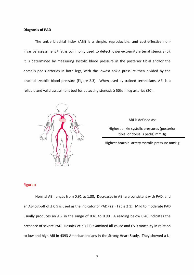
Diagnosis of PAD
The ankle brachial index (ABI) is a simple, reproducible, and cost-effective non-
invasive assessment that is commonly used to detect lower-extremity arterial stenosis (5).
It is determined by measuring systolic blood pressure in the posterior tibial and/or the
dorsalis pedis arteries in both legs, with the lowest ankle pressure then divided by the
brachial systolic blood pressure (Figure 2.3). When used by trained technicians, ABI is a
reliable and valid assessment tool for detecting stenosis ≥ 50% in leg arteries (20).
Figure x
Normal ABI ranges from 0.91 to 1.30. Decreases in ABI are consistent with PAD, and
an ABI cut-off of 0.9 is used as the indicator of PAD (22) (Table 2 1). Mild to moderate PAD
usually produces an ABI in the range of 0.41 to 0.90. A reading below 0.40 indicates the
presence of severe PAD. Resnick et al (22) examined all-cause and CVD mortality in relation
to low and high ABI in 4393 American Indians in the Strong Heart Study. They showed a U-
ABI is defined as:
Highest ankle systolic pressures (posterior
tibial or dorsalis pedis) mmHg
Highest brachial artery systolic pressure mmHg

8
shaped association between ABI and mortality, with significantly increased risk in both the
<0.90 and >1.40 groups.
ABI Interpretation
>1.30 Normal, but considered incompressible (calcified) arteries 0.91–1.30 Normal range 0.41–0.90 Mild to moderate PAD 0.5–0.74 Consistent with moderate peripheral arterial disease (intermittent
claudication or rest pain) < 0.4 Severe PAD
Table 2.1: Interpretation of ankle-brachial index (ABI) values
Invasive methods such as duplex scanning, magnetic resonance imaging (MRI), and
digital subtraction angiography are commonly used for anatomical localisation of arterial
disease prior to intervention rather than for initial diagnosis.
Intermittent Claudication
Intermittent claudication (IC), defined as exercise-induced muscle pain that is
relieved with rest, is one the most common symptoms in patients with lower extremity PAD
(4). Symptoms may be described as pain, achiness, a sense of fatigue, or nonspecific
discomfort that occurs with exercise. Symptoms normally dissipate following several
minutes of rest (23).
The location of the pain is an indication of the site of arterial occlusion (Figure 2.4)
(24). Claudication of the calf is usually the result of an occlusion in the superficial femoral
artery. The most frequently affected artery in intermittent claudication is the popliteal
artery; symptoms are most common in the calf muscles (25). This artery is an extension of
the femoral artery and continues below the knee where it branches off and carries blood to

9
the muscles in the calf and foot. Hip, thigh, and buttock claudication are associated with
occlusion of the aorta and iliac arteries (25).
Figure 2.4: Location of the pain associated with PAD
Critical Limb Ischemia
CLI is defined as PAD causing lower-extremity pain at rest or imminent limb loss
caused by severe impairment of blood flow to the affected limb, and is classified as Fontaine
Class III (26). The Fontaine Classification is the method by which chronic peripheral
ischaemia is classified. CLI is a manifestation of PAD that describes patients with chronic
ischemic rest pain, foot and leg ulcers, or gangrene. Discomfort is often worse in a supine
position and pain can be reduced when the limb is placed in a dependant position. CLI
results from the presence of multilevel occluded vessels that impair blood flow and distal
perfusion pressure to a level insufficient to satisfy the nutritive needs of the limb at rest
(13). Approximately 500–1000 people per million of the population are diagnosed with CLI
(27). PAD patients with diabetes are at a greater risk of CLI, and risk of amputation is
increased 5 fold in this population (28).
The clinical diagnosis of CLI should be confirmed by haemodynamic parameters such
as the ankle or toe systolic pressure. Ischemic rest pain most commonly occurs below an

10
ankle pressure of 50 mmHg or a toe pressure <30 mmHg. Up to 30% of patients with lower
extremity PAD will progress from IC to CLI over the course of their disease (27, 29).
Approximately 25% of patients die and a further 30% require a major amputation one year
after CLI diagnosis (27). Owing to the high risk of limb loss and fatal and nonfatal vascular
events, it is vitally important to diagnose CLI quickly. The severity of the symptoms of PAD
can be classified according to either the Fontaine or Rutherford scales (Table 2.2).
Table 2.2: Classification of peripheral artery disease (Fontaine and Rutherford scale adapted from Norgren et al, 2007)
Fontaine Rutherford
Stage Clinical Grade Category Clinical
I Asymptomatic 0 0 Asymptomatic
IIa Mild claudication I 1 Mild claudication
IIb Moderate to severe claudication I 2 Moderate claudication
III Ischemic rest pain I 3 Severe claudication
IV Ulceration or gangrene II 4 Ischemic rest pain
III 5 Minor tissue loss
III 6 Major tissue loss
Risk Factors in PAD
The primary etiology of PAD is atherosclerosis, and the risk factors are similar to
those for coronary artery and cerebrovascular disease. Age, smoking and diabetes are the
most powerful risk factors for PAD. Others include African American ethnicity, dyslipidemia,
hypertension, hyperhomocysteinemia, elevated C-reactive protein (CRP) levels, and chronic
renal insufficiency (30).

11
The prevalence of PAD increases with age. A strong association between advanced
age (≥ 70years) and PAD prevalence has been shown in the National Health and Nutrition
Examination Survey (NHANES) 1999–2000. The prevalence of PAD was 4.3% in subjects
aged 40 years or older compared with 14.5% in those aged 70 years or older (30). In 1985,
Criqui et al., reported the prevalence of PAD to be 2% to 3% in individuals aged 50 years or
less compared with 20% in those aged greater than 75 years (33).
Diabetes and smoking are the two most common modifiable risk factors for PAD.
Results from a survey conducted in Sweden found that 21% of diabetic patients had signs of
PAD (34). In a cross-sectional analysis of a 4153 Greek adults (37), the odds ratio for
vascular disease was 1.94 for patients with the metabolic syndrome, 3.04 for patients with
the metabolic syndrome and diabetes, and 1.48 for patients with the metabolic syndrome
but no diabetes. The association of smoking and PAD is dose-dependent; the risk of PAD
increases with years smoked and packs smoked per year. (38). The incidence of PAD
increases from 2.6% in never smokers to 4.5% in moderate smokers (0-25 pack/year) to
9.8% in heavy smokers (>25 pack/year) (39). More than 80% of individuals with PAD are
current or former smokers (35, 40). Individuals of African American ethnicity are
approximately 3-4 times more likely to develop PAD compared to their non-African white
counterparts (31, 32).
The common dyslipidemia found in PAD is similar to insulin resistance, enforcing the
strong association between diabetes with PAD (41). Patients with PAD have higher
circulating levels of triglycerides (42), lower levels of high-density lipoprotein cholesterol
(HDL-C) and higher levels of total cholesterol than healthy individual’s subjects (42). The risk

12
of PAD increases by 5-10% for each 10mg/dL increase in total cholesterol (43). In a cross
sectional study involving 708 men, aged 55-74., Planas et al., (44) found that the waist-to-
hip ratio was independently associated with PAD. CRP, an acute-phase reactant produced
by the liver in response to inflammation, is one of many circulating inflammatory markers
that is related to CAD and may play a role in its pathogenesis. There is evidence of a linear
relation between CRP concentrations and the severity of PAD (34).
Treatment of PAD
The goals of treatment for individuals with PAD are to prevent progression of PAD,
prevent coronary or cerebrovascular events, and for those with IC to relive symptoms,
improve functional capacity and improve quality of life (34). For all patients across the PAD
spectrum risk factor modification is recommended, including weight loss, smoking
cessation, lipid-lowering therapy, anti-platelet therapy and increased exercise (23).
Recommendations for individuals with IC include supervised exercise interventions and
pharmacological therapies (Cilostazol and pentoxifyline). Cilostazol reduces the pain of
intermittent claudication by dilating the arteries, thereby improving the flow of blood and
oxygen to the legs. Pentoxifylline improves blood flow by making it easier for red blood cells
to pass through vessels. It also decreases the viscosity of blood. Surgical or endovascular
intervention is usually reserved for patients with severe, lifestyle-limiting symptoms that do
not adequately respond to conservative management, including pharmacologic treatment.
Treatment of PAD – Exercise
The physiological, metabolic, and mechanical alterations that occur during periods of
exercise presumably stimulate an adaptive response that ultimately reduces claudication

13
symptoms and improves functional capacity. Exercise training improves maximal walking
ability by an average of 150% (45-51). Improvements in walking ability are most often
attained when exercise sessions >30 min are undertaken at least 3 times per week for 6
months and involve walking to near maximal pain (52, 53). The exact mechanisms by which
exercise training yields improvements in walking ability remain unclear.
The increased blood flow in response to repeated periods of exercise-induced
hyperemia may alter vascular structure and function (54). Angiogenesis in response to
exercise training may increase blood flow to skeletal muscle, distal to the stenosis (55).
However, studies examining the effect of exercise training on leg blood flow have been
equivocal. Gardner et al., 2001 found that 6 months of exercise training increased reactive
hyperaemic blood flow by 27% and max calf blood flow by 30% in patients with PAD. The
improvements in blood flow were associated with improved walking ability. However,
resting blood flow and ABI were not altered in response to training (56). In contrast, a
number of studies have reported no changes in leg blood flow in patients with improved
walking ability after an exercise program (57, 58), suggesting that other mechanisms may be
responsible for the large improvements observed following exercise training.
Impaired endothelial dependent vasodilation (EDD) has been demonstrated in
patients with PAD (59) and in patients with risk factors for atherosclerosis, such as type 2
diabetes, (60) hypertension, and hyperlipidemia (61). Results from studies in patients with
other chronic diseases, suggest that exercise training improves endothelial-dependent
vasodilatation (62, 63). Brachial artery flow mediated dilation (FMD) was found to be

14
significantly decreased in men with PAD, 30 min and 2 h following 10 min of treadmill
exercise to intolerable ischaemic pain in the affected lower extremity exercise (64).
Exercise training may improve abnormal hemorheology in patients with claudication,
thereby facilitating oxygen delivery (65). Hemorheology refers to the properties of flowing
blood and its elements specifically, plasma viscosity, hematocrit, red cell deformability, and
red cell aggregation. Ernst & Matrai 1987 et al., found that treadmill walking at 3 km/hr to
absolute claudication twice a day, five days per week for 2 months resulted in significant
normalization of blood and plasma viscosity, blood cell filterability, and red cell aggregation.
No changes occurred in a non-exercise control group (66).
Chronic ischemia of PAD results in metabolic abnormalities in the affected skeletal
muscle. PAD is associated with increased plasma and skeletal muscle short-chain
acylcarnitine content (23). Resting muscle short-chain acylcarnitine content is inversely
correlated with claudication limited functional capacity (23). The degree of metabolic
dysfunction is a better predictor of functional capacity than haemodynamic measurements.
In patients with unilateral claudication, the increase in acylcarnitines only occurred in the
affected skeletal muscle, indicating that this metabolic abnormality was specific to the limb
with reduced blood flow (67). Patients with the greatest accumulation of acylcarnitines had
the lowest treadmill exercise performance. Treadmill exercise training, but not resistance
training, reduces plasma levels of acylcarnitine accumulation (68). Improvements in
maximal walking ability may also be related to changes in walking efficiency or improvement
in the tolerance of claudication pain (23).

15
Treatment of PAD - Pharmacotherapy
Effective drug therapies include aspirin, pentoxifylline and cilostazol (71).
Pentoxifylline is a methylxanthine derivative with hemorrheologic and immunomodulating
properties. It reduces blood viscosity, changes the morphology of red blood cells, and
decreases the potential for platelet aggregation and thrombus formation. Cilostazol is a
quinolinone derivative that inhibits cellular phosphodiesterase III. It suppresses platelet
aggregation, activates lipoprotein lipase, and causes arterial dilation (69). A large randomly
controlled prospective trial found that cilostazol was significantly better than pentoxifylline
or placebo for increasing walking distances in patients with moderate to severe PAD (70).
The improvement in treadmill walking performance with pentoxifylline was not significantly
different from the placebo (70). Data supporting use of pentoxifylline for claudication is
weak, and pentoxifylline is not generally accepted as efficacious.
Aspirin and clopidogrel are often used as antithrombotic therapies in PAD. They
have not been shown to improve symptoms of intermittent claudication but are important
in reducing cardiovascular complications associated with the presence of atherosclerosis
and PAD. The Antithrombotic Trialist Collaboration (ATT) found that even a low dose of
aspirin (75–150mg) reduced vascular events by 32% in patients with PAD (71). However, the
Clopidogrel versus Aspirin in Patients at Risk of Ischaemic Events (CAPRIE) trial found that
clopidogrel treatment in patients with symptomatic PAD was more effective than aspirin in
preventing ischemic events, 5.83% and 5.32% for clopidogrel and aspirin, respectively (72).
Treatment for CLI can be quite complex, but the primary aim should be to reduce the
pain and improve blood flow in order to minimize the need for amputation. The majority of

16
patients with CLI will ultimately require a revascularization procedure. Endovascular
therapy is now the primary strategy for management of CLI. Patients with CLI not eligible
for arterial reconstruction, prostanoids are the only vasoactive drugs with proven efficacy.
Creutzig et al., (73) concluded in a recent meta-analysis of randomized placebo-controlled
trials, of patients with stage III or IV PAD that PGE1 therapy not only had significant
beneficial effects over placebo on ulcer healing and pain relief, but also increased the
number of patients surviving with both legs at 6-months follow-up.
Atherosclerosis and Endothelium
Atherosclerosis is the most frequent underlying cause of PAD (6). It is characterized
by the accumulation of monocyte-derived macrophages within the vessel wall and the
accompanying inflammatory response. This process is initiated by the transmigration and
retention of low density lipoprotein cholesterol (LDL-C) in the sub-endothelial extracellular
matrix, where it is subjected to chemical modification, and converted to oxidized LDL
(OxLDL), a key pathogenic mediator of atherosclerosis (Figure 4.5) (107).
OxLDL stimulates inflammatory signaling by endothelial cells, resulting in the
expression of membrane-bound adhesion molecules that facilitate the attachment of
circulating leukocytes to the vessel wall, and induce smooth muscle cell proliferation.
Attachment of monocytes facilitates their migration across the endothelium into the
subendothelial space where they differentiate into macrophages. Macrophages engulf
OxLDL, leading to the formation of large foam cells, a major component of early lesions.
Systemic inflammatory markers of vascular inflammation include an elevated white blood
cell count (WBC), and high circulating levels of CRP and TNF-α.

17
Figure 4.5: Summary of the atherosclerotic disease progression and developmnet
Endothelium and Endothelial Function
Endothelial cells form a continuous monolayer that line blood vessels of the entire
vascular tree, and represent a surface area of approximately 4000 to 7000 m2. Malpighi's
discovery in the 17th century of the endothelium as a physical separation between blood
and tissue with no substantial functionality persisted throughout the nineteenth and
twentieth centuries (74). Landmark studies in the 1980’s demonstrated the obligatory role
of endothelial cells in acetylcholine (ACh)-mediated vasodilation (75) and in the paradoxical
ACh-mediated vasoconstriction of atherosclerotic vessels (76). The endothelium is now
viewed as a complex dynamic barrier that plays a crucial role in maintaining vascular
integrity.
The endothelium-dependent vasodilatory response to exogenously administered
acetylcholine (ACh) is attributable to the production and diffusion of nitric oxide (NO), a
hydrophobic diatomic gas produced in response to changes in shear forces, or via a variety
of agonists acting on specific endothelial cell membrane receptors (Figure 4.6) (81). In
addition to its vasodilatory effects, NO counteracts leukocyte adhesion to the endothelium,
attenuates vascular smooth muscle proliferation and migration, influences the production

18
of superoxide anion, suppresses platelet aggregation and protects against vascular injury,
inflammation, and thrombosis, key events in the development and progression of
atherosclerosis (77).
Figure 4.6: Molecular structure of nitric oxide
Cardiovascular disease risk factors such as aging and a family history of CVD, active
and passive smoking, lipid disorders, hypertension, diabetes mellitus, obesity, and physical
inactivity, among others, have been shown to promote the development of atherosclerosis
through their deleterious effects on endothelial structure and function (78). Damage to the
endothelium caused by cardiovascular disease risk factors results in the reduction of nitric
oxide (NO) bioavailability. The progressive inability of endothelial cells, exposed to risk
factors, to generate sufficient NO promotes a vascular phenotype prone to atherogenesis
(79).
The functional and structural integrity of the endothelium is critical in maintaining
vascular homoeostasis, and the presence of atherosclerosis has been shown to affect the
vasomotor responses of the diseased vessel to shear stress, and a number of vasoactive
compounds (80). Flow-mediated dilation (FMD) is a commonly used non-invasive procedure
to assess endothelial function. It is based on the assumption that healthy, intact endothelial
cells can detect, and dilate, to changes in shear stress following a brief period of occlusion
(62). The percentage change in arterial diameter in response to shear stress can be
measured using high-resolution B mode ultrasonography, and is believed to be endothelial-

19
dependent and mediated by NO. Diameter changes are also measured after administration
of glyceryl-trinitrate to assess the response of the vessel to endothelium-independent
vasodilation (81).
The prognostic significance of endothelial function as a predictor of cardiovascular
events in healthy individuals and in patients with CAD and those with normal coronary
arteries and risk factors for atherosclerosis has been addressed in a number of studies (82,
83, 84,). Interventions proven to reduce cardiovascular risk, such as weight loss, smoking
cessation, lipid-lowering therapy, and angiotensin-converting enzyme (ACE) inhibitors been
shown to improve endothelial function and decrease cardiovascular risk (85).
Endothelial Function – Exercise
Evidence from both cross-sectional and longitudinal studies indicate that
cardiorespiratory fitness delays the decrease in endothelial function associated with ageing
(86) and reverses impaired endothelial function in individuals with atherosclerotic CVD (87).
Exercise-induced improvements in vascular function appear to occur more readily and with
remarkable consistency in vessels with antecedent functional impairment. Improvements in
endothelial function induced by exercise training are attributable to a combination of
enhanced vasodilatory capacity and arterial remodelling (62).
Observations that a single bout of exercise can transiently alter atherosclerotic CVD
risk factors (89) have led to the notion that perhaps some of the effects of exercise on
endothelial function may be attributable to the acute effect of exercise. A number of
studies have investigated the effect of an acute bout of exercise on endothelial function in

20
healthy and disease populations. Table 2.4 provides a summary overview of studies that
have examined the effect of acute exercise on endothelial function.
Cycling for 30 min at 50% VO2peak increases brachial artery FMD in young female
smokers (90), and treadmill exercise for 34 min at 60% VO2max almost doubled brachial
artery FMD in premenopausal women (91) but had no effect on brachial artery FMD in
postmenopausal women. Both low volume high-intensity interval exercise and moderate-
intensity endurance exercise significantly increased absolute FMD and normalized brachial
artery FMD 1 h post exercise in men and women with CAD (92). Brachial artery FMD is
enhanced 1 h following 45 min acute bouts of low-, moderate- and high-intensity treadmill
exercise in active overweight men, but is attenuated in inactive overweight men (93).
In contrast a number of studies have found a transitory functional deterioration in
FMD in healthy individuals 1 h after a single bout of high-intensity interval running (94), non-
elite runners 1 h after completing a marathon, and healthy male smokers compared to non-
smokers (7.7 v 4.1%) immediately following 40 min of submaximal steady-state exercise on
a cycle ergometer (95).
Brachial artery FMD was found to be significantly decreased in men with PAD, 30
min and 2 h following 10 min of treadmill exercise to intolerable ischaemic pain in the
affected lower extremity exercise (64). In contrast, Silvestro et al., found that an acute bout
of treadmill exercise to the onset of claudication has no effect on brachial artery FMD
measured 5 min post exercise in men and women (62 ± 2 year) with PAD (96). In contrast,
FMD was significantly impaired following treadmill exercise to maximal claudication pain,
demonstrating that exercise-induced ischemia further deteriorates FMD. Intravenous

21
administration of vitamin C ameliorates the impaired FMD response following treadmill
exercise to maximal claudication pain only. Others have shown that antioxidant treatment
can prevent acute impairment in endothelial function. The benefits of vitamin C on
endothelial function are believed to be due to superoxide scavenging, and/or inhibition of
LDL-C that takes place during high-intensity exercise. Vitamin C is also an important
regulator of the intracellular redox (96).

22
Table 2.3: Studies that examined the effect of acute exercise on endothelial function
Author Patient Cohort Mode of Exercise Duration Intensity FMD
Cosio-Lima (2006) Renal Transplant TM Walking 30 min 70 - 85% HRmax
Gaenzer (2001) Smoking Cycling 40 min 50% VO2peak
Silvestro (2002) PAD TM Walking Max claudication 3 km.h-1 @ 3%
Harvey (2005) Pre & post-menopausal TM Walking
Rooks (2011) Smoking/non-smokers Cycling 59% VO2peak 30 min
Jones (2008) Diurnal Variation Intermittent Cycling 3 x 10 min 70% VO2peak
Jones (2010) Diurnal Variation Intermittent Cycling 3 x 10 min 70% VO2peak
Rognmo (2005) CV Fitness HII Running 5 x 5 min 90% HRmax high fit
low fit
Harris (2008)
Overweight (8 active) Intermittent Cycling 45 min
25% VO2peak
50% VO2peak
75% VO2peak
Llewellyn (2012) Healthy TM Running 30 min 60% VO2max
Currie (2012) CAD Intermittent vs. Continuous 10 x 1min vs 30 min 80% vs. 55% POpeak
Dawson (2008) Nonelite Runners Marathon 42.2 km n/a
Joras PAD TM Walking Max claudication 3.2 km.h-1 @ 12%
CAD, coronary artery disease; FMD, flow-mediated vasodilation; Tm, Treadmill; HII, High intensity interval; HRmax, Heart rate max; PAD, peripheral arterial
disease; VO2, oxygen consumption; PO, power output.

Effect of Acute Exercise on Postprandial Lipemia and Endothelial Dysfunction in Men with Peripheral Arterial Disease
__________________________________________________________________________________
23
Supervised treadmill exercise has been shown to improve vascular function, in
patients with PAD. McDermott et al., randomly assigned 156 patients with PAD to 6 months
of supervised treadmill exercise, lower extremity resistance training, or a sedentary control
group. Improvements in brachial artery FMD were significantly greater at 6 months in the
treadmill group than the resistance training, or the control group. Changes in brachial
artery FMD in the resistance training group were not different from the control group.
Improvements in the 6-min walk test and maximum treadmill walking time were
significantly greater in the treadmill exercise group than the resistance training group (97).
Intermittent treadmill walking at 2.0 mph to near-maximal claudication pain 3 days a week
for 6 months has also been shown to significantly improve brachial artery FMD. Time to
onset of IC pain increased by 94%, and the time to maximal claudication pain increased 43%
with exercise (98).
Another acute study also showed reduced FMD response following acute exercise in
patients with PAD (100). The exercise protocol involved walking at 3 km/hr and a 3% grade
with a 3% grade increase every 2 minutes up to a maximum of 15%. Patients exercise was
stopped when claudication pain became intolerable, and FMD was assessed 5 minutes later.
In patients with peripheral arterial disease, calf pain during walking, followed by a period of
rest to allow reperfusion of the ischemic limb, induces increased oxidative stress and a
marked inflammatory response (42, 48, 100). By reducing nitric oxide availability both these
events may be responsible for the acute systemic endothelial insult observed in the present
acute studies.

Effect of Acute Exercise on Postprandial Lipemia and Endothelial Dysfunction in Men with Peripheral Arterial Disease
__________________________________________________________________________________
24
Lipids
Lipids are a heterogeneous group of hydrophobic organic molecules that have a
number of important physiological functions including the formation of membranes, energy
storage, cellular signalling, protection, insulation and the production of steroid hormones
and bile acids. They can be broadly classified as fatty acids, acylglycerols, phospholipids,
eicosanoids, steroids and lipoproteins (128).
Acylglycerols are composed of a glycerol molecule with one (monoacylglycerol), two
(diacylglycerol) or three (triacylglycerol) fatty acids attached at the hydroxyl group.
Triacylglycerols, also termed triglycerides (TG), are the most abundant lipid in the body.
Triglyceride and cholesterol are solubilised by their incorporation into small lipid-protein
complexes termed lipoproteins. Lipoproteins are classified according to their density as
chylomicrons, very low density lipoproteins (VLDLs), intermediate density lipoproteins
(IDLs), low density lipoproteins (LDLs) or high density lipoproteins (HDLs). They contain
different concentrations of protein, triglyceride, cholesterol, cholesterol esters and
phospholipids (129).
Triglyceride is the principal form in which fat is present in the diet. Following their
hydrolysis by pancreatic lipase, TGs combine with proteins, free cholesterol and
phospholipids in the intestinal mucosal cells to form nascent chylomicrons. The TG rich
nascent chylomicrons are secreted into the lymphatic system and enter the circulation at
the thoracic duct (129).
Circulating nascent chylomicrons acquire additional apoproteins, Apo E and Apo C-II,
from high density lipoprotein cholesterol (HDL-C) and become mature chylomicrons (128)

Effect of Acute Exercise on Postprandial Lipemia and Endothelial Dysfunction in Men with Peripheral Arterial Disease
__________________________________________________________________________________
25
that are approximately 90% triglyceride, 5% cholesterol, 4% phospholipid and 1% protein.
During their passage through the circulation, chylomicrons bind to lipoprotein lipase (LPL)
on the surface of capillary endothelial cells resulting in the hydrolysis of TG to fatty acids
and glycerol in a reaction that requires the presence of Apo C-II (128). Fatty acids can then
be oxidised as fuel by muscle or other cells, or alternatively they can be re-esterified to
triglycerides and stored in adipose tissue (128). Following removal of triglyceride the
chylomicron is termed a chylomicron remnant. Remnants are removed from circulation by
hepatic cell surface receptor proteins.
In addition to dietary TG, the liver also plays a key role in the synthesis and transport
of lipids. Hepatic derived cholesterol and triglyceride along with Apo B-100, phospholipid
and small amounts of Apo E and Apo C are packaged into nascent VLDL particles (128) that
consists of approximately 50% triglyceride, 20% phospholipids, 20% cholesterol and 10%
protein (130). Following their release into the circulation, nascent VLDL particles acquire
Apo E and Apo C from circulating HDL. As with chylomicrons, TG is hydrolysed to free fatty
acids and glycerol by lipoprotein lipase (130). Some of the VLDL remnants ar cleared from
circulation by binding of Apo B-100 and Apo E to hepatic LDL receptors (128). The remaining
triglyceride-depleted VLDL remnants are termed intermediate density lipoprotein (IDL).
Through the action of hepatic lipase (HL) IDL can be further metabolised to produce low
density lipoprotein (LDL) (128).
LDL is made up of approximately 50% cholesterol, 25% protein, 20% phospholipids
and 5% triglycerides (130) and functions to deliver free cholesterol to peripheral tissues
(131). LDL particles are cleared from circulation primarily by hepatic LDL receptors.

Effect of Acute Exercise on Postprandial Lipemia and Endothelial Dysfunction in Men with Peripheral Arterial Disease
__________________________________________________________________________________
26
Postprandial Lipemia
PPL is characterized by an increase in plasma levels of TGRL for up to 8 h following
the consumption of a HFM. Studies in healthy populations and those with CVD and CVD risk
factors and clinical populations have shown that the postprandial period has a significant
deleterious effect on endothelial function, as evidenced in reduced brachial artery FMD,
increases in circulating adhesion molecules, cytokines and endothelial microparticles (1,
102, 103, 9). The maximum impairment in EDD occurs 4 h postprandial, coinciding with the
peak TG concentration, and continues for up to 8 h (9, 95). Consecutive HFM causes further
dysfunction to the endothelium, and greater oxidative stress for each consecutive meal (11).
The concept of atherosclerosis as a postprandial phenomenon was first proposed by
Zilversmit (1979) (104), and is related to the increased production of reactive oxygen species
(ROS) including superoxide anion O2-, (105, 106, 103) which in turn reduces NO
bioavailabilty. A reduction in NO bioavailability has been identified as a primary pathogenic
factor responsible for endothelial dysfunction. Nitric Oxide (NO) is produced by nitric oxide
synthase (NOS) which catalyzes the conversion of L-arginine to NO and L-citrulline. Shear
stress produced by laminar blood flow stimulate eNOS phosphorylation and is responsible
for basal endothelial NO tone. A decrease in eNOS activity or oxidative inactivation of NO
have been shown to reduce NO bioavailability (38) Peluffo G, Radi R. Biochemistry of protein
tyrosine nitration in cardiovascular pathology. Cardiovasc Res 75: 291–302, 2007.
The reaction of NO reacts with superoxide radicals (O2-) results in a decrease in its
concentration and the formation of peroxynitrite (-OONO). Cardiomyocytes and endothelial
and smooth muscle cells are major sites of O2- production in the cardiovascular system.

Effect of Acute Exercise on Postprandial Lipemia and Endothelial Dysfunction in Men with Peripheral Arterial Disease
__________________________________________________________________________________
27
When O2- levels increase, NO is consumed and peroxynitrite is formed, hampering the
diffusion of NO. The reaction of NO with O2- results in a loss of its protective action and the
formation of peroxynitrite, a potent pro-oxidant. This may result in a shift in the anti-
atherogenic actions of NO to a pro-atherogenic oxidative phenotype.
Prospective studies in healthy population groups indicate that the postprandial
triglyceride (TG) level is a more reliable predictor of future CVD events than fasting TG
levels. Specific TGRL remnants such as VLDL, LDL and lipoprotein B–containing particles are
related to initiation and progression of atherosclerosis (107). Transmigration of LDL
particles across the vascular endothelium and their subsequent oxidation is a primary event
in the atherosclerotic process. Postprandial LDL particles are more easily oxidised than
fasting LDL. Similarly, postprandial VLDL particles may also be more prone to oxidation than
fasting VLDL (108).
The lipolysis products of oxidised chylomicrons have been identified as potential
components of postprandial lipoproteins that increase endothelial permeability and
stimulate adhesion molecule expression (132; 133). Chylomicrons obtained following meals
rich in polyunsaturated fats were more easily oxidised in vitro, and consistently induced
higher levels of adhesion molecule expression (132, 133).
Postprandial Lipemia and PAD
PAD is associated with several lipid abnormalities including increased levels of fasting
TGs and low levels of HDL-C. In PAD patients with a normal lipid profile, administration of
an oral fat load alters the lipid profile to one considered atherogenic, and characterized by

Effect of Acute Exercise on Postprandial Lipemia and Endothelial Dysfunction in Men with Peripheral Arterial Disease
__________________________________________________________________________________
28
an increased magnitude of postprandial lipemia (increased AUIC-TG) and a reduction of
HDL-C (22).
Postprandial Lipemia and Exercise
Moderate-intensity exercise performed 18 to 20 h before a HFM meal attenuates the
postprandial increase in plasma TG (23). The mechanisms responsible for the exercise
related attenuation of postprandial TG are not fully understood. Exercise reduces the
fasting TG pool size, but the incremental area (above baseline) under the TG-time curve is
also reduced compared with control conditions. The exercise-induced plasma TG lowering
effect may be due to increased activity of lipoprotein lipase, which could enhance the
removal of TG-rich lipoproteins from the blood. LPL activity is up-regulated after exercise in
a time-course consistent with the postprandial reduction of TG. Other putative mechanisms
include hormonal changes (44), alterations in cellular homeostasis (45), and muscle
contraction (43) may play a role in the exercise induced attenuation in TG following a HFM.
The lower postprandial TG concentrations after exercise also reflect an enhanced rate of
removal by peripheral tissues of TG-rich lipoproteins. A decreased rate of VLDL synthesis
and secretion from the liver is also a contributing factor and accounts, and may also play a
role in reducing circulating postprandial TG (24).
Postprandial lipemia and intermittent exercise
A number of studies have examined the effect of exercise intensity and durations on
attenuating the postprandial increase in plasma TG (25, 26). Regardless of exercise duration
and intensity, total energy expenditure (TEE) is the primary factor that influences PPL.
Hardman(109) et al., compared the lipemic response to a standard OFTT consumed 16–18 h

Effect of Acute Exercise on Postprandial Lipemia and Endothelial Dysfunction in Men with Peripheral Arterial Disease
__________________________________________________________________________________
29
following a moderate intensity (60% VO2 max) and low intensity (30% VO2max) exercise
bout. The moderate intensity exercise session resulted in a larger attenuation in
postprandial TG-AUC when compared to the low intensity exercise. When the duration of
low intensity exercise was increased so that the TEE was identical to the moderate intensity
trial, each bout of exercise reduced lipemia to the same degree.
The energy expenditure can also be accumulated over a number of exercise sessions
Gill et al., 1998 compared the accumulative effect of multiple short duration bouts of
exercise to one continuous bout on PPL in healthy men. Participants exercised on a
treadmill at 60% VO2max in either one 90-min session (continuous exercise trial), or three
30-min sessions at intervals of several hours (intermittent exercise trial). Total caloric
expenditure was similar in both trials. The TG-AUC was similar in both trials, indicating that
both intermittent and continuous exercise can similarly reduce PPL (109).
The possibility of an accumulating benefit with multiple short bouts is particularly important
for patients with PAD, as in many instances the disease restricts their ability to exercise for
extended periods. Individuals with PAD exhibit a postprandial lipid profile that is considered
atherogenic, and also have an exaggerated postprandial lipemic response following a HFM.
Based on findings from previous studies involving individuals with other forms of CVD,
continuous and intermittent exercise may attenuate the postprandial response. To date, no
studies have examined the acute effects of exercise on PPL and endothelial function in men
with PAD.

Effect of Acute Exercise on Postprandial Lipemia and Endothelial Dysfunction in Men with Peripheral Arterial Disease
__________________________________________________________________________________
30
Inflammation and Exercise
Inflammation plays an important role in the pathogenesis of cardiovascular disease.
(Blum A, Miller HI. The role of inflammation in atherosclerosis. Isr J Med Sci 1996;32:1059–
1065.) There is an inverse association between levels of physical activity and the blood
concentration of inflammatory markers (Hammett et al 2006, Kohut et al 2006, Markovitch
et al 2008, Plaisance et al 2007). The mechanisms responsible for the decrease in the
circulating levels of inflammatory biomarkers in response to exercise are not completely
understood (Kohut et al 2006). Inflammatory markers have been found to transiently
increase after a single bout of high-intensity exercise (Markovitch et al 2008, Olson et al
2007, Plaisance et al 2007). This transient increase results in the subsequent up-regulation
of anti-inflammatory pathways, such as CRP (Markovitch et al 2008). Plaisance et al., (2007)
found no significant change in the circulating levels of selected serum inflammatory markers
in response to an acute bout of exercise at 70% VO2 (until they expended 500 kcal) in 21
moderately/highly fit men.
There is evidence that unhealthy individuals may express increased concentrations
of inflammatory markers immediately post exercise (Plaisance et al 2007) and experience
greater tissue damage. Obese individuals often exhibit elevated levels of inflammatory
markers (Olson et al 2007). Waist circumference is positively associated (p<0.05) with
baseline levels of CRP (Plaisance et al 2007). Undertaking resistance training 2 times per
week for I year had been shown to significantly increase lean body mass and significantly
reduce the circulating levels of CRP in overweight women (Olson et al 2007). Relatively

Effect of Acute Exercise on Postprandial Lipemia and Endothelial Dysfunction in Men with Peripheral Arterial Disease
__________________________________________________________________________________
31
small increased in lean body mass may decrease circulating inflammatory markers (Olson et
al 2007).
Inflammatory biomarkers
Inflammation plays an important role in the onset and development of
atherosclerotic lesions (110,111). C-reactive protein (CRP) is part of a group of substances
known as “acute phase reactants” synthesised primarily by hepatic tissue, in response to
tissue injury, including that caused by infection, malignant disease and chronic inflammatory
conditions. It plays an active role in vascular inflammation and development of
atherosclerosis (112). Elevated levels of CRP increase the expression of cell adhesion
molecules and interleukin-6 (IL-6). CRP can also help facilitate LDL-C uptake by endothelial
macrophages, (113-115) and decrease the expression of endothelial derived nitric oxide
synthase (116). CRP also increases the expression and activity of the serine protease
inhibitor plasminogen activator inhibiter (PAI-1).
Tumour necrosis factor alpha (TNF-α) is a proinflammatory cytokine produced by
monocytes/macrophages (117), that is involved in the development of atherosclerosis. TNF-
α stimulated the expression of adhesion molecules and IL-6, a major inductor of hepatic CRP
synthesis (118). High circulating levels of TNF-α increases the risk of recurrent events in
stable post-myocardial infarction patients (119).
Conclusion
PAD affects 3-12% and IC affects 1-3% of the general population, (15, 16) and its
prevalence increases significantly with age, affecting up to 20% of patients over the age of
75 years. Patients with PAD have an exaggerated PPL response to a HFM. Exercise

Effect of Acute Exercise on Postprandial Lipemia and Endothelial Dysfunction in Men with Peripheral Arterial Disease
__________________________________________________________________________________
32
attenuates the postprandial response to HFM in obese patients and those with CAD and
diabetes. To date, no studies have examined the acute effects of exercise on PPL and
endothelial function in men with PAD.

Effect of Acute Exercise on Postprandial Lipemia and Endothelial Dysfunction in Men with Peripheral Arterial Disease
__________________________________________________________________________________
33
Chapter 3
METHODOLOGY
Participants
Eight men ≥ 50 year with diagnosed PAD who had been participating in a community-
based phase IV cardiac rehabilitation programme, for a minimum of 6 months, were
recruited. Participants were excluded if they had a ratio of arm blood pressure to ankle
blood pressure ≥ 0.95 at rest or ≥ 0.85 after exercise, Fontaine Stage III/Fontaine Stage II
PAD (intermittent claudication upon ambulation) for 3 months, unstable angina,
uncontrolled hypertension (systolic blood pressure (BP) >180 mmHg, diastolic BP >100
mmHg), resting tachycardia, unstable/acute heart failure and in good health for a minimum
of two weeks prior to beginning the study.
Research Design
The study used a randomised cross-over design. Participants visited the
Cardiovascular Research Unit at Dublin City University on three occasions and involved one
screening visit and two experimental trials. They subsequently underwent two oral fat
tolerance tests (OFTT) with a 4 h observation period, separated by approximately 7 d. On
the evening prior to the OFTT, the partcipants either rested at home (CON trial) or
completed a 200 kcal treadmill walk (EX trial) at a self-regulated intensity. Participants did
not participate in the community-based phase IV cardiac rehabilitation program for 2 d
before each experimental trial. They recorded their normal diet the day prior to the first
OFTT and repeated this diet on the day prior to the second OFTT.

Effect of Acute Exercise on Postprandial Lipemia and Endothelial Dysfunction in Men with Peripheral Arterial Disease
__________________________________________________________________________________
34
Screening
The nature and risks of the study were explained. A plain language statement was
read and informed consent was obtained in accordance with the Research Ethics Committee
at Dublin City University. Participants then completed a general health questionnaire
(appendix D), and had their blood pressure, height and weight measured.
Experimental Trials
Participants reported to the Cardiovascular Research Unit in DCU at 8 am following
an overnight fast. An intravenous catheter (21G) was inserted into a prominent forearm
vein. Following a 10 min rest period, a baseline blood sample was obtained. Participants
underwent (under supervision) an OFTT. The test meal consisted of croissants, butter,
chocolate and potato crisps with a macronutrient composition per 2m2 body surface area of
38 g fat, 57 g carbohydrate, which amounted to a total meal count of 639 Kcal. Water was
consumed ad libitum following the first OFTT. The volume of water consumed and the
time(s) of consumption relative to the meal were recorded and replicated following the
subsequent OFTT. Blood samples were obtained before the test meal was ingested and in
the postprandial period at 30, 60, 120, 180, and 240 min. Participants rested quietly during
this period, but were permitted to read or watch television.

Effect of Acute Exercise on Postprandial Lipemia and Endothelial Dysfunction in Men with Peripheral Arterial Disease
__________________________________________________________________________________
35
Acute Exercise Bout
Participants exercised on a treadmill (Woodway ELG 55, Waukesha,WI) at a self-
selected intensity until they expended 200 kcal. The velocity and gradient control arrows
were visible and participants were allowed to alter the treadmill grade and velocity ad
libitum. A discontinuous protocol was used due to nature of PAD. Energy expenditure and
exercise intensity were recorded continuously throughout the exercise bout. Metabolic
measurements were recorded throughout the exercise bout using open circuit spirometry
(Sensormedics Vmax 229 metabolic system, SensorMedics Corp., Yorba Linda CA).
Blood Pressure
Resting blood pressure was taken from a upright sitting position using a mercurial
sphygmomanometer (Dekamet model Accoson Sphygmomanometers, Harlow Essex) and
stethoscope (Classic II 3M Littmann, St. Paul, MN).
Height and Weight
Height and body mass were measured to the nearest 0.1 cm and 0.1 kg
respectively using the SECA Stadiomiter. Participants were barefoot for the measurement.
Overview of Endothelial Function Assessment
Flow mediated dilation (FMD) was assessed using high-resolution ultrasonography,
by the same investigator, in a quiet, temperature-controlled room. Ultrasound
measurements were performed on a SonoSite MicroMaxx® (SonoSite Inc., Bothell,

Effect of Acute Exercise on Postprandial Lipemia and Endothelial Dysfunction in Men with Peripheral Arterial Disease
__________________________________________________________________________________
36
Washington, US) ultrasound system with a linear array transducer (Figure 3.1), operating at
a frequency of 12.0 MHz.
Figure 3.1: SonoSite MicroMaxx® Ultrasound system and 12.0 MHz linear array transducer
Participants arrived to the Vascular Research Unit, DCU at approximately 8.00 am after an 8
h overnight fast. Water consumption was permitted during the fasting period and where
possible, all vasoactive medications were withheld for at least 4 half-lives.
All brachial artery images were acquired with the participants in a supine position.
A baseline brachial artery image was acquired following a 10 min rest period and was
immediately followed by assessment of FMD. The right arm of the participant rested on an
examination table perpendicular to the bed, and was extended and externally rotated to
permit imaging of the right brachial artery. An automated blood pressure cuff was placed
on the right forearm, distal to the brachial artery (Figure 3.2) and electrodes for a 3-lead
ECG were placed on their chest. The ECG tracing was activated and settings adjusted to
ensure clear identification of the R wave which corresponds to the end of diastole in the
cardiac cycle.

Effect of Acute Exercise on Postprandial Lipemia and Endothelial Dysfunction in Men with Peripheral Arterial Disease
__________________________________________________________________________________
37
Figure 3.2: Arm position and cuff placement.
Ultrasound Technique and Image Acquisition
Anatomic landmarks such as veins and fascial planes were noted and used to
ensure that all M-mode images and Doppler measurements were recorded at the same site.
A longitudinal image of the brachial artery was obtained using B mode ultrasound. The
brachial artery was insonated 3-7 cm above the antecubital crease. Great care was taken to
maximize vessel diameter and provide optimal blood vessel wall definition. Depth and gain
settings were optimized to delineate the lumen-arterial interface optimally on both the near
(anterior) and far (posterior) wall. The boundaries were clearly visualized with the angle of
insonation perpendicular to the vessel. The imaging plane should bisect the vessel in the
longitudinal direction to ensure diameter measurements obtained from these images
reflect the true diameter of the vessel (Corretti et al., 2002).
M mode Imaging
The brachial artery was imaged using M mode function to facilitate arterial
diameter measurements at appropriate time points (figure 3.3). The baseline brachial
artery image was named and saved for subsequent off-line analysis of arterial diameter

Effect of Acute Exercise on Postprandial Lipemia and Endothelial Dysfunction in Men with Peripheral Arterial Disease
__________________________________________________________________________________
38
using a custom-designed, semi-automated ultrasound arterial measurement software. Each
image acquired, incorporated a minimum of 2 ECG R waves. The diameter of the brachial
artery was measured at a minimum of two and maximum of three consecutive R waves on
the ECG, a process referred to as “gating”. The R wave represents the end of diastole in the
cardiac cycle. Gating allows diameter estimates to be taken at specified vertical cross-
sections of the artery. The mean of the 2-3 measurements was taken as the brachial artery
diameter.
Figure 3.3: M mode ultrasound image of the brachial artery
Doppler Imaging
Doppler imaging was used to measure blood flow velocity (cm/s) in the brachial
artery. The Doppler scale was adjusted to accommodate the spectral signal and the
expected increase in blood flow following cuff release. The scale was maintained at the
minimum range to decrease measurement error. The Doppler gate was set to minimum
(1.5 mm) and was positioned in the center of the artery lumen. The Doppler gate was

Effect of Acute Exercise on Postprandial Lipemia and Endothelial Dysfunction in Men with Peripheral Arterial Disease
__________________________________________________________________________________
39
aligned with the direction of flow and the transducer was adjusted to achieve an angle of
60. The insonation angle between the pulsed-wave Doppler beam and the vessel walls was
adjusted by manipulation of the transducer, to allow the beam to be steered and the angle
corrected in alignment with the vessel orientation/parallel, and blood flow axis at a discrete
segment of vessel 60. The Doppler function traced the spectral wave form (Figure 3.4).
The investigator froze the image and peak systolic velocity was manually measured using
the in-built ultrasound calipers (SonoSite MicroMaxx®). The “Doppler” function traced the
spectral wave form.
Figure 3.4: Frozen screen shot of a Doppler image
Endothelial-Dependent Dilation (EDD)
Figure 3.5 illustrates the endothelial-dependent dilation assessment protocol.
After baseline measurements were recorded, the pneumatic cuff was inflated to 250 mmHg
for 5.0 min. Following 5 min of occlusion, the cuff was rapidly released resulting in reactive

Effect of Acute Exercise on Postprandial Lipemia and Endothelial Dysfunction in Men with Peripheral Arterial Disease
__________________________________________________________________________________
40
hyperemia of the hand and a subsequent increase in brachial artery blood flow. Post-
deflation peak systolic velocity was measured within 15 seconds of cuff release and vessel
diameter was recorded to allow calculation of hyperaemic flow. M-mode images of the
brachial artery were named and recorded every 30 sec post-deflation for 5 min. These
images were saved for post-analysis, using a custom-designed, semi-automated ultrasound
arterial measurement software.
Figure 3.5: Overview of the protocol used to assess endothelial dependent dilation
Endothelial Independent Dilation (EID)
Figure 3.6 illustrates the endothelial-independent dilation assessment protocol
after a resting period of 15 min post-deflation; the brachial artery was imaged using M-
mode ultrasound. This image was named and recorded as a new baseline. Participants
were administered with a single spray of sublingual gylceryl trinitrate (GTN) (400 g). M-
mode images of the brachial artery were once again named and recorded every 30 sec post
GTN for 5 min. Similarly, images were saved for post-analysis.

Effect of Acute Exercise on Postprandial Lipemia and Endothelial Dysfunction in Men with Peripheral Arterial Disease
__________________________________________________________________________________
41
Figure 3.6: Overview of the protocol used to assess endothelial independent dilation
All images were selected for analysis using a standard dialog box (Figure 3.7). For
each image, the artery was located and the area between the anterior and posterior arterial
walls was manually selected. The software used this point to segment the arterial boundary
using a constrained region-growing algorithm, and the result was depicted visually in that
the segmented arterial lumen was highlighted using grey shading. The segmentation of the
artery was updated in real-time. The automated values could be overridden by selecting a
new seed point or using a slider to change the threshold intensity values of the
segmentation.
Figure 3.7: Standard dialog box

Effect of Acute Exercise on Postprandial Lipemia and Endothelial Dysfunction in Men with Peripheral Arterial Disease
__________________________________________________________________________________
42
Gated measurements of the brachial artery diameter were recorded using a
minimum of two and maximum of 3 consecutive R waves on an ECG. The mean of the 2-3
measurements was taken as the brachial artery diameter.
Cardiorespiratory and Metabolic Measures
Expired air was collected continuously during the exercise bout for determination
of O2 and CO2 via a breath by breath metabolic system (Sensormedics Vmax 229,
SensorMedics Corp., CA). Prior to testing, the gas analysers were calibrated with standard
gases of known concentration. Energy expenditure and substrate oxidation was estimated
using indirect calorimetry.
Energy expenditure during the acute bout of exercise was calculated using the
equation of Weir:134 EE (kJ.min-1) = 16.5 O2 (L.min-1) + 4.63 CO2 (L.min-1). The rate of
carbohydrate and fat oxidation was calculated from gas exchange measurements according
to the table of non-protein respiratory quotient:135 CHO oxidation = 4.85 x CO2 – 3.226 x
O2, and lipid oxidation = 1.695 x O2 - 1.701 x CO2, with mass expressed in g.min-1 and
gas volume in L.min-1. The percentage of carbohydrate and lipid oxidation during exercise
was calculated using the McGilvery & Goldstein equations:136 [%lipid oxidation = ((1-
RER)/0.29) x 100, and %CHO oxidation = ((RER-0.71)/0.29) x 100. Values for O2, CO2 and
RER were averaged every minute throughout the exercise period.
Mass Flow Sensor Heated wire Anemometer - Mode of Operation
A mass flow sensor (Sensormedics, Loma Linda, CA, USA) was used to collect
breath-by-breath measurement of ventilation. The mass flow sensor is a low resistance

Effect of Acute Exercise on Postprandial Lipemia and Endothelial Dysfunction in Men with Peripheral Arterial Disease
__________________________________________________________________________________
43
tube with a tapered internal diameter extending from both ends of a laminar flow throat. A
cold and hot stainless steel wire electrically heated to -180°C and -240°C respectively, are
centered in the flow stream. These wires are elements in a servo-controller bridge circuit
that maintain the resistance ratio of the two wires at a constant value. If only the
temperature of the inspired gases change, then both wires lose heat at the same rate and
no current change is required to keep the bridge balanced. As air flows across the wires,
the hot air loses heat more rapidly than the cold air and the current must be added to keep
the bridges balanced at a 3:4 ratio. The amount of current required is proportional to the
mass flow of the gas. This method ensures that the sensor measures only the heat loss
from the molecular convection of the moving gas stream, and not the artefact due to
cooling of the gas as it passes through a breathing assembly. The mass flow meter responds
to instantaneous flow rates between 0-16 L.sec-1 and integrated flow between 0-350 L.min-
1 with flow resistance <1.5 cmH2O.L-1.sec-1. The mass flow sensor was outputted to the
analyser module of the Vmax 229 and was sampled at a rate of 125 Hz.
Mass Flow Sensor Calibration
A 3 L volume syringe (Sensormedics, Loma Linda, CA, USA) was used to calibrate
the mass flow sensor prior to each test. The syringe was connected to the mass flow sensor,
and stroked four times in order to measure inspired and expired volumes. The volumes
were calculated by expressing 3 L as a fraction of each measured inspired and expired
volume achieved during calibration. An average correction factor was calculated for inspired
and expired volumes, and used to fine tune the volume measurement. A verification
procedure was performed. This involved stroking the 3 L volume syringe four times. Inspired

Effect of Acute Exercise on Postprandial Lipemia and Endothelial Dysfunction in Men with Peripheral Arterial Disease
__________________________________________________________________________________
44
and expired volumes were measured using the newly calculated correction factors. In order
to pass the calibration procedure, one of the four strokes had to have an average flow rate
< 0.5L.sec-1, and at least one of the four strokes had to have an average flow > 3.0L.sec-1.
Gas Analysers
The Vmax 229 utilizes a rapid response infrared measurement technique. An O2
and CO2 analyzer is integrated with the Vmax 229. A small sample of inspired air is drawn
through a sample cell, and exposed to an infrared light through an optical that it passed
through a band pass filter and the sample cell. An infrared detector responds to the amount
of infrared light that passes through the sample cell. The amount of light that passes
through the sample cell varies according to the concentration of CO2 in the sample cell.
Based on measured levels of infrared light intensity, the analyzer computes the PCO2 in the
gas sample. The CO2 analyzer is linearly scaled across the 0-100% range with a resolution of
0.01% CO2, and a response time of < 130ms (10-90%) at 500ml.min-1 flow. The O2 analyzer is
based on the high paramagnetic susceptibility of O2. A diamagnetic glass dumbbell
suspended in a magnetic field rotates in proportion to the PO2. The analyzer is linearly
scaled across the 0-100% range with a resolution of 0.01% O2 and a response time of <
130ms (10-90%) at 500ml.min-1 flow.
Calibration of CO2and O2 Analyzers
The gas analyzers were calibrated with standard gases of known concentration
(BOC gases, Dublin, Ireland). The first calibration gas contained 26.00±0.02% oxygen (O2)
and the balance nitrogen (N2). The second calibration gas contained 4.00±0.02% carbon

Effect of Acute Exercise on Postprandial Lipemia and Endothelial Dysfunction in Men with Peripheral Arterial Disease
__________________________________________________________________________________
45
dioxide (CO2), 16.00±0.02% O2, and the balance N2. A small bore drying tube connected to
the CO2 and O2 analyzers sampled the calibration gases. The absorption and evaporative
properties of the drying tube ensured that the relative humidity of the calibration gas was
equilibrated to ambient conditions prior to sampling by the O2 and CO2 analyzers. The
calibration gas was sampled at a rate of 125 Hz. The response time was similar between O2
and CO2 analyzer.
Blood Sampling and Processing
Blood samples were collected into vacutainers containing either potassium EDTA or
without any additive (Becton Dickinson and Co., UK). Serum was separated by
centrifugation (1620 g for 15 min at 4 ºC) (PK121R centrifuge, ALC, VA) within 30 min of
collection. Plasma and Serum samples were stored at -80C. Repeated measures from all
subjects were defrosted together and analysed in the same run.
Biochemical Assays
Immediately following collection, a complete blood count was performed on whole
blood samples drawn at baseline, 30 min, 60 min, 120 min, 180 min, 240 min in an EDTA-
coated tube using an automatic hematology analyser (Ac-T Diff 2, Beckman Coulter, CA).
Serum total cholesterol, TG, HDL-C, LDL-C and high-sensitivity CRP were determined using
enzymatic assays (Randox laboratories, Northern Ireland). Commercially available enzyme-
linked immunoassays (R&D Systems, UK) were used to determine plasma TNF-α. Intra-assay
coefficient of variation was 8.1%.

Effect of Acute Exercise on Postprandial Lipemia and Endothelial Dysfunction in Men with Peripheral Arterial Disease
__________________________________________________________________________________
46
Statistical Analysis
Statistical analysis was performed using SPSS 19.0 computer software (SPSS, Chicago,
IL). The dependent variables were EDD and EID, TG, total cholesterol, HDL-C, LDL-C,
leukocytes, hsCRP, and TNF-. A mixed model (time x experimental condition) repeated
measures ANOVA was used to compare changes in endothelial function, blood lipids,
leukocytes and inflammatory markers. Where indicated by a significant F value, post hoc
tests, with a Bonferroni correction for multiple comparisons, was performed to compare
specific group means. Data are presented as mean ± standard deviation. A probability of p ≤
0.05 was accepted for statistical significance.
Summary postprandial responses to the test meal are reported as time-averaged
postprandial values (Appendix B) using the approach of Gill et al (2006). Time-averaged
postprandial values represent the total area under the concentration vs. time curve (AUC),
calculated using the trapezium rule, and divided by the length of the postprandial period.
The TG increment (Appendix B) represents the AUC above baseline, divided by the length of
the postprandial period.

Effect of Acute Exercise on Postprandial Lipemia and Endothelial Dysfunction in Men with Peripheral Arterial Disease
__________________________________________________________________________________
47
Chapter 4
RESULTS
Participant’s physical characteristics are summarized in Table 1. Participants were
classified as being overweight and borderline hypertensive. Fasting concentrations of total
cholesterol, HDL-C, LDL-C and TG were within the normal range. The total caloric count of
the meal was 639.5 ± 35.3 Kcal and consisted of 38.1 ± 2.2 g of fat and 57.0 ± 3.5 g of
carbohydrate. Participants exercised for 38.0 ± 3.6 min and expended 195.2 ± 8.8 kcal·hr-1
during the acute bout of intermittent exercise. The treadmill velocity and gradient was 4.7 ±
0.3 km·hr-1 and 0.9 ± 0.1%, respectively. Total oxidation of carbohydrate was 0.63 ± 0.10
g/min and fat was 0.25 ± 0.04 g/min.
Triglycerides
Circulating TG levels were significantly higher (p<0.05) in both the exercise and
control trials at 120 min, 180 min and 240 min compared to baseline (Figure 4.1). There
was no significant difference in TG at any time points between the exercise and control trial.
The TG-AUC was 12% lower in the exercise than the control trial (7.1 vs 8.1 mmol/L). This
difference did not however reach statistical significance. There were no within or between
trial differences in total cholesterol, LDL-C or HDL-C at any time point (Table 2).
Inflammatory Markers
Compared to baseline, circulating leukocytes were significantly higher (p<0.05) in
both the exercise and control trial at 120 min, 180 min and 240 min. There was no
significant difference in leukocyte number between the exercise and control trial at any

Effect of Acute Exercise on Postprandial Lipemia and Endothelial Dysfunction in Men with Peripheral Arterial Disease
__________________________________________________________________________________
48
time point during the study. Plasma levels of hsCRP did not change in response to the OFTT
during the control or exercise condition. Plasma concentrations of TNF-α were significantly
higher (p<0.05) than baseline at 120 min, 180 min and 240 min following the OFTT in both
the exercise and control trials. There was no significant difference in plasma concentrations
of TNF-α between the exercise and control trial at any time point.
Endothelial Function
FMD results are presented in figure 4.4. A significant main effect for condition
(P<0.05) was observed but there was no significant condition-by-time interaction. There
was a significant decrease (p<0.05) in FMD 2 h postprandial in both conditions. There was
no significant difference due to the exercise condition. There was no significant difference
in endothelial-independent dilation in ether condition. A significant percentage increase
(p<0.05) was observed in brachial artery Doppler flow between 2 h and 4 h postprandial in
the control trial. Endothelial dependent dilation (EDD) and endothelial independent dilation
(EID) were not significantly different between the groups.

Effect of Acute Exercise on Postprandial Lipemia and Endothelial Dysfunction in Men with Peripheral Arterial Disease
__________________________________________________________________________________
49
Table 4.1: Subjects physical characteristics (n=8)
Values are mean ± SD
Age (yrs) 62.3 ± 2.4 Height (cm) 167.9 ± 8.9 Body mass (kg·m-2) 28.8 ± 3.4 Ankle Brachial Index 0.6 ± 0.1 Body surface area (m2) 1.9 ± 0.1 Systolic BP (mmHg) 138.0 ± 7.8 Diastolic BP (mmHg) 84.9 ± 3.6 Cholesterol (mmol/L-1) 5.0 ± 1.1 HDL-C (mmol/L-1) 3.0 ± 1.4
LD-C (mmol/L-1) 1.3 ± 0.2 Triglycerides (mmol/L-1) 1.1 ± 0.5

Effect of Acute Exercise on Postprandial Lipemia and Endothelial Dysfunction in Men with Peripheral Arterial Disease
__________________________________________________________________________________
50
Table 4.2: Effect of acute exercise on lipids and inflammatory biomarkers
Control (n=8) Exercise (n=8)
0 h 4 h 0 h 4 h
Total cholesterol (mmol.L-1) 4.8 ± 1.2 4.9 ± 1.1 5.0 ± 1.2 4.8 ± 1.1
LDL-cholesterol (mmol.L-1) 1.2 ± 0.2 1.1 ± 0.2 1.2 ± 0.3 1.1 ± 0.2
HDL-cholesterol (mmol.L-1) 3.0 ± 1.1 2.8 ± 1.1 3.0 ± 1.3 2.9 ± 1.1
TG (mmol.L-1) 1.2 ± 0.5 2.6 ± 0.8* 1.1 ± 0.4 2.1 ± 0.8*
CRP (mg.L-1) 3.5 ± 2.3 3.5 ± 2.4 3.6 ± 2.0 3.4 ± 2.1
TNF-α (pg.mL-1) 2.4 ± 1.2 2.6 ± 1.2* 2.4 ± 1.1 2.7 ± 1.2*
WBC (cells x 10e3.μL-1) 6.8 ± 1.3 8.3 ± 1.5* 6.9 ± 1.1 8.1 ± 0.8*
Values are mean ± SD; *p<0.05 vs. 0 h (within group)

Effect of Acute Exercise on Postprandial Lipemia and Endothelial Dysfunction in Men with Peripheral Arterial Disease
__________________________________________________________________________________
51
Figure 4.1 Circulating levels of TG in the postprandial period following the control and exercise trial. Values are mean ± SD; *P<0.05 vs. baseline in exercise and control trial
0
1
2
3
4
0 30 60 120 180 240
Exercise
Control
Trig
lyce
rid
e (
mm
ol/
L)
Time (mins)
*
* *
* * *
(n=8)
(n=8)

Effect of Acute Exercise on Postprandial Lipemia and Endothelial Dysfunction in Men with Peripheral Arterial Disease
__________________________________________________________________________________
52
Figure 4.2 Circulating leukocytes in the postprandial period following the control and exercise trial. Values are mean ± SD; *P<0.05 vs. baseline in exercise and control trial
5
6
7
8
9
10
0 30 60 120 180 240
Exercise
Control
Time (mins)
WB
C (
10
^3/u
l)
* *
*
* * *
(n=8)
(n=8)

Effect of Acute Exercise on Postprandial Lipemia and Endothelial Dysfunction in Men with Peripheral Arterial Disease
__________________________________________________________________________________
53
Figure 4.3. Circulating levels of TNF-α in the postprandial period following the control
and exercise trial. Values are mean ± SD; *P<0.05 vs. baseline in exercise and control trial
1
2
3
4
0 30 60 120 180 240
Exercise
Control
Time (mins)
TNF-α
(p
g/m
l)
* * *
* * *
(n=8)
(n=8)

Effect of Acute Exercise on Postprandial Lipemia and Endothelial Dysfunction in Men with Peripheral Arterial Disease
__________________________________________________________________________________
54
Figure 4.4 Brachial artery flow mediated dilation in control and exercise trial before the HFM, and 2 h and 4 h following the HFM. Values are mean ± SD; *P<0.05 vs. baseline in exercise and control trial
0
5
10
15
20
25
0 120 240
Exercise
Control
Time (mins)
FMD
(%
)
*
*
(n=8)
(n=8)

Effect of Acute Exercise on Postprandial Lipemia and Endothelial Dysfunction in Men with Peripheral Arterial Disease
__________________________________________________________________________________
55
Chapter 5
DISCUSSION
Postprandial lipemia (PPL) is characterized by an increase in triglyceride-rich
lipoproteins for up to 8 h following the consumption of a HFM. Regular exposure to high
postprandial TG concentrations is positively associated with atherosclerosis through its
adverse effect on vascular integrity and inflammation.1. Epidemiological studies also
indicate that elevated postprandial lipemia is an independent predictor of CVD2.
Reducing the accumulation of TG rich lipoproteins during the postprandial period
presents a viable target for lowering arteriosclerotic risk. The benefits of aerobic exercises
in acute PPL have been documented in several studies (11, 100, and 120). Several exercise
variations have been tested, such as light, moderate and intense continuous and
intermittent exercises. To date, no studies have examined the effect of exercise on PPL in
patients with PAD. It was hypothesised that in men with PAD, an acute exercise bout would
reduce the postprandial TG response, decrease circulating levels of inflammatory markers
and preserve brachial artery FMD in response to a standardised HFM.
The major findings of the present study are that acute exercise does not i) attenuate
the PPL in response to a HFM in patients with PAD, and ii) ameliorate the reduction in
brachial artery FMD in response to a HFM, or iii) alter the endothelial independent
1 Patsch, J. R., Miesenböck, G., Hopferwieser, T., Mühlberger, V., Knapp, E., Dunn, J. K., Gotto, A. M. and
Patsch, W. (1992) Relation of triglyceride metabolism and coronary artery disease. Studies in the postprandial state. Arterioscler. Thromb. 12, 1336-1345 2 estgaard, B. G., Benn, M., Schnohr, P. and Tybjaerg-Hansen, A. (2007) Nonfasting triglycerides and risk of
myocardial infarction, ischemic heart disease, and death in men and women. JAMA, J. Am. Med. Assoc. 298, 299-308

Effect of Acute Exercise on Postprandial Lipemia and Endothelial Dysfunction in Men with Peripheral Arterial Disease
__________________________________________________________________________________
56
vasomotor responses. These findings may be partly explained by the fact that the total
caloric expenditure (200 kcal) may have been insufficient to induce a significant decrease in
PPL. Previous studies that have found an attenuated postprandial response to a HFM
involved moderate to vigorous levels of exercise at 60% VO2max that resulted in a caloric
expenditure between 600 – 900 kcal (1,109). The functional limitations associated with PAD
precluded these individuals from undertaking a prolonged bout of exercise that would be
sufficient to meet the caloric threshold required to attenuate the PPL response to a HFM. It
was not possible to assess the relative metabolic demand during exercise due to the fact
that participants were unable to undertake a maximal exercise test. However, the fact that
the mean treadmill walking velocity was 4.7 km·h-1 would indicate that the exercise intensity
was light to moderate. Pilot work, prior to the study indicated that on average, PAD
patients were only able to sustain discontinuous exercise at light to moderate levels of
activity for > 30 min.
Postprandial TG levels were similar in both the exercise and control trials up to 3 h.
The fact that TG levels began to decrease towards baseline values at 4 h in the exercise
group, but were still rising in the control group, suggests a delayed clearance of TG, in the
control trial. The TG-AUC pattern was similar to that found in other studies that compared
the effect of an acute bout of exercise on postprandial TG-AUC (120, 121).
Impaired EDD has been linked to the pathogenesis of atherosclerotic vascular
diseases and acute cardiovascular events. A number of previous studies have found a
transient impairment in endothelial vasomotor function in response to a high fat meal
(HFM) (1). An acute bout of exercise performed prior to HFM can ameliorate this

Effect of Acute Exercise on Postprandial Lipemia and Endothelial Dysfunction in Men with Peripheral Arterial Disease
__________________________________________________________________________________
57
impairment in endothelial vasomotor function (112). In the present study, brachial artery
FMD was impaired for 4 h following consumption of a single HFM, and acute exercise had no
effect on the FMD response. In fact, there is evidence that a single bout of exercise can
transiently impair brachial artery FMD in patients with PAD. Elevated TG increases
superoxide anion production and may reduce endothelial vasomotor function through
direct inactivation of NO and increases in lipid oxidation (123).
In patients with PAD, calf-pain during walking, followed by a period of rest to allow
reperfusion of the ischemic limb, induces an increase in oxidative stress and a marked
inflammatory response (63,64). Oral administration of vitamins C and vitamin E ameliorates
the decrease in endothelial function in response to a HFM in healthy individuals (96)
indicating that an oxidative stress mechanism may be responsible for the impaired
endothelial function after a HFM (21).
The increase in circulating levels of WBC and TNF-α at 4 h following a HFM are
consistent with a number of previous studies (1,124), and have been shown to contribute to
a pro-inflammatory environment. The inflammatory response in the postprandial state was
not attenuated by acute exercise. Previous studies that have investigated the effects of a
HFM on plasma levels of TNF-α have been equivocal. A study by Esposito et al., (125) found
that a single HFM further impaired endothelial function in individuals with the metabolic
syndrome, and increased TNF-α levels were significantly related to this impairment. In
contrast, others have found no change in TNF-α levels following a HFM. Gill et al., (126)
found that the decrease in endothelial function in the cutaneous microcirculation after a
Effect of Acute Exercise on Postprandial Lipemia and Endothelial Dysfunction in Men with Peripheral Arterial Disease
__________________________________________________________________________________
58
HFM was not associated with increased circulating levels of TNF-α. However, this study
involved endurance-trained athletes, and may not be representative of patients with CVD.
CRP is linked to endothelial cell activation (112). Studies examining CRP responses
after an acute bout of exercise have used strenuous and prolonged activity bouts (112,114).
A single session of exercise triggers an increase in circulating levels of CRP. There was no
change in CRP at 4 h after the OFTT in either condition, in the present study. It is possible
that the acute inflammatory response was much lower when compared to studies that used
prolonged and/or high intensity exercise.
Study Limitations
The most accurate method for normalizing FMD responses is believed to be the area
under the curve shear response from post-occlusion to the time at which peak
diameter occurs. Presently, the Vascular Research laboratory in DCU is not equipped
to measure viscosity and the ultrasound system does not have the functionality for
simultaneous acquisition of B-mode diameter and pulsed-wave Doppler velocity
signals, or the continuous measurement of blood flow velocity. Duplex ultrasound
for simultaneous capture is recommended where available. For these reasons shear
rate was calculated as an accepted estimate of shear stress, using peak blood flow
velocity (equation: shear rate = 4*peak velocity/peak diameter)
Due to dropouts only 8 participants completed the research study
Conclusion

Effect of Acute Exercise on Postprandial Lipemia and Endothelial Dysfunction in Men with Peripheral Arterial Disease
__________________________________________________________________________________
59
In summary, the present study found that an OFTT significantly increases post
prandial TG, leukocytes and TNF-α and significantly reduces brachial artery FMD in men with
PAD. An acute bout of discontinuous exercise that resulted in a 200 Kcal expenditure does
not attenuate the post prandial TG response or ameliorate the decrease in endothelial
vasomotor function.

Effect of Acute Exercise on Postprandial Lipemia and Endothelial Dysfunction in Men with Peripheral Arterial Disease
__________________________________________________________________________________
60
Chapter 6
Bibliography
1. Vogel RA, Corretti MC, Plotnick GD. Effect of a single high-fat meal on endothelial
function in healthy subjects. Am J Cardiol 1997. 79: p. 350–354.
2. Vink A, Schoneveld AH, Borst C, et al. The contribution of plaque and arterial
remodeling to de novo atherosclerotic luminal narrowing in the femoral artery. J
Vasc Surg. 2002. 36: p. 1194–1198.
3. Dormandy J, Heeck L, Vig S. The natural history of claudication: risk to life and
limb. Semin Vasc Surg. 1999. 12: p. 123–137.
4. Dahloff A, Holm J, Schersten T, Sivertsson R: Peripheral arterial insufficiency:
Effect of physical training on walking tolerance, calf blood flow and blood
resistance. Scand J Rehabil Med 1976. 8: p. 19-26.
5. C. Cimminiello. PAD: Epidemiology and pathophysiology. Thromb Res 2002. 106:
p. 295–301.
6. C.D. Lewis. Peripheral arterial disease of the lower extremity. J Cardiovasc Nurs.
2001. 15: p. 45–63.
7. Adiels M, Olofsson SO, Taskinen MR, Boren J. Overproduction of very low-density
lipoproteins is the hallmark of the dyslipidemia in the metabolic syndrome.
Arterioscler Thromb Vasc Biol 2008. 28: p. 1225–1236.

Effect of Acute Exercise on Postprandial Lipemia and Endothelial Dysfunction in Men with Peripheral Arterial Disease
__________________________________________________________________________________
61
8. Elena Bravo, Mariarosaria Napolitano and Kathleen M. Botham. Postprandial Lipid
Metabolism: The Missing Link between Life-Style Habits and the Increasing
Incidence of Metabolic Diseases in Western Countries. The Open Translational
Medicine Journal 2010. 2; p. 1-13.
9. Ceriello A, Taboga C, Tonutti L, Quagliaro L, Piconi L, Bais B, Da Ros R, Motz E.
Evidence for an independent and cumulative effect of postprandial
hypertriglyceridemia and hyperglycemia on endothelial dysfunction and oxidative
stress generation: effects of short- and long-term simvastatin treatment.
Circulation 2002 106(10): p. 1211-8.
10. Berlin, J. A. and G. A. Colditz. A meta-analysis of physical activity in the prevention
of coronary heart disease. Am J Epidemiol 1990. 132: p. 612-628.
11. Graham TE. Exercise, postprandial triacylglyceridemia, and cardiovascular disease
risk. Can J Appl Physiol 2004. 29(6): p. 781-99.
12. Hardman AE. The influence of exercise on postprandial triacylglycerol metabolism.
Atherosclerosis 1998. 141(1): p. 93-100.
13. Lawrence A. Garcia. Epidemiology and Pathophysiology of Lower Extremity
Peripheral Arterial Disease. Journal of Endovascular Therapy: 2006. 13: p. II3-II9.
14. Hertzer NR, Beven EG, Young JR, O’Hara PJ, Ruschhaupt WF , Graor RA, Dewolfe
VG, Maljovec LC. Coronary artery disease in peripheral vascular patients. A
classification of 1000 coronary angiograms and results of surgical management.
Ann Surg 1984. 199: p. 223–233.

Effect of Acute Exercise on Postprandial Lipemia and Endothelial Dysfunction in Men with Peripheral Arterial Disease
__________________________________________________________________________________
62
15. Murabito JM, Evans JC, Nieto K, Larson MG, Levy D, Wilson PW. Prevalence and
clinical correlates of peripheral arterial disease in the Framingham Offspring
Study. Am Heart J 2002. 143(6): p. 961-5.
16. Sigvant B, Wiberg-Hedman K, Bergqvist D et al.: A population-based study of
peripheral arterial disease prevalence with special focus on critical limb ischemia
and sex differences. J. Vasc Surg 2007. 45(6): p. 1185—1191.
17. Hiatt WR, Hoag S, Hamman RF. Effect of diagnostic criteria on the prevalence of
peripheral arterial disease. The San Luis Valley Diabetes Study. Circulation 1995.
91: p. 1472–9.
18. Selvin E, Erlinger TP. Prevalence of and risk factors for peripheral arterial disease
in the United States: results from the national health and nutrition survey, 1999–
2000. Circulation 2004. 110: p. 738–43.
19. Ness J, Aronow WS. Prevalence of coexistence of coronary artery disease,
ischemic stroke, and peripheral arterial disease in older persons, mean age 80
years, in an academic hospital-based geriatrics practice. J Am Geriatr Soc 1999.
47: p. 1255–6.
20. Ouriel K, McDonnell AE, Metz CE, Zarins CK. Critical evaluation of stress testing in
the diagnosis of peripheral vascular disease. Surgery 1982; 91: p. 686–693.
21. Grenon SM, Gagnon J, Hsiang Y. Ankle-brachial index for assessment of peripheral
arterial disease. N Engl J Med. 2009. 361(19): e40.

Effect of Acute Exercise on Postprandial Lipemia and Endothelial Dysfunction in Men with Peripheral Arterial Disease
__________________________________________________________________________________
63
22. Helaine E. Resnick, Robert S. Lindsay, Mary McGrae McDermott, Richard B.
Devereux, Kristina L. Jones, Richard R. Fabsitz, Barbara V. Howard. Relationship of
High and Low Ankle Brachial Index to All-Cause and Cardiovascular Disease
Mortality. Circulation 2004. 109: p. 733-739.
23. Hiatt WR, Regensteiner JG, Hargarten ME, Wolfel EE, Brass EP. Benefit of exercise
conditioning for patients with peripheral arterial disease. Circulation 1990. 81(2):
p. 602-9.
24. Omran Abul-Khoudoud, Diagnosis and Risk Assessment of Lower Extremity
Peripheral Arterial Disease. Journal of Endovascular Therapy 2006. 13; p. II10-II18.
25. Jonathan D Beard. Chronic lower limb ischemia. West J Med. 2000. 173(1): p. 60–
63
26. JD. Santilli, and SM. Santilli. Chronic Critical Limb Ischemia: Diagnosis, Treatment
and Prognosis. Am Fam Physician 1999. 59(7): p. 1899-1908.
27. Dormandy J, Heeck L, Vig S. The natural history of claudication: risk to life and
limb. SeminVasc Surg 1999. 12: p. 123–137.
28. Jude EB, Oyibo SO, Chalmers N, et al. Peripheral arterial disease in diabetic and
non-diabetic patients: a comparison of severity and outcome. Diabetes Care 2001.
24: p. 1433–1337.
29. Dormandy J, Heeck L, Vig S. Major amputations: clinical patterns and predictors.
Semin Vasc Surg 1999. 12: p. 154–161.

Effect of Acute Exercise on Postprandial Lipemia and Endothelial Dysfunction in Men with Peripheral Arterial Disease
__________________________________________________________________________________
64
30. Elizabeth Selvin, MPH; Thomas P. Erlinger, MD, MPH. Prevalence of and Risk
Factors for Peripheral Arterial Disease in the United States. Circulation 2004. 110:
p. 738-743.
31. Kullo, I.J., Bailey, K.R., Kardia, S.L., Mosley, T.H., Jr, Boerwinkle, E. and Turner, S.T.
Ethnic differences in peripheral arterial disease in the NHLBI Genetic
Epidemiology Network of Arteriopathy (GENOA) study. Vasc Med 2003. 8: p. 237–
242.
32. Newman, A.B., Sutton-Tyrrell, K., Vogt, M.T. and Kuller, L.H. Morbidity and
mortality in hypertensive adults with a low ankle/arm blood pressure index. JAMA
1993. 270: p. 487–489.
33. M H Criqui, S S Coughlin and A Fronek. Noninvasively diagnosed peripheral
arterial disease as a predictor of mortality: results from a prospective study.
Circulation 1985. 72: p. 768-773.
34. Michael H Criqui. Peripheral arterial disease - epidemiological aspects. Vasc Med
2001 6.
35. Wouter T. Meijer, Arno W. Hoes, Dominique Rutgers, Michiel L. Bots, Albert
Hofman and Diederick E. Grobbee. Peripheral Arterial Disease in the Elderly : The
Rotterdam Study. Arterioscler Thromb Vasc Biol 1998. 18: p. 185-192.
36. McDermott MM, Liu K, Criqui MH, et al. Ankle-brachial index and subclinical
cardiac and carotid disease: the multi-ethnic study of atherosclerosis. Am J
Epidemiol 2005. 162: p. 33–41.

Effect of Acute Exercise on Postprandial Lipemia and Endothelial Dysfunction in Men with Peripheral Arterial Disease
__________________________________________________________________________________
65
37. Athyros VG, Mikhailidis DP, Papageorgiou AA, et al. METS-GREECE Collaborative
Group Prevalence of atherosclerotic vascular disease among subjects with the
metabolic syndrome with or without diabetes mellitus: the METS-GREECE
Multicentre Study. Curr Med Res Opin 2004. 20: p. 1691–701.
38. Powell JT. Smoking. In: Fowkes FGR, ed. Epidemiology of Peripheral Vascular
Disease. Berlin: Springer-Verlag 1991. p. 141–53.
39. J. F. Price, P. I. Mowbray, A. J. Lee, A. Rumley, G. D. O. Lowe and F. G. R. Fowkes.
Relationship between smoking and cardiovascular risk factors in the development
of peripheral arterial disease and coronary artery disease. Eur Heart J 1999. 20(5):
p. 344-353.
40. G D Smith, M J Shipley and G Rose. Intermittent claudication, heart disease risk
factors, and mortality. The Whitehall Study. Circulation 1990. 82: p. 1925-1931.
41. L. Norgren,A W.R. Hiatt,b J.A. Dormandy, M.R. Nehler, K.A. Harris, and F.G.R.
Fowkes on behalf of the TASC II Working Group. Inter-Society Consensus for the
Management of Peripheral Arterial Disease (TASC II). Journal of Vascular Surgery
2007. 45(1); p. 5-67.
42. Graziana Lupattelli, Leonella Pasqualini, Elmo Mannarino; Increased postprandial
lipemia in patients with normolipemic peripheral arterial disease. American Heart
Journal 2002. 143(4); p. 733-738.
43. A B Newman, D S Siscovick, T A Manolio, J Polak, L P Fried, N O Borhani, S K
Wolfson. Ankle-arm index as a marker of atherosclerosis in the Cardiovascular

Effect of Acute Exercise on Postprandial Lipemia and Endothelial Dysfunction in Men with Peripheral Arterial Disease
__________________________________________________________________________________
66
Health Study. Cardiovascular Heart Study (CHS) Collaborative Research Group.
Circulation 1993. 88: p. 837-845.
44. Planas A, Clara A, Pou JM, et al. Relationship of obesity distribution and peripheral
arterial occlusive disease in elderly men. Int J Obes Relat Metab Disord. 2001. 25:
p. 1068–70.
45. L Lassen N: Effect of daily muscular exercise inpatients with intermittent
claudication. Lancet 1966. 2: p. 1093-1095.
46. Ekroth R, Dahloff A, Gunderall B, Holm J, Schersten T: Physical training of patients
with intermittent claudication: Indications, methods, and results. Surgery 1978.
84: p. 640-643.
47. Alpert J. Larsen 0, Lassen N: Exercise and intermittent claudication. Blood flow in
the calf muscle during walking studied by the zenon-133 clearance method.
Circulation 1969. 39: p. 353-359.
48. Skinner J, Strandness E: Exercise and intermittent claudication. II: Effect of
physical training. Circulation 1967. 36: p. 23-29.
49. Holm J, Dahloff A, Bjorntorp P, Schersten T: Enzyme studies in muscles of patients
with intermittent claudication. Effect of training. Scand J Clin Lab Invest 1973.
31(128): p. 201-205.
50. Ernst EE, Matrai A: Intermittent claudication, exercise, and blood rheology.
Circulation 1987. 76: p. 1110-1114.

Effect of Acute Exercise on Postprandial Lipemia and Endothelial Dysfunction in Men with Peripheral Arterial Disease
__________________________________________________________________________________
67
51. Leng GC, Fowler B, Ernst E. Exercise for intermittent claudication. Cochrane
Database Syst Rev 2000 (2).
52. Gardner AW, Poehlman ET. Exercise rehabilitation programs for the treatment of
claudication pain. A meta-analysis. JAMA 1995. 274: p. 975–80.
53. Bernstein EF, Fronek A. Current status of noninvasive tests in the diagnosis of
peripheral arterial disease. Surg Clin North Am 1982. 62: p. 473–87.
54. Dick H. J. Thijssen, Andrew J. Maiorana, Gerry O’Driscoll, Nigel T. Cable, Maria T. E.
Hopman, and Daniel J. Green. Impact of inactivity and exercise on the vasculature
in humans. Eur J Appl Physiol 2010. 108(5): p. 845–875.
55. Zhao, Yan PhD; Li, Jianan MD; Lin, Aicui PhD; Xiao, Mingyue MD; Xiao, Bo MD;
Wan, Chunxiao MD. Improving Angiogenesis and Muscle Performance in the
Ischemic Limb Model by Physiological Ischemic Training in Rabbits. American
Journal of Physical Medicine & Rehabilitation 2011. 90:12. p 1020–1029.
56. Gardner AW, Katzel LI, Sorkin JD, Bradham DD, Hochberg MC, Flinn WR, Goldberg
AP. Exercise rehabilitation improves functional outcomes and peripheral
circulation in patients with intermittent claudication: a randomized controlled
trial. J Am Geriatr Soc. 2001. 49: p. 755–762.
57. Lundgren F, Dahllof AG, Lundholm K, Schersten T, Volkmann R. Intermittent
claudication -- surgical reconstruction or physical training? A prospective
randomized trial of treatment efficiency. Ann Surg 1989. 209: p. 346-355.

Effect of Acute Exercise on Postprandial Lipemia and Endothelial Dysfunction in Men with Peripheral Arterial Disease
__________________________________________________________________________________
68
58. Johnson EC, Voyles WF, Atterbom HA, Pathak D, Sutton MF, Greene ER. Effects of
exercise training on common femoral artery blood flow in patients with
intermittent claudication. Circulation 1989. 80(III): p. II-59.
59. Yataco AR, Corretti MC, Gardner AW, Womack CJ, Katzel LI. Endothelial reactivity
and cardiac risk factors in older patients with peripheral arterial disease. Am J
Cardiol 1999. 83: p. 754–758.
60. Caballero AE. Long-term benefits of insulin therapy and glycemic control in
overweight and obese adults with type 2 diabetes. J Diabetes Complications 2008.
23(2): p. 143-52.
61. Libby P. The forgotten majority: unfinished business in cardiovascular risk
reduction. J Am Coll Cardiol 2005. 46: p. 1225–1228.
62. Lavrencic A, Salobir BG, Keber I: Physical training improves flow-mediated dilation
in patients with the polymetabolic syndrome. Arterioscler Thromb Vasc Biol 2000.
20: p. 551–555.
63. Higashi Y, Sasaki S, Kurisu S, et al. Regular aerobic exercise augments
endothelium-dependent vascular relaxation in normotensive as well as
hypertensive subjects: role of endothelium-derived nitric oxide. Circulation 1999.
100: p. 1194 –202.
64. Majda J and Poredoš P. The association of acute exercise-induced ischaemia with
systemic vasodilator function in patients with peripheral arterial disease. Vascular
Medicine 2008. 13: p. 255–262.

Effect of Acute Exercise on Postprandial Lipemia and Endothelial Dysfunction in Men with Peripheral Arterial Disease
__________________________________________________________________________________
69
65. Kerry J. Stewart, William, R. Hiatt, Judith Gregendsteiner and Alan Thirsch.
Exercise training for claudication. N Engl J Med 2002. 347(24).
66. Ernst EE, Matrai A. Intermittent claudication, exercise, and blood rheology.
Circulation 1987. 76(5): p. 1110-1114.
67. Hiatt, W.R., E.E. Wolfel, J.G. Regensteiner et al. Skeletal muscle carnitine
metabolism in patients with unilateral peripheral arterial disease. J. Appl. Physiol
1992. 73: p. 346–353.
68. W. R. Hiatt, J. G. Regensteiner,E. E. Wolfel, M. R. Carry and E. P. Brass. Effect of
exercise training on skeletal muscle histology and metabolism in peripheral
arterial disease. Journal of Applied Physiology 1996. 81(2); p. 780-788.
69. Schrör K. The pharmacology of cilostazol. Diabetes Obes Metab 2002. 2: p. 14-19.
70. Dawson DL, Cutler BS, Hiatt WR, Hobson RW, Martin JD, Bortey EB, Forbes WP,
Strandness DE Jr. A comparison of cilostazol and pentoxifylline for treating
intermittent claudication. Am J Med. 2000. 109(7): p. 523-30.
71. Antithrombotic Trialists' Collaboration. Collaborative meta-analysis of randomised
trials of antiplatelet therapy for prevention of death, myocardial infarction, and
stroke in high risk patients. BMJ 2002. 12(324); p. 71-86.
72. CAPRIE Steering Committee. A randomised, blinded, trial of clopidogrel versus
aspirin in patients at risk of ischaemic events (CAPRIE). The Lancet 1996.
348(9038); p. 1329–1339.

Effect of Acute Exercise on Postprandial Lipemia and Endothelial Dysfunction in Men with Peripheral Arterial Disease
__________________________________________________________________________________
70
73. Creutzig A, Bullinger M, Cachovan M, et al: Improvement in the quality of life after
IV PGE1 therapy for intermittent claudication. VASA 1997. 26; 122-127.
74. Laubichler MD, Aird WC, Maienschein J. The endothelium in history. Endothelial
Biomedicine 2007: p. 5–19.
75. Furchgott RF, Zawadzki JV. The obligatory role of endothelial cells in the
relaxation of arterial smooth muscle by acetylcholine. Nature 1980. 27: p.373-
376.
76. Ludmer PL, Selwyn AP, Shook TL, et al. Paradoxical vasoconstriction induced by
acetylcholine in atherosclerotic coronary arteries. N Engl J Med. 1986; 315: p.
1046–1051.
77. Förstermann U. Nitric oxide and oxidative stress in vascular disease. Pflugers
Arch 2010. 459(6): p. 923-39.
78. Glasser SP, Selwyn AP, Ganz P. Atherosclerosis: risk factors and the vascular
endothelium. Am Heart J 1996. 131(2): p. 379-84.
79. Vanhoutte PM, Shimokawa H, Tang EH, Feletou M. Endothelial dysfunction and
vascular disease. Acta Physiol 2009 196(2):193-222.
80. Shi Pan. Molecular Mechanisms Responsible for the Atheroprotective Effects of
Laminar Shear Stress. Antioxid Redox Signal 2009. 11(7): p. 1669–1682.

Effect of Acute Exercise on Postprandial Lipemia and Endothelial Dysfunction in Men with Peripheral Arterial Disease
__________________________________________________________________________________
71
81. Doshi SN, Naka KK, Payne N, Jones CJ, Ashton M, Lewis MJ & Goodfellow J. Flow-
mediated dilatation following wrist and upper arm occlusion in humans: the
contribution of nitric oxide. Clin Sci 2001. 101, p. 629–635.
82. Schächinger V, Britten MB, Zeiher AM. Prognostic impact of coronary vasodilator
dysfunction on adverse long-term outcome of coronary heart disease. Circulation
2000. 101(16); 102-104.
83. Hadi AR Hadi, Cornelia S Carr, and Jassim Al Suwaidi. Endothelial Dysfunction:
Cardiovascular Risk Factors, Therapy, and Outcome. Vasc Health Risk Manag
2005. 1(3): p. 183–198.
84. Yeboah J, Crouse JR, Hsu FC, Burke GL, Herrington DM. Brachial flow-mediated
dilation predicts incident cardiovascular events in older adults: the Cardiovascular
Health Study. Circulation 2007 115(18); 2390-2397.
85. Smith SC Jr, Blair SN, Criqui MH Starke RD. et al. Preventing heart attack and
death in patients with coronary disease. Endorsed by the board of trustees of the
American College of Cardiology. Cardiovasc Nurs 1996. 32(4): p. 26-8.
86. Rinder MR, Spina RJ, Ehsani AA. Enhanced endothelium-dependent vasodilation
in older endurance-trained men. J Appl Physiol 2000. 88(2): p. 761-6.
87. Hambrecht, R., Hillbrich, L, Erbs , S et al. Correction of endothelial dysfunction in
chronic heart failure: additional effects of exercise training and oral L-arginine
supplementation. Journal american college cardiology 2000. 35; p. 706-713

Effect of Acute Exercise on Postprandial Lipemia and Endothelial Dysfunction in Men with Peripheral Arterial Disease
__________________________________________________________________________________
72
88. Green paper highlighted
89. Thompson PD, Crouse SF, Goodpaster B, Kelley D, Moyna N, Pescatello L. The
acute versus the chronic response to exercise. Med. Sci. Sports Exerc 2001. 33:
p.202-214.
90. Cherie R. Rooks, Kevin K. McCully, Rod K. Dishman. Acute exercise improves
endothelial function despite increasing vascular resistance during stress in
smokers and nonsmokers. Psychophysiology 48(9); p. 1299–1308.
91. Harvey PJ, Picton PE, Su WS, Morris BL, Notarius CF, Floras JS. 2005. Exercise as an
alternative to oral estrogen for amelioration of endothelial dysfunction in
postmenopausal women. Am Heart J. 149: p. 291-297.
92. Currie KD, McKelvie RS, Macdonald MJ. Flow-Mediated Dilation Is Acutely
Improved following High-Intensity Interval Exercise. Med Sci Sports Exerc 2012.
93. Padilla J, Harris RA, Rink LD, Wallace JP. Characterization of the brachial artery
shear stress following walking exercise. Vasc Med 2008. 13(2): p. 105-11.
94. Rognmo Ø, Hetland E, Helgerud J, Hoff J, Slørdahl SA. High intensity aerobic
interval exercise is superior to moderate intensity exercise for increasing aerobic
capacity in patients with coronary artery disease. European Journal of
Cardiovascular Prevention and Rehabilitation. 2004. 11(3): p. 216–222.
95. Hannes Gaenzer, Guenther Neumayr, Peter Marschang, Wolfgang Sturm, Rudolf
Kirchmair, Josef R. Patsch. Flow-Mediated Vasodilation of the Femoral and

Effect of Acute Exercise on Postprandial Lipemia and Endothelial Dysfunction in Men with Peripheral Arterial Disease
__________________________________________________________________________________
73
Brachial Artery Induced by Exercise in Healthy Nonsmoking and Smoking Men.
Journal of the American College of Cardiology 2001 38(5).
96. Silvestro A, Scopacasa F, Oliva G, de Cristofaro T, Iuliano L, Brevetti G. Vitamin C
prevents endothelial dysfunction induced by acute exercise in patients with
intermittent claudication. Atherosclerosis 2002. 165(2): p. 277–283.
97. Mary M. McDermott, Michael H. Criqui et al. Treadmill Exercise and Resistance
Training in Patients with Peripheral Arterial Disease with and without Intermittent
Claudication. A Randomized Controlled Trial. JAMA 2009. 301(2).
98. Brendle DC, Joseph LJO, Corretti MC, Gardner AW, Katzel LI. Effects of exercise
rehabilitation on endothelial reactivity in older patients with peripheral arterial
disease. The American Journal of Cardiology 2001. 87(3): p. 324–329.
99. Silvestro A, Schiano V, Bucur R, Brevetti G, Scopacasa F, Chiariello M. Effect of
propionylcarnitine on changes in endothelial function and plasma levels of
adhesion molecules induced by acute exercise in patients with intermittent
claudication. Angiology 2006. 57(2): p. 145-54.
100. Tsetsonis NV, Hardman AE, Mastana SS: Acute effects of exercise on postprandial
lipemia: A comparative study in trained and untrained middle-aged women. Am J
Clin Nutr 1997. 65: p. 525-533.
101. American College of Sports Medicine. ACSM Guidelines for Exercise Testing and
Prescription. Baltimore: Williams and Wilkins, 1995.

Effect of Acute Exercise on Postprandial Lipemia and Endothelial Dysfunction in Men with Peripheral Arterial Disease
__________________________________________________________________________________
74
102. Subodh Verma, Michael R. Buchanan and Todd J. Anderson. Endothelial Function
Testing as a Biomarker of Vascular Disease. Circulation 2003. 108: p. 2054-2059.
103. Bae JH, Bassinge E, Kim KB, Kim YN, Kim KS, Lee HJ, Moon KC, Lee MS, Park KY,
Schewemmer M. Postprandial hypertriglyceridemia impairs endothelial function
by enhanced oxidant stress. Atherosclerosis 2001 155: p. 517–523.
104. D B Zilversmit. Atherogenesis: a postprandial phenomenon. Circulation 1979; 60:
p. 473-485.
105. Ceriello A, Taboga C, Tonutti L, et al. Evidence for an independent and cumulative
effect of postprandial hypertriglyceridemia and hyperglycemia on endothelial
dysfunction and oxidative stress generation: effects of short- and long-term
simvastatin treatment. Circulation 2002. 106: p.1211–8.
106. Van Oostrom AJ, Sijmonsma TP, Verseyden C, Jansen EH, de Koning EJ, Rabelink
TJ, Castro Cabezas M. Postprandial recruitment of neutrophils may contribute to
endothelial dysfunction. J Lipid Res 2003. 44(3): p. 576-83.
107. Howard N. Hodis. Triglyceride-Rich Lipoprotein Remnant Particles and Risk of
Atherosclerosis. Circulation 1999. 99: p. 2852-2854.
108. Bjarne Østerud and Eirik Bjorklid. Role of Monocytes in Atherogenesis. Physiol
2003. 83(4) p. 1069-1112.
109. Gill JM, Murphy MH, Hardman AE. Postprandial lipemia: effects of intermittent
versus continuous exercise. Med Sci Sports Exerc. 1998. 30(10): p. 1515-20.

Effect of Acute Exercise on Postprandial Lipemia and Endothelial Dysfunction in Men with Peripheral Arterial Disease
__________________________________________________________________________________
75
110. Maseri A. Inflammation, atherosclerosis, and ischemic events: exploringthe
hidden side of the moon. N Engl J Med. 1997;336:1014 –1016.
111. Libby P, Geng YJ, Sukhova GK, et al. Molecular determinants of atherosclerotic
plaque vulnerability. Ann N Y Acad Sci. 1997. 811: p 134 –145.
112. Pasceri V, Willerson J.T., Yeh E.T.H. Direct proinflammatory effect of C- reactive
protein on human endothelial cells. Circulation 2000. 102: p. 2165-8.
113. Libby P. Inflammation in atherosclerosis. Nature 2002. 440: p. 868-74.
114. Ridker PM, Buring J.E., Cook N.R., Rifai N. C-reactive protein, the metabolic
syndrome, and risk of incident cardiovascular events. Circulation 2003. 107:391-7.
115. Yeh ETH. CRP as a mediator of disease. Circulation 2004. 109(II).
116. Devaraj S, Singh U, Jialal I. The evolving role of C-reactive protein in
atherothrombosis. Clin Chem. 2009. 55: p.229 –238.
117. Beutler B, Cerami A. The biology of cachectin/TNF—a primary mediator of the
host response. Annu Rev Immunol 1989; 7: p 625–55.
118. Libby P, Ridker PM. Novel inflammatory markers of coronary risk: theory versus
practice. Circulation 1999. 100: p 1148–50.
119. Ridker P.M., Rifai N., Pfeffer M et al. Elevation of tumor necrosis factor alpha and
increased risk of recurrent coronary events after myocardial infarction. Circulation
2000. 101: p.2149-53.

Effect of Acute Exercise on Postprandial Lipemia and Endothelial Dysfunction in Men with Peripheral Arterial Disease
__________________________________________________________________________________
76
120. Aparecido Pimentel Ferreira, Cristiane Batisti Ferreira, Nancí Maria de França et
al: The influence of intense intermittent versus moderate continuous exercise on
postprandial lipemia. Clinics (Sao Paulo). 2011 April; 66(4): 535–541.
121. Arpit Singhal, Jennifer L. Trilk, Kirk J. Cureton et al: Effect of intensity of resistance
exercise on postprandial lipemia. J Appl Physiol 106: 823–829, 2009.
122. Padilla J, Harris RA, Fly AD, Rink LD, Wallace JP. The effect of acute exercise on
endothelial function following a high-fat meal. Eur J Appl Physiol 2006. 98(3):p.
256-62.
123. Vane JR, Anggard EE, Botting RM. Regulatory function of the vascular
endothelium. New Engl. J. Med 1990. 323: p. 27-31.
124. Tushuizen ME, Nieuwland R, Scheffer PG et al. Two consecutive high-fat meals
affect endothelial-dependent vasodilation, oxidative stress and cellular
microparticles in healthy men. J Thromb Homeost 2006; 4: 1003–10.
125. Katherine Esposito, Miryam Ciotola, Dario Giugliano et al: Effect of a single high-
fat meal on endothelial function in patients with the metabolic syndrome: Role of
tumor necrosis factor-α.
126. Gill JM, Caslake MJ, McAllister C, Tsofliou F, Ferrell WR, Packard CJ, Malkova D.
Effects of short-term detraining on postprandial metabolism, endothelial function,
and inflammation in endurance-trained men: dissociation between changes in
triglyceride metabolism and endothelial function. J Clin Endocrinol Metab.
2003.88(9): p. 4328-35.

Effect of Acute Exercise on Postprandial Lipemia and Endothelial Dysfunction in Men with Peripheral Arterial Disease
__________________________________________________________________________________
77
127. Marchesi S, Lupattelli G, Schillaci G, et al. Impaired flow-mediated vasoactivity
during post-prandial phase in young healthy men. Atherosclerosis. 2000. 153: p.
397–402.
128. Betteridge DJ, Morrell JM. Clinician’s guide to lipids and coronary heart
disease.Arnold. London. 2003.
129. Ferguson EE. Preventing, stopping, or reversing coronary artery disease.
Triglceriderich lipoproteins and associated lipoprotein and metabolic
abnormalities: The need for recognition and treatment. Disease a Month
2000.46(7): p. 422-503.
130. Tortora GJ, Grabowski SR. Principles of Anatomy and Physiology. John Wiley &
sons,Inc. USA. 2000.
131. Barter PJ. Cardioprotective effects of high-density lipoproteins: The
evidencestrengths. Arterioclerosis, Thrombosis and Vascular Biology 2005.25: p.
1305-1306.
132. Jagla A, Schrezenmeir J: Induction of E-Selectin expression by oxidized
chylomicrons. Diabetologia 1998. 41 (1).
133. Mabile L, Salvayre R, BonnafØ MJ, N›gre-Salvayre A: Oxidizability and subsequent
cytotoxicity of chylomicrons to monocytic U937 and endothelial cells are
dependent on dietaryfatty acid composition. Free Radical Biol Med 1995: 599-
607.

Effect of Acute Exercise on Postprandial Lipemia and Endothelial Dysfunction in Men with Peripheral Arterial Disease
__________________________________________________________________________________
78
134. Weir JB. New methods for calculating metabolic rate with special reference to
protein metabolism. J Physiol 1949;109:1-9.
135. Peronnet F, Massicote D. Table of nonprotein respiratory quotient: an update.
Can J Sport Sci 1991;16:23-9.
136. McGilvery RW, Goldstein GW. Biochemistry: A Functional Approach. Philadelphia:
Saunders; 1983.

Effect of Acute Exercise on Postprandial Lipemia and Endothelial Dysfunction in Men with Peripheral Arterial Disease
__________________________________________________________________________________
79
Appendices
Appendix A: Submission to Ethics Committee
Appendix B: Plain Language Statement
Appendix C: Informed Consent
Appendix D: Data Collection Sheets
Appendix E: Meal Composition
Appendix F: Endothelial dependant and independent dilation tables

Effect of Acute Exercise on Postprandial Lipemia and Endothelial Dysfunction in Men with Peripheral Arterial Disease
__________________________________________________________________________________
80
APPENDIX A
This application form is to be used by researchers seeking ethics approval for individual
projects and studies. The signed original and an electronic copy of your completed
application must be submitted to the DCU Research Ethics Committee.
NB - The hard copy must be signed by the PI. The electronic copy should consist of one file
only, which incorporates all supplementary documentation. The completed application
must be proofread and spellchecked before submission to the REC. All sections of the
application form should be completed. Applications which do not adhere to these
requirements will not be accepted for review and will be returned directly to the
applicant.
Applications must be completed on the form; answers in the form of attachments will not
be accepted, except where indicated. No handwritten applications will be accepted.
Research must not commence until written approval has been received from the Research
Ethics Committee.
Dublin City University
RESEARCH ETHICS COMMITTEE
APPLICATION FOR APPROVAL OF A PROJECT INVOLVING HUMAN PARTICIPANTS
Application No. (office use only) DCUREC/2010/
Period of Approval (office use only) ....../....../...... to
....../....../....

Effect of Acute Exercise on Postprandial Lipemia and Endothelial Dysfunction in Men with Peripheral Arterial Disease
__________________________________________________________________________________
81
PROJECT TITLE Effect of Acute Exercise on Postprandial Lipemia in Peripheral Arterial
Disease
PRINCIPAL
INVESTIGATOR(S)
Prof. Niall M. Moyna
Please confirm that all supplementary information is included in your application (in both
signed original and electronic copy). If questionnaire or interview questions are submitted in
draft form, a copy of the final documentation must be submitted for final approval when
available.
INCLUDED NOT
APPLICABLE Bibliography Recruitment advertisement Plain language statement/Information Statement Informed Consent form Evidence of external approvals related to the research
Questionnaire draft
final
Interview Schedule draft
final
Debriefing material Other
Please note:
1. Any amendments to the original approved proposal must receive prior REC approval. 2. As a condition of approval investigators are required to document and report
immediately to the Secretary of the Research Ethics Committee any adverse events, any issues which might negatively impact on the conduct of the research and/or any complaint from a participant relating to their participation in the study

Effect of Acute Exercise on Postprandial Lipemia and Endothelial Dysfunction in Men with Peripheral Arterial Disease
__________________________________________________________________________________
82
Please submit the signed original, plus the electronic copy of your completed application to: Ms. Fiona Brennan, Research Officer, Office of the Vice-President for Research ([email protected], Ph. 01-7007816)
1. ADMINISTRATIVE DETAILS
THIS PROJECT IS: Research Project Funded Consultancy
(tick as many as
apply)
Practical Class Clinical Trial
Student Research Project
(please give details)
Other - Please Describe:
Resear
chMast
ers
Taught Masters
PhD Undergraduate
Project Start
Date:
01/02/11 Project End
date:
01/07/12
1.1 INVESTIGATOR CONTACT DETAILS (see Guidelines)
PRINCIPAL INVESTIGATOR(S):
TITLE SURNAME FIRST NAME PHONE FAX EMAIL
Prof Moyna Niall 01
7008802
01 7008888 [email protected]

Effect of Acute Exercise on Postprandial Lipemia and Endothelial Dysfunction in Men with Peripheral Arterial Disease
__________________________________________________________________________________
83
OTHER INVESTIGATORS:
TITLE SURNAME FIRST NAME PHONE FAX EMAIL
Dr. Mc Caffrey Noel 087
2797597
01 7008888
Dr. Woods Catherine 01 7008008
01 7008888
Dr. Murphy Ronan 01 7008824
01 7008888
Ms. Hughes Sarah 086
8673608
01 7008888
u.ie
Ms. Furlong Bróna 086
3687961
01 7008888
u.ie
Mr. Kevin O’Hara 085
1687637
01 7008888 [email protected]
e
FACULTY/DEPARTMENT/SCHOOL/
CENTRE:
(NB – if Nursing, please note all students
including PhD’s must attach the letter from
the Nursing Ethics Advisory Committee to
this application)

Effect of Acute Exercise on Postprandial Lipemia and Endothelial Dysfunction in Men with Peripheral Arterial Disease
__________________________________________________________________________________
84
1.2 WILL THE RESEARCH BE UNDERTAKEN ON-SITE AT DUBLIN CITY UNIVERSITY?
YES NO (If NO, give details of off-campus location.)
1.3 IS THIS PROTOCOL BEING SUBMITTED TO ANOTHER ETHICS COMMITTEE, OR HAS IT
BEEN PREVIOUSLY SUBMITTED TO AN ETHICS COMMITTEE?)
YES NO (If YES, please provide details and copies of approval(s)
received etc.)
DECLARATION BY INVESTIGATORS The information contained herein is, to the best of my knowledge and belief, accurate. I
have read the University’s current research ethics guidelines, and accept responsibility for
the conduct of the procedures set out in the attached application in accordance with the
guidelines, the University’s policy on Conflict of Interest and any other condition laid down
by the Dublin City University Research Ethics Committee or its Sub-Committees. I have
attempted to identify all risks related to the research that may arise in conducting this
research and acknowledge my obligations and the rights of the participants.
If there any affiliation or financial interest for researcher(s) in this research or its outcomes
or any other circumstances which might represent a perceived, potential or actual conflict of
interest this should be declared in accordance with Dublin City University policy on Conflicts
of Interest.
I and my co-investigators or supporting staff have the appropriate qualifications, experience
and facilities to conduct the research set out in the attached application and to deal with
any emergencies and contingencies related to the research that may arise.

Effect of Acute Exercise on Postprandial Lipemia and Endothelial Dysfunction in Men with Peripheral Arterial Disease
__________________________________________________________________________________
85
Signature(s):
Principal investigator(s):
Print name(s) in block letters: Niall M. Moyna
Date: 01/12/10

Effect of Acute Exercise on Postprandial Lipemia and Endothelial Dysfunction in Men with Peripheral Arterial Disease
__________________________________________________________________________________
86
2. PROJECT OUTLINE
2.1 LAY DESCRIPTION
High levels of fat in the blood following a meal can impair the ability of blood vessels
to function properly. A single session of exercise that occurs 4-24 hours prior to a
high fat meal can substantially reduce the subsequent increase in the amount of fat
in the blood. Whether the impaired ability of blood vessels to function properly can
be reduced with an acute bout of exercise has not been extensively investigated in
patients with peripheral arterial disease (PAD). The purpose of this study is to
investigate if a single bout of exercise prior to feeding can counteract the
impairment in blood vessel function following a high fat meal in patients with PAD.
2.2 AIMS OF AND JUSTIFICATION FOR THE RESEARCH
The vascular endothelium is a primary target for injury from mechanical forces and
processes related to cardiovascular risk factors including, aging, hypertension,
dyslipidemia impaired fasting glucose, insulin resistance and smoking. Vascular
injury provoked by pathophysiological stimuli reduces the bioavailability of
endothelium-derived NO resulting in endothelial dysfunction. This in turn
contributes to lesion development through inflammation, vasoconstriction, smooth
muscle cell proliferation, platelet activation, leukocyte adhesion, mitogenesis, pro-
oxidation impaired coagulation, and thrombogenesis. There is now accumulating
evidence that endothelial dysfunction represents one of the earliest events in the
pathogenesis of cardiovascular disease.
The available evidence suggests that high levels of circulating triglycerides in
response to a fatty meal (postprandial triglycerides) impairs endothelial function for
up to 6 h (Gaenzer et al. 2001; Plotnick et al. 1997; Vogel et al. 1997). Since a
significant part of the day is spent in the postprandial state (Sies et al. 2005),
strategies that counteract this state may be effective in preventing cardiovascular
disease. A single bout of prolonged exercise that occurs 4-24 hours prior to feeding
can reduce postprandial triglycerides. This acute effect typically results in a 20-30%
reduction in the area under the triglyceride-time curve. To our knowledge no
studies have examined the efficacy of a single exercise bout in attenuating
postprandial lipemia and endothelial dysfunction in patients with peripheral arterial
disease. The purpose of this study is to investigate if a single bout of exercise prior
to feeding can counteract the postprandial impairment on endothelial function in
patients with PAD.

Effect of Acute Exercise on Postprandial Lipemia and Endothelial Dysfunction in Men with Peripheral Arterial Disease
__________________________________________________________________________________
87
2.3 PROPOSED METHOD
Overview
The study will take place in Dublin City University. Subjects will visit the Vascular Research Unit (VRU) in the School of Health and Human Performance on 4 separate occasions. During the first visit subjects will undergo a brief physical examination, have their body composition assessed and perform a treadmill exercise test. The second and third visit will take place within 12-14 h of each other. Subjects will visit the VRU between 6 and 7 pm to undertake a bout of exercise (visit 2). Subjects will return to the VRU the next morning to undergo a 4 h oral fat tolerance test (visit 3). On the evening prior the fourth visit the subjects will rest quietly at home and visit the VRU the next morning to undergo a 4 h oral fat tolerance test. Subjects will consume their normal diet for 2 d before and will not undertake strenuous physical activity for 24 h prior to each oral fat tolerance test (OFTT). They will fast for 12 h before the OFTT. Blood sample will be taken and endothelial function will be assessed at regular intervals for 4 hours.
Visit 1: The nature and risks of the study will be explained, and informed consent will be obtained. Subjects will undergo a brief physical examination. The treadmill test will involve an incremental walking to volitional fatigue (2mph: 0%grade, increasing 2% every 2min until absolute claudication distance). A physician will be present during the test. Subjects will wear a mouthpiece or facemask during the test to familiarize them with the equipment. A 12 lead ECG will be used to continuously monitor the electrical activity of the heart. Rating of perceived exertion will be measured every min.
Visit 2: During the second visit subjects will walk on a treadmill at a self selected intensity until they expend 300Kcal within 1hr. The treadmill exercise will be undertaken by having the subjects exercise until claudication symptoms develop, then rest until symptoms subside. The exercise-rest cycle will be repeated until the subjects expend 300 kcal. Subjects will wear a mouthpiece or facemask during the test to measure caloric expenditure. Heart rate will be continuously measured and rating of perceived exertion will be recorded every 5 min.
Visit 3: Oral fat tolerance tests
Subjects will visit the VRU between 8-10 am the following morning in a fasted state. An intravenous catheter will be inserted into a forearm vein and a fasting blood sample obtained (0 h). This catheter will kept patent during the 4 h postprandial follow-up period by flushing regularly with a 0.9% saline solution. Endothelial dependent dilation (EDD) and endothelial independent dilation (EID) will then be measured. The test meal will consist of a macronutrient composition per 2m2 body surface area of 97 g fat, 124 g CHO and 1450 kcal. Subjects will rest quietly in the laboratory during the observation period with blood sampled at 0.5, 1, 2, 4h postprandially. Endothelial function will be measured at 0.5, 2, and 4hr.
Visit 4: On the evening prior the fourth visit the subjects will rest quietly at home and visit the VRU the next morning to undergo a 4 h oral fat tolerance test. Subjects will undergo the same experimental protocol as described for visit 3.

Effect of Acute Exercise on Postprandial Lipemia and Endothelial Dysfunction in Men with Peripheral Arterial Disease
__________________________________________________________________________________
88
Dietary Control
Subjects will be required to abstain from alcohol and not to engage in exercise or heavy physical work, for 3 d prior to each OFTT. On the day prior to each OFTT, diet will bestrictly controlled with subjects consuming 3 meals provided by the laboratory. The meals will be individualized to provide an energy content equal to 1.4 times BMR, with 56% as CHO, 30% as fat and 30% as protein. BMR will be estimated from the Harris-Benedict equation (12). On the two days prior to this, subjects will consume their normal diet. Food items consumed along with portion size will be recorded on sheets provided, prior to the first OFTT. This diet was then replicated in advance of the second OFTT.
Brachial Artery Reactivity (BAR):
Endothelial dependent dilation will be determined in response to reactive hyperemia following 5 min of lower arm occlusion. A blood pressure cuff will be placed on the left arm for blood pressure monitoring and another on the right lower arm for occlusion. ECG leads will be attached to monitor heart rate. Subjects will rest for 10 min in a supine position. Blood pressure will be determined during the final 2 minutes of the rest period. Baseline blood flow and brachial artery diameter (SonoSite, MicroMaxx) will be recorded. The right arm blood pressure cuff will then be inflated to approximately 220-230 mmHg and maintained at that pressure for 5 minutes. The cuff will then be rapidly deflated after 5 min of occlusion. Doppler blood flow measurement will be obtained during the first minute following cuff deflation. Brachial artery diameter will be assessed at one and three minutes post occlusion. Subjects will then rest for 15 minutes to eliminate endothelium dependent effects on brachial artery diameter. After this period, endothelial independent dilation will be assessed. Baseline blood flow and brachial artery diameter will be recorded and used as a baseline prior to sublingual nitroglycerine administration. Nitroglycerin (0.4mg) will be placed under the subjects tongue. Doppler blood flow measurement will be obtained three minutes following the sublingual nitroglycerin administration and brachial artery diameter measurements will be assessed 3 and 5 minutes post nitroglycerin administration.
2.4 PARTICIPANT PROFILE
Men and women aged ≥ 40 yr with diagnosed PAD, who have been referred to the SmartSteps programme by the vascular surgeons in Beaumont Hospital and the Mater Misericordiae Hospital will be recruited.
Inclusion Criteria:
Referred by the vascular departments in Beaumont Hospital and The Mater Hospital
Stable angina
Ratio of arm blood pressure to ankle blood pressure <0.95 at rest or <0.85 after exercise
Fontaine Stage II PAD (intermittent claudication upon ambulation) for > 3 months
Effect of Acute Exercise on Postprandial Lipemia and Endothelial Dysfunction in Men with Peripheral Arterial Disease
__________________________________________________________________________________
89
Clinically stable and in good health for a minimum of two weeks prior to beginning the study
Exclusion Criteria:
Fontaine Stage I PAD (ambulation not limited by claudication)
Fontaine Stage III PAD (pain at rest)
Ulceration or gangrene
Vascular surgery or angioplasty in past 6 months
Co-morbidities contradictive to exercise
Factors other than intermittent claudication limiting exercise tolerance
Diabetes mellitus
Unable to walk on a treadmill
Current smoker
Unstable angina
Systolic blood pressure >180 mmHg and/or diastolic blood pressure > 100 mmHg
Resting tachycardia
Unstable or acute heart failure
2.5 MEANS BY WHICH PARTICIPANTS ARE TO BE RECRUITED
Men and women referred to the SmartSteps programme by the vascular surgeons in Beaumont Hospital and the Mater Misericordiae Hospital will be informed of the research study. Participants must complete an induction day before commencing the SmartSteps programme. Participants will be informed of the research study at the induction day. A brief summary of the study will be provided to explain the study to the individuals and provide contact details. Following an expression of interest, potential subjects will be asked to visit the Vascular Research Unit in the School of Health and Human Performance. They will be told by agreeing to attend the first session they are not obligated to participate in the study. An explanation will be given to each potential subject to explain the nature, benefits, risks and discomforts of the study. They will be provided with a plain language statement, and the informed consent will be explained. They will be encouraged to ask questions, and any individual with doubts about participating in the study will have an opportunity to ask questions. Individuals who wish to participate in the study will have to provide written informed consent. Contact details will be provided to ensure all queries or concerns of the participant can be dealt with immediately.
2.6 PLEASE EXPLAIN WHEN, HOW, WHERE, AND TO WHOM RESULTS WILL BE DISSEMINATED, INCLUDING WHETHER PARTICIPANTS WILL BE PROVIDED WITH ANY INFORMATION AS TO THE FINDINGS OR OUTCOMES OF THE PROJECT?

Effect of Acute Exercise on Postprandial Lipemia and Endothelial Dysfunction in Men with Peripheral Arterial Disease
__________________________________________________________________________________
90
The results will form the basis for a postgraduate masters and will be presented at scientific meetings and published in scientific journals. The identity of individual participants will not be divulged. Group information will only be presented. Participants will be provided with a copy of their results, summarising information such as body mass index, blood pressure and cholesterol levels.
2.7 OTHER APPROVALS REQUIRED Has permission to gain access to another location,
organisation etc. been obtained? Copies of letters of approval to be provided when
available.
YES NO NOT APPLICABLE
(If YES, please specify from whom and attach a copy. If NO, please explain when this
will be obtained.)
2.8 HAS A SIMILAR PROPOSAL BEEN PREVIOUSLY APPROVED BY THE REC?
YES NO
DCUREC/2005/004: Effect of acute exercise and muscle glycogen on postprandial
lipemia
DCUREC/2006/? OPEEC Study - Obesity, Prandrandial Lipemia, Endothelial function
and Exercise in Children Study
3. RISK AND RISK MANAGEMENT
3.1 ARE THE RISKS TO SUBJECTS AND/OR RESEARCHERS ASSOCIATED WITH YOUR
PROJECT GREATER THAN THOSE ENCOUNTERED IN EVERYDAY LIFE?
YES NO If YES, this proposal will be subject to full REC review
If NO, this proposal may be processed by expedited
administrative review
3.2 DOES THE RESEARCH INVOLVE:

Effect of Acute Exercise on Postprandial Lipemia and Endothelial Dysfunction in Men with Peripheral Arterial Disease
__________________________________________________________________________________
91
YES N
O
use of a questionnaire? (attach copy)?
interviews (attach interview questions)?
observation of participants without their knowledge?
participant observation (provide details in section 2)?
audio- or video-taping interviewees or events?
access to personal and/or confidential data (including student, patient or client data) without the participant’s specific consent?
administration of any stimuli, tasks, investigations or procedures which may be experienced by participants as physically or mentally painful, stressful or unpleasant during or after the research process?
performance of any acts which might diminish the self-esteem of participants or cause them to experience embarrassment, regret or depression?
investigation of participants involved in illegal activities?
procedures that involve deception of participants?
administration of any substance or agent?
use of non-treatment of placebo control conditions?
collection of body tissues or fluid samples?
collection and/or testing of DNA samples?
participation in a clinical trial?
administration of ionising radiation to participants?
3.3 POTENTIAL RISKS TO PARTICIPANTS AND RISK MANAGEMENT PROCEDURES
1. Exercise carries with it a very small risk of discomfort, abnormal heart rhythms, heart attack, or death in less than one in 30,000 patients. Subjects will be continuously monitored using a 12 lead ECG.
2. Drawing blood may cause a slight pain where the needle is inserted and can leave a bruise. A person trained to take blood will be used to decrease these risks. The amount of blood drawn is not harmful.

Effect of Acute Exercise on Postprandial Lipemia and Endothelial Dysfunction in Men with Peripheral Arterial Disease
__________________________________________________________________________________
92
3. Assessment of BF, VR and PORH will require restriction of blood flow for up to 10 minutes. This may cause slight discomfort in the leg, which will go away after the blood pressure cuff in deflated.
Alternatives to the risks: Analysis of circulating levels of blood lipids cannot be
undertaken without a sample of blood. The investigators are certified and
experienced in phlebotomy and plethysmography.
3.4 ARE THERE LIKELY TO BE ANY BENEFITS (DIRECT OR INDIRECT) TO PARTICIPANTS
FROM THIS RESEARCH?
YES NO Participants will be provided with a copy of their results,
summarising information such as blood pressure and
cholesterol levels
3.5 ARE THERE ANY SPECIFIC RISKS TO RESEARCHERS? (e.g. risk of infection or where
research is undertaken at an off-campus location)
YES NO Working with blood and needles carries risks, however
the exposure to blood and needles is minimal and the
School of Health and Human Performance has standard
operating procedures for the handling of biological
products.
3.6 ADVERSE/UNEXPECTED OUTCOMES (see Guidelines)
The School of Health and Human Performance has the facilities to implement all aspects of this study and has an emergency plan for adverse events. In the unlikely event of a major adverse outcome, an ambulance will be called and the participant will immediately be sent to Beaumont Hospital. In the unlikely event of a minor adverse outcome, the situation will be dealt with by the attending study physician with subsequent attention at the on-campus VHI SwiftCare clinic if required.
3.7 MONITORING (see Guidelines)
The principal investigator will be involved in all aspects of the research, including participant recruitment and data collection. The research team will have weekly meetings to update on all aspects of the study. The School of Health and Human Performance has a detailed list of Standard Operating Procedures for each of the protocols in this study. All researchers, including students, must be familiar with the procedures and the Safety Statement before beginning data collection.
3.8 SUPPORT FOR PARTICIPANTS

Effect of Acute Exercise on Postprandial Lipemia and Endothelial Dysfunction in Men with Peripheral Arterial Disease
__________________________________________________________________________________
93
This project does not require additional support for participants
3.9 DO YOU PROPOSE TO OFFER PAYMENTS OR INCENTIVES TO PARTICIPANTS?
YES NO (If YES, please provide further details.)

Effect of Acute Exercise on Postprandial Lipemia and Endothelial Dysfunction in Men with Peripheral Arterial Disease
__________________________________________________________________________________
94
4. INVESTIGATORS’ QUALIFICATIONS, EXPERIENCE AND SKILLS (Approx. 200 words –
see Guidelines)
Prof. Moyna is an exercise physiologist and has extensive experience in
cardiovascular research.
Dr. Noel McCaffrey is a physician with extensive experience in exercise related research
Mr. Kevin O’Hara is a graduate student in the School of Health and Human Performance, DCU. He has extensive experience in studies involving humans.
Ms. Sarah Hughes is a graduate student in the School of Health and Human Performance, DCU. She has extensive experience in studies involving human experimentation, and has undertaken extensive training in ultrasonography under the guidance of Cleona Gray, Chief Vascular Technologist in the Department of Vascular Surgery in the Mater Hospital, Dublin. Sarah is also a certified phlebotomist.
Dr Ronan Murphy has 12 years of post PhD experience and training in cell and molecular biology, vascular biology, and thrombosis & haemostasis. He received his undergraduate degree and Ph.D. with NUI Galway. Following this he worked for two years as a Clinical Research Scientist in the field of Pharmacogenomics. He was awarded a Fellowship from the HRB to work on bleeding disorders. Thereafter, he went to work for Prof. S.J. Shattil, at The Scripps Research Institute, San Diego (2000-2003). He has also been a visiting scientist to the Blood Research Institute, Milwaukee, USA.
Dr. Catherine Woods is Head of the School of Health and Human Performance, Catherine and Dr. Noel McCaffrey established the community based Smart Steps rehabilitation program in DCU. Dr Woods is actively involved in the co-ordination of the HeartSmart classes.
Brona Furlong has recently graduated first in her class with a 1st class honours degree in Sports Science and Health. She is currently enrolled as a PhD student in the School of Health and Human Performance. She has extensive experience in blood sampling and research involving human subjects. Brona will assist with data collection and supervising classes.

Effect of Acute Exercise on Postprandial Lipemia and Endothelial Dysfunction in Men with Peripheral Arterial Disease
__________________________________________________________________________________
95
5. CONFIDENTIALITY/ANONYMITY
5.1 WILL THE IDENTITY OF THE PARTICIPANTS BE PROTECTED?
YES NO (If NO, please explain)
IF YOU ANSWERED YES TO 5.1, PLEASE ANSWER THE FOLLOWING QUESTIONS: 5.2 HOW WILL THE ANONYMITY OF THE PARTICIPANTS BE RESPECTED? (see Guidelines)
Confidentiality is an important issue during data collection. Participant’s identity and other personal information will not be revealed, published or used in further studies. Subjects will be assigned an ID number under which all personal information will be stored in a secure locked cabinet and saved in a password-protected file in a computer at DCU. The principal investigator, and collaborators listed on this ethics application will have access to the data.
5.3 LEGAL LIMITATIONS TO DATA CONFIDENTIALITY: (Have you included appropriate information in the plain language statement and consent form? See Guidelines)
YES NO (If NO, please advise how participants will be advised.)
6 DATA/SAMPLE STORAGE, SECURITY AND DISPOSAL (see Guidelines)
6.1 HOW WILL THE DATA/SAMPLES BE STORED? (The REC recommends that all data be stored on campus)
Stored at DCU Stored at another site (Please explain where and for what purpose)
6.2 WHO WILL HAVE ACCESS TO DATA/SAMPLES?

Effect of Acute Exercise on Postprandial Lipemia and Endothelial Dysfunction in Men with Peripheral Arterial Disease
__________________________________________________________________________________
96
Access by named researchers only Access by people other than named researcher(s) (Please explain who and for what purpose) Other : (Please explain)
6.3 IF DATA/SAMPLES ARE TO BE DISPOSED OF, PLEASE EXPLAIN HOW, WHEN AND BY
WHOM THIS WILL BE DONE?
The principal investigator will be responsible for security of the data. The data will
be kept in locked cabinet in the Cardiovascular Research Unit in the School of Health
and Human Performance in DCU. Access to the data will only be attainable by the
named researchers. Data will be kept for a minimum of five years from the date of
publication of the research. Aside from the named researchers, no others will have
access to the raw data. Data will be shredded by Prof. Moyna after 5 years.
Effect of Acute Exercise on Postprandial Lipemia and Endothelial Dysfunction in Men with Peripheral Arterial Disease
__________________________________________________________________________________
97
7. FUNDING
7.1 HOW IS THIS WORK BEING FUNDED? NA 7.2 PROJECT GRANT NUMBER (If relevant and/or known)
NA 7.3 DOES THE PROJECT REQUIRE APPROVAL BEFORE CONSIDERATION FOR FUNDING
BY A GRANTING BODY?
YES NO
7.4 HOW WILL PARTICIPANTS BE INFORMED OF THE SOURCE OF THE FUNDING?
NA
7.5 DO ANY OF THE RESEARCHERS, SUPERVISORS OR FUNDERS OF THIS PROJECT HAVE
A PERSONAL, FINANCIAL OR COMMERCIAL INTEREST IN ITS OUTCOME THAT
MIGHT COMPROMISE THE INDEPENDENCE AND INTEGRITY OF THE RESEARCH, OR
BIAS THE CONDUCT OR RESULTS OF THE RESEARCH, OR UNDULY DELAY OR
OTHERWISE AFFECT THEIR PUBLICATION?
YES NO (If Yes, please specify how this conflict of interest will be
addressed.)
Effect of Acute Exercise on Postprandial Lipemia and Endothelial Dysfunction in Men with Peripheral Arterial Disease
__________________________________________________________________________________
98
APPENDIX: B
8. PLAIN LANGUAGE STATEMENT (Approx. 400 words – see Guidelines)
Plain Language Statement
Dublin City University
Project Title: Effect of Acute Exercise on Postprandial Lipemia in Peripheral Arterial Disease
The Research Study will take place in the School of Health and Human Performance, DCU.
The principle investigator is: Prof. Niall M. Moyna,
(Tel: 7008802, Fax: 7008888, Email: [email protected])
I. The way the body processes the fat we eat is one of the factors that influences our risk of developing heart disease. After a single fatty meal, the level of fat in the bloodstream increases at first and then decreases again over an 8 hour period. Individuals who are not capable of processing fatty meals efficiently have a high concentration of fat in their bloodstream over a prolonged period which may impair blood vessel function. This study will investigate if a single bout of exercise prior to feeding can counteract the impairment in blood vessel function following a high fat meal in patients with PAD. The study will require that you make 4 visits to the Vascular Research Unit in the School of Health and Human Performance in DCU.
II. Visit 1: During the first visit you will undergo a brief physical examination and have your weight, height and body fat measured using fat calipers. You will then undertake a treadmill test. This will involve you walking until the pain in your legs forces you to stop. The electrical activity of you heart will be assessed by a 12 lead electrocardiogram. This is a special machine that takes 12 different views of your heart (like photographs) while you’re exercising. You will have electrodes placed on your chest to measure the electrical activity of my heart. For this, and all subsequent exercise tests, you will be fitted with a mouthpiece connected to a machine to measure the composition of gases in your breath.
Visit 2 and 3: The second and third visit will take place within 12-14 h of each other. You will visit the VRU between 6 and 7 pm and walk on a treadmill. You will walk until you get claudication pain and then you will rest until it goes away. You will repeat this cycle of walking and recovery until you burn the 500 calories.

Effect of Acute Exercise on Postprandial Lipemia and Endothelial Dysfunction in Men with Peripheral Arterial Disease
__________________________________________________________________________________
99
You will return to the VRU the next morning to undergo a 6 h oral fat tolerance test (visit 3). Before the fat meal, you will have a small plastic tube, called a catheter, inserted into a vein in your arm to facilitate the taking of blood samples. You will eat a fatty meal and then have a blood sample taken and 30 min, I hour, 2 hours, 4 hours and 6 hours following the meal. The health of your arteries will be assessed at the same time that the blood samples are taken. This will be done by using an ultrasound to take an image of your blood vessel. This involves blocking the blood flow to your arm for 5 minutes using a blood pressure cuff and taking one spray of glyceryl trinitrate under your tongue. During this time you will be free to read and work quietly. The total amount of blood taken during this visit will be approximately 7 tablespoons.
On the evening prior the fourth visit you will rest quietly at home and visit the VRU the next morning to undergo a 6 h oral fat tolerance test. The same measurements will be taken after the meal as in visit 3.
You will consume your normal diet for 3 d before and will not undertake strenuous physical activity for 24 hours prior to each fat test. You will fast for 12 hours before each fat test. For 2 days before the first fatty meal test, you will record the food you eat in a diary. You will follow the same diet for the 2 days before the second fatty meal test.
III. 1. Exercise carries with it a very small risk of discomfort, abnormal heart rhythms, heart attack, or death in less than 1 in 30,000 patients. Your heart rate will be continuously monitored using a 12 lead ECG.
2. Drawing blood may cause a slight pain where the needle is inserted and may leave a bruise. A person trained to take blood will be used to decrease these risks.
3. Taking an ultrasound image of your arm requires blocking the blood flow to your arm for 5 minutes using a blood pressure cuff. This may cause slight discomfort in your arm, which will go away after the blood pressure cuff in deflated. The glyceryl trinitrate used in this study may cause a headache that could last 5 to 10 min.
IV. Your confidentiality will be guarded. All information we gather will be stored in a secure filing cabinet. The results of the study will be used for a postgraduate project and may be published in academic journals. You will not be identified, as your information will be presented as part of a group. You will be assigned an ID number under which all personal information will be stored in the secure locked filing cabinet and saved in a password protected file in a computer at DCU. You need to be aware that confidentiality of information provided can only be protected within the limitations of the law. It is possible for data to be subject to subpoena, freedom of information claim or mandated reporting by some professions.

Effect of Acute Exercise on Postprandial Lipemia and Endothelial Dysfunction in Men with Peripheral Arterial Disease
__________________________________________________________________________________
100
VI. Involvement in this study is completely voluntary. You may withdraw from the Research Study at any time. Withdrawal from the study will not affect your participation in the SmartSteps Programme or the medical management of your condition.
VIII. If you have concerns about this study and wish to contact an independent person, please contact: The Secretary, Dublin City University Research Ethics Committee, c/o Office of the Vice-President for Research, Dublin City University, Dublin 9. Tel 01-7008000

Effect of Acute Exercise on Postprandial Lipemia and Endothelial Dysfunction in Men with Peripheral Arterial Disease
__________________________________________________________________________________
101
APPENDIX: C
9. INFORMED CONSENT FORM (Approx. 300 words – see Guidelines)
Informed Consent
Dublin City University
Project Title Effect of Acute Exercise on Postprandial Lipemia in Peripheral Arterial Disease
Principle Investigator Prof. Niall M. Moyna
Introduction to this study
The way the body processes the fat we eat is one of the factors that influences our
risk of developing heart disease. After a single fatty meal, the level of fat in the
bloodstream increases at first and then decreases again over an 8 hour period.
Individuals who are not capable of processing fatty meals efficiently have a high
concentration of fat in their bloodstream over a prolonged period which may impair
blood vessel function. The purpose of this study is to investigate if a single bout of
exercise prior to feeding can counteract the impairment in blood vessel function
following a high fat meal in patients with PAD.
Participants Requirements
1. I will visit the Vascular Research Unit in the School of Health and Human Performance DCU on 4 separate days.
2. During the first visit I will undergo a brief physical examination and I will also have my weight, height and body fat measured using fat calipers. I will then walk on a treadmill. The treadmill speed will stay at 2.0 miles per hour and the incline will increase by 2.0% every 2 minutes. I will continue walking until the pain in my leg forces me to stop. The electrical activity of my heart will be assessed by a 12 lead electrocardiogram. This is a special machine that takes 12 different views of my heart (like photographs) while I am exercising. I will have electrodes placed on my chest to measure the electrical activity of my heart. For this, and all subsequent exercise tests, I will be fitted with a mouthpiece connected to a machine to measure the composition of gases in my breath
3. The main part of the study will involve the collection of blood samples following consumption of two high fat meals. Before each fat meal, I will have a small plastic tube, called a catheter, inserted into a vein in my arm to facilitate the taking of blood samples. I will eat a fatty meal and then have a blood sample taken and 30 min, I hour, 2 hours, 4 hours and 6 hours following the meal. The health of my arteries will be assessed at the same time that the blood samples are taken. During

Effect of Acute Exercise on Postprandial Lipemia and Endothelial Dysfunction in Men with Peripheral Arterial Disease
__________________________________________________________________________________
102
this time I will be free to read and work quietly. The total amount of blood taken during this visit will be approximately 100 ml or 7 tablespoons.
4. On the evening before one of the fatty meals, I will go to DCU to walk until I burn between 500 calories. I will walk until I get claudication pain and then I will rest until it goes away. I will repeat this cycle of walking and recovery until I burn the 500 calories. On the evening before the other fatty meal test, I will rest quietly at home.
5. For 2 days before each fatty meal test I will not be able to do any exercise or strenuous physical work (e.g. gardening). I will also not be able to drink alcohol.
6. For 2 days before the first fatty meal test, I will record the food I eat in a diary. I will follow the same diet for the 2 days before the second fatty meal test.
7. To test the health of the arteries in my arms I will lie on my back, and an ultrasound will be placed on my upper arm to create an image of my artery. After the first image is recorded, a blood pressure cuff will be inflated on my forearm to block blood flow for five minutes. This may be uncomfortable. The cuff will be released and the images of my arteries repeated. I will rest for 15 minutes and then have one spray of glyceryl trinitrte sprayed under my tongue. The glyceryl trinitrate will cause my arm arteries to enlarge and how much they enlarge will again be documented by taking a third set of pictures. If I am taking Viagra I will notify Kevin O’Hara. I will not take iagra for at least 24 hours before the two visits involving the high fat meal.
Potential risks to participants from involvement in the Research Study
1. Exercise testing carries with it a very small risk of abnormal heart rhythms, heart
attack, or death in less than one in 30,000 patients. Your heart will be continuously
monitored using a 12 lead ECG and a physician will be present.
2. Drawing blood may cause a slight pain where the needle is inserted and can leave a
bruise. A person trained to take blood will be used to decrease these risks.
3. The amount of blood drawn is not harmful; however, if I have a history of anaemia, I
should inform the investigator.
4. To assess the resistance vessels in my calf it will require blocking the blood flow to
my calf for 5 minutes. This may cause slight discomfort in the leg, which will go away
after the blood pressure cuff in deflated
Benefits (direct or indirect) to participants from involvement in the Research Study
After completing the study I will be provided with a copy of my results, summarising
information such as my body mass index, blood pressure and cholesterol levels. There
are no other direct benefits to me.

Effect of Acute Exercise on Postprandial Lipemia and Endothelial Dysfunction in Men with Peripheral Arterial Disease
__________________________________________________________________________________
103
Participant – please complete the following (circle Yes or No for each question)
Have you read or had read to you the Plain Language Statement? Yes No
Do you understand the information provided? Yes No
Have you had an opportunity to ask questions and discuss this study? Yes No
Have you received satisfactory answers to all your questions? Yes No
Advice as to arrangements to be made to protect confidentiality of data, including that
confidentiality of information provided is subject to legal limitations.
Your identity and other personal information will not be revealed, published or used in
further studies. You will be assigned an ID number under which all personal information
will be stored in a secure locked cabinet and saved in a password protected file in a
computer at DCU. The named investigators will have access to the data. Data will be
shredded after 5 years by Prof. Moyna.
Confidentiality is insured, but you must be aware that confidentiality of information
provided can only be protected within the limitations of the law. It is possible for data
to be subject to subpoena, freedom of information claim or mandated reporting by
some professions.
If you are in a dependent relationship with any of the researchers their involvement in
the project will not affect ongoing assessment/grades/management or treatment of
health at DCU. Withdrawal from this study will not affect your participation in the
HeartSmart programme or the medical treatment of your condition.
Signature:
I have read and understood the information in this form. The researchers have answered
my questions and concerns, and I have a copy of this consent form. Therefore, I (print
name) ______________________ consent to take part in this research project entitled
Effect of Acute Exercise on Postprandial Lipemia in Peripheral Arterial Disease
Participants Signature: ____________________________________
Name in Block Capitals
Witness:
Date:

Effect of Acute Exercise on Postprandial Lipemia and Endothelial Dysfunction in Men with Peripheral Arterial Disease
__________________________________________________________________________________
104

Effect of Acute Exercise on Postprandial Lipemia and Endothelial Dysfunction in Men with Peripheral Arterial Disease
__________________________________________________________________________________
105
Appendix D
Data Collection sheets
Screening Visit
Last Name: ________________ Date: _________
First Name: _______________ Subject ID: _________
Date of Birth: ____/____/____ Age: _ yrs
Contact Details
Contact number (home) ______________ Contact number (mobile) ________
Email Address ______________
Informed consent signed Yes □ No □
Medications: _________________________________________________________
_________________________________________________________
_________________________________________________________
Anthropometrics
Height: _________ cm BMI/BSA ___________________
Weight: _________ kg BP: ______ mmHg
Any comments: _________________________________________________________
_________________________________________________________
_________________________________________________________
_________________________________________________________

Effect of Acute Exercise on Postprandial Lipemia and Endothelial Dysfunction in Men with Peripheral Arterial Disease
__________________________________________________________________________________
106
General Health Questionnaire
Name:……………………………….. Occupation:……………………………
Address:……………………………………………………………………………….
Telephone: (Home)………………….. (Work):………………………………..
_____________________________________________________________________
Do you have, or have you ever suffered from: -Diabetes? Yes / No
-Asthma? Yes / No
-Epilepsy? Yes / No
Have you ever had pains in your chest or heart? Yes / No
Do you ever feel faint or have spells of dizziness? Yes / No
Do you have or have you ever had high blood pressure? Yes / No
Have you ever been told you have high cholesterol or been on medication for cholesterol?
Yes / No
Do you have a muscle, back or joint problem that could be aggravated by physical
activity or made worse with exercise? Yes / No

Effect of Acute Exercise on Postprandial Lipemia and Endothelial Dysfunction in Men with Peripheral Arterial Disease
__________________________________________________________________________________
107
Do you have any current injuries? Yes / No
In the past week, have you suffered from any illness which required you to be in bed or off
work for one day or more? Yes / No
Do you smoke? If yes, how many per day? Yes / No
Do you drink? If yes, how many units per week? Yes / No
Is there a good physical reason not mentioned here why you should not carry out
laboratory testing? Yes / No
Are you currently taking vitamin supplements? If so please state brand and dosage
___________________________________________________________________
___________________________________________________________________
Please provide any further information concerning any condition/complaints which you
suffer from and any medication which you may be taking by prescription or otherwise:
……………………………………………………………………………………………………………
……………………………………………………………………………………………………………
……………………………………………………………………………………………………………
Date: Signature:
Authorising Signature:

Effect of Acute Exercise on Postprandial Lipemia and Endothelial Dysfunction in Men with Peripheral Arterial Disease
__________________________________________________________________________________
108
Oral Fat Tolerance Test
Date: ______________ Trial (Control/Exercise):_____________
Supervised by: _______________ Canula Insertion: __________________
Control of prior exercise, diet and alcohol
Have you consumed any alcohol during the last 48 hours? Yes / No
Apart from the treadmill walk in the laboratory, have you undertaken any exercise or
prolonged strenuous physical work over the last 48 hours? Yes / No
Have you closely followed the diet you recorded prior to your fatty meal test? Yes / No
Test Meal
Body Surface Area (m2) ________________
Croissants (100g/2m2) ________________
Butter (35g/2m2) ________________
Chocolate (45g/2m2) ________________
Pringles (24g/2m2) ________________
Total fat content (g) ________________
Total carbohydrate content (g) ________________
Test meal started ________________
Test meal finished ________________
Water consumed with meal (ml) ________________
Meal finished completely ____________

Effect of Acute Exercise on Postprandial Lipemia and Endothelial Dysfunction in Men with Peripheral Arterial Disease
__________________________________________________________________________________
109
Blood Sample Due time Time taken if different Vacutainers
0Hr __________ ___________________ RR_P___
30min post meal __________ ___________________ RR_P___
1Hr post meal __________ ___________________ RR_P___
2Hr post meal ___________ ___________________ RR_P___
3Hr post meal ___________ ___________________ RR_P___
4Hr post meal ___________ ___________________ RR_P___

Effect of Acute Exercise on Postprandial Lipemia and Endothelial Dysfunction in Men with Peripheral Arterial Disease
__________________________________________________________________________________
110
Self Regulated Exercise
Name: _______________________
Date: _______________________
Bout Speed (km/hr) Grade(%) IC (mins) AC (mins) RPE RQ Rest EE
(Kcal) 1 2 3 4 5 6 7
Total Energy expended (Kcal) ___________
Total mins exercised __________________
Total Fat Oxidation (g) ____________________
Total Carbohydrate oxidation (g) _______________
Comments:
___________________________________________________________________________
___________________________________________________________________________
___________________________________________________________________________
___________________________________________________________________________

Effect of Acute Exercise on Postprandial Lipemia and Endothelial Dysfunction in Men with Peripheral Arterial Disease
__________________________________________________________________________________
111
Appendix E
Meal composition and nutritional information
Energy Energy Protein CHO Fat
Name (KJ/100g) (Kcal/100g) (g/100g) (g/100g) (g/100g)
Cadburys Dairymilk Buttons 2205 525 7.5 56.8 30
Mayfair Croissants 1647 393 8.6 48.4 18.3
Kerrygold spreadable Butter 2982 725 0.7 0.6 80
Original Pringles 2192 526 3.9 52 34

Effect of Acute Exercise on Postprandial Lipemia and Endothelial Dysfunction in Men with Peripheral Arterial Disease
__________________________________________________________________________________
112
APPENDIX F
Table 0.1: Exercise Brachial artery diameter (cm)
Baseline (cm)
Peak Dilation (cm)
Absolute (cm)
Percentage (cm)
Baseline 0.43 ± 0.06 0.50 ± 0.07 0.07 ± 0.02 15.1 ± 4.3
2 h post 0.46 ± 0.06 0.49 ± 0.06 0.04 ± 0.02 8.2 ± 5.7*
4 h post 0.47 ± 0.08 0.52 ± 0.06 0.05 ± 0.03 12.4 ± 9.4
Values are mean ± SD; *p<0.05 v Baseline
Table 0.2: Control Brachial artery diameter (cm)
Baseline
(cm)
Peak Dilation
(cm)
Absolute
(cm)
Percentage
(cm)
Baseline 0.42 ± 0.06 0.47 ± 0.06 0.05 ± 0.02 12.51 ± 2.94
2 h post 0.45 ± 0.06 0.48 ± 0.07 0.03 ± 0.01 6.85 ± 1.85*
4 h post 0.46 ± 0.08 0.50 ± 0.06 0.04 ± 0.02 10.43 ± 6.7
Values are mean ± SD; *p<0.05 v Baseline
Table 0.3: Exercise Brachial artery diameter (cm) GTN
Baseline (cm)
Peak Dilation (cm)
Absolute (cm)
Percentage (cm)
Baseline 0.46 ± 0.04 0.52 ± 0.07 0.06 ± 0.04 12.27 ± 8.85
2 h post 0.46 ± 0.07 0.54 ± 0.05 0.08 ± 0.02 17.36 ± 6.92
4 h post 0.46 ± 0.07 0.53 ± 0.07 0.07 ± 0.03 16.32 ± 8.16
Values are mean ± SD;

Effect of Acute Exercise on Postprandial Lipemia and Endothelial Dysfunction in Men with Peripheral Arterial Disease
__________________________________________________________________________________
113
Table 0.4: Control Brachial artery diameter (cm) GTN
Baseline (cm)
Peak Dilation (cm)
Absolute (cm)
Percentage (cm)
Baseline 0.46 ± 0.05 0.53 ± 0.06 0.07 ± 0.03 14.25 ± 6.10
2 h post 0.46 ± 0.05 0.53 ± 0.04 0.07 ± 0.03 15.56 ± 6.84
4 h post 0.47 ± 0.07 0.53 ± 0.07 0.07 ± 0.05 14.58 ± 11.36
Values are mean ± SD;
Table 0.5: Percentage changes in brachial artery Doppler flow - Exercise
Baseline (cm)
Peak Dilation (cm)
Absolute (cm)
Percentage (cm/s)
Baseline 60.79 ± 10.62 99.68 ± 27.02 38.89 ± 20.50 62.94 ± 16.63
2 h post 59.24 ± 8.52 101.21 ± 13.56 41.97 ± 10.69 71.90 ± 19.72
4 h post 54.10 ± 8.50 89.63 ± 19.94 35.54 ± 19.80 67.85 ± 36.43
Table 0.6: Percentage changes in brachial artery Doppler flow - Control
Baseline (cm)
Peak Dilation (cm)
Absolute (cm)
Percentage (cm)
Baseline 56.53 ± 13.73 104.96 ± 18.83 48.43 ± 19.69 92.98 ± 52.60
2 h post 54.61 ± 10.47 111.78 ± 13.73 57.16 ± 20.15 112.63 ± 56.56*
4 h post 51.10 ± 10.40 87.61 ± 7.37 36.52 ± 12.46 77.57 ± 38.58
Values are mean ± SD; *p<0.05 v Baseline

Effect of Acute Exercise on Postprandial Lipemia and Endothelial Dysfunction in Men with Peripheral Arterial Disease
__________________________________________________________________________________
114
Related Documents