ELECTROENCEPHALOGRAM (EEG)

Welcome message from author
This document is posted to help you gain knowledge. Please leave a comment to let me know what you think about it! Share it to your friends and learn new things together.
Transcript

ELECTROENCEPHALOGRAM(EEG)

The Source of EEG
• The generator source is WITHIN the cerebral cortex by pyramidal cells. EEG is a measure of cerebral electrical activity
• Electrical activity recorded is produced by extracellular current flow associated with summated excitatory and inhibitory postsynaptic potential.
• EEG does not record individual action potentials.

The Synaptic Potentials
• Synaptic potentials have lower voltage value than the action potentials, but the resultant current has larger distribution
•Post-synaptic potentials:• Longer duration• Larger amount of membrane surface areas

EPSPs/IPSPs(Excitatory/Inhibitory Post-synaptic Potentials)
•EPSP – produces a change in membrane permeability within a select portion of the cell membrane resulting in a net influx of +ve ions that depolarizes the cell
• IPSP – selective activation of either Cl- or K+
channels resulting in a net outward ionic current with hyperpolarization of the cell

WHAT DOES AN EEG REFLECT?Spontaneous EEG activity occurs when currents flow across charged neuronal membranes. An EEG waveforms reflects the summation of PSPs
Rhythmic and Arrhythmic EEG activity•When EEG waves are rhythmical, most of the cells
within the given neuronal pool are behaving similarly.
With arrhythmic activity, there is less correlation with individual cell behavior

FACTORS AFFECTING EEG WAVEFORMS
• Voltage of cortical discharge
• Area involved in synchronous activity
• Degree of synchrony
• Location of the dipole generators in relation to the convolutions of the cortical mantle


EEG vs SEIZURES
• This test is particularly useful for recording uncontrollable, abnormal brain wave activity associated with epileptic seizures.
• Doctors refer to some specific EEG patterns as ‘epileptiform abnormalities’ or ‘epilepsy waves’.
• These EEG wave patterns consist of Spikes, Sharp Waves, and Spike-and-Wave discharges, Slow Waves etc.
• They help to assist classification of a seizure disorder type.


EEG WAVEFORMS
• NORMAL / ABNORMAL?
• ABNORMAL – SPECIFIC/NON-SPECIFIC?
• NON-SPECIFIC – seen under conditions like trauma, stroke, brain tumor. Example: slowing
• SPECIFIC – indicate tendency towards seizures.
a.k.a EPILEPSY WAVES• Spikes
• Sharp
• Spike-and-waves
• Polyspike
• Polyspike-wave
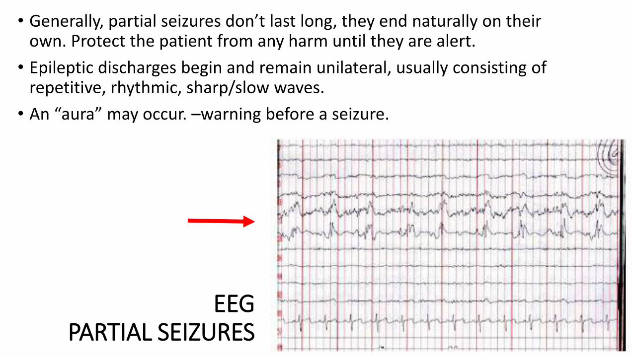
EEGPARTIAL SEIZURES
• Generally, partial seizures don’t last long, they end naturally on their own. Protect the patient from any harm until they are alert.
• Epileptic discharges begin and remain unilateral, usually consisting of repetitive, rhythmic, sharp/slow waves.
• An “aura” may occur. –warning before a seizure.


GENERALISED SEIZURES
Absence seizures – Petit-mal seizures• Start and end abruptly, usually lasts <30s
• Staring, unresponsive, frequent eye blinking, lip smacking
Voltage often max in F-C regionsSudden onset of generalized spike-and-wave complexes
Irregular slow spike-and-wave complexesNo characteristic location

TONIC-CLONIC• Tonic-clonic
• Tonic – sudden stiffness of limb, lose balance and fall
Lasts < 20s ; EEG: Rapid, high amplitude spikes
• Clonic – Repetitive jerks of muscles
Lasts longer ; EEG: rhythmic spike-wave matching the f of jerks


Myoclonic • – Single jerks/ quick series of jerks (upper shoulders)
EEG: High vol poly-spikes, last <1s, followed by slow-waves
Can be rhythmic or non-rhythmic
*Refer PBL EEG given*


•Atonic – sudden loss of muscle tone
• Eye drooping, head nodding, shoulders slumping
• Severe form “drop attack”, last < 15s
• EEG: irregular mixed waveforms. Intermittent desynchronized flattening/polyspikes

Related Documents