104.141015 104 Edvo-Kit #104 Size Determination of DNA Restriction Fragments Experiment Objective: The objective of this experiment module is to develop an understanding of principles involved in estimating the size of unknown DNA fragments by agarose gel electrophoresis. See page 3 for storage instructions.

Welcome message from author
This document is posted to help you gain knowledge. Please leave a comment to let me know what you think about it! Share it to your friends and learn new things together.
Transcript

104.141015
104Edvo-Kit #104
Size Determination of DNARestriction FragmentsExperiment Objective:
The objective of this experiment module is to develop an understanding of principles involved in estimating the size of unknown DNA fragments by agarose gel electrophoresis.
See page 3 for storage instructions.

Page
Experiment Components 3
Experiment Requirements 3
Background Information 4
Experiment Procedures Experiment Overview 6 Module I: Agarose Gel Electrophoresis 8 Module II: Staining Agarose Gels 10 Module III: Size Determination of DNA Restriction Fragments 12 Study Questions 15 Instructor's Guidelines 16 Pre-Lab Preparations 17 Experiment Results and Analysis 19 Study Questions and Answers 20
Appendices 21
Safety Data Sheets can be found on our website: www.edvotek.com/safety-data-sheets
EDVOTEK, The Biotechnology Education Company, and InstaStain are registered trademarks of EDVOTEK, Inc. Ready-to-Load, QuickStrip, FlashBlue, and UltraSpec-Agarose are trademarks of EDVOTEK, Inc.
Table of Contents
SIZE DETERMINATION OF DNA RESTRICTION FRAGMENTS EDVO-Kit 104
1.800.EDVOTEK • Fax 202.370.1501 • [email protected] • www.edvotek.com
2
Duplication of any part of this document is permitted for non-profi t educational purposes only. Copyright © 1989-2014 EDVOTEK, Inc., all rights reserved. 104.141015
EDVO-Kit 104SIZE DETERMINATION OF DNA RESTRICTION FRAGMENTS

Experiment Components
Experiment #104 is designed for 8 gels if stained with FlashBlue™ or InstaStain® Blue (both included) or 16 gels if stained with SYBR® Safe or InstaStain® Ethidium Bromide (not included).
Store QuickStrip™ samples in the refrigerator immedi-ately upon receipt. All other components can be stored at room temperature.
• Horizontal gel electrophoresis apparatus• D.C. power supply• Automatic micropipets with tips• Balance• Microwave, hot plate or burner• Pipet pump• 250 ml fl asks or beakers• Hot gloves• Safety goggles and disposable laboratory gloves• Small plastic trays or large weigh boats (for gel destaining)• DNA visualization system (white light)• Distilled or deionized water
All experiment components are intended for educational research only. They are not to be used for diagnostic or drug purposes, nor admin-istered to or consumed by humans or animals.
Requirements
READY-TO-LOAD™ SAMPLES FOR ELECTROPHORESISStore QuickStrip™ samples in the refrigerator immediately upon receipt.
All other components can be stored at room temperature.
Components (in QuickStrip™ format) Check (√)
A or D Standard DNA Marker ❑B or E Unknown DNA 1 ❑C or F Unknown DNA 2 ❑
REAGENTS & SUPPLIES
• UltraSpec-Agarose™ ❑• Electrophoresis Buffer (50x) ❑• 10x Gel Loading Solution ❑• FlashBlue™ DNA Stain ❑• InstaStain® Blue cards ❑• 1 ml pipet ❑• Microtipped Transfer Pipets ❑
SIZE DETERMINATION OF DNA RESTRICTION FRAGMENTSEDVO-Kit 104
3
1.800.EDVOTEK • Fax 202.370.1501 • [email protected] • www.edvotek.com
Duplication of any part of this document is permitted for non-profi t educational purposes only. Copyright © 1989-2014 EDVOTEK, Inc., all rights reserved. 104.141015
EDVO-Kit 104 SIZE DETERMINATION OF DNA RESTRICTION FRAGMENTS

Size determination of DNA fragments is essential to DNA mapping and analyzing restriction enzyme cleavage patterns. Restriction enzymes are endonucleases that cleave both strands of DNA at very specifi c sequences within DNA. Locations of their cleavage sites are important for DNA fi ngerprinting, determination of genetic diseases and for DNA analysis.
Agarose gel electrophoresis is a convenient analytical method for determining the size of DNA molecules in the range of 500 to 30,000 base pairs. Samples of DNA are delivered in wells made in an agarose gel, which is placed in an electrophoresis chamber containing a buffer solution and electrodes. Direct current (D.C.) is applied from a power source. Since DNA is negatively charged at neutral pH, it will migrate through the gel towards the positive electrode. The agarose gel consists of microscopic pores that act as a molecular sieve that separates DNA molecules according to their size and shape. The migration rate of DNA molecules of the same shape is inversely proportional to their size. This results in smaller DNA molecules to migrate faster through the gel. The charge to mass ratio is the same for different sized DNA molecules.
Nucleotides in DNA are linked together by negatively charged phosphodiester bonds. For every base pair (average molecular weight of approximately 660) there are two charged phosphodiester linkages. Therefore, negative charges in DNA is accompanied by approximately the same mass. The absolute amount of charge in DNA is not a critical factor in the separation process. Separation occurs because smaller molecules pass through the gel pores more easily than larger ones (i.e., the gel is sensitive to the physical size of the molecule). DNA fragment migration rate is inversely proportional to the log10 of its size in base pairs. In this experiment, DNA fragments of unknown size and Standard DNA fragments are submitted to electro-phoretic separation. The unknown DNA fragments will migrate through the gel according to their respective sizes and relative to the Standard DNA fragments. After electrophoresis, the gel is stained and the DNA bands are visualized. The migration distances of the known and unknown fragments are measured and plotted on semi-log graph paper according to their size on the y-axis versus the migration distance on the x-axis. The size of the fragments on the y-axis are expressed as the log of the number of base pairs. This allows the data to be plotted as a straight line. The DNA fragments of known size (Standard DNA fragments) are used to plot a standard curve. The migration distance of the unknown DNA fragments are estimated by extrapolation from the standard curve.
The standard fragments are used to make a standard curve by plotting their size on the y-axis versus the migra-tion distance on the x-axis. The size of the fragments on the y-axis are expressed as the log of the number of base pairs they contain or the log of their molecular weight. Most of the plotted data obtained from the mark-ers will yield a straight line. The migration distance of the unknown DNA fragment(s) are found on the X-axis and their size is estimated from the standard curve.
After determining the size of the DNA fragments generated by single and combinations of restriction enzymes, a DNA map is constructed as previously described.
In this experiment, you will determine the relative locations of three restriction enzyme cleavage sites on a circular plasmid DNA. The plasmid has been cleaved with two restriction enzymes. Enzyme 1 cleaves the plasmid once. Assume that the Enzyme 1 site is at position 0. Enzyme 2 cuts the plasmid twice. The objec-tive is to calculate the distances in base pairs between the points of cleavage and to determine whether the Enzyme 1 site is in between the Enzyme 2 sites.
Background Information
SIZE DETERMINATION OF DNA RESTRICTION FRAGMENTS EDVO-Kit 104
1.800.EDVOTEK • Fax 202.370.1501 • [email protected] • www.edvotek.com
4
Duplication of any part of this document is permitted for non-profi t educational purposes only. Copyright © 1989-2014 EDVOTEK, Inc., all rights reserved. 104.141015
EDVO-Kit 104SIZE DETERMINATION OF DNA RESTRICTION FRAGMENTS
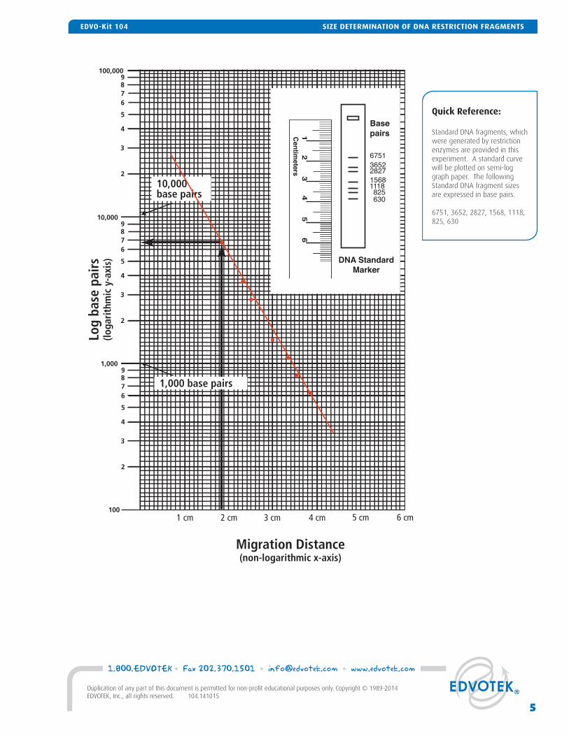
Quick Reference:
Standard DNA fragments, which were generated by restriction enzymes are provided in this experiment. A standard curve will be plotted on semi-log graph paper. The following Standard DNA fragment sizes are expressed in base pairs.
6751, 3652, 2827, 1568, 1118, 825, 630
1 cm 2 cm 3 cm 4 cm 5 cm
Migration Distance(non-logarithmic x-axis)
Log
base
pai
rs(lo
gari
thm
ic y
-axi
s)
10,000 base pairs
1,000 base pairs
1 2
5
6
3 4
Cen
timeters
67513652282715681118 825 630
Basepairs
DNA StandardMarker
6 cm
5
1.800.EDVOTEK • Fax 202.370.1501 • [email protected] • www.edvotek.com
Duplication of any part of this document is permitted for non-profi t educational purposes only. Copyright © 1989-2014 EDVOTEK, Inc., all rights reserved. 104.141015
SIZE DETERMINATION OF DNA RESTRICTION FRAGMENTSEDVO-Kit 104
EXPERIMENT OBJECTIVE:
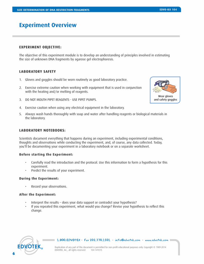
The objective of this experiment module is to develop an understanding of principles involved in estimating the size of unknown DNA fragments by agarose gel electrophoresis.
LABORATORY SAFETY
1. Gloves and goggles should be worn routinely as good laboratory practice.
2. Exercise extreme caution when working with equipment that is used in conjunction with the heating and/or melting of reagents.
3. DO NOT MOUTH PIPET REAGENTS - USE PIPET PUMPS.
4. Exercise caution when using any electrical equipment in the laboratory.
5. Always wash hands thoroughly with soap and water after handling reagents or biological materials in the laboratory.
LABORATORY NOTEBOOKS:
Scientists document everything that happens during an experiment, including experimental conditions, thoughts and observations while conducting the experiment, and, of course, any data collected. Today, you’ll be documenting your experiment in a laboratory notebook or on a separate worksheet.
Before starting the Experiment:
• Carefully read the introduction and the protocol. Use this information to form a hypothesis for this experiment.
• Predict the results of your experiment.
During the Experiment:
• Record your observations.
After the Experiment:
• Interpret the results – does your data support or contradict your hypothesis? • If you repeated this experiment, what would you change? Revise your hypothesis to refl ect this
change.
Experiment Overview
Wear gloves and safety goggles
SIZE DETERMINATION OF DNA RESTRICTION FRAGMENTS EDVO-Kit 104
1.800.EDVOTEK • Fax 202.370.1501 • [email protected] • www.edvotek.com
6
Duplication of any part of this document is permitted for non-profi t educational purposes only. Copyright © 1989-2014 EDVOTEK, Inc., all rights reserved. 104.141015
EDVO-Kit 104SIZE DETERMINATION OF DNA RESTRICTION FRAGMENTS

Experiment Overview
After electrophoresis, transfer gel for staining
InstaStain® Blue or FlashBlue™DNA stain.
Attach safety cover,connect
leads to power source and conduct
electrophoresis
Load eachsample in
consecutive wells
Remove end blocks & comb, then submerge
gel under buffer in electrophoresis
chamber
Prepare agarose gel in
casting tray
5
4
3
2
1
Gel pattern will vary depending upon experiment.
( - )( - )
( + )( + )
1 2 3 4 5 6
Analysis onwhite light
source.
7
1.800.EDVOTEK • Fax 202.370.1501 • [email protected] • www.edvotek.com
Duplication of any part of this document is permitted for non-profi t educational purposes only. Copyright © 1989-2014 EDVOTEK, Inc., all rights reserved. 104.141015
SIZE DETERMINATION OF DNA RESTRICTION FRAGMENTSEDVO-Kit 104

Module I: Agarose Gel Electrophoresis
60°C
1:001. 3.
4. 5.
7.
Caution! Flask will be HOT!
Concentratedbuffer
Distilledwater
Agarose
2.50x
Flask
60°C20min.
WAIT6.
Pour
IMPORTANT:
If you are unfamiliar with agarose gel prep and electrophoresis, detailed instructions and helpful resources are available at www.edvotek.com
Wear gloves and safety goggles
CASTING THE AGAROSE GEL
1. DILUTE concentrated 50X Electrophoresis buffer with distilled water (refer to Table A for correct volumes depending on the size of your gel casting tray).
2. MIX agarose powder with buffer solution in a 250 ml fl ask (refer to Table A).3. DISSOLVE agarose powder by boiling the solution. MICROWAVE the solution on high for 1 minute. Care-
fully REMOVE the fl ask from the microwave and MIX by swirling the fl ask. Continue to HEAT the solution in 15-second bursts until the agarose is completely dissolved (the solution should be clear like water).
4. COOL agarose to 60° C with careful swirling to promote even dissipation of heat.5. While agarose is cooling, SEAL the ends of the gel-
casting tray with the rubber end caps. PLACE the well template (comb) in the appropriate notch.
6. POUR the cooled agarose solution into the pre-pared gel-casting tray. The gel should thoroughly solidify within 20 minutes. The gel will stiffen and become less transparent as it solidifi es.
7. REMOVE end caps and comb. Take particular care when removing the comb to prevent damage to the wells.
ConcentratedBuffer (50x)
Size of GelCasting tray
7 x 7 cm
7 x 10 cm
7 x 14 cm
0.6 ml
1.0 ml
1.2 ml
+DistilledWater
29.4 ml
49.0 ml
58.8 ml
+TOTALVolume
30 ml
50 ml
60 ml
=
Individual 0.8% UltraSpec-Agarose™ Gel
Amt ofAgarose
0.23 g
0.39 g
0.46 g
Table
A
SIZE DETERMINATION OF DNA RESTRICTION FRAGMENTS EDVO-Kit 104
1.800.EDVOTEK • Fax 202.370.1501 • [email protected] • www.edvotek.com
8
Duplication of any part of this document is permitted for non-profi t educational purposes only. Copyright © 1989-2014 EDVOTEK, Inc., all rights reserved. 104.141015
EDVO-Kit 104SIZE DETERMINATION OF DNA RESTRICTION FRAGMENTS

Module I: Agarose Gel Electrophoresis
1X DilutedBuffer
8. 9.
10. 11.
Pour
Lane 1
2
3
Tube A or D
Tube B or E
Tube C or F
Table 1: Gel Loading
Standard DNA Marker
Unknown DNA 1
Unknown DNA 2
REMINDER:Before loading the samples, make sure the gel is properly oriented in the ap-paratus chamber.
Wear gloves and safety goggles
RUNNING THE GEL
8. PLACE the gel (still on the tray) into the electrophoresis chamber. COVER the gel with 1X Electrophoresis Buffer (See Table B for recommended volumes). The gel should be completely submerged.
9. PUNCTURE the foil overlay of the QuickStrip™ with a pipet tip. LOAD the entire sample (35 μl) into the well in the order indicated by Table 1, at right.
10. PLACE safety cover on the unit. CHECK that the gel is properly oriented. Remember, the DNA samples will migrate toward the positive (red) electrode.
11. CONNECT leads to the power source and PERFORM electrophoresis (See Table C for time and voltage guidelines). Allow the tracking dye to migrate at least 3.5 cm from the wells.
12. After electrophoresis is complete, REMOVE the gel and casting tray from the electrophoresis chamber and proceed to instruc-tions for STAINING the agarose gel.
Time & Voltage Guidelines (0.8% Agarose Gel)
Min. / Max.Volts
150 125 75
15/20 min. 20/30 min. 35 / 45 min.
Table
CElectrophoresis Model
M6+ M12 (classic)& M36
Min. / Max.
20/30 min. 30/35 min. 55/70 min.
M12 (new)
Min. / Max.
25 / 35 min. 35 / 45 min. 60 / 90 min.
50x Conc.Buffer
DistilledWater+
EDVOTEKModel #
Total Volume Required
1x Electrophoresis Buffer (Chamber Buffer)
M6+ & M12 (new)
M12 (classic)
M36
300 ml
400 ml
1000 ml
Dilution
Table
B
6 ml
8 ml
20 ml
294 ml
392 ml
980 ml
9
1.800.EDVOTEK • Fax 202.370.1501 • [email protected] • www.edvotek.com
Duplication of any part of this document is permitted for non-profi t educational purposes only. Copyright © 1989-2014 EDVOTEK, Inc., all rights reserved. 104.141015
SIZE DETERMINATION OF DNA RESTRICTION FRAGMENTSEDVO-Kit 104

Module II-A: Staining Agarose Gels Using FlashBlue™
STAIN
1.
4.3.
ConcentratedFlashBlue™ Stain
Distilledwater
2.10x
Pour
Flask
5.
5min.
DESTAIN
20min.
Pour
( - )( - )
( + )( + )
1 2 3 4 5 6
1. DILUTE 10 ml of 10x concentrated FlashBlue™ with 90 ml of water in a fl ask and MIX well.2. REMOVE the agarose gel and casting tray from the electrophoresis chamber. SLIDE the gel off of the cast-
ing tray into a small, clean gel-staining tray. 3. COVER the gel with the 1x FlashBlue™ stain solution. STAIN the gel for 5 minutes. For best results, use an
orbital shaker to gently agitate the gel while staining. STAINING THE GEL FOR LONGER THAN 5 MINUTES WILL REQUIRE EXTRA DESTAINING TIME.
4. TRANSFER the gel to a second small tray. COVER the gel with water. DESTAIN for at least 20 minutes with gentle shaking (longer periods will yield better results). Frequent changes of the water will acceler-ate destaining.
5. Carefully REMOVE the gel from the destaining liquid. VISUALIZE results using a white light visualization system. DNA will appear as dark blue bands on a light blue background.
ALTERNATIVE PROTOCOL:
1. DILUTE one ml of concentrated FlashBlue™ stain with 149 ml dH2O. 2. COVER the gel with diluted FlashBlue™ stain. 3. SOAK the gel in the staining liquid for at least three hours. For best results, stain gels overnight.
Wear gloves and safety goggles
SIZE DETERMINATION OF DNA RESTRICTION FRAGMENTS EDVO-Kit 104
1.800.EDVOTEK • Fax 202.370.1501 • [email protected] • www.edvotek.com
10
Duplication of any part of this document is permitted for non-profi t educational purposes only. Copyright © 1989-2014 EDVOTEK, Inc., all rights reserved. 104.141015
EDVO-Kit 104SIZE DETERMINATION OF DNA RESTRICTION FRAGMENTS

Module II-B: Staining Agarose Gels Using InstaStain® Blue
Wear gloves and safety goggles
1. Carefully REMOVE the agarose gel and casting tray from the electrophoresis chamber. SLIDE the gel off of the casting tray on to a piece of plastic wrap on a fl at surface.
2. MOISTEN the gel with a few drops of electrophoresis buffer.3. Wearing gloves, PLACE the blue side of the InstaStain® Blue card on the gel. 4. With a gloved hand, REMOVE air bubbles between the card and the gel by fi rmly run-
ning your fi ngers over the entire surface. Otherwise, those regions will not stain.5. PLACE the casting tray on top of the gel/card stack. PLACE a small weight (i.e. an
empty glass beaker) on top of the casting tray. This ensures that the InstaStain® Blue card is in direct contact with the gel surface. STAIN the gel for 10 minutes.
6. REMOVE the InstaStain® Blue card. If the color of the gel appears very light, reapply the InstaStain® Blue card to the gel for an additional fi ve minutes.
7. TRANSFER the gel to a small, clean gel-staining tray. COVER the gel with about 75 mL of distilled water and DESTAIN for at least 20 minutes. For best results, use an orbital shaker to gently agitate the gel while staining. To accelerate destaining, warm the distilled water to 37°C and change it frequently.
8. Carefully REMOVE the gel from the destaining liquid. VISUALIZE results using a white light visualization system. DNA will appear as dark blue bands on a light blue background.
75 ml
Moistenthe gel
1. 2. 4.
5. 6.
3.
10min.
STAIN
InstaStain® Blue
U.S. Patent Pending
InstaStain® Ethid
U.S. Patent Pending
InstaStain® Ethidium Bromide
U.S. Patent Pending
-----
InstaStain® Blue
U.S. Patent Pending
( - )( - )
( + )( + )
7. 8.
( - )
( + )
20min.
DESTAIN
or overnight
ALTERNATIVE PROTOCOL:
1. Carefully SLIDE the agarose gel from its casting tray into a small, clean tray containing about 75 ml of dis-tilled/deionized water or used electrophoresis buffer. The gel should be completely submerged.
2. Gently FLOAT the InstaStain® Blue card(s) on top of the liquid with the stain (blue side) facing toward the gel. Each InstaStain® Blue card will stain 49 cm2 of gel (7 x 7 cm).
3. COVER the tray with plastic wrap to prevent evaporation. SOAK the gel in the staining liquid for at least 3 hours. The gel can remain in the liquid overnight if necessary.
4. Carefully REMOVE the gel from the staining tray. VISUALIZE results using a white light visualization system. DNA will appear as dark blue bands on a light blue background.
NOTE:DO NOT STAIN GELS IN THE
ELECTROPHORESIS APPARATUS.
SIZE DETERMINATION OF DNA RESTRICTION FRAGMENTSEDVO-Kit 104
11
1.800.EDVOTEK • Fax 202.370.1501 • [email protected] • www.edvotek.com
Duplication of any part of this document is permitted for non-profi t educational purposes only. Copyright © 1989-2014 EDVOTEK, Inc., all rights reserved. 104.141015
EDVO-Kit 104 SIZE DETERMINATION OF DNA RESTRICTION FRAGMENTS

Quick Reference:Standard DNA fragment sizes - length is expressed in base pairs.
6751, 3652, 2827, 1568, 1118, 825, 630
Agarose gel electrophoresis separates biomolecules into discrete bands, each com-prising molecules of the same size. How can these results be used to determine the lengths of different fragments? Remember, as the length of a biomolecule increases, the distance to which the molecule can migrate decreases because large molecules cannot pass through the channels in the gel with ease. Therefore, the migration rate is inversely proportional to the length of the molecules—more specifi cally, to the log10 of molecule's length. To illustrate this, we ran a sample that contains bands of known lengths called a “standard”. We will measure the distance that each of these bands traveled to create a graph, known as a “standard curve”, which can then be used to extrapolate the size of unknown molecule(s).
1. Measure and Record Migration Distances
Measure the distance traveled by each Stan-dard DNA Fragment from the lower edge of the sample well to the lower end of each band. Record the distance in centimeters (to the nearest millimeter) in your note-book. Repeat this for each DNA fragment in the standard.
Measure and record the migration distances of each of the fragments in the unknown samples in the same way you measured the standard bands.
2. Generate a Standard Curve.
Because migration rate is inversely propor-tional to the log10 of band length, plotting the data as a semi-log plot will produce a straight line and allow us to analyze an exponential range of fragment sizes. You will notice that the vertical axis of the semi-log plot appears atypical at fi rst; the dis-tance between numbers shrinks as the axis progresses from 1 to 9. This is because the axis represents a logarithmic scale. The fi rst cycle on the y-axis corresponds to lengths from 100-1,000 base pairs, the second cycle measures 1,000-10,000 base pairs, and so on. To create a standard curve on the semi-log paper, plot the distance each Standard DNA fragment migrated on the x-axis (in mm) versus its size on the y-axis (in base pairs). Be sure to label the axes!
Figure 1: Semilog graph example
Module III: Size Determination of DNA Restriction Fragments
SIZE DETERMINATION OF DNA RESTRICTION FRAGMENTS EDVO-Kit 104
1.800.EDVOTEK • Fax 202.370.1501 • [email protected] • www.edvotek.com
12
Duplication of any part of this document is permitted for non-profi t educational purposes only. Copyright © 1989-2014 EDVOTEK, Inc., all rights reserved. 104.141015
EDVO-Kit 104SIZE DETERMINATION OF DNA RESTRICTION FRAGMENTS

After all the points have been plotted, use a ruler or a straight edge to draw the best straight line possible through the points. The line should have approximately equal numbers of points scattered on each side of the line. It is okay if the line runs through some points (see Figure 1 for an example).
3. Determine the length of each unknown fragment.
a. Locate the migration distance of the unknown fragment on the x-axis of your semi-log graph. Draw a vertical line extending from that point until it intersects the line of your standard curve.
b. From the point of intersection, draw a second line, this time horizontally, toward the y-axis. The value at which this line intersects the y-axis represents the approximate size of the fragment in base pairs (refer to Figure 1 for an example). Make note of this in your lab notebook.
c. Repeat for each fragment in your unknown sample.
13
1.800.EDVOTEK • Fax 202.370.1501 • [email protected] • www.edvotek.com
Duplication of any part of this document is permitted for non-profi t educational purposes only. Copyright © 1989-2014 EDVOTEK, Inc., all rights reserved. 104.141015
SIZE DETERMINATION OF DNA RESTRICTION FRAGMENTSEDVO-Kit 104

8,000
10,000
7,000
6,000
5,000
4,000
3,000
2,000
9,000
80 70
60
50
40
30
20
10
90 100
1,000
800 700
600
500
400
300
200
900
X-axis: Migration distance (cm)
1 cm 2 cm 3 cm 4 cm 5 cm 6 cm
Y-a
xis:
Ba
se P
airs
1.800.EDVOTEK • Fax 202.370.1501 • [email protected] • www.edvotek.com
14
Duplication of any part of this document is permitted for non-profi t educational purposes only. Copyright © 1989-2014 EDVOTEK, Inc., all rights reserved. 104.141015
SIZE DETERMINATION OF DNA RESTRICTION FRAGMENTS EDVO-Kit 104

Study Questions
1. How should the x and y axes of the semi-log graph paper used in this experiment, be labeled?
2. Determine Unknown 1 and Unknown 2 DNA fragment sizes in base pairs according to your stan-dard curve, then determine your percentage error.
3. Use the example of the standard curve to determine:
a. The base pair size if the migration distance is 2.8 centimeters.
b. The migration distance if the fragment contains 5,500 base pairs.
SIZE DETERMINATION OF DNA RESTRICTION FRAGMENTSEDVO-Kit 104
15
1.800.EDVOTEK • Fax 202.370.1501 • [email protected] • www.edvotek.com
Duplication of any part of this document is permitted for non-profi t educational purposes only. Copyright © 1989-2014 EDVOTEK, Inc., all rights reserved. 104.141015
EDVO-Kit 104 SIZE DETERMINATION OF DNA RESTRICTION FRAGMENTS
Instructor's Guide
1.800.EDVOTEK • Fax 202.370.1501 • [email protected] • www.edvotek.com
16
Duplication of any part of this document is permitted for non-profi t educational purposes only. Copyright © 1989-2014 EDVOTEK, Inc., all rights reserved. 104.141015
INSTRUCTOR'S GUIDE SIZE DETERMINATION OF DNA RESTRICTION FRAGMENTS EDVO-Kit 104
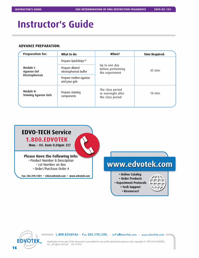
ADVANCE PREPARATION:
Preparation for: What to do: Time Required:When?
Prepare QuickStrips™Up to one day before performingthe experiment
45 min.Module I: Agarose Gel Electrophoresis
Module II: Staining Agarose Gels
Prepare diluted electrophoresis buffer
The class periodor overnight afterthe class period
10 min.Prepare stainingcomponents
Prepare molten agaroseand pour gels

Pre-Lab Preparations: Module I
AGAROSE GEL ELECTROPHORESIS
This experiment requires a 0.8% agarose gel per student group. You can choose whether to prepare the gels in advance or have the students prepare their own. Allow approximately 30-40 minutes for this procedure.
Individual Gel Preparation:
Each student group can be responsible for casting their own individual gel prior to conducting the experiment. See Module I in the Student’s Experimental Procedure. Students will need 50x Electrophoresis Buffer, distilled water and agarose powder.
Batch Gel Preparation:
To save time, a larger quantity of agarose solution can be prepared for sharing by the class. Electrophoresis buffer can also be prepared in bulk. See Appendix B.
Preparing Gels in Advance:
Gels may be prepared ahead and stored for later use. Solidifi ed gels can be stored under buffer in the refrigerator for up to 2 weeks.
Do not freeze gels at -20º C as freezing will destroy the gels.
Gels that have been removed from their trays for storage should be “anchored” back to the tray with a few drops of molten agarose before being placed into the tray. This will prevent the gels from sliding around in the trays and the chambers.
FOR MODULE IEach Student Groupshould receive:• 50x Electrophoresis Buffer• Distilled Water • UltraSpec-Agarose™• QuickStrip™ Samples
NOTE:Accurate pipetting is critical for maximizing successful experi-ment results. EDVOTEK Series 100 experiments are designed for students who have had previous experience with micropipetting techniques and agarose gel electrophoresis.
If students are unfamiliar with using micropipets, we recom-mended performing Cat. #S-44, Micropipetting Basics or Cat. #S-43, DNA DuraGel™ prior to conducting this advanced level experiment.
SAMPLES FORMAT: PREPARING THE QUICKSTRIPS™
QuickStrip™ tubes consist of a microtiter block covered with a pro-tective overlay. Each well contains pre-aliquoted DNA.
Using sharp scissors, fi rst divide the block of tubes into individual strips by cutting between the rows (see diagram at right). Next, cut each individual strip between wells C and D and wells F and G. Take care not to damage the protective overlay while separating the samples.
Each lab group will receive one set of tubes, either rows A-C or D-F. • A & D contain Standard DNA Marker • B & E Unknown DNA 1 • C & F Unknown DNA 2 • G & H are intentionally left blank. (Discard these tubes.)
If using SYBR® Safe or InstaStain® Ethidium Bromide for DNA vi-sualization, each QuickStrip™ is shared by two groups. 18 μl of the DNA sample will be loaded into each well. Proceed to visualize the results as specifi ed by the DNA stain literature.
Carefully cut between each set of tubes.Then, between rows C & D and F & G.
A
B
C
D
E
F
G
H
CU
T H
ERE
A
B
C
D
E
F
G
H
CU
T H
ERE
A
B
C
D
E
F
G
H
CU
T H
ERE
CU
T H
ERE
A
B
C
D
E
F
G
H
CU
T H
ERE
A
B
C
D
E
F
G
H
A
B
C
D
E
F
G
H
CUT HERE
CUT HERE
17
1.800.EDVOTEK • Fax 202.370.1501 • [email protected] • www.edvotek.com
Duplication of any part of this document is permitted for non-profi t educational purposes only. Copyright © 1989-2014 EDVOTEK, Inc., all rights reserved. 104.141015
INSTRUCTOR'S GUIDEEDVO-Kit 104 SIZE DETERMINATION OF DNA RESTRICTION FRAGMENTS

Pre-Lab Preparations: Module II
MODULE II-A: STAINING AGAROSE GELS WITH INSTASTAIN® BLUE
The easiest and most convenient DNA stain available is InstaStain® Blue. InstaStain® Blue does not require the formulation, storage and disposal of large volumes of liquid stain. Each InstaStain® Blue card contains a small amount of blue DNA stain. When the card is placed in water, the DNA stain is released. This solution simultaneously stains and destains the gel, providing uniform gel staining with minimal liquid waste and mess.
You can use a White Light Visualization System (Cat. #552) to visualize gels stained with InstaStain® Blue.
MODULE II-B: STAINING AGAROSE GELS WITH FLASHBLUE™
FlashBlue™ stain is optimized to shorten the time required for both staining and de-staining steps. Agarose gels can be stained with diluted FlashBlue™ for 5 minutes and destained for only 20 minutes. For the best results, leave the gel in liquid overnight. This will allow the stained gel to “equilibrate” in the destaining solution, resulting in dark blue DNA bands contrasting against a uniformly light blue background. A white light box (Cat. #552) is recommended for visualizing gels stained with FlashBlue™.
• Stained gels may be stored in destaining liquid for several weeks with refrigera-tion, although the bands may fade with time. If this happens, re-stain the gel.
• Destained gels can be discarded in solid waste disposal. Destaining solutions can be disposed of down the drain.
MODULE II: PHOTODOCUMENTATION OF DNA (OPTIONAL)
Once gels are stained, you may wish to photograph your results. There are many different photodocumentation systems available, including digital systems that are interfaced directly with computers. Specifi c instructions will vary depending upon the type of photodocumentation system you are using.
FOR MODULE II-AEach Student Groupshould receive:• 1 InstaStain® card per 7 x 7 cm gel
FOR MODULE II-BEach Student Groupshould receive:• 10 ml 10X concentrated FlashBlue OR 100 mL
1x diluted FlashBlue• Small plastic tray or
weight boat• Distilled or deionized
water
Wear gloves and safety goggles
1.800.EDVOTEK • Fax 202.370.1501 • [email protected] • www.edvotek.com
18
Duplication of any part of this document is permitted for non-profi t educational purposes only. Copyright © 1989-2014 EDVOTEK, Inc., all rights reserved. 104.141015
INSTRUCTOR'S GUIDE SIZE DETERMINATION OF DNA RESTRICTION FRAGMENTS EDVO-Kit 104

Experiment Results and Analysis
In the idealized schematic, the relative positions of DNA fragments are shown but are not depicted to scale.
Lane Tube Sample Molecular Weights (in bp)
1 A or D DNA Standard --------- Markers
2 B or E Unknown DNA 1 4282
3 C or F Unknown DNA 2 3000, 1282
19
1.800.EDVOTEK • Fax 202.370.1501 • [email protected] • www.edvotek.com
Duplication of any part of this document is permitted for non-profi t educational purposes only. Copyright © 1989-2014 EDVOTEK, Inc., all rights reserved. 104.141015
INSTRUCTOR'S GUIDEEDVO-Kit 104 SIZE DETERMINATION OF DNA RESTRICTION FRAGMENTS

Please refer to the kit insert for the Answers to
Study Questions
A EDVOTEK® Troubleshooting Guide
B Bulk Preparation of Agarose Gels
Safety Data Sheets can be found on our website: www.edvotek.com/safety-data-sheets
Appendices
21
1.800.EDVOTEK • Fax 202.370.1501 • [email protected] • www.edvotek.com
Duplication of any part of this document is permitted for non-profi t educational purposes only. Copyright © 1989-2014 EDVOTEK, Inc., all rights reserved. 104.141015
APPENDICESEDVO-Kit 104 SIZE DETERMINATION OF DNA RESTRICTION FRAGMENTS

Appendix AEDVOTEK® Troubleshooting Guides
PROBLEM: CAUSE: ANSWER:
After staining the gel, the DNA bands are faint.
The gel was not stained for a sufficient period of time. Repeat staining protocol.
Bands are not visible on the gel.
The gel was not prepared properly.
The gel was not stained properly.
Ensure that the electrophoresis buffer was correctly diluted.
Repeat staining.
The background of gel is too dark. Destain the gel for 5-10 minutes in distilled water.
Contact the manufacturer of the electrophoresis unit or power source.
DNA bands were not resolved.
Tracking dye should migrate at least 3.5 cm (if using a 7x7 cm tray), and at least 6 cm (if using a 7x14 cm tray) from the wells to ensure adequate separation.
Be sure to run the gel at least 6 cm before staining and visualizing the DNA (approximately one hour at 125 V).
There’s not enough sample in my QuickStrip™. The QuickStrip™ has dried out. Add 40 µL water, gently pipet up and down to mix before
loading.
DNA bands fade when gels are kept at 4°C.
DNA stained with FlashBlue™ may fade with time Re-stain the gel with FlashBlue™
There is no separationbetween DNA bands,even though the trackingdye ran the appropriate distance.
The wrong percent gel was used for electrophoretic separation.
Be sure to prepare the correct percent agarose gel. Forreference, the Ready-to-Load™ DNA samples should be analyzed using a 0.8% agarose gel.
Malfunctioning electrophoresis unit orpower source.
1.800.EDVOTEK • Fax 202.370.1501 • [email protected] • www.edvotek.com
22
Duplication of any part of this document is permitted for non-profi t educational purposes only. Copyright © 1989-2014 EDVOTEK, Inc., all rights reserved. 104.141015
APPENDICES SIZE DETERMINATION OF DNA RESTRICTION FRAGMENTS EDVO-Kit 104

Appendix B
To save time, the electrophoresis buffer and agarose gel solution can be prepared in larger quantities for sharing by the class. Unused diluted buffer can be used at a later time and solidifi ed agarose gel solution can be remelted.
Bulk Preparation of Agarose Gels
Bulk Electrophoresis Buffer
Quantity (bulk) preparation for 3 liters of 1x electro-phoresis buffer is outlined in Table D.
Batch Agarose Gels (0.8%)
For quantity (batch) preparation of 0.8% agarose gels, see Table E.
1. Use a 500 ml fl ask to prepare the diluted gel buffer.
2. Pour 3.0 grams of UltraSpec-Agarose™ into the prepared buffer. Swirl to disperse clumps.
3. With a marking pen, indicate the level of solution volume on the outside of the fl ask.
4. Heat the agarose solution as outlined previously for individual gel preparation. The heating time will require adjustment due to the larger total volume of gel buf-fer solution.
5. Cool the agarose solution to 60°C with swirling to promote even dissipation of heat. If evaporation has occurred, add distilled water to bring the solution up to the original volume as marked on the fl ask in step 3.
6. Dispense the required volume of cooled agarose solution for casting each gel. Mea-sure 30 ml for a 7 x 7 cm tray, 50 ml for a 7 x 10 cm tray, and 60 ml for a 7 x 14 cm tray. For this experiment, 7 x 7 cm gels are recommended.
7. Allow the gel to completely solidify. It will become fi rm and cool to the touch after approxi-mately 20 minutes. Then proceed with preparing the gel for electrophoresis.
60˚C
Note: The UltraSpec-Agarose™ kit component is usually labeled with the amount it contains. Please read the label care-fully. If the amount of aga-rose is not specifi ed or if the bottle's plastic seal has been broken, weigh the agarose to ensure you are using the correct amount.
50x Conc.Buffer +
DistilledWater
Total Volume Required
60 ml 2,940 ml 3000 ml (3 L)
Bulk Preparation of Electrophoresis BufferTable
D
Batch Prep of 0.8% UltraSpec-Agarose™Table
EAmt ofAgarose
(g)
ConcentratedBuffer (50X)
(ml)+
DistilledWater(ml)
TotalVolume
(ml)+
3.0 7.5 382.5 390
23
1.800.EDVOTEK • Fax 202.370.1501 • [email protected] • www.edvotek.com
Duplication of any part of this document is permitted for non-profi t educational purposes only. Copyright © 1989-2014 EDVOTEK, Inc., all rights reserved. 104.141015
APPENDICESEDVO-Kit 104 SIZE DETERMINATION OF DNA RESTRICTION FRAGMENTS
Related Documents