Stargardt Disease: The View From 2011 Janet S. Sunness, MD Education and Research from Envision Volume 5 | Issue 4 S targardt disease is the most common cause of macular degeneration and central visual loss in young people. Stargardt disease often develops in the teens or twen- ties, but may develop in younger children (the youngest child I have seen is 5) or somewhat later in life. It affects both eyes at the same time, though one may be somewhat worse than the other. In most people it affects only central vision (read- ing, recognizing faces and other fine tasks), but in certain more rare types, peripheral vision may be affected as well. Vision loss tends to progress rapidly at first, but for many people it tends to stabilize at about 20/200. There is also a form of this condition that begins later in life and tends to progress more slowly. DIAGNOSING STARGARDT DISEASE Patients with Stargardt disease have findings that are different from patients with age-related macular degeneration, and some terminology is worth knowing. Many patients with Stargardt disease have yellow flecks in their retina. In these patients, the disease is also known as fundus flavimaculatus (the retina with the yellow spots). The macula, the cen- ter of the retina, often has a shiny appearance, which is referred to as a “beaten bronze appearance,” and unlike age-related macular degeneration, it is often difficult to determine just by looking at some- one how much of a blind spot the patient may have. The fluorescein angiogram (when dye is injected into the arm and pictures of the retina are taken) often shows an unusual appearance called a “dark choroid,” meaning that the initial flush of dye which goes through the blood vessel system beneath the retina cannot be seen. Some patients with Stargardt disease start out with a bullseye appearance to the macula, in which there is a small island of spared vision surrounded by a donut- shaped blind spot. In other patients, or later in the course of the disease,

Welcome message from author
This document is posted to help you gain knowledge. Please leave a comment to let me know what you think about it! Share it to your friends and learn new things together.
Transcript
Stargardt Disease: The View From 2011 Janet S. Sunness, MD
Education and Research from Envision
Volume 5 | Issue 4
Stargardt disease is the most common cause of macular
degeneration and central visual loss in young people. Stargardt disease often develops in the teens or twen-ties, but may develop in younger children (the youngest child I have seen is 5) or somewhat later in life. It affects both eyes at the same time, though one may be somewhat worse than the other. In most people it affects only central vision (read-ing, recognizing faces and other fine tasks), but in certain more rare types, peripheral vision may be affected as well. Vision loss tends to progress rapidly at first, but for many people it tends to stabilize at about 20/200. There is also a form
of this condition that begins later in life and tends to progress more slowly.
DIAGNOSING STARGARDT DISEASE
Patients with Stargardt disease have findings that are different from patients with age-related macular degeneration, and some terminology is worth knowing. Many patients with Stargardt disease have yellow flecks in their retina. In these patients, the disease is also known as fundus flavimaculatus (the retina with the yellow spots). The macula, the cen-ter of the retina, often has a shiny appearance, which is referred to as a “beaten bronze appearance,”
and unlike age-related macular degeneration, it is often difficult to determine just by looking at some-one how much of a blind spot the patient may have. The fluorescein angiogram (when dye is injected into the arm and pictures of the retina are taken) often shows an unusual appearance called a “dark choroid,” meaning that the initial flush of dye which goes through the blood vessel system beneath the retina cannot be seen. Some patients with Stargardt disease start out with a bullseye appearance to the macula, in which there is a small island of spared vision surrounded by a donut-shaped blind spot. In other patients, or later in the course of the disease,

Professional Education
Feature Article
ABOUT ENVISION FOUNDATIONThe mission of the Envision Foundation is to secure funding for the successful delivery of services offered by the Envision Vision Rehabilitation Center and the education programs of the Foundation. Envision Foundation focuses on fundraising to ensure that no patient is ever turned away—regardless of ability to pay; public education to help prevent blindness; and professional education to determine best practices in order to serve patients who are blind or low vision.
To submit an article or case study to be considered for publication in Visibility, please contact Michael Epp, Director of Professional Education, at (316) 440-1515 or [email protected].
REQUEST COPIES OF VISIBILITYIf you would like to share Visibility with a colleague, please request a copy from Michael Epp, Director of Professional Education, at [email protected] or call (316) 440-1515. Visibility is also available online at www.envisionus.com/Visibility.
The viewpoints expressed by the guest authors of Visibility do not necessarily reflect the viewpoints of Envision or its staff.
Visibility is a quarterly publication of the Envision Foundation.
610 N. Main, Wichita, KS 67203(316) 440-1600www.envisionus.comServicios bilingües disponibles:(316) 440-1660
EDITORAL STAFFMichael Epp, MS, Director of Professional Education Kelsey Rawson, Professional Education Associate
Shannon Riley, MA, Research and Analytics Associate
Kathi A. Buche, Graphic Design Manager
GUEST CONTRIBUTORS Janet S. Sunness, MD William L. Park, MD
ENVISION
2 | Table of Contents
Stargardt Disease: The View From 2011Janet S. Sunness, MD
Utilizing Red Contact Lenses for Retinal DystrophiesWilliam Park, OD, FAAO
Case Report: How the Amber-Red Contact Lens Changed My LifeMarica Harris, CLVT, St. Louis Veterans Medical Center
Cataracts: An Overview for the Low Vision ProfessionalDasa V. Gangadhar, MD
Envision Conference 2011: Resounding Success!
Envision Conference Research Session Highlights
16
8
17
Case Study
12
Copyright © 2011 Envision Foundation. Individual articles are Copyright © 2011 of the indicated authors, printed with permission. All rights reserved. No part of this publication may be reproduced or transmitted in any form or by any means, electronic or mechanical, including photocopying, recording or any information storage or retrieval system, without written permission of Envision Foundation.
Marica Harris, CLVTDasa V. Gangadhar, MD
Disease Etiology
Continuing Education
18Focus on Research
Stargardt Disease: The View From 2011 continued from front page
Vis ib i l i ty | Vol . 5 , Issue 4
Feature Ar t ic le | 3
there is a solid area of blind spot which does not spare the very center.
DEALING WITH BLIND SPOTS IN THE CENTRAL VISION
It is critical for any patient with blind spots in or near the center of vision to learn how to move the eye so that the object of interest is imaged onto a seeing part of the retina. Most Stargardt patients do this quite well and tend to look slightly above what they want to see. This has the effect of moving the blind spot up and out of the way. The difficulty is that as one uses retina farther from the center of the macula, larger letters are needed in order to see; magnification, using a variety of low vision devices, can be very helpful.
A diagnostic device called a microperimeter can allow us to plot out the exact location of the blind spot directly on a video image of the macula. This information helps us to advise patients on strategies for using the remaining seeing retina in the most effective way. Other spe-cialized imaging, using infrared and autofluorescence imaging, allows us to determine the stage and progres-sion of the condition more compre-hensively.
LIVING WITH STARGARDT DISEASE
Most patients whom I see with Stargardt disease have adapted quite well; they lead busy and active lifestyles. Among these patients is a scientist, radio announcer, a social worker, and a tandem bike racing
competitor. They have rewarding personal and professional lives. Improved low vision interventions in the future will help patients to adapt even better to this condition. It is important to remember that a loss of vision is a real loss to the indi-vidual, and some people may react to this with anger and depression. While these are normal stages many
patients go through, these emotions may interfere with the person’s abil-ity to adapt and see around the blind spot. If there is difficulty in working through these emotions, support groups and counseling may be beneficial. Support groups are very helpful in general for understanding the symptoms of central visual loss and for learning different ways of coping.
Some patients with Stargardt disease are able to drive. Each state has different rules in terms of the mini-mum visual acuity required to get a restricted license. Patients who meet this vision requirement can work with special driving instructors who can instruct them and determine whether they can drive safely. GPS systems are probably the best device for drivers
with somewhat reduced central vision. Patients can also learn how to use a bioptic telescope to read street signs and traffic signals.
THE GENETICS OF STARGARDT DISEASE
Stargardt disease is an inherited, or genetic, disease; that is, it is caused by a mutation of a gene. Most patients I see with Stargardt
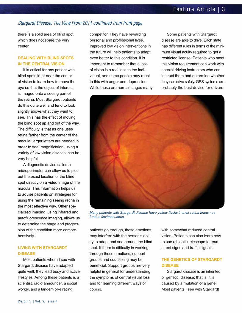
Many patients with Stargardt disease have yellow flecks in their retina known as fundus flavimaculatus.

4 | Feature Ar t ic le
ENVISION
are the only ones in their family to have the condition, and they are likely to have the autosomal reces-sive form, meaning that both copies of the gene (one from the mother and one from the father) are abnor-mal. The children of these patients in all likelihood will be carriers of the disease (have one abnormal gene) but will not be affected, assuming that the other parent is not a car-rier. Patients may have siblings who are affected, but their parents and children are not likely to be affected. There is also a rarer autosomal
dominant form, meaning that only one copy of the gene has to be abnormal. In the dominant form, one parent of the patient also has the disease and other siblings or family members may have it as well.
Researchers have found that mutations in a gene called ABCA4 are responsible for at least 75 percent of the cases of Stargardt disease. This gene is responsible for helping transport vitamin A derivatives (retinoids) from the visual cycle, so that these retinoids can be regenerated and reused. When the retinoids cannot be properly trans-
ported, it leads to an accumulation of a material called lipofuscin that may be toxic to the retinal pigment epithelium, the cells under the retina needed to sustain vision. The high levels of accumulation begin at birth; it is not clear what triggers the cells to start degenerating.
ADVANCES IN POTENTIAL TREATMENTS FOR STARGARDT DISEASE
The ABCA4 gene responsible for Stargardt disease was discovered in 1997.1 A great deal of progress has
been made since that time. Scientists have been able to develop mice that are missing the ABCA4 gene (so-called knockout mice). These mice accumulate large amounts of lipofus-cin, as do people. But since mice retinas do not have
the specific structure of a macula, they do not undergo the degenera-tion seen in people. Scientists have studied whether various drugs or other interventions can slow or stop the accumulation of the abnormal material in these mice. They have found that if you do not expose the mice to light, there is less lipofus-cin accumulation. It is difficult to know how this applies to people, but some investigators recommend that patients wear sunglasses when they are outdoors. They also found that giving the mice Acutane (a vitamin A derivative used for severe acne that
has serious complications such as birth defects and psychiatric side ef-fects) slowed down the accumulation of the abnormal material. Research is ongoing to find safe therapies that may help in this condition.
In the last few years, the first successful animal and human gene therapy trials have been conducted for a severe retinal degeneration that begins at birth (Lebers Con-genital Amaurosis, associated with an RPE65 mutation). Normal copies of the gene are attached to virus vectors, which help to gain entry of the genetic material into the cells of the patient. This is injected underneath the retina. Similar gene therapy, using normal copies of the ABCA4 gene and a different virus vector, has been successful in the knockout mouse model. Since there is no naturally occurring Stargardt disease known in animals, scientists have used dogs that do not have a mutation to test that the gene can enter the desired cell type. These studies have gone well.2 In 2011, the StarGen study of gene therapy for Stargardt disease began. The first phase is a safety study. It will involve follow up of patients for about a year to look for any evidence of side effects or complications affect-ing the eye or any other part of the person. For this phase, only patients with very severe vision loss will be enrolled.3
A second clinical trial that began in 2011 uses stem cells to treat Stargardt disease (NCT01345006; Advanced Cell Technology; see clini-caltrials.gov). Stem cells are trans-formed into retinal pigment epithelial
A student with Stargardt disease uses a computer with assistive technology

cells. These cells are then injected under the retina. The initial phase is to assess safety with various num-bers of cells injected (beginning with 50,000). Since this has not before been performed, it is not known to what extent there will be an inflam-matory reaction to or rejection of the cells, or what other side effects will occur. This study is therefore only enrolling patients with very severe vision loss at this time. While the ul-timate goal would be for the injected cells to replace the degenerated cells of the patient, this will be very difficult to achieve. A more modest goal would be for the injected cells to secrete growth factors that slow down cell death.4
Researchers are also studying ways to slow down cell death in gen-eral. They are studying, in patients
with a severe type of retinal degen-eration, whether cell death can be slowed by growth factors. They have made a small reservoir containing cells that have been genetically engineered to make a growth factor. They will implant this reservoir in the eye, and, like an internal factory, the growth factor that is made will be able to diffuse out of the reservoir and into the eye.
Investigators are working on dif-ferent types of “artificial retinas” that could take advantage of the fact that the nerve connection system from the retina to the brain is still normal in patients with Stargardt disease. What are diseased are the retinal pigment epithelium and the photo-receptors, the cells that “catch” the light and send a signal to the other nerve cells in the retina. If one could
replace the photoreceptors with an electrical or other type of light sensor, and could send the signal to the nerve network in the retina, the patient would be able to see. The development of artificial retinas is still in the early stages5, but prog-ress continues to be made.
CONCLUSIONStargardt disease, for the
foreseeable future, will remain the primary cause of central vision loss in young people. With appropriate low vision intervention, these patients generally do well, and they are generally open to using new high-tech options. There is great potential for future treatment of Stargardt disease in clinical trials that are beginning now.
Feature Ar t ic le | 5
Vis ib i l i ty | Vol . 5 , Issue 4
References: 1. Allikmets R, Singh N, Sun H, et al. A photoreceptor cell-specific ATP-binding
transporter gene (ABCR) is mutated in recessive Stargardt macular dystrophy. Nat. Genet., 1997; 15(3), 236–46.
2. National Eye Institute; University of Pennsylvania. Phase I Trial of Ocular Subretinal Injection of a Recombinant Adeno-Associated Virus (rAAV2-CBSB-hRPE65) Gene Vector to Patients With Retinal Disease Due to RPE65 Mutations (Clinical Trials of Gene Therapy for Leber Congenital Amaurosis). ClinicalTrials.gov http://www.clinicaltrials.gov/ct2/show/NCT00481546?term=NCT+00481546&rank=1. Bethesda, MD: National Library of Medicine. NLM Identifier: NCT00481546. Published May 31, 2007. Updated on May 23, 2011. Accessed October 4, 2011.
3. Oxford BioMedica. A Phase I/IIa Dose Escalation Safety Study of Subretinally Injected StarGen Administered to Patients With Stargardt Macular Degeneration. ClinicalTrials.gov http://www.clinicaltrials.gov/ct2/show/NCT01367444?term=NCT01367444&rank=1. Bethesda, MD: National Library of Medicine. NLM Identifier: NCT01367444. Published June 3, 2011. Updated on June 10, 2011. Accessed October 4, 2011.
4. Advanced Cell Technology. A Phase I/II, Open-Label, Multi-Center, Prospective Study to Determine the Safety and Tolerability of Sub-retinal Transplantation of Human Embryonic Stem Cell Derived Retinal Pigmented Epithelial (MA09-hRPE) Cells in Patients With Stargardt’s Macular Dystrophy (SMD). ClinicalTrials.gov http://www.clinicaltrials.gov/ct2/show/NCT01345006?term=NCT01345006&rank=1. Bethesda, MD: National Library of Medicine. NLM Identifier: NCT01345006. Published April 28, 2011. Updated on May 16, 2011. Accessed October 4, 2011.
5. Artificial Retina Project. US. Department of Energy. http://artificialretina.energy.gov.
Dr. Sunness is a leading international expert in macular degeneration and in methods of measuring macular function, with particular emphasis on dry age-related macular degeneration and Stargardt disease. She is an oph-thalmologist, with specialties in medical retinal disease and low vision. She has pioneered the use of the scanning laser ophthalmoscope to learn more about macular disease and how patients use their peripheral retina when they have a central scotoma. She was awarded the Gass Medal for outstanding work on macular disease at the 2011 annual meeting of the Macula Society. Dr. Sunness is the Medical Director of the Richard E. Hoover Low Vision Rehabilitation Services at the Greater Baltimore Medical Center and a member of the Foundation Fighting Blindness working committee on Stargardt disease.
6 | Cont inuing Educat ion
ENVISION
Utilizing Red Contact Lenses for Retinal Dystrophies William Park, OD, FAAO
A retinal cone disorder is a general term used to describe a group of rare inherited ocular disorders characterized by the loss or dysfunction of cone cells, the photoreceptors responsible for both central and color vision. The most common symptoms of cone dys-trophy are visual loss, sensitivity to bright lights, and poor color vision. Achromatopsia is cone dysfunction that begins at birth. A cone dystro-phy is a disease that progresses gradually over time, with an age of onset ranging from the late teens into the forties. Color vision HHR testing (Hardy, Rand and Rittler, Pseudoisochromatic Plate Test Series) reveals many errors on both red-green and blue-yellow plates. 1-2
Patients with cone disorders often have debilitating photophobia over and above their decreased visual acuity. This prevents them from optimal visual functioning in the daytime and even in ordinary indoor illumination. Patients often need to wear a baseball cap or visor in addition to light-absorbing glasses; even then, they may still find it difficult to function.
No treatment for the disease is available, but low vision aids, including red-tinted contact lenses, can be helpful in selected patients.
In the early 1990s, this author became perplexed with two brother and sister pairs who had a high compound hyperopic astigmatic
correction in one family, compound myopia/astigmatism (CMA) in the other family; each patient was legally blind. All four were diag-nosed and validated to have cone dystrophies (achromatopsia) based on electro-diagnostic testing, objec-tive clinical findings and subjective history. All four demonstrated nys-tagmus, BCVA ranging from 20/200 to 20/400 and high refractive error greater than 5 diopters of hyperopia or myopia along with 3 diopters or more of astigmatism with dark 500 series CPF lenses incorporated in spectacles to counteract the incapacitating aversion to normal illumination.
The four children were 6, 9, 10 and 12 years of age; they were self-conscious because of their appear-ance which consisted of high pre-scription, brown-tinted lenses and a hat (being worn in the classroom – unlike their peers). The contact lens was originally designed with a 6 mm pupil “to hide” the red tint rela-tive to peer pressure (all had blue irises). Upon routine CL follow-up, one of the children asked for a full tint to block out light further, stating, “she had even more boyfriends as a result of her red eye presentation.”
RESEARCH INTO RED CONTACT LENS USE
In 2004, Park and Sunness3 reviewed 23 patients with cone dis-orders and significant photophobia
Release Date: December 2011
Expiration Date: December 1, 2014
Instruction Level: Introductory
Goal Statement: Retinal cone dystrophy affects the quality of life with debilitating photophobia and decreased visual acuity of thousands of people in the United States. No treatment for retinal dystrophies is available, but low vision aids, including red-tinted contact lenses, can be helpful in selected patients. This educational activity reviews the diagnosis, evaluation and therapeutic interventions via contact lens fitting available to patients with retinal dystrophies. Post-fitting management and patient experience will also be presented.
Faculty/Editorial Board: William L. Park, OD
Target Audience: Optometrists
Credit Statement: This course is COPE approved for 1 hour of CE credit. COPE ID is 33197-CL. Please check with your state licensing board to see if this approval counts towards your CE requirement for relicensure.
Sponsorship Statement: This continuing education course is sponsored by Envision. Envision is a COPE-approved administrator/provider of CE courses.
Disclosure Statement: Dr. Park has no financial interest.

Cont inuing Educat ion | 7
Vis ib i l i ty | Vol . 5 , Issue 4
not ameliorated with standard methods. Two patients had cone-rod degeneration, one had cone dystrophy, and the remaining 20 patients had achromatopsia.
The mean visual acuity with
Before and after. After being fitted for contact lenses, this patient went on to complete her GED, obtain two undergraduate degrees and became gainfully employed with the Social Security Administration.
glasses was 20/200 (range, 20/80–20/400). The mean age was 17 years (range, 4–55 years). Mean visual acuity with the red contact lenses improved to 20/125. Eight patients became newly eligible for
a motor vehicle license, with visual acuity of 20/100 or better.
All patients were fitted with soft contact lenses of 55 percent water content, methafilicon material (Kontur Kontact Lens, Hercules, California, USA) using a translucent red (K21/20) 11-mm zone in a 15-mm overall diameter. These lenses allowed for approximately 30 percent light transmission. The tint density prescribed (medium, dark or very dark; percent trans-mission was not available from the company) depended on the degree of alleviation of photophobia indoors and outdoors.
The use of red contact lenses to alleviate photophobia capital-izes on both the tint of the lens and the advantages of a contact lens versus spectacles. Contact lenses are advantageous, in that complete pupillary coverage limits peripheral glare, reduces optical surfaces, and eliminates rear sur-face reflections. Furthermore, due to the nystagmus present in all the patients with achromatopsia, the visual axis is never aligned with the optical center of the spectacle lens, resulting in degradation of vision. Therefore, contact lenses are the primary mode of obtaining best-corrected visual acuity if tolerated subjectively.4-6
MANAGING EXPECTATIONS FOR SUCCESS
My philosophy for successful fit-ting and long-term wear is that I do not fit patients whom I do not think will be successful. The success is
continued on page 9

8 | Case Study
ENVISION
Case Report: How the Amber-Red Contact Lens Changed My Life
As a Certified Low Vision Thera-pist and a person with Stargardt disease, I’ve learned there are many myths about visual impair-ment: that people will eventually go blind, prescription eyewear will not help, and enhancing contrast will not improve a patient’s visual acu-ity. Those of us in the field know most patients maintain a useful amount of vision and prescrip-tion eyewear can improve a patient’s visual function-ing. Interestingly, contact lenses are rarely considered as viable options for improving the low vision patient’s visual acuity. In my per-sonal journey, I found prescription glasses without contrast enhance-ment offered little benefit. However, the combination of both improves my visual acuity, decreases the appearance of my blind spot and reduces light sensitivity. Fortunately, low vision specialists are now recognizing tinted contact lenses as a valuable therapeutic intervention for photophobic low vision patients like me. That’s how I met William Park, OD.
Let’s be honest; as a young professional, I don’t want my visual impairment to be the first topic of conversation when I meet someone new. Unfortunately, wearing polar-ized amber sunglasses indoors
makes that difficult. I often thought to myself, “It would be great if someone could make me a pair of tinted contact lenses.” I did
some investigating and learned Dr. Park and his fellow researchers were doing just that. As I read on, I discovered tinted contact lenses were ideal for patients like me who have difficulty looking directly through the center of their eye-glasses. For example, my blind spot, or central scotoma, requires me
to look off to the side to see objects when the prescription is actually in the center of the eyeglass lens. I scheduled an appointment with Dr. Park and was anxious for my contact lens evaluation.
In October 2010, Dr. Park performed a thorough evaluation, which included a scanning laser ophthalmoscope (SLO) visual field test, checking my eyeglass pre-scription, and a tinted contact lens evaluation. He noted a change in my prescription and my 5-8 degree central scotoma. I tried several shades of contact lenses and selected an amber-red tint. Dr. Park and his staff had me evaluate the contact lenses indoors and out-doors before making my final selec-tion. The amber-red contact lens is the best low vision aid I’ve ever experienced. My blind spot is less noticeable, I am no longer affected
William Park, OD and Marcia Harris, CLVT, St. Louis Veterans Medical Center
28 YO AAF
Occupation: MS, ED, CLVT at St. Louis VA
CC: Extreme photophobia
Allergies: PCN
FOHx: AMD – Grandmother
VAsc: OD 20/300 OS 20/125-1
VAcc: OD 20/100+2 OS 20/100+1
K’s: 43.50 x 44 @ 076 43.25 x 45.00 @ 092
EOMs: WNL/Full & SmoothColor/Isihara 2/17 red/green deficiencyAnterior Segment NAP OUIOP OD 13 OS 15Dilated Fundus ExamPosterior Pole Beaten Bronze presentation with circumscribing flecks OU Peripheral Retina NAP OU Optic Nerve NAP OUPrescribed contacts1 month later Kontur Kontact Lenses Narcissus Red after 20 minutes wear time 8.6 15.0 -1.50 20/70 8.6 15.0 -1.25 20/80 OU 20/70
Patient Ocular History

Cont inuing Educat ion | 9
Vis ib i l i ty | Vol . 5 , Issue 4
derived from a three- to five-hour trial in the outside environment (based on the initial low vision examination) with lenses fabri-cated according to the findings of that visit. It is my opinion that the contact lens trial should always be scheduled at a second visit, with subjective environmental appraisal of the contact lenses as the primary focus.
Activities encouraged are: travel in an automobile, going to a mall or shopping, eating a meal at a res-taurant, attending a movie, visiting a museum or outdoor play. The sole objective is to have the support team interact with and evaluate the likelihood of the patient’s successful wear based on “real ADLs.” This is especially important with pediatric patients as indicated in other stud-ies.7, 8
All my patients have suffered moderate to high degree of visual impairment or worse. Substan-tiation of the subject’s motivation (regardless of age) for potential of improved outcome measures in
visual acuity, contrast sensitivity, binocularity, stereopsis, activities of daily living (ADL) function and quality of life (short and long term) are considered.
Contact lenses should be an important consideration for best-corrected visual acuity for high refractive errors and/or anatomical ocular disease manifestations such as aniridia (prosthetic iris contact lens). They can be critical as a component of short and long-term BCVA considerations for infants due to nystagmus and their high refractive error. Contact lenses may also be instrumental in enhancing daily visual function for persons with retinal dystrophies.
Pictured: 6mm vs. 14.5 mm red contact lens button on green iris eye
by indoor glare, and I can navigate social and professional situations with greater confidence.
Since the contact lenses stay on the center of my eye, that means I am always looking through the best part of the lens. When I wore prescription sunglasses, my blind spot covered a person’s entire face when I looked directly at them. I had to look slightly to the right to read a person’s facial expression. With my contact lenses, the blind spot appears smaller, so I can look directly at the person to whom I’m talking. The blind spot is still there, but it’s so small that it only covers a small part of their face. For those of us in the field, this means my preferred retinal locus (PRL) is closer to central fixation. I no longer have to wear sunglasses to reduce the glare from my television or computer screen. I can also recog-nize coworkers from farther away and perform work-related tasks with more ease. As a result, I have less visual fatigue after a full day’s work. The lenses are light enough that they do not significantly affect my color vision and I can typically wear a pair of store-bought sun-glasses outdoors. The dramatic difference tinted contact lenses has made in my life makes me wonder how many other patients could benefit? My hope is that tinted contact lenses are considered as a potential therapeutic intervention for any low vision patient with severe light sensitivity and poor contrast sensitivity.
References:1. The University of Arizona, College of Medicine,
Hereditary Ocular Disease. Available at: http://disorders.eyes.arizona.edu/category/alternate-names/retinal-cone-dystrophy. Accessed October 21, 2011.
2. Ryan SJ, ed. Retina, 4th ed. Philadelphia, PA: Elsevier; 2006:394-485.
3. Park WL, Sunness JS. Red contact lenses for alleviation of photophobia in patients with cone disorders. Am J Ophthalmol 2004;137:774-75.
continued on page 11

1. Cone disorders ________. a. Often manifest in patients as a debilitating photophobia b. Is a group of rare inherited ocular disorders c. Both a and b d. None of the above 2. A common symptom of retinal cone dystrophy is poor color vision revealed by testing errors with a ________. a. Congenital stationary night blindness test b. HHR series color vision test plates c. Distance visual acuity of count fingers test d. Both a and b
3. In the Park and Sunness study3 the mean visual acuity with glasses of the subjects was_______. a. 20/400 b. 20/200 c. 20/100 d. 20/80
4. One therapeutic intervention for pa-tients with cone dystrophy is _______. a. Accommodative spectacles b. Multifocal IOL c. The use of red contact lenses d. Video magnifiers
5. Using red contact lenses to alleviate photophobia capitalizes on _______. a. The tint of the lens b. Advantages of contact lenses versus spectacles c. BCVA d. All of the above
6. The use of red contact lenses is advantageous for _______. a. Hyperopia control b. Myopia control c. Changing iris color d. Limiting peripheral glare
7. According to the author, successful fitting and long-term wear of contact lenses is achieved by ______. a. A successful BCVA result on the initial low vision examination b. A second patient visit lens trial, with subjective environmental appraisal c. Pre-fabricated red contact lenses d. All of the above
8. During the post-fitting management phase of a red contact lens patient, which activities should be evaluated? a. Evaluation of a three- to five- hour trial in the outside environment b. ADLs c. Interaction with the support team d. All of the above
9. Which of the following is NOT a factor that would affect a patient’s candidacy to be prescribed red contact lenses? a. Non-compliance b. Active eye disease, injury or any abnormal state of the cornea, conjunctiva or eyelids c. A diagnosis of Stargardt disease d. Both b and c
10. The patient in the Case Report experienced _______ as a result of being fitted with red contact lenses. a. Alleviation of central scotoma b. A preferred retinal locus (PRL) closer to central fixation c. Less visual fatigue d. Both b and c
10 | Cont inuing Educat ion
ENVISION
Utilizing Red Contact Lenses for Retinal Dystrophies
Directions: To obtain 1 hour of continuing education credit, complete the exam by recording the best answer to each self-assessment question on the Examination Answer Sheet. Mail answer sheet to Envision-CE, Attn: Michael Epp, 610 N. Main, Wichita, KS 67203. A minimum score of 70% is required to obtain a certificate of completion. There is no fee for this course.

Cont inuing Educat ion | 11
Vis ib l i ty | Vol . 5 , Issue 4
William L. Park, OD, FAAO, is in private practice in Wichita, Kan. Dr. Park is committed to outreach efforts in stemming the epidemic of diabetes. He works exclusively with patients referred for low vision evaluation, low vision rehabilitation and neurological vision loss. He is a past Director of Low Vision Services, Lions Research & Rehabilitation Center, Wilmer Eye Institute-Johns Hopkins University. Dr. Park can be reached at William L. Park, OD, LLC, www.parklowvision.com., 610 N. Main, Suite 201 Wichita, KS 67203, (316) 440-1690 or [email protected].
Examination Answer SheetValid for credit through December 1, 2014
Utilizing Red Contact Lenses for Retinal DystrophiesDirections: Select one answer for each question in the exam and completely darken the appropriate
circle. A minimum score of 70% is required to earn credit.
Mail to: Envision CE, Attn: Michael Epp, 610 N. Main, Wichita, KS 67203
COPE approved for 1 hour of CE credit. COPE ID is 33197-CL. Please check with your state licensing board to see if this approval counts towards your CE
requirement for relicensure.
This continuing education course is sponsored by Envision. Envision is a COPE-approved administrator/provider of CE courses.
There is a six- to eight-week processing time for this exam.
1. A B C D
2. A B C D
3. A B C D
4. A B C D
5. A B C D
6. A B C D
7. A B C D
8. A B C D
9. A B C D
10. A B C D
Please retain a copy for your records. Please print clearly.You must choose and complete one of the following three identifier types:
1 SS#
Last 4 digits of your SS# and date of birth State Code and license #: (Example: NY 12345678)
First Name
Last Name
The following is your: Home Address Business Address
Business Name
Address
City State
Zip –
Telephone # – –
Fax# – –
By submitting this answer sheet, I certify that I have read the lesson in its entirety and completed the self-assessment exam personally based on the material presented. I have not obtained the answers to this exam by any fraudulent or improper means.
Signature Date
How long did it take to complete this course
Comments on this course:
Topics you would like it he future CE articles:
11. The goal statement was achieved: Very Well Adequately Poorly
12. The information presented was: Very Useful Useful Not Very Useful
13. The difficulty of the course was Complex Appropriate Basic
14. Your knowledge of the subject was increased Greatly Somewhat Hardly
15. The quality of the course was: Excellent Fair Poor
2 3
Lesson 111201.1 EU-VISI-5:4
4. Apte RS, Sunness JS. Goldstein BG, Park WL, Raden RZ, Ellman MJ. Bilateral macular staphylomas in a patient with cone dystrophy. Br J Ophthalmol 2003;87:1049-1051.
5. Park WL. Specialty contact lenses an important treatment for the visually impaired. Primary Care Optometry News 1997;2: 39–46.
6. Schiefer U, Kurtenbach A, Braun, et al. Centrally tinted contact lenses: a useful visual aid for patients with achromatopsia. German J Ophthalmol 1995;4:52–56.
7. Young RSL, Krefman RA, Fishman GA. Visual improvements with red-tinted glasses in a patient with cone dystrophy. Arch Ophthalmol 1982;100:268–271.
8. Zisman F, Harris MG. Therapeutically tinted contact lenses: In: Harris MG, London R, editors: Contact lenses: treatment options for ocular disease. St. Louis: Mosby, 1996:105–122.

12 | Disease Et io logy
ENVISION
NORMAL EYE ANATOMYVision is a complicated process
with light rays passing through vari-ous structures of the eye (cornea, pupil and lens) before becoming focused on the retina, which serves much like film in a camera. The lens is one of the primary focusing structures of the eye, and sits just behind the colored part of the eye.
WHAT IS A CATARACT?A common myth is that a cata-
ract is a growth or film that forms on the eye’s surface. However, a cataract is the clouding of the normally clear lens inside the eye. The normal lens is formed of neatly arranged protein fibers that allow light to pass easily and become focused clearly. In a cataract, these fibers clump together to form opaque clusters and the entire lens becomes cloudy and disrupts the passage of light into the eye. This can make images seem dull or blurry and interfere with a patient’s ability to see clearly.
WHAT CAUSES CATARACTS?For the vast majority of individu-
als, cataract development is part of the normal aging process. Heredity,
however, can play a role in how early or late in life a patient develops a cataract.
Risk Factors: Certain risk factors, alone or in combination with age, can increase the risk of developing cataracts as an adult.
• Smoking• Excessive exposure to UV rays (sunlight exposure)• Significant eye injuries• Poorly controlled diabetes• Long-term use of certain medications, primarily corticosteroids
SIGNS AND SYMPTOMS OF CATARACTS
The word cataract is derived from the Greek word for “waterfall.” If a cataract becomes advanced enough, a patient’s vision can fail and they may feel like they are seeing everything through a sheet of rushing water. Today, it is rare for a patient’s cataract to advance that far because treatment is usually sought at a much earlier stage.
Classic symptoms include:• Blurred vision: Fuzzy vision is a common first symptom
Cataracts: An Overview for the Low Vision Professional Dasa V. Gangadhar, MD
Low vision rehabilitation is, by nature, a collaboration of medical professionals and other low vision
specialists. Not all involved with the continuum of care know the exact science of cataract formation or
removal. For this reason, the basics are necessary in order to fully understand the etiology and treatment
options for patients who may have vision loss due to cataracts.

Disease Et io logy | 13
Vis ib i l i ty | Vol . 5 , Issue 4
of cataracts. Street signs are more difficult to see and reading becomes a challenge. Many patients even present the complaint that they can no longer see the golf ball.
• Night vision difficulties: Patients with cataracts may feel unsafe driving at night. Glare from oncoming head- lights can become blinding and streetlights can take on a halo effect.
• Excessive brightness and glare: Even sunlight can be blinding and create intoler- able glare for patients.
• Prescription changes: Frequent eyeglass prescrip- tion changes are needed. With early cataract formation, it is appropriate to change a patient’s prescription in order to improve vision. However, with more advanced cataracts, eyeglass changes are no longer beneficial.
• Dimness of colors: Cataracts cause colors to become washed out and dull. Because cataracts usually develop slowly, patients are often unaware of this color change. It is only after cataract surgery that many patients realize the profound impact that the cataract had on color perception and brightness.
• Increased eyestrain: For many cataract patients, reading, working on a computer, or using their eyes for daily activities can create strain or fatigue.
• Double vision: Patients may see “ghost” images or an out- of-focus shadow effect.
CATARACT TREATMENT There are no non-surgical medi-
cal treatments for cataracts. No eyedrops, exercises, medications, or glasses will cause cataracts to regress or disappear. Surgery is the only definitive treatment for cataracts. The surgical technique has progressed so much that the incision can be as small as two millimeters and a patient’s return to excellent vision can often occur within a few days. Cataract surgery has become the most commonly performed surgery and one of the most successful. Surgery can have a life-changing impact for many patients.
Not everyone that has a cataract needs surgery. “If it is not broken, don’t fix it.” If a patient is not yet struggling or limited in their daily activities, treatment may not be necessary. Patients may be able to compensate by changing glasses, using magnifying lenses or utilizing improved lighting when reading or working.
Patients can be trained on these simple modifications through low vision rehabilitation. Through train-ing in activities of daily living, assis-tive technology, adaptive aids and
appropriate lighting, patients can continue to utilize their remaining functional vision.
When a patient’s cataract pro-gresses to a point where it is signifi-cantly interfering with daily living or compromising their lifestyle (driving, cooking, reading, performing their job, sewing or golfing) it may be time to recommend surgery.
Several decades ago, doctors waited until a cataract became “ripe”– when the lens became fully opaque – before surgery was per-formed. With modern surgical techniques and excellent surgical results, it is better to proceed earlier than was customary in the past.
SURGICAL TECHNIQUEModern cataract surgery is per-formed on an outpatient basis, either in an ambulatory surgical center or in a hospital. Surgery isperformed with anesthetic eye
“Even in individuals with low vision,
cataract surgery may still be appropriate.
At minimum, cataract surgery can improve
peripheral vision, brightness and
colors.”

drops or local anesthesia. Patients do not need to undergo general anesthesia. Surgery is typically pain-less, both during and after surgery. Using an operating microscope, tiny surgical instruments are used to break apart and remove the cloudy cataractous lens from the eye. Ultrasonic techniques (called phacoemulsification) are the most modern techniques of cataract removal. Lasers are not used in cataract removal. The back mem-brane of the lens (called the posterior capsule) is usually left in
place. A man-made artificial lens is then implanted into the eye to replace the natural lens that was removed. Visual recovery is usually very rapid and patients can resume normal daily activities almost immediately.
The lens capsule (the membrane that holds the artificial lens in place) can become cloudy several months or years after the original cataract operation. If the cloudy capsule
blurs a patient’s vision, a laser pro-cedure (posterior capsulotomy) can be painlessly performed to make an opening in the capsule, thereby restoring normal vision.
LENS IMPLANTSModern lens implants are usu-
ally made of a foldable material (acrylic or silicone) that can be implanted into the eye via very small incisions. The power of the lens must be individually calculated for each surgery. Lens implants today can correct near-sightedness and far-sightedness. The most modern specialized lenses can even correct astigmatism and can have multifocal powers for best uncor-rected distance and near vision. Surgery can make one much less dependent on glasses, but often reading glasses or glasses with a low prescription are still needed for patients to obtain their best vision.
LOW VISION AND CATARACTSThere are many causes of com-promised vision beyond cataracts. Macular degeneration, inheritedocular disorders, glaucoma, and other eye diseases can create uncorrectable visual compromise. Individuals with these diseases can, and will, still develop cataracts. Even
14 | Disease Et io logy
ENVISION

Disease Et io logy | 15
Vis ib i l i ty | Vol . 5 , Issue 4
Dasa V. Gangadhar, MD, is an ophthalmologist practicing with Grene Vision Group, Wichita, Kan. Dr. Gangadahr’s areas of clinical inter-est include cataract and implant sur-gery, corneal transplanta-tion and cor-nea, refractive and external diseases. He is also the Medical Director for the Kansas Eye Bank and Cornea Research Center.
The Envision Foundation recently announced that the Envision Vision Rehabilitation Center (EVRC) has been chosen as a site for a clinical trial research study to evaluate the safety and efficacy of the BrainPort® vision device in subjects who are blind. The EVRC is one of seven sites across the nation, selected by Wicab, Inc., the study sponsor, to participate in this clinical trial research study. This 12-month study allows subjects to use the device at home after completion of initial clinic screening and training.
The BrainPort® vision system consists of a postage-stamp-size elec-trode that is placed on the tongue, a base unit, a digital video camera and a hand-held controller for zoom and contrast inversion. Visual information is collected from the user-adjustable head-mounted camera and sent to the BrainPort® base unit. The base unit translates the visual information into a stimulated pattern that is “displayed” on the tongue. The tactile image is created by presenting white pixels from the camera as strong stimulation, black pixels as no stimulation, and gray levels as medium lev-els of stimulation, with the ability to invert contrast when appropriate. Users often report the sensation as pictures that are painted on the tongue with Champagne bubbles.
Thus far, study participants have been able to recognize high-contrast objects, their location, movement, and some aspects of perspective and depth. Trained participants use information from the tongue “display” to augment their understanding of their immediate environment.
Eligible subjects must be between the ages of 18 and 79 years of age, and have had a medical diagnosis of blindness for at least six months. Blindness may not be a result of cortical injury, such as a traumatic brain injury or stroke. Subjects must have completed rehabilitation (such as orientation and mobility training with a white cane or guide dog). Previous use of the BrainPort® vision device, pregnancy, regular tobacco use and allergies to nickel or steel exclude participation in this study. Participants should be able to easily commute to and from downtown Wichita, and are required to make four quarterly clinic visits in addition following their initial training sessions.
Envision Seeks Subjects to Participate in National Clinical Research for the BrainPort® Vision Device Clinical Study
Potential study participants for this clinical trial should be referred to http://vision.wicab.com/index.php, and contact Shannon Riley, Research & Analytics Associate, Envision Foundation, at (316) 440-1528 or email [email protected].
in individuals with low vision, cata-ract surgery may still be appropriate. At minimum, cataract surgery can improve peripheral vision, bright-ness and colors.
SUMMARYToday’s surgical innovations
have effectively eliminated the fear of vision loss from cataracts. Mod-ern cataract surgery is performed on millions of patients annually and is successful in over 95 percent of cases. Even patients with low vision from other causes may still benefit, to a limited extent, from cataract surgery. Regular eye exams are necessary to diagnose and treat eye diseases before they become more serious. Modern medicine continues to expand the boundaries of eye care and eye surgery. This article was originally published in Visiblity 2009: 3(3):6-8

16 | Profess ional Educat ion
ENVISION
2012 Visibility Guide for AuthorsSUBMISSION CATEGORIESLead ArticleTheory, themed focus, or general article highlighting relevant issues facing the vision rehabilitation field. Length: Approximately 1,500 wordsFigures and Tables: May include up to 2 photos, figures or tables
Disease Etiology or DiagnosisGeneral article outlining new findings in the diagnosis and etiology of vision-related diseases.Length: Approximately 1,500 wordsFigures and tables: May include up to 2 photos, figures or tables
Case StudyPresentation of a patient case study (HIPAA compliant, de-identified patient information) that shows how a presenting problem was approached with vision rehabilitation interventions, follow-up and/or referrals. Length: Approximately 1,000 wordsFigures and Tables: May include up to 3 photos, figures or tables
ResearchResearch Review: Review of past and/or current vision research and its potential for vision rehabilitation clinical applications.
Research Abstract: An early concept, short research abstract that encompasses the exploratory stages of vision research and its potential for vision rehabilitation clinical interventions, technology applications, or surgical, pharmaceutical or gene therapy interventions. Include hypothesis, purpose or objective, research methods, results/expected results, discussion, conclusion, and future directions of research and acknowledgement.
Human Subjects Research: IRB and HIPAA compliant, de-indentified patient information. If animals are used in study, the acknowledgement section should describe the animal care protocol that was followed, name the institution that sponsored the study and identify relevant IRB approval. Biomedical research involving animals must conform to generally accepted principles of animal maintenance and care, such as those of the Association for Research in Vision and Ophthalmology (ARVO).
Length: Approximately 1,000 wordsFigures and Tables: May include up to 4 photos, figures or tables
Product AdvertorialArticle highlighting a company, product or service of significance to the vision rehabilitation and research fields. Each issue of Visibility will publish only one advertorial. Advertorials are sold on a first come, first served basis. All advertorials will be marked as paid advertisements. Length: Up to 800 wordsFigures and Tables: May include up to 2 photos, figures or tablesPrice: $1,000
Issue Number Submissions and Advertorial Deadline Issue Publish DateVolume 6, Issue 1 11/18/2011 2/3/2012Volume 6, Issue 2 2/17/2012 5/4/2012Volume 6, Issue 3 5/18/2012 8/3/2012Volume 6, Issue 4 8/10/2012 11/9/2012
SUBMISSION DEADLINES
For more information regarding submission requirements, visit www.envisionus.com/visibility.

Profess ional Educat ion | 17
Vis ib i l i ty | Vol . 5 , Issue 4
ENVISION CONFERENCE 2011:
Nearly 300 low vision clinicians, researchers, educators and advocates gathered at the Hilton St. Louis at the Ballpark, St. Louis, Missouri, for the sixth annual Envision Conference September 21-24. Fifty-six clinical education sessions and 10 low vision research sessions were presented by leaders in the field. A total of 130 hours of continuing education units were offered representing the multi-disciplinary nature of the conference: ACCME, ACVREP, AOTA, COPE and CRCC.
The focus of Envision Conference 2011 was “Excellence in Research.” The plenary “Excellence in Research” Keynote was delivered by Gary Rubin, PhD, FARVO. Dr. Rubin is a
Gold Fellow of the Association for Research in Vision and Ophthalmol-ogy and an Honorary Fellow of the College of Optometrists in the UK. In addition to publishing more than 100 scientific papers and book chapters, some of his more noted research includes reading and face recogni-tion in people with impaired vision, a study sponsored by the National Eye Institute. He has also researched the effect of visual impairment on older people’s daily lives, a study spon-sored by the National Institute on Aging. Rubin has devoted consider-able time to the development and validation of new clinical vision tests used in a wide range of eye diseases including cataract, macular degen-eration and diseases of the optic nerve. In addition to being this year’s keynote speaker, Dr. Rubin also moderated a vision research sym-posium titled “Clinical Trials for Low Vision Rehabilitation: Interventions & Methodologies.”
EXCEPTIONAL SPEAKERS Many thanks are also due to the
skilled presenters in clinical educa-tion and low vision research who
helped to make this year’s program the best to-date. Additional thanks go to the Envision peer review com-mittees who reviewed the hundreds of clinical education and research abstracts that ultimately lead to the first-rate program. For a review of this year’s program, see the “Previ-ous Conferences” page at www.envisionconference.org.
“This conference is unique in its focus on low vision. The combination of research and workshop platforms is critical to those of us in the teaching field who are required to use information and strategies that are ‘research based’.” – Envision Conference 2011 Attendee
Resounding Success!Thank You to everyone that made Envision Conference 2011 a resounding success for vision rehabilitation and research! Many hours of planning and executing went into designing the leading venue for low vision clinical education and research.
“This was my first experience with Envision. It was a very rewarding experience and will help me in my efforts to provide the best low vision services to my patients. The team effort of disciplines was embraced by this conference and it was a great opportunity for networking and professional growth! The Envision Conference will be on my list of ‘must attend’ conferences!” – Shirley Anderson, OTR/L, CLVT, Envision Conference 2011 Attendee
continued on page 20
18 | Focus on Research
ENVISION
WEDNESDAY, SEPTEMBER 21Wednesday provided intensive workshops on a
number of diverse topics for those arriving early to the conference. The first of the workshops presented was Workshop on Prism Adaptation Therapy for Hemis-patial Neglect Associated With Brain Injury or Stroke, presented by Kevin Houston, OD, and Kia Eldred, OD. The complex relationship between diabetes and vision rehabilitation was addressed by Emilie Hagan, ARNP-CNS, MS, CDE, William Park, OD, and Karen Kendrick, OTR/L, CLVT, in Diabetes and the Low Vision Reha-bilitation Team: The Great Escape, while Adding Low Vision Services in Your Private Practice was presented by Kendall Krug, OD, and Lori Grover, OD. Five other diverse workshops completed the day’s activities.
THURSDAY, SEPTEMBER 22The first day of concurrent sessions for Envision
Conference 2011 got off to a great start with research Health States of Patients Seeking Outpatient Low Vision Rehabilitation Services Within LOVRNET, moderated by Judith Goldstein, OD. This session provided presenta-tions on the health states of nearly 800 patients within the Low Vision Research Network (LOVRNET), includ-ing The Relationship Between Physical Ability and Functional Ability in Patients Seeking Outpatient Services Within the Low Vision Research Network (LOVRNET), by Kim Schoessow, OTD, Functional Ability and Depressed Mood Among Patients in the Low Vision Research Network (LOVRNET) by Judith Goldstein, OD, and The Measurement of Personal-ity and Social Support Systems Among Patients in the Low Vision Research Network (LOVRNET) by K. Bradley Kehler, OD. In the Afternoon, Trouble Seeing...Trouble Sleeping: How Are They Related?, moderated
by Olga Overbury, PhD, presented research on the timely and previously unconsidered issues related to the relationships between sleep disorder and visual impair-ment. A few of the presentations included Eyes Wide Shut: Lens Properties and Sleep Disturbances, presented by Walter Wittich, PhD, and Prevalence of Circadian Rhythm Disorders Among Blind People With and Without Light Perception, presented by Erin Flynn-Evans, PhD. Presentations that also gener-ated a great deal of interest were found in Dual Sensory Impairment, moderated by Walter Wittich, PhD. These included The Relevance of Providing Both Auditory and Visual Speech Cues to Older Adults With Dual Sensory Loss Impairments by Jean-Pierre Gagne, PhD, and Psychosocial Adjustment for Persons Aging With Hearing and Vision Loss, presented by B.J. LeJeune, CRC, CVRT.
FRIDAY, SEPTEMBER 23On Friday, the high quality of research sessions
continued, beginning with research Mobility Panel moderated by Shirin Hassan, OD, PhD. Included in this session was a very interesting presentation entitled, Enhanced Orientation and Mobility in the Visually Impaired Using Virtual Audio-Based Environments, by Erin Connors, and a pair of presentations by Pradeep Ramulu, MD, MHS, PhD: Restriction of Travel Out-side the Home in Glaucoma and AMD: Direct Mea-surement Using a Cellular Network-Based Tracking Device and Objective Measurement of Real-world Physical Activity in Glaucoma and Macular Degen-eration Using Accelerometer Devices.
Gary Rubin, PhD, this years’ winner of the Envision Award in Low Vision Research, moderated Difficulties in Designing Clinical Trials: Finding Solutions with
ENVISION CONFERENCE 2011 RESEARCH SESSION HIGHLIGHTS
Envision Conference 2011 took place in St. Louis, Missouri, September 21-24 with pre-conference educational workshops taking place on Wednesday, September 21. The Envision Conference is one of the largest and most respected gatherings of multi-disciplinary professionals engaged in clinical practice and research related to low vision and blind rehabilitation. The presentation of eight pre-conference workshops, 10 research panel sessions and 56 clinical education sessions ensured that there were topics of interest for all professionals present. Below are just some of the highlights from this year’s research sessions.

Focus on Research | 19
Vis ib i l i ty | Vol . 5 , Issue 4
presentations by Alan Morse, JD, PhD: The Efficacy of Clinical Trials in the Context of Public Policy; Joan Stelmack, OD: Recruitment and Retention in Clinical Trials; and Graham Strong, OD, MSc: Abandonment of Assistive Devices at the Completion of a Clinical Trial.
In the afternoon, Ronald Schuchard, PhD, moder-ated Functional and Performance Outcomes. A few of the presentations included Impact of Glaucoma on Activity Participation, by Monica Perlmutter, OT, CLVT, and Anjali Bhorade, MD, Key Life Stages and Transi-tion Points for Young People With Sensory Impair-ments Between Birth and Age 25 Years, by Carolyn Palmer, PhD, and Increased Visual Field Variability in Newly Diagnosed Glaucoma Patients is Partly Related to Increased Depressive Symptoms or Worry About Blindness, by Eva Bittner, OD, PhD.
Friday closed out, with an applied research session Research on Employment, moderated by Deborah Gold, PhD. Presentations addressed varied topics related to employment of people with visual impairments, includ-ing Pre-employment Skills for Children Aged 5-14 With Vision Loss: An Evidence-based Model for Resource Development, presented by Biljana Zuvela, MA; Transition Services that Lead to Competitive Employment Outcomes for Transition-age Individu-als With Blindness or Visual Impairments, presented by B.J. LeJeune, CRC, CVRT; and Supervising People Who Are Blind or Visually Impaired: A Qualitative Study of Employer Attitudes, presented by Deborah Gold, PhD.
SATURDAY, SEPTEMBER 24The excellent research sessions on the last day of
Envision Conference 2011 provided plenty of reason to stay to the end, as some of the most intense interest and animated discussions took place on Saturday. Low Vision Research: Hot Off the Press, presented late-breaking low vision research findings. Moderated by George Timberlake, PhD, and Ronald Schuchard, PhD, this session provided an early start to the last day of the conference with presentations on Barriers to Vision Rehabilitation: The Montreal Study, by Olga Over-bury, PhD, and Walter Wittich, PhD; Does Improving Vision Reduce the Risk of Falls? A Review, by
J. Vernon Odom, PhD; Effect of Bilateral Macular Scotomas From AMD on Reach-to-Grasp Hand Movement, by George Timberlake, PhD; and A Test of Face Discrimination Ability in Aging and Vision Loss, by Ronald Schuchard, PhD.
The research session Diagnostics and Assistive Technology, moderated by Robert Massof, PhD, was next and included presentations by Joshua Pratt, OD: The Retinal Locus for Reading: What We Can Learn From Text Location on the Retina Just Before Word Enunciation; Maria Enzo Vingolo, MD, PhD and Serena Salvatore, MD: Evaluation of Fixation Stabil-ity by Bivariate Contour Ellipse Area (BCEA) Using the MP-1 Microperimeter in Mild Visually Impaired Patients; and Lisa Mauney, MS: Developing a Model to Predict Low Vision Users’ Performance When Operating Small Visual Displays.
Low Vision and Psychological Functioning, mod-erated by Laura Dreer, PhD, closed out the day and another successful Envision Conference, with research on the impact of low vision on various aspects of psychological functioning. Presentations included The Impact of Depression on the Actual and Perceived Effects of Reading, by Patricia Grant, MS, Vision Impairment: The Impact on Social Cognition and Social Ability, by Carolyn Palmer, PhD, and On-Road Driving Performance of Moderate/Advanced Glau-coma Patients, by Anajali Bhorade, MD.
As can be seen from these brief highlights, the quality, relevance, variety and number of this year’s research sessions were the best to date, and next year promises to be even better as Envision Conference 2012 returns to St. Louis, Missouri, on September 12-15. Make your plans to attend and present at this one-of-a-kind growing, multi-disciplinary, low vision research event.
Submissions for clinical education and research sessions are open now through March 19, 2012
via the online submissions form. Visit www.envisionconference.org
to register and submit.

Envision Professional Education Calendar
For more information, visit the Education and Resources page at www.envisionus.com.
ONE-STOP-SHOP FOR LOW VISION RESOURCES AND SERVICES
Thanks to our exhibitors and sponsors, the Envision Conference 2011 Exhibit Hall was the place to be for special events and breaks. Leading-edge low vision technologies were on display for attendees to experience and add to their practices.
Ready Yourself for Envision Confer-ence 2012. Registration for attendees, exhibitors and sponsors is already underway as we make plans to return to St. Louis, Missouri, September 12-15, 2012. See you in St. Louis for Envision Conference 2012!
March 19, 2012 Deadline for Clinical Education and Research Submissions
June 29, 2012 Deadline for Early Bird Registration
July 6, 2012 Deadline for Advance Price Exhibitor Registration
August 17, 2012 Hotel Room Block Deadline
September 12-15, 2012 Envision Conference 2012 at the Hilton St. Louis at the Ballpark
To register, or learn more about Envision Conference, visit the web-site at www.envisionconference.org or contact Michael Epp, Director, Professional Education, at [email protected]. For updates from Envision Conference, follow us on Twitter (@EnvisionConf) or find us on Facebook.
Important Dates
“This is the only annual meeting where low vision researchers and clinicians from all disciplines can come together and spend four days discussing low vision rehabilitation as committed professionals, without time pressure, distractions or competing agendas. It is a must-attend meeting for me.”
– Robert W. Massof, PhD, Envision Conference 2011 Attendee
Envision Conference 2011: Resounding Success! continued from page 17
January 12, 2012 Low Vision Grand Rounds – New Treatment Strategy (Bevacizumab) for Advanced Retinopathy of Prematurity. Wichita, KS. CE – ACCME, AOTA, COPE
March 15, 2012Early Intervention and the Role of Occupational Therapy. Wichita, KS. CE – AOTA
April 12, 2012Low Vision Grand Rounds – Update on AMD. Wichita, KS. CE – ACCME, AOTA, COPE
June 15, 2012Neurological Vision Loss. Wichita, KS. CE – AOTA, COPE
July 12, 2012Low Vision Grand Rounds – What’s New and Exciting in Corneal Surgery. Wichita, KS. CE – ACCME, AOTA, COPE
September 12-15, 2012 Envision Conference 2012. Hilton St. Louis at the Ballpark, St. Louis, MO. CE – ACCME, ACVREP, AOTA, CRCC, COPE
October 11, 2012 Low Vision Grand Rounds – Research on Contact Lenses. Wichita, KS. CE – ACCME, AOTA, COPE
November 9, 2012Assistive Technology. Wichita, KS. CE – ACVREP, AOTA, CRCC, COPE
Related Documents