Echinococcus granulosus antigen B: A Hydrophobic Ligand Binding Protein at the host–parasite interface Valeria Silva-Álvarez a,b , Ana Maite Folle b , Ana Lía Ramos b , Fernando Zamarreño c , Marcelo D. Costabel c , Eduardo García-Zepeda d , Gustavo Salinas b,1 , Betina Córsico a , Ana María Ferreira b,n a Instituto de Investigaciones Bioquímicas de La Plata (INIBIOLP), Consejo Nacional de Investigaciones Científicas y Técnicas (CONICET)-Universidad Nacional de La Plata (UNLP), La Plata, Argentina b Cátedra de Inmunología, Facultad de Ciencias/Facultad de Química, Universidad de la República (UdelaR), Montevideo, Uruguay c Grupo de Biofísica, Departamento de Física, Universidad Nacional del Sur (UNS), Bahía Blanca, Argentina d Departamento de Inmunología, Instituto de Investigaciones Biomédicas, Universidad Nacional Autónoma de México (UNAM), Ciudad de México, Mexico Keywords: Cestode Echinococcus HLBP Lipoprotein Antigen B Tapeworm abstract Lipids are mainly solubilized by various families of lipid binding proteins which participate in their transport between tissues as well as cell compartments. Among these families, Hydrophobic Ligand Binding Proteins (HLBPs) deserve special consideration since they comprise intracellular and extra- cellular members, are able to bind a variety of fatty acids, retinoids and some sterols, and are present exclusively in cestodes. Since these parasites have lost catabolic and biosynthetic pathways for fatty acids and cholesterol, HLBPs are likely relevant for lipid uptake and transportation between parasite and host cells. Echinococcus granulosus antigen B (EgAgB) is a lipoprotein belonging to the HLBP family, which is very abundant in the larval stage of this parasite. Herein, we review the literature on EgAgB composition, structural organization and biological properties, and propose an integrated scenario in which this parasite HLBP contributes to adaptation to mammalian hosts by meeting both metabolic and immunomodulatory parasite demands. & 2014 Elsevier Ltd. All rights reserved. 1. Introducing Echinococcus granulosus antigen B: an abundant HLBP of a tissue-dwelling larvae Echinococcus (Phylum Platyhelminthes, class Cestoda, family Taeniidae) is a tapeworm parasite, which has a complex, indirect life-cycle comprising different mammalian hosts and parasite stages [1]. The definitive and intermediate hosts harbor the adult worms and the larvae respectively, and have a predator–prey relation- ship between them, which ensure to uphold the parasite life cycle. The taxonomy of Echinococcus spp. has been a controversial issue for decades. Recently, molecular techniques and phylogenetic analysis allowed the identification of new species of Echinococcus morphologically indistinguishable. Particularly, some of the tradi- tionally Echinococcus granulosus strains or genotypes (named G1– G10) are now recognized as different species within Echinococcus genus. Thus, nine valid species of Echinococcus are accepted at this time: Echinococcus multilocularis, Echinococcus vogeli, Echinococcus oligarthra, Echinococcus shiquicus, E. granulosus sensu stricto (tradi- tionally named G1–G3), Echinococcus equinus (traditionally named G4), Echinococcus ortleppi (traditionally named G5), Echinococcus canadensis (traditionally named G6–G10) and Echinococcus felidis [2,3]. Therefore, the originally defined E. granulosus is a species complex; the term E. granulosus sensu lato (E. granulosus s. l.) can be used to group the different species and strains comprising E. granulosus complex. The larval stage of E. granulosus s. l. is the causative agent of cystic echinococcosis (hydatid disease) in a wide range of mammalian species (mainly domestic ungulates) as well as humans. It establishes and gradually grows within host viscera (mainly liver and lungs). Fig. 1 illustrates the cyst structure; it is a unilocular fluid-filled bladder limited by a two-layer wall [1]. The cyst external layer is an acellular, carbohydrate rich material permeable to macromolecules, which is synthesized by the under- lying cellular layer called germinal (GL). The latter is a syncitium, Contents lists available at ScienceDirect journal homepage: www.elsevier.com/locate/plefa Prostaglandins, Leukotrienes and Essential Fatty Acids http://dx.doi.org/10.1016/j.plefa.2014.09.008 0952-3278/& 2014 Elsevier Ltd. All rights reserved. Abbreviations: DCs, dendritic cells; EgAgB, Echinococcus granulosus antigen B; GL, germinal layer; HF, hydatid fluid; HLBP, Hydrophobic Ligand Binding Protein; LL, laminated layer; SEC-MALLS, size exclusion chromatography coupled to multi- angle laser light scattering n Corresponding author. E-mail address: [email protected] (A.M. Ferreira). 1 Present address: Institut Pasteur de Montevideo, Mataojo 2020, Montevideo, 11400, Uruguay. Please cite this article as: V. Silva-Álvarez, et al., Echinococcus granulosus antigen B: A Hydrophobic Ligand Binding Protein at the host– parasite interface, Prostaglandins Leukotrienes Essent. Fatty Acids (2014), http://dx.doi.org/10.1016/j.plefa.2014.09.008i Prostaglandins, Leukotrienes and Essential Fatty Acids ∎ (∎∎∎∎) ∎∎∎–∎∎∎

Welcome message from author
This document is posted to help you gain knowledge. Please leave a comment to let me know what you think about it! Share it to your friends and learn new things together.
Transcript

Echinococcus granulosus antigen B: A Hydrophobic Ligand BindingProtein at the host–parasite interface
Valeria Silva-Álvarez a,b, Ana Maite Folle b, Ana Lía Ramos b, Fernando Zamarreño c, MarceloD. Costabel c, Eduardo García-Zepeda d, Gustavo Salinas b,1,Betina Córsico a, Ana María Ferreira b,n
a Instituto de Investigaciones Bioquímicas de La Plata (INIBIOLP), Consejo Nacional de Investigaciones Científicas y Técnicas (CONICET)-Universidad Nacionalde La Plata (UNLP), La Plata, Argentinab Cátedra de Inmunología, Facultad de Ciencias/Facultad de Química, Universidad de la República (UdelaR), Montevideo, Uruguayc Grupo de Biofísica, Departamento de Física, Universidad Nacional del Sur (UNS), Bahía Blanca, Argentinad Departamento de Inmunología, Instituto de Investigaciones Biomédicas, Universidad Nacional Autónoma de México (UNAM), Ciudad de México, Mexico
Keywords:CestodeEchinococcusHLBPLipoproteinAntigen BTapeworm
a b s t r a c t
Lipids are mainly solubilized by various families of lipid binding proteins which participate in theirtransport between tissues as well as cell compartments. Among these families, Hydrophobic LigandBinding Proteins (HLBPs) deserve special consideration since they comprise intracellular and extra-cellular members, are able to bind a variety of fatty acids, retinoids and some sterols, and are presentexclusively in cestodes. Since these parasites have lost catabolic and biosynthetic pathways for fattyacids and cholesterol, HLBPs are likely relevant for lipid uptake and transportation between parasite andhost cells. Echinococcus granulosus antigen B (EgAgB) is a lipoprotein belonging to the HLBP family, whichis very abundant in the larval stage of this parasite. Herein, we review the literature on EgAgBcomposition, structural organization and biological properties, and propose an integrated scenario inwhich this parasite HLBP contributes to adaptation to mammalian hosts by meeting both metabolic andimmunomodulatory parasite demands.
& 2014 Elsevier Ltd. All rights reserved.
1. Introducing Echinococcus granulosus antigen B: anabundant HLBP of a tissue-dwelling larvae
Echinococcus (Phylum Platyhelminthes, class Cestoda, familyTaeniidae) is a tapeworm parasite, which has a complex, indirectlife-cycle comprising different mammalian hosts and parasite stages[1]. The definitive and intermediate hosts harbor the adult wormsand the larvae respectively, and have a predator–prey relation-ship between them, which ensure to uphold the parasite life cycle.The taxonomy of Echinococcus spp. has been a controversial issuefor decades. Recently, molecular techniques and phylogeneticanalysis allowed the identification of new species of Echinococcus
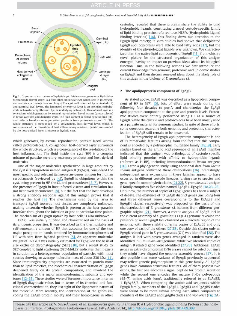
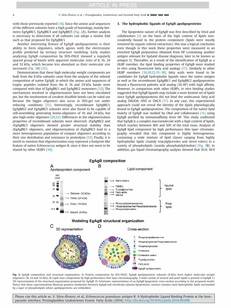
morphologically indistinguishable. Particularly, some of the tradi-tionally Echinococcus granulosus strains or genotypes (named G1–G10) are now recognized as different species within Echinococcusgenus. Thus, nine valid species of Echinococcus are accepted at thistime: Echinococcus multilocularis, Echinococcus vogeli, Echinococcusoligarthra, Echinococcus shiquicus, E. granulosus sensu stricto (tradi-tionally named G1–G3), Echinococcus equinus (traditionally namedG4), Echinococcus ortleppi (traditionally named G5), Echinococcuscanadensis (traditionally named G6–G10) and Echinococcus felidis[2,3]. Therefore, the originally defined E. granulosus is a speciescomplex; the term E. granulosus sensu lato (E. granulosus s. l.) can beused to group the different species and strains comprisingE. granulosus complex. The larval stage of E. granulosus s. l. is thecausative agent of cystic echinococcosis (hydatid disease) in a widerange of mammalian species (mainly domestic ungulates) as well ashumans. It establishes and gradually grows within host viscera(mainly liver and lungs). Fig. 1 illustrates the cyst structure; it isa unilocular fluid-filled bladder limited by a two-layer wall [1].The cyst external layer is an acellular, carbohydrate rich materialpermeable to macromolecules, which is synthesized by the under-lying cellular layer called germinal (GL). The latter is a syncitium,
Contents lists available at ScienceDirect
journal homepage: www.elsevier.com/locate/plefa
Prostaglandins, Leukotrienes and EssentialFatty Acids
http://dx.doi.org/10.1016/j.plefa.2014.09.0080952-3278/& 2014 Elsevier Ltd. All rights reserved.
Abbreviations: DCs, dendritic cells; EgAgB, Echinococcus granulosus antigen B; GL,germinal layer; HF, hydatid fluid; HLBP, Hydrophobic Ligand Binding Protein; LL,laminated layer; SEC-MALLS, size exclusion chromatography coupled to multi-angle laser light scattering
n Corresponding author.E-mail address: [email protected] (A.M. Ferreira).1 Present address: Institut Pasteur de Montevideo, Mataojo 2020, Montevideo,
11400, Uruguay.
Please cite this article as: V. Silva-Álvarez, et al., Echinococcus granulosus antigen B: A Hydrophobic Ligand Binding Protein at the host–parasite interface, Prostaglandins Leukotrienes Essent. Fatty Acids (2014), http://dx.doi.org/10.1016/j.plefa.2014.09.008i
Prostaglandins, Leukotrienes and Essential Fatty Acids ∎ (∎∎∎∎) ∎∎∎–∎∎∎
which generates, by asexual reproduction, parasite larval wormscalled protoscoleces. A collagenous, host-derived layer surroundsthe whole structure, which is a consequence of the resolution of thehost inflammation. The fluid inside the cyst (HF) is a complexmixture of parasite secretory–excretory products and host-derivedmolecules.
One of the major molecules synthesized in large amounts bythe cyst is a lipoprotein named antigen B (EgAgB), considered themost specific and relevant Echinococcus-genus antigen for humanserodiagnosis (reviewed by [4]). EgAgB is ubiquitous within thecyst, being present in HF, the GL and protoscoleces [5–7]. However,the presence of EgAgB in host infected viscera and circulation hasnot been well documented [8], but the fact that the host developsa strong antibody response against this antigen proves that itreaches the host [9]. The mechanisms used by the larva totransport EgAgB towards host tissues are completely unknown,making uncertain whether EgAgB is present at the host–parasiteinterface throughout the infection or just at particular time points.The mechanism of EgAgB uptake by host cells is also unknown.
EgAgB was initially purified and characterized on the basis ofits antigenic properties. It was described as the thermostable andself-aggregating antigen of HF that accounts for one of the twomajor precipitation bands obtained by immunoelectrophoresis ofHF with sera from hydatid patients [5]. An apparent molecularweight of 160 kDa was initially estimated for EgAgB on the basis ofsize exclusion chromatography (SEC) [10], but a recent study bySEC coupled to light scattering (SEC-MALLS) indicates that it existsin solution as a heterogeneous population of particles with mostspecies showing an average molecular mass of about 230 kDa [11].Since immunogenicity properties are associated to protein morethan to lipid moieties, the biochemical characterization of EgAgBdeepened firstly on its protein composition, and involved theidentification of the major immunodominant subunits and epi-topes [12–15]. These studies were of foremost importance in termsof EgAgB diagnostic value, but in terms of its chemical and fun-ctional characterization, they lost sight of the lipoprotein nature ofthe molecule. More recently, the identification of the genes en-coding the EgAgB protein moiety and their homologous in other
cestodes, revealed that these proteins share the ability to bindhydrophobic ligands, constituting a novel cestode-specific familyof lipid binding proteins referred to as HLBPs (Hydrophobic LigandBinding Proteins) [16]. This finding drew our attention to theEgAgB lipid moiety; in vitro studies had shown that delipidatedEgAgB apolipoproteins were able to bind fatty acids [17], but theidentity of the physiological ligands was unknown. We character-ized then the native lipid components of EgAgB [11], from which anovel picture for the structural organization of this antigenemerged, having an impact on previous ideas about its biologicalfunction. Thus, in the following sections we first introduce thecurrent knowledge from genomic, proteomic and lipidomic studieson EgAgB, and then discuss renewed ideas about the likely role ofthis antigen in the biology of E. granulosus s.l.
2. The apolipoprotein component of EgAgB
As stated above, EgAgB was described as a lipoprotein compo-nent of HF in 1971 [5]. Lots of effort were made during thefollowing four decades to purify and characterize the EgAgBapolipoprotein component at the gene and protein levels. Proteo-mic studies were entirely performed using HF as a source ofEgAgB, while the cyst GL and protoscoleces have been mostly usedas a parasite material for genomic studies. In spite of these efforts,some questions regarding both genomic and proteomic character-ization of EgAgB still remain to be answered.
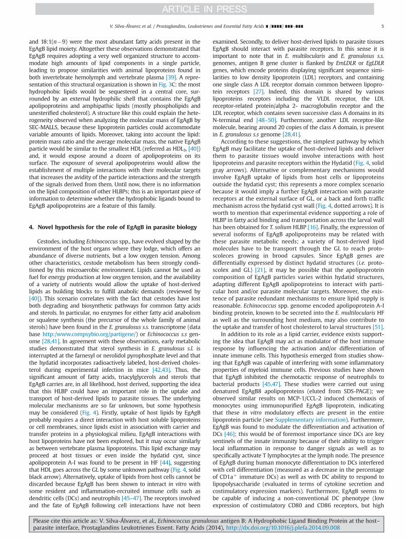
The heterogeneity of EgAgB apolipoprotein component is oneof its remarkable features arising from the fact that this compo-nent is encoded by a polymorphic multigene family [18,19]. Earlystudies based on the amino acid sequence of an EgAgB memberindicated that this antigen was orthologous of cestode-specificlipid binding proteins with affinity to hydrophobic ligands(referred as HLBP), including immunodominant Taenia antigens[20]. Later, a phylogenetic study using additional data from Taeniasolium antigens confirmed these observations [16] Interestingly,independent gene expansions in these families appear to haveoccurred in different cestode lineages, giving rise to species andgene-specific monophyletic clades [21,22]. E. granulosus s.l. antigenB family comprises five clades named EgAgB1–EgAgB5 [18,23–26].Until now, the number of copies of EgAgB genes has been a subjectof debate. The existence of 10 EgAgB distinct genes (including fourand three different genes corresponding to the EgAgB3 andEgAgB4 clades, respectively) was proposed on the basis of thecharacterization of E. granulosus s.l. isolates from different geo-graphic origins [21]. However, a recent analysis of EgAgB loci inthe current assembly of E. granulosus s.s (G1) genome revealed thepresence of seven EgAgB loci clustered on a discrete region of thegenome, with three slightly differing copies of EgAgB3 and onlyone copy of each of the others [27,28]. Outside this cluster only anEgAgB related gene in E. granulosus s.s (G1) was identified [28]. Theantigen B loci with seven genes arranged in tandem were alsoidentified in E. multilocularis genome, while two identical copies ofantigen B related gene were identified [27,28]. Additional EgAgBgenes in extra-chromosomal DNA arrays cannot be ruled out sincethey might have slipped the genome assembly process [27]. It isalso possible that some variants of EgAgB previously sequencedmay reflect genetic polymorphism in this gene family. All EgAgBgenes have common structural features. All of them possess twoexons, the first one encodes a signal peptide for protein secretionwhile the second one encodes the mature 8 kDa polypeptide(65–71 amino acids long), traditionally referred to as EgAgB8/1–EgAgB8/5. When comparing the amino acid sequences withinEgAgB family, members of the EgAgB1, EgAgB3 and EgAgB5 cladeswere found to be more similar among each other compared tomembers of the EgAgB2 and EgAgB4 clades and vice versa (Fig. 2A).
Fig. 1. Diagrammatic structure of hydatid cyst. Echinococcus granulosus Hydatid orMetacestode (larval stage) is a fluid-filled unilocular cyst which sets in intermedi-ate host viscera (mainly liver and lungs). The cyst wall is formed by laminated (LL)and germinal (GL) layers. The laminated or external layer is an acellular, carbohy-drate rich material synthesized by the underlying cellular GL. This internal layer is asyncytium, which generates by asexual reproduction larval worms (protoscoleces)in brood capsules and daughter cysts. The fluid content is called hydatid fluid (HF)and collects larval excretion/secretion products from protoscoleces and GL. Thewhole structure is surrounded by a collagenous, host-derived layer, which isconsequence of the resolution of host inflammatory reaction. Hydatid surroundedby the host-derived layer is known as hydatid cyst.
V. Silva-Álvarez et al. / Prostaglandins, Leukotrienes and Essential Fatty Acids ∎ (∎∎∎∎) ∎∎∎–∎∎∎2
Please cite this article as: V. Silva-Álvarez, et al., Echinococcus granulosus antigen B: A Hydrophobic Ligand Binding Protein at the host–parasite interface, Prostaglandins Leukotrienes Essent. Fatty Acids (2014), http://dx.doi.org/10.1016/j.plefa.2014.09.008i
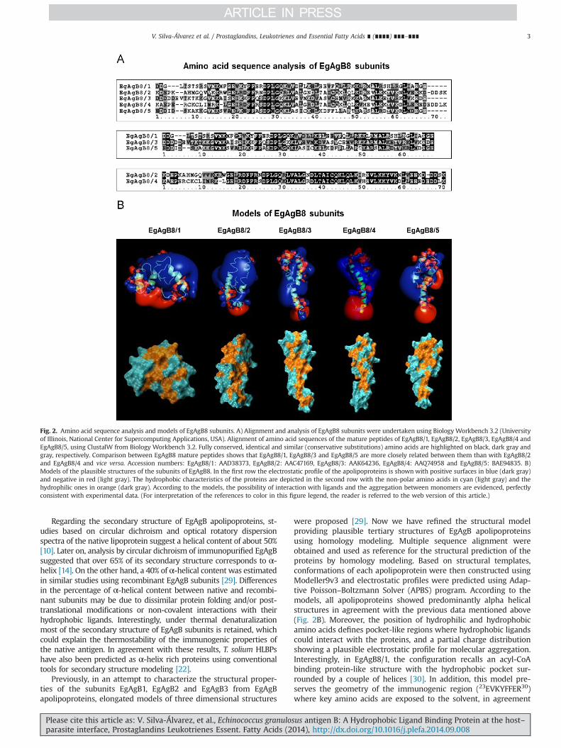
Regarding the secondary structure of EgAgB apolipoproteins, st-udies based on circular dichroism and optical rotatory dispersionspectra of the native lipoprotein suggest a helical content of about 50%[10]. Later on, analysis by circular dichroism of immunopurified EgAgBsuggested that over 65% of its secondary structure corresponds to α-helix [14]. On the other hand, a 40% of α-helical content was estimatedin similar studies using recombinant EgAgB subunits [29]. Differencesin the percentage of α-helical content between native and recombi-nant subunits may be due to dissimilar protein folding and/or post-translational modifications or non-covalent interactions with theirhydrophobic ligands. Interestingly, under thermal denaturalizationmost of the secondary structure of EgAgB subunits is retained, whichcould explain the thermostability of the immunogenic properties ofthe native antigen. In agreement with these results, T. solium HLBPshave also been predicted as α-helix rich proteins using conventionaltools for secondary structure modeling [22].
Previously, in an attempt to characterize the structural proper-ties of the subunits EgAgB1, EgAgB2 and EgAgB3 from EgAgBapolipoproteins, elongated models of three dimensional structures
were proposed [29]. Now we have refined the structural modelproviding plausible tertiary structures of EgAgB apolipoproteinsusing homology modeling. Multiple sequence alignment wereobtained and used as reference for the structural prediction of theproteins by homology modeling. Based on structural templates,conformations of each apolipoprotein were then constructed usingModeller9v3 and electrostatic profiles were predicted using Adap-tive Poisson–Boltzmann Solver (APBS) program. According to themodels, all apolipoproteins showed predominantly alpha helicalstructures in agreement with the previous data mentioned above(Fig. 2B). Moreover, the position of hydrophilic and hydrophobicamino acids defines pocket-like regions where hydrophobic ligandscould interact with the proteins, and a partial charge distributionshowing a plausible electrostatic profile for molecular aggregation.Interestingly, in EgAgB8/1, the configuration recalls an acyl-CoAbinding protein-like structure with the hydrophobic pocket sur-rounded by a couple of helices [30]. In addition, this model pre-serves the geometry of the immunogenic region (23EVKYFFER30)where key amino acids are exposed to the solvent, in agreement
Fig. 2. Amino acid sequence analysis and models of EgAgB8 subunits. A) Alignment and analysis of EgAgB8 subunits were undertaken using Biology Workbench 3.2 (Universityof Illinois, National Center for Supercomputing Applications, USA). Alignment of amino acid sequences of the mature peptides of EgAgB8/1, EgAgB8/2, EgAgB8/3, EgAgB8/4 andEgAgB8/5, using ClustalW from Biology Workbench 3.2. Fully conserved, identical and similar (conservative substitutions) amino acids are highlighted on black, dark gray andgray, respectively. Comparison between EgAgB8 mature peptides shows that EgAgB8/1, EgAgB8/3 and EgAgB8/5 are more closely related between them than with EgAgB8/2and EgAgB8/4 and vice versa. Accession numbers: EgAgB8/1: AAD38373, EgAgB8/2: AAC47169, EgAgB8/3: AAK64236, EgAgB8/4: AAQ74958 and EgAgB8/5: BAE94835. B)Models of the plausible structures of the subunits of EgAgB8. In the first row the electrostatic profile of the apolipoproteins is shown with positive surfaces in blue (dark gray)and negative in red (light gray). The hydrophobic characteristics of the proteins are depicted in the second row with the non-polar amino acids in cyan (light gray) and thehydrophilic ones in orange (dark gray). According to the models, the possibility of interaction with ligands and the aggregation between monomers are evidenced, perfectlyconsistent with experimental data. (For interpretation of the references to color in this figure legend, the reader is referred to the web version of this article.)
V. Silva-Álvarez et al. / Prostaglandins, Leukotrienes and Essential Fatty Acids ∎ (∎∎∎∎) ∎∎∎–∎∎∎ 3
Please cite this article as: V. Silva-Álvarez, et al., Echinococcus granulosus antigen B: A Hydrophobic Ligand Binding Protein at the host–parasite interface, Prostaglandins Leukotrienes Essent. Fatty Acids (2014), http://dx.doi.org/10.1016/j.plefa.2014.09.008i
with those previously reported [14]. Since the amino acid sequencesof the different subunits have a high grade of homology, mainly bet-ween EgAgB8/1, EgAgB8/3 and EgAgB8/5 (Fig. 2A), further analysisis necessary to determine if all subunits can adopt a similar foldsuch as that proposed for EgAgB8/1.
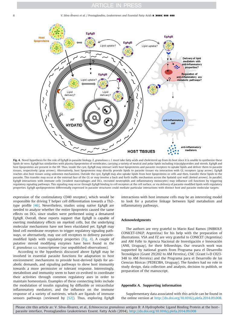
Another interesting feature of EgAgB apolipoproteins is theirability to form oligomers, which agrees with the electrostaticprofile predicted by tertiary structure modeling. Early studiesanalyzing EgAgB composition by SDS-PAGE showed a regularlyspaced group of bands with apparent molecular sizes of 8, 16, 24and 32 kDa, which became less abundant as their molecular sizeincreased (Fig. 3A) [31].
Demonstration that these high molecular weight components arebuilt from the 8 kDa subunits came from the analysis of the subunitcomposition of native EgAgB, in which the amino acid sequences oftryptic peptides isolated from the 8, 16 and 24 kDa bands werecompared with that of EgAgB8/1 and EgAgB8/2 monomers [32]. Themechanisms involved in oligomerization have not been elucidatedyet, but the involvement of covalent disulfide bonds can be ruled outbecause the bigger oligomers also occur in SDS-gel run underreducing conditions [31]. Interestingly, recombinant EgAgB8/1,EgAgB8/2 and EgAgB8/3 subunits were also found to be capable ofself-assembling generating homo-oligomers of 16 and 24 kDa, butalso high-order oligomers [29,33]. Differences in the oligomerizationproperties of recombinant subunits were observed: rEgAgB8/2 andrEgAgB8/3 oligomers showed greater structural stability thanrEgAgB8/1 oligomers, and oligomerization of rEgAgB8/3 lead to amore heterogeneous population of compact oligomers according totheir size distribution and resistance to proteolysis [33]. Finally, it isworth to mention that oligomerization may represent a footprint-likefeature of native Echinococcus antigen B, since it does not seem to beshared by other HLBPs [34].
3. The hydrophobic ligands of EgAgB apolipoproteins
The lipoprotein nature of EgAgB was first described by Oriol andcollaborators [5] on the basis of the high content of lipids non-covalently bound to the protein component (lipids were mostlyremoved by organic solvent extraction); this was a logical conclusioneven though in this work these properties were measured in anenriched EgAgB preparation obtained from HF (containing anotherantigen relevant for hydatid disease diagnosis, later to be known asantigen 5). Thereafter, as a result of the identification of EgAgB as aHLBP member, the lipid binding properties of EgAgB were studiedin vitro using fluorescent fatty acid analogs [17]. Similarly to otherHLBP members [16,20,22,35–38], fatty acids were found to becandidates for EgAgB hydrophobic ligands since the native antigenas well as the recombinant EgAgB8/1 and EgAgB8/2 apolipoproteinsbound a fluorescent palmitic acid analog (16-AP) with high affinity.However, in comparison with other HLBPs, in vitro binding studiessuggested that EgAgB ligands may include a more limited set of lipidssince EgAgB apolipoproteins did not bind the undecanoic fatty acidanalog DAUDA, ANS or DACA [17]. In any case, this experimentalapproach could not reveal the identity of the lipids physiologicallybound to EgAgB apolipoproteins. The composition of the native lipidmoiety of EgAgB was studied by Obal and collaborators [11] usingEgAgB purified by immunoaffinity from HF. This study confirmedthat EgAgB is a complex macromolecule with a high content of lipids,which reaches between 40% and 50% of the total mass. Analysis ofEgAgB lipid component by high performance thin layer chromato-graphy revealed that this component is highly heterogeneous,containing a wide mixture of lipid classes ranging from highlyhydrophobic lipids (mainly triacylglyceroles and sterol esters) to avariety of phospholipids (mainly phosphatidylcholine) (Fig. 3B). Inaddition, gas liquid chromatography analysis showed that 16:0, 18:0
Fig. 3. EgAgB composition and structural organization. A) Protein composition by SDS-PAGE: EgAgB apolipoprotein subunits (8 kDa) form higher molecular weightoligomers (16, 24 and 32 kDa). B) Lipid class composition by high performance thin layer chromatography. A wide variety of neutral and polar lipids is present in EgAgB. C)3D representation of the structural organization proposed for EgAgB. D) Schematic representation of an EgAgB lipoprotein cross-section according to the proposed model.Notice that these representations illustrate putative similarities between EgAgB and vertebrates plasma lipoproteins; nucleus contains most hydrophobic lipids surroundedby a layer of phospholipids where apolipoproteins are embedded.
V. Silva-Álvarez et al. / Prostaglandins, Leukotrienes and Essential Fatty Acids ∎ (∎∎∎∎) ∎∎∎–∎∎∎4
Please cite this article as: V. Silva-Álvarez, et al., Echinococcus granulosus antigen B: A Hydrophobic Ligand Binding Protein at the host–parasite interface, Prostaglandins Leukotrienes Essent. Fatty Acids (2014), http://dx.doi.org/10.1016/j.plefa.2014.09.008i
and 18:1(n�9) were the most abundant fatty acids present in theEgAgB lipid moiety. Altogether these observations demonstrated thatEgAgB requires adopting a very well organized structure to accom-modate high amounts of lipid components in a single particle,leading to propose similarities with animal lipoproteins found inboth invertebrate hemolymph and vertebrate plasma [39]. A repre-sentation of this structural organization is shown in Fig. 3C: the mosthydrophobic lipids would be sequestered in a central core, sur-rounded by an external hydrophilic shell that contains the EgAgBapolipoproteins and amphipathic lipids (mostly phospholipids andunesterified cholesterol). A structure like this could explain the hete-rogeneity observed when analyzing the molecular mass of EgAgB bySEC-MALLS, because these lipoprotein particles could accommodatevariable amounts of lipids. Moreover, taking into account the lipid:protein mass ratio and the average molecular mass, the native EgAgBparticle would be similar to the smallest HDL (referred as HDL3, [40])and, it would expose around a dozen of apolipoproteins on itssurface. The exposure of several apolipoproteins would allow theestablishment of multiple interactions with their molecular targetsthat increases the avidity of the particle interactions and the strengthof the signals derived from them. Until now, there is no informationon the lipid composition of other HLBPs; this is an important piece ofinformation to determine whether the hydrophobic ligands bound toEgAgB apolipoproteins are a feature of this family.
4. Novel hypothesis for the role of EgAgB in parasite biology
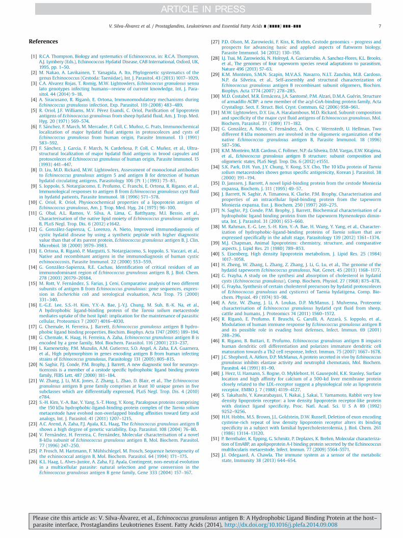
Cestodes, including Echinococcus spp., have evolved shaped by theenvironment of the host organs where they lodge, which offers anabundance of diverse nutrients, but a low oxygen tension. Amongother characteristics, cestode metabolism has been strongly condi-tioned by this microaerobic environment. Lipids cannot be used asfuel for energy production at low oxygen tension, and the availabilityof a variety of nutrients would allow the uptake of host-derivedlipids as building blocks to fulfill anabolic demands (reviewed by[40]). This scenario correlates with the fact that cestodes have lostboth degrading and biosynthetic pathways for common fatty acidsand sterols. In particular, no enzymes for either fatty acid anabolismor squalene synthesis (the precursor of the whole family of animalsterols) have been found in the E. granulosus s.s. transcriptome (database http:/www.compsybio.org/partigene/) or Echinococcus s.s gen-ome [28,41]. In agreement with these observations, early metabolicstudies demonstrated that sterol synthesis in E. granulosus s.l. isinterrupted at the farnesyl or nerolidol pyrophosphate level and thatthe hydatid incorporates radioactively labeled, host-derived choles-terol during experimental infection in mice [42,43]. Thus, thesignificant amount of fatty acids, triacylglycerols and sterols thatEgAgB carries are, in all likelihood, host derived, supporting the ideathat this HLBP could have an important role in the uptake andtransport of host-derived lipids to parasite tissues. The underlyingmolecular mechanisms are so far unknown, but some hypothesismay be considered (Fig. 4). Firstly, uptake of host lipids by EgAgBprobably requires a direct interaction with host soluble lipoproteinsor cell membranes, since lipids exist in association with carrier andtransfer proteins in a physiological milieu. EgAgB interactions withhost lipoproteins have not been explored, but it may occur similarlyas between vertebrate plasma lipoproteins. This lipid exchange mayproceed at host tissues or even inside the hydatid cyst, sinceapolipoprotein A-I was found to be present in HF [44], suggestingthat HDL goes across the GL by some unknown pathway (Fig. 4, solidblack arrow). Alternatively, uptake of lipids from host cells cannot bediscarded because EgAgB has been shown to interact in vitro withsome resident and inflammation-recruited immune cells such asdendritic cells (DCs) and neutrophils [45–47]. The receptors involvedand the fate of EgAgB following cell interactions have not been
examined. Secondly, to deliver host-derived lipids to parasite tissuesEgAgB should interact with parasite receptors. In this sense it isimportant to note that in E. multilocularis and E. granulosus s.s.genomes, antigen B gene cluster is flanked by EmLDLR or EgLDLRgenes, which encode proteins displaying significant sequence simi-larities to low density lipoprotein (LDL) receptors, and containingone single class A LDL receptor domain common between lipopro-tein receptors [27]. Indeed, this domain is shared by variouslipoproteins receptors including the VLDL receptor, the LDLreceptor-related protein/alpha 2- macroglobulin receptor and theLDL receptor, which contains seven successive class A domains in itsN-terminal end [48–50]. Furthermore, another LDL receptor-likemolecule, bearing around 20 copies of the class A domain, is presentin E. granulosus s.s genome [28,41].
According to these suggestions, the simplest pathway by whichEgAgB may facilitate the uptake of host-derived lipids and deliverthem to parasite tissues would involve interactions with hostlipoproteins and parasite receptors within the Hydatid (Fig. 4, solidgray arrows). Alternative or complementary mechanisms wouldinvolve EgAgB uptake of lipids from host cells or lipoproteinsoutside the hydatid cyst; this represents a more complex scenariobecause it would imply a further EgAgB interaction with parasitereceptors at the external surface of GL, or a back and forth trafficmechanism across the hydatid cyst wall (Fig. 4, dotted arrows). It isworth to mention that experimental evidence supporting a role ofHLBP in fatty acid binding and transportation across the larval wallhas been obtained for T. solium HLBP [16]. Finally, the expression ofseveral isoforms of EgAgB apolipoproteins may be related withthese parasite metabolic needs; a variety of host-derived lipidmolecules have to be transport through the GL to reach proto-scoleces growing in brood capsules. Since EgAgB genes aredifferentially expressed by distinct hydatid structures (i.e. proto-scolex and GL) [21], it may be possible that the apolipoproteincomposition of EgAgB particles varies within hydatid structures,adapting different EgAgB apolipoproteins to interact with parti-cular host and/or parasite molecular targets. Moreover, the exis-tence of parasite redundant mechanisms to ensure lipid supply isreasonable. Echinococcus spp. genome encoded apolipoprotein A-Ibinding protein, known to be secreted into the E. multilocularis HFas well as the surrounding host medium, may also contribute tothe uptake and transfer of host cholesterol to larval structures [51].
In addition to its role as a lipid carrier, evidence exists support-ing the idea that EgAgB may act as modulator of the host immuneresponse by influencing the activation and/or differentiation ofinnate immune cells. This hypothesis emerged from studies show-ing that EgAgB was capable of interfering with some inflammatoryproperties of myeloid immune cells. Previous studies have shownthat EgAgB inhibited the chemotactic response of neutrophils tobacterial products [45,47]. These studies were carried out usingdenatured EgAgB8 apolipoproteins (eluted from SDS-PAGE); weobserved similar results on MCP-1/CCL-2 induced chemotaxis ofmonocytes using immunopurified EgAgB lipoprotein, indicatingthat these in vitro modulatory effects are present in the entirelipoprotein particle (see Supplementary information). Furthermore,EgAgB was found to modulate the differentiation and activation ofDCs [46]; this would be of foremost importance since DCs are keysentinels of the innate immunity because of their ability to triggerlocal inflammation in response to danger signals as well as tospecifically activate T lymphocytes at the lymph node. The presenceof EgAgB during human monocyte differentiation to DCs interferedwith cell differentiation (measured as a decrease in the percentageof CD1aþ immature DCs) as well as with DC ability to respond tolipopolysaccharide (evaluated in terms of cytokine secretion andcostimulatory expression markers). Furthermore, EgAgB seems tobe capable of inducing a non-conventional DC phenotype (lowexpression of costimulatory CD80 and CD86 receptors, but high
V. Silva-Álvarez et al. / Prostaglandins, Leukotrienes and Essential Fatty Acids ∎ (∎∎∎∎) ∎∎∎–∎∎∎ 5
Please cite this article as: V. Silva-Álvarez, et al., Echinococcus granulosus antigen B: A Hydrophobic Ligand Binding Protein at the host–parasite interface, Prostaglandins Leukotrienes Essent. Fatty Acids (2014), http://dx.doi.org/10.1016/j.plefa.2014.09.008i
expression of the costimulatory CD40 receptor), which would beresponsible for driving T helper cell differentiation towards a Th2-type profile [46]. Nevertheless, studies using native EgAgB areneeded to analyze whether the entire lipoprotein caused the sameeffects on DCs, since studies were performed using a denaturedEgAgB. Overall, these reports support that EgAgB is capable ofexerting modulatory effects on myeloid cells, but the underlyingmolecular mechanisms have not been elucidated yet. EgAgB maybind cell membrane receptors to trigger regulatory signaling path-ways, or alternatively, may use cell receptors to delivery parasite-modified lipids with regulatory properties (Fig. 4). A couple ofputative steroid modifying enzymes have been found in theE. granulosus s.s. transcriptome (our unpublished observations).
According to the hypothesis discussed above EgAgB may beinvolved in essential parasite functions for adaptation to hostenvironment: mechanisms to provide host-derived lipids for an-abolic demands, and signaling pathways to skew host immunitytowards a more permissive or tolerant response. Interestingly,metabolism and immunity seem to have co-evolved to coordinatetheir activities through common regulatory axes in order tomaintain homeostasis. Examples of these connecting links includethe modulation of insulin signaling by diffusible or intracellularinflammatory mediators, and the influence on the immuneresponse of a variety of nutrients, which are ligands of immunesensors pathways (reviewed by [52]). Thus, exploring EgAgB
interactions with host immune cells may be an interesting modelto look for a putative linkage between lipid metabolism andinflammatory pathways.
Acknowledgments
The authors are very grateful to Mario Raul Ramos (INIBIOLP,CONICET-UNLP, Argentina) for his help with the preparation ofillustrations. VSA and FZ are very grateful to CONICET (Argentina)and AM Folle to Agencia Nacional de Investigación e Innovación(ANII, Uruguay), for their fellowships. Our research work wassupported by national grants from Programa para el DesarrolloTecnológico (Grant 29/202 to AM Ferreira), CSIC (Grant I+D C023-348 to AM Ferreira) and the Programa para el Desarrollo de lasCiencias Básicas (PEDECIBA, Uruguay). The funders had no role instudy design, data collection and analysis, decision to publish, orpreparation of the manuscript.
Appendix A. Supporting information
Supplementary data associated with this article can be found inthe online version at http://dx.doi.org/10.1016/j.plefa.2014.09.008.
Fig. 4. Novel hypothesis for the role of EgAgB in parasite biology. E. granulosus s. l. must take fatty acids and cholesterol up from its host since it is unable to synthesize theselipids de novo. EgAgB has similarities with plasma lipoproteins of vertebrates, carrying a variety of neutral and polar lipids including triacylglycerides and sterols. EgAgB andhost lipoproteins are present in the HF. Thus, inside the cyst, EgAgB may interact with host lipoproteins and parasite receptors to uptake lipids and deliver them to parasitetissues, respectively (gray arrows). Alternatively, host lipoproteins may directly provide lipids to parasite tissues via interaction with GL receptors (gray arrow). EgAgBreaches also host tissues using unknown mechanisms. Outside the cyst, EgAgB may also uptake lipids from host lipoproteins or cells and then, transfer these lipids to theparasite. This transfer may occur at the external face of the GL or may involve a back and forth traffic mechanism across the hydatid cyst wall (dotted arrows). In parallel,EgAgB interactions with immune cells (resident macrophages and DCs, recruited neutrophils and inflammatory monocytes) may influence cell functions by triggeringregulatory signaling pathways. This signaling may occur through EgAgB binding to cell receptors at the cell surface, or via delivery of parasite-modified lipids with regulatoryproperties. EgAgB apolipoproteins differentially expressed in parasite structures could mediate particular interactions with distinct host and parasite molecular targets.
V. Silva-Álvarez et al. / Prostaglandins, Leukotrienes and Essential Fatty Acids ∎ (∎∎∎∎) ∎∎∎–∎∎∎6
Please cite this article as: V. Silva-Álvarez, et al., Echinococcus granulosus antigen B: A Hydrophobic Ligand Binding Protein at the host–parasite interface, Prostaglandins Leukotrienes Essent. Fatty Acids (2014), http://dx.doi.org/10.1016/j.plefa.2014.09.008i
References
[1] R.C.A. Thompson, Biology and systematics of Echinococcus, in: R.C.A. Thompson,A.J. Lymbery (Eds.), Echinococcus Hydatid Disease, CAB International, Oxford, UK,1995, pp. 1–50.
[2] M. Nakao, A. Lavikainen, T. Yanagida, A. Ito, Phylogenetic systematics of thegenus Echinococcus (Cestoda: Taeniidae), Int. J. Parasitol. 43 (2013) 1017–1029.
[3] C.A. Alvarez Rojas, T. Romig, M.W. Lightowlers, Echinococcus granulosus sensulato genotypes infecting humans—review of current knowledge, Int. J. Para-sitol. 44 (2014) 9–18.
[4] A. Siracusano, R. Riganò, E. Ortona, Immunomodulatory mechanisms duringEchinococcus granulosus infection, Exp. Parasitol. 119 (2008) 483–489.
[5] R. Oriol, J.F. Williams, M.V. Pérez Esandi, C. Oriol, Purification of lipoproteinantigens of Echinococcus granulosus from sheep hydatid fluid, Am. J. Trop. Med.Hyg. 20 (1971) 569–574.
[6] F. Sánchez, F. March, M. Mercader, P. Coll, C. Muñoz, G. Prats, Immunochemicallocalization of major hydatid fluid antigens in protoscoleces and cysts ofEchinococcus granulosus from human origin, Parasite Immunol. 13 (1991)583–592.
[7] F. Sánchez, J. Garcia, F. March, N. Cardeñosa, P. Coll, C. Muñoz, et al., Ultra-structural localization of major hydatid fluid antigens in brood capsules andprotoscoleces of Echinococcus granulosus of human origin, Parasite Immunol. 15(1993) 441–447.
[8] D. Liu, M.D. Rickard, M.W. Lightowlers, Assessment of monoclonal antibodiesto Echinococcus granulosus antigen 5 and antigen B for detection of humanhydatid circulating antigens, Parasitology 106 (Pt 1) (1993) 75–81.
[9] S. Ioppolo, S. Notargiacomo, E. Profumo, C. Franchi, E. Ortona, R. Rigano, et al.,Immunological responses to antigen B from Echinococcus granulosus cyst fluidin hydatid patients, Parasite Immunol. 18 (1996) 571–578.
[10] C. Oriol, R. Oriol, Physiocochemical properties of a lipoprotein antigen ofEchinococcus granulosus, Am. J. Trop. Med. Hyg. 24 (1975) 96–100.
[11] G. Obal, A.L. Ramos, V. Silva, A. Lima, C. Batthyany, M.I. Bessio, et al.,Characterisation of the native lipid moiety of Echinococcus granulosus antigenB, PLoS Negl. Trop. Dis. 6 (2012) e1642.
[12] G. González-Sapienza, C. Lorenzo, A. Nieto, Improved immunodiagnosis ofcystic hydatid disease by using a synthetic peptide with higher diagnosticvalue than that of its parent protein, Echinococcus granulosus antigen B, J. Clin.Microbiol. 38 (2000) 3979–3983.
[13] E. Ortona, R. Riganò, P. Margutti, S. Notargiacomo, S. Ioppolo, S. Vaccari, et al.,Native and recombinant antigens in the immunodiagnosis of human cysticechinococcosis, Parasite Immunol. 22 (2000) 553–559.
[14] G. González-Sapienza, R.E. Cachau, Identification of critical residues of animmunodominant region of Echinococcus granulosus antigen B, J. Biol. Chem.278 (2003) 20179–20184.
[15] M. Rott, V. Fernández, S. Farias, J. Ceni, Comparative analysis of two differentsubunits of antigen B from Echinococcus granulosus: gene sequences, expres-sion in Escherichia coli and serological evaluation, Acta Trop. 75 (2000)331–340.
[16] E.-G.E. Lee, S.S.-H. Kim, Y.Y.-A. Bae, J.-Y.J. Chung, M. Suh, B.-K. Na, et al.,A hydrophobic ligand‐binding protein of the Taenia solium metacestodemediates uptake of the host lipid: implication for the maintenance of parasiticcellular, Proteomics 7 (2007) 4016–4030.
[17] G. Chemale, H. Ferreira, J. Barrett, Echinococcus granulosus antigen B hydro-phobic ligand binding properties, Biochim. Biophys. Acta 1747 (2005) 189–194.
[18] G. Chemale, K. Haag, H. Ferreira, A. Zaha, Echinococcus granulosus antigen B isencoded by a gene family, Mol. Biochem. Parasitol. 116 (2001) 233–237.
[19] L. Kamenetzky, P.M. Muzulin, A.M. Gutierrez, S.O. Angel, A. Zaha, E.A. Guarnera,et al., High polymorphism in genes encoding antigen B from human infectingstrains of Echinococcus granulosus, Parasitology 131 (2005) 805–815.
[20] N. Saghir, P.J. Conde, P.M. Brophy, J. Barrett, A new diagnostic tool for neurocys-ticercosis is a member of a cestode specific hydrophobic ligand binding proteinfamily, FEBS Lett. 487 (2000) 181–184.
[21] W. Zhang, J. Li, M.K. Jones, Z. Zhang, L. Zhao, D. Blair, et al., The Echinococcusgranulosus antigen B gene family comprises at least 10 unique genes in fivesubclasses which are differentially expressed, PLoS Negl. Trop. Dis. 4 (2010)e784.
[22] S.-H. Kim, Y.-A. Bae, Y. Yang, S.-T. Hong, Y. Kong, Paralogous proteins comprisingthe 150 kDa hydrophobic-ligand-binding-protein complex of the Taenia soliummetacestode have evolved non-overlapped binding affinities toward fatty acidanalogs, Int. J. Parasitol. 41 (2011) 1207–1215.
[23] A.C. Arend, A. Zaha, F.J. Ayala, K.L. Haag, The Echinococcus granulosus antigen Bshows a high degree of genetic variability, Exp. Parasitol. 108 (2004) 76–80.
[24] V. Fernández, H. Ferreira, C. Fernández, Molecular characterisation of a novel8-kDa subunit of Echinococcus granulosus antigen B, Mol. Biochem. Parasitol.77 (1996) 247–250.
[25] P. Frosch, M. Hartmann, F. Mühlschlegel, M. Frosch, Sequence heterogeneity ofthe echinococcal antigen B, Mol. Biochem. Parasitol. 64 (1994) 171–175.
[26] K.L. Haag, L. Alves-Junior, A. Zaha, F.J. Ayala, Contingent, non-neutral evolutionin a multicellular parasite: natural selection and gene conversion in theEchinococcus granulosus antigen B gene family, Gene 333 (2004) 157–167.
[27] P.D. Olson, M. Zarowiecki, F. Kiss, K. Brehm, Cestode genomics – progress andprospects for advancing basic and applied aspects of flatworm biology,Parasite Immunol. 34 (2012) 130–150.
[28] I.J. Tsai, M. Zarowiecki, N. Holroyd, A. Garciarrubio, A. Sanchez-Flores, K.L. Brooks,et al., The genomes of four tapeworm species reveal adaptations to parasitism,Nature 496 (2013) 57–63.
[29] K.M. Monteiro, S.M.N. Scapin, M.V.A.S. Navarro, N.I.T. Zanchin, M.B. Cardoso,N.P. da Silveira, et al., Self-assembly and structural characterization ofEchinococcus granulosus antigen B recombinant subunit oligomers, Biochim.Biophys. Acta 1774 (2007) 278–285.
[30] M.D. Costabel, M.R. Ermácora, J.A. Santomé, P.M. Alzari, D.M.A. Guérin, Structureof armadillo ACBP: a new member of the acyl-CoA-binding protein family, ActaCrystallogr. Sect. F. Struct. Biol. Cryst. Commun. 62 (2006) 958–961.
[31] M.W. Lightowlers, D.Y. Liu, A. Haralambous, M.D. Rickard, Subunit compositionand specificity of the major cyst fluid antigens of Echinococcus granulosus, Mol.Biochem. Parasitol. 37 (1989) 171–182.
[32] G. González, A. Nieto, C. Fernández, A. Orn, C. Wernstedt, U. Hellman, Twodifferent 8 kDa monomers are involved in the oligomeric organization of thenative Echinococcus granulosus antigen B, Parasite Immunol. 18 (1996)587–596.
[33] K.M. Monteiro, M.B. Cardoso, C. Follmer, N.P. da Silveira, D.M. Vargas, E.W. Kitajima,et al., Echinococcus granulosus antigen B structure: subunit composition andoligomeric states, PLoS Negl. Trop. Dis. 6 (2012) e1551.
[34] S.K. Park, D.H. Yun, J.Y. Chung, Y. Kong, S.Y. Cho, The 10 kDa protein of Taeniasolium metacestodes shows genus specific antigenicity, Korean J. Parasitol. 38(2000) 191–194.
[35] D. Janssen, J. Barrett, A novel lipid-binding protein from the cestode Monieziaexpansa, Biochem. J. 311 (1995) 49–57.
[36] J. Barrett, N. Saghir, A. Timanova, K. Clarke, P.M. Brophy, Characterisation andproperties of an intracellular lipid-binding protein from the tapewormMoniezia expansa, Eur. J. Biochem. 250 (1997) 269–275.
[37] N. Saghir, P.J. Conde, P.M. Brophy, J. Barrett, Biochemical characterisation of ahydrophobic ligand binding protein from the tapeworm Hymenolepis dimin-uta, Int. J. Parasitol. 31 (2001) 653–660.
[38] M. Rahman, E.-G. Lee, S.-H. Kim, Y.-A. Bae, H. Wang, Y. Yang, et al., Character-ization of hydrophobic-ligand-binding proteins of Taenia solium that areexpressed specifically in the adult stage, Parasitology 139 (2012) 1361–1374.
[39] M.J. Chapman, Animal lipoproteins: chemistry, structure, and comparativeaspects, J. Lipid Res. 21 (1980) 789–853.
[40] S. Eisenberg, High density lipoprotein metabolism, J. Lipid Res. 25 (1984)1017–1058.
[41] H. Zheng, W. Zhang, L. Zhang, Z. Zhang, J. Li, G. Lu, et al., The genome of thehydatid tapeworm Echinococcus granulosus, Nat. Genet. 45 (2013) 1168–1177.
[42] G. Frayha, A study on the synthesi and absorption of cholesterol in hydatidcysts (Echinococcus granulosus), Comp. Biochem. Physiol. 27 (1968) 875–878.
[43] G. Frayha, Synthesis of certain cholesterol precursors by hydatid protoscolecesof Echinococcus granulosus and cysticerci of Taenia hydatigena, Comp. Bio-chem. Physiol. 49 (1974) 93–98.
[44] A. Aziz, W. Zhang, J. Li, A. Loukas, D.P. McManus, J. Mulvenna, Proteomiccharacterisation of Echinococcus granulosus hydatid cyst fluid from sheep,cattle and humans, J. Proteomics 74 (2011) 1560–1572.
[45] R. Riganò, E. Profumo, F. Bruschi, G. Carulli, A. Azzarà, S. Ioppolo, et al.,Modulation of human immune response by Echinococcus granulosus antigen Band its possible role in evading host defenses, Infect. Immun. 69 (2001)288–296.
[46] R. Rigano, B. Buttari, E. Profumo, Echinococcus granulosus antigen B impairshuman dendritic cell differentiation and polarizes immature dendritic cellmaturation towards a Th2 cell response, Infect. Immun. 75 (2007) 1667–1678.
[47] J.C. Shepherd, A. Aitken, D.P. McManus, A protein secreted in vivo by Echinococcusgranulosus inhibits elastase activity and neutrophil chemotaxis, Mol. Biochem.Parasitol. 44 (1991) 81–90.
[48] J. Herz, U. Hamann, S. Rogne, O. Myklebost, H. Gausepohl, K.K. Stanley, Surfacelocation and high affinity for calcium of a 500-kd liver membrane proteinclosely related to the LDL-receptor suggest a physiological role as lipoproteinreceptor, EMBO J. 7 (1988) 4119–4127.
[49] S. Takahashi, Y. Kawarabayasi, T. Nakai, J. Sakai, T. Yamamoto, Rabbit very lowdensity lipoprotein receptor: a low density lipoprotein receptor-like proteinwith distinct ligand specificity, Proc. Natl. Acad. Sci. U S A 89 (1992)9252–9256.
[50] H.H. Hobbs, M.S. Brown, J.L. Goldstein, D.W. Russell, Deletion of exon encodingcysteine-rich repeat of low density lipoprotein receptor alters its bindingspecificity in a subject with familial hypercholesterolemia, J. Biol. Chem. 261(1986) 13114–13120.
[51] P. Bernthaler, K. Epping, G. Schmitz, P. Deplazes, K. Brehm, Molecular characteriza-tion of EmABP, an apolipoprotein A-I binding protein secreted by the Echinococcusmultilocularis metacestode, Infect. Immun. 77 (2009) 5564–5571.
[52] J.I. Odegaard, A. Chawla, The immune system as a sensor of the metabolicstate, Immunity 38 (2013) 644–654.
V. Silva-Álvarez et al. / Prostaglandins, Leukotrienes and Essential Fatty Acids ∎ (∎∎∎∎) ∎∎∎–∎∎∎ 7
Please cite this article as: V. Silva-Álvarez, et al., Echinococcus granulosus antigen B: A Hydrophobic Ligand Binding Protein at the host–parasite interface, Prostaglandins Leukotrienes Essent. Fatty Acids (2014), http://dx.doi.org/10.1016/j.plefa.2014.09.008i
Related Documents