Echinococcus equinus and Echinococcus granulosus sensu stricto from the United Kingdom: genetic diversity and haplotypic variation q Belgees Boufana a,⇑ , Wai San Lett a , Samia Lahmar b , Imad Buishi c , Anthony J. Bodell a , Antonio Varcasia d , Adriano Casulli e , Nicholas J. Beeching f,g , Fiona Campbell h , Monica Terlizzo i , Donald P. McManus j , Philip S. Craig a a Cestode Zoonoses Research Group, School of Environment and Life Sciences, University of Salford, Manchester M5 4WT, United Kingdom b Parasitology Laboratory, National School of Veterinary Medicine, Sidi Thabet, Tunisia c The University of Tripoli, P.O. Box 606, Tripoli, Libya d Laboratorio di Parassitologia, Ospedale Didattico Veterinaria, Università degli Studi di Sassari, Via Vienna 2, 07100 Sassari, Italy e Department of Infectious, Parasitic and Immunomediated Diseases, Intituto Superiore di Sanità, viale Regina Elena 299, 00161 Rome, Italy f Liverpool School of Tropical Medicine, Liverpool L3 5QA, United Kingdom g NIHR Funded Health Protection Research Unit in Emerging and Zoonotic Infections, Liverpool L69 7BE, United Kingdom h Royal Liverpool and Broadgreen University Hospitals NHS Trust, Liverpool L7 8XP, United Kingdom i Aintree University Hospital, Liverpool L9 7AL, United Kingdom j Molecular Parasitology Laboratory, QIMR Berghofer Medical Research Institute, Brisbane, Queensland 4029, Australia article info Article history: Received 24 July 2014 Received in revised form 7 October 2014 Accepted 13 October 2014 Available online xxxx Keywords: Echinococcus equinus Echinococcus granulosus sensu stricto Genetic diversity Haplotypes United Kingdom abstract Cystic echinococcosis is endemic in Europe including the United Kingdom. However, information on the molecular epidemiology of Echinococcus spp. from the United Kingdom is limited. Echinococcus isolates from intermediate and definitive animal hosts as well as from human cystic echinococcosis cases were analysed to determine species and genotypes within these hosts. Echinococcus equinus was identified from horse hydatid isolates, cysts retrieved from captive UK mammals and copro-DNA of foxhounds and farm dogs. Echinococcus granulosus sensu stricto (s.s.) was identified from hydatid cysts of sheep and cattle as well as in DNA extracted from farm dog and foxhound faecal samples, and from four human cystic echinococcosis isolates, including the first known molecular confirmation of E. granulosus s.s. infec- tion in a Welsh sheep farmer. Low genetic variability for E. equinus from various hosts and from different geographical locations was detected using the mitochondrial cytochrome c oxidase subunit 1 gene (cox1), indicating the presence of a dominant haplotype (EQUK01). In contrast, greater haplotypic variation was observed for E. granulosus s.s. cox1 sequences. The haplotype network showed a star-shaped network with a centrally placed main haplotype (EgUK01) that had been reported from other world regions. Ó 2014 Australian Society for Parasitology Inc. Published by Elsevier Ltd. All rights reserved. 1. Introduction Echinococcus granulosus sensu lato (s.l.) species complex is the causative agent of cystic echinococcosis (CE), a zoonotic disease of worldwide importance and global distribution (Craig et al., 2007). Members of this species complex have been shown by molecular genotyping to include E. granulosus sensu stricto (s.s.) (genotypes G1–G3), Echinococcus equinus (G4), Echinococcus ortle- ppi (G5) and Echinococcus canadensis (G6–G10) (Nakao et al., 2007; Thompson, 2008). The epidemiology and biology of echino- coccosis in the United Kingdom (UK) has been the subject of histor- ical reports that indicated the presence of one or two species or strains of Echinococcus (Williams and Sweatman, 1963; Ku maratilake et al., 1986; McManus et al., 1989). Molecular studies have confirmed the distinctiveness of two sympatric Echinococcus spp. in the UK (McManus and Rishi, 1989; Bowles et al., 1992; Le et al., 2002). Echinococcus equinus (genotype G4, horse strain) and E. granulosus (genotype G1, sheep strain) are known to be endemic in the UK, maintained primarily in horse/foxhound and sheep/dog transmission cycles, respectively (McManus et al., 1989). In the UK, equine hydatidosis was reported historically to be widespread (Thompson and Smyth, 1975), whereas ovine hydatid- osis is more localised, being primarily restricted to Wales and its bordering areas as well as the Hebridean Islands in Scotland http://dx.doi.org/10.1016/j.ijpara.2014.10.005 0020-7519/Ó 2014 Australian Society for Parasitology Inc. Published by Elsevier Ltd. All rights reserved. q Note: Nucleotide sequence data are available in NCBI database under the following Accession Nos. KP101614 – KP101622 ⇑ Corresponding author at: Department of Zoology, University of Benghazi, P.O. Box 1308, Benghazi, Libya. Tel.: +44 61 295 4563; fax: +44 61 295 5015. E-mail address: [email protected] (B. Boufana). International Journal for Parasitology xxx (2014) xxx–xxx Contents lists available at ScienceDirect International Journal for Parasitology journal homepage: www.elsevier.com/locate/ijpara Please cite this article in press as: Boufana, B., et al. Echinococcus equinus and Echinococcus granulosus sensu stricto from the United Kingdom: genetic diver- sity and haplotypic variation. Int. J. Parasitol. (2014), http://dx.doi.org/10.1016/j.ijpara.2014.10.005

Welcome message from author
This document is posted to help you gain knowledge. Please leave a comment to let me know what you think about it! Share it to your friends and learn new things together.
Transcript
International Journal for Parasitology xxx (2014) xxx–xxx
Contents lists available at ScienceDirect
International Journal for Parasitology
journal homepage: www.elsevier .com/locate / i jpara
Echinococcus equinus and Echinococcus granulosus sensu stricto from theUnited Kingdom: genetic diversity and haplotypic variation q
http://dx.doi.org/10.1016/j.ijpara.2014.10.0050020-7519/� 2014 Australian Society for Parasitology Inc. Published by Elsevier Ltd. All rights reserved.
q Note: Nucleotide sequence data are available in NCBI database under thefollowing Accession Nos. KP101614 – KP101622⇑ Corresponding author at: Department of Zoology, University of Benghazi, P.O.
Box 1308, Benghazi, Libya. Tel.: +44 61 295 4563; fax: +44 61 295 5015.E-mail address: [email protected] (B. Boufana).
Please cite this article in press as: Boufana, B., et al. Echinococcus equinus and Echinococcus granulosus sensu stricto from the United Kingdom: genetisity and haplotypic variation. Int. J. Parasitol. (2014), http://dx.doi.org/10.1016/j.ijpara.2014.10.005
Belgees Boufana a,⇑, Wai San Lett a, Samia Lahmar b, Imad Buishi c, Anthony J. Bodell a, Antonio Varcasia d,Adriano Casulli e, Nicholas J. Beeching f,g, Fiona Campbell h, Monica Terlizzo i, Donald P. McManus j,Philip S. Craig a
a Cestode Zoonoses Research Group, School of Environment and Life Sciences, University of Salford, Manchester M5 4WT, United Kingdomb Parasitology Laboratory, National School of Veterinary Medicine, Sidi Thabet, Tunisiac The University of Tripoli, P.O. Box 606, Tripoli, Libyad Laboratorio di Parassitologia, Ospedale Didattico Veterinaria, Università degli Studi di Sassari, Via Vienna 2, 07100 Sassari, Italye Department of Infectious, Parasitic and Immunomediated Diseases, Intituto Superiore di Sanità, viale Regina Elena 299, 00161 Rome, Italyf Liverpool School of Tropical Medicine, Liverpool L3 5QA, United Kingdomg NIHR Funded Health Protection Research Unit in Emerging and Zoonotic Infections, Liverpool L69 7BE, United Kingdomh Royal Liverpool and Broadgreen University Hospitals NHS Trust, Liverpool L7 8XP, United Kingdomi Aintree University Hospital, Liverpool L9 7AL, United Kingdomj Molecular Parasitology Laboratory, QIMR Berghofer Medical Research Institute, Brisbane, Queensland 4029, Australia
a r t i c l e i n f o
Article history:Received 24 July 2014Received in revised form 7 October 2014Accepted 13 October 2014Available online xxxx
Keywords:Echinococcus equinusEchinococcus granulosus sensu strictoGenetic diversityHaplotypesUnited Kingdom
a b s t r a c t
Cystic echinococcosis is endemic in Europe including the United Kingdom. However, information on themolecular epidemiology of Echinococcus spp. from the United Kingdom is limited. Echinococcus isolatesfrom intermediate and definitive animal hosts as well as from human cystic echinococcosis cases wereanalysed to determine species and genotypes within these hosts. Echinococcus equinus was identifiedfrom horse hydatid isolates, cysts retrieved from captive UK mammals and copro-DNA of foxhoundsand farm dogs. Echinococcus granulosus sensu stricto (s.s.) was identified from hydatid cysts of sheepand cattle as well as in DNA extracted from farm dog and foxhound faecal samples, and from four humancystic echinococcosis isolates, including the first known molecular confirmation of E. granulosus s.s. infec-tion in a Welsh sheep farmer. Low genetic variability for E. equinus from various hosts and from differentgeographical locations was detected using the mitochondrial cytochrome c oxidase subunit 1 gene (cox1),indicating the presence of a dominant haplotype (EQUK01). In contrast, greater haplotypic variation wasobserved for E. granulosus s.s. cox1 sequences. The haplotype network showed a star-shaped networkwith a centrally placed main haplotype (EgUK01) that had been reported from other world regions.
� 2014 Australian Society for Parasitology Inc. Published by Elsevier Ltd. All rights reserved.
1. Introduction
Echinococcus granulosus sensu lato (s.l.) species complex is thecausative agent of cystic echinococcosis (CE), a zoonotic diseaseof worldwide importance and global distribution (Craig et al.,2007). Members of this species complex have been shown bymolecular genotyping to include E. granulosus sensu stricto (s.s.)(genotypes G1–G3), Echinococcus equinus (G4), Echinococcus ortle-ppi (G5) and Echinococcus canadensis (G6–G10) (Nakao et al.,
2007; Thompson, 2008). The epidemiology and biology of echino-coccosis in the United Kingdom (UK) has been the subject of histor-ical reports that indicated the presence of one or two species orstrains of Echinococcus (Williams and Sweatman, 1963; Kumaratilake et al., 1986; McManus et al., 1989). Molecular studieshave confirmed the distinctiveness of two sympatric Echinococcusspp. in the UK (McManus and Rishi, 1989; Bowles et al., 1992; Leet al., 2002). Echinococcus equinus (genotype G4, horse strain) andE. granulosus (genotype G1, sheep strain) are known to be endemicin the UK, maintained primarily in horse/foxhound and sheep/dogtransmission cycles, respectively (McManus et al., 1989).
In the UK, equine hydatidosis was reported historically to bewidespread (Thompson and Smyth, 1975), whereas ovine hydatid-osis is more localised, being primarily restricted to Wales and itsbordering areas as well as the Hebridean Islands in Scotland
c diver-
2 B. Boufana et al. / International Journal for Parasitology xxx (2014) xxx–xxx
(McManus et al., 1989). The main focus of CE in humans is in thesheep farming communities of central and southern Wales(Walters, 1977; Palmer and Biffin, 1987). The incidence of humanCE for the period 1974–1983 was 2 and 5.6 cases per million forWales and Powys county (central Wales), respectively(Stallbaumer et al., 1986). During the period 2005–2009, a totalof 62 CE cases, many non-indigenous, were recorded from Englandand Wales with a mean incidence of 0.024/100,000 population(Halsby et al., 2014).
Surprisingly, however, there has been only one molecular con-firmation of a human CE case acquired in the UK. Echinococcusgranulosus (G1) was identified from a hydatid cyst removed fromthe liver of a patient with a long history of working as a foxhoundhandler in southwestern England (Craig et al., 2012). Regardingdiagnosis of infection in the definitive host, an Echinococcusgenus-specific copro-antigen ELISA has been used to determinethe prevalence of infection in dogs from Wales (Walters andCraig, 1992; Palmer et al., 1996; Buishi et al., 2005; Mastin et al.,2011). A similar copro-antigen approach, together with copro-PCR, has now been applied to investigate the prevalence of echino-coccosis in foxhound packs, with confirmation of the presence ofechinococcosis in foxhounds from England and Wales (Lett, W.S.,2013. Detection of E. granulosus and E. equinus in dogs and epide-miology of canine echinococcosis in the UK. PhD thesis, Universityof Salford, UK). Nevertheless, molecular data on Echinococcus iso-lates from the UK remains limited and information on Echinococcushaplotypes infecting intermediate and definitive hosts is lacking.The present study was therefore conducted to identify Echinococ-cus spp. and their haplotypes from various hosts from the UKand to examine genetic variability within these isolates.
2. Materials and methods
2.1. Hydatid cysts and samples
Echinococcus isolates used in this study were derived from live-stock intermediate (sheep, cattle, horses) and definitive hosts(farm dogs, foxhounds) from various parts of the UK (Table 1).Horse hydatid isolates (n = 30) were collected from eight infectedhorse livers at an abattoir in Nantwich, Cheshire (England)between 2010 and 2011 together with ‘horse passports’ whichincluded information on age, gender and the last place of residencefor each animal. Horse hydatid protoscoleces were also collectedfrom an infected horse at Bristol (England) abattoir (n = 1). The ger-minal layer from a horse (n = 1) originally from Ireland was alsoincluded in this study. This horse was reported to have been rearedin Ireland and then lived in Scotland (Methlick and Balmedie, thenWest Hill in Aberdeenshire) from the age of 4–8 years.
Sheep hydatid cysts (n = 10) were collected from the liver andlungs of four slaughtered animals from Gaerwen abattoir in Angle-sey (northern Wales) between 2006 and 2009. Farm dog faecal sam-ples (n = 8) originated from a Welsh hydatid study carried out inPowys county (Wales) between May 2008 and July 2010. Theremaining farm dog faecal samples (n = 12) were collected fromfarms in the Welsh counties of Powys and Gwent between Julyand November 2002 (Buishi et al., 2005). Foxhound faecal samples(n = 6) were collected from the ground of penned areas of anony-mised hunts from England and Wales between 2010 and 2011 (Lett,W.S., 2013, PhD thesis, University of Salford, UK (cited earlier)).Hydatid cyst histopathology blocks (n = 3) from UK patients whowere considered to have acquired CE infection in the UK and weretreated at the Royal Liverpool and Aintree Hospitals (Liverpool,UK), as well as a human-derived E. granulosus (G1) DNA sample froma hydatid cyst removed from a UK foxhound hunt worker (Craiget al., 2012), were available for the current study (Table 2). DNA of
Please cite this article in press as: Boufana, B., et al. Echinococcus equinus and Echsity and haplotypic variation. Int. J. Parasitol. (2014), http://dx.doi.org/10.1016
E. equinus confirmed from two captive UK mammals (zebra, Equusburchellii; lemur, Varecia rubra) (Boufana et al., 2012), as well ashydatid cysts derived from a second UK born lemur, were alsoincluded. For comparison, two E. equinus hydatid isolates from thelungs and liver of a Sardinian horse (Varcasia et al., 2008) and E. equi-nus DNA (n = 22) from Tunisian donkeys (Boufana et al., 2014) wereused in this study. Additionally, two E. equinus UK horse-derivedNational Center for Biotechnology Information (NCBI) databasenucleotide sequences (Accession number (No.) AF346403, Le et al.,2002; Accession No. AB786665, M. Nakao, unpublished data) wereincluded in the dataset.
2.2. DNA extraction, PCR and sequencing
Genomic DNA was extracted from ethanol-fixed Echinococcusprotoscoleces and/or germinal layer using the Qiagen DNeasyBlood and Tissue Kit (Qiagen, Hilden, Germany) according to themanufacturer’s instructions. DNA was extracted from histopathol-ogy wax-embedded hydatid cysts using the Qiagen GeneRead DNAFFPE Kit (Qiagen). Copro-DNA was extracted from faecal samplesusing the QIAamp DNA stool mini kit (Qiagen). The amplificationof a fragment within the cytochrome c oxidase subunit 1 mito-chondrial gene (cox1, 828 bp) was carried out using a publishedprotocol (Nakao et al., 2000). Amplified products were commer-cially sequenced in both directions (Source Bioscience, Notting-ham, UK). FinchTV viewer (Geospiza, Seattle, WA, USA) was usedto view chromatograms and generated sequences were comparedagainst the NCBI database using the BLAST algorithm (http://www.ncbi.nlm.nih.gov/BLAST/).
2.3. Data analysis
Data analysis was accomplished using methods described pre-viously (Boufana et al., 2014). In brief, sequences were trimmedin Proseq 3.5 (Filatov, 2002), aligned in MEGA 5 (Tamura et al.,2011) and ClustalX2 (Larkin et al., 2007), and exported into DnaSP4.5 software (Rozas et al., 2003). Haplotype networks were gener-ated using HapView (Salzburger et al., 2011) with maximum like-lihood trees constructed using the DNAML program (PHYLIP)(Felsenstein, 1989) which was then run from HapView. The popu-lation genetics package Arlequin 3.1 (Excoffier et al., 2005) wasused to calculate population diversity indices (haplotype andnucleotide diversities), neutrality indices, Tajima’s D (Tajima,1989) and Fu’s Fs (Fu, 1997), and to estimate the degree of popula-tion differentiation using the pairwise fixation index (Fst). The flat-worm mitochondrial code (Nakao et al., 2000) was used to inferamino acid sequences from the nucleotide sequences.
3. Results
3.1. Echinococcus spp. from UK hosts
In this study 76 Echinococcus isolates originating from 15 live-stock intermediate hosts (n = 44), 26 definitive hosts (n = 26), threecaptive mammals (n = 3) and four human CE cases (n = 4) from theUK were analysed. Species analysis for these UK isolates using theBLAST algorithm (http://www.ncbi.nlm.nih.gov/BLAST/) identified42 E. equinus and 34 E. granulosus s.s. infections (Tables 1 and 2).All UK horse isolates (n = 31) and captive mammals (lemurs,n = 2; zebra, n = 1) had 100% sequence identity with E. equinus(NCBI Accession No. AB786665). Isolates from sheep (n = 10) andcattle (n = 2) all belonged to E. granulosus s.s. The four human CEisolates were confirmed as E. granulosus s.s. DNA extracted fromfaeces of Welsh farm dogs and foxhounds confirmed these canidswere infected with either E. equinus or E. granulosus s.s. Three of
inococcus granulosus sensu stricto from the United Kingdom: genetic diver-/j.ijpara.2014.10.005
Table 1Number, source and species confirmation of Echinococcus spp. isolates collected in the United Kingdom from intermediate and definitive hosts for this study.
Host (n) DNA source No. of isolates Origin/last residence Species confirmation
Echinococcus equinus Echinococcus granulosus s.s.
Sheep (4) GL/PSC 10 Anglesey, North Wales 0 10Cattle (2) GL 2 Wales 0 2Horse (9) GL/PSC 31 United Kingdom regionsa 31 0Farm dogs (20) Faeces 20 Wales 3 17Foxhounds (6) Faeces 6 Hunts in Wales and England 5 1Lemur (2) GL 2 Suffolk/Essex, England 2 0Zebra (1) GL 1 West Midlands, England 1 0
Total 72 42 30
s.s., sensu stricto; GL, germinal layer; PSC, protoscoleces.a England (Bristol, Hampshire, West Sussex, Shropshire, Yorkshire), Wales (Swansea, Ceredigion).
Table 2Details of human cystic echinococcosis cases from the United Kingdom used in this study.
Patient ID Gender Age Organ Hydatid cyst size (cm) United Kingdom region Occupation Species confirmation Source
1572 Female 20 Lung NA NA NA Echinococcus granulosus s.s. This study1477 Male 43 Spleen 14 Cumbriaa Engineer Echinococcus granulosus s.s. This study7203 Male 47 Liver 9.5 Powysb Farmer Echinococcus granulosus s.s. This study6701 Male 41 Liver 7.5 Wiltshire/
GloucestershireaFoxhound handler Echinococcus granulosus
(G1 genotype)Craig et al. (2012)
NA, not available; s.s., sensu stricto.a England.b Wales.
B. Boufana et al. / International Journal for Parasitology xxx (2014) xxx–xxx 3
20 (15%) Welsh farm dogs had E. equinus DNA in their faeces and17 (85%) indicated the presence of E. granulosus s.s. Echinococcusequinus DNA was also confirmed in the faeces of five (83.3%) ofthe six foxhounds tested. Echinococcus granulosus s.s. DNA was con-firmed in one foxhound (16.7%). The Irish horse isolate included inthis study had a 99% sequence identity with E. equinus (NCBI Acces-sion No. AB786665).
3.2. Nucleotide sequence variation
Within the analysed mitochondrial cox 1 fragment (827 bp) forUK E. equinus and E. granulosus s.s. no insertion mutations or dele-tions were detected. There were 12 and seven polymorphic sitesrecorded for E. equinus and E. granulosus s.s. of which zero and five,respectively, were parsimony informative.
3.3. Haplotype networks for E. equinus and E. granulosus (s.s.) fromthe UK
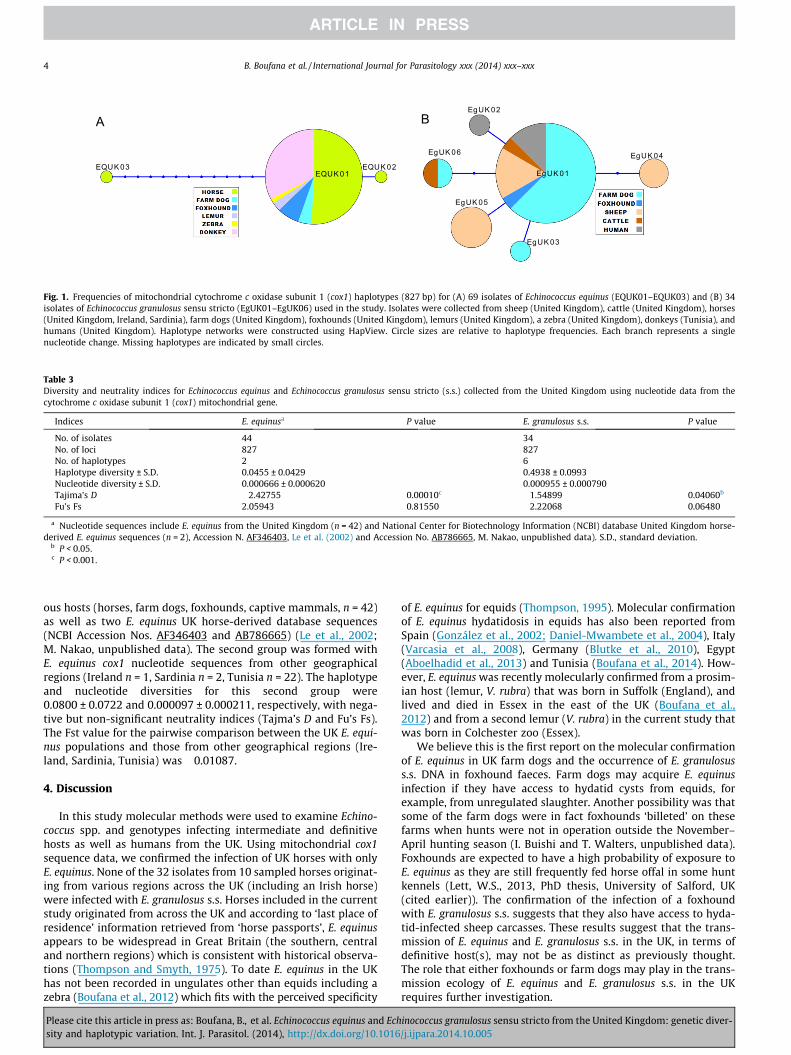
Only three haplotypes (EQUK01–EQUK03) were detected withinthe haplotype network generated using the UK E. equinus cox1mitochondrial DNA sequences (including NCBI Accession Nos.AF346403 and AB786665), as well as E. equinus nucleotidesequences from Sardinia, Tunisia and Ireland. Of these, 97.1% (67/69) of the DNA sequences, including E. equinus isolates from UKhosts (horses, n = 31; farm dogs, n = 3; foxhounds, n = 5; captivemammals, n = 3), isolates from a Sardinian horse (n = 2), donkeyhydatid isolates from Tunisia (n = 22) as well as a UK horse-deriveddatabase sequence (NCBI Accession No. AB786665, M. Nakao,unpublished data) all belonged to the main E. equinus haplotype(EQUK01) (Fig. 1A). One sequence derived from a horse that origi-nated from Ireland, and which differed from the main E. equinushaplotype (EQUK01) by a one-step mutation, occupied the secondE. equinus haplotype (EQUK02). An E. equinus UK horse-deriveddatabase sequence (Accession No. AF346403, Le et al., 2002)included in this study occupied the third haplotype (EQUK03).
In contrast, a total of six haplotypes (EgUK01–EgUK06) sepa-rated by one to two stepwise mutations were detected within
Please cite this article in press as: Boufana, B., et al. Echinococcus equinus and Echsity and haplotypic variation. Int. J. Parasitol. (2014), http://dx.doi.org/10.1016
the 34 cox1 E. granulosus s.s. mitochondrial DNA sequences fromthe UK isolates (Fig. 1B). The network displayed a star-like config-uration with a centrally positioned haplotype (EgUK01) constitut-ing 70.6% (24/34) of the total number of isolates. A BLAST searchshowed the E. granulosus common haplotype (EgUK01) to be100% identical to the main E. granulosus haplotype from China(G01: AB491414; 789 bp) (Nakao et al., 2010), Europe (EG1:JF513058; 351 bp) (Casulli et al., 2012) and the Middle East (Iran)(Eg01: JQ250806; 1609 bp) (Yanagida et al., 2012). Haplotypes of E.equinus and E. granulosus s.s. generated in this study were depos-ited into the NCBI database under Accession numbers KP101614–KP101616 and KP101617–KP101622 respectively.
3.4. Diversity and neutrality indices for UK E. equinus and E.granulosus s.s.
The haplotype diversity for isolates of E. granulosus s.s.(0.4938 ± 0.0993) were at least ten times those recorded for E. equi-nus populations (Table 3). Tajima’s D was significantly negative forE. granulosus s.s. populations, indicating the presence of variantnucleotides and population expansion. This was further supportedby the negative (but non-significant) Fu’s Fs value for E. granulosuss.s. populations which indicated the presence of excess alleles asexpected following recent population expansion or genetic hitch-hiking. The highly significant Tajima’s D recorded for E. equinuspopulations is due to the inclusion of an UK horse-derived E. equi-nus database sequence (haplotype EQUK03; NCBI Accession No.AF346403, Le et al., 2002) which differed from the main UK E. equi-nus haplotype (EQUK01) by 12 bp. The positive Fu’s Fs valuerecorded for E. equinus populations is indicative of haplotype/alleledeficiency expected from recent population bottleneck events.
3.5. Comparison of E. equinus isolates from different geographicallocations
The degree of population differentiation (Fst) was determinedfor two sets of E. equinus isolates. The first set included all UKderived E. equinus cox1 nucleotide sequences generated from vari-
inococcus granulosus sensu stricto from the United Kingdom: genetic diver-/j.ijpara.2014.10.005
EgUK01
A B
EQUK03EQUK01
EQUK02
EgUK02
EgUK04EgUK06
EgUK05
EgUK03
Fig. 1. Frequencies of mitochondrial cytochrome c oxidase subunit 1 (cox1) haplotypes (827 bp) for (A) 69 isolates of Echinococcus equinus (EQUK01–EQUK03) and (B) 34isolates of Echinococcus granulosus sensu stricto (EgUK01–EgUK06) used in the study. Isolates were collected from sheep (United Kingdom), cattle (United Kingdom), horses(United Kingdom, Ireland, Sardinia), farm dogs (United Kingdom), foxhounds (United Kingdom), lemurs (United Kingdom), a zebra (United Kingdom), donkeys (Tunisia), andhumans (United Kingdom). Haplotype networks were constructed using HapView. Circle sizes are relative to haplotype frequencies. Each branch represents a singlenucleotide change. Missing haplotypes are indicated by small circles.
Table 3Diversity and neutrality indices for Echinococcus equinus and Echinococcus granulosus sensu stricto (s.s.) collected from the United Kingdom using nucleotide data from thecytochrome c oxidase subunit 1 (cox1) mitochondrial gene.
Indices E. equinusa P value E. granulosus s.s. P value
No. of isolates 44 34No. of loci 827 827No. of haplotypes 2 6Haplotype diversity ± S.D. 0.0455 ± 0.0429 0.4938 ± 0.0993Nucleotide diversity ± S.D. 0.000666 ± 0.000620 0.000955 ± 0.000790Tajima’s D �2.42755 0.00010c �1.54899 0.04060b
Fu’s Fs 2.05943 0.81550 �2.22068 0.06480
a Nucleotide sequences include E. equinus from the United Kingdom (n = 42) and National Center for Biotechnology Information (NCBI) database United Kingdom horse-derived E. equinus sequences (n = 2), Accession N. AF346403, Le et al. (2002) and Accession No. AB786665, M. Nakao, unpublished data). S.D., standard deviation.
b P < 0.05.c P < 0.001.
4 B. Boufana et al. / International Journal for Parasitology xxx (2014) xxx–xxx
ous hosts (horses, farm dogs, foxhounds, captive mammals, n = 42)as well as two E. equinus UK horse-derived database sequences(NCBI Accession Nos. AF346403 and AB786665) (Le et al., 2002;M. Nakao, unpublished data). The second group was formed withE. equinus cox1 nucleotide sequences from other geographicalregions (Ireland n = 1, Sardinia n = 2, Tunisia n = 22). The haplotypeand nucleotide diversities for this second group were0.0800 ± 0.0722 and 0.000097 ± 0.000211, respectively, with nega-tive but non-significant neutrality indices (Tajma’s D and Fu’s Fs).The Fst value for the pairwise comparison between the UK E. equi-nus populations and those from other geographical regions (Ire-land, Sardinia, Tunisia) was �0.01087.
4. Discussion
In this study molecular methods were used to examine Echino-coccus spp. and genotypes infecting intermediate and definitivehosts as well as humans from the UK. Using mitochondrial cox1sequence data, we confirmed the infection of UK horses with onlyE. equinus. None of the 32 isolates from 10 sampled horses originat-ing from various regions across the UK (including an Irish horse)were infected with E. granulosus s.s. Horses included in the currentstudy originated from across the UK and according to ‘last place ofresidence’ information retrieved from ‘horse passports’, E. equinusappears to be widespread in Great Britain (the southern, centraland northern regions) which is consistent with historical observa-tions (Thompson and Smyth, 1975). To date E. equinus in the UKhas not been recorded in ungulates other than equids including azebra (Boufana et al., 2012) which fits with the perceived specificity
Please cite this article in press as: Boufana, B., et al. Echinococcus equinus and Echsity and haplotypic variation. Int. J. Parasitol. (2014), http://dx.doi.org/10.1016
of E. equinus for equids (Thompson, 1995). Molecular confirmationof E. equinus hydatidosis in equids has also been reported fromSpain (González et al., 2002; Daniel-Mwambete et al., 2004), Italy(Varcasia et al., 2008), Germany (Blutke et al., 2010), Egypt(Aboelhadid et al., 2013) and Tunisia (Boufana et al., 2014). How-ever, E. equinus was recently molecularly confirmed from a prosim-ian host (lemur, V. rubra) that was born in Suffolk (England), andlived and died in Essex in the east of the UK (Boufana et al.,2012) and from a second lemur (V. rubra) in the current study thatwas born in Colchester zoo (Essex).
We believe this is the first report on the molecular confirmationof E. equinus in UK farm dogs and the occurrence of E. granulosuss.s. DNA in foxhound faeces. Farm dogs may acquire E. equinusinfection if they have access to hydatid cysts from equids, forexample, from unregulated slaughter. Another possibility was thatsome of the farm dogs were in fact foxhounds ‘billeted’ on thesefarms when hunts were not in operation outside the November–April hunting season (I. Buishi and T. Walters, unpublished data).Foxhounds are expected to have a high probability of exposure toE. equinus as they are still frequently fed horse offal in some huntkennels (Lett, W.S., 2013, PhD thesis, University of Salford, UK(cited earlier)). The confirmation of the infection of a foxhoundwith E. granulosus s.s. suggests that they also have access to hyda-tid-infected sheep carcasses. These results suggest that the trans-mission of E. equinus and E. granulosus s.s. in the UK, in terms ofdefinitive host(s), may not be as distinct as previously thought.The role that either foxhounds or farm dogs may play in the trans-mission ecology of E. equinus and E. granulosus s.s. in the UKrequires further investigation.
inococcus granulosus sensu stricto from the United Kingdom: genetic diver-/j.ijpara.2014.10.005
B. Boufana et al. / International Journal for Parasitology xxx (2014) xxx–xxx 5
The low haplotype and nucleotide diversities for E. equinus(cox1) sequences seen in the current study are indicative of the lackof population expansion, which is further supported by the struc-ture of the network that included only three haplotypes, with acommon main haplotype (EQUK01) encompassing sequences fromUK hosts as well as those from hosts originating from other geo-graphical regions (Sardinia, horse; Tunisia, donkeys). A similarabsence of genetic variation for the cox1 mitochondrial locus aswell as for a nuclear locus (ef1a) of E. equinus in Tunisian donkeyshas been reported recently (Boufana et al., 2014). Further, the lowvalue (�0.01087) for the pairwise fixation index (Fst) for E. equinusfrom different regions reported here suggests that these popula-tions are not genetically differentiated.
Conversely, the presence of a common founder haplotype of E.granulosus (EgUK01) within a star-shaped network with single stepmutations combined with negative neutrality indices indicatespopulations sharing a recent history of bottleneck or founderevents and population expansion. It has been suggested that acommon genetic structure of geographically isolated populationsindicates the worldwide distribution of a single lineage of E. gran-ulosus s.s. and that expansion occurred following anthropogenicmovement of domesticated animal hosts (Nakao et al., 2010).
Three human CE cases molecularly confirmed in the currentstudy as E. granulosus s.s. were indigenous to the UK. Two of thesethree patients were identified as a Welsh farmer and an engineer,and their clinical records revealed that they had probably acquiredinfection in Powys (Wales) and Cumbria (northwestern England),respectively. This is the first known molecular identification of ahuman CE case in a Welsh farmer who had lived all his life in anendemic CE region (Powys) in the UK. It is also the first molecularconfirmation of human CE (acquired in England outside the knownCE ‘hotspot’ endemic area) that was not related to the patient’soccupation. This supports the suggestion of a more widespread dis-tribution of E. granulosus transmission in the UK, and is consistentwith the report of human CE in a Japanese female which theauthors suspected was acquired in the UK during visits to severalrural areas in the Cotswold region of south central England(Nakamura et al., 2011).
In summary, canine echinococcosis due to E. equinus and E.granulosus s.s. was confirmed for the first time, to our knowledge,by molecular genotyping in both farm dogs and foxhounds in theUK. Echinococcus equinus hydatid isolates were confirmed by DNAanalyses only from equids and captive lemurs, while E. granulosusDNA was confirmed in sheep, cattle and human hydatid isolates.Further studies are required to elucidate the apparent wider thanexpected geographic distribution of E. granulosus s.s. in the UKand it would be of interest to apply other neutral nuclear DNAmarkers that may provide further information on the populationstructure of E. equinus in the British Isles (Great Britain andIreland).
Acknowledgements
The authors wish to acknowledge the following individuals forproviding material used in this study: Mark Stidworthy, Interna-tional Zoo Veterinary Group, West Yorkshire, UK (hydatid cystsfrom lemurs); Sian Mitchell, AHVLA Carmarthen, Johnstown, UK(bovine hydatid cyst from Wales); Janina Kutscha, Ardene HouseVeterinary Practice, Scotland (hydatid cyst from Irish horse);Angela Potter and Bob Lawrence, West Midlands Safari & LeisurePark, Worcestershire, UK (hydatid cyst from a zebra); Kevin Shad-dick, Bristol abattoir, Bristol, UK (protoscoleces from horse hydatidcyst); Marion Woods, Royal Brisbane Women’s Hospital, Australia(ribbons of wax-embedded UK human CE hydatid cyst). Weacknowledge the use of eight farm dog faecal samples from aWelsh hydatid study funded by the Welsh Assembly Government.
Please cite this article in press as: Boufana, B., et al. Echinococcus equinus and Echsity and haplotypic variation. Int. J. Parasitol. (2014), http://dx.doi.org/10.1016
The support of Arjen Brouwer (Welsh Assembly Government) isgratefully acknowledged.
References
Aboelhadid, S.M., El-Dakhly, K.M., Yanai, T., Fukushi, H., Hassanin, K.M., 2013.Molecular characterization of Echinococcus granulosus in Egyptian donkeys. Vet.Parasitol. 193, 292–296.
Blutke, A., Hamel, D., Hüttner, M., Gehlen, H., Romig, T., Pfister, K., Hermanns, W.,2010. Cystic echinococcosis due to Echinococcus equinus in a horse fromsouthern Germany. J. Vet. Diagn. Invest. 22, 458–462.
Boufana, B., Lahmar, S., Rebai, W., Ben Safta, Z., Jebabli, L., Ammar, A., Kachti, M.,Aouadi, S., Craig, P.S., 2014. Genetic variability and haplotypes of Echinococcusisolates from Tunisia. Trans. R. Soc. Trop. Med. Hyg. 108, 706–714.
Boufana, B., Stidworthy, M.F., Bell, S., Chantrey, J., Masters, N., Unwin, S., Wood, R.,Lawrence, R.P., Potter, A., McGarry, J., Redrobe, S., Killick, R., Foster, A.P.,Mitchell, S., Greenwood, A.G., Sako, Y., Nakao, M., Ito, A., Wyatt, K., Lord, B.,Craig, P.S., 2012. Echinococcus and Taenia spp. from captive mammals in theUnited Kingdom. Vet. Parasitol. 190, 95–103.
Bowles, J., Blair, D., McManus, D.P., 1992. Genetic variants within the genusEchinococcus identified by mitochondrial DNA sequencing. Mol. Biochem.Parasitol. 54, 165–173.
Buishi, I., Walters, T., Guildea, Z., Craig, P., Palmer, S., 2005. Reemergence of canineEchinococcus granulosus infection, Wales. Emerg. Infect. Dis. 11, 568–571.
Casulli, A., Interisano, M., Sreter, T., Chitimia, L., Kirkova, Z., La Rosa, G., Pozio, E.,2012. Genetic variability of Echinococcus granulosus sensu stricto in Europeinferred by mitochondrial DNA sequences. Infect. Genet. Evol. 12, 377–383.
Craig, P.S., McManus, D.P., Lightowlers, M.W., Chabalgoity, J.A., Garcia, H.H., Gavidia,C.M., Gilman, R.H., Gonzalez, A.E., Lorca, M., Naquira, C., Nieto, A., Schantz, P.M.,2007. Prevention and control of cystic echinococcosis. Lancet Infect. Dis. 7, 385–394.
Craig, P.S., Woods, M.L., Boufana, B., O’Loughlin, B., Gimpel, J., Lett, W.S., McManus,D.P., 2012. Cystic echinococcosis in a fox-hound hunt worker, UK. Pathog. Glob.Health 106, 373–375.
Daniel-Mwambete, K., Ponce-Gordo, F., Cuesta-Bandera, C., 2004. Geneticidentification and host range of the Spanish strains of Echinococcus granulosus.Acta Trop. 91, 87–93.
Excoffier, L., Laval, G., Schneider, S., 2005. Arlequin ver. 3.0: an integrated softwarepackage for population genetics data analysis. Evol. Bioinform. Online 1, 47–50.
Felsenstein, J., 1989. PHYLIP – phylogeny inference package (version 3.2). Cladistics5, 164–166.
Filatov, D.A., 2002. ProSeq: A software for preparation and evolutionary analysis ofDNA sequence data sets. Mol. Ecol. Notes 2, 621–624.
Fu, Y.X., 1997. Statistical tests of neutrality of mutations against population growth,hitchhiking and background selection. Genetics 147, 915–925.
González, L.M., Daniel-Mwambete, K., Montero, E., Rosenzvit, M.C., McManus, D.P.,Gárate, T., Cuesta-Bandera, C., 2002. Further molecular discrimination ofSpanish strains of Echinococcus granulosus. Exp. Parasitol. 102, 46–56.
Halsby, K.D., Walsh, A.L., Smith, R., Said, B., Kirkbride, H., Smyth, B., Browning, L.,Larkin, L., Morgan, D., 2014. The health burden of orphan zoonotic disease in theUnited Kingdom, 2005–2009. Zoonoses Public Health 61, 39–47.
Kumaratilake, L.M., Thompson, R.C., Eckert, J., 1986. Echinococcus granulosus ofequine origin from different countries possess uniform morphologicalcharacteristics. Int. J. Parasitol. 16, 529–540.
Larkin, M.A., Blackshields, G., Brown, N.P., Chenna, R., McGettigan, P.A., McWilliam,H., Valentin, F., Wallace, I.M., Wilm, A., Lopez, R., Thompson, J.D., Gibson, T.J.,Higgins, D.G., 2007. Clustal W and Clustal X version 2.0. Bioinformatics 23,2947–2948.
Le, T.H., Pearson, M.S., Blair, D., Dai, N., Zhang, L.H., McManus, D.P., 2002. Completemitochondrial genomes confirm the distinctiveness of the horse–dog andsheep–dog strains of Echinococcus granulosus. Parasitology 124, 97–112.
Mastin, A., Brouwer, A., Fox, M., Craig, P., Guitián, J., Li, W., Stevens, K., 2011. Spatialand temporal investigation of Echinococcus granulosus coproantigen prevalencein farm dogs in South Powys, Wales. Vet. Parasitol. 178, 100–107.
McManus, D.P., Rishi, A.K., 1989. Genetic heterogeneity within Echinococcusgranulosus: isolates from different hosts and geographical areas characterizedwith DNA probes. Parasitology 99, 17–29.
McManus, D.P., Thompson, R.C., Lymbery, A.J., 1989. Comment on the status ofEchinococcus granulosus in the UK. Parasitol. Today 5, 365–367.
Nakamura, K., Ito, A., Yara, S., Haranaga, S., Hibiya, K., Hirayasu, T., Sako, Y., Fujita, J.,2011. Case report: a case of pulmonary and hepatic cystic Echinococcosis of CE1stage in a healthy Japanese female that was suspected to have been acquiredduring her stay in the United Kingdom. Am. J. Trop. Med. Hyg 85, 456–459.
Nakao, M., Li, T., Han, X., Ma, X., Xiao, N., Qiu, J., Wang, H., Yanagida, T., Mamuti, W.,Wen, H., Moro, P.L., Giraudoux, P., Craig, P.S., Ito, A., 2010. Geneticpolymorphisms of Echinococcus tapeworms in China as determined bymitochondrial and nuclear DNA sequences. Int. J. Parasitol. 40 (379–3), 85.
Nakao, M., McManus, D.P., Schantz, P.M., Craig, P.S., Ito, A., 2007. A molecularphylogeny of the genus Echinococcus inferred from complete mitochondrialgenomes. Parasitology 134, 713–722.
Nakao, M., Sako, Y., Yokoyama, N., Fukunaga, M., Ito, A., 2000. Mitochondrial geneticcode in cestodes. Mol. Biochem. Parasitol. 111, 415–424.
Palmer, S.R., Biffin, A.H., 1987. The changing incidence of human hydatid disease inEngland and Wales. Epidemiol. Infect. 99, 693–700.
inococcus granulosus sensu stricto from the United Kingdom: genetic diver-/j.ijpara.2014.10.005
6 B. Boufana et al. / International Journal for Parasitology xxx (2014) xxx–xxx
Palmer, S.R., Biffin, A.H., Craig, P.S., Walters, T.M., 1996. Control of hydatid disease inWales. BMJ 312, 674–675.
Rozas, J., Sánchez-DelBarrio, J.C., Messeguer, X., Rozas, R., 2003. DnaSP, DNApolymorphism analyses by the coalescent and other methods. Bioinformatics19, 2496–2497.
Salzburger, W., Ewing, G.B., Haeseler, A., 2011. The performance of phylogeneticalgorithms in estimating haplotype genealogies with migration. Mol. Ecol. 20,1952–1963.
Stallbaumer, M.F., Clarkson, M.J., Bailey, J.W., Pritchard, J.E., 1986. The epidemiologyof hydatid disease in England and Wales. J. Hyg. 96, 121–127.
Tajima, F., 1989. Statistical method for testing the neutral mutation hypothesis byDNA polymorphism. Genetics 123, 585–595.
Tamura, K., Peterson, D., Peterson, N., Stecher, G., Nei, M., Kumar, S., 2011. MEGA5:molecular evolutionary genetics analysis using maximum likelihood,evolutionary distance, and maximum parsimony methods. Mol. Biol. Evol. 28,2731–2739.
Thompson, R.C.A., 1995. Biology and systematics of Echinococcus. In: Thompson,R.C.A., Lymbery, A.J. (Eds.), Echinococcus and Hydatid Disease. CAB International,Wallingford, Oxon, UK, pp. 1–50.
Please cite this article in press as: Boufana, B., et al. Echinococcus equinus and Echsity and haplotypic variation. Int. J. Parasitol. (2014), http://dx.doi.org/10.1016
Thompson, R.C.A., 2008. The taxonomy, phylogeny and transmission ofEchinococcus. Exp. Parasitol. 119, 439–446.
Thompson, R.C., Smyth, J.D., 1975. Equine hydatidosis: a review of the current statusin Great Britain and the results of an epidemiological survey. Vet. Parasitol. 1,107–127.
Varcasia, A., Garippa, G., Pipia, A.P., Scala, A., Brianti, E., Giannetto, S., Battelli, G.,Poglayen, G., Micagni, G., 2008. Cystic echinococcosis in equids in Italy.Parasitol. Res. 102, 815–818.
Walters, T.M., 1977. Hydatid disease in Wales. Trans. R. Soc. Trop. Med. Hyg. 71,105–108.
Walters, T.M., Craig, P.S., 1992. Diagnosis of Echinococcus granulosus infection indogs. Vet. Rec. 131, 39–40.
Williams, R.J., Sweatman, G.K., 1963. On the transmission, biology and morphologyof Echinococcus granulosus equinus, a new subspecies of hydatid tapeworm inhorses in Great Britain. Parasitology 53, 391–407.
Yanagida, T., Mohammadzadeh, T., Kamhawi, S., Nakao, M., Sadjjadi, S.M., Hijjawi,N., Abdel-Hafez, S.K., Sako, Y., Okamoto, M., Ito, A., 2012. Geneticpolymorphisms of Echinococcus granulosus sensu stricto in the Middle East.Parasitol. Int. 61, 599–603.
inococcus granulosus sensu stricto from the United Kingdom: genetic diver-/j.ijpara.2014.10.005
Related Documents


