ECG Rhythm Interpretation AV Junctional Blocks

Welcome message from author
This document is posted to help you gain knowledge. Please leave a comment to let me know what you think about it! Share it to your friends and learn new things together.
Transcript

ECG Rhythm Interpretation
AV Junctional Blocks

Course Objectives
• To recognize the normal rhythm of the heart - “Normal Sinus Rhythm.”
• To recognize the 13 most common rhythm disturbances.
• To recognize an acute myocardial infarction on a 12-lead ECG.

Learning Modules
• ECG Basics
• How to Analyze a Rhythm
• Normal Sinus Rhythm
• Heart Arrhythmias
• Diagnosing a Myocardial Infarction
• Advanced 12-Lead Interpretation

Arrhythmias
• Sinus Rhythms
• Premature Beats
• Supraventricular Arrhythmias
• Ventricular Arrhythmias
• AV Junctional Blocks

AV Nodal Blocks
• 1st Degree AV Block
• 2nd Degree AV Block, Type I
• 2nd Degree AV Block, Type II
• 3rd Degree AV Block

Rhythm #10
60 bpm• Rate?• Regularity? regular
normal
0.08 s
• P waves?
• PR interval? 0.36 s• QRS duration?
Interpretation? 1st Degree AV Block

1st Degree AV Block
• Deviation from NSR– PR Interval > 0.20 s

1st Degree AV Block
• Etiology: Prolonged conduction delay in the AV node or Bundle of His.

Rhythm #11
50 bpm• Rate?• Regularity? regularly irregular
nl, but 4th no QRS
0.08 s
• P waves?
• PR interval? lengthens• QRS duration?
Interpretation? 2nd Degree AV Block, Type I

2nd Degree AV Block, Type I
• Deviation from NSR– PR interval progressively lengthens,
then the impulse is completely blocked (P wave not followed by QRS).

2nd Degree AV Block, Type I
• Etiology: Each successive atrial impulse encounters a longer and longer delay in the AV node until one impulse (usually the 3rd or 4th) fails to make it through the AV node.

Rhythm #12
40 bpm• Rate?• Regularity? regular
nl, 2 of 3 no QRS
0.08 s
• P waves?
• PR interval? 0.14 s• QRS duration?
Interpretation? 2nd Degree AV Block, Type II

2nd Degree AV Block, Type II
• Deviation from NSR– Occasional P waves are completely
blocked (P wave not followed by QRS).

2nd Degree AV Block, Type II
• Etiology: Conduction is all or nothing (no prolongation of PR interval); typically block occurs in the Bundle of His.
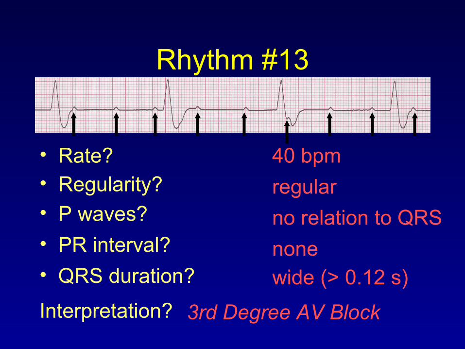
Rhythm #13
40 bpm• Rate?• Regularity? regular
no relation to QRS
wide (> 0.12 s)
• P waves?
• PR interval? none• QRS duration?
Interpretation? 3rd Degree AV Block

3rd Degree AV Block
• Deviation from NSR– The P waves are completely blocked in
the AV junction; QRS complexes originate independently from below the junction.

3rd Degree AV Block
• Etiology: There is complete block of conduction in the AV junction, so the atria and ventricles form impulses independently of each other. Without impulses from the atria, the ventricles own intrinsic pacemaker kicks in at around 30 - 45 beats/minute.

Remember• When an impulse originates in a ventricle,
conduction through the ventricles will be inefficient and the QRS will be wide and bizarre.
Related Documents