Cell Transplantation, Vol. 18, pp. 245–254, 2009 0963-6897/09 $90.00 + .00 Printed in the USA. All rights reserved. E-ISSN 1555-3892 Copyright 2009 Cognizant Comm. Corp. www.cognizantcommunication.com REVIEW Early Translation of Adipose-Derived Cell Therapy for Cardiovascular Disease Ricardo Sanz-Ruiz,* Eugenia Ferna ´ndez-Santos,* Marta Domı ´nguez-Mun ˜oa,* Radoslaw Parma,† Adolfo Villa,* Lucı ´a Ferna ´ndez,* Pedro L. Sa ´nchez,* and Francisco Ferna ´ndez-Avile ´s* *Cardiology Department, Hospital General Universitario Gregorio Maran ˜o ´n, Madrid, Spain †Third Division of Cardiology, Medical University of Silesia, Katowice, Poland Over the past decade, cell therapy has emerged as a new approach to reversing myocardial ischemia. Several types of adult stem cells have been studied in both preclinical and clinical conditions for this purpose: bone marrow cells, circulating cells, and myoblasts. Nevertheless, the quest for the ideal “anti-ischemic” cell is still ongoing. Recently, the existence of a population of stem cells located in adipose tissue (adipose-derived stem cells) has been observed. These are able to differentiate into multiple cell lineages including cardiomyo- cytic differentiation. In this review we discuss the basic principles of adipose-derived stem cells (types and characteristics, harvesting, and expansion), the initial experimental studies, and the currently ongoing clinical trials. Key words: Stem cells; Cardiovascular repair; Adipose-derived stem cells; Adipose tissue; APOLLO trial; PRECISE trial ADIPOSE TISSUE: A NEW SOURCE though results have shown an improvement in cardiac function due mostly to paracrine effects (8), formation OF STEM CELLS of a new myocardial mass has only been established for embryonic stem cells. Recent advances in the diagnosis and treatment of coronary artery disease have improved the prognosis of Recently, it has been shown that in the adipose tissue stroma, besides committed adipogenic, endothelial, and patients in the acute phase of the disease, the acute myo- cardial infarction (AMI). However, the loss of myocar- pluripotent vascular progenitor cells, we can also find multipotent cell types (adipose-derived stem cells, ASCs). dial tissue and the ventricular negative remodeling pro- cess will lead this growing population of AMI survivors In this review we will focus on the types, isolation, char- acterization, and preclinical and clinical application of to the syndrome known as chronic heart failure (CHF). This major health problem and its enormous economic ASCs in the cardiovascular field. consequences have guided cardiovascular research to- WORKING IN THE BENCH: wards new therapeutic approaches, among which cell ADIPOSE-DERIVED STEM therapy has been extensively studied as one of the more CELLS CHARACTERISTICS promising ones. Nomenclature The quest for the best cell type is still ongoing. This ideal cell type should be capable of differentiating into In recent years it has been shown that mesenchymal stem cells (MSCs) have an inherent ability for self-renew- functional cardiomyocytes and of forming new vessels to nourish the damaged area. Cell types from several al, proliferation, and differentiation towards mature tis- sues depending on the microenvironment by which they different sources have already been tested in animal models, and bone marrow-derived cells and skeletal my- are surrounded. Such characteristics, which define them as stem cells, make them very interesting for their use oblasts have been used in clinical trials (13,28). Al- Address correspondence to Prof. Francisco Ferna ´ndez-Avile ´s, Hospital General Universitario Gregorio Maran ˜o ´n, Doctor Esquerdo 46, 28007, Madrid, Spain. Tel: 91 426 5880; Fax: 91 586 8276; E-mail: [email protected] 245

Welcome message from author
This document is posted to help you gain knowledge. Please leave a comment to let me know what you think about it! Share it to your friends and learn new things together.
Transcript

Cell Transplantation, Vol. 18, pp. 245–254, 2009 0963-6897/09 $90.00 + .00Printed in the USA. All rights reserved. E-ISSN 1555-3892Copyright 2009 Cognizant Comm. Corp. www.cognizantcommunication.com
REVIEW
Early Translation of Adipose-Derived Cell Therapyfor Cardiovascular Disease
Ricardo Sanz-Ruiz,* Eugenia Fernandez-Santos,* Marta Domınguez-Munoa,* Radoslaw Parma,†Adolfo Villa,* Lucıa Fernandez,* Pedro L. Sanchez,* and Francisco Fernandez-Aviles*
*Cardiology Department, Hospital General Universitario Gregorio Maranon, Madrid, Spain†Third Division of Cardiology, Medical University of Silesia, Katowice, Poland
Over the past decade, cell therapy has emerged as a new approach to reversing myocardial ischemia. Severaltypes of adult stem cells have been studied in both preclinical and clinical conditions for this purpose: bonemarrow cells, circulating cells, and myoblasts. Nevertheless, the quest for the ideal “anti-ischemic” cell isstill ongoing. Recently, the existence of a population of stem cells located in adipose tissue (adipose-derivedstem cells) has been observed. These are able to differentiate into multiple cell lineages including cardiomyo-cytic differentiation. In this review we discuss the basic principles of adipose-derived stem cells (types andcharacteristics, harvesting, and expansion), the initial experimental studies, and the currently ongoing clinicaltrials.
Key words: Stem cells; Cardiovascular repair; Adipose-derived stem cells; Adipose tissue; APOLLO trial;PRECISE trial
ADIPOSE TISSUE: A NEW SOURCE though results have shown an improvement in cardiacfunction due mostly to paracrine effects (8), formationOF STEM CELLSof a new myocardial mass has only been established forembryonic stem cells.Recent advances in the diagnosis and treatment of
coronary artery disease have improved the prognosis of Recently, it has been shown that in the adipose tissuestroma, besides committed adipogenic, endothelial, andpatients in the acute phase of the disease, the acute myo-
cardial infarction (AMI). However, the loss of myocar- pluripotent vascular progenitor cells, we can also findmultipotent cell types (adipose-derived stem cells, ASCs).dial tissue and the ventricular negative remodeling pro-
cess will lead this growing population of AMI survivors In this review we will focus on the types, isolation, char-acterization, and preclinical and clinical application ofto the syndrome known as chronic heart failure (CHF).
This major health problem and its enormous economic ASCs in the cardiovascular field.consequences have guided cardiovascular research to-
WORKING IN THE BENCH:wards new therapeutic approaches, among which cell
ADIPOSE-DERIVED STEMtherapy has been extensively studied as one of the more
CELLS CHARACTERISTICSpromising ones.
NomenclatureThe quest for the best cell type is still ongoing. Thisideal cell type should be capable of differentiating into In recent years it has been shown that mesenchymal
stem cells (MSCs) have an inherent ability for self-renew-functional cardiomyocytes and of forming new vesselsto nourish the damaged area. Cell types from several al, proliferation, and differentiation towards mature tis-
sues depending on the microenvironment by which theydifferent sources have already been tested in animalmodels, and bone marrow-derived cells and skeletal my- are surrounded. Such characteristics, which define them
as stem cells, make them very interesting for their useoblasts have been used in clinical trials (13,28). Al-
Address correspondence to Prof. Francisco Fernandez-Aviles, Hospital General Universitario Gregorio Maranon, Doctor Esquerdo 46, 28007,Madrid, Spain. Tel: 91 426 5880; Fax: 91 586 8276; E-mail: [email protected]
245

246 SANZ-RUIZ ET AL.
in regenerative medicine. It was originally thought that derived stem cells” (ASCs), according to the Interna-tional Fat Applied Technology Society Consensus (14).these MSCs were found exclusively in bone marrow, but
many scientists have been searching for cells with a sim- There are no standards regarding the minimum biologi-cal criteria that each cell type must meet to be classifiedilar profile in other adult tissues (9,54,55) and have com-
pared the characteristics of MSCs derived from different as one type or the other. In the case of MSCs, the Inter-national Society for Cellular Therapy (ISCT) defines thetissues (15,16,38,52).
The problem lies in the low number in which these minimal criteria to be met for cells to be consideredMSCs as the following (6): 1) adherence to plastic in stan-cells are found in adult tissues, as well as in the diffi-
culty in isolating and maintaining them in vitro. Adipose dard culture conditions; 2) phenotype positive (≥95%+):CD105, CD73, CD90 and phenotype negative (<2%+):tissue has been deemed one of the most attractive tis-
sues, as it is easily available after liposuction proce- CD45, CD34, CD14 or CD11b, CD79α or CD19 andHLA-DR; 3) in vitro differentiation: osteoblast, adipo-dures. In addition, isolating MSCs from adipose tissue
is a simple and reproducible process with an optimal cell cytes, chondroblast (demonstrated by staining of in vitrocell culture).yield given the amount of tissue from which they are
derived (54). Adherence to these minimal criteria by scientistswould help to standardize the protocols used in obtain-In mammals there are two types of adipose tissue,
brown (BAT) and white (WAT). BAT functions as an ing the cells and to the characterization of each cell type,therefore ensuring a more efficient progression in bothenergy-dissipating organ while WAT serves as the prin-
cipal energy storage for the organism. It has been re- preclinical and clinical trials.ported that the heterogeneity to plasticity in adipose tis-
Harvesting and Expansionsue depends on the type of tissue from which the stemThere are several expansion protocols and differentia-cells are obtained (35). It has also been demonstrated
tion stages for cells obtained from adipose tissue, givingthe immense plasticity of stroma-vascular fractionyield to a great diversity of results. Normally, ASCs are(SVF) cells collected from inguinal fat in their abilityused in one of two stages of differentiation: immediatelyto give rise to osteoblasts, endothelial cells, adipocytes,following tissue digestion with collagenase, referred tohematopoietic cells, and cardiomyocytes, compared toas the fresh fraction (SVF), or cells that have been ex-SVF cells derived from BAT. Thus, inguinal WAT has apanded for three or four passages (PLA). It is also possi-larger resident population of stem cells and also greaterble to find preclinical studies in which subpopulationsplasticity, making it an optimal choice for cell therapy.of the SVF fraction are used: preadipocytes (41), multi-Nevertheless, data confirming these theories in humanspotent adipose tissue-derived mesenchymal stem cellare not yet available. It seems that neither the type of(AT-MSC) (2), etc.surgical procedure nor the anatomical location from
which the fat is collected affect the total number of via- SVF Isolation. The protocol used by different au-thors for the isolation of SVF is a modified version ofble cells obtained via SVF (31,42). However, it has been
observed that factors such as age, the type of adipose the methods described by Zuk et al. (54,55). However,as previously noted, there are many protocols describedtissue and its location, the surgical procedure used in
obtaining the cells, and the cultivation conditions can for isolating the SVF and the following expansion (2,16,19,24,31).affect the differentiation and proliferation capacity of
ASCs (5,16,17,48,54,55). To isolate the SVF from adipose tissue, the tissue iswashed with phosphate-buffered saline (PBS), and treatedThere is no accordance when it comes to the nomen-
clature used in describing progenitor cells from adipose with collagenase under continuous shaking during a pe-riod of 30–60 min. Collagenase activity is usually neu-tissue-derived stroma, which can sometimes lead to con-
fusion. Thus, the terms adipose tissue-derived stromal tralized with an equal volume of Dulbecco’s modifiedEagle’s medium (DMEM) supplemented with 10% fetalcell (ADSC), adipose stroma vascular cell fraction
(SVF), and adipose-derived regenerative cells (ADRCs) bovine serum (FBS). The digested solution is centri-fuged at low speed for 10 min, the cell pellet resus-correspond to cells obtained immediately after adipose
tissue collagenase digestion; on the other hand, pro- pended in DMEM/10% FBS, and the resulting solutionis filtered through a 40–200-µm nylon mesh filter. Thecessed lipoaspirate cells (PLA) and plastic-adherent adi-
pose-derived stem cells (ASCS) describe cells obtained solution is centrifuged again and the cells are resus-pended in complete expansion medium.after culture of the aforementioned ones. Other terms
that have been used are multipotent adipose tissue-derived All of these protocols must be carried out in ex-tremely sterile conditions, and if at any point in timemesenchymal stem cell (AT-MSC), which includes SVF
subpopulations, or even “preadipocytes” (adipocyte pro- there were to arise a possible clinical application, thesetechniques would be continued in Good Manufacturinggenitors). Both cell types can be referred to as “adipose-

ADIPOSE-DERIVED STEM CELLS FOR CARDIOVASCULAR REPAIR 247
Practices (GMP) facilities. With this premise, many lab- streptomycin/1% L-glutamine) at varying cell concentra-tions, the initial passage of the primary cell culture beingoratories have limited use of these cells due to low avail-
ability and the high costs associated with the installation referred to as passage 0 (P0). The cultures are main-tained in a 5% CO2, humidified incubator at 37°C. Theof GMP facilities. As a result, cell separation systems
have been developed that optimize the time involved nonadherent cells are removed at 12–24 h and freshcomplete expansion medium is added. The medium isand the quality of the product, when deriving cells from
adipose tissue under GMP conditions. An example is the changed twice a week until observing 75–90% conflu-ence, upon which the cells are passed with 0.5 mMCelutionTM System (Cytori Therapeutics Inc., San Diego,
CA, USA), a completely automatic cell isolation CE EDTA/0.05% trypsin, incubated for 1–5 min at 37°C.The cells are plated at concentrations between 5,000 andmarked system with applications in cardiac pathologies.
The CelutionTM System enzymatically digests the adi- 30,000 cells/cm2 (P1). The expansion can be continued,repeating the same process to obtain cells in differentpose tissue in a single cellular solution, which is fol-
lowed by a wash to separate the adult adipocytes and passages (P2, P3, P4, etc.) (Fig. 2).debris from the desired cell fraction, ASCs. This fraction
Immunophenotypecontains adult mesenchymal-like stromal cells, endothe-lial progenitor cells, and other adipose tissue stromal Cytometry is the most frequently used tool in identi-cells (16,54,55). Once the ASCs fraction is isolated it fying distinct cell populations. Unlike other cell typesis immediately delivered to the patient by percutaneous such as hematopoietic cells, there are no specific mark-intracoronary or transendocardial injection (7) (Fig. 1). ers for SVF or PLA cells. Because of this, stromal-asso-
ciated and stem cell-associated markers are used forPLA Expansion. The PFA expansion protocols arejust as diverse as the protocols for isolating SVF cells. identification (Table 1). PLA cells express CD105 (en-
dogline or MAb SH2), CD73 (ecto 5′ nucleotidase orAs a general rule, nucleated cells in the SVF are countedand the viability of the cells is assessed using the trypan MAb SH3), and CD90 (or Thy-1), markers that also
identify bone marrow-derived MSCs. In fact, the expres-blue exclusion assay. The cells are cultured in completeexpansion medium (DMEM/10% FBS/1% penicillin/ sion of these antigens is one of the minimum criteria
Figure 1. An example of protocol using freshly isolated adipose-derived stem cells. After abdominal liposuction, fat tissue isdigested and centrifuged with the CelutionTM System (Cytori Therapeutics Inc., San Diego, CA, USA) to obtain stem cells that willbe delivered percutaneously through transendocardial injections.

248 SANZ-RUIZ ET AL.
Figure 2. Autologous adipose-derived mesenchymal stem cells growing in colonies, in the sixthday of culture (magnification 10×). These cells were expanded from the adipose tissue stroma-vascular fraction (SVF).
outlined by the ISCT for MSCs classification (14). Other markers such as CD34 (60%), a marker that is eventu-ally lost with expansion (24). The SVF cells also containsurface antigens expressed by these cells include CD166
(adhesion molecule) or CD44 (hyaluronate receptor), as low/medium levels of subpopulations of hematopoieticlineages including both T and B lymphocytes (CD45+well as endothelial markers CD105 and CD146. How-
ever, PLA cells are negative for the following stem cell CD34−CD3+; CD45+CD34−CD19+), granulocytes (CD45+
CD11b+), monocytes (CD45+CD14), as well as subpopu-and hematopoietic lineage markers: CD34, CD133, CD117,and CD11b (granulocytes, monocytes, natural killer), lations that express markers found on endothelial cells
(CD34+CD31+CD146+) and vascular smooth muscle cellsCD14 (monocytes), CD19 (lymphocytes), and CD45(pan-leucocyte) (2,24,31,40). They are also negative for (CD34dimCD31−CD146+) (31).histocompatible locus antigen-DR (HLA-DR), making
Differentiationthese cells very attractive for allogenic clinical use dueto low immunity reactions. Surprisingly, HLA-DR ex- The stem cells found in adipose tissue have a large
plasticity towards different tissues depending on the mi-pression is detected within heterogeneous SVF cell pop-ulations, although a decrease is observed with subse- croenvironment that surrounds them. The first confirma-
tion of multilineage differentiation observed in PLAquent passages. The expression of this antigen reducesthe applications of these cells to strictly autologous use, cells was done using protocols previously described for
MSCs (54). Currently, there are commercial mediabecause allogenic use would have a risk of rejection(22). available for “in vitro” differentiation, being possible to
differentiate PLA cells to adipocytes, chondrocytes, os-HLA-DR is not the only marker in adipose cells thatvaries in expression depending on whether the cells are teocytes, neurons, and cardiomyocytes.
This cardiomyogenic differentiation is carried outfreshly isolated or already expanded. The detection ofother antigens also depends on the stage of the cell. The with confluent PLA cells. These cells are cultivated for
3–6 weeks with myogenic medium: DMEM, 10% FBS,initial SVF cell population expresses high levels ofCD166 (64%) and medium/low levels of CD44, CD29, 5% horse serum (HS), dexamethasone, hydrocortisone,
and 1% antibiotic/antimitotic (26,54). Medium is changedCD73, CD90, and CD105 (5–54%). These values in-crease in passage 4 (P4) expanded cells to levels be- twice a week and myogenic differentiation is confirmed
by immunohistochemical staining for the muscle-specifictween 69% (CD166) and 98% (CD44). There is a sub-population of SVF cells positive for stem cell-associated transcription factors Myo-D1 and myosin heavy chain.
ADIPOSE-DERIVED STEM CELLS FOR CARDIOVASCULAR REPAIR 249
Table 1. Cell Surface Characteristics of Adipose-Derived in global ejection fraction and in capillary density in theStem Cells in the Two Stages of Differentiation: myocardial border area of treated animals compared withStroma-Vascular Fraction (SVF) and Processed controls (45).Lipoaspirate (PLA) Cardiac stem cells (CSCs) isolated from BAT have
also been investigated in acute myocardial ischemia. InSVF PLAan experimental AMI model in rats (49), CD29+ CSCs
CD44 + +++ were injected at the infarct border area. A reduction inCD73 + +++ the amount of collagen with an improvement of ventric-CD90 + +++ ular function and remodeling by echocardiography wereCD166 +++ + reported. This cardiac repair rate was of a higher effi-CD34 SP − ciency compared with that in reports that used BM-CD11b SP − MNCs, and was mediated through differentiation ofCD14 SP −
CSCs into ECs, smooth muscle cells (SMCs), and cardi-CD19 SP −omyocytes (CMs), and through secretion of angiogenicCD45 SP −and antiapoptotic factors [ vascular endothelial growthCD105 + +factor (VEGF), b-fibroblast growth factor (b-FGF), andCD146 + +hepatocyte growth factor (HGF) ]. To further investigateHLA-DR + −the mechanisms by which BAT-derived cells (BATDCs)
SP: subpopulations. contribute to myocardial repair, coculture experimentswere performed by this group. First, they coculturedcord blood cells (CBCs) with BATDCs, and demon-
In 2004, the transformation of PLA cells to cardio- strated that these educated CBCs were able to differenti-myocytes by adding 5-azacytidine to the culture medium ate into CMs and to improve the cardiac function (50).was described (36). In the same year, another group ob- Then they cultured CD133+ CSCs isolated from BATtained beating cells with characteristics typical of cardi- with BM-MNCs to induce cardiomyogenic differentia-omyocytes, cultivating SVF cells in MethoCult: 1% tion (51). When injected after an AMI in rats, these edu-methylcellulose in Iscove’s MDM with 1% BSA, 15% cated BM-MNCs effectively differentiated into CMs andFBS, 2-mercaptoethanol, L-glutamine, human transfer- improved cardiac function by means of differentiatingring, IL-3, IL-6, and SCF, without the addition of 5- into C31+ ECs and SMCs and producing VEGF andazacytidine (33). HGF.
Finally, ASCs have been used by intracoronary ad-TRANSLATION WITH ADIPOSE-DERIVED ministration in a swine AMI model, with or without cul-
STEM CELLS IN CARDIOVASCULAR ture (1,46,47). In the first two studies freshly isolatedDISEASES: PRECLINICAL BACKGROUND ASCs were applied, thus improving left ventricle (LV)Since the first experiences in isolation and culture of ejection fraction compared to the control group at 6-
ASCs, numerous articles have been published studying month follow-up (1,47). The study with cultured ASCstheir plasticity and applying them in both small- and reported similar results (46). Four weeks after treatment,large-animal models for many purposes in regenerative left ventricular perfusion, function, and remodeling weremedicine. Focusing on the topic of this review—cardio- substantially improved by means of apoptosis inhibitionvascular disease—an important number of animal stud- and differentiation into ECs and SMCs (angiogenesis).ies have explored the use, behavior, and effect of ASCs This improvement was similar to that observed after in-in the three main scenarios of this pathology: AMI, tracoronary administration of BM-MNCs, though theCHF, and peripheral vascular disease (PVD). latter did not have any effect on ventricular remodeling.
Acute Myocardial Infarction Chronic Heart Failure
Another type of cell, in this case CD29+/CD90+The first study in this setting investigated freshly iso-lated ASCs that were delivered into the left ventricular MSCs from murine adipose tissue, were grown to create
a monolayer sheet using temperature-responsive culturechamber (an approximation of intracoronary delivery)immediately after cryoinjury myocardial damage in rats dishes. These monolayers were transferred and grafted
onto scarred myocardium 8 weeks after AMI (25). This(44). This study demonstrated for the first time myocar-dial engraftment of ASCs with expression of cardiac-spe- treatment resulted in decreased scarring and enhanced
cardiac structure and function compared with controls,cific products. In addition, functional and pathological as-sessments were done that showed significant improvements translating in a higher survival rate in the treated group.

250 SANZ-RUIZ ET AL.
The improvement was explained by the authors by in murine ischemic hindlimb models with CD31- ASCs(3,23,27,29,34,37).growth factor-mediated paracrine effects and by a de-
crease in left ventricle wall stress resulting from the In the first study, SVF cells from mice and humanswere administered by intramuscular injection in the is-thick MSC tissue, which included newly formed vessels,
CMs, and undifferentiated MSCs. chemic leg (34). An improvement in angiographic scoresand cutaneous blood flow were observed in treated ani-ASCs have also been investigated in a CHF model
in pigs by transendocardial injection with the NOGATM mals, the best results being obtained when culturing thecells before injection. Differentiation into endothelialnavigation system (BDS, Cordis Corporation, Johnson
and Johnson) (4). Four weeks after ameroid implanta- cells (ECs) and vascular regeneration were invoked bythe authors as probable mechanisms.tion, freshly isolated ASCs were delivered into the
chronic ischemic tissue with preservation of wall thick- A different subpopulation of ASCs, CD34+/CD31−
cells from the human SVF, were isolated and injectedness in the infarcted area in treated animals compared tocontrols. DAPI-labeled cells were seen in all animals 30 intravenously in nude mice, demonstrating an increase
in blood flow and capillary density (23). Again, the in-days after injections.Finally, ASCs have been proved to provide a superior corporation of the ASCs into the mouse vasculature
(vasculogenesis), possibly through differentiation intobenefit in terms of cardiac function and tissue viabilityto cardiac predifferentiated ASCs and bone marrow mo- ECs, was suggested as the mechanism of this beneficial
effect. Interestingly, both studies showed that ASCs ef-nonuclear cells in a chronic model of myocardial infarc-tion in rats (21). fects on vascular regeneration were similar to that ob-
tained with bone marrow-derived mononuclear stemIn conclusion, ASCs have been proved to engraft andsurvive in the myocardium, having a beneficial effect on cells (BM-MNCs), a conclusion later confirmed in the
same model by other groups (29).perfusion and structural and functional parametersthrough angiogenesis and paracrine mechanisms. Another study with human ASCs showed that cul-
tured Flk1+/CD34−/CD31− cells were able to differentiatePeripheral Artery Disease into functional ECs (expressing human CD34) and to
augment recovery of perfusion in ischemic hindlimbsThe first preclinical studies with ASCs in the cardio-vascular application were performed by several groups when intravenously injected (3).
Figure 3. Electromechanical mapping of the left ventricle with the NOGA XPTM System (BDS, Cordis Corporation, Johnson andJohnson). Myocardial areas with low contractility and impaired endocardial voltage are identified as viable and targeted for cellinjection (brown dots).
ADIPOSE-DERIVED STEM CELLS FOR CARDIOVASCULAR REPAIR 251
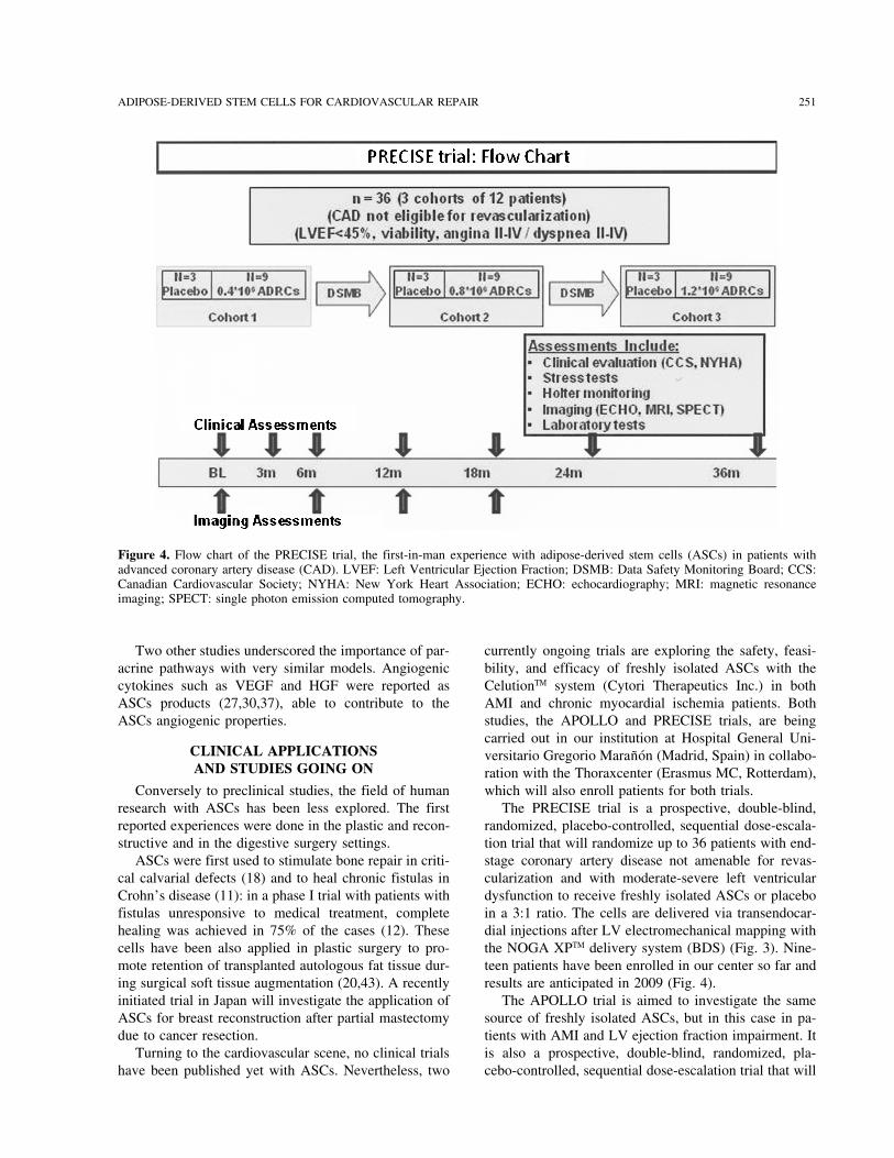
Figure 4. Flow chart of the PRECISE trial, the first-in-man experience with adipose-derived stem cells (ASCs) in patients withadvanced coronary artery disease (CAD). LVEF: Left Ventricular Ejection Fraction; DSMB: Data Safety Monitoring Board; CCS:Canadian Cardiovascular Society; NYHA: New York Heart Association; ECHO: echocardiography; MRI: magnetic resonanceimaging; SPECT: single photon emission computed tomography.
Two other studies underscored the importance of par- currently ongoing trials are exploring the safety, feasi-bility, and efficacy of freshly isolated ASCs with theacrine pathways with very similar models. Angiogenic
cytokines such as VEGF and HGF were reported as CelutionTM system (Cytori Therapeutics Inc.) in bothAMI and chronic myocardial ischemia patients. BothASCs products (27,30,37), able to contribute to the
ASCs angiogenic properties. studies, the APOLLO and PRECISE trials, are beingcarried out in our institution at Hospital General Uni-
CLINICAL APPLICATIONS versitario Gregorio Maranon (Madrid, Spain) in collabo-AND STUDIES GOING ON ration with the Thoraxcenter (Erasmus MC, Rotterdam),
which will also enroll patients for both trials.Conversely to preclinical studies, the field of humanresearch with ASCs has been less explored. The first The PRECISE trial is a prospective, double-blind,
randomized, placebo-controlled, sequential dose-escala-reported experiences were done in the plastic and recon-structive and in the digestive surgery settings. tion trial that will randomize up to 36 patients with end-
stage coronary artery disease not amenable for revas-ASCs were first used to stimulate bone repair in criti-cal calvarial defects (18) and to heal chronic fistulas in cularization and with moderate-severe left ventricular
dysfunction to receive freshly isolated ASCs or placeboCrohn’s disease (11): in a phase I trial with patients withfistulas unresponsive to medical treatment, complete in a 3:1 ratio. The cells are delivered via transendocar-
dial injections after LV electromechanical mapping withhealing was achieved in 75% of the cases (12). Thesecells have been also applied in plastic surgery to pro- the NOGA XPTM delivery system (BDS) (Fig. 3). Nine-
teen patients have been enrolled in our center so far andmote retention of transplanted autologous fat tissue dur-ing surgical soft tissue augmentation (20,43). A recently results are anticipated in 2009 (Fig. 4).
The APOLLO trial is aimed to investigate the sameinitiated trial in Japan will investigate the application ofASCs for breast reconstruction after partial mastectomy source of freshly isolated ASCs, but in this case in pa-
tients with AMI and LV ejection fraction impairment. Itdue to cancer resection.Turning to the cardiovascular scene, no clinical trials is also a prospective, double-blind, randomized, pla-
cebo-controlled, sequential dose-escalation trial that willhave been published yet with ASCs. Nevertheless, two

252 SANZ-RUIZ ET AL.
include up to 48 patients. In the APOLLO trial ASCs will shed light on the safety and efficacy issues of thecardiovascular application of these progenitor cells.are delivered through intracoronary infusion after appro-
priate infarct-related artery repair with stent implanta-REFERENCEStion. The first patient has been included in our center
and the enrollment is planned to be completed by next 1. Alt, E.; Scharlau, M.; Pinkernell, K.; Amadi, C.; Reddy,K.; Mathias, N. Uncultured, autologous adipose-derivedyear.stromal cells-a novel cell source for cardiac repair. Am. J.Cardiol. 96(Suppl. I):71H; 2005.
PROS AND CONS OF ADIPOSE-DERIVED 2. Astori, G.; Vignati, F.; Bardelli, S.; Tubio, M.; Gola, M.;STEM CELLS Albertini, V.; Bambi, F.; Scali, G.; Castelli, D.; Rasini,
V.; Soldati, G.; Moccetti, T. “In vitro” and multicolor phe-ASCs have emerged as a new and promising type of notypic characterization of cell subpopulations identified
stem cells with two clear advantages over the previously in fresh human adipose tissue stromal vascular fractionand in the derived mesenchymal stem cells. J. Transl.used from bone marrow, blood, or skeletal muscle.Med. 5:55; 2007.Firstly, an easy and repeatable access that allows the
3. Cao, Y.; Sun, Z.; Liao, L.; Meng, Y.; Han, Q.; Zhao, R. C.harvesting of high amounts of adipose tissue by a mini-Human adipose tissue-derived stem cells differentiate into
mally invasive method and secondly, an increased pro- endothelial cells in vitro and improve postnatal neovascu-liferative potential to expand themselves in culture, ei- larization in vivo. Biochem. Biophys. Res. Commun. 332:
370–379; 2005.ther because of properties of the cells or as a result of a4. Cardoso, C.; Silva, G. V.; Fernandes, M.; Schreiber, R.;greater frequency of stem cells within the population
Oliveira, E. M.; Jimenez-Quevedo, P.; Sanz-Ruiz, R.;used to initiate the culture.Angeli, F. S.; Pinkernell, K.; Perin, E. Catheter based de-
Moreover, ASCs can be efficiently cryoperserved livery of adipose-derived stem cells in a large animalwith different solutions, keeping their viability and the chronic ischemia model improves myocardial healing.
Eur. Heart J. 28(Suppl. I):227; 2007.adipo/osteogenic potential (32), and can be safely and5. Dicker, A.; Le Blanc, K.; Astrom, G.; van Harmelen, V.;efficiently transfected with Sendai virus (SeV) vectors
Gotherstrom, C.; Blomqvist, L.; Arner, P.; Ryden, M.for gene transfer (53).Functional studies of mesenchymal stem cells derived
An interesting point that deserves deep preclinical re- from adult human adipose tissue. Exp. Cell Res. 308:283–search is the immunogenicity of ASCs. As we have 290; 2005.
6. Dominici, M.; Le Blanc, K.; Mueller, I.; Slaper-Corten-seen, class I and II major histocompatibility complexesbach, I.; Marini, F.; Krause, D.; Deans, R.; Keating, A.;(MHC) are expressed in only 1% of ASCs, and thereforeProckop, D. J.; Horwitz, E. Minimal criteria for definingit has been hypothesized that these could behave as uni-multipotent mesenchymal stromal cells. The International
versal donor cells and could be used for autologous and Society for Cellular Therapy position statement. Cytother-for allogenic transplantation. apy 8:315–317; 2006.
7. Duckers, H. J.; Pinkernell, K.; Milstein, A. M.; Hedrick,Nevertheless, there are some scientific and medicalM. H. The Bedside CelutionTM system for isolation of adi-questions related to ASCs that need to be answered be-pose derived regenerative cells. EuroIntervention 2:395–fore their clinical systematic application. First is the de-398; 2006.
velopment of large-scale manufacturing techniques ac- 8. Fazel, S.; Cimini, M.; Chen, L.; Li, S.; Angoulvant, D.;cording to the GMP requirements. Thus, we will able to Fedak, P.; Verma, S.; Weisel, R. D.; Keating, A.; Li, R. K.
Cardioprotective c-kit+ cells are from the bone marrowdetermine how to manipulate, store, and ship ASCs, al-and regulate the myocardial balance of angiogenic cyto-ways under GMP conditions to avoid contamination.kines. J. Clin. Invest. 116:1865–1877; 2006.For safety purposes, the possibility of adverse events
9. Fickert, S.; Fiedler, J.; Brenner, R. E. Identification ofwill need to be ruled out definitely. These include basi- subpopulations with characteristics of mesenchymal pro-cally two concerns: hemostasis when harvesting adipose genitor cells from human osteoarthritic cartilage using tri-
ple staining for cell surface markers. Arthritis Res. Ther.tissue in patients with AMI who are on antiaggregant6:422–432; 2004.and anticoagulant treatment (10), and the exceptional
10. Fraser, J. K.; Schreiber, R.; Strem, B.; Zhu, M.; Alfonso,chance of long-term tumor development (39). No casesZ.; Wulur, I.; Hedrick, M. H. Plasticity of human adipose
of arrhythmias have been reported in any of the studies stem cells toward endothelial cells and cardiomyocytes.performed so far. Nat. Clin. Pract. Cardiovasc. Med. 3(Suppl. I):S33–37;
2006.11. Garcıa-Olmo, D.; Garcıa-Arranz, M.; Garcıa, L. G.; Cuel-CONCLUSIONS
lar, E. S.; Blanco, I. F.; Prianes, L. A.; Montes, J. A.;Pinto, F. L.; Marcos, D. H.; Garcıa-Sancho, L. AutologousAs a summary, we have seen in this review that hu-stem cell transplantation for treatment of rectovaginal fis-man adipose tissue is a novel promising alternativetula in perianal Crohn’s disease: A new cell-based ther-source of stem cells for cardiovascular repair that hasapy. Int. J. Colorectal Dis. 18:451–454; 2003.
showed encouraging results in the preclinical field. 12. Garcıa-Olmo, D.; Garcıa-Arranz, M.; Herreros, D.; Pascual,However, all this evidence needs adequate translation I.; Peiro, C.; Rodrıguez-Montes, J. A. A phase I clinical
trial of the treatment of Crohn’s fistula by adipose mesen-into humans. The ongoing PRECISE and APOLLO trials

ADIPOSE-DERIVED STEM CELLS FOR CARDIOVASCULAR REPAIR 253
chymal stem cell transplantation. Dis. Colon Rectum 48: 25. Miyahara, Y.; Nagaya, N.; Kataoka, M.; Yanagawa, B.;Tanaka, K.; Hao, H.; Ishino, K.; Ishica, H.; Shimizu, T.;1416–1423; 2005.
13. Gavira, J. J.; Herreros, J.; Perez, A.; Garcia-Velloso, M. J.; Kangawa, K.; Sano, S.; Okano, T.; Kitamura, S.; Mori,H. Monolayered mesenchymal stem cells repair scarredBarba, J.; Martin-Herrero, F.; Canizo, C.; Martin-Arnau,
A.; Martı-Climent, J. M.; Hernandez, M.; Lopez-Holgado, myocardium after myocardial infarction. Nat. Med. 12:459–465; 2006.N.; Gonzalez-Santos, J. M.; Martın-Luengo, C.; Alegria,
E.; Prosper, F. Autologous skeletal myoblast transplanta- 26. Mizuno, H.; Zuk, P. A.; Zhu, M.; Lorenz, H. P.; Benhaim,P.; Hedrick, M. H. Myogenic differentiation by humantion in patients with nonacute myocardial infarction: 1-
year follow-up. J. Thorac. Cardiovasc. Surg. 131:799– processed lipoaspirate cells. Plast. Reconstr. Surg. 109:199–209; 2002.804; 2006.
14. Gimble, J. M.; Katz, A. J.; Bunnell, B. A. Adipose-derived 27. Moon, M. H.; Kim, S. Y.; Kim, Y. J.; Kim, S. J.; Lee,J. B.; Bae, Y. C. Human adipose tissue-derived mesenchy-stem cells for regenerative medicine. Circ. Res. 100:
1249–1260; 2007. mal stem cells improve postnatal neovascularization in amouse model of hindlimb ischemia. Cell. Physiol. Bio-15. Izadpanah, R.; Trygg, C.; Patel, B.; Kriedt, C.; Dufour,
J.; Gimble, J. M.; Bunnell, B. A. Biologic properties of chem. 17:279–290; 2006.28. Murry, C. E.; Field, L. J.; Menasche, P. Cell-based cardiacmesenchymal stem cells derived from bone marrow and
adipose tissue. J. Cell. Biochem. 99:1285–1297; 2006. repair: Feflections at the 10-year point. Circulation 112:3174–3183; 2005.16. Kern, S.; Eichler, H.; Stoeve, J.; Kluter, H.; Bieback, K.
Comparative analysis of mesenchymal stem cells from 29. Nakagami, H.; Morishita, R.; Maeda, K.; Kikuchi, Y.;Ogihara, T.; Kaneda, Y. Adipose tissue-derived stromalbone marrow, umbilical cord blood, or adipose tissue.
Stem Cells 24:1294–1301; 2006. cells as a novel option for regenerative cell therapy. J.Atheroscler. Thromb. 13:77–81; 2006.17. Lee, R. H.; Kim, B.; Choi, I.; Kim, H.; Choi, H. S.; Suh,
K.; Bae, Y. C.; Jung, J. S. Characterization and expression 30. Nakagami, H.; Maeda, K.; Morishita, R.; Iguchi, S.;Nishikawa, T.; Takami, Y.; Kikuchi, Y.; Saito, Y.; Tamai,analysis of mesenchymal stem cells from human bone
marrow and adipose tissue. Cell. Physiol. Biochem. 14: K.; Ogihara, T.; Kaneda, Y. Novel autologous cell therapyin ischemic limb disease through growth factor secretion311–324; 2004.
18. Lendeckel, S.; Jodicke, A.; Christophis, P.; Heidinger, K.; by cultured adipose tissue-derived stromal cells. Arterios-cler. Thromb. Vasc. Biol. 25:2542–2547; 2005.Wolff, J.; Fraser, J. K.; Hedrick, M. H.; Berthold, L.;
Howaldt, H. P. Autologous stem cells (adipose) and fibrin 31. Oedayrajsingh-Varma, M. J.; van Ham, S. M.; Knippen-berg, M.; Helder, M. N.; Klein-Nulend, J.; Schouten, T. E.;glue used to treat widespread traumatic calvarial defects:
Case report. J. Craniomaxillofac. Surg. 32:370–373; 2004. Ritt, M. J.; van Milligen, F. J. Adipose tissue-derived mes-enchymal stem cell yield and growth characteristics are19. Liu, T. M.; Martina, M.; Hutmacher, D. W.; Hui, J. H.;
Lee, E. H.; Lim, B. Identification of common pathways affected by the tissue-harvesting procedure. Cytotherapy8:166–177; 2006.mediating differentiation of bone marrow and adipose tis-
sue-derived human mesenchymal stem cells into three 32. Oishi, K.; Noguchi, H.; Yukawa, H.; Miyazaki, T.; Kato,R.; Kitagawa, Y.; Ueda, M.; Hayasi, S. Cryopreservationmesenchymal lineages. Stem Cells 25:750–760; 2007.
20. Matsumoto, D.; Sato, K.; Gonda, K.; Takaki, Y.; Shigeura, of mouse adipose tissue-derived stem/progenitor cells.Cell Transplant. 17:35–41; 2008.T.; Sato, T.; Aiba-Kojima, E.; Iizuka, F.; Inoue, K.; Suga,
H.; Yoshimura, K. Cell-assisted lipotransfer: Supportive 33. Planat-Benard, V.; Menard, C.; Andre, M.; Puceat, M.;Perez, A.; Garcia-Verdugo, J. M.; Penicaud, L.; Casteilla,use of human adipose-derived cells for soft tissue aug-
mentation with lipoinjection. Tissue Eng. 12:3375–3382; L. Spontaneous cardiomyocyte differentiation from adi-pose tissue stroma cells. Circ. Res. 94:223–229; 2004.2006.
21. Mazo, M.; Planat-Benard, V.; Abizanda, G.; Pelacho, B.; 34. Planat-Benard, V.; Silvestre, J. S.; Cousin, B.; Andre, M.;Nibbelink, M.; Tamarat, R.; Clergue, M.; Manneville, C.;Leobon, B.; Gavira, J. J.; Penuelas, I.; Cemborain, A.;
Penicaud, L.; Laharrague, P.; Joffre, C.; Boisson, M.; Saillan-Barreau, C.; Duriez, M.; Tedgui, A.; Levy, B.;Penicaud, L.; Casteilla, L. Plasticity of human adipose lin-Ecay, M.; Collantes, M.; Barba, J.; Casteilla, L.; Prosper,
F. Transplantation of adipose derived stromal cells is asso- eage cells toward endothelial cells. Physiological and ther-apeutic perspectives. Circulation 109:656–663; 2004.ciated with functional improvement in a rat model of
chronic myocardial infarction. Eur. J. Heart Fail. 10:454– 35. Prunet-Marcassus, B.; Cousin, B.; Caton, D.; Andre, M.;Penicaud, L.; Casteilla, L. From heterogeneity to plasticity462; 2008.
22. McIntosh, K.; Zvonic, S.; Garrett, S.; Mitchell, J. B.; in adipose tissues: Site-specific differences. Exp. Cell Res.312:727–736; 2006.Floyd, Z. E.; Hammill, L.; Kloster, A.; Di Halvorsen, Y.;
Ting, J. P.; Storms, R. W.; Goh, B.; Kilroy, G.; Wu, X.; 36. Rangappa, S.; Fen, C.; Lee, E. H.; Bongso, A.; Sim, E. K.Transformation of adult mesenchymal stem cells isolatedGimble, J. M. The immunogenicity of human adipose-
derived cells: Temporal changes in vitro. Stem Cells 24: from the fatty tissue into cardiomyocytes. Ann. Thorac.Surg. 75:775–779; 2003.1246–1253; 2006.
23. Miranville, A.; Heeschen, C.; Sengenes, C.; Curat, C. A.; 37. Rehman, J.; Traktuev, D.; Li, J.; Merfeld-Clauss, S.;Temm-Grove, C. J.; Bovenkerk, J. E.; Pell, C. L.; John-Busse, R.; Bouloumie, A. Improvement of postnatal neo-
vascularization by human adipose tissue-derived stem stone, B. H.; Considine, R. V.; March, K. L. Secretionof angiogenic and antiapoptotic factors by human adiposecells. Circulation 110:349–355; 2004.
24. Mitchell, J. B.; McIntosh, K.; Zvonic, S.; Garrett, S.; stromal cells. Circulation 109:1292–1298; 2004.38. Rider, D. A.; Dombrowski, C.; Sawyer, A. A.; Ng, G. H.;Floyd, Z. E.; Kloster, A.; Di Halvorsen, Y.; Storms, R. W.;
Goh, B.; Kilroy, G.; Wu, X.; Gimble, J. M. Immunophe- Leong, D.; Hutmacher, D. W.; Nurcombe, V.; Cool, S. M.Autocrine FGF2 increases the multipotentiality of humannotype of human adipose-derived cells: Temporal changes
in stromal-associated and stem cell-associated markers. adipose-derived mesenchymal stem cells. Stem Cells 26:1598–1608; 2008.Stem Cells 24:376–385; 2006.

254 SANZ-RUIZ ET AL.
39. Rubio, D.; Garcia-Castro, J.; Martin, M. C.; de la Fuente, 48. Xu, Y.; Malladi, P.; Wagner, D. R.; Longaker, M. T. Adi-pose-derived mesenchymal cells as a potential cell sourceR.; Cigudosa, J. C.; Lloyd, A. C.; Bernad, A. Spontaneous
human adult stem cell transformation. Cancer Res. 65: for skeletal regeneration. Curr. Opin. Mol. Ther. 7:300–305; 2005.3035–3039; 2005.
40. Schaffler, A.; Buchler, C. Concise review: Adipose tissue- 49. Yamada, Y.; Wang, X. D.; Yokoyama, S.; Fukuda, N.;Takakura, N. Cardiac progenitor cells in brown adiposederived stromal cells—basic and clinical implications for
novel cell-based therapies. Stem Cells 25:818–827; 2007. tissue repaired damaged myocardium. Biochem. Biophys.Res. Commun. 342:662–670; 2006.41. Sengenes, C.; Lolmede, K.; Zakaroff-Girard, A.; Busse,
R.; Bouloumie, A. Preadipocytes in the human subcutane- 50. Yamada, Y.; Yokoyama, S.; Fukuda, N.; Kidoya, H.; Huang,X. Y.; Naitoh, H.; Satoh, N.; Takakura, N. A novel ap-ous adipose tissue display distinct features from the adult
mesenchymal and hematopoietic stem cells. J. Cell. Phys- proach for myocardial regeneration with educated bloodcells cocultured with cells from brown adipose tissue. Bio-iol. 205:114–122; 2005.
42. Smith, P.; Adams, Jr., W. P.; Lipschitz, A. H.; Chau, B.; chem. Biophys. Res. Commun. 353:182–188; 2007.51. Yamada, Y.; Yokoyama, S.; Wang, X. D.; Fukuda, N.;Sorokin, E.; Rohrich, R. J.; Brown, S. A. Autologous hu-
man fat grafting: Effect of harvesting and preparation Takakura, N. Cardiac stem cells in brown adipose tissueexpress CD133 and induce bone marrow nonhematopoi-techniques on adipocyte graft survival. Plast. Reconstr.
Surg. 117:1836–1844; 2006. etic cells to differentiate into cardiomyocytes. Stem Cells25:1326–1333; 2007.43. Stosich, M. S.; Mao, J. J. Adipose tissue engineering from
human adult stem cells: Clinical implications in plastic 52. Yoshimura, H.; Muneta, T.; Nimura, A.; Yokoyama, A.;Koga, H.; Sekiya, I. Comparison of rat mesenchymal stemand reconstructive surgery. Plast. Reconstr. Surg. 119:71–
83; 2007. cells derived from bone marrow, synovium, periosteum,adipose tissue, and muscle. Cell Tissue Res. 327:449–462;44. Strem, B. M.; Zhu, M.; Alfonso, Z.; Daniels, E. J.; Schreib-
er, R.; Beygui, R.; MacLellan, W. R.; Hedrick, M. H.; 2007.53. Yukawa, H.; Noguchi, H.; Oishi, K.; Miyazaki, T.; Kita-Fraser, J. K. Expression of cardiomyocytic markers on ad-
ipose tissue-derived cells in a murine model of acute myo- gawa, Y.; Inoue, M.; Hasegawa, M.; Hayasi, S. Recombi-nant sendai virus-mediated gene transfer to adipose tissue-cardial injury. Cytotherapy 7:282–291; 2005.
45. Strem, B. M.; Jordan, M.; Kim, J.; Yang, J.; Anderson, derived stem cells (ASCs). Cell Transplant. 17:43–50;2008.C. D.; Daniels, E. Adipose tissue-derived stem cells en-
hance cardiac function following surgically-induced myo- 54. Zuk, P. A.; Zhu, M.; Mizuno, H.; Huang, J.; Futrell,J. W.; Katz, A. J.; Benhaim, P.; Lorenz, H. P.; Hedrick,cardial infarction. Circulation 112(Suppl. II):274; 2005.
46. Valina, C.; Pinkernell, K.; Song, Y. H.; Bai, X.; Sadat, S.; M. H. Multilineage cells from human adipose tissue: Im-plications for cell-based therapies. Tissue Eng. 7:211–Campeau, R. J.; Le Jemtel, T. H.; Alt, E. Intracoronary
administration of autologous adipose tissue-derived stem 228; 2001.55. Zuk, P. A.; Zhu, M.; Ashjian, P.; De Ugarte, D. A.;cells improves left ventricular function, perfusion and re-
modeling after acute myocardial infarction. Eur. Heart J. Huang, J. I.; Mizuno, H.; Alfonso, Z. C.; Fraser, J. K.;Benhaim, P.; Hedrick, M. H. Human adipose tissue is a28:2667–2677; 2007.
47. Watanabe, C. T.; Lee, S.; Daniela, E.; Naqvi, T. Z.; Shah, source of multipotent stem cells. Mol. Biol. Cell 13:4279–4295; 2002.P. K.; Shah, A. Intracoronary adipose tissue-derived stem
cell therapy preserves left ventricular function in a porcineinfarct model. Am. J. Cardiol. 94(Suppl. I):188E; 2004.
Related Documents