10.1128/JVI.79.9.5665-5675.2005. 2005, 79(9):5665. DOI: J. Virol. Rowland-Jones and Jeremy Farrar Thu Thao, Nguyen Thi Dung, Tran Tinh Hien, Sarah Nguyen Thi Phuong Dung, Tran Nguyen Bich Chau, Le Thi Cameron P. Simmons, Tao Dong, Nguyen Vinh Chau, Secondary Dengue Virus Infections Epitopes in Vietnamese Adults with Early T-Cell Responses to Dengue Virus http://jvi.asm.org/content/79/9/5665 Updated information and services can be found at: These include: REFERENCES http://jvi.asm.org/content/79/9/5665#ref-list-1 at: This article cites 51 articles, 35 of which can be accessed free CONTENT ALERTS more» articles cite this article), Receive: RSS Feeds, eTOCs, free email alerts (when new http://journals.asm.org/site/misc/reprints.xhtml Information about commercial reprint orders: http://journals.asm.org/site/subscriptions/ To subscribe to to another ASM Journal go to: on December 9, 2013 by guest http://jvi.asm.org/ Downloaded from on December 9, 2013 by guest http://jvi.asm.org/ Downloaded from

Welcome message from author
This document is posted to help you gain knowledge. Please leave a comment to let me know what you think about it! Share it to your friends and learn new things together.
Transcript

10.1128/JVI.79.9.5665-5675.2005.
2005, 79(9):5665. DOI:J. Virol. Rowland-Jones and Jeremy FarrarThu Thao, Nguyen Thi Dung, Tran Tinh Hien, SarahNguyen Thi Phuong Dung, Tran Nguyen Bich Chau, Le Thi Cameron P. Simmons, Tao Dong, Nguyen Vinh Chau, Secondary Dengue Virus InfectionsEpitopes in Vietnamese Adults with Early T-Cell Responses to Dengue Virus
http://jvi.asm.org/content/79/9/5665Updated information and services can be found at:
These include:
REFERENCEShttp://jvi.asm.org/content/79/9/5665#ref-list-1at:
This article cites 51 articles, 35 of which can be accessed free
CONTENT ALERTS more»articles cite this article),
Receive: RSS Feeds, eTOCs, free email alerts (when new
http://journals.asm.org/site/misc/reprints.xhtmlInformation about commercial reprint orders: http://journals.asm.org/site/subscriptions/To subscribe to to another ASM Journal go to:
on Decem
ber 9, 2013 by guesthttp://jvi.asm
.org/D
ownloaded from
on D
ecember 9, 2013 by guest
http://jvi.asm.org/
Dow
nloaded from
JOURNAL OF VIROLOGY, May 2005, p. 5665–5675 Vol. 79, No. 90022-538X/05/$08.00�0 doi:10.1128/JVI.79.9.5665–5675.2005Copyright © 2005, American Society for Microbiology. All Rights Reserved.
Early T-Cell Responses to Dengue Virus Epitopes in VietnameseAdults with Secondary Dengue Virus Infections
Cameron P. Simmons,1* Tao Dong,2 Nguyen Vinh Chau,1,3 Nguyen Thi Phuong Dung,1Tran Nguyen Bich Chau,1 Le Thi Thu Thao,3 Nguyen Thi Dung,3 Tran Tinh Hien,3
Sarah Rowland-Jones,2 and Jeremy Farrar1
Oxford University Clinical Research Unit,1 Hospital for Tropical Diseases,3 Ho Chi Minh City, Vietnam, andInstitute for Molecular Medicine, Nuffield Department of Clinical Medicine, John Radcliffe Hospital,
University of Oxford, Oxford, United Kingdom2
Received 21 October 2004/Accepted 17 December 2004
T-cell responses to dengue viruses may be important in both protective immunity and pathogenesis. Thisstudy of 48 Vietnamese adults with secondary dengue virus infections defined the breadth and magnitude ofperipheral T-cell responses to 260 overlapping peptide antigens derived from a dengue virus serotype 2 (DV2)isolate. Forty-seven different peptides evoked significant gamma interferon enzyme-linked immunospot (ELIS-POT) assay responses in 39 patients; of these, 34 peptides contained potentially novel T-cell epitopes. NS3 andparticularly NS3200-324 were important T-cell targets. The breadth and magnitude of ELISPOT responses toDV2 peptides were independent of the infecting dengue virus serotype, suggesting that cross-reactive T cellsdominate the acute response during secondary infection. Acute ELISPOT responses were weakly correlatedwith the extent of hemoconcentration in individual patients but not with the nadir of thrombocytopenia oroverall clinical disease grade. NS3556-564 and Env414-422 were identified as novel HLA-A*24 and B*07-restrictedCD8� T-cell epitopes, respectively. Acute T-cell responses to natural variants of Env414-422 and NS3556-564 werelargely cross-reactive and peaked during disease convalescence. The results highlight the importance of NS3and cross-reactive T cells during acute secondary infection but suggest that the overall breadth and magnitudeof the T-cell response is not significantly related to clinical disease grade.
Dengue viruses are positive-strand RNA viruses belongingto the family Flaviviridae. Many tropical and subtropical re-gions have become areas where dengue viruses are endemic;such endemicity is an important cause of morbidity and mor-tality (17, 40). Primary infection with any of the four serotypesof dengue virus (DV1 to DV4) typically results in either anasymptomatic illness or dengue fever (DF), a self-limitingacute viral infection characterized by high fever and myalgia.Epidemiological studies suggest that individuals who are in-fected a second time with a different dengue virus serotype areat significantly greater risk of developing dengue hemorrhagicfever (DHF), a serious illness characterized by plasma leakage,thrombocytopenia, and occasionally hypovolemic shock (5, 12,43, 45). While children carry most of the symptomatic diseaseburden in countries where dengue viruses are endemic, youngadults are also significantly affected.
Antibody-dependent enhancement (ADE) of secondary vi-ral infection is a prominent explanation for the associationbetween severe disease and preexisting dengue virus immuneresponses (19). It is hypothesized that ADE promotes highersystemic viral loads, a theory supported by studies of culturedcells (20, 21) and of experimentally infected primates (19).Correspondingly, high dengue viral loads (31, 47) and antigen-emia (32) have been associated with DHF in children. How-ever, the mechanisms through which relatively higher dengue
viral loads might mediate the vascular leakage that is so prom-inent in patients with severe disease remains unclear. Otherparameters, such as the age, nutritional status, and geneticbackground of the host, may also be important (1, 10, 30, 34,35, 44, 46). Excessive immune activation during secondary in-fection has also been hypothesized to act in concert with theseother risk factors and to promote the development of severevascular leakage. In particular, the rapid mobilization of sero-type cross-reactive memory T cells that release vasodilatoryinflammatory molecules has been suggested to explain someaspects of the clinical syndrome (28). Accordingly, relativelyhigher frequencies of activated CD8� T cells in peripheralblood have been detected in patients with DHF than in thosewith DF (14, 41, 51). Other plasma markers of cellular immuneactivation, including tumor necrosis factor (23), gamma inter-feron (IFN-�) (26), interleukin-6 (IL-6) (22), soluble IL-2 re-ceptor (15, 26), and tumor necrosis factor receptor II (15)levels, are also elevated in DHF compared to DF. Collectively,these studies suggest a role for the immune response in diseasepathogenesis. How T cells contribute to this process is incom-pletely defined, however, since only a few studies have directlyexamined T-cell responses during acute disease (14, 41). Amore comprehensive understanding of the relationship be-tween T-cell response and disease severity in acute dengueillness would be obtained by measuring ex vivo responses to aspectrum of dengue virus antigens in a patient population witha mixed HLA background. To this end, this study measured themagnitude and specificity of T-cell responses to 260 peptideantigens in 48 Vietnamese adults experiencing a secondarydengue virus infection. Results from these experiments were
* Corresponding author. Mailing address: Oxford University Clini-cal Research Unit, Hospital for Tropical Diseases, 190 Ben Ham Tu,District 5, Ho Chi Minh City, Vietnam. Phone: 84 8 9237954. Fax: 848 9238904. E-mail: [email protected].
5665
on Decem
ber 9, 2013 by guesthttp://jvi.asm
.org/D
ownloaded from
used as a foundation from which to define two novel T-cellepitopes and to assess the relationship between cellular im-mune response and clinical and virological parameters.
MATERIALS AND METHODSStudy design and patients. Venous blood samples were obtained from adult
patients enrolled in a prospective study of dengue virus infection at The Hospitalfor Tropical Diseases, Ho Chi Minh City, Vietnam. Patients were recruited ifthere was a clinical suspicion of dengue virus infection. World Health Organi-zation classification criteria (49) were applied to each case after a review of thestudy notes. However, the strict classification scheme could not be applied to twocases because of conflicting clinical presentations. For example, two subjects hadclinical manifestations of plasma leakage (e.g., pleural effusions) and mucosal
bleeding, but platelet counts were between 100,000 and 120,000. We classifiedthese cases as DHF II. Several cases also demonstrated hemoconcentration of�20% during hospitalization but nevertheless had clinical signs of plasma leak-age, e.g., pleural effusions or ascites. These cases were classified as DHF I or II,depending on whether or not hemorrhagic manifestations were present.
Peripheral blood samples were collected from patients between 8 and 11 a.m.on the first morning after admission (study day 1), on study day 3, and again onstudy day 5 unless the patient had been discharged earlier. Convalescent sampleswere obtained at 2 weeks and 1 month postadmission. Platelet counts andhematocrit values were recorded regularly during hospitalization. The extent ofhemoconcentration during symptomatic illness was determined by comparing themaximum hematocrit recorded during hospitalization with the value recorded atconvalescence (study day 14 or 30). These data were available for 24 patients.
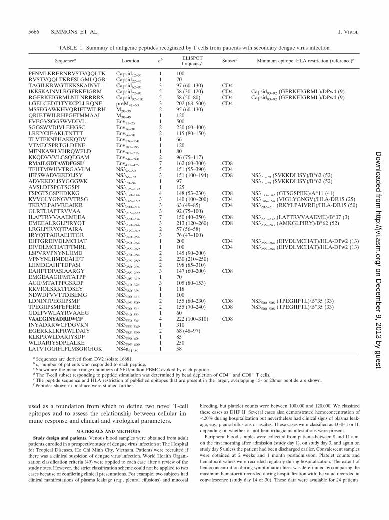
TABLE 1. Summary of antigenic peptides recognized by T cells from patients with secondary dengue virus infection
Sequencea Location nb ELISPOTfrequencyc Subsetd Minimum epitope, HLA restriction (reference)e
PFNMLKRERNRVSTVQQLTK Capsid12–31 1 100RVSTVQQLTKRFSLGMLQGR Capsid22–41 1 70TAGILKRWGTIKKSKAINVL Capsid62–81 3 97 (60–130) CD4IKKSKAINVLRGFRKEIGRM Capsid72–91 5 58 (30–120) CD4 Capsid83–92 (GFRKEIGRML)/DPw4 (9)RGFRKEIGRMLNILNRRRRS Capsid82–101 5 58 (50–80) CD4 Capsid83–92 (GFRKEIGRML)/DPw4 (9)LGELCEDTITYKCPLLRQNE preM41–60 3 202 (68–500) CD4MSSEGAWKHVQRIETWILRH M20–39 2 95 (60–130)QRIETWILRHPGFTMMAAI M30–49 1 120FVEGVSGGSWVDIVL Env11–25 1 500SGGSWVDIVLEHGSC Env16–30 2 230 (60–400)LRKYCIEAKLTNTTT Env56–70 2 115 (80–150)TLVTFKNPHAKKQDV Env136–150 1 66VTMECSPRTGLDFNE Env181–195 1 120MENKAWLVHRQWFLD Env201–215 1 80KKQDVVVLGSQEGAM Env246–260 2 96 (75–117)RMAILGDTAWDFGSLf Env411–425 7 162 (60–300) CD8TFHTMWHVTRGAVLM NS345–59 5 151 (55–390) CD4IEPSWADVKKDLISY NS365–79 3 151 (100–194) CD8 NS371–79 (SVKKDLISY)/B*62 (52)ADVKKDLISYGGGWK NS370–84 1 97 NS371–79 (SVKKDLISY)/B*62 (52)AVSLDFSPGTSGSPI NS3125–139 1 125FSPGTSGSPIIDKKG NS3130–144 4 148 (53–230) CD8 NS3133–142 (GTSGSPIIK)/A*11 (41)KVVGLYGNGVVTRSG NS3145–159 3 140 (100–200) CD4 NS3146–154 (VIGLYGNGV)/HLA-DR15 (25)TKRYLPAIVREAIKR NS3200–214 3 63 (49–85) CD4 NS3202–211 (RKYLPAIVRE)/HLA-DR15 (50)GLRTLIAPTRVVAA NS3215–229 3 92 (75–100)ILAPTRVVAAEMEEA NS3220–234 7 150 (40–350) CD8 NS3221–232 (LAPTRVVAAEME)/B*07 (3)EMEEALRGLPIRYQT NS3230–244 3 213 (120–260) CD8 NS3235–243 (AMKGLPIRY)/B*62 (52)LRGLPIRYQTPAIRA NS3235–249 2 57 (56–58)IRYQTPAIRAEHTGR NS3240–254 3 76 (47–100)EHTGREIVDLMCHAT NS3250–264 1 200 CD4 NS3255–264 (EIVDLMCHAT)/HLA-DPw2 (13)EIVDLMCHATFTMRL NS3255–269 1 100 CD4 NS3255–264 (EIVDLMCHAT)/HLA-DPw2 (13)LSPVRVPNYNLIIMD NS3270–284 2 145 (90–200)VPNYNLIIMDEAHFT NS3275–289 2 230 (210–250)LIIMDEAHFTDPASI NS3280–294 2 198 (85–310)EAHFTDPASIAARGY NS3285–299 3 147 (60–200) CD8EMGEAAGIFMTATPP NS3305–319 1 70AGIFMTATPPGSRDP NS3310–324 3 105 (80–153)KKVIQLSRKTFDSEY NS3380–394 1 118NDWDFVVTTDISEMG NS3400–414 1 100LDNINTPEGIIPSMF NS3495–509 2 155 (80–230) CD8 NS3500–508 (TPEGIIPTL)/B*35 (33)TPEGIIPSMFEPERE NS3500–514 2 155 (70–240) CD8 NS3500–508 (TPEGIIPTL)/B*35 (33)GDLPVWLAYRVAAEG NS3540–554 1 60VAAEGINYADRRWCFf NS3550–564 4 222 (100–310) CD8INYADRRWCFDGVKN NS3555–569 1 310EGERKKLKPRWLDAIY NS3585–599 2 68 (48–97)KLKPRWLDARIYSDP NS3590–604 1 85WLDARIYSDPLALKE NS3595–609 1 250LATVTGGIFLFLMSGRGIGK NS4a61–80 1 58
a Sequences are derived from DV2 isolate 16681.b n, number of patients who responded to each peptide.c Shown are the mean (range) numbers of SFU/million PBMC evoked by each peptide.d The T-cell subset responding to peptide stimulation was determined by bead depletion of CD4� and CD8� T cells.e The peptide sequence and HLA restriction of published epitopes that are present in the larger, overlapping 15- or 20mer peptide are shown.f Peptides shown in boldface were studied further.
5666 SIMMONS ET AL. J. VIROL.
on Decem
ber 9, 2013 by guesthttp://jvi.asm
.org/D
ownloaded from
Written informed consent was obtained from the patient. The study protocol wasapproved by the Scientific and Ethical Committee at The Hospital for TropicalDiseases and the Oxford Tropical Research Ethical Committee.
Dengue virus PCR. Dengue virus RNA in acute plasma samples was isolatedwith RNAgents (Promega, Madison, Wis.). RNA was reverse transcribed, andtwo rounds of PCR were performed using primers and methods described pre-viously (29). In samples containing virus, the PCR yielded DNA products theunique sizes of which were diagnostic for each dengue virus serotype.
Serology, cell isolation, and HLA typing. Dengue virus infection was con-firmed via serological testing of acute- and early-convalescent-phase plasmasamples collected at least 3 days apart with a commercial capture-immunoglob-ulin M (IgM) and IgG enzyme-linked immunosorbent assay (ELISA) (Panbio,Brisbane, Australia). The ELISA was performed and the results were interpretedaccording to the manufacturer’s instructions. This ELISA assay has been vali-dated as both sensitive and specific for primary and secondary dengue virusinfections (48). In this study, the term “secondary infection” is used to describethe nature of the serological response and does not imply that this was neces-sarily the second dengue virus infection experienced by the patient. Peripheralblood mononuclear cells (PBMC) were isolated from whole blood by Ficoll-Hypaque (Lymphoprep; Axis-Shield, Oslo, Norway) density gradient centrifuga-tion and used directly in enzyme-linked immunospot (ELISPOT) assays or cryo-preserved for future use. Molecular HLA typing was performed on most study
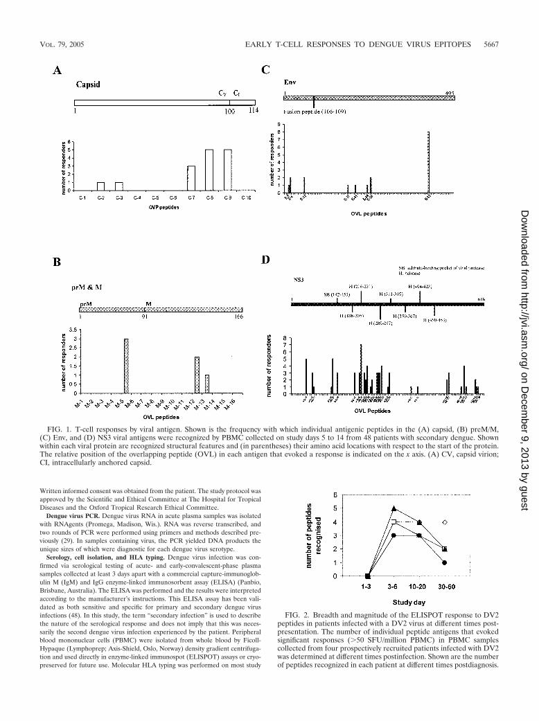
FIG. 1. T-cell responses by viral antigen. Shown is the frequency with which individual antigenic peptides in the (A) capsid, (B) preM/M,(C) Env, and (D) NS3 viral antigens were recognized by PBMC collected on study days 5 to 14 from 48 patients with secondary dengue. Shownwithin each viral protein are recognized structural features and (in parentheses) their amino acid locations with respect to the start of the protein.The relative position of the overlapping peptide (OVL) in each antigen that evoked a response is indicated on the x axis. (A) CV, capsid virion;CI, intracellularly anchored capsid.
FIG. 2. Breadth and magnitude of the ELISPOT response to DV2peptides in patients infected with a DV2 virus at different times post-presentation. The number of individual peptide antigens that evokedsignificant responses (�50 SFU/million PBMC) in PBMC samplescollected from four prospectively recruited patients infected with DV2was determined at different times postinfection. Shown are the numberof peptides recognized in each patient at different times postdiagnosis.
VOL. 79, 2005 EARLY T-CELL RESPONSES TO DENGUE VIRUS EPITOPES 5667
on Decem
ber 9, 2013 by guesthttp://jvi.asm
.org/D
ownloaded from
subjects with amplification refractory mutation system-PCR with sequence-spe-cific primers, as described previously (4). With samples from some subjects, acommercial typing application, the Dynal RELI SSO HLA typing kit (DynalBiotech, Wirral, United Kingdom) was used.
ELISPOT and peptides. In this study, we were unable to collect enough bloodfrom patients to study T-cell responses to peptide antigens spanning the entiregenome. Consequently, we focused our investigations on structural antigens(capsid, preM/M, and Env) and those nonstructural viral antigens previouslynominated as being T-cell immunogens (NS3 and NS4a). A total of 260 peptides(220 15mers and 40 20mers, all overlapping by 10 amino acids [aa]), spanning thecapsid, preM, M, Env, NS3, and NS4a sequences from a dengue virus serotype2 isolate (strain 16681) were synthesized by standard, solid-phase 9-fluorenyl-methoxy carbonyl chemistry. Purity ranged from 30 to 90% as determined byhigh-performance liquid chromatography (2). PBMC samples were obtainedbetween study days 5 to 14 from 48 patients experiencing a secondary denguevirus infection. PBMC samples were tested in IFN-� ELISPOT assays against the260 peptides arranged into a matrix of 32 peptide pools, with 16 peptides in eachpool (each at 1 �M) and each peptide present in two different pools. Thisapproach allows rapid identification of the peptide within a pool that is respon-sible for evoking a response in the IFN-� ELISPOT assay, since this peptideshould drive a response in two different pools. In every case, peptides identifiedas being antigenic in this manner were retested as individual peptides. Thus, eachpositive response was confirmed twice, by means of the internal control affordedby the matrix and then individually by a confirmatory assay. The ELISPOT assaywas performed essentially according to the manufacturer’s instructions (MabtechAB, Stockholm, Sweden). Briefly, 96-well polyvinylidene difluoride-backedplates (MAIPS45; Millipore), precoated with 15 �g of anti-IFN-� monoclonalantibody 1-D1K (Mabtech)/ml, were blocked with R10 (RPMI 1640 containing10% heat-inactivated fetal calf serum, 2 mM glutamine, 100 �g of streptomycin/ml, and 100 U of penicillin) for 2 h. A total of 1 � 105 to 3 � 105 PBMC wereadded in 100 �l of R10 per well, and peptide pools or individual peptides weresubsequently added to a final concentration of 16 �M (peptide pools) or 1 �M(individual peptides). After an overnight incubation at 37°C and 5% CO2 in air,plates were washed with phosphate-buffered saline (PBS) containing 0.05%Tween-20 (Sigma). Next, 100 �l of PBS containing 1 �g of biotinylated anti-IFN-� monoclonal antibody 7-B6-1-biotin (Mabtech)/ml was added. After 2 h,plates were washed, and streptavidin-alkaline phosphatase conjugate (Mabtech)was added at a dilution of 1:1,000 in PBS. The number of spot-forming units(SFU) in each well was counted with the aid of a dissecting microscope, and thebackground (no antigen stimulation) was subtracted. Wells containing PBMCstimulated with phytohemagglutinin (Sigma, Poole, United Kingdom) served asa positive assay control. A positive response to a pool of peptides was defined ashaving �50 SFU per million PBMC after subtraction of background. This cutoffwas used on the basis that 50 SFU per million PBMC was 2 standard deviations(SD) above the mean of unstimulated PBMC from early-convalescent-phasedengue virus infection cases.
Cell depletions. T-cell depletions from PBMC were performed using anti-CD8or anti-CD4 antibody-coated immunomagnetic beads (Dynal, Oslo, Norway)
according to the manufacturer’s instructions. Flow cytometry analysis of thedepleted cells indicated this approach routinely achieved �95% depletion of thetarget cells.
Cytotoxicity assay. A T-cell line from BC503 was generated by pulsing 2 � 106
PBMC with 100 �M of Env414–422 for 1 h. Cells were cultured in R10 supple-mented with 25 ng of IL-7/ml for 3 days, and then 100 U of IL-2/ml was addedevery 3 to 4 days thereafter. After 14 days, cells were harvested and used aseffectors in a 51Cr-labeled release assay. Targets cells in the chromium releaseassay consisted of a 51Cr-labeled B-cell line (BCL) that was HLA matched witheffectors only at the B*07 locus. The BCL was pulsed for 1 h with the Env414–422
peptide, washed, and aliquoted in microtiter plates (5,000 cells/well), and theneffector cells were added at a range of effector-to-target ratios. Unpulsed cellswere used as negative controls. The 51Cr release was calculated from the follow-ing equation: ([experimental release�spontaneous release]/[maximumrelease�spontaneous release]) � 100%. Nonspecific killing of unpulsed targetcells was subtracted from that of pulsed target cells.
Short-term stimulation of PBMC with peptide-pulsed B-cell lines. A rapid,flow cytometry-based method was employed to define the HLA restriction ofcandidate T-cell epitopes as described previously (11). Firstly, Epstein-Barrvirus-transformed B-cell lines (BCL) that matched or mismatched the effectorcells to be studied (patients’ PBMC) at given HLA alleles were selected. TheseBCLs were pulsed with test or control peptides (10 �M) for 1 h and then washedthree times. A total of 5 � 105 peptide-pulsed and washed BCL were then addedto 2 � 106 PBMC (effectors) in a 0.3-ml volume and cultured at 37°C with 5%CO2 in air for 6 h. During the last 4 h, brefeldin A (10 �g/ml) was added. After6 h, cells were washed, surface stained for CD3 and CD8, then fixed (FACSlyse;Becton Dickinson, San Diego, Calif.), and made permeable with Facsperm buffer(Becton Dickinson). Cells were then washed and stained for intracellular IFN-�and the early activation marker CD69. After being stained and fixed, CD3�
CD8� cells were gated on a FACSCalibur flow cytometer (Becton Dickinson),and the percentage of cells that was CD69� IFN-�� was determined for eachBCL-peptide combination. PBMC stimulated with phorbol myristate acetate-ionomycin were used as positive controls, and PBMC stimulated with mediumalone were used as negative controls.
Statistics. Spearman correlation analysis was used to measure associationsbetween nonnormally distributed variables. Analysis of variance was used forcomparisons across multiple groups. A P value of �0.05 was regarded as signif-icant. The SPSS software package SPSS, version 10 (SPSS, Inc., Chicago, Ill.),was used for all analyses.
RESULTS
Characteristics of study population. This study was con-ducted at The Hospital for Tropical Diseases, Ho Chi MinhCity, Vietnam. The Hospital for Tropical Diseases serves thelocal community and acts as a tertiary referral hospital forpatients in southern Vietnam with infectious diseases. BetweenJune 2002 and April 2003, we recruited 48 patients suspectedof having a dengue virus infection from the adult intensive careunit into a prospective study of cellular immune responses. Themean age was 19 years (SD, 3 years; range, 15 to 27 years) andthe mean length of illness before admission was 5 days (SD, 1day; range, 1 to 10 days). Serology of paired plasma samplesindicated that all patients were experiencing a secondary den-gue virus infection. During hospital admission, the averagemaximum hematocrit attained was 49% (SD, 4.7%; range, 41to 60%) and the mean nadir of the platelet count was 47,700 �106/ml (SD, 33,900 � 106; range, 13,900 � 106 to 115,000 �106).
IFN-� ELISPOT responses against a panel of peptides span-ning the capsid, preM, M, Env, NS3, and NS4a viral antigens.To measure the breadth and magnitude of T-cell responses tomultiple dengue virus antigens, overlapping peptides spanningthe capsid, preM, M, Env, NS3, and NS4a viral antigens froma DV2 isolate were employed in IFN-� ELISPOT assays withPBMC samples from dengue virus-immune Vietnamese adults.In 39 patients, we identified one or more individual peptides
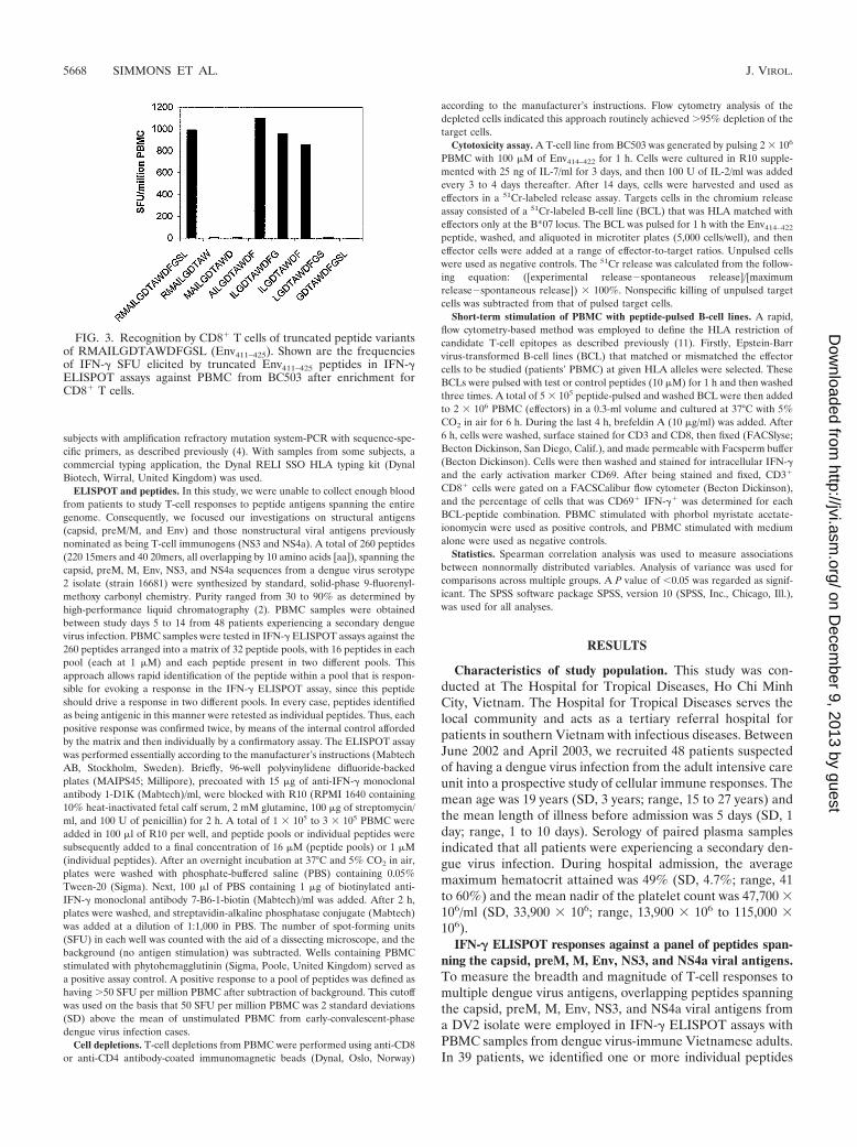
FIG. 3. Recognition by CD8� T cells of truncated peptide variantsof RMAILGDTAWDFGSL (Env411–425). Shown are the frequenciesof IFN-� SFU elicited by truncated Env411–425 peptides in IFN-�ELISPOT assays against PBMC from BC503 after enrichment forCD8� T cells.
5668 SIMMONS ET AL. J. VIROL.
on Decem
ber 9, 2013 by guesthttp://jvi.asm
.org/D
ownloaded from
that reproducibly evoked an IFN-� response in the ELISPOTassay that exceeded 50 SFU/million PBMC. In total, T cellsfrom 39 patients recognized 47 different peptides. The magni-tude of responses to individual peptides ranged from 50 to 500SFU/million PBMC. The breadth of the response in individualpatients ranged from 1 to 10 peptides. A summary of theindividual peptides that reproducibly evoked responses inIFN-� ELISPOT assays is provided in Table 1. In several cases,two adjacent and overlapping peptides evoked a response inthe same patient, suggesting the peptides contained a sharedepitope or alternatively suggesting the presence of overlappingepitopes. For several of the peptide antigens, we used IFN-�ELISPOT assays with PBMC depleted of either CD4� orCD8� T cells to identify the responding T-cell subset. Theseexperiments identified several peptides (capsid62–81, preM41–60,Env411–425, NS345–59, NS3285–299, and NS3550–564) that containnovel CD4� or CD8� T-cell determinants (Table 1). Thirteen(23%) of the 47 peptides recognized by PBMC contain se-quences that have previously been characterized as denguevirus- or flavivirus-specific CD4� or CD8� T-cell epitopes(Table 1).
Antigen specificity of IFN-� ELISPOT responses and theirrelationship to recognized structural motifs. The spectrum ofpeptides recognized by T cells ex vivo was examined in thecontext of known biological features in each of the respectiveviral antigens. Several antigenic peptides were identified in thecapsid protein; these were primarily located near to the con-served cleavage point of the C-terminal hydrophobic signalsequence (Table 1 and Fig. 1A). Antigenic peptides were alsodetected in preM/M (Table 1 and Fig. 1B), Env (Table 1 andFig. 1C), and NS4a (Table 1 and data not shown). Theseantigenic peptides were not associated with any recognizedconserved structural motifs. NS3, a 618-aa-long serine proteaseand helicase, contained the highest number (n � 30) of anti-genic peptides (Table 1 and Fig. 1D). A total of 14 (47%) outof 30 of these antigenic peptides were clustered within a 124-aa-long stretch of NS3 (NS3200–324) (Fig. 1D). Alignment ofconsensus amino acid sequences from all four dengue virusserotypes indicated that this region of NS3 is more conserved(78%) than NS3 as a whole (68%). NS3 was the most fre-quently recognized viral antigen, with 56% of all tested pa-tients responding to at least one antigenic peptide in NS3compared to 27% for Env, 23% for capsid, 10% for preM/M,and 2% for NS4a.
Relationship between the serotype of the infecting virus andthe breadth and magnitude of responses to overlapping DV2peptides in IFN-� ELISPOT assays. A dengue virus serotype-specific PCR performed on acute plasma samples collectedfrom all 48 patients identified the serotype of the infectingvirus in 33 individuals (68%). Among these 32 patients, DV1was detected in 9 (28%), DV2 was detected in 15 (47%), DV3was detected in 1 (3%), and DV4 was detected in 7 (22%). Onepatient had a mixed infection comprising DV1 and DV4 (3%).We sought to determine whether a relationship existed be-
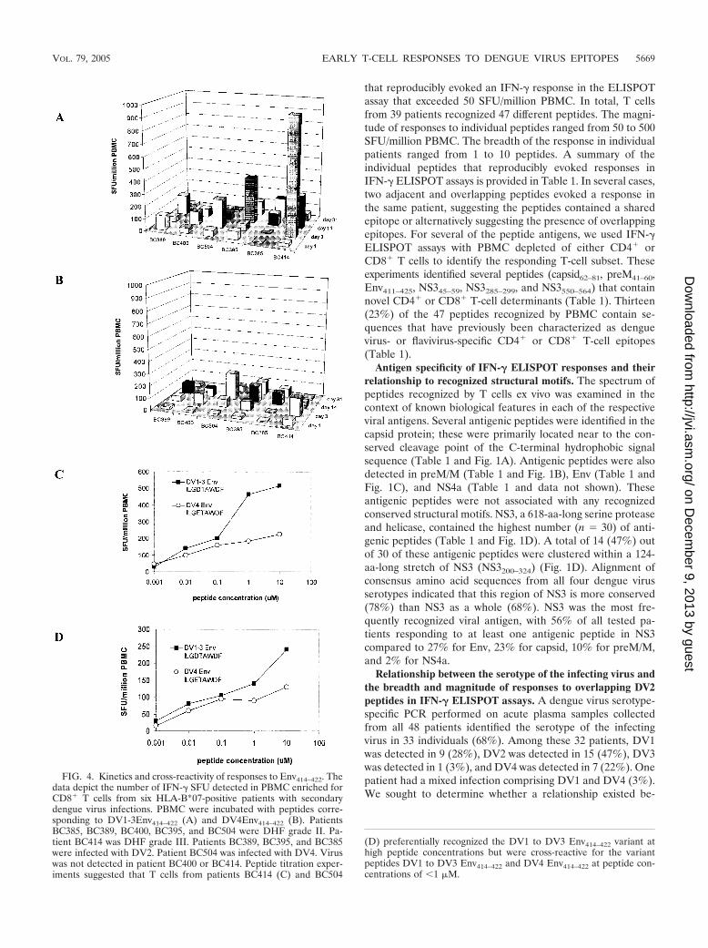
FIG. 4. Kinetics and cross-reactivity of responses to Env414–422. Thedata depict the number of IFN-� SFU detected in PBMC enriched forCD8� T cells from six HLA-B*07-positive patients with secondarydengue virus infections. PBMC were incubated with peptides corre-sponding to DV1-3Env414–422 (A) and DV4Env414–422 (B). PatientsBC385, BC389, BC400, BC395, and BC504 were DHF grade II. Pa-tient BC414 was DHF grade III. Patients BC389, BC395, and BC385were infected with DV2. Patient BC504 was infected with DV4. Viruswas not detected in patient BC400 or BC414. Peptide titration exper-iments suggested that T cells from patients BC414 (C) and BC504
(D) preferentially recognized the DV1 to DV3 Env414–422 variant athigh peptide concentrations but were cross-reactive for the variantpeptides DV1 to DV3 Env414–422 and DV4 Env414–422 at peptide con-centrations of �1 �M.
VOL. 79, 2005 EARLY T-CELL RESPONSES TO DENGUE VIRUS EPITOPES 5669
on Decem
ber 9, 2013 by guesthttp://jvi.asm
.org/D
ownloaded from
tween the breadth and magnitude of the T-cell response toindividual peptides during early convalescence (study day 5)and the serotype of the infecting virus. Study day 5 was selectedbecause it allowed us to assess the breadth and magnitude ofthe T-cell response as close as possible to when the patient wasill but the viremia had nonetheless been resolved. It also al-lowed us to measure responses in a large number of patients,since not all patients returned for follow-up visits at study day14. We did not include the patient with a mixed DV1-DV4infection in this analysis. In cases where patients made re-sponses to adjacent peptides, we included responses to both inthe analysis. Among the 47 patients analyzed, the breadth ofthe response was not significantly associated with the serotypeof the infecting virus. Thus, patients infected with DV2 (n �15) did not respond to significantly more peptides (median, 3;range, 0 to 10) than patients infected with either DV1 (n � 9,median, 1.5; range, 1 to 4), DV3 (n � 1, median, 0), DV4 (n �7; median, 2; range, 0 to 4) or patients in whom a dengue viruscould not be detected (n � 15; median, 3; range, 0 to 5). Likethe breadth of the response, the mean sum of ELISPOT fre-quencies to individual DV2 peptides in patients infected withDV2 did not significantly exceed (93 42 SFU/million PBMC[values are means SD) those in patients infected with either
DV1 (185 135 SFU) or DV4 (112 60 SFU) or in patientsin whom a dengue virus could not be detected (108 78 SFU).
One possible explanation for the absence of significantlystronger responses to DV2 peptides in patients infected with aserotype 2 virus is that the DV2-specific response had notsufficiently matured at the time of analysis (study day 5). Toexplore this hypothesis further, ELISPOT responses to poolsof overlapping DV2 peptides were measured in serial PBMCsamples collected from five prospectively recruited patientsinfected with DV2. Responses detected against pooled pep-tides were verified using individual peptides. In the four pa-tients who had responses to one or more peptide antigens, thebreadth of the response actually contracted rather than ex-panded as time since infection elapsed (Fig. 2). The narrowingof the breadth of responses coincided with a reduction in themagnitude of responses to each antigenic peptide (data notshown). This suggests that a significant DV2-specific responsedoes not evolve with increased time since secondary infection.
Relationship between clinical parameters and the breadthand magnitude of responses to overlapping DV2 peptides inIFN-� ELISPOT assays. Clinical disease grade had no appar-ent relationship to the breadth or magnitude of the T-cellresponse. With respect to the breadth of responses, samplesfrom patients with DHF grade I (n � 11) recognized 2.5peptides on average (range, 0 to 5) in ELISPOT assays, whichwas virtually identical to samples from patients with grade II (n� 16; 2.3 peptides; range, 0 to 7) or grade III (n � 20; 2.4peptides; range, 0 to 5). Only one patient with DHF IV wasrecruited to the study, although interestingly, PBMC from thisindividual recognized 10 different peptides. Like the breadth ofthe response, there were no significant differences between themean sum of ELISPOT responses recorded for patients ineach of the disease grades (grade I, 138 148; grade II, 143 71; grade III, 103 74; grade IV, 110).
The magnitude of responses to individual peptides in eachpatient was also compared to the minimum platelet count andmaximum relative increase in hemoconcentration recordedduring hospitalization. The extent of acute hemoconcentrationwas determined by comparing the maximum hematocrit re-corded during hospitalization to the hematocrit recorded atfollow-up in each patient; these data were available for 24patients, all of whom had detectable ELISPOT responses dur-ing the acute phase of illness. Correlation analysis revealed asignificant relationship (r � 0.41; P � 0.04) between the mag-nitude of IFN-� ELISPOT responses measured on study day 5and the extent of hemoconcentration in individual patients.Conversely, the nadir of thrombocytopenia did not significantlycorrelate with the sum of responses to peptides in individualpatients (r � 0.13; P � 0.56).
The minimum determinant and HLA restriction of T cellsresponding to Env411–425. The peptide RMAILGDTAWDFGSL(Env411–425) was among the most frequently recognized bypatient T cells in ELISPOT assays. Seven of 39 patients rec-ognized Env411–425 with a mean response of 153 76 SFU per106 PBMC. HLA typing revealed that HLA-B*07 was the onlyHLA antigen shared by all seven responders. In one patient(BC503), depletion of CD4� or CD8� T cells from PBMCprior to the ELISPOT assay revealed that only the CD8�
T-cell-enriched fraction responded to Env414–422 peptide stim-ulation (data not shown). Truncated synthetic peptides and
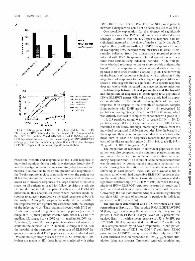
FIG. 5. NS3555–564 is a CD8� T-cell epitope. (A) In IFN-� ELIS-POT assays, PBMC (study day 3) from subject BC313 responded tothe DV2 NS3 peptide VAAEGINYADRRWCF (NS3550–564) in aCD8-dependent fashion. (B) The truncated peptide INYADRRWCF(NS3550–564) was the minimum peptide that evoked the strongestELISPOT response at the lowest peptide concentration.
5670 SIMMONS ET AL. J. VIROL.
on Decem
ber 9, 2013 by guesthttp://jvi.asm
.org/D
ownloaded from
ELISPOT assays indicated ILGDTAWDF (Env414–422) wasthe smallest peptide recognized by CD8� T cells from patientBC503 (Fig. 3). The HLA restriction of Env414–422 in patientBC503 was confirmed via a cytotoxic T lymphocyte killingassay. A short term T-cell line generated from Env414–422 pep-tide-stimulated PBMC from BC503 was lytic to a peptide-pulsed BCL that shared only B*07 in common with the effectorcells (25% specific lysis at an effector:target ratio of 10:1 forEnv414–422 peptide pulsed targets versus �2.5% for targetsalone). Env411–425 did not elicit ELISPOT responses in B*07-positive, flavivirus-naı̈ve individuals (United Kingdom resi-dents).
Kinetics and cross-reactivity of responses to Env414–422. Wesought to define the kinetics of T-cell responses to the B*07-restricted Env414–422 epitope in prospectively recruited dengue
patients. First, the extent of sequence variation present atEnv414–422 across all four dengue virus serotypes was exam-ined. The sequence of Env414–422 in DV1 to DV3 was invariant(ILGDTAWDF), while DV4 Env414–422 differed by only 1 aa(ILGETAWDF). The dynamic of T-cell responses to thesevariant peptides were measured in six prospectively identifiedHLA-B*07-positive patients with serologically confirmed sec-ondary dengue virus infections. All six patients responded tothe HLA-B*07-restricted epitopes Env414–422 (Fig. 4A and B)in ELISPOT assays, with the peak response measured on studyday 3 or 14. ELISPOT responses to each of the epitope vari-ants were generally similar. In samples from patient BC414,however, we observed much stronger responses (6-fold) tothe DV1 to DV3 Env414–422 (ILGDTAWDF) epitope variantthan to DV4 Env414–422 (ILGETAWDF). Stronger responses
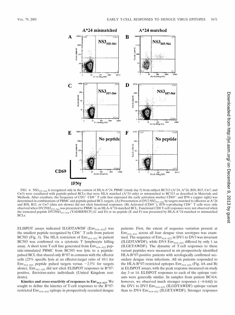
FIG. 6. NS3555–564 is recognized only in the context of HLA-A*24. PBMC (study day 5) from subject BC313 (A*24, A*26, B38, B15, Cw7, andCw3) were cocultured with peptide-pulsed BCLs that were HLA matched (A*24 only) or mismatched to BC313 as described in Materials andMethods. After coculture, the frequency of CD3� CD8� T cells that expressed the early activation marker CD69� and IFN-� (upper right) wasdetermined in combinations of PBMC and peptide-pulsed BCL targets. (A) Presentation of DV2 NS3555–564 by targets matched to effectors at A*26and B38, B15, or Cw7 (data not shown) did not elicit functional responses. (B) Activated (CD69�), IFN-�-producing CD8� T cells were onlyobserved when DV2NS3555–564 was presented to PBMC in an HLA-A*24-matched BCL. Functional CD8 T-cell responses were not observed whenthe truncated peptide DV2NS3557–564 (YADRRWCF) (C and D) or no peptide (E and F) was presented by HLA-A*24-matched or mismatchedBCLs.
VOL. 79, 2005 EARLY T-CELL RESPONSES TO DENGUE VIRUS EPITOPES 5671
on Decem
ber 9, 2013 by guesthttp://jvi.asm
.org/D
ownloaded from

to DV1 to DV3 Env414–422 were also observed in patientBC504 (3.5 fold at days 3 and 14) and patient BC395 (2-fold atday 14) but were less pronounced. Measurement of T-cellresponses to these variant peptides at lower peptide concen-trations, akin to those likely to be found in vivo, revealed muchmore homogenous responses, however. For example, at pep-tide concentrations of �1 �m, PBMC from patients BC414and BC504 recognized Env414–422 peptide variants equallywell; similar results were observed for PBMC from the otherstudy subjects (Fig. 4C and D).
The minimum determinant and HLA restriction of T cellsresponding to NS3550–564. In IFN-� ELISPOT assays, subjectBC313 (HLA-A*24, HLA-A*26, HLA-B38, HLA-B15, HLA-Cw7, and HLA-Cw3) responded to three nonoverlapping NS3peptides: VAAEGINYADRRWCF (NS3550–564), EAHFTDPASIAARGY (NS3285–299), and IEPSWADVKKDLISY(NS365–79). Depletion of CD4� or CD8� T cells from PBMCprior to ELISPOT assay indicated the responding cells wereCD8� T cells (Fig. 5A). The peptide spanning NS365–79 con-tained a previously defined HLA-B*15-restricted epitope,NS371–79 (52); since patient BC313 was also HLA-B*15-posi-tive, these findings were not investigated further. Two lines ofevidence suggested the NS3550–564 peptide contained an HLA-A*24 epitope. First, a peptide containing this sequence hadpreviously been shown to evoke T-cell responses in HLA-A*24-positive Vietnamese dengue patients (35). Second, thesequence NYADRRWCF (NS3556–564) conformed to the con-sensus A*24 sequence, which predicts peptides with a tyrosineor phenylalanine at the second position and an uncharged
amino acid at the C-terminal anchor position. The minimumdeterminant in NS3550–564 that elicited a response from patientBC313, who was HLA-A*24 positive, was identified by usingtruncated peptides spanning the predicted epitope in ELISPOTassays. INYADRRWCF (NS3555–564) was the minimum pep-tide that evoked the strongest response at the lowest peptideconcentration in samples from BC313 (Fig. 5B). We used in-tracellular IFN-� staining to confirm the HLA restriction ofDV2NS3556–564. PBMC from BC313 was cocultured withbrefeldin A and peptide-pulsed BCLs matched or mismatchedto effectors at the A*24 locus. After 6 h, the frequency ofIFN-�-staining CD8� T cells was determined for each peptideand BCL combination. The results confirmed that functionalCD8� T-cell responses to DV2 NS3555–564 were dependent onpeptide presentation through the A*24 locus (Fig. 6). Presen-tation of DV2 NS3555–564 by targets matched to effectors atA*26, B38, B15, or Cw7 did not elicit functional responses(Fig. 6 and data not shown). NS3555–564 did not elicit ELISPOTresponses in A*24-positive, flavivirus-naı̈ve individuals (UnitedKingdom residents).
Kinetics and cross-reactivity of responses to NS3556–564. T-cell responses to peptides corresponding to all known naturalvariants of the A*24-restricted NS3556–564 epitope were mea-sured in 16 prospectively recruited, HLA-A*24-positive den-gue patients during early convalescence (i.e., at hospital dis-charge). T-cell responses to any one of the NS3556–564 variantswere detected in 4 (25%) of 16 subjects, suggesting that thiswas not a dominant epitope. In all patients, the peak responseto each of the epitope variants was recorded on study day 14
FIG. 7. Kinetics of T-cell responses to NS3555–564. Shown are the kinetics and magnitude of IFN-� ELISPOT responses in samples from fourpatients with secondary dengue virus infection to each of the recognized peptide variants of NS3555–564. PBMC from each patient were stimulatedwith DV1NS3555–564 (FQYSDRRWCF) (A), DV2NS3555–564 (INYADRRWCF) (B), DV3NS3555–564 (IKYTDRKWCF) (C), and DV4NS3555–564(ISYKDREWCF) (D). Subject BC311 was infected with DV4, and subject BC398 was infected with DV2.
5672 SIMMONS ET AL. J. VIROL.
on Decem
ber 9, 2013 by guesthttp://jvi.asm
.org/D
ownloaded from

(Fig. 7). The spectrum of responses detected in each of thefour patients varied according to the peptide antigen (Fig. 7).Thus, some patients made monotypic responses (patientBC313), while others had responses against two peptide vari-ants (patient s BC311, BC398, and BC404). In both patientswhere information on the infecting viral serotype was available(BC311 [DV4] and BC398 [DV2]), there was an ELISPOTresponse to the NS3556–564 epitope matching the serotype ofvirus mediating the current infection, but also a response to asecond natural NS3556–564 epitope sequence (Fig. 8). BC311and BC398 also responded to the second NS3556–564 epitopesequence at low-peptide concentrations that better reflected invivo conditions (Fig. 8).
DISCUSSION
The breadth and specificity of T-cell responses to dengueviral antigens in populations living in areas where dengue isendemic remain relatively poorly characterized. This serolog-ical and virological study of Vietnamese adults with secondarydengue virus infection measured the breadth and frequency ofT-cell responses to 260 overlapping peptides from a DV2 virus.The large numbers of subjects and peptides included repre-sents a strength of this study. Neither the breadth nor magni-tudes of the recorded peptide-specific T-cell responses weresignificantly associated with clinical disease grade or the sero-type of dengue virus mediating the current infection. A second
key finding was the identification of 34 different peptide se-quences that potentially contained many novel T-cell epitopes.These assays facilitated the identification of a novel HLA-B*07-restricted epitope in Env and an HLA-A*24-restrictedepitope in NS3.
Most studies of dengue virus-specific T cells have occurredin the context of T-cell clones generated from live attenuateddengue virus vaccinees, or less frequently, from dengue pa-tients (6, 9, 13, 24, 27, 37). This study represents the firstattempt to define the relative antigenicity of peptides frommultiple dengue viral antigens in a large number of patientsfrom a hyperendemic country. NS3 was recognized by morethan half of all Vietnamese adult patients and also containedthe largest number of antigenic peptides. These results areconsistent with previous studies that have emphasized the im-portance of NS3 as a T-cell target (37). Almost half the anti-genic peptides in NS3 were clustered within an amino acidregion (NS3200–324) that represents just 20% of the whole pro-tein. NS3200–324 is highly conserved (78%) across all four se-rotypes of dengue virus and contains motifs and charged res-idues that are essential for helicase activity and viralreplication (38, 39). Given the conserved nature of NS3 (68%),and particularly NS3200–324, it seems likely that many of theNS3-derived antigenic peptides identified in this study weretargets of reactivated, dengue virus cross-reactive T cells thatwere primed by a previous dengue virus infection. That Viet-namese patients infected with either DV1 or DV4 mountedELISPOT responses to DV2 peptides that were equivalent inbreadth and strength to patients infected with a DV2 virussupports the contention that serotype cross-reactive, ratherthan serotype-specific, T cells dominate the acute responseduring secondary infections. In patients infected with DV2,there was no evidence that the T-cell response to DV2 pep-tides increased in breadth with time since secondary infection.This suggests the early, cross-reactive T-cell response remainsdominant in late convalescence when the memory T-cell poolhas been established. This is supported by a previous study thatdemonstrated the dominance of cross-reactive T cells amongthe dengue virus-specific memory T-cell population in Thaichildren 12 months after secondary dengue virus infection(36).
An important question is whether T-cell responses to den-gue virus antigens are associated with clinical disease. In thisstudy, no significant differences were observed in either thebreadth or magnitude of the total T-cell response betweenpatients with different clinical disease grades. We did, however,observe that the sum of peptide-specific responses measured inELISPOT assays correlated, albeit weakly, with the extent ofhemoconcentration recorded in each patient. Other studieshave found significant associations between the magnitude ofepitope-specific T-cell responses and clinical disease grade.For example, relatively higher frequencies of activated, cross-reactive CD8� T cells (41, 51) and a range of direct andindirect markers of CD4� and CD8� T-cell activation havebeen associated with increasing disease severity during second-ary infection (8, 14–16, 22, 23, 26). An important differencebetween this study and that of others is that we performed acomprehensive analysis of the functional T-cell response inadult patients to 260 peptides spanning five viral antigens asopposed to an analysis of responses in children to a single
FIG. 8. T-cell responses to NS3555–564 variants during acute second-ary dengue virus infection. (A) Peptide titration curves in IFN-�ELISPOT assays for PBMC collected in the acute phase of infection(study day 5) from patient BC311 (infected with DV4) and stimulatedwith DV4NS3555–564 (ISYKDREWCF) or DV2NS3555–564 (INYADRRWCF). (B) Peptide titration curves in IFN-� ELISPOT assays forPBMC collected in the acute phase of infection (study day 5) frompatient BC398 (infected with DV2) and stimulated with DV2NS3555–564 (INYADRRWCF) and DV1NS3555–564 (FQYSDRRWCF).
VOL. 79, 2005 EARLY T-CELL RESPONSES TO DENGUE VIRUS EPITOPES 5673
on Decem
ber 9, 2013 by guesthttp://jvi.asm
.org/D
ownloaded from
CD8� T-cell epitope (41, 51). Other confounding influences,such as ethnicity, HLA background, and viral factors, may alsoexplain differences between this study and those that have beenreported previously.
Two novel CD8� T-cell epitopes were identified in thisstudy. The rationale for studying the NS3550–564 peptide wasthreefold. First, previous studies had suggested this peptidemight contain an HLA-A*24 epitope (35). Second, A*24 is oneof the commonest alleles present in the Vietnamese popula-tion (33%) and hence an A*24 epitope might be widelyrecognized. Third, A*24 has been associated with susceptibilityto DHF in Vietnamese children (35), suggesting immune re-sponses restricted through this allele might be poorly protec-tive and/or pathogenic. Responses to the A*24-restrictedNS3556–564 epitope were detected in a minority of HLA-A*24-positive patients, suggesting this is not a dominant epitope.Consistent with other studies on unrelated epitopes (41, 51),responses to the CD8� T-cell epitopes defined in this studywere generally strongest after the patient had been dischargedfrom hospital, suggesting that antigen may continue to persist,possibly in the form of immune complexes, well after the res-olution of viremia. The establishment of a library of definedT-cell epitopes from dengue viruses will help to define the roleof T cells in immunity and disease pathogenesis. They shouldalso prove useful in measuring the immunogenicity of livedengue virus vaccine candidates (7, 18, 42).
Two Vietnamese adults whose samples recognized theA*24-restricted NS3556–564 epitope also had samples that rec-ognized an NS3556–564 epitope different from that present inthe infecting dengue virus serotype. Mongkolsapaya et al. haverecently suggested that infection history and what they termed“original antigenic sin” by dengue virus-specific memory Tcells may explain the hierarchy of responses to an A*11-re-stricted dengue virus T-cell epitope following secondary infec-tion (41). Such skewed T-cell responses could hypotheticallylimit or delay viral elimination, leading to higher viral loads,increased immune activation, and immunopathology. In thecontext of A*24-restricted NS3556–564-specific T cells, furtherstudies of T-cell affinity and functional phenotype will be re-quired to determine whether this represents original antigenicsin in the T-cell response.
Future studies of CD8� T-cell responses to dengue virusantigens are likely to employ HLA class I tetramers that can beused to evaluate the functional phenotype of serotype-specificand cross-reactive CD8� T cells. Integrating these cellular datawith viremia parameters and carefully collected clinical phe-notypes should reveal novel insights into disease pathogenesisand immunity.
ACKNOWLEDGMENTS
The generous support of the Wellcome Trust is acknowledged.S.R.-J. and T. D. are funded by Medical Research Council, UK.
Special thanks to the patients, nurses, and clinicians who made thisstudy possible. We acknowledge Gavin Screaton and Juthathip Mon-gkolsapaya for sharing the overlapping peptides and Kati DiGleria forsynthesis of peptides.
REFERENCES
1. Bethell, D. B., J. Gamble, P. L. Pham, M. D. Nguyen, T. H. Tran, T. H. Ha,T. N. Tran, T. H. Dong, I. B. Gartside, N. J. White, and N. P. Day. 2001.Noninvasive measurement of microvascular leakage in patients with denguehemorrhagic fever. Clin. Infect. Dis. 32:243–253.
2. Blok, J., S. M. McWilliam, H. C. Butler, A. J. Gibbs, G. Weiller, B. L.Herring, A. C. Hemsley, J. G. Aaskov, S. Yoksan, and N. Bhamarapravati.1992. Comparison of a dengue-2 virus and its candidate vaccine derivative:sequence relationships with the flaviviruses and other viruses. Virology 187:573–590.
3. Brinton, M. A., I. Kurane, A. Mathew, L. Zeng, P. Y. Shi, A. Rothman, andF. A. Ennis. 1998. Immune mediated and inherited defences against flavivi-ruses. Clin. Diagn. Virol. 10:129–139.
4. Bunce, M., C. M. O’Neill, M. C. Barnardo, P. Krausa, M. J. Browning, P. J.Morris, and K. I. Welsh. 1995. Phototyping: comprehensive DNA typing forHLA-A, B, C, DRB1, DRB3, DRB4, DRB5 and DQB1 by PCR with 144primer mixes utilizing sequence-specific primers (PCR-SSP). Tissue Anti-gens 46:355–367.
5. Burke, D. S., A. Nisalak, D. E. Johnson, and R. M. Scott. 1988. A prospectivestudy of dengue infections in Bangkok. Am. J. Trop. Med. Hyg. 38:172–180.
6. Dharakul, T., I. Kurane, N. Bhamarapravati, S. Yoksan, D. W. Vaughn,C. H. Hoke, and F. A. Ennis. 1994. Dengue virus-specific memory T cellresponses in human volunteers receiving a live attenuated dengue virus type2 candidate vaccine. J. Infect. Dis. 170:27–33.
7. Edelman, R., S. S. Wasserman, S. A. Bodison, R. J. Putnak, K. H. Eckels, D.Tang, N. Kanesa-Thasan, D. W. Vaughn, B. L. Innis, and W. Sun. 2003.Phase I trial of 16 formulations of a tetravalent live-attenuated denguevaccine. Am. J. Trop. Med. Hyg. 69:48–60.
8. Gagnon, S. J., M. Mori, I. Kurane, S. Green, D. W. Vaughn, S. Kalayanarooj,S. Suntayakorn, F. A. Ennis, and A. L. Rothman. 2002. Cytokine geneexpression and protein production in peripheral blood mononuclear cells ofchildren with acute dengue virus infections. J. Med. Virol. 67:41–46.
9. Gagnon, S. J., W. Zeng, I. Kurane, and F. A. Ennis. 1996. Identification oftwo epitopes on the dengue 4 virus capsid protein recognized by a serotype-specific and a panel of serotype-cross-reactive human CD4� cytotoxic T-lymphocyte clones. J. Virol. 70:141–147.
10. Gamble, J., D. Bethell, N. P. Day, P. P. Loc, N. H. Phu, I. B. Gartside, J. F.Farrar, and N. J. White. 2000. Age-related changes in microvascular per-meability: a significant factor in the susceptibility of children to shock? Clin.Sci. (London) 98:211–216.
11. Goulder, P. J., M. M. Addo, M. A. Altfeld, E. S. Rosenberg, Y. Tang, U.Govender, N. Mngqundaniso, K. Annamalai, T. U. Vogel, M. Hammond, M.Bunce, H. M. Coovadia, and B. D. Walker. 2001. Rapid definition of fivenovel HLA-A*3002-restricted human immunodeficiency virus-specific cyto-toxic T-lymphocyte epitopes by ELISPOT and intracellular cytokine stainingassays. J. Virol. 75:1339–1347.
12. Graham, R. R., M. Juffrie, R. Tan, C. G. Hayes, I. Laksono, C. Ma’roef,Erlin, Sutaryo, K. R. Porter, and S. B. Halstead. 1999. A prospective sero-epidemiologic study on dengue in children four to nine years of age inYogyakarta, Indonesia I. Studies in 1995–1996. Am. J. Trop. Med. Hyg.61:412–419.
13. Green, S., I. Kurane, R. Edelman, C. O. Tacket, K. H. Eckels, D. W. Vaughn,C. H. Hoke, Jr., and F. A. Ennis. 1993. Dengue virus-specific human CD4�
T-lymphocyte responses in a recipient of an experimental live-attenuateddengue virus type 1 vaccine: bulk culture proliferation, clonal analysis, andprecursor frequency determination. J. Virol. 67:5962–5967.
14. Green, S., S. Pichyangkul, D. W. Vaughn, S. Kalayanarooj, S. Nimmannitya,A. Nisalak, I. Kurane, A. L. Rothman, and F. A. Ennis. 1999. Early CD69expression on peripheral blood lymphocytes from children with dengue hem-orrhagic fever. J. Infect. Dis. 180:1429–1435.
15. Green, S., D. W. Vaughn, S. Kalayanarooj, S. Nimmannitya, S. Suntayakorn,A. Nisalak, R. Lew, B. L. Innis, I. Kurane, A. L. Rothman, and F. A. Ennis.1999. Early immune activation in acute dengue illness is related to develop-ment of plasma leakage and disease severity. J. Infect. Dis. 179:755–762.
16. Green, S., D. W. Vaughn, S. Kalayanarooj, S. Nimmannitya, S. Suntayakorn,A. Nisalak, A. L. Rothman, and F. A. Ennis. 1999. Elevated plasma inter-leukin-10 levels in acute dengue correlate with disease severity. J. Med.Virol. 59:329–334.
17. Guzman, M. G., and G. Kouri. 2002. Dengue: an update. Lancet Infect. Dis.2:33–42.
18. Gwinn, W., W. Sun, B. L. Innis, J. Caudill, and A. D. King. 2003. Serotype-specific T(H)1 responses in recipients of two doses of candidate live-atten-uated dengue virus vaccines. Am. J. Trop. Med. Hyg. 69:39–47.
19. Halstead, S. B. 1979. In vivo enhancement of dengue virus infection in rhesusmonkeys by passively transferred antibody. J. Infect. Dis. 140:527–533.
20. Halstead, S. B., and E. J. O’Rourke. 1977. Antibody-enhanced dengue virusinfection in primate leukocytes. Nature 265:739–741.
21. Halstead, S. B., and E. J. O’Rourke. 1977. Dengue viruses and mononuclearphagocytes. I. Infection enhancement by non-neutralizing antibody. J. Exp.Med. 146:201–217.
22. Juffrie, M., G. M. Meer, C. E. Hack, K. Haasnoot, Sutaryo, A. J. Veerman,and L. G. Thijs. 2001. Inflammatory mediators in dengue virus infection inchildren: interleukin-6 and its relation to C-reactive protein and secretoryphospholipase A2. Am. J. Trop. Med. Hyg. 65:70–75.
23. Kittigul, L., W. Temprom, D. Sujirarat, and C. Kittigul. 2000. Determina-tion of tumor necrosis factor-alpha levels in dengue virus infected patients by
5674 SIMMONS ET AL. J. VIROL.
on Decem
ber 9, 2013 by guesthttp://jvi.asm
.org/D
ownloaded from

sensitive biotin-streptavidin enzyme-linked immunosorbent assay. J. Virol.Methods 90:51–57.
24. Kurane, I., M. A. Brinton, A. L. Samson, and F. A. Ennis. 1991. Denguevirus-specific, human CD4� CD8� cytotoxic T-cell clones: multiple patternsof virus cross-reactivity recognized by NS3-specific T-cell clones. J. Virol.65:1823–1828.
25. Kurane, I., B. L. Innis, C. H. Hoke, Jr., K. H. Eckels, A. Meager, J. Janus,and F. A. Ennis. 1995. T cell activation in vivo by dengue virus infection.J. Clin. Lab. Immunol. 46:35–40.
26. Kurane, I., B. L. Innis, S. Nimmannitya, A. Nisalak, A. Meager, J. Janus,and F. A. Ennis. 1991. Activation of T lymphocytes in dengue virus infec-tions. High levels of soluble interleukin 2 receptor, soluble CD4, solubleCD8, interleukin 2, and interferon-gamma in sera of children with dengue.J. Clin. Investig. 88:1473–1480.
27. Kurane, I., Y. Okamoto, L. C. Dai, L. L. Zeng, M. A. Brinton, and F. A.Ennis. 1995. Flavivirus-cross-reactive, HLA-DR15-restricted epitope on NS3recognized by human CD4� CD8� cytotoxic T lymphocyte clones. J. Gen.Virol. 76:2243–2249.
28. Kurane, I., A. L. Rothman, P. G. Livingston, S. Green, S. J. Gagnon, J.Janus, B. L. Innis, S. Nimmannitya, A. Nisalak, and F. A. Ennis. 1994.Immunopathologic mechanisms of dengue hemorrhagic fever and dengueshock syndrome. Arch. Virol. Suppl. 9:59–64.
29. Lanciotti, R. S., C. H. Calisher, D. J. Gubler, G. J. Chang, and A. V.Vorndam. 1992. Rapid detection and typing of dengue viruses from clinicalsamples by using reverse transcriptase-polymerase chain reaction. J. Clin.Microbiol. 30:545–551.
30. Leitmeyer, K. C., D. W. Vaughn, D. M. Watts, R. Salas, I. Villalobos, C. de,C. Ramos, and R. Rico-Hesse. 1999. Dengue virus structural differences thatcorrelate with pathogenesis. J. Virol. 73:4738–4747.
31. Libraty, D. H., T. P. Endy, H. S. Houng, S. Green, S. Kalayanarooj, S.Suntayakorn, W. Chansiriwongs, D. W. Vaughn, A. Nisalak, F. A. Ennis, andA. L. Rothman. 2002. Differing influences of virus burden and immuneactivation on disease severity in secondary dengue-3 virus infections. J. In-fect. Dis. 185:1213–1221.
32. Libraty, D. H., P. R. Young, D. Pickering, T. P. Endy, S. Kalayanarooj, S.Green, D. W. Vaughn, A. Nisalak, F. A. Ennis, and A. L. Rothman. 2002.High circulating levels of the dengue virus nonstructural protein NS1 early indengue illness correlate with the development of dengue hemorrhagic fever.J. Infect. Dis. 186:1165–1168.
33. Livingston, P. G., I. Kurane, L. C. Dai, Y. Okamoto, C. J. Lai, R. Men, S.Karaki, M. Takiguchi, and F. A. Ennis. 1995. Dengue virus-specific, HLA-B35-restricted, human CD8� cytotoxic T lymphocyte (CTL) clones. Recog-nition of NS3 amino acids 500 to 508 by CTL clones of two different serotypespecificities. J. Immunol. 154:1287–1295.
34. Loke, H., D. Bethell, C. X. T. Phuong, N. Day, N. White, J. Farrar, and A.Hill. 2002. Susceptibility to dengue hemorrhagic fever in Vietnam: evidenceof an association with variation in the vitamin D receptor and Fc� receptorIIa genes. Am. J. Trop. Med. Hyg. 67:102–106.
35. Loke, H., D. B. Bethell, C. X. Phuong, M. Dung, J. Schneider, N. J. White,N. P. Day, J. Farrar, and A. V. Hill. 2001. Strong HLA class I-restricted T cellresponses in dengue hemorrhagic fever: a double-edged sword? J. Infect.Dis. 184:1369–1373.
36. Mathew, A., I. Kurane, S. Green, H. A. Stephens, D. W. Vaughn, S. Kalay-anarooj, S. Suntayakorn, D. Chandanayingyong, F. A. Ennis, and A. L.Rothman. 1998. Predominance of HLA-restricted cytotoxic T-lymphocyteresponses to serotype-cross-reactive epitopes on nonstructural proteins fol-lowing natural secondary dengue virus infection. J. Virol. 72:3999–4004.
37. Mathew, A., I. Kurane, A. L. Rothman, L. L. Zeng, M. A. Brinton, and F. A.Ennis. 1996. Dominant recognition by human CD8� cytotoxic T lympho-
cytes of dengue virus nonstructural proteins NS3 and NS1.2a. J. Clin. Inves-tig. 98:1684–1691.
38. Matusan, A. E., P. G. Kelley, M. J. Pryor, J. C. Whisstock, A. D. Davidson,and P. J. Wright. 2001. Mutagenesis of the dengue virus type 2 NS3 pro-teinase and the production of growth-restricted virus. J. Gen. Virol. 82:1647–1656.
39. Matusan, A. E., M. J. Pryor, A. D. Davidson, and P. J. Wright. 2001.Mutagenesis of the Dengue virus type 2 NS3 protein within and outsidehelicase motifs: effects on enzyme activity and virus replication. J. Virol.75:9633–9643.
40. Monath, T. P. 1994. Dengue: the risk to developed and developing countries.Proc. Natl. Acad. Sci. USA 91:2395–2400.
41. Mongkolsapaya, J., W. Dejnirattisai, X. N. Xu, S. Vasanawathana, N.Tangthawornchaikul, A. Chairunsri, S. Sawasdivorn, T. Duangchinda, T.Dong, S. Rowland-Jones, P. T. Yenchitsomanus, A. McMichael, P. Malasit,and G. Screaton. 2003. Original antigenic sin and apoptosis in the patho-genesis of dengue hemorrhagic fever. Nat. Med. 9:921–927.
42. Sabchareon, A., J. Lang, P. Chanthavanich, S. Yoksan, R. Forrat, P. Attan-ath, C. Sirivichayakul, K. Pengsaa, C. Pojjaroen-Anant, L. Chambonneau,J. F. Saluzzo, and N. Bhamarapravati. 2004. Safety and immunogenicity ofa three dose regimen of two tetravalent live-attenuated dengue vaccines infive- to twelve-year-old Thai children. Pediatr. Infect. Dis. J. 23:99–109.
43. Sangkawibha, N., S. Rojanasuphot, S. Ahandrik, S. Viriyapongse, S. Jata-nasen, V. Salitul, B. Phanthumachinda, and S. B. Halstead. 1984. Riskfactors in dengue shock syndrome: a prospective epidemiologic study inRayong, Thailand. I. The 1980 outbreak. Am. J. Epidemiol. 120:653–669.
44. Stephens, H. A., R. Klaythong, M. Sirikong, D. W. Vaughn, S. Green, S.Kalayanarooj, T. P. Endy, D. H. Libraty, A. Nisalak, B. L. Innis, A. L.Rothman, F. A. Ennis, and D. Chandanayingyong. 2002. HLA-A and -Ballele associations with secondary dengue virus infections correlate withdisease severity and the infecting viral serotype in ethnic Thais. TissueAntigens 60:309–318.
45. Thein, S., M. M. Aung, T. N. Shwe, M. Aye, A. Zaw, K. Aye, K. M. Aye, andJ. Aaskov. 1997. Risk factors in dengue shock syndrome. Am. J. Trop. Med.Hyg. 56:566–572.
46. Thisyakorn, U., and S. Nimmannitya. 1993. Nutritional status of childrenwith dengue hemorrhagic fever. Clin. Infect. Dis. 16:295–297.
47. Vaughn, D. W., S. Green, S. Kalayanarooj, B. L. Innis, S. Nimmannitya, S.Suntayakorn, T. P. Endy, B. Raengsakulrach, A. L. Rothman, F. A. Ennis,and A. Nisalak. 2000. Dengue viremia titer, antibody response pattern, andvirus serotype correlate with disease severity. J. Infect. Dis. 181:2–9.
48. Vaughn, D. W., A. Nisalak, T. Solomon, S. Kalayanarooj, M. D. Nguyen, R.Kneen, A. Cuzzubbo, and P. L. Devine. 1999. Rapid serologic diagnosis ofdengue virus infection using a commercial capture ELISA that distinguishesprimary and secondary infections. Am. J. Trop. Med. Hyg. 60:693–698.
49. World Health Organization,. 1997. Dengue haemorrhagic fever: diagnosis,treatment, prevention and control. World Health Organization, Geneva,Switzerland.
50. Zeng, L., I. Kurane, Y. Okamoto, F. A. Ennis, and M. A. Brinton. 1996.Identification of amino acids involved in recognition by dengue virus NS3-specific, HLA-DR15-restricted cytotoxic CD4� T-cell clones. J. Virol. 70:3108–3117.
51. Zivna, I., S. Green, D. W. Vaughn, S. Kalayanarooj, H. A. Stephens, D.Chandanayingyong, A. Nisalak, F. A. Ennis, and A. L. Rothman. 2002. T cellresponses to an HLA-B*07-restricted epitope on the dengue NS3 proteincorrelate with disease severity. J. Immunol. 168:5959–5965.
52. Zivny, J., M. DeFronzo, W. Jarry, J. Jameson, J. Cruz, F. A. Ennis, and A. L.Rothman. 1999. Partial agonist effect influences the CTL response to aheterologous dengue virus serotype. J. Immunol. 163:2754–2760.
VOL. 79, 2005 EARLY T-CELL RESPONSES TO DENGUE VIRUS EPITOPES 5675
on Decem
ber 9, 2013 by guesthttp://jvi.asm
.org/D
ownloaded from
Related Documents