1 Vol.:(0123456789) SCIENTIFIC REPORTS | (2020) 10:13674 | https://doi.org/10.1038/s41598-020-70754-z www.nature.com/scientificreports Early postoperative seizures (EPS) in patients undergoing brain tumour surgery Tunc Faik Ersoy 1* , Sami Ridwan 1,2 , Alexander Grote 1 , Roland Coras 3 & Matthias Simon 1 Early postoperative seizures (EPS) are a common complication of brain tumour surgery. This paper investigates risk factors, management and clinical relevance of EPS. We retrospectively analysed the occurrence of EPS, clinical and laboratory parameters, imaging and histopathological findings in a cohort of 679 consecutive patients who underwent craniotomies for intracranial tumours between 2015 and 2017. EPS were observed in 34/679 cases (5.1%), with 14 suffering at least one generalized seizure. Patients with EPS had a worse postoperative Karnofsky performance index (KPI; with EPS, KPI < 70 vs. 70–100: 11/108, 10.2% vs. 23/571, 4.0%; p = 0.007). Preoperative seizure history was a predictor for EPS (none vs. 1 vs. ≥ 2 seizures: p = 0.037). Meningioma patients had the highest EPS incidence (10.1%, p < 0.001). Cranial imaging identified a plausible cause in most cases (78.8%). In 20.6%, EPS were associated with a persisting new neurological deficit that could not otherwise be explained. 34.6% of the EPS patients had recurrent seizures within one year. EPS require an emergency work-up. Multiple EPS and recurrent seizures are frequent, which indicates that EPS may also reflect a more chronic condition i.e. epilepsy. EPS are often associated with persisting neurological worsening. Early postoperative seizures (EPS) are a common complication of brain tumour surgery. EPS are oſten categorized as acute symptomatic seizures 1,2 . ey are usually felt to reflect acute medical or surgical conditions that may require emergency treatment. is includes haemorrhages, infectious complications and electrolyte disturbances, but also systemic infections and cardiopulmonary disorders resulting in hypotension and hypoxia. Hence, EPS may have potentially severe consequences. ey may result in significant and oſten persisting (neurological) morbidity and reduced quality of life. Furthermore, they usually prolong the patient’s hospital stay. Potential negative consequences include a delayed transfer for rehabilitation therapy, an overall prolonged rehabilitation and, importantly, delayed adjuvant therapy. is latter aspect is of considerable importance e.g. in patients with gliomas and metastasis who will oſten not realize the benefits of surgery if adjuvant therapy is withhold. In addition, many patients with brain metastases require more or less urgent treatment for their systemic disease. ere is also the issue of distinguishing between incidental or acute symptomatic seizures with no or a very low risk of recurrent seizures and true postoperative chronic epilepsy 1–3 . e latter condition requires chronic treatment with antiepileptic drugs and comes with relevant socioeconomic sequelae such as restriction of driving privileges. is may be a particularly important issue for patients with benign tumours such as many menin- giomas who have a good chance of a surgical cure of their tumour. In such cases, the risk of recurrent seizures may well be their only (neurological) health concern 4,5 . ere is a growing interest in tumour-associated epilepsy 6,7 . However, relatively few investigators have focused specifically on EPS 8,9 . Consequently, questions regarding the necessary diagnostic work-up and the use of antie- pileptic drugs in cases with EPS are difficult to answer. For the present study, we have therefore reviewed our recent institutional experience with EPS aſter brain tumour surgery between 2015 and 2017. OPEN 1 Department of Neurosurgery, Evangelisches Klinikum Bethel, Kantensiek 11, 33617 Bielefeld, Germany. 2 Department of Neurosurgery, Paracelsus-Klinik Osnabrück, Am Natruper Holz 69, 49076 Osnabrück, Germany. 3 Institute of Neuropathology, University Hospital Erlangen, Schwabachanlage 6, 91054 Erlangen, Germany. * email: [email protected]

Welcome message from author
This document is posted to help you gain knowledge. Please leave a comment to let me know what you think about it! Share it to your friends and learn new things together.
Transcript
1
Vol.:(0123456789)
Scientific RepoRtS | (2020) 10:13674 | https://doi.org/10.1038/s41598-020-70754-z
www.nature.com/scientificreports
early postoperative seizures (epS) in patients undergoing brain tumour surgerytunc faik ersoy 1*, Sami Ridwan1,2, Alexander Grote1, Roland coras3 & Matthias Simon1
early postoperative seizures (epS) are a common complication of brain tumour surgery. this paper investigates risk factors, management and clinical relevance of epS. We retrospectively analysed the occurrence of EPS, clinical and laboratory parameters, imaging and histopathological findings in a cohort of 679 consecutive patients who underwent craniotomies for intracranial tumours between 2015 and 2017. EPS were observed in 34/679 cases (5.1%), with 14 suffering at least one generalized seizure. patients with epS had a worse postoperative Karnofsky performance index (Kpi; with epS, KPI < 70 vs. 70–100: 11/108, 10.2% vs. 23/571, 4.0%; p = 0.007). Preoperative seizure history was a predictor for EPS (none vs. 1 vs. ≥ 2 seizures: p = 0.037). Meningioma patients had the highest EPS incidence (10.1%, p < 0.001). Cranial imaging identified a plausible cause in most cases (78.8%). In 20.6%, EPS were associated with a persisting new neurological deficit that could not otherwise be explained. 34.6% of the EPS patients had recurrent seizures within one year. EPS require an emergency work-up. Multiple EPS and recurrent seizures are frequent, which indicates that EPS may also reflect a more chronic condition i.e. epilepsy. EPS are often associated with persisting neurological worsening.
Early postoperative seizures (EPS) are a common complication of brain tumour surgery. EPS are often categorized as acute symptomatic seizures1,2. They are usually felt to reflect acute medical or surgical conditions that may require emergency treatment. This includes haemorrhages, infectious complications and electrolyte disturbances, but also systemic infections and cardiopulmonary disorders resulting in hypotension and hypoxia. Hence, EPS may have potentially severe consequences. They may result in significant and often persisting (neurological) morbidity and reduced quality of life. Furthermore, they usually prolong the patient’s hospital stay. Potential negative consequences include a delayed transfer for rehabilitation therapy, an overall prolonged rehabilitation and, importantly, delayed adjuvant therapy. This latter aspect is of considerable importance e.g. in patients with gliomas and metastasis who will often not realize the benefits of surgery if adjuvant therapy is withhold. In addition, many patients with brain metastases require more or less urgent treatment for their systemic disease.
There is also the issue of distinguishing between incidental or acute symptomatic seizures with no or a very low risk of recurrent seizures and true postoperative chronic epilepsy1–3. The latter condition requires chronic treatment with antiepileptic drugs and comes with relevant socioeconomic sequelae such as restriction of driving privileges. This may be a particularly important issue for patients with benign tumours such as many menin-giomas who have a good chance of a surgical cure of their tumour. In such cases, the risk of recurrent seizures may well be their only (neurological) health concern4,5.
There is a growing interest in tumour-associated epilepsy6,7. However, relatively few investigators have focused specifically on EPS8,9. Consequently, questions regarding the necessary diagnostic work-up and the use of antie-pileptic drugs in cases with EPS are difficult to answer. For the present study, we have therefore reviewed our recent institutional experience with EPS after brain tumour surgery between 2015 and 2017.
open
1Department of Neurosurgery, Evangelisches Klinikum Bethel, Kantensiek 11, 33617 Bielefeld, Germany. 2Department of Neurosurgery, Paracelsus-Klinik Osnabrück, Am Natruper Holz 69, 49076 Osnabrück, Germany. 3Institute of Neuropathology, University Hospital Erlangen, Schwabachanlage 6, 91054 Erlangen, Germany. *email: [email protected]

2
Vol:.(1234567890)
Scientific RepoRtS | (2020) 10:13674 | https://doi.org/10.1038/s41598-020-70754-z
www.nature.com/scientificreports/
Materials and methodspatients. We identified all 679 consecutive patients who underwent a craniotomy for an intracranial tumour between January 2015 and August 2017 in the Department of Neurosurgery, Evangelisches Klinikum Bethel, Bielefeld, Germany, by searching the departmental electronic database. Patients operated for medication-refrac-tory epilepsy within the epilepsy surgery program were excluded from this analysis, i.e. all cases specifically referred to us following a presurgical work-up aiming at the establishment of a surgical concept for epilepsy rather than tumour control10. During the study period, we used the current (= 2009) ILAE (International League Against Epilepsy) definition of pharmacoresistant epilepsy, i.e. “… failure of adequate trials of two tolerated, appropriately chosen and used antiepileptic drug schedules (whether as monotherapies or in combination) to achieve sustained seizure freedom”11.
clinical data. Pertinent clinical data and follow-up information were retrospectively retrieved through a chart review and entered in an electronic database. Clinical parameters included age at surgery, gender, his-topathological diagnosis, surgery for tumour recurrence, extent of resection, pre- and postoperative (time of discharge) Karnofsky Performance Index (KPI), occurrence of seizures prior to operation and their frequency, type of the seizure (according to the current ILAE (International League Against Epilepsy) classification)12 and preoperative anticonvulsive therapy. The preoperative MRI work-up was reviewed in each case and the respec-tive tumour location was recorded including specifically the following items: supra- vs. infratentorial and intra- vs. extra-axial growth as well as involvement or compression of the frontal, temporal, central or insular lobe. Tumours affecting the latter cerebral structures are believed to be particularly prone to cause epilepsy13. EPS were defined as (generalized) involuntary movements, abnormal sensory phenomena or an altered mental status that could not otherwise be explained, occurring within 30 days post-surgery, and categorized according to the current ILAE classification8,12,14. Acute electroencephalograms were ordered in cases in which the diagnosis of an EPS was questionable8. We specifically reviewed all clinical, laboratory and in particular imaging data from all patients with EPS in order to identify their likely cause.
Maximum tumour diameter and perifocal oedema > 1 cm were assessed in all meningioma patients using axial contrast-enhanced T1- and axial FLAIR-weighted scans, respectively4. Oedema formation and large tumour size have been associated with preoperative seizures in meningioma patients by some authors4,15,16. We also reviewed all available early (< 24 h) postoperative imaging studies from cases undergoing supratentorial meningioma surgery. Specifically, we recorded extra-axial (epi- or subdural) bleeds and pneumocephalus with mass effect, cerebral contusions, and measurable (i.e. > 0.5 cm) hematomas or bleeds with mass effect in the resection cavity as well as new small/perforator and territorial infarcts. Such imaging findings may cause neurological deficits and/or have clinical consequences ranging from prolonged observation to revision surgery17. Of note, they would also be regarded as plausible causes for an acute symptomatic seizure2.
Histopathology. All neuropathological analyses were performed at the Institute of Neuropathology, Uni-versity Hospital Erlangen, Germany. This included immunohistochemistry and molecular genetic analysis if required by the WHO 2016 classification or its 2007 predecessor, or whenever such additional investigations were deemed useful by the responsible neuropathologist.
Statistical analysis. For statistical analyses, we used a commercially available software (IBM SPSS Statis-tics for Windows, Version 25.0, IBM Corp., Armonk, NY). Standard procedures (Fisher exact test, chi-square test, linear-by-linear association [Mantel–Haenszel test] and Student-t-test) were used for univariate analyses as indicated. Two-sided tests were employed throughout and p values < 0.05 were considered significant. For multivariate analyses, we used logistic regression modelling (inclusion procedure).
ethical approval. The study was approved by the responsible institutional research committee and all procedures were in accordance with its ethical standards and with the 1964 Helsinki declaration and its later amendments (Ethikkommission der Ärztekammer Westfalen-Lippe und der Westfälischen Wilhelms-Univer-sität Münster, Germany, Az 2018-484-f-S).
informed consent. The responsible institutional research committee and local law do not require informed consent for this study.
Resultspatient cohort. We studied a total of 679 surgical cases, i.e. 679 procedures performed in 630 patients. Median age was 61.0 years. The series comprises more females (379, 60.2%) than males (251, 39.8%). The median pre- and postoperative KPI was both 90. 103 (15.2%) patients had surgery for a recurrent tumour. 137 (20.2%) surgeries were performed for infratentorial and 542 (79.8%) surgeries for supratentorial tumours. The latter figure includes 4/16 tentorial meningiomas with at least some extension into the supratentorial compartment. Twenty-six cases had open microsurgical biopsies (3.8%). The most frequent histology was meningioma (all WHO grades, N = 218, 32.1%), followed by glioblastoma (N = 177, 26.1%), and metastasis (N = 138, 20.5%). The histopathological diagnoses are detailed in Table 1.
In total, 155 patients (22.8%) presented with seizures prior to surgery, with approximately half of them (82, 12.1%) reporting a history of multiple (≥ 2) seizures. Preoperative generalized seizures occurred in 83 cases (12.2%). Antiepileptic treatment was prescribed in 141 of the 155 cases (93.0%) with preoperative seizures (levetiracetam: 121; other monotherapy: 6; combination therapy including levetiracetam: 13; other combination

3
Vol.:(0123456789)
Scientific RepoRtS | (2020) 10:13674 | https://doi.org/10.1038/s41598-020-70754-z
www.nature.com/scientificreports/
therapy: 1). The remaining 14 cases were not felt to have presented with seizures by their referring physicians and the treating neurosurgeons at the time of their surgery. Patients without a seizure history did not receive (prophylactic) anticonvulsive treatment. A postoperative steroid taper starting with 16–24 mg dexamethasone on day 1 was routinely prescribed in cases with significant brain oedema and mass effect.
patients with early postoperative seizures. EPS seizures were observed in 34/679 cases (5.1%), with 17 patients (2.5%) suffering multiple seizures. 14 patients (2.1%) had at least one generalized seizure. More than half of these patients had seizures within the first three days following surgery and 29/34 (85.3%) within the first 7 days. All cases suffering an EPS are described in detail in Table 2.
We found no correlations between age, sex or preoperative KPI with the occurrence of EPS in the overall cohort (Table 3). Patients with EPS had a worse postoperative KPI (patients with EPS, KPI < 70 vs. 70–100: 11/108, 10.2% vs. 23/571, 4.0%; p = 0.007). Preoperative seizure history was a risk factor for EPS. EPS were seen in 22/524 (4.2%) cases with no preoperative seizure, but in 4/73 (5.5%) patients with a single and 8/82 (9.8%) cases with multiple preoperative seizures (none vs. 1 vs. ≥ 2 seizures: p = 0.037; none vs. any preoperative seizure: p = ns; 0–1 vs. ≥ 2 seizures: p = 0.035; Table 3). EPS were observed in 10/141 (7.1%) patients on anticonvulsive medication and in 2/14 (14.3%) cases who had preoperative seizures, yet no anticonvulsive treatment.
Only 1/137 (0.7%) patient with an infratentorial tumour, but 33/542 (6.1%, p = 0.007) patients with supraten-torial growths suffered an EPS. EPS were seen significantly more often following surgery for tumours involving or compressing the frontal, central, temporal and/or insular lobes (32/452, 7.1% vs. 2/227, 0.9%, p < 0.001). At least some involvement of the primary sensorimotor cortex was seen in 83 patients, 4 of which experienced an EPS (4.8%). Extra-axial growths carried an increased risk for EPS (extra-axial vs. intra-axial: 23/273, 8.4% vs. 11/406, 2.7%, p = 0.001). More precisely, the risk of EPS varied significantly with tumour histology. The majority of the patients suffering an EPS underwent surgery for a meningioma (22/34, 64.7%). EPS were seen in 22/218 (10.1%) cases with meningioma, but only in 7/177 (4.0%) with glioblastoma, 2/64 (3.1%) with other gliomas (i.e. diffuse astrocytoma or oligodendroglioma WHO grades II and III, pilocytic astrocytoma, pleomorphic xan-thoastrocytoma and glioneuronal tumours), 2/139 (1.4%) with metastasis, and 1/81 (1.2%) with other histologies (p = 0.001). Repeat surgery was not associated with an increased risk for EPS (Table 3).
Table 1. Histopathological diagnoses. a Due to round-off error, the percentages add up to < 100%. b WHO, World Health Organization; SFT, solitary fibrous tumour; IDH, isocitrate dehydrogenase 1 and 2; NOS, not otherwise specified (= no molecular genetic studies performed); PNET, primitive neuroectodermal tumour.
Histologyb N %
Meningioma WHO grade I 168 24.7
Atypical meningioma WHO grade II 47 6.9
Anaplastic meningioma WHO grade III 3 0.4
Hemangiopericytoma/ SFT WHO grades II & III 3 0.4
(Vestibular) schwannoma 18 2.7
Glioblastoma multiforme, IDH wildtype, WHO grade IV 159 23.4
Glioblastoma multiforme, IDH mutated, WHO grade IV 7 1.0
Giant cell glioblastoma WHO grade IV 6 0.9
Gliosarcoma WHO grade IV 5 0.7
Anaplastic astrocytoma, IDH mutated, WHO grade III 12 1.8
Anaplastic astrocytoma, IDH wildtype, WHO grade III 5 0.7
Astrocytoma, IDH mutated, WHO grade II 10 1.5
(Anaplastic) astrocytoma, NOS, WHO grades II & III 2 0.3
Anaplastic oligodendroglioma, IDH mutated, WHO grade III 12 1.8
Oligodendroglioma, IDH mutated, WHO grade II 7 1.0
Glioneuronal tumours 8 1.2
Pilocytic astrocytoma 7 1.0
Pleomorphic xanthoastrocytoma WHO grade II 1 0.1
Ependymoma WHO grades II & III 12 1.8
Subependymoma WHO grade I 2 0.3
Medulloblastoma & PNET 3 0.4
Craniopharyngioma 5 0.7
Pituitary adenoma 6 0.9
Hemangioblastoma WHO grade I 10 1.5
Lymphoma 17 2.5
Metastasis 139 20.5
Other 5 0.7
Total 679 100.0a

4
Vol:.(1234567890)
Scientific RepoRtS | (2020) 10:13674 | https://doi.org/10.1038/s41598-020-70754-z
www.nature.com/scientificreports/
No Sexa/age Histologyb LocationcPreoperative seizuresd EPSd,e Seizure causef
Surgical revision
KPIg (preop./discharge)
Seizure-related deficits and complications
Seizure w/in 1 yearj
1 f/74 Meningioma °I R medial sphe-noidal wing No D6: Focal cogni-
tive (aphasia) sz ? No 100/100Temporary aphasia & confusion
N/A
2 f/60 Meningioma °I R temporooc-cipital convexity No D3: Focal aware
motor szs ICB No 90/90 No Yes
3 f/74 Meningioma °I L frontal falx No D1: Gen tonic–clonic sz ? No 90/90 No Yes
4 f/68 Meningioma °I R frontal con-vexity No
D5: Unknown onset tonic–clonic sz
ICB No 20/70 No No
5 f/42 Meningioma °I L > R olfactory groove No D6: Gen tonic–
clonic sz ? No 100/90 No No
6 f/61 Meningioma °I R medial sphe-noidal wing No D1: Focal aware
motor szs Oedema No 50/30 Persisting stupor No
7 m/43 Meningioma °I L frontal con-vexity No
D1: Unknown onset tonic–clonic + focal aware motor szs
Infarction No 100/100 No Yes
8 f/69 Meningioma °I L frontal con-vexity No D1: Focal aware
motor szs SDH Yes 90/70Temporary aphasia & confusion
No
9 f/71 Meningioma °IPlanum sphenoidale (+ l petrosal)
No D0: Focal aware motor szs Oedema, ICB No 90/70 No No
10 f/71 Meningioma °I (2x)
L frontal con-vexity
Focal cognitive (aphasia) sz
D4: Focal cogni-tive (aphasia) sz EDH Yes 90/80 No N/A
11 f/41 Meningioma °IR temporal convexity (multiple)
Focal impaired awareness szs
D6: Focal aware motor szs
Multiple tumours No 40/40 No No
12 f/77 Meningioma °I L frontal con-vexity
Focal aware motor szs
D1: Gen tonic–clonic szs (progress-ing to status epilepticus)
ICB No 90/0 h No N/A
13 f/66 Meningioma °I (recurrent)
R central par-asagittal No D1: Focal aware
motor sz Oedema No 100/90Temporary hemiparesis, persisting foot drop
No
14 f/43 Meningioma °I (recurrent) R fronto-basal No D2: Gen tonic–
clonic sz ICB No 90/90 No No
15 f/70 Atyp. Meningi-oma °II
L > R central falx No D2: Focal aware
motor szs ICB No 100/100 No No
16 f/78 Atyp. Meningi-oma °II L frontal falx No
D1: Focal aware motor szs (progressing to tonic–clonic)
ICB No 100/50
Temporary hemiparesis, persisting aphasia and confusion
No
17 f/6 k Atyp. Meningi-oma °II L frontal falx No D7: Unknown
onset motor sz Oedema No 100/100 No No
18 m/81 Atyp. Meningi-oma °II
L frontotempo-ral convexity
Focal impaired awareness szs
D1: Focal aware motor szs Infarction, SDH No 60/0i Pneumonia No
19 f/75 Atyp. Meningi-oma °II
L > R frontal convexity (multiple)
Focal aware motor szs
D8: Focal aware motor szs
Multiple tumours No 80/80 No Yes
20 f/81 Atyp. Meningi-oma °II
R lateral sphe-noid wing
Focal aware motor szs
D5: Gen tonic–clonic sz Oedema No 20/50 No No
21 f/37 Atyp. Meningi-oma °II
L frontal con-vexity
Gen tonic–clonic sz
D2: Unknown onset motor szs
Oedema, hyper-natremia No 20/40 No Yes
22 m/69Atyp. Men-ingioma, °II (recurrent)
R > L parieto-occipital parasagittal
Focal aware sensory szs (progressing to bilat tonic–clonic)
D10: Gen atonic szs ? No 90/60 Persisting
confusion Yes
23 m/36Astrocytoma °II, IDH mt, no 1p/19q del
L temporal No D2: Focal cogni-tive (aphasia) sz
Awake crani-otomy No 100/100 Temporary
aphasia Yes
24 m/23Astrocytoma °II, IDH mt, no 1p/19q del
L temporoin-sular
Focal aware autonomic szs (progressing to bilat tonic–clonic)
D8: Focal aware motor + cogni-tive (aphasia) szs
SDH, awake craniotomy No 90/80
Temporary hemiparesis and confusion, per-sisting aphasia
Yes
Continued
5
Vol.:(0123456789)
Scientific RepoRtS | (2020) 10:13674 | https://doi.org/10.1038/s41598-020-70754-z
www.nature.com/scientificreports/
Next, we performed a multivariate binary logistic regression analysis. Using preoperative seizures (none, sin-gle, ≥ 2), tumour location (infra- vs. supratentorial growth and involvement/ compression of the frontal, central, temporal and/or insular lobes vs. not) and histology (meningioma, glioblastoma, other glioma vs. metastases and all “other” histologies combined) as covariates, the analysis revealed only histology (p = 0.007) as an independ-ent predictor of EPS. Specifically, patients with meningiomas had a 4.79-fold (95% CI 1.37–16.78, p = 0.014) increased relative risk for suffering EPS using the combined metastases and “other” histologies subgroup as reference category.
early postoperative seizures in meningioma patients. The majority of our EPS cases had surgery for a meningioma. Preoperative seizure history, supratentorial tumour location, growth in association with the frontal, central, temporal and/or insular lobes and a worse postoperative KPI were associated with EPS not only in the overall cohort but also in the meningioma patient subgroup. Likely due to the limited sample size, only the latter two correlations proved statistically significant (Table 4). We found no correlation between oedema and EPS, while patients with EPS had indeed somewhat larger tumours (largest tumour diameter, axial T1-weighted contrast-enhanced images, EPS vs. no EPS: 471.4 ± 243.3 vs. 391.8 ± 184.2 mm, p = ns; Table 4).
All meningioma cases with EPS had supratentorial surgery. Early postoperative imaging studies could be made available in 166/186 (89.2%) patients (151 cCT, 15 MRI) with supratentorial meningiomas. Neither extra-axial hematomas, nor brain contusions, postoperative infarcts, resection cavity hematomas or pneumocephalus were found to significantly predict EPS. However, there was a statistical trend for an association between EPS and resection cavity hematoma (p = 0.055).
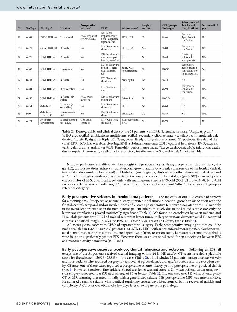
Early postoperative seizures: work-up, clinical relevance and outcome. Following an EPS, all except one of the 34 patients received cranial imaging within 24 h. MR and/or CT scans revealed a plausible cause for the seizure in 26/33 (78.8%) of the cases (Table 2). This includes 22 patients managed conservatively and four patients who required surgery for removal of epidural, subdural and/or bleeds into the resection cav-ity. Of note, one of these cases reported a preoperative seizure history, yet no postoperative or postictal deficit (Fig. 1). However, the size of the (epidural) bleed was felt to warrant surgery. Only two patients undergoing revi-sion surgery recovered to a KPI at discharge of 80 or better (Table 2). The one case (no. 34) without emergency CT or MR scanning presented initially with a generalized seizure. His postoperative MRI was unremarkable. He suffered a second seizure with identical semiology several days later, from which he recovered quickly and completely. A CT scan was obtained a few days later showing no acute pathology.
No Sexa/age Histologyb LocationcPreoperative seizuresd EPSd,e Seizure causef
Surgical revision
KPIg (preop./discharge)
Seizure-related deficits and complications
Seizure w/in 1 yearj
25 m/66 sGBM, IDH mt R temporal Focal impaired awareness szs
D5: Focal impared aware-ness + cognitive (aphasia) szs
EDH, ICB No 90/90Temporary dysarthria & confusion
No
26 m/70 sGBM, IDH mt R frontal No D3: Gen tonic–clonic sz EDH, ICB Yes 80/80 Temporary
confusion No
27 m/76 GBM, IDH wt R frontal NoD8: Focal aware motor + cogni-tive (aphasia) sz
ICB No 70/40Persisting aphasia & hemiparesis
N/A
28 m/60 GBM, IDH wt L temporal NoD3: Focal aware motor + cogni-tive (aphasia) szs
SDH, ICB, hyponatremia Yes 100/60
Temporary hemiparesis & confusion, per-sisting aphasia
Yes
29 m/42 GBM, IDH wt R frontal No D7: Gen tonic–clonic sz Meningitis No 70/70 No No
30 m/84 GBM, IDH wt R postcentral No D7: Unclassi-fied sz ICB No 90/90
Temporary aphasia & confusion
N/A
31 m/57 GBM, IDH wt R frontal cin-gulum
Focal aware motor sz
D0: Focal aware motor szs Infarction No 100/100 No N/A
32 m/54 Metastasis R central (+ l cerebellar) No D3: Gen tonic–
clonic sz EDH No 90/60 No N/A
33 f/50 Metastasis (recurrent)
L temporome-sial No D4: Gen tonic–
clonic sz Meningitis No 90/80 No N/A
34 m/29 Vestibular schwannoma
R cerebellopon-tine angle
Gen tonic–clonic sz
D13: Gen tonic–clonic sz
Hydrocephalus, EVD No 80/70 No No
Table 2. Demographic and clinical data of the 34 patients with EPS. a f, female; m, male. b Atyp., atypical; °, WHO grade; GBM, glioblastoma multiforme; sGBM, secondary glioblastoma; wt, wildtype; mt, mutated; del, deleted. c L, left; R, right; multiple, ≥ 2. d Gen, generalized; sz/szs; seizure/seizures. e D, postoperative day of the (first) EPS. f ICB, intracerebral bleeding; SDH, subdural hematoma; EDH, epidural hematoma; EVD, external ventricular drain; ?, unknown. g KPI, Karnofsky performance index. h Large cardiogenic MCA infarction, death due to sepsis. i Pneumonia, death due to respiratory insufficiency. j w/in, within; N/A, not available.
6
Vol:.(1234567890)
Scientific RepoRtS | (2020) 10:13674 | https://doi.org/10.1038/s41598-020-70754-z
www.nature.com/scientificreports/
Laboratory testing diagnosed relevant serum electrolyte disturbances in two cases (1 hyponatremia: Na+ = 126 mmol/l, 1 hypernatremia: Na+ = 159 mmol/l; Table 2). Meningitis was believed to be responsible for seizures in two cases. These two cases made a good recovery following appropriate antibiotic treatment (Table 2). Awake craniotomies and intraoperative cortical electrostimulation were thought to play a causative role in two other cases, and a temporary external ventricular drain in one patient.
Medical management of EPS patients relied heavily on levetiracetam. 20/22 (90.9%) of cases with de-novo EPS were treated with levetiracetam. In the remaining two cases, no anticonvulsive treatment was initiated. Ten EPS patients with preoperative seizures had their levetiracetam dose increased or adjusted based on serum level determinations. In two cases with preoperative epilepsy and multiple EPS, who were already on levetiracetam, concomitant medication with lacosamide was initiated.
EPS were associated with very relevant morbidities and even mortality. Thirteen patients (38.2%) displayed a new neurological deficit following EPS not explained by imaging or other findings, with seven of those persisting at discharge (20.6%). One patient developed pneumonia subsequent to multiple seizures and ultimately a fatal respiratory insufficiency.
Seizure recurrence after EPS was frequent. As pointed out above, 17/34 (50.0%) patients experienced multiple seizures, i.e. suffered already a recurrent seizure within 30 days of the index surgery. Patients were routinely man-aged by outside neurologists after discharge. Nevertheless, seizure follow-up for at least one year could be made available for 26 of these cases. Nine patients (34.6%) reported at least one other seizure within a year. Interestingly, five of these nine patients (55.6%) had a negative seizure history prior to their surgery.
Table 3. Risk factors for EPS in N = 679 brain tumour operations. a KPI, Karnofsky performance index. b Age: Mean ± SD, standard deviation; all other variables: N, number of surgical cases. c ns, not significant. *p < 0.05.
Variablea Mean ± SD/ Nb EPS No EPS pc
Age (years) 59.8 ± 15.0 60.6 ± 16.4 59.8 ± 14.9 ns
Sex
Female 404 20/404 (5.0%) 384/404 (95.0%)ns
Male 275 14/275 (5.1%) 261/275 (94.9%)
Preoperative KPI
KPI < 70 69 6/69 (10.2%) 63/69 (91.3%)ns
KPI 70–100 610 28/610 (4.6%) 582/610 (95.4%)
Postoperative KPI
KPI < 70 108 11/108 (10.2%) 97/108 (89.8%)0.007*
KPI 70–100 571 23/571 (4.0%) 548/571 (96.0%)
Preoperative seizures
No seizure 524 22/524 (4.2%) 502/524 (95.6%)ns
≥ 1 seizure 155 12/155 (7.7%) 143/155 (92.3%)
No or 1 seizure 597 26/597 (4.4%) 571/597 (95.6%)0.035*
≥ 2 seizures 82 8/82 (9.8%) 74/82 (90.2%)
Anticonvulsive 141 10/141 (7.1%) 131/141 (92.9%)ns
No anticonvulsive 537 24/537 (4.5%) 513/537 (95.5%)
Histology
Glioblastoma 177 7/177 (4.0%) 170/177 (96.0%)
< 0.001*
Other glioma 64 2/64 (3.1%) 62/64 (96.9%)
Meningioma 218 22/218 (10.1%) 198/218 (89.9%)
Metastasis 139 2/139 (1.4%) 137/139 (98.6%)
Other 81 1/81 (1.2%) 80/81 (98.8%)
Location
Supratentorial 542 33/542 (6.1%) 509/542 (93.9%)0.007*
Infratentorial 137 1/137 (0.7%) 136/137 (99.3%)
Intra-axial 406 11/406 (2.7%) 395/406 (97.3%)0.001*
Extra-axial 273 23/273 (8.4%) 250/273 (91.6%)
Frontal, central, temporal and/or insular lobe involvement/compression 452 32/452 (7.1%) 420/452 (92.9%)< 0.001*
No 227 2/227 (0.9%) 225/227 (99.1%)
Surgical procedure
Resection 653 34/653 (5.2%) 619/653 (94.8%)ns
Biopsy 26 0/26 (0%) 26/26 (100%)
Surgery for recurrence 103 4/103 (3.9%) 99/7,103 (96.1%)ns
First tumours surgery 576 30/576 (5.2%) 546/576 (94.8%)

7
Vol.:(0123456789)
Scientific RepoRtS | (2020) 10:13674 | https://doi.org/10.1038/s41598-020-70754-z
www.nature.com/scientificreports/
Table 4. Risk factors for EPS in N = 218 meningioma operations. a KPI, Karnofsky performance index; complete resection, Simpson grade 1–3; STR/biopsy, Simpson grade 4–5. b Age & tumour size: Mean ± SD, standard deviation; all other variables: N, number of surgical cases. c ns, not significant. d supratentorial tumours only. *p < 0.05. **p = 0.055.
Variablea Mean ± SD/ Nb EPS No EPS pc
Age (years) 61.3 ± 14.3 64.3 ± 14.0 61.0 ± 14.3 ns
Sex
Female 169 19/169 (11.2%) 150/169 (88.8%)ns
Male 49 3/49 (6.1%) 46/49 (93.9%)
Preoperative KPI
KPI < 70 21 6/21 (28.6%) 15/21 (71.4%)ns
KPI 70–100 197 16/197 (8.1%) 181/197 (91.9%)
Postoperative KPI
KPI < 70 40 9/40 (22.5%) 31/40 (77.5%)0.004*
KPI 70–100 178 13/178 (7.3%) 165/178 (92.7%)
Preoperative seizures
No seizure 163 14/163 (8.6%) 149/163 (91.4%)ns
≥ 1 seizure 55 8/55 (14.5%) 47/55 (85.5%)
No or 1 seizure 188 16/188 (8.5%) 172/188 (91.5%)ns
≥ 2 seizures 30 6/30 (20.0%) 24/30 (80.0%)
Anticonvulsive 46 6/46 (13.0%) 40/46 (87.0%)ns
No anticonvulsive 172 16/172 (9.3%) 156/172 (90.7%)
Location
Supratentorial 186 22/186 (11.8%) 164/186 (88.2%)ns
Infratentorial 32 0/32 (0%) 32/32 (100%)
Convexity/falx 125 16/125 (12.8%) 109/125 (87.2%)
nsSupratentorial skull-base 56 5/56 (8.9%) 51/56 (91.1%)
Other 37 1/37 (2.7%) 36/37 (97. 3%)
Frontal, central, temporal and/or insular lobe compression 166 21/166 (12.7%) 145/166 (87.3%)0.031*
No 52 1/52 (1.9%) 51/52 (98.1%)
Surgical procedure
Resection 217 22/217 (10.1%) 195/217 (89.9%)ns
Biopsy 1 0/1 (0%) 1/1 (100%)
Complete resection 194 21/194 (10.8%) 173/194 (88.2%)ns
STR or biopsy 24 1/24 (4.2%) 23/24 (95.8%)
Surgery for recurrence 30 3/30 (10.0%) 27/30 (90.0%)ns
First tumour surgery 188 19/188 (10.1%) 169/188 (89.9%)
Histology
WHO grade I 168 14/168 (8.3%) 154/168 (91.7%)
nsWHO grade II 47 8/47 (17.0%) 39/47 (83.0%)
WHO grade III 3 0/3 (0%) 3/3 (0%)
Tumour size (mm) 400.0 ± 191.9 471.4 ± 243.3 391.8 ± 184.2 ns
Postoperative imagingd
Extra-axial hematoma 19 3/19 (15.8%) 16/19 (84.2%)ns
No 147 19/147 (12.9%) 128/147 (87.1%)
Resection cavity hematoma 67 13/67 (19.4%) 54/67 (80.6%)ns**
No 99 9/99 (9.1%) 90/99 (90.9%)
Brain contusion 67 8/67 (11.9%) 59/67 (88.1%)ns
No 99 14/99 (14.1%) 85/99 (85.9%)
Infarct 31 4/31 (12.9%) 27/31 (87.1%)ns
No 135 18/135 (13.3%) 117/135 (86.7%)
Pneumocephalus 24 2/24 (8.3%) 22/24 (91.7%)ns
No 142 20/142 (14.1%) 122/142 (85.9%)

8
Vol:.(1234567890)
Scientific RepoRtS | (2020) 10:13674 | https://doi.org/10.1038/s41598-020-70754-z
www.nature.com/scientificreports/
DiscussionThe rate of early postoperative (= perioperative or in-hospital) seizures is usually reported as < 5–10% in most studies with some authors detailing EPS rates only for seizure-naïve patients4,8,9,15,18,19. We observed a 5.1% overall and 6.1% rate in patients with supratentorial tumours. Posterior fossa operations were included in our analysis primarily in order to avoid selection bias. Seizures in patients with infratentorial tumours are generally rare19. However, some patients with posterior fossa tumours require (temporary) ventricular drains, i.e. an (albeit minor) supratentorial operation, and certain perioperative complications thought to underlie EPS such as meningitis occur after both infratentorial and supratentorial surgeries. Interestingly, our series includes a patient undergo-ing surgery for a large vestibular schwannoma who required temporary CSF drainage and suffered a generalized seizure on postoperative day 13.
EPS are not benign. In a substantial number of cases, they are associated with and reflect major complications such as bleeds requiring operative revision (4/34, 11.8%) or meningitis (2/34, 5.9%). Also, early postoperative seizures per se can result in neurological worsening (13/34, 38.2%) which often persists (7/34, 20.6%). Seizures can result in pulmonary complications due to acute or silent aspirations in confused or stuporous patients. One of our cases ultimately succumbed to a pulmonary sepsis initially triggered by an early postoperative seizure.
Risk factors for EPS in our cohort included a positive preoperative seizure history, a supratentorial tumour location, tumour growth involving or compressing particularly epileptogenic brain tissues (i.e. the frontal, cen-tral, temporal and insular lobes)13 and, importantly, tumour histology. Meningioma patients accounted for the majority of cases and the highest overall rate (10.1%). However, contradictory results, i.e. an association with glioma histology and infiltrating growth and relatively lower perioperative seizure rates in meningioma patients have also been reported8. Awake craniotomy (i.e. intraoperative cortical electrostimulation) figured prominently as a risk factor for perioperative seizures in a recent study by Oushy and co-workers and was therefore felt to contribute to seizure formation in two of our cases8.
In accordance with the literature, we found an (albeit not statistically significantly) increased risk for EPS in patients with WHO grade II/III meningiomas, a convexity/parasagittal tumour location, perifocal oedema > 1 cm and an association with a worse postoperative KPI4,15. Meningioma-associated epilepsy has recently attracted some attention in the neurosurgical community. A non-skull base location, tumour size, peritumoral oedema, malignancy, tumour progression and recurrence, age and sex (as a possible corollary of a higher WHO grade), sei-zure history and EEG findings, clinical symptoms and surgical complications have all been (variably) associated with the risk of preoperative and postoperative seizures in meningioma patients1,4,5,15,16,20. We also investigated if early postoperative neuroimaging following supratentorial meningioma surgery might predict EPS. We were unable to identify significant correlations, however, there was a statistical trend for an association between EPS and the presence of a hematoma in the resection cavity.
Are EPS simply acute symptomatic seizures? By definition, acute symptomatic seizures occur within 7 days of the underlying brain insult2. Indeed, the majority of our cases reported seizures within the first three days following surgery and 29/34 (85.3%) within the first 7 days. Some authors have reported statistical correlations between surgical complications (including new neurological deficits) and the occurrence of early postoperative seizures1,4. We identified surgical complications such as haemorrhages and increased oedema, meningitis, and electrolyte disorders as the most likely cause of the seizure in 27/34 (79.4%) of cases. Clinical worsening (a pos-sible corollary of a structural postoperative or other e.g. infectious complication) also correlated with the EPS rate in our series.
On the other hand, EPS tend to recur. 17/34 (50.0%) of our cases with EPS had a recurrent seizure within 30 days of the surgery (i.e. multiple EPS). The one year-recurrence rate was 34.6%, i.e. in a sizable proportion of
Figure 1. (a) A 71-year old female patient (no. 10) with a history of one (focal) preoperative seizure, who underwent surgery for a left frontal convexity meningioma. (b) On the fourth postoperative day, the patient suffered another focal seizure, yet exhibited no focal or other neurological deficit. The emergency CT scan showed a relevant epidural hematoma, which was surgically removed. (c) Postoperative CT scan after epidural clot removal. The patient’s further clinical course was uneventful.

9
Vol.:(0123456789)
Scientific RepoRtS | (2020) 10:13674 | https://doi.org/10.1038/s41598-020-70754-z
www.nature.com/scientificreports/
EPS patients the first seizure is simply the first manifestation of epilepsy. High recurrence rates have also been published by Wirsching and co-workers (29/46, 63.0%)1 and Chen et al. (13/36, 36.1%)4. Also, the risk of EPS varies with the preoperative seizure history in this as well as in several other published cohorts1,4. These data suggest that not only acute perioperative brain insults but also more chronic factors such as the anatomic and metabolic alterations caused by the brain tumour contribute to early postoperative epileptogenesis.
In our view, these figures together with the adverse clinical course seen in several cases with multiple periop-erative seizures justifies institution of anticonvulsive treatment already following a single perioperative seizure. Of note, we have no longer term follow-up from our patients and while we use antiepileptics quite liberally during the early postoperative period, we strongly urge patients and their physicians to re-evaluate this medication at the latest after one year. Antiepileptic medication may occasionally carry significant adverse effects. Side effects of levetiracetam, which is the current drug of choice for most neuro-oncological patients requiring anticonvulsive treatment, include e.g. fatigue, insomnia, mood and behaviour changes, headaches and decreased white blood counts21,22. These effects of have to be weighed against the risk of recurrent seizures with its attendant socioeco-nomic sequelae (e.g. restriction of driving privileges).
Given the rather high rates of adverse outcomes and recurrence after EPS, it is tempting to speculate about a role for prophylactic anticonvulsive medication. This is a controversial topic. Most believe that routine use of prophylactic anticonvulsants does not lower the risk of postoperative seizures in patients without preopera-tive seizures14,23–25. However, there are some data including a small randomized prospective trial suggesting a benefit from prophylactic levetiracetam9. Based on our experience and the data just outlined, we consider prophylactic levetiracetam in vulnerable patients with a high risk of EPS, e.g. an elderly patient with a large convexity meningioma.
Our study allows some conclusions with respect to the proper management of EPS. Establishing a specific diagnostic algorithm for such emergencies is important. Neuroimaging revealed the presumed cause of the seizure in the great majority (26/33, 78.8%) of our cases. In four patients, the imaging finding prompted an operative revision. We therefore feel that obtaining a CT or MRI scan after an EPS is mandatory. Meningitis and electrolyte disorders may also contribute to the formation of perioperative seizures, i.e. laboratory blood testing and a very low threshold for a lumbar puncture should be part of the diagnostic work-up.
The major limitation of our study is its retrospective design. Not all seizures reported by the patients or observed by the attending staff may have been properly documented. In addition, it is quite possible that sub-clinical seizures have simply escaped detection. Another shortcoming is that this is a single institutional series which somewhat limits the generalizability of our findings. Nevertheless, we present a sizable, unselected and recent experience with the diagnosis and management of EPS.
conclusionEarly postoperative seizures (EPS) following brain tumour surgery are common. EPS often reflect serious compli-cations of brain tumour surgery and are associated with a relatively high rate of adverse neurological and medi-cal sequelae. We found that structural causes not infrequently requiring surgical revision are a common cause, which suggests CT (or MR) imaging as a mandatory part of the work-up of an EPS. Multiple EPS and recurrent seizures during follow-up are frequent, which indicates that EPS are not just acute symptomatic seizures, but not uncommonly reflect a more chronic condition i.e. epilepsy. Finally, our data indicate that meningioma surger-ies may carry a particularly high risk for EPS, which raises the question of an antiepileptogenic prophylaxis in selected cases.
Data availabilityThe datasets generated during and/or analysed during the current study are available from the corresponding author on reasonable request.
Received: 25 January 2020; Accepted: 31 July 2020
References 1. Wirsching, H.-G. et al. Predicting outcome of epilepsy after meningioma resection. Neuro. Oncol. 18, 1002–1010 (2016). 2. Beghi, E. et al. Recommendation for a definition of acute symptomatic seizure. Epilepsia 51, 671–675 (2010). 3. Hesdorffer, D. C., Logroscino, G., Cascino, G., Annegers, J. F. & Hauser, W. A. Risk of unprovoked seizure after acute symptomatic
seizure: Effect of status epilepticus. Ann. Neurol. 44, 908–912 (1998). 4. Chen, W. C. et al. Factors associated with pre- and postoperative seizures in 1033 patients undergoing supratentorial meningioma
resection. Neurosurgery 81, 297–306 (2017). 5. Schneider, M. et al. Preoperative tumor-associated epilepsy in patients with supratentorial meningioma: factors influencing seizure
outcome after meningioma surgery. J. Neurosurg. https ://doi.org/10.3171/2019.7.JNS19 455 (2019). 6. Maschio, M. et al. Management of epilepsy in brain tumors. Neurol. Sci. 40, 2217–2234 (2019). 7. Chen, D. Y., Chen, C. C., Crawford, J. R. & Wang, S. G. Tumor-related epilepsy: epidemiology, pathogenesis and management. J.
Neurooncol. 139, 13–21 (2018). 8. Oushy, S. et al. New-onset seizure during and after brain tumor excision: a risk assessment analysis. J. Neurosurg. 128, 1713–1718
(2018). 9. Iuchi, T. et al. Levetiracetam versus phenytoin for seizure prophylaxis during and early after craniotomy for brain tumours: a phase
II prospective, randomised study. J. Neurol. Neurosurg. Psychiatry 86, 1158–1162 (2015). 10. Cloppenborg, T. et al. Trends in epilepsy surgery: Stable surgical numbers despite increasing presurgical volumes. J. Neurol. Neu-
rosurg. Psychiatry 87, 1322–1329 (2016). 11. Kwan, P. et al. Definition of drug resistant epilepsy: consensus proposal by the ad hoc Task Force of the ILAE Commission on
Therapeutic Strategies. Epilepsia 51, 1069–1077 (2010).
10
Vol:.(1234567890)
Scientific RepoRtS | (2020) 10:13674 | https://doi.org/10.1038/s41598-020-70754-z
www.nature.com/scientificreports/
12. Fisher, R. S. et al. Operational classification of seizure types by the international league against epilepsy: position paper of the ILAE Commission for Classification and Terminology. Epilepsia 58, 522–530 (2017).
13. Kurzwelly, D., Herrlinger, U. & Simon, M. Seizures in patients with low-grade gliomas—incidence, pathogenesis, surgical manage-ment, and pharmacotherapy. Adv. Tech. Stand. Neurosurg. 35, 81–111 (2010).
14. Wu, A. S. et al. A prospective randomized trial of perioperative seizure prophylaxis in patients with intraparenchymal brain tumors. J. Neurosurg. 118, 873–883 (2013).
15. Skardelly, M. et al. Risk factors of preoperative and early postoperative seizures in patients with meningioma: a retrospective single-center cohort study. World Neurosurg. 97, 538–546 (2017).
16. Englot, D. J. et al. Seizures in supratentorial meningioma: a systematic review and meta-analysis. J. Neurosurg. 124, 1552–1561 (2016).
17. Geßler, F. et al. Is postoperative imaging mandatory after meningioma removal? Results of a prospective study. PLoS ONE 10, e0124534 (2015).
18. Lwu, S., Hamilton, M. G., Forsyth, P. A., Cairncross, J. G. & Parney, I. F. Use of peri-operative anti-epileptic drugs in patients with newly diagnosed high grade malignant glioma: a single center experience. J. Neurooncol. 96, 403–408 (2010).
19. Skardelly, M. et al. Predictors of preoperative and early postoperative seizures in patients with intra-axial primary and metastatic brain tumors: a retrospective observational single center study. Ann. Neurol. 78, 917–928 (2015).
20. Chaichana, K. L. et al. Seizure control for patients undergoing meningioma surgery. World Neurosurg. 79, 515–524 (2013). 21. Pourzitaki, C. et al. Efficacy and safety of prophylactic levetiracetam in supratentorial brain tumour surgery: a systematic review
and meta-analysis. Br. J. Clin. Pharmacol. 82, 315–325 (2016). 22. Englot, D. J., Chang, E. F. & Vecht, C. J. Epilepsy and brain tumors. in Handbook of Clinical Neurology 134, 267–285 (Elsevier B.V.,
2016). 23. Islim, A. I. et al. Postoperative seizures in meningioma patients: improving patient selection for antiepileptic drug therapy. J.
Neurooncol. 140, 123–134 (2018). 24. Ansari, S. F., Bohnstedt, B. N., Perkins, S. M., Althouse, S. K. & Miller, J. C. Efficacy of postoperative seizure prophylaxis in intra-
axial brain tumor resections. J. Neurooncol. 118, 117–122 (2014). 25. Dewan, M. C. et al. The influence of perioperative seizure prophylaxis on seizure rate and hospital quality metrics following glioma
resection. Neurosurgery 80, 563–570 (2017).
Author contributionsT.F.E. (conceptualization, data curation, formal analysis, investigation, methodology, resources, writing—origi-nal draft, writing—review & editing). M.S. (conceptualization, data curation, formal analysis, methodology, resources, writing—original draft, writing—review & editing, project administration, and supervision). R.C. (data curation: histopathological analysis, methodology, resources, writing—review & editing). S.R. (formal analysis, methodology, writing—review & editing), A.G. (formal analysis, methodology, writing—review & editing).
competing interests The authors declare no competing interests.
Additional informationCorrespondence and requests for materials should be addressed to T.F.E.
Reprints and permissions information is available at www.nature.com/reprints.
Publisher’s note Springer Nature remains neutral with regard to jurisdictional claims in published maps and institutional affiliations.
Open Access This article is licensed under a Creative Commons Attribution 4.0 International License, which permits use, sharing, adaptation, distribution and reproduction in any medium or
format, as long as you give appropriate credit to the original author(s) and the source, provide a link to the Creative Commons license, and indicate if changes were made. The images or other third party material in this article are included in the article’s Creative Commons license, unless indicated otherwise in a credit line to the material. If material is not included in the article’s Creative Commons license and your intended use is not permitted by statutory regulation or exceeds the permitted use, you will need to obtain permission directly from the copyright holder. To view a copy of this license, visit http://creat iveco mmons .org/licen ses/by/4.0/.
© The Author(s) 2020
Related Documents