Early growth responsive gene 3 in human breast carcinoma: a regulator of estrogen- meditated invasion and a potent prognostic factor Takashi Suzuki, Akio Inoue 1 , Yasuhiro Miki, Takuya Moriya, Jun-ichi Akahira, Takanori Ishida 2 , Hisashi Hirakawa 3 , Yuri Yamaguchi 1 , Shin-ichi Hayashi 4 and Hironobu Sasano Department of Pathology, Tohoku University School of Medicine, 2-1 Seiryo-machi, Aoba-ku, Sendai 980-8575, Japan 1 Research Institute for Clinical Oncology, Saitama Cancer Center, Saitama, Japan 2 Department of Surgery, Tohoku University School of Medicine, Sendai, Japan 3 Department of Surgery, Tohoku Kosai Hospital, Sendai, Japan 4 Department of Molecular Medical Technology, Tohoku University School of Medicine, Sendai, Japan (Requests for offprints should be addressed to T Suzuki; Email: [email protected]) A Inoue is now at InfoGenes Co. Ltd, Tsukuba, Japan Abstract Early growth responsive gene 3 (EGR3) is a zinc-finger transcription factor and plays important roles in cellular growth and differentiation. We recently demonstrated estrogen-mediated induction of EGR3 in breast carcinoma cells. However, EGR3 has not yet been examined in breast carcinoma tissues and its significance remains unknown. Therefore, in this study, we examined biological functions of EGR3 in the breast carcinoma by immunohistochemistry, in vitro study, and nude mouse xenograft model. EGR3 immunoreactivity was detected in carcinoma cells in 99 (52%) out of 190 breast carcinoma tissues and was associated with the mRNA level. EGR3 immunoreactivity was positively associated with lymph node status, distant metastasis into other organs, estrogen receptor a, or EGR3 immunoreactivity in asynchronous recurrent lesions in the same patients, and was negatively correlated with tubule formation. EGR3 immunoreactivity was significantly associated with an increased risk of recurrence and adverse clinical outcome by both uni- and multivariate analyses. Egr3-expressing transformant cell lines derived from MCF-7 Tet-Off cells (Eg-10 and Eg-11) significantly enhanced the migration and invasion properties according to the treatment of doxycyclin, but did not significantly change the cell proliferation. Moreover, Eg-11 cells injected into athymic mice irregularly invaded into the adjacent peritumoral tissues, although Clt-7, which was stably transfected with empty vector as a control, demonstrated a well-circumscribed tumor. Eg-11 cells were significantly associated with invasive components and less tubule formation in the xenograft model. These results suggest that EGR3 plays an important role in estrogen-meditated invasion and is an independent prognostic factor in breast carcinoma. Endocrine-Related Cancer (2007) 14 279–292 Introduction Breast carcinoma is one of the most common malignancies in women worldwide. Human breast tissue is a target for estrogens, and these sex steroids play an important role in development of hormone- dependent breast carcinomas (Thomas 1984, Vihko & Apter 1989). The biological effects of estrogens are mediated through an initial interaction with estrogen receptor (ER) a and/or b, members of a nuclear receptor superfamily (designated NR3A1 and NR3A2 respectively). ERs function as dimers, and activate transcription in a ligand-dependent manner by binding to estrogen responsive elements (EREs) located in the promoter region of various target genes (Tsai & O’Malley 1994). A variety of estrogenic functions Endocrine-Related Cancer (2007) 14 279–292 Endocrine-Related Cancer (2007) 14 279–292 1351-0088/07/014–279 q 2007 Society for Endocrinology Printed in Great Britain DOI:10.1677/ERC-06-0005 Online version via http://www.endocrinology-journals.org

Welcome message from author
This document is posted to help you gain knowledge. Please leave a comment to let me know what you think about it! Share it to your friends and learn new things together.
Transcript

Endocrine-Related Cancer (2007) 14 279–292
Early growth responsive gene 3 in humanbreast carcinoma: a regulator of estrogen-meditated invasion and a potent prognosticfactor
Takashi Suzuki, Akio Inoue1, Yasuhiro Miki, Takuya Moriya, Jun-ichi Akahira,Takanori Ishida2, Hisashi Hirakawa3, Yuri Yamaguchi1, Shin-ichi Hayashi 4
and Hironobu Sasano
Department of Pathology, Tohoku University School of Medicine, 2-1 Seiryo-machi, Aoba-ku, Sendai 980-8575, Japan1Research Institute for Clinical Oncology, Saitama Cancer Center, Saitama, Japan2Department of Surgery, Tohoku University School of Medicine, Sendai, Japan3Department of Surgery, Tohoku Kosai Hospital, Sendai, Japan4Department of Molecular Medical Technology, Tohoku University School of Medicine, Sendai, Japan
(Requests for offprints should be addressed to T Suzuki; Email: [email protected])
A Inoue is now at InfoGenes Co. Ltd, Tsukuba, Japan
Abstract
Early growth responsive gene 3 (EGR3) is a zinc-finger transcription factor and plays important rolesin cellular growth and differentiation. We recently demonstrated estrogen-mediated induction ofEGR3 in breast carcinoma cells. However, EGR3 has not yet been examined in breast carcinomatissues and its significance remains unknown. Therefore, in this study, we examined biologicalfunctions of EGR3 in the breast carcinoma by immunohistochemistry, in vitro study, and nudemousexenograft model. EGR3 immunoreactivity was detected in carcinoma cells in 99 (52%) out of 190breast carcinoma tissues and was associated with the mRNA level. EGR3 immunoreactivity waspositively associatedwith lymph node status, distantmetastasis into other organs, estrogen receptora, or EGR3 immunoreactivity in asynchronous recurrent lesions in the same patients, and wasnegatively correlatedwith tubule formation. EGR3 immunoreactivitywas significantly associatedwithan increased risk of recurrence and adverse clinical outcome by both uni- and multivariate analyses.Egr3-expressing transformant cell lines derived from MCF-7 Tet-Off cells (Eg-10 and Eg-11)significantly enhanced themigrationand invasionproperties according to the treatment of doxycyclin,but did not significantly change the cell proliferation.Moreover, Eg-11 cells injected into athymicmiceirregularly invaded into the adjacent peritumoral tissues, althoughClt-7, whichwas stably transfectedwith empty vector as a control, demonstrated a well-circumscribed tumor. Eg-11 cells weresignificantly associated with invasive components and less tubule formation in the xenograft model.These results suggest that EGR3 plays an important role in estrogen-meditated invasion and is anindependent prognostic factor in breast carcinoma.
Endocrine-Related Cancer (2007) 14 279–292
Introduction
Breast carcinoma is one of the most common
malignancies in women worldwide. Human breast
tissue is a target for estrogens, and these sex steroids
play an important role in development of hormone-
dependent breast carcinomas (Thomas 1984, Vihko &
Apter 1989). The biological effects of estrogens are
Endocrine-Related Cancer (2007) 14 279–292
1351-0088/07/014–279 q 2007 Society for Endocrinology Printed in Great B
mediated through an initial interaction with estrogen
receptor (ER) a and/or b, members of a nuclear
receptor superfamily (designated NR3A1 and NR3A2
respectively). ERs function as dimers, and activate
transcription in a ligand-dependent manner by binding
to estrogen responsive elements (EREs) located in the
promoter region of various target genes (Tsai &
O’Malley 1994). A variety of estrogenic functions
ritain
DOI:10.1677/ERC-06-0005
Online version via http://www.endocrinology-journals.org

T Suzuki et al.: EGR3 in breast cancer
are characterized by the expression of these genes
(Hayashi et al. 2003), and therefore, it is very
important to examine the expression and roles of
estrogen responsive genes to obtain a better under-
standing of estrogenic actions in human breast cancer.
Early growth responsive gene 3 (EGR3) belongs to
the EGR family of zinc-finger transcription factors,
and shares a common sequence termed the EGR
responsive element with other members involved in
DNA binding and transactivation (Patwardhan et al.
1991, O’Donovan et al. 1999). Previous studies
revealed that EGR3 was involved in the development
of muscle spindle (Tourtellotte & Milbrandt 1998,
Tourtellotte et al. 2001) and thymocyte proliferation
(Xi & Kersh 2004), indicating that EGR3 plays
important roles in cellular growth and differentiation.
We have recently demonstrated that EGR3 was
induced by estradiol in MCF-7 breast carcinoma cells
from the cDNA microarray analysis (Inoue et al.
2004). These findings suggest a possible role for EGR3
in estrogen-dependent human breast carcinomas.
However, EGR3 has not been examined in human
breast carcinoma tissues, and its biological and clinical
significance remains unknown. Therefore, in this
study, we examined biological functions of EGR3 in
the breast carcinoma using immunohistochemistry,
in vitro study, and nude mouse xenograft model. From
these results, here, we first report that EGR3 is a
regulator of estrogen-mediated invasion, and is a
potent prognostic factor in human breast carcinomas.
Materials and methods
Patients and tissues
About 190 specimens of invasive ductal carcinoma of
the breast were obtained from female patients who
underwent mastectomy from 1984 to 1992 in the
Department of Surgery, Tohoku University Hospital,
Sendai, Japan. Breast tissue specimens were obtained
from patients with a mean age of 53.5 years (range
22–82). The patients did not receive chemotherapy or
irradiation prior to surgery. About 62 patients received
tamoxifen therapy after the surgery. The mean follow-
up time was 102 months (range 3–157 months). The
histological grade and tubule formation of each
specimen was evaluated based on the method of Elston
& Ellis (1991). Asynchronous recurrent lesions of the
breast carcinoma were also available for examination
in 13 cases (breast, 3 cases; lymph node, 3 cases; skin,
2 cases; liver, 2 case; lung, 1 case; bone, 1 case; and
chest wall, 1 case). All specimens were fixed in 10%
formalin and embedded in paraffin wax.
280
Thirty-one specimens of invasive ductal carcinoma
were obtained from patients who underwent mastect-
omy in 2000 in the Departments of Surgery at Tohoku
University Hospital and Tohoku Kosai Hospital,
Sendai, Japan. Specimens for RNA isolation were
snap-frozen and stored at K80 8C, and those for
immunohistochemistry were fixed in 10% formalin and
embedded in paraffin wax. Informed consent was
obtained from all patients prior to their surgery and
examination of specimens used in this study.
Research protocols for this study were approved by
the Ethics Committee at both Tohoku University
School of Medicine and Tohoku Kosai Hospital.
Antibodies
A rabbit polyclonal antibody for EGR3 (C-24 (sc-191))
was purchased from Santa Cruz Biotechnology (Santa
Cruz, CA, USA). This antibody was raised against a
peptide mapping at the carboxy terminus of human
EGR3. The EGR3 antibody specially recognized human
EGR3 by immunoblotting and immunohistochemistry,
and was non-cross-reactive with EGR1, EGR2, or
Wilms’ tumor proteins (data from Santa Cruz Bio-
technology). Monoclonal antibodies for ERa (ER1D5),
progesterone receptor (PR; MAB429), and Ki-67
(MIB1) were purchased from Immunotech (Marseille,
France), Chemicon (Temecula, CA, USA), and DAKO
(Carpinteria, CA, USA) respectively. Rabbit polyclonal
antibodies for ERb (06-629) and HER2 (A0485) were
obtained from Upstate Biotechnology (Lake Placid,
NY, USA) and DAKO respectively.
Immunohistochemistry
A Histofine Kit (Nichirei, Tokyo, Japan), which
employs the streptavidin–biotin amplification method
was used in this study. Antigen retrieval was performed
by heating the slides in an autoclave at 120 8C for 5 min
in citric acid buffer (2 mM citric acid and 9 mM
trisodium citrate dehydrate (pH 6.0)). Dilutions of
primary antibodies used in this study were as follows:
EGR3, 1/500; ERa, 1/50; ERb, 1/50; PR, 1/30; HER2,
1/200; and Ki-67, 1/50. The antigen–antibody complex
was visualized with 3,3 0-diaminobenzidine
(DAB) solution (1 mM DAB, 50 mM Tris–HCl buffer
(pH 7.6), and 0.006% H2O2) and counterstained with
hematoxylin. As a negative control, normal mouse or
rabbit IgG was used instead of the primary antibodies.
Immunohistochemical preabsorption test was also
performed for EGR3 immunohistochemistry using the
blocking peptide (sc-191 P; Santa Cruz Biotechnology).
www.endocrinology-journals.org

Endocrine-Related Cancer (2007) 14 279–292
Scoring of immunoreactivity and statistical
analysis
EGR3, ERa, ERb, PR, and Ki-67 immunoreactivity
was detected in the nucleus, and the immunoreactivity
was evaluated in more than 1000 carcinoma cells for
each case, and subsequently the percentage of
immunoreactivity, i.e. labeling index (LI), was
determined. Cases with EGR3 or ERa LI of more
than 10% were considered EGR3- or ERa-positive
breast carcinomas in this study, according to a report
on ERa (Goldhirsch et al. 2005).
An association between EGR3 immunoreactivity
and clinicopathological factors was evaluated using a
Student’s t-test, cross-table using the c2-test, or
correlation coefficient (r) and regression equation.
Overall and disease-free survival curves were gener-
ated according to the Kaplan–Meier method and the
statistical significance was calculated using the log-
rank test. Uni- and multivariate analyses were
evaluated by a Cox’s proportional hazard model
using PROC PHREG in our SAS software.
Cells and chemicals
MCF-7 human breast cancer cell line and LY-2, which
is a tamoxifen-resistant MCF-7 cell variant (Paik et al.
1994), were cultured in RPMI-1640 (Sigma–Aldrich)
with 10% fetal bovine serum (FBS; JRH Biosciences,
Lenexa, KS, USA). We also used Eg-10 and Eg-11
cells which are Egr3-expressing transformants derived
from MCF-7 Tet-Off cells (Inoue et al. 2004), and Ctl-
7 cells which are MCF-7 Tet-Off cells stably
transfected with empty vector (Inoue et al. 2004).
Overexpression of Egr3 in Eg-10 and Eg-11 cells was
dramatically repressed by the treatment of doxycyclin
(50 ng/ml; Inoue et al. 2004). These cells were also
cultured in RPMI-1640 (Sigma–Aldrich) with 10%
FBS. All the cells used in this study were cultured with
phenol red-free RPMI-1640 medium containing 10%
dextran-coated charcoal–FBS for 3 days before
treatment of the experiment. Estradiol and tamoxifen
were purchased from Sigma–Aldrich, while ICI
182 780 was obtained from Tocris Cookson Inc.
(Ellisville, MO, USA).
Real-time PCR
Total RNA was extracted from breast carcinoma
tissues or cultured cells using TRIzol reagent (Invi-
trogen Life Technologies), and a reverse transcription
kit (Superscript II Preamplification system; Gibco-
BRL) was used in the synthesis of cDNA.
www.endocrinology-journals.org
The LightCycler System (Roche Diagnositics
GmbH) was used to semi-quantify the mRNA
expression levels by real-time PCR (Dumoulin et al.
2000). Settings for the PCR thermal profile were as
follows: initial denaturation at 95 8C for 1 min
followed by 40 amplification cycles of 95 8C for 1 s,
annealing at 68 8C (EGR3 and ribosomal protein L 13a
(RPL13A)) for 15 s, and elongation at 72 8C for 15 s.
Oligonucleotide primers for EGR3 (NM_004430) were
designed in different exons to avoid the amplification
of genomic DNA, and the primer sequences
were FWD: 5 0-CTGCCTGACAATCTGTACCC-3 0
(cDNA position; 416–435) and REV: 5 0-GTAGGT-
CACGGTCTTGTTGC-3 0 (cDNA position; 594–613).
The primer sequences for RPL13A (NM_012423) were
FWD: 5 0-CCTGGAGGAGAAGAGGAAAGAGA-3 0
(cDNA position; 487–509) and REV: 5 0-TTGAG-
GACCTCTGTGTATTTGTCAA-3 0 (cDNA position;
588–612; Vandesompele et al. 2002). To verify
amplification of the correct sequences, PCR products
were purified and subjected to direct sequencing.
Negative control experiments lacked cDNA substrate
to check for the possibility of exogenous contaminant
DNA. EGR3 mRNA level was summarized as the ratio
of RPL13A mRNA level (%).
Immunoblotting
The cell protein was extracted in triple detergent lysis
buffer (LK-18) at 4 8C. About 25 mg (immunoblotting
for EGR3) or 5 mg (immunoblotting for b-actin) of the
protein (whole cell extracts) were subjected to
SDS–PAGE (10% acrylamide gel). Following SDS–
PAGE, proteins were transferred onto Hybond P
polyvinylidene difluoride membrane (Amersham Bios-
ciences). The blots were blocked in 5% nonfat dry skim
milk for 1 h at room temperature, and were then
incubated with a primary antibody for EGR3 (C-24
(sc-191), Santa Cruz Biotechnology) or b-actin (AC-15
(Sigma #A-5411), Sigma–Aldrich) for 18 h at 4 8C.
After incubation with anti-rabbit or anti-mouse IgG
horseradish peroxidase (Amersham Biosciences) for
1 h at room temperature, antibody/protein complexes
on the blots were detected using ECL plus western
blotting detection reagents (Amersham Biosciences).
Immunointensity of specific bands was measured by
LAS-1000 imaging system (Fuji Photo Film, Tokyo,
Japan). Immunointensity of EGR3 in each sample was
normalized to that of b-actin, and subsequently,
relative immunointensity ratio of EGR3 was sum-
marized as a ratio compared with that of MCF-7 cells
in the absence of estradiol or tamoxifen.
281

T Suzuki et al.: EGR3 in breast cancer
Migration assay and invasion assay
Cell migration assay was performed using a 24-well
tissue culture plate (Becton Dickinson, Franklin Lakes,
NJ, USA) and Chemotaxicell (8 mm pore size; Kurabo,
Osaka, Japan). The membrane of Chemotaxicell was
coated with 0.3 mg/ml of collagen I (CELLGEN,
Tokyo, Japan). After 3 days of the treatment with or
without doxycyclin (50 ng/ml) in serum-free RPMI-
1640 medium, 5!105 cells were plated at the upper
chamber, while NIH/3T3 conditioned medium was in
the lower chamber. After incubation for 6 h at 37 8C,
cells on the upper surface of membrane were removed
by wiping with a cotton swab, and those on the lower
surface were subsequently fixed with 70% ethanol and
stained with hematoxylin and eosin. The migration
ability was evaluated as a total number of cells on the
lower surface of membrane, which was counted under
microscopy.
The cell invasion assay was performed by a modified
migration assay. In this experiment, upper surface of
the membrane of Chemotaxicell was coated with
80 mg/cm2 of Matrigel basement membrane matrix
(BD Biosciences, Two Oak Park, MA, USA; Albini
et al. 1987, Taniguchi et al. 1989). About 5!105 cells
at the upper chamber were incubated with 24 h at
37 8C, and the invasion ability was subsequently
evaluated as the total number of cells on the lower
surface of membrane.
Cell proliferation assay and apoptosis analysis
The status of cell proliferation of cells was measured
using a WST-8 (2-(2-methoxy-4-nitrophenyl)-3-
(4-nitrophenyl)-5-(2,4-disulfophenyl)-2H-tetrazolium,
monosodium salt) method (Cell Counting Kit-8;
Dojindo, Kumamoto, Japan). The apoptotic status of
cells was evaluated by an apoptosis screening kit
(Wako, Osaka, Japan), which employed a modified
TdT-mediated dUTP nick-end labeling (TUNEL)
method. Optical densities (ODZ450 nm for cell
proliferation assay and ODZ490 nm for apoptosis
analysis) were obtained with a Model 680 microplate
reader (Bio-Rad Laboratories). The cell number and
apoptosis index were calculated according to the
following equation: (cell OD value after test materials
treated/vehicle control cell OD value), and sub-
sequently evaluated as a ratio (%) compared with
that at 0 day after the treatment.
Athymic mouse xenograft model
Eg-11 and Ctl-7 cells were resuspended in phenol-red
free Matrigel (Becton Dickinson; 1!107 (0.1 ml)/site)
282
and placed on superior side of BALB/c-nu/nu athymic
female mice (5 weeks of age; Charles River Labora-
tories, Tokyo, Japan). Tumor tissues were resected
after 2 months, and were subsequently fixed in 10%
formalin and embedded in paraffin wax.
Results
Immunohistochemistry for EGR3 in breast
carcinoma tissues
Immunoreactivity for EGR3 was detected in the nuclei
of invasive ductal carcinoma cells (Fig. 1A). A mean
value of EGR3 LI in the 190 breast carcinoma tissues
examined was 19.1% (range 0–96%), and a number of
EGR3-positive breast carcinomas (i.e. EGR3 LI of
more than 10%) were 99 out of 190 cases (52%). EGR3
immunoreactivity was weakly and focally detected in
epithelial cells of morphologically normal glands
(Fig. 1B). Immunohistochemical preabsorption test
for EGR3 demonstrated no specific immunoreactivity
in a negative control (Fig. 1C). We also examined
mRNA expression of EGR3 in 31 cases of invasive
ductal carcinoma tissues using real-time PCR. EGR3
mRNA expression level was significantly (PZ0.003,
rZ0.52) correlated with the EGR3 immunoreactivity
in these cases examined (Fig. 1D).
Associations between EGR3 immunoreactivity and
clinicopathological parameters in 190 breast carci-
nomas were summarized in Table 1. EGR3-positive
breast carcinoma was significantly associated with
synchronous lymph node status (PZ0.002), distant
metastasis into other organs (PZ0.02), ERa status
(PZ0.01), ERa LI (PZ0.02), or EGR3 immunor-
eactivity in asynchronous recurrent lesions in the same
patients (PZ0.03). On the other hand, a negative
correlation was detected between EGR3 immunoreac-
tivity and tubule formation (PZ0.01). In this study,
there were no significant correlations between EGR3
immunoreactivity and other clinicopathological par-
ameters, including the patient age, menopausal status,
clinical stage, tumor size, histological grade, ERb LI,
PR LI, HER2 status, and Ki-67 LI. Similar tendency
was detected when EGR3 immunoreactivity was
evaluated as a continuous variable (i.e. LI; Table 1).
Correlation between EGR3 immunoreactivity and
clinical outcome of the breast carcinoma patients
In order to examine an association between EGR3
immunoreactivity and prognosis precisely, we excluded
stage IV cases and used stages I to III breast carcinoma
patients (nZ169) in the following analyses. EGR3
immunoreactivity was significantly associated with an
www.endocrinology-journals.org
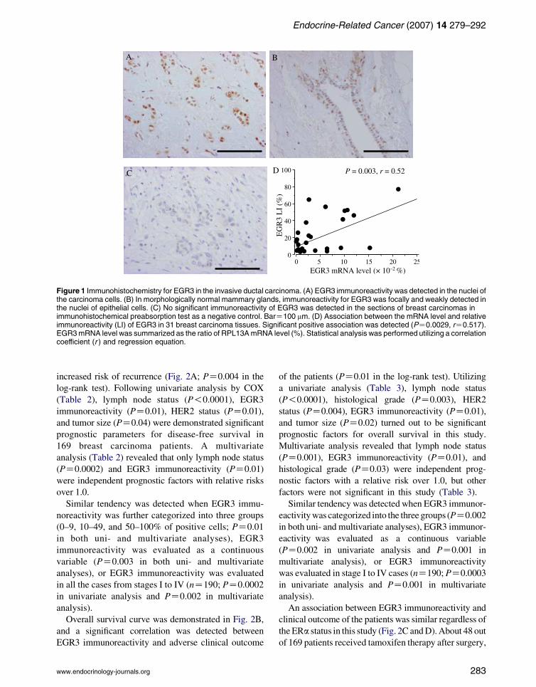
Figure 1 Immunohistochemistry for EGR3 in the invasive ductal carcinoma. (A) EGR3 immunoreactivity was detected in the nuclei ofthe carcinoma cells. (B) In morphologically normal mammary glands, immunoreactivity for EGR3 was focally and weakly detected inthe nuclei of epithelial cells. (C) No significant immunoreactivity of EGR3 was detected in the sections of breast carcinomas inimmunohistochemical preabsorption test as a negative control. BarZ100 mm. (D) Association between the mRNA level and relativeimmunoreactivity (LI) of EGR3 in 31 breast carcinoma tissues. Significant positive association was detected (PZ0.0029, rZ0.517).EGR3mRNA level was summarized as the ratio of RPL13AmRNA level (%). Statistical analysis was performed utilizing a correlationcoefficient (r ) and regression equation.
Endocrine-Related Cancer (2007) 14 279–292
increased risk of recurrence (Fig. 2A; PZ0.004 in the
log-rank test). Following univariate analysis by COX
(Table 2), lymph node status (P!0.0001), EGR3
immunoreactivity (PZ0.01), HER2 status (PZ0.01),
and tumor size (PZ0.04) were demonstrated significant
prognostic parameters for disease-free survival in
169 breast carcinoma patients. A multivariate
analysis (Table 2) revealed that only lymph node status
(PZ0.0002) and EGR3 immunoreactivity (PZ0.01)
were independent prognostic factors with relative risks
over 1.0.
Similar tendency was detected when EGR3 immu-
noreactivity was further categorized into three groups
(0–9, 10–49, and 50–100% of positive cells; PZ0.01
in both uni- and multivariate analyses), EGR3
immunoreactivity was evaluated as a continuous
variable (PZ0.003 in both uni- and multivariate
analyses), or EGR3 immunoreactivity was evaluated
in all the cases from stages I to IV (nZ190; PZ0.0002
in univariate analysis and PZ0.002 in multivariate
analysis).
Overall survival curve was demonstrated in Fig. 2B,
and a significant correlation was detected between
EGR3 immunoreactivity and adverse clinical outcome
www.endocrinology-journals.org
of the patients (PZ0.01 in the log-rank test). Utilizing
a univariate analysis (Table 3), lymph node status
(P!0.0001), histological grade (PZ0.003), HER2
status (PZ0.004), EGR3 immunoreactivity (PZ0.01),
and tumor size (PZ0.02) turned out to be significant
prognostic factors for overall survival in this study.
Multivariate analysis revealed that lymph node status
(PZ0.001), EGR3 immunoreactivity (PZ0.01), and
histological grade (PZ0.03) were independent prog-
nostic factors with a relative risk over 1.0, but other
factors were not significant in this study (Table 3).
Similar tendency was detected when EGR3 immunor-
eactivity was categorized into the three groups (PZ0.002
in both uni- and multivariate analyses), EGR3 immunor-
eactivity was evaluated as a continuous variable
(PZ0.002 in univariate analysis and PZ0.001 in
multivariate analysis), or EGR3 immunoreactivity
was evaluated in stage I to IV cases (nZ190; PZ0.0003
in univariate analysis and PZ0.001 in multivariate
analysis).
An association between EGR3 immunoreactivity and
clinical outcome of the patients was similar regardless of
the ERa status in this study (Fig. 2C and D). About 48 out
of 169 patients received tamoxifen therapy after surgery,
283

Table 1 Association between Early growth responsive gene 3 (EGR3 ) immunoreactivity and clinicopathological parameters in 190
breast carcinomas
EGR3 immunoreactivity
C(nZ99) K (nZ91) P value EGR3 LI P value
Age (years) 52.3G1.2 54.4G1.2 0.23 0.59
Menopausal status
Premenopausal 46 (24%) 38 (20%) 19.1G2.3
Postmenopausal 53 (28%) 53 (28%) 0.61 19.2G2.2 0.96
Stage
I 18 (10%) 26 (14%) 15.8G2.8
II 56 (30%) 51 (27%) 18.8G2.1
III 9 (5%) 9 (5%) 16.1G5.2
IV 16 (8%) 5 (3%) 0.07 30.6G6.4 0.07
Tumor size (mm) 34.6G2.7 30.0G3.4 0.29 0.20
Lymph node status
Positive 54 (28%) 29 (15%) 24.6G2.8
Negative 45 (24%) 62 (33%) 0.002 14.9G1.7 0.003
Distant metastasis
Positive 16 (8%) 5 (3%) 30.6G6.4
Negative 83 (44%) 86 (45%) 0.02 17.7G1.6 0.01
Histological grade
1 (well) 26 (14%) 24 (13%) 19.7G3.1
2 (moderate) 40 (21%) 31 (16%) 21.5G2.7
3 (poor) 33 (17%) 36 (19%) 0.60 16.3G2.6 0.48
Tubule formation
1 (O75%) 12 (6%) 22 (12%) 13.2G2.9
2 (10–75%) 23 (12%) 29 (15%) 14.9G2.5
3 (!10%) 64 (34%) 40 (21%) 0.01 23.2G2.4 0.20
ERa status
Positive 78 (41%) 57 (30%) 22.0G2.0
Negative 21 (11%) 34 (18%) 0.01 12.2G2.3 0.01
ERa LI (%) 47.9G3.4 37.3G3.7 0.02 (rZ0.23) 0.001
ERb LI (%) 16.2G2.3 15.2G2.1 0.82 0.37
PR LI (%) 42.9G3.3 36.9G3.6 0.23 0.33
HER2 status
Positive 29 (15%) 28 (15%) 15.4G2.5
Negative 70 (37%) 63 (33%) 0.82 20.8G2.0 0.23
Ki-67 LI (%) 24.8G1.8 24.4G1.9 0.89 0.93
EGR3 immunoreac-
tivity in recurrent
lesions (nZ13)
(rZ0.65) 0.02
Positive 9 (69%) 1 (8%)
Negative 1 (8%) 2 (15%) 0.03
Data are presented as meanG95% confidence interval (95% CI) or the number of cases with percentage. P values !0.05 wereconsidered significant, and described as boldface.
T Suzuki et al.: EGR3 in breast cancer
and these cases were ERa-positive breast cancers.
The disease-free and overall survival curves in these
patients were summarized in Fig. 2E and F respectively.
EGR3 immunoreactivity was also associated with an
increased risk of recurrence and worse prognosis in the
group of breast cancer patients who received tamoxifen
therapy (PZ0.01 and 0.03 in the log-rank test
respectively). An association between EGR3 immunor-
eactivity and clinical outcome of the patients was not
significantly changed regardless of the status of adjuvant
chemotherapy after surgery in this study (data not
shown).
284
Estrogen-mediated expression of EGR3 in MCF-7
breast carcinoma cells
As shown in Fig. 3A, EGR3 mRNA expression was
induced by estradiol in a dose-dependent manner in
MCF-7 cells. This induction became significant from
1 nM estradiol (P!0.001 versus control (non-treat-
ment with estradiol)), and EGR3 mRNA level of MCF-
7 cells treated with 10 nM estradiol (100.0G2.0%) was
14-fold increased when compared with the control
level (7.2G2.0%). The estradiol-mediated mRNA
expression of EGR3 was suppressed by addition of
www.endocrinology-journals.org

Figure 2 Disease-free and overall survival of 169 patients withbreast carcinoma according to EGR3 immunoreactivity(Kaplan–Meier method). (A and B) EGR3 immunoreactivity wassignificantly associated with an increased risk of recurrence(PZ0.0036, log-rank test) (A) and worse prognosis (PZ0.0090,log-rank test) (B). (C and D) EGR3 immunoreactivity wassignificantly correlated with a risk of recurrence regardlessof the ERa status (PZ0.0154 in ERa-positive cases (C), andPZ0.0348 in ERa-negative cases (D)). (E and F) EGR3immunoreactivity was significantly associated with anincreased risk of recurrence (PZ0.0123) (E) and worseprognosis (PZ0.0316) (F) in the 48 patients received tamoxifentherapy.
Table 2 Uni- and multivariate analyses of disease-free survival
in stages I–III breast cancer patients examined (nZ169)
Uni-
variateMultivariate
Variable P P
Relative risk
(95% CI)
Lymph node status
(positive/negative)
!0.0001 0.0002 3.8 (1.9–7.7)
EGR3 immunoreactivity
(positive/negative)
0.01 0.01 2.5 (1.3–4.8)
HER2 status
(positive/negative)
0.01 0.08
Tumor size
(S20 mm/!20 mm)
0.04 0.48
Ki-67 LI (S10/!10) 0.15
Adjuvant chemotherapy
(no/yes)
0.34
ERa status
(negative/positive)
0.44
Tamoxifen therapy (no/yes) 0.46
Histological grade (3/1, 2) 0.78
Data considered significant (P!0.05) in the univariate analyseswere described as boldface, and were examined in themultivariate analyses.
Table 3 Uni- and multivariate analyses of overall survival in
stages I–III breast cancer patients examined (nZ169)
Uni-
variateMultivariate
Variable P P
Relative risk
(95% CI)
Lymph node status
(positive/negative)
!0.0001 0.001 6.0 (2.2–16.5)
Histological grade (3/1, 2) 0.003 0.03 2.5 (1.1–4.7)
HER2 status (positive/
negative)
0.004 0.38
EGR3 immunoreactivity
(positive/negative)
0.01 0.01 3.0 (1.3–7.0)
Tumor size (S20 mm/
!20 mm)
0.02 0.28
Ki-67 LI (S10/!10) 0.11
Adjuvant chemotherapy
(no/yes)
0.21
Tamoxifen therapy
(no/yes)
0.37
ERa status (negative/
positive)
0.42
Data considered significant (P!0.05) in the univariate analyseswere described as boldface and were examined in themultivariate analyses.
Endocrine-Related Cancer (2007) 14 279–292
tamoxifen in a dose-dependent manner (Fig. 3B).
EGR3 mRNA level in MCF-7 cells treated with 10 nM
estradiol and 10 mM tamoxifen (21.8G6.2%) was
decreased into 22% of that treated with 10 nM estradiol
alone (100.0G2.0%), but its level remained
significantly higher (P!0.001 and threefold) than
the control level (neither estradiol nor tamoxifen;
7.2G2.0%). Tamoxifen (10 mM) alone did not signi-
ficantly change the EGR3 mRNA level in MCF-7 cells.
Estradiol also induced EGR3 mRNA expression in
LY-2 cells, a tamoxifen-resistant MCF-7 cell variant,
in a dose-dependent manner (Fig. 3C), which was
www.endocrinology-journals.org
significant from 10 pM estradiol (P!0.05 versus the
control level). The level of EGR3 mRNA in LY-2 cells
treated with 10 nM estradiol (1245G222%) was
31-fold higher than the control level (39.7G14.2%),
and was 12-fold higher than that in MCF-7 cells treated
285

Figure 3 Induction of EGR3 expression by estradiol in MCF-7cells. (A and C) MCF-7 (A) or LY-2 (C) cells were treated withindicated concentrations of estradiol for 3 days, and the EGR3mRNA was evaluated as the ratio of RPL13A mRNA level (%).(B and D) MCF-7 (B) or LY-2 (D) cells were treated withestradiol (10 nM) with indicated concentrations of tamoxifen for3 days, and the EGR3 mRNA was evaluated as the ratio ofRPL13A mRNA level (%). Data are presented as meanGS.D.(nZ4). *P!0.05 and ***P!0.001 versus control (no treatmentwith estradiol or tamoxifen for 3 days; left column) respectively.The statistical analyses were performed using a one-wayANOVA and Bonferroni test. (E and F) Immunoblotting forEGR3 in MCF-7 (E) and LY-2 (F) cells. Cells were treated withestradiol (10 nM) and/or tamoxifen (10 mM) for 3 days. Amountof protein loaded in each lane was 25 mg (immunoblotting forEGR3) or 5 mg (immunoblotting for b-actin). Immunointensity ofEGR3 in each sample was normalized to that of b-actin, andrelative immunointensity ratio of EGR3 was summarized as aratio compared with that of MCF-7 cells treated without estradiolor tamoxifen (left lane in E).
Figure 4 Effects of ICI 182 780 on EGR3 mRNA expression inMCF-7 cells. (A and C) MCF-7 (A) or LY-2 (C) cells were treatedwith indicated concentrations of ICI 182 780 for 3 days in theabsence of exogenous estradiol. (B and D) MCF-7 (B) or LY-2(D) cells were treated with indicated concentrations of estradioland ICI 182 780 (10 nM) for 3 days. Data are presented asmeanGS.D. (nZ4). *P!0.05, **P!0.01, and ***P!0.001versus control (non-treatment with ICI 182 780 (A and C) or10 nM ICI 182 780 in the absence of estradiol (B and D) for3 days (left column in each figure)). The statistical analyseswere performed using a one-way ANOVA and Bonferroni test.
T Suzuki et al.: EGR3 in breast cancer
under the same condition. Tamoxifen dose-depen-
dently suppressed the estradiol-mediated mRNA
expression of EGR3 in LY-2 cells (Fig. 3D). EGR3
mRNA level in LY-2 cells treated with 10 nM estradiol
and 10 mM tamoxifen (278G35.0%) was decreased
into 22% of that treated with 10 nM estradiol
alone (1,245G222%), but was still significantly higher
(P!0.001 and sevenfold) than the control level
(39.7G14.2%). Tamoxifen (10 mM) did not signi-
ficantly change the EGR3 mRNA level also in LY-2
cells. Similar tendency was detected at EGR3 protein
levels both in MCF-7 and LY-2 cells by immuno-
blotting analyses (Fig. 3E and F).
286
Pure anti-estrogen, ICI 182 780, alone did not
significantly change EGR3 mRNA level in MCF-7
cells (Fig. 4A). EGR3 mRNA level was slightly
increased by addition of estradiol in MCF-7 cells
under the treatment with 10 nM ICI 182 780, but
no significant association was detected (2.3-fold and
PZ0.06, 10 nM estradiol versus control (Fig. 4B;
non-treatment with estradiol).
On the other hand, as shown in Fig. 4C, ICI 182 780
alone significantly inhibited the EGR3 mRNA level of
LY-2 cells in a dose-dependent manner, and the EGR3
mRNA level of LY-2 cells treated with 10 mM
ICI 182 780 was decreased into 21% (7.8G3.6%,
P!0.001) of the basal level (non-treatment with ICI
182 780; 37.8G11.8%). When LY-2 cells were treated
with 10 nM ICI 182 780, the EGR3 mRNA level was
significantly induced by estradiol in a dose-dependent
manner (43-fold and P!0.001, 10 nM estradiol versus
control (Fig. 4D; non-treatment with estradiol).
Estradiol (10 nM) did not significantly induce the
EGR3 mRNA expression when MCF-7 or LY-2
cells were treated with 10 mM ICI 182 780 (0.9-fold
and PZ0.83 in MCF-7, and 1.2-fold and PZ0.77
in LY-2).
www.endocrinology-journals.org

Endocrine-Related Cancer (2007) 14 279–292
Increased invasion properties in Egr3-expressing
MCF-7 Tet-Off cells
In order to further characterize the biological functions
of EGR3 in breast carcinoma cells, we then employed
Eg-10 and Eg-11 Egr3-expressing transformants
derived from MCF-7 Tet-Off cells (Inoue et al.
2004). EGR3 mRNA levels of these transformants
were 57 and 540% in Eg-10 and Eg-11 respectively.
As a control, we used Ctl-7, which was stably
Figure 5 (A andB) Immunohistochemistry for EGR3 inEgr3-expressin thenuclei of Eg-11 cells (A), but not inCtl-7 cells (B). Immunohistocparaffin-embedded specimens. BarZ100 mm. (C–F) Migration assaapoptosis analysis (F) inEgr3-expressing cells. Eg-10,Eg-11, andCtrespectively, without or with the treatment of doxycyclin (50 ng/ml).Chemotaxicell was counted in the migration and invasion assays (Ccalculated and evaluated as a ratio (%) compared with that at 0 dayanalysis (E andF). Data are presented asmeanGS.D. (nZ4). An opea closed bar shows that under the treatment with doxycyclin. The stabsence and presence of doxycyclin using a one-way ANOVA and
www.endocrinology-journals.org
transfected with empty vector in the MCF-7 Tet-Offcells (Inoue et al. 2004), and the EGR3 mRNA levelwas negligible (2.2!10K3%). In the immunohisto-chemistry, EGR3 immunoreactivity was detected inthe nuclei of Eg-10 and Eg-11 cells (Fig. 5A), butnot in Ctl-7 cells (Fig. 5B). The EGR3 mRNAlevels decreased into negligible levels in both Eg-10(5.0!10K3%) and Eg-11 (1.4!10K2%) cells, whenthese cells were treated with doxycyclin (50 ng/ml)for 3 days.
ingMCF-7Tet-Off cells. Immunoreactivity of EGR3wasdetectedhemistrywas performedusing cell blocks from formalin-fixedandy (C), invasion assay (D), cell proliferation assay (E), andl-7 cellswere incubatedwith 6 h, 24 h, 3 days, and3daysat 37 8CA total number of cells on the lower surface of membrane ofand D). While, the cell number and apoptosis index wereafter the treatment in the cell proliferation assay and apoptosisn bar represents the value of cells treatedwithout doxycyclin, andatistical analyses were performed between the values in theBonferroni test, and **P!0.01 and ***P!0.001 respectively.
287

T Suzuki et al.: EGR3 in breast cancer
Figure 5C shows the result of migration assay in
Egr3-expressing MCF-7 cells. The number of migrated
cells was significantly higher in Eg-10 (P!0.001) and
Eg-11 (P!0.01) when compared with that in the
treatment with doxycyclin. However, a number of
migrated Ctl-7 cells were not significantly altered
between the absence and presence of doxycyclin.
Moreover, the number of invaded cells was signi-
ficantly higher in Eg-10 and Eg-11 (P!0.001) than
that under the treatment with doxycyclin, and it was
3.3-fold higher in Eg-10 and 3.7-fold in Eg-11
(Fig. 5D). Invasion property was not significantly
altered in Ctl-7 cells according to the treatment with
doxycyclin.
Cell proliferation (Fig. 5E) and apoptosis index
(Fig. 5F) of these three cells were not significantly
altered between the absence and presence of doxycy-
clin for 3 days.
Morphological features of Egr3-expressing
MCF-7 cells in athymic mice xenograft model
In order to study the biological roles of EGR3 in breast
carcinoma cells in vivo, we injected Eg-11 and Ctl-7
cells into female nude mice, and the tumor tissues were
resected after 2 months. As shown in Fig. 6A and B,
Figure 6 Histological features of Egr3-expressing MCF-7 cells in atremarkable in the Ctl-7 carcinoma and no invasive components wein clusters and trabeculae, and frequently invaded into a tissue wshows invasive components of Eg-11 cells in the adipose tissue astain. Tumor tissues were resected at 2 months after the injection,BarZ100 mm.
288
Ctl-7 showed a well-circumscribed tumor, and tubule
formation was remarkable in the carcinoma tissue.
Invasion into the surrounding tissue was not detected in
any of the cases examined. On the other hand, Eg-11
cells arranged in clusters and trabeculae with focal
glandular formation, and irregularly invaded into the
adjacent peritumoral tissue such as adipose tissue in all
of the cases examined (Fig. 6C and D). As shown in
Table 4, Eg-11 carcinoma tissues significantly demon-
strated invasion (PZ0.01) and less tubule formation
(PZ0.03) when compared with the Ctl-7 carcinoma
tissues. However, tumor volume, largest dimension
histologically determined, and Ki-67 LI of the tumor
were not significantly different between these two
types of carcinoma. The tumor volumes of Eg-11 and
Ctl-7 were monitored weekly, but no significant
changes were detected when compared with the
original volume (data not shown).
Discussion
In this study, EGR3 immunoreactivity was closely
correlated with the mRNA level, and was significantly
associated with the ERa status, but not with ERb, in the
breast carcinoma tissues. In the previous studies using
cDNA microarray, EGR3 mRNA was significantly
hymic mouse xenograft model. (A and B) Tubule formation wasre detected. (C and D) Eg-11 carcinoma cells mainly arrangedhich surrounded the primary carcinoma lesion (arrows). (D)djacent to the primary carcinoma site. Hematoxylin and eosinand were fixed in 10% formalin and embedded in paraffin wax.
www.endocrinology-journals.org

Table 4 Histological features of Early growth responsive gene 3 (Egr3 )-expressing MCF-7 cells injected into athymic mice
Eg-11 (nZ4) Ctl-7 (nZ4) P value
Tumor volumea (mm3) 399G101 357G74 0.75
Histologically determined largest
dimensiona (mm)
4.8G0.6 5.1G0.4 0.61
Tubule formation
1 (O75%) 0 (7%) 1 (13%)
2 (10–75%) 0 (11%) 3 (15%)
3 (!10%) 4 (31%) 0 (23%) 0.03
Invasive lesions
Present 4 (100%) 0 (0%)
Absent 0 (0%) 4 (100%) 0.01
Ki-67 LI of the tumora (%) 54.5G4.6 49.5G7.1 0.58
Tumor tissues were resected at 2 months after the injection, and were subsequently fixed in 10% formalin and embedded in paraffinwax. Tumor volume was evaluated by a formula for a semiellipsoid (4/3pr 3/2). P values !0.05 were considered significant anddescribed as boldface.aData are presented as meanGS.D. All other values represent the number of cases and percentage.
Endocrine-Related Cancer (2007) 14 279–292
(the relative value of more than 2.0) induced by
estradiol in various carcinoma cell lines derived from
breast (MCF-7 and MCF-7 c9), endometrium (Ishi-
kawa), ovary (SK-OV-3), and stomach (MKN-28;
Inoue et al. 2002, 2004, Hayashi et al. 2003). Induction
of EGR3 mRNA was detected at 6 h after estradiol
treatment (10 nM) and reached the maximal level at
24–72 h in MCF-7 cells by northern blot analysis
(Inoue et al. 2004), and ERE sequence was identified at
2.3 kb from the most upstream mRNA 5 0 end of Egr3
(Bourdeau et al. 2004). The biological estrogenic
actions are mainly mediated through ERa (Korach
1994, Hayashi et al. 2003), and MCF-7 cells highly
express ERa but low level of ERb (Vladusic et al.
2000). Therefore, results from our present study
suggest that EGR3 is expressed in the breast carcinoma
cells, mainly through ERa, as a result of estrogenic
action.
We also found that 21 out of 55 cases were
immunopositive for EGR3 in breast carcinoma tissues
negative for ERa (LI of !10%). This is partly because
EGR3 expression was induced by a low or undetect-
able level of ERa. However, EGR3 was also reported
to be induced by various factors, including mitogenic
stimulation (Patwardhan et al. 1991, O’Donovan et al.
1998, Mercier et al. 2001, Jouvert et al. 2002).
Therefore, factors other than estrogen may also be
partly involved in the regulation of EGR3 expression in
some breast carcinomas.
In our study, EGR3 immunoreactivity was
inversely associated with tubule formation, and
positively correlated with metastatic lesions of
lymph nodes or other organs in the breast carci-
nomas. Moreover, overexpression of Egr3 signi-
ficantly enhanced invasion properties in MCF-7
www.endocrinology-journals.org
cells in both in vitro study and nude mouse xenograft
model. Therefore, EGR3 is postulated to play a
pivotal role in carcinoma cell invasion mediated by
estrogens in breast carcinomas. Metastasis is the
major cause of treatment failure and death of
carcinoma patients, and it is a multi-step process
that involves not only invasion of carcinoma cells but
also lymphogenous and/or hematogenous spread and
cell proliferation in the metastatic sites. In our
present study, EGR3 immunoreactivity was not
associated with tumor size or Ki-67 LI in the breast
carcinoma tissues, and overexpression of Egr3 was
not necessarily involved in the cell proliferation or
apoptosis status in MCF-7 cells. Therefore, co-
operation with EGR3 and other factors may be
required for the metastasis of ER-positive breast
carcinoma. It awaits further examinations for the
detailed clarification of estrogen-mediated metastatic
process, because biological function of a great
majority of estrogen-responsive genes currently
remains unclear. However, for instance, cyclin D
(Steeg & Zhou 1998) and estrogen-responsive finger
protein (Efp; Urano et al. 2002, Suzuki et al. 2005b)
were shown to induce the estrogen-mediated prolifer-
ation in breast carcinoma cells, and histone deacetyl-
ase (HDAC) 6 were reported as a regulator of cell
motility in ER-positive breast carcinoma cells (Saji
et al. 2005).
Both uni- and multivariate analyses in our study
have demonstrated that EGR3 immunoreactivity is a
potent prognostic factor for both the recurrence and
overall survival in breast carcinoma patients, and
similar tendency was also detected in the patients who
received tamoxifen therapy. Estradiol is well known to
be locally produced and act in breast carcinomas
289

T Suzuki et al.: EGR3 in breast cancer
regardless of the menopausal status (Suzuki et al.
2005a). In the present in vitro experiments, tamox-
ifen suppressed estradiol-mediated expression of
EGR3 mRNA in a dose-dependent manner, but the
EGR3 mRNA level in MCF-7 cells treated with
estradiol and 10 mM tamoxifen was significantly higher
than the control level. Optimal concentrations of
tamoxifen were generally considered 10 nM to
10 mM in in vitro studies (Vendrell et al. 2005), and
serum concentration of tamoxifen was reported at
1.8 mM in patients who received high-dose tamoxifen
(320 mg), nevertheless 20 mg tamoxifen is usually
administrated in breast carcinoma patients. Therefore,
tamoxifen may not completely block the estradiol-
mediated EGR3 expression in the breast carcinoma
patients.
Regarding the molecular mechanism leading to
tamoxifen resistance, recent studies demonstrated that
breast carcinoma cells adapt by changing their
response to estradiol and developing an increased
sensitivity to the growth-stimulating action (Martin
et al. 2003, Berstein et al. 2004, Santen et al. 2004).
These processes are called ‘hypersensitivity to estra-
diol’, and the potential association with increased
concentrations of ERa and ER-mediated events is
proposed (Santen et al. 2001, Chan et al. 2002,
Vendrell et al. 2005). In this study, EGR3 mRNA
level in LY-2 cells was 5.5-fold higher than that in
MCF-7 cells in the absence of exogenous estradiol, but
it was dose-dependently decreased by ICI 182 780. In
addition, LY-2 cells showed marked amplitude of
estradiol-mediated EGR3 mRNA expression when
compared with MCF-7 cells. Therefore, it is suggested
that EGR3 expression is mainly mediated through ER
in LY-2 cells, and these findings of our present study
are possibly explained by the hypersensitivity to
estradiol in tamoxifen-resistant state of MCF-7 cells.
Considering that the EGR3 mRNA level in LY-2 cells
treated with estradiol and 10 mM tamoxifen was 2.8-
fold higher than that in MCF-7 cells treated with
estradiol alone, EGR3 may play an important role also
in the tamoxifen-resistant breast carcinoma patients.
Therefore, residual carcinoma cells following surgical
treatment in EGR3-positive breast carcinomas could
rapidly invade in the presence of local estrogens
regardless of the tamoxifen therapy, thereby resulting
in an increased recurrence and poor prognosis in these
patients.
In summary, EGR3 immunoreactivity was detected
in carcinoma cells in 52% of breast carcinoma tissues
in this study, and it was associated with its mRNA
level. EGR3 immunoreactivity was positively associ-
ated with lymph node status, distant metastasis into
290
other organs, ERa, or EGR3 immunoreactivity in the
recurrent lesions, and negatively correlated with tubule
formation. EGR3 immunoreactivity was significantly
associated with an increased risk of recurrence or
worse prognosis, regardless of the tamoxifen therapy.
Estradiol significantly induced EGR3 mRNA
expression in a dose-dependent manner in MCF-7
cells, which was markedly amplified in a tamoxifen-
resistant MCF-7 cell variant (LY-2). Tamoxifen
suppressed the estradiol-meditated induction of
EGR3 mRNA in a dose-dependent manner in these
cells, but tamoxifen could not inhibit its expression
completely. Egr3-expressing MCF-7 cells significantly
increased the invasion property, but not cell prolifer-
ation, both in vitro and in vivo experiments.
These results from our present study suggest that
EGR3 plays an important role in estrogen-meditated
invasion and is a potent prognostic factor in human
breast carcinoma.
Acknowledgements
We appreciate the skillful technical assistance of Ms
Chika Kaneko, Mr Katsuhiko Ono, Ms Toshie Suzuki,
and Ms Ikumi Miura (Department of Pathology,
Tohoku University School of Medicine). The authors
declare that there is no conflict of interest that would
prejudice the impartiality of this scientific work.
References
Albini A, Iwamoto Y, Kleinman HK, Martin GR, Aaronson
SA, Kozlowski JM & McEwan RN 1987 A rapid in vitro
assay for quantitating the invasive potential of tumor
cells. Cancer Research 47 3239–3245.
Berstein LM, Wang JP, Zheng H, Yue W, Conaway M &
Santen RJ 2004 Long-term exposure to tamoxifen induces
hypersensitivity to estradiol. Clinical Cancer Research 10
1530–1534.
Bourdeau V, Deschenes J, Metivier R, Nagai Y, Nguyen D,
Bretschneider N, Gannon F, White JH & Mader S 2004
Genome-wide identification of high-affinity estrogen
response elements in human and mouse. Molecular
Endocrinology 18 1411–1427.
Chan CM, Martin LA, Johnston SR, Ali S & Dowsett M 2002
Molecular changes associated with the acquisition of
oestrogen hypersensitivity in MCF-7 breast cancer
cells on long-term oestrogen deprivation.
Journal of Steroid Biochemistry and Molecular
Biology 81 333–341.
Dumoulin FL, Nischalke HD, Leifeld L, von dem Bussche A,
Rockstroh JK, Sauerbruch T & Spengler U 2000 Semi-
quantification of human C–C chemokine mRNAs with
www.endocrinology-journals.org

Endocrine-Related Cancer (2007) 14 279–292
reverse transcription/real-time PCR using multi-specific
standards. Journal of Immunological Methods 241
109–119.
Elston CW & Ellis IO 1991 Pathological prognostic factors in
breast cancer. I. The value of histological grade in breast
cancer. Experience from a large study with long-term
follow-up. Histopathology 19 403–410.
Goldhirsch A, Glick JH, Gelber RD, Coates AS, Thurlimann
B & Senn HJ 2005 Panel members. Meeting highlights:
international expert consensus on the primary therapy of
early breast cancer. Annals of Oncology 16 1569–1583.
Hayashi SI, Eguchi H, Tanimoto K, Yoshida T, Omoto Y,
Inoue A, Yoshida N & Yamaguchi Y 2003 The expression
and function of estrogen receptor alpha and beta in human
breast cancer and its clinical application. Endocrine-
Related Cancer 10 193–202.
Inoue A, Yoshida N, Omoto Y, Oguchi S, Yamori T, Kiyama
R & Hayashi S 2002 Development of cDNA microarray
for expression profiling of estrogen-responsive genes.
Journal of Molecular Endocrinology 29 175–192.
Inoue A, Omoto Y, Yamaguchi Y, Kiyama R & Hayashi SI
2004 Transcription factor EGR3 is involved in the
estrogen-signaling pathway in breast cancer cells.
Journal of Molecular Endocrinology 32 649–661.
Jouvert P, Dietrich JB, Aunis D & Zwiller J 2002 Differential
rat brain expression of EGR proteins and of the
transcriptional corepressor NAB in response to acute or
chronic cocaine administration. Neuromolecular
Medicine 1 137–151.
Korach KS 1994 Insights from the study of animals
lacking functional estrogen receptor. Science 266
1524–1527.
Martin LA, Farmer I, Johnston SR, Ali S, Marshall C &
Dowsett M 2003 Enhanced estrogen receptor (ER) alpha,
ERBB2, and MAPK signal transduction pathways operate
during the adaptation of MCF-7 cells to long term
estrogen deprivation. Journal of Biological Chemistry
278 30458–30468.
Mercier G, Turque N & Schumacher M 2001 Early activation
of transcription factor expression in Schwann cells by
progesterone. Brain Research. Molecular Brain Research
97 137–148.
O’Donovan KJ, Wilkens EP & Baraban JM 1998 Sequential
expression of Egr-1 and Egr-3 in hippocampal granule
cells following electroconvulsive stimulation. Journal of
Neurochemistry 70 1241–1248.
O’Donovan KJ, Tourtellotte WG, Millbrandt J & Baraban JM
1999 The EGR family of transcription-regulatory
factors: progress at the interface of molecular and
systems neuroscience. Trends in Neurosciences 22
167–173.
Paik S, Hartmann DP, Dickson RB & Lippman ME 1994
Antiestrogen resistance in ER positive breast cancer cells.
Breast Cancer Research and Treatment 31 301–307.
Patwardhan S, Gashler A, Siegel MG, Chang LC, Joseph LJ,
Shows TB, Le Beau MM & Sukhatme VP 1991 EGR3, a
www.endocrinology-journals.org
novel member of the Egr family of genes encoding
immediate-early transcription factors. Oncogene 6
917–928.
Saji S, Kawakami M, Hayashi S, Yoshida N, Hirose M,
Horiguchi S, Itoh A, Funata N, Schreiber SL, Yoshida M
et al. 2005 Significance of HDAC6 regulation via
estrogen signaling for cell motility and prognosis in
estrogen receptor-positive breast cancer. Oncogene 24
4531–4539.
Santen R, Jeng MH, Wang JP, Song R, Masamura S,
McPherson R, Santner S, Yue W & Shim WS 2001
Adaptive hypersensitivity to estradiol: potential
mechanism for secondary hormonal responses in breast
cancer patients. Journal of Steroid Biochemistry and
Molecular Biology 79 115–125.
Santen RJ, Song RX, Zhang Z, Yue W & Kumar R 2004
Adaptive hypersensitivity to estrogen: mechanism for
sequential responses to hormonal therapy in breast cancer.
Clinical Cancer Research 10 337S–345S.
Steeg PS & Zhou Q 1998 Cyclins and breast cancer. Breast
Cancer Research and Treatment 52 17–28.
Suzuki T, Miki Y, Nakamura Y, Moriya T, Ito K, Ohuchi N
& Sasano H 2005a Sex steroid-producing enzymes in
human breast cancer. Endocrine-Related Cancer 12
701–720.
Suzuki T, Urano T, Tsukui T, Horie-Inoue K, Moriya T,
Ishida T, Muramatsu M, Ouchi Y, Sasano H & Inoue S
2005b Estrogen-responsive finger protein as a new
potential biomarker for breast cancer. Clinical Cancer
Research 11 6148–6154.
Taniguchi S, Tatsuka M, Nakamatsu K, Inoue M, Sadano H,
Okazaki H, Iwamoto H & Baba T 1989 High invasiveness
associated with augmentation of motility in a fos-
transferred highly metastatic rat 3Y1 cell line. Cancer
Research 49 6738–6744.
Thomas DB 1984 Do hormones cause cancer? Cancer 53
595–604.
Tourtellotte WG & Milbrandt J 1998 Sensory ataxia and
muscle spindle agenesis in mice lacking the transcription
factor Egr3. Nature Genetics 20 87–91.
Tourtellotte WG, Keller-Peck C, Milbrandt J & Kucera J
2001 The transcription factor Egr3 modulates sensory
axon–myotube interactions during muscle spindle
morphogenesis. Developmental Biology 232 388–399.
Tsai MJ & O’Malley BW 1994 Molecular mechanisms of
action of steroid/thyroid receptor superfamily members.
Annual Review of Biochemistry 63 451–486.
Urano T, Saito T, Tsukui T, Fujita M, Hosoi T, Muramatsu
M, Ouchi Y & Inoue S 2002 Efp targets 14-3-3 sigma
for proteolysis and promotes breast tumour growth.
Nature 417 871–875.
Vandesompele J, De Preter K, Pattyn F, Poppe B, Van Roy N,
De Paepe A & Speleman F 2002 Accurate normalization
of real-time quantitative RT-PCR data by geometric
averaging of multiple internal control genes. Genome
Biology 3 research0034.1–11.
291

T Suzuki et al.: EGR3 in breast cancer
Vendrell JA, Bieche I, Desmetz C, Badia E, Tozlu S, Nguyen
C, Nicolas JC, Lidereau R & Cohen PA 2005 Molecular
changes associated with the agonist activity of hydroxy-
tamoxifen and the hyper-response to estradiol in hydroxy-
tamoxifen-resistant breast cancer cell lines. Endocrine-
Related Cancer 12 75–92.
Vihko R & Apter D 1989 Endogenous steroids in the
pathophysiology of breast cancer. CRC Critical Reviews
in Oncology/Hematology 9 1–16.
292
Vladusic EA, Hornby AE, Guerra-Vladusic FK,
Lakins J & Lupu R 2000 Expression an
d regulation of estrogen receptor beta in human
breast tumors and cell lines. Oncology Reports
7 157–167.
Xi H & Kersh GJ 2004 Early growth response gene 3
regulates thymocyte proliferation during the transition
from CD4KCD8K to CD4CCD8C. Journal of
Immunology 172 964–971.
www.endocrinology-journals.org
Related Documents