Early effects of transcatheter aortic valve implantation and aortic valve replacement on myocardial function and aortic valve hemodynamics: Insights from cardiovascular magnetic resonance imaging Gareth Crouch, MBBS, a,b Jayme Bennetts, MBBS, a,b Ajay Sinhal, MD, c Phillip J. Tully, PhD, b Darryl P. Leong, PhD, a Craig Bradbrook, MRS, c Amy L. Penhall, BSc, c Carmine G. De Pasquale, PhD, a,c Adhiraj Chakrabarty, MBBS, c,d Robert A. Baker, PhD, a,b and Joseph B. Selvanayagam, DPhil a,c,d Objectives: There remains a paucity of mechanistic data on the effect of transcatheter aortic valve implantation (TAVI) on early left and right ventricular function and quantitative aortic valve regurgitation. We sought to assess and compare the early effects on myocardial function and aortic valve hemodynamics of TAVI and aortic valve replacement (AVR) using serial cardiovascular magnetic resonance (CMR) imaging and echocardiography. Methods: A prospective comparison study of 47 patients with severe aortic stenosis undergoing either TAVI (n ¼ 26) or high-risk AVR (n ¼ 21). CMR (for left ventricle/right ventricle function, left ventricular mass, left atrial volume, and aortic regurgitation) was carried out before the procedure and early postprocedure ( < 14 days). Results: Groups were similar with respect to Society of Thoracic Surgeons score (TAVI, 7.7 vs AVR, 5.9; P ¼ .11). Preoperative left ventricular (TAVI, 69% 13% vs AVR, 73% 10%; P ¼ .10) and right ventricular (TAVI, 61% 11% vs AVR, 59% 8%; P ¼ .5) ejection fractions were similar. Postoperative left ventricular ejection fraction was preserved in both groups. In contrast, decline in right ventricular ejection fraction was more significant in the TAVI group (61%-54% vs 59%-58%; P ¼ .01). Postprocedure aortic regurgitant fraction was significantly greater in the TAVI group (16% vs 4%; P ¼ .001), as was left atrial size (110 vs 84 mL; P ¼ .02). Further analysis revealed a significant relationship between the increased aortic regurgitant fraction and greater left atrial size (P ¼ .006), and a trend toward association between the decline in right ventricle dysfunction and increased postprocedure aortic regurgitation (P ¼ .08). Conclusions: There was no significant difference in early left ventricular systolic function between techniques. Whereas right ventricle systolic function was preserved in the AVR group, it was significantly impaired early after TAVI, possibly reflecting a clinically important pathophysiologic consequence of paravalvular aortic regur- gitation. (J Thorac Cardiovasc Surg 2014;-:1-9) Aortic stenosis is an increasingly common condition asso- ciated with significant morbidity and mortality, and conse- quent public health burden. 1 Although surgical aortic valve replacement (AVR) has been the mainstay of treatment over the past 4 decades, transcatheter aortic valve implantation (TAVI) has emerged as an attractive option, especially in pa- tients with high or prohibitive surgical risk. 2-6 Despite widespread clinical use there remains limited data on hemodynamics early post-TAVI, which may have important prognostic implications. Paravalvular aortic regurgitation (PAR) in particular has emerged as a potentially important determinant of short- and medium-term clinical outcomes after TAVI 7 ; however, there is a paucity of mechanistic data on the effect of TAVI-related aortic regurgitation (AR) on ventricle structure and function. Furthermore, the inci- dence, extent, and temporal sequence of myocardial reversible and irreversible injury are poorly characterized after both TAVI and AVR. 8,9 The few studies to date that have examined early left ventricle (LV) function and aortic valve hemodynamic effects following TAVI have used transthoracic echocardiography (TTE), which has From Flinders University, a Adelaide, Australia; Departments of Cardiothoracic Sur- gery b and Cardiology, c Flinders Medical Centre, Adelaide, Australia; and the South Australian Health and Medical Research Institute, d Adelaide, Australia. Supported by unencumbered research grants from St Jude Medical and Edwards Lifesciences. Disclosures: Ajay Sinhal reports consulting fees from Edwards and lecture fees from Medtronic and Astra Zeneca. Carmine G. De Pasquale reports consulting fees from Novartis and lecture fees from Servier. Jayme Bennetts is an advisor for Medtronic. All other authors have nothing to disclose with regard to commercial support. Received for publication Sept 3, 2014; revisions received Oct 7, 2014; accepted for publication Oct 11, 2014. Address for reprints: Joseph B. Selvanayagam, DPhil, Department of Cardiology, Flinders Medical Centre, Flinders Dr, Bedford Park, South Australia, 5042 Australia (E-mail: [email protected]). 0022-5223/$36.00 Copyright Ó 2014 by The American Association for Thoracic Surgery http://dx.doi.org/10.1016/j.jtcvs.2014.10.064 The Journal of Thoracic and Cardiovascular Surgery c Volume -, Number - 1 Crouch et al Acquired Cardiovascular Disease ACD

Welcome message from author
This document is posted to help you gain knowledge. Please leave a comment to let me know what you think about it! Share it to your friends and learn new things together.
Transcript

Early effects of transcatheter aortic valve implantation and aorticvalve replacement on myocardial function and aortic valvehemodynamics: Insights from cardiovascular magneticresonance imaging
Gareth Crouch, MBBS,a,b Jayme Bennetts, MBBS,a,b Ajay Sinhal, MD,c Phillip J. Tully, PhD,b
Darryl P. Leong, PhD,a Craig Bradbrook, MRS,c Amy L. Penhall, BSc,c Carmine G. De Pasquale, PhD,a,c
Adhiraj Chakrabarty, MBBS,c,d Robert A. Baker, PhD,a,b and Joseph B. Selvanayagam, DPhila,c,d
Objectives: There remains a paucity of mechanistic data on the effect of transcatheter aortic valve implantation(TAVI) on early left and right ventricular function and quantitative aortic valve regurgitation. We soughtto assess and compare the early effects on myocardial function and aortic valve hemodynamics of TAVIand aortic valve replacement (AVR) using serial cardiovascular magnetic resonance (CMR) imaging andechocardiography.
Methods: A prospective comparison study of 47 patients with severe aortic stenosis undergoing either TAVI(n ¼ 26) or high-risk AVR (n ¼ 21). CMR (for left ventricle/right ventricle function, left ventricular mass,left atrial volume, and aortic regurgitation) was carried out before the procedure and early postprocedure(<14 days).
Results: Groups were similar with respect to Society of Thoracic Surgeons score (TAVI, 7.7 vs AVR, 5.9;P¼ .11). Preoperative left ventricular (TAVI, 69%" 13% vs AVR, 73%" 10%; P¼ .10) and right ventricular(TAVI, 61%" 11% vs AVR, 59%" 8%; P¼ .5) ejection fractions were similar. Postoperative left ventricularejection fraction was preserved in both groups. In contrast, decline in right ventricular ejection fraction was moresignificant in the TAVI group (61%-54% vs 59%-58%; P¼ .01). Postprocedure aortic regurgitant fraction wassignificantly greater in the TAVI group (16% vs 4%; P ¼ .001), as was left atrial size (110 vs 84 mL; P ¼ .02).Further analysis revealed a significant relationship between the increased aortic regurgitant fraction and greaterleft atrial size (P ¼ .006), and a trend toward association between the decline in right ventricle dysfunction andincreased postprocedure aortic regurgitation (P ¼ .08).
Conclusions: There was no significant difference in early left ventricular systolic function between techniques.Whereas right ventricle systolic function was preserved in the AVR group, it was significantly impaired earlyafter TAVI, possibly reflecting a clinically important pathophysiologic consequence of paravalvular aortic regur-gitation. (J Thorac Cardiovasc Surg 2014;-:1-9)
Aortic stenosis is an increasingly common condition asso-ciated with significant morbidity and mortality, and conse-quent public health burden.1 Although surgical aortic valvereplacement (AVR) has been the mainstay of treatment over
the past 4 decades, transcatheter aortic valve implantation(TAVI) has emerged as an attractive option, especially in pa-tients with high or prohibitive surgical risk.2-6 Despitewidespread clinical use there remains limited data onhemodynamics early post-TAVI, which may have importantprognostic implications.Paravalvular aortic regurgitation (PAR) in particular
has emerged as a potentially important determinant ofshort- and medium-term clinical outcomes after TAVI7;however, there is a paucity of mechanistic data on theeffect of TAVI-related aortic regurgitation (AR) onventricle structure and function. Furthermore, the inci-dence, extent, and temporal sequence of myocardialreversible and irreversible injury are poorly characterizedafter both TAVI and AVR.8,9 The few studies to date thathave examined early left ventricle (LV) function andaortic valve hemodynamic effects following TAVI haveused transthoracic echocardiography (TTE), which has
From Flinders University,a Adelaide, Australia; Departments of Cardiothoracic Sur-geryb and Cardiology,c Flinders Medical Centre, Adelaide, Australia; and the SouthAustralian Health and Medical Research Institute,d Adelaide, Australia.
Supported by unencumbered research grants from St Jude Medical and EdwardsLifesciences.
Disclosures: Ajay Sinhal reports consulting fees from Edwards and lecture fees fromMedtronic and Astra Zeneca. Carmine G. De Pasquale reports consulting fees fromNovartis and lecture fees from Servier. Jayme Bennetts is an advisor for Medtronic.All other authors have nothing to disclose with regard to commercial support.
Received for publication Sept 3, 2014; revisions received Oct 7, 2014; accepted forpublication Oct 11, 2014.
Address for reprints: Joseph B. Selvanayagam, DPhil, Department of Cardiology,Flinders Medical Centre, Flinders Dr, Bedford Park, South Australia, 5042Australia (E-mail: [email protected]).
0022-5223/$36.00Copyright ! 2014 by The American Association for Thoracic Surgeryhttp://dx.doi.org/10.1016/j.jtcvs.2014.10.064
The Journal of Thoracic and Cardiovascular Surgery c Volume -, Number - 1
Crouch et al Acquired Cardiovascular Disease
ACD
substantial limitations related to image quality andsensitivity, especially in a postprocedure setting.10 Further,TTE is severely restricted in right ventricle (RV) assess-ment, particularly in the perioperative setting.11
High-resolution cardiovascular magnetic resonance(CMR) imaging is a safe, noninvasive technique that allowsserial assessment of myocardial function and tissuecharacterization in the periprocedure setting.9,12-16 Givenits 3-dimensional nature and superior signal to noiseratio, cine CMR is highly superior to 2-dimensionalechocardiography and has become the gold standard inves-tigation for measurement of LV/RV volumes, mass, andfunction of both normal and abnormal ventricles.9,17-19
Finally, it allows quantitative assessment of both nativeand prosthetic aortic valve flow dynamics, includingparameters such as peak velocity and regurgitationvolume.20 Using this highly accurate and reproducibletechnique, in a single-center prospective cohort trial, wecompared the extent of perioperative LVand RVmyocardialinjury in patients undergoing TAVI with those undergoinghigh-risk AVR. Furthermore, we sought to characterize theassociation between postprocedure AR (as assessed byCMR imaging) with effects on the RVand LV. We hypothe-sized that TAVI would result in significantly less LVand RVmyocardial stunning compared with AVR. Furthermore, wespeculated that the occurrence of PAR would be correlatedwith worse myocardial function following the procedure.
METHODSEthics
This study was approved by the Human Research Ethics Committee ofFlinders Medical Centre (approval No. 237.11) and conducted in accor-dance with the Declaration of Helsinki. All patients gave written informedconsent.
Patient SelectionTranscatheter aortic valves remain an investigational device in Australia
and are approved for use in patients deemed inoperable or high-risk for
AVR. Patients with severe symptomatic aortic stenosis referred for inter-vention were assessed by the heart team, taking into consideration age, co-morbidities, risk scores, and frailty. A clinical decision then determinedwhether the individual proceeded to AVR or TAVI. Patients undergoingTAVI who were included were all from the high-risk cohort, with inoper-able patients excluded.
To limit bias, a high-risk cohort of patients with AVR was selected.Inclusion criteria were EuroSCORE>12 or Society of Thoracic Surgeonsrisk score>4, age>70 years, and subjective frailty assessment.21 Addition-ally, patients with a preprocedure LVejection fraction<45%were excludedto maintain homogeneity in periprocedural functional assessment.
Study ProtocolConsenting patients who met selection criteria had preprocedural inves-
tigation within 14 days of their procedure. This included biochemistry,echocardiography, and CMR. Postprocedure, patients had echocardiogra-phy and CMR within 14 days (Figure 1). All patients underwent preproce-dure coronary angiography.
AVR and TAVI TechniquesAll open surgery was performed by experienced cardiothoracic sur-
geons. Techniques were similar, being standard median sternotomy andcardiopulmonary bypass with diastolic arrest achieved by antegrade tepidblood cardioplegia. Three tissue valve prostheses were used: MedtronicMosaic (Medtronic Inc, Minneapolis, Minn), St Jude Medical Epic(St Jude Medical Inc, St Paul, Minn), and Trifecta (St Jude Medical Inc).
Transcatheter valve procedures were performed by an interventionalcardiologist (A.S.) and cardiac surgeon (J.B.) with implantation of80 valves before this study. All TAVIs were performed using combinedangiography and transoesophegeal echocardiography guidance. Allprocedures used the Edwards Sapien XT prosthesis (Edwards Lifesciences,Irvine, Calif) deployed transfemorally.
CMR Imaging ProtocolPatients were studied in a 1.5-T CMR imaging scanner (Siemens Aera,
Munich, Germany), and steady-state free precession cine images (TE/TR1.5/3.0 ms, flip angle 60#) were acquired in 2 long-axis and 8 to 10short-axis views. The acquisition of short-axis views began 1 cm belowthe level of the mitral valve insertion plane and continued in 1-cm incre-ments through the LV and RV.
Forward and regurgitant aortic flows were measured using through-planephase-contrast velocity mapping (free breathing, retrospective gating). Theimage plane was placed approximately 0.5 cm above the aortic valve atend-diastole, and maintained throughout the cardiac cycle. Commerciallyavailable gadolinium-based contrast agent (Gadovist 1.0, Gadobutrol; BayerHealthcare, Berlin, Germany) was given to those patients with a glomerularfiltration rate>45 mL/min/m2. Images were acquired after a 6-minute delaywith the use of an inversion-recovery segmented gradient echo sequence.Late gadolinium enhancement (LGE) images were acquired in identicallong- and short-axis planes to the cine images, except for the most apicalshort-axis slice, whichwas excluded. Regional wall motion analysis was per-formed by two blinded observers using a 16 segment American Heart Asso-ciation model and the following 0 to 4 scale, where 0 ¼ normal, 1 ¼ mildlyhypokinetic, 2 ¼ severely hypokinetic, 3 ¼ akinetic, and 4 ¼ dyskinetic.
Postprocessing AnalysisA standardizedmethod for analyzing and calculatingLVandRVvolumes
was used. These methods along with their reproducibility have been previ-ously published.22 Analysis was performed using commercially availablesoftware CMR42 (Circle Cardiovascular Imaging, Calgary, Canada). Leftatrial volume was measured using the biplane area-length method usingCMR 2- and 4-chamber views.23 Preoperative CMR LV short axis imageswere analyzed for regional wall motion abnormalities using a standard 17
Abbreviations and AcronymsANOVA ¼ analysis of varianceAR ¼ aortic regurgitationAVR ¼ aortic valve replacementCMR ¼ cardiovascular magnetic resonanceLGE ¼ late gadolinium enhancementLV ¼ left ventricleLVEDV ¼ left ventricular end-diastolic volumeLVESV ¼ left ventricular end-systolic volumePAR ¼ paravalvular aortic regurgitationQCA ¼ quantitative coronary angiographyRV ¼ right ventricleTAVI ¼ transcatheter aortic valve implantationTTE ¼ transthoracic echocardiography
Acquired Cardiovascular Disease Crouch et al
2 The Journal of Thoracic and Cardiovascular Surgery c - 2014
ACD
segment American Heart Association model grading wall motion from 0 to4 according to previously published criteria.14 Areas of myocardial infarc-tion were quantified using the 5 standard deviation method.24
Echocardiography ProtocolTTE was performed before and after the procedure concurrent with
CMR, to examine LV diastolic function. Vivid E9 ultrasound machineswere used and results were analyzed offline using EchoPAC PC Version7 (General Electric-Vingmed Ultrasound, Milwaukee, Wis). TransmitralE- and A-wave velocities were measured using pulse wave Doppler atthe mitral valve leaflet tips. Mitral E0 was measured using pulse wave Tis-sue Doppler by positioning the sample volume at the septal mitral annulus.
Coronary Angiography AnalysisSeverity of coronary artery lesions was quantified using quantitative
coronary angiography (QCA) by automated software and assessed visuallywhen not suitable for QCA. A cutoff of>50% diameter stenosis was usedto classify single, double, or triple vessel disease. Any lesion (>70% diam-eter stenosis by QCA) that was not revascularized was labelled incom-pletely revascularized.
Statistical AnalysisValues are expressed as mean " standard deviation or median (inter-
quartile range) as appropriate. All values were checked for normality andthe following transformations were made: LV ejection fraction beforeand after the procedure (inverse), left ventricular end-diastolic volume(LVEDV) (square root), left ventricular end-systolic volume (LVESV)(log), RV ejection fraction (RVEF) (square root), right ventricular end-diastolic volume (RVEDV) (square root), right ventricular end-systolic
volume (RVESV) (square root), brain natriuretic peptide (log), and regur-gitation fraction (log).
Descriptive comparisons (TAVI vs AVR) were made with independentsamples t test and the c2 or Fisher exact test as appropriate. Changes inindices of cardiac function were evaluated with repeated measures analysisof variance (ANOVA). The ANOVAmodel specified main effects for time,between group effects (TAVI vs AVR), and interaction effect time by group.ANOVA findings were corroborated using linear mixed effects models,with patient identity incorporated as a random effect. Additionally, in ancil-lary analysis adjustments were made for propensity score to reduce biasdue to the nonrandomized TAVI allocation. Calculated with logistic regres-sion, the propensity score determines the likelihood that a patient wastreated with TAVI based on demographic and comorbid conditions. Giventhe small numbers, Society of Thoracic Surgeons risk score was chosen asthe single characteristic that represented all factors influencing preferencefor TAVI (eg, age, reoperation, LV function, chronic obstructive pulmonarydisease, and cerebrovascular accident). Given the study focus on RV sys-tolic function, change in RV ejection fraction was analyzed with repeatedmeasures ANOVA in a full-factorial model and consideration was givento a number of indices selected a priori for their influence on RV ejectionfraction (age, N-terminal pro B-type natriuretic peptide, postprocedureLVejection fraction, and AR). Covariates were entered as interaction terms(covariate3 group3 time) in addition to main and between group effects.The area under the curve for serial high-sensitivity troponin T measure-ments was calculated according to Matthews and colleagues25 andanalyzed with Mann-Whitney U test. The difference in proportion of pa-tients with N-terminal pro B-type natriuretic peptide values exceedingnormal reference range of 125 ng/L was analyzed with Fisher exact test.Statistical analyses were performed with SPSS version 20.00 (IBM-SPSSInc, Armonk, NY) and STATA (StataCorp, College Station, Tex).
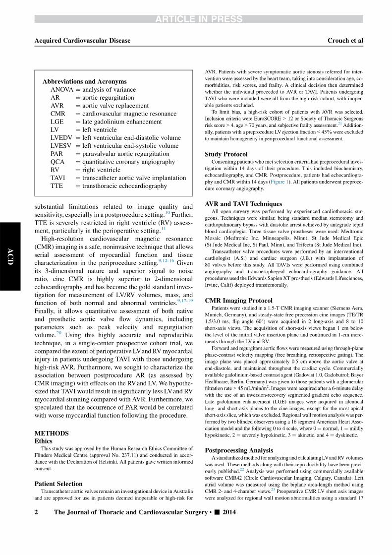
RESULTSBaseline Clinical CharacteristicsA total of 48 patients were recruited, with 1 exclusion due
pacemaker implantation, leaving 47 patients: 26 TAVI and21 AVR. The preoperative characteristics of the 2 groupswere similar with regards to Society of Thoracic Surgeonsrisk score and comorbidities (Table 1). Patients in theTAVI group were significantly older (85 " 6 years vs 80" 4 years; P $ .001). Overall surgical risk factors weresimilar between cohorts. The TAVI group did have a signif-icantly higher rate of previous cardiac surgery. All patientswith prior cardiac surgery had previously undergone coro-nary artery bypass surgery, with coronary angiographydemonstrating patent mammary artery grafts in each. Therewas no significant difference between groups whencomparing incompletely revascularized coronary territories(TAVI, 8 out of 78 vs AVR, 3 out of 63; P ¼ .20). All pa-tients proceeded as clinically indicated to TAVI or AVR in-tended group.There was no procedure-related mortality in either group.
All patients had a successful procedure and no patient in theTAVI group required conversion to AVR in the first 30 daysafter the procedure. Mean valve size was larger in the TAVIgroup (25.3 " 1.8 mm vs 22.6 " 1.9 mm; P<.001). In thesurgical group the mean cardiopulmonary bypass and cross-clamp times were 65 " 13 minutes and 50 " 11 minutes,respectively. Transcatheter patients required between 3 and4 bursts of rapid ventricular pacing during the procedure.
FIGURE 1. Study pathway. CMR, Cardiovascular magnetic resonance
imaging; TTE, transthoracic echocardiography; AVR, aortic valve replace-
ment TAVI, transcatheter aortic valve implantation.
Crouch et al Acquired Cardiovascular Disease
The Journal of Thoracic and Cardiovascular Surgery c Volume -, Number - 3
ACD

CMR Imaging ResultsAll patients completed pre- and postprocedure scans.
Three patients had inadequate image quality for assessmentof aortic valve flow. A total of 47 patients had scans beforeand after the procedure (100%) with 44 (94%) having com-plete imaging of LV/RV volumes, function, and aortic valveflow. Mean time to postoperative scan was 4.7 " 4 daysversus 5.8 " 2 days for TAVI and AVR patients, respec-tively (P ¼ .14).
Change in LV/RV Function After TAVI and AVRThe CMR changes over time are illustrated in Table 2. LV
ejection fraction was preserved postoperatively in bothgroups (TAVI, 68% " 12% vs AVR, 71% " 13%;P ¼ .31) with no significant difference in thegroup 3 time interaction (P ¼ .5). There was a significantdifference in LV remodeling between TAVI and CMRgroups. Despite similar baseline LVEDV (129 " 39 mLvs 118 " 27 mL, respectively; P ¼ .13), LV dilation wasgreater following TAVI than AVR (LVEDV, 133 " 42 mLvs 97 " 21 mL, respectively; P<.001; group 3 time inter-action, P<.001) (P<.01).
RV ejection fraction was similar between groups prepro-cedure (TAVI, 61% " 11% vs AVR, 59% " 8%;P ¼ .6). Unlike LV ejection fraction, RV ejection fractiondecreasedmore in the TAVI group than the AVRgroup (post-procedure 54% " 13% vs 58% " 8%; P ¼ .1;group 3 time interaction, P ¼ .008). This difference wasdriven by differences in RVESV between the groups.Whereas baseline RVESVs were similar between TAVIand AVR groups (41" 24 mL vs 40" 11 mL; P¼ .8), post-procedure RVESV was significantly larger in the TAVI
group (51 " 31 mL vs 39 " 14 mL: P ¼ .05;group3 time interaction, P ¼ .005). The significant associ-ations between ventricular remodeling/function and proce-dure type remained significant after adjusting for serumpro brain natriuretic peptide concentration.
Sensitivity analysis was performed to determine whetheradjustment for propensity score altered the Cine magneticresonance imaging results. TheLVEDVmain effectswere un-changed forgroup3 time interaction (P¼ .002).TheRVejec-tion fraction and RVESV main effects were also unchangedfor group3 time interaction (P ¼ .009 and P¼ .005).
Changes to Left Atrial VolumeLeft atrial volume was significantly greater at follow-up
in the TAVI group compared with the AVR group (110 "35 mL vs 84 " 24 mL; P ¼ .02). There was a trend towardmore favorable reverse remodeling in the AVR group(group 3 time interaction, P ¼ .09).
Changes to Aortic Valve FlowAortic valve flow parameters on CMR imaging are shown
in Table 3. Aortic stenosis, as assessed by aortic valve peakvelocity and aortic valve area improved over time and weresimilar between groups. Postprocedure regurgitant fractionrevealed significantly greater AR in the TAVI cohort whencompared the AVR group (TAVI, 16%" 16% vs AVR, 4%" 2%; P ¼ .005).
Myocardial InjuryRegional wall motion. Thirteen AVR patients (62%) and14 TAVI patients (54%) had normal regional wall motionin all segments preoperatively (P ¼ .56). The number of
TABLE 1. Baseline patient characteristics
Characteristic
Aortic valve
replacement (n ¼ 21)
Transcatheter aortic
valve replacement (n ¼ 26) P value
Age, y 79.6 " 4.0 84.6 " 5.6 <.001
Male 8 (38.1) 17 (65.4) .06
Society of Thoracic Surgeons risk score 5.9 " 3.4 7.7 " 3.9 .11
Brain natriuretic peptide 1084 " 1783 2854 " 4944 <.001
Hypertension 19 (90.5) 24 (92.3) .82
Hypercholesterolemia 15 (71.4) 21 (80.8) .51
Previous myocardial infarction 4 (19.0) 6 (23.1) 1.0
Previous percutaneous coronary intervention 3 (14.3) 8 (30.8) .30
Chronic obstructive pulmonary disease 9 (42.9) 11 (42.3) .97
Renal impairment 8 (38.1) 9 (34.6) .81
Atrial fibrillation 5 (23.8) 6 (23.1) .95
Diabetes 9 (42.9) 10 (38.5) .76
Redo — 11 (42.3) <.001
Previous cerebrovascular accident/transient ischemic attack 1 (4.8) 9 (34.6) .02
Angina 10 (47.6) 8 (30.8) .24
New York Heart Association functional class 2.7 " 0.6 2.5 " 0.8 .14
Pulmonary arterial hypertension 7 (33.3) 5 (17.2) .19
Nonrevascularized coronary artery disease 3 (14.3) 8 (30.8) .21
Values are presented as mean " standard deviation or n (%).
Acquired Cardiovascular Disease Crouch et al
4 The Journal of Thoracic and Cardiovascular Surgery c - 2014
ACD

patients in each groupwith 3 ormore dysfunctional segmentspreprocedure was also similar (TAVI 35% and AVR 29%;P ¼ .65). In addition, there was no difference in the changein segmental wall motion between groups (group 3 timeinteraction, P ¼ .42 for wall motion score index).Late gadolinium enahancement imaging. LGE imagingwas obtained preprocedure in 81% and 86% (P ¼ .47)and postprocedure in 76% and 86% (P ¼ .73) of AVRand TAVI patients, respectively. Preoperatively 9 TAVI pa-tients (45%) and 5 AVR patients (33%) demonstrated LGEin at least 1 LV slice (P ¼ .49). Postoperatively there wasnew LGE in 2 TAVI patients (11%) and 2 AVR patients(13%) (P ¼ .72).
Diastolic LV FunctionThe E/E0 ratio—an index of diastolic LV function—
showed a significant reduction in the AVR group compared
with the TAVI patients (baseline E/E0 in the AVR group was23" 9 vs 23" 8 in the TAVI group [P¼ .4]; postprocedureE/E0 19" 7 in the AVR group vs 23" 7, in the TAVI group[P ¼ .05]; group-time interaction [P ¼ .03]).
Post Hoc AnalysesFollowing the finding of right ventricular dysfunction in
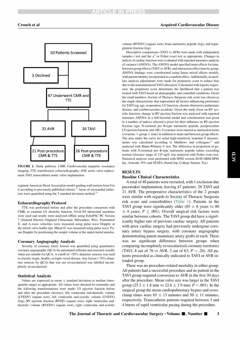
the TAVI cohort, ancillary statistical analyses assessed for in-fluences on the change in RV function. There was a trend to-ward a negative association between regurgitant fraction andRVejection fraction (coefficient,%0.10; 95% confidence in-terval,%0.22 to 0.011; P¼ .08) (Figure 2). To further assessthis finding the TAVI cohort was divided into those with mildor less paravalvular aortic regurgitation (PAR) and thosewithmoderate or greater PAR. Therewas no significant differencein preprocedure RVejection fraction between groups ($mild62% " 10% vs & moderate 59% " 13%; P ¼ .16). Post-procedure there was significantly worse RVejection fractionin those patients with moderate or greater PAR ($mild 58%" 11% vs&moderate 48%" 13%; P¼ .03). Additionallythere was a correlation between change in aortic valve regur-gitant fraction and left atrial dilation postprocedure (coeffi-cient, 2.65; 95% confidence interval, 0.75-4.55; P ¼ .006),although we were unable to show a significant associationbetween left atrial volume and RV ejection fraction. Giventhe potential for ischemia-induced RV dysfunction furtheranalysis of the angiographic QCA data were sought. Thesedemonstrated no difference in the number of incompletelyrevascularized territories (P ¼ .91).
DISCUSSIONThe principal findings from our study are that LVejection
fraction was equally well preserved in both TAVI and AVR
TABLE 2. Preoperative and postoperative ventricular function
Function Aortic valve replacement Transcatheter aortic valve implantation P for group 3 time interaction
Left ventricle ejection fraction .48
Preoperative 73% " 9% 69% " 13%
Postoperative 71% " 13% 68% " 12%
Left ventricular end-diastolic volume, mL <.01
Preoperative 118 " 27 129 " 39
Postoperative 97 " 21 133 " 42
Left ventricular end-systolic volume, mL .54
Preoperative 33 " 15 43 " 28
Postoperative 30 " 17 44 " 32
Right ventricle ejection fraction .01
Preoperative 59% " 8% 61% " 11%
Postoperative 58% " 8% 54% " 13%
Right ventricular end-diastolic volume, mL .28
Preoperative 98 " 22 104 " 37
Postoperative 95 " 27 108 " 42
Right ventricular end-systolic volume, mL <.01
Preoperative 40 " 11 41 " 24
Postoperative 39 " 14 51 " 31
Values are presented as mean " standard deviation.
TABLE 3. Aortic valve flow
Variable
Aortic valve
replacement
Transcatheter
aortic valve
implantation
P for
group 3 time
interaction
Forward volume, mL .07
Preoperative 59 " 24 57 " 22
Postopoperative 66 " 24 76 " 26
Regurgitant fraction .51
Preoperative 12% " 16% 18% " 15%
Postoperative 4.0% " 2.4% 16% " 16%
Peak velocity, m/s .71
Preoperative 3.6 " 0.81 3.9 " 1.0
Postoperative 2.6 " 0.62 2.4 " 0.49
Aortic valve area, cm2 .12
Preoperative 0.74 " 0.18 0.83 " 0.21
Postoperative 1.8 " 0.40 1.7 " 0.29
Values are presented as mean " standard deviation.
Crouch et al Acquired Cardiovascular Disease
The Journal of Thoracic and Cardiovascular Surgery c Volume -, Number - 5
ACD

groups early following the procedure and that RV systolicfunction was impaired early after TAVI, and potentiallyassociated with PAR. Given the prognostic importanceof RV dysfunction, our findings provide a potentialmechanism for the recent observation of increasedmortalityin the setting of post-TAVI AR.
No previous study has used CMR imaging for serialassessment and comparison of perioperative (<14 days)myocardial effects of AVR and TAVI. Previous CMR-based studies have conducted scans at preprocedure and 6months postprocedure offering a midterm outlook.26,27
Our study findings do not support the primary hypothesisthat LV function would be better preserved by TAVI whencompared with AVR. Imaging at similar intervalspostprocedure revealed no significant difference in globalleft ventricular systolic function (ie, LV ejection fraction).Indeed, volumetric measures of LV remodeling favoredAVR. Ewe and colleagues28 used TTE to assess early($48 hours) postprocedure LV global function in 147patients undergoing TAVI and compared this to aretrospective surgical control group of 99 patients. Theseinvestigators reported no significant change in LV ejectionfraction in patients with baseline LV ejection fraction> 50% who underwent TAVI.28 Earlier work by Claveland colleagues29 in a TAVI population with matchedsurgical controls revealed similar findings with nosignificant difference or change in LV function.29 Themajority of patients in our cohort had normal (>55%) LVejection fraction preprocedure (85% of TAVI and 95% ofAVR patients). Hence, despite the use of a more sensitiveand reproducible technique (ie, CMR), our findings onglobal LV function support previous echocardiographicstudies. Our hypothesis was based on an expected relativedecrease in LV ejection fraction in the AVR groupsecondary to the ischemic insult of cardiopulmonary bypass
and aortic crossclamping. Our results suggest that there isno left ventricular functional benefit of a transcatheterapproach despite the absence of ischemic insult.
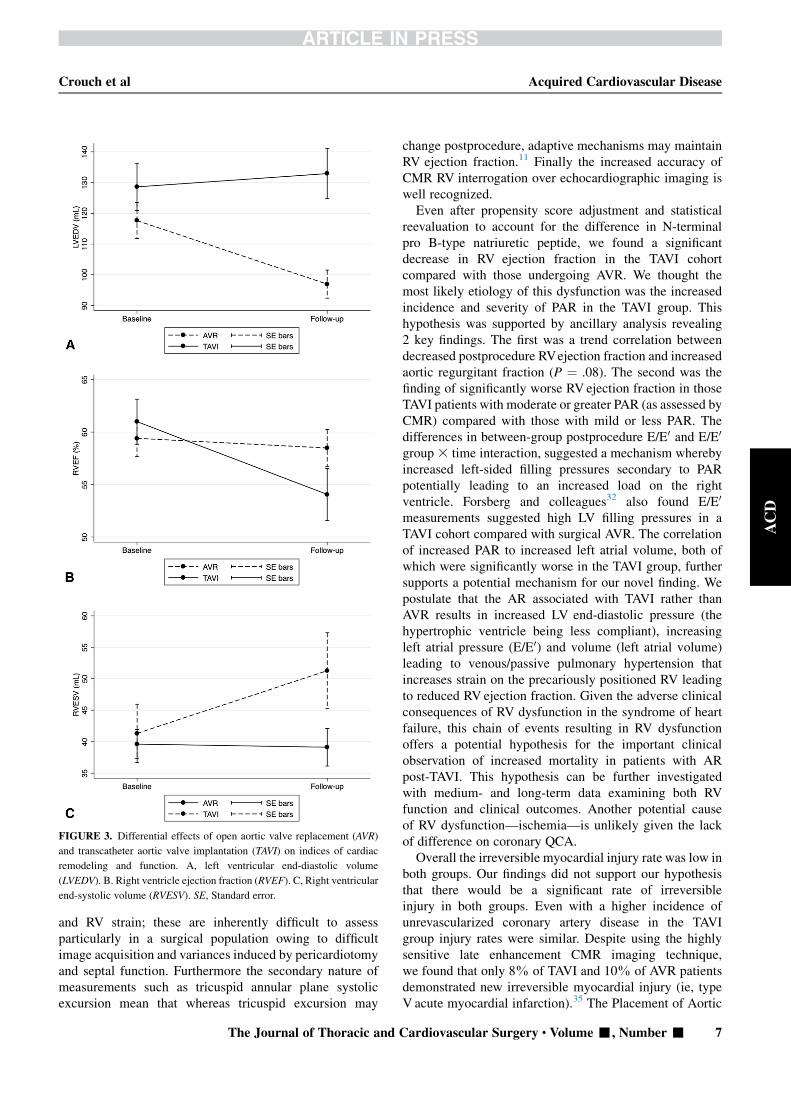
There were differences in LV volumetric indices betweenthe 2 groups, but contrary to our initial hypothesis, thisfavored the AVR group. Postoperative LVEDV demon-strated a trend favoring the AVR group (Figure 3) and thisreduction was partially matched by a nonsignificantreduction in LVESV in the AVR group accounting for thelack of change in LV ejection fraction. The presence ofreduced postoperative LVEDV can be explained simplyby the reduced outflow obstruction leading to a lowerend-diastolic pressure and subsequently reduced end-diastolic volume. Successful resolution of aortic stenosiswould be expected to have this effect in both groups;however, this was not seen in the TAVI group. We proposethis may be an effect of acutely increased postprocedure ARincreasing end-diastolic pressure. This may offset thebenefit that aortic valve obstruction resolution providesand result in static LV volumetric measures after TAVI.Previous echocardiographic studies in TAVI cohorts havenot shown any significant reduction in LVEDV (or LVend-diastolic diameter) early postprocedure in eithernormal or impaired LV ejection fraction.28,30 The nextgeneration of TAVI devices may confirm the mechanismof volumetric changes post TAVI should they decrease theincidence and severity of postprocedure AR.
Despite the fact that both groups had preserved RVfunction at baseline, there was significantly greater RVdysfunction in the TAVI group. RV ejection fractiondecreased significantly in TAVI patients compared withthe AVR patients, driven largely by an increase in RVESVin TAVI patients. The RV is recognized as a thin-walled,highly compliant structure with a primary compensatorymechanism to injury—irrespective of the cause—ofdilation.31 The AVR cohort demonstrated a decrease inboth RVEDV and RVESV. To the best of our knowledgethis study is the first to report a decrease in RV ejectionfraction among TAVI patients when compared withAVR and tellingly the only report of CMR-derived RVparameters. None of the large trials or registries has focusedon RV function and several small echocardiographic studiesdid not report significant changes.7,32 Kempny andcolleagues33 and Zhao and colleagues34 had findingscontradictory to ours with echocardiography assessed RVfunction preserved in a TAVI cohort and decreased in asurgical control group. The discrepancy between findingsis likely explained by several methodologic limitations,namely unmatched baseline characteristics, mixture of me-chanical and bioprosthetic valves, mixture of transfemoraland transapical valves, and concomitant bypass surgery inthe surgical group. Moreover, the echocardiographic studiesreported elsewhere largely used secondary measures of RVfunction such as tricuspid annular plane systolic excursion
FIGURE 2. Graphic representation of the relationship between aortic
regurgitation assessed by cardiovascular magnetic resonance imaging
and right ventricle ejection fraction (RVEF) assessed by cardiovascular
magnetic resonance imaging.
Acquired Cardiovascular Disease Crouch et al
6 The Journal of Thoracic and Cardiovascular Surgery c - 2014
ACD
and RV strain; these are inherently difficult to assessparticularly in a surgical population owing to difficultimage acquisition and variances induced by pericardiotomyand septal function. Furthermore the secondary nature ofmeasurements such as tricuspid annular plane systolicexcursion mean that whereas tricuspid excursion may
change postprocedure, adaptive mechanisms may maintainRV ejection fraction.11 Finally the increased accuracy ofCMR RV interrogation over echocardiographic imaging iswell recognized.Even after propensity score adjustment and statistical
reevaluation to account for the difference in N-terminalpro B-type natriuretic peptide, we found a significantdecrease in RV ejection fraction in the TAVI cohortcompared with those undergoing AVR. We thought themost likely etiology of this dysfunction was the increasedincidence and severity of PAR in the TAVI group. Thishypothesis was supported by ancillary analysis revealing2 key findings. The first was a trend correlation betweendecreased postprocedure RVejection fraction and increasedaortic regurgitant fraction (P ¼ .08). The second was thefinding of significantly worse RV ejection fraction in thoseTAVI patients with moderate or greater PAR (as assessed byCMR) compared with those with mild or less PAR. Thedifferences in between-group postprocedure E/E0 and E/E0
group 3 time interaction, suggested a mechanism wherebyincreased left-sided filling pressures secondary to PARpotentially leading to an increased load on the rightventricle. Forsberg and colleagues32 also found E/E0
measurements suggested high LV filling pressures in aTAVI cohort compared with surgical AVR. The correlationof increased PAR to increased left atrial volume, both ofwhich were significantly worse in the TAVI group, furthersupports a potential mechanism for our novel finding. Wepostulate that the AR associated with TAVI rather thanAVR results in increased LV end-diastolic pressure (thehypertrophic ventricle being less compliant), increasingleft atrial pressure (E/E0) and volume (left atrial volume)leading to venous/passive pulmonary hypertension thatincreases strain on the precariously positioned RV leadingto reduced RV ejection fraction. Given the adverse clinicalconsequences of RV dysfunction in the syndrome of heartfailure, this chain of events resulting in RV dysfunctionoffers a potential hypothesis for the important clinicalobservation of increased mortality in patients with ARpost-TAVI. This hypothesis can be further investigatedwith medium- and long-term data examining both RVfunction and clinical outcomes. Another potential causeof RV dysfunction—ischemia—is unlikely given the lackof difference on coronary QCA.Overall the irreversible myocardial injury rate was low in
both groups. Our findings did not support our hypothesisthat there would be a significant rate of irreversibleinjury in both groups. Even with a higher incidence ofunrevascularized coronary artery disease in the TAVIgroup injury rates were similar. Despite using the highlysensitive late enhancement CMR imaging technique,we found that only 8% of TAVI and 10% of AVR patientsdemonstrated new irreversible myocardial injury (ie, typeV acute myocardial infarction).35 The Placement of Aortic
FIGURE 3. Differential effects of open aortic valve replacement (AVR)
and transcatheter aortic valve implantation (TAVI) on indices of cardiac
remodeling and function. A, left ventricular end-diastolic volume
(LVEDV). B. Right ventricle ejection fraction (RVEF). C, Right ventricular
end-systolic volume (RVESV). SE, Standard error.
Crouch et al Acquired Cardiovascular Disease
The Journal of Thoracic and Cardiovascular Surgery c Volume -, Number - 7
ACD

Transcatheter Valves (PARTNER) trial reported infarctrates of 0% and 0.6% for TAVI and AVR, respectively, us-ing clinical and biochemical data; and the higher rate re-flects the higher sensitivity of the CMR technique. Theclinical significance of these small areas of myocardialinjury is uncertain, although our previous work in a popula-tion undergoing coronary artery bypass grafting indicatesthat it may not be entirely benign.36
LimitationsThe most significant limitation of this trial is the
nonrandomized design. Regulatory approval limitationsmeanTAVI remains utilized largely in the inoperable cohort,limiting randomization. As expected, the TAVI group mem-bers were significantly older because they consisted of bothtechnically andmedically high-risk patients. The higher rateof reoperative cases in the TAVI groupwas attributable to theevolution of TAVI as the preferred treatment for patientswith patent bypass grafts and multiple comorbidities. Thequestion of increased susceptibility to periproceduralischemic injury in these patients was addressed with thefindings that neitherQCAor regionalwallmotion abnormal-ity assessment demonstrated differences between groups.Despite the difference in age and redo status, the groupswere matched regarding Society of Thoracic Surgeons riskscore, as they were for CMR-derived preprocedure cardiacindices. Moreover, our attempt to adjust for risk factors us-ing propensity score and post-hoc mixed effects regressionmodeling demonstrated no change in the significant find-ings. Overall the 2 groups were sufficiently matched andpost-hoc analysis was sufficiently robust that results seenin this study reflect the effects of procedural technique ratherthan preexisting patient characteristics.
The small cohort numbers is also an obvious limitation.One of the inherent advantages of CMR over echocardiog-raphy or other modalities is the accuracy and reducedobserver variability it provides, allowing smaller researchcohorts. Although the ability to postulate a link betweenAR and RV dysfunction is hampered by our small numbers,we believe this new observation warrants focused study in afuture mechanistic study. Finally, the inherent effects of car-diovascular loading states need to be considered. Patientsunderwent scanning during the early morning and allwere at least 48 hours postprocedure with mean time of 6to 7 days. The similarity in time of day and time postproce-dure to allow for settling of fluid shifts we believeminimizes the variation from cardiovascular loading. Thiswould minimize the influence on factors such as left atrialvolume. We do acknowledge the difficulties in measuringleft atrial volume, regardless of the methodology.
CONCLUSIONSPatients undergoing either AVR or TAVI have preserved
LV function after the procedure. Furthermore although
irreversible injury rates were very low in both groups,they are higher than has been reported using clinical and/or biochemical methods in large trials.
Our results demonstrate for the first time that TAVI isassociated with early RV dysfunction. This may reflectthe higher incidence of AR with TAVI and explain therecent observation of increased long-term mortality in thissetting.
The authors thank Suchi Grover for assistance with the second-ary CMR analysis.
References1. Dweck MR, Boon NA, Newby DE. Calcific aortic stenosis: a disease of the valve
and the myocardium. J Am Coll Cardiol. 2012;60:1854-63.2. Beckmann A, Hamm C, Figulla HR, Cremer J, Kuck KH, Lange R, et al. The
German Aortic Valve Registry (GARY): a nationwide registry for patients under-going invasive therapy for severe aortic valve stenosis. Thorac Cardiovasc Surg.2012;60:319-25.
3. Makkar RR, Fontana GP, Jilaihawi H, Kapadia S, Pichard AD, Douglas PS, et al.Transcatheter aortic-valve replacement for inoperable severe aortic stenosis. NEngl J Med. 2012;366:1696-704.
4. Onorati F, D’Errigo P, Grossi C, Barbanti M, Ranucci M, Covello DR, et al. Ef-fect of severe left ventricular systolic dysfunction on hospital outcome aftertranscatheter aortic valve implantation or surgical aortic valve replacement: re-sults from a propensity-matched population of the Italian OBSERVANT multi-center study. J Thorac Cardiovasc Surg. 2014;147:568-75.
5. Smith CR, Leon MB, Mack MJ, Miller DC, Moses JW, Svensson LG, et al.Transcatheter versus surgical aortic-valve replacement in high-risk patients. NEngl J Med. 2011;364:2187-98.
6. Tice JA, Sellke FW, Schaff HV. Transcatheter aortic valve replacement in pa-tients with severe aortic stenosis who are at high risk for surgical complications:summary assessment of the California Technology Assessment Forum. J ThoracCardiovasc Surg. 2014;148:482-91.e6.
7. Kodali SK, Williams MR, Smith CR, Svensson LG, Webb JG, Makkar RR, et al.Two-year outcomes after transcatheter or surgical aortic-valve replacement. NEngl J Med. 2012;366:1686-95.
8. Bondarenko O, Beek AM, Nijveldt R, McCann GP, van Dockum WG,Hofman MB, et al. Functional outcome after revascularization in patients withchronic ischemic heart disease: a quantitative late gadolinium enhancementCMR study evaluating transmural scar extent, wall thickness and periproceduralnecrosis. J Cardiovasc Magn Reson. 2007;9:815-21.
9. Selvanayagam JB, Petersen SE, Francis JM, RobsonMD, Kardos A, Neubauer S,et al. Effects of off-pump versus on-pump coronary surgery on reversible andirreversible myocardial injury: a randomized trial using cardiovascular magneticresonance imaging and biochemical markers. Circulation. 2004;109:345-50.
10. Goncalves A, Marcos-Alberca P, Almeria C, Feltes G, Rodriguez E, Hernandez-Antolin RA, et al. Acute left ventricle diastolic function improvement after trans-catheter aortic valve implantation. Eur J Echocardiogr. 2011;12:790-7.
11. Lindqvist P, Calcutteea A, Henein M. Echocardiography in the assessment ofright heart function. Eur J Echocardiogr. 2008;9:225-34.
12. Pegg TJ, Selvanayagam JB, Karamitsos TD, Arnold RJ, Francis JM, Neubauer S,et al. Effects of off-pump versus on-pump coronary artery bypass grafting onearly and late right ventricular function. Circulation. 2008;117:2202-10.
13. Pegg TJ, Maunsell Z, Karamitsos TD, Taylor RP, James T, Francis JM, et al. Util-ity of cardiac biomarkers for the diagnosis of type V myocardial infarction aftercoronary artery bypass grafting: insights from serial cardiac MRI. Heart. 2011;97:810-6.
14. Selvanayagam JB, Kardos A, Francis JM, Wiesmann F, Petersen SE, Taggart DP,et al. Value of delayed-enhancement cardiovascular magnetic resonance imagingin predicting myocardial viability after surgical revascularization. Circulation.2004;110:1535-41.
15. Selvanayagam JB, Porto I, Channon K, Petersen SE, Francis JM, Neubauer S,et al. Troponin elevation after percutaneous coronary intervention directly repre-sents the extent of irreversible myocardial injury: insights from cardiovascularmagnetic resonance imaging. Circulation. 2005;111:1027-32.
16. Selvanayagam JB, Cheng AS, Jerosch-Herold M, Rahimi K, Porto I, van Gaal W,et al. Effect of distal embolization on myocardial perfusion reserve after
Acquired Cardiovascular Disease Crouch et al
8 The Journal of Thoracic and Cardiovascular Surgery c - 2014
ACD
percutaneous coronary intervention: a quantitative magnetic resonance perfusionstudy. Circulation. 2007;116:1458-64.
17. Chandran KB, Udaykumar HS, Reinhardt JM. Image-based computationalmodeling of the human circulatory and pulmonary systems: methods and appli-cations. Iowa City, IA: Springer; 2010.
18. Pennell DJ. Ventricular volume and mass by CMR. J Cardiovasc Magn Reson.2002;4:507-13.
19. Pennell DJ. Cardiovascular magnetic resonance. Circulation. 2010;121:692-705.20. Myerson SG. Heart valve disease: investigation by cardiovascular magnetic reso-
nance. J Cardiovasc Magn Reson. 2012;14:7.21. Michel P, Roques F, Nashef SA. Logistic or additive EuroSCORE for high-risk
patients? Eur J Cardiothorac Surg. 2003;23:684-7; discussion 687.22. Leong DP, Chakrabarty A, Shipp N, Molaee P, Madsen PL, Joerg L, et al. Effects
of myocardial fibrosis and ventricular dyssynchrony on response to therapy innew-presentation idiopathic dilated cardiomyopathy: insights from cardiovascu-lar magnetic resonance and echocardiography. Eur Heart J. 2012;33:640-8.
23. Lang RM, Bierig M, Devereux RB, Flachskampf FA, Foster E, Pellikka PA, et al.Recommendations for chamber quantification. Eur J Echocardiogr. 2006;7:79-108.
24. Amado LC, Gerber BL, Gupta SN, Rettmann DW, Szarf G, Schock R, et al. Accu-rate and objective infarct sizing by contrast-enhanced magnetic resonance imagingin a canine myocardial infarction model. J Am Coll Cardiol. 2004;44:2383-9.
25. Matthews JN, Altman DG, Campbell MJ, Royston P. Analysis of serial measure-ments in medical research. BMJ. 1990;300:230-5.
26. Fairbairn TA, Steadman CD, Mather AN, Motwani M, Blackman DJ, Plein S,et al. Assessment of valve haemodynamics, reverse ventricular remodellingand myocardial fibrosis following transcatheter aortic valve implantationcompared to surgical aortic valve replacement: a cardiovascular magnetic reso-nance study. Heart. 2013;99:1185-91.
27. La Manna A, Sanfilippo A, Capodanno D, Salemi A, Cadoni A, Cascone I, et al.Left ventricular reverse remodeling after transcatheter aortic valve implanta-
tion: a cardiovascular magnetic resonance study. J Cardiovasc Magn Reson.2013;15:39.
28. Ewe SH, Ajmone Marsan N, Pepi M, Delgado V, Tamborini G, Muratori M, et al.Impact of left ventricular systolic function on clinical and echocardiographic out-comes following transcatheter aortic valve implantation for severe aortic steno-sis. Am Heart J. 2010;160:1113-20.
29. Clavel MA,Webb JG, Pibarot P, Altwegg L, Dumont E, Thompson C, et al. Com-parison of the hemodynamic performance of percutaneous and surgical bio-prostheses for the treatment of severe aortic stenosis. J Am Coll Cardiol. 2009;53:1883-91.
30. Kempny A, Diller GP, Kaleschke G, Orwat S, Funke A, Schmidt R, et al. Impactof transcatheter aortic valve implantation or surgical aortic valve replacement onright ventricular function-the reply. Heart. 2012;98:1299-304.
31. Sidebotham D. Cardiothoracic critical care. Philadelphia: Butterworth-Heine-mann; 2007.
32. Forsberg LM, Tamas E, Vanky F, Nielsen NE, Engvall J, Nylander E. Left andright ventricular function in aortic stenosis patients 8 weeks post-transcatheteraortic valve implantation or surgical aortic valve replacement. Eur J Echocar-diogr. 2011;12:603-11.
33. Kempny A, Diller GP, Kaleschke G, Orwat S, Funke A, Schmidt R, et al. Impactof transcatheter aortic valve implantation or surgical aortic valve replacement onright ventricular function. Heart. 2012;98:1299-304.
34. Zhao Y, Lindqvist P, Nilsson J, Holmgren A, Naslund U, Henein MY. Trans-cath-eter aortic valve implantation–early recovery of left and preservation of rightventricular function. Interact Cardiovasc Thorac Surg. 2011;12:35-9.
35. Thygesen K, Alpert JS, Jaffe AS, Simoons ML, Chaitman BR, White HD.Third universal definition of myocardial infarction. Eur Heart J. 2012;33:2551-67.
36. Rahimi K, Banning AP, Cheng AS, Pegg TJ, Karamitsos TD, Channon KM, et al.Prognostic value of coronary revascularisation-related myocardial injury: a car-diac magnetic resonance imaging study. Heart. 2009;95:1937-43.
Crouch et al Acquired Cardiovascular Disease
The Journal of Thoracic and Cardiovascular Surgery c Volume -, Number - 9
ACD
000 Early effects of transcatheter aortic valve implantation and aortic valvereplacement on myocardial function and aortic valve hemodynamics: Insightsfrom cardiovascular magnetic resonance imagingGareth Crouch, MBBS, Jayme Bennetts, MBBS, Ajay Sinhal, MD, Phillip J. Tully, PhD, Darryl P.
Leong, PhD, Craig Bradbrook, MRS, Amy L. Penhall, BSc, Carmine G. De Pasquale, PhD, Adhiraj
Chakrabarty, MBBS, Robert A. Baker, PhD, and Joseph B. Selvanayagam, DPhil, Adelaide,
Australia
The effect of transcatheter aortic valve implantation on ventricle function and valve hemodynamics
was assessed by cardiac magnetic resonance imaging. Our data demonstrated similar effects on
global left ventricle function but worse global right ventricle function in the transcatheter aortic
valve implantation group. Further analysis revealed an association between worse right ventricle
dysfunction, increased left atrial volume, and increased paravalvular leak in the transcatheter aortic
valve implantation group.
Acquired Cardiovascular Disease Crouch et al
The Journal of Thoracic and Cardiovascular Surgery c - 2014
ACD
Related Documents