Biomaterials 27 (2006) 4192–4203 Early bone apposition in vivo on plasma-sprayed and electrochemically deposited hydroxyapatite coatings on titanium alloy Hao Wang a, , Noam Eliaz b , Zhou Xiang c,d , Hu-Ping Hsu c,d , Myron Spector c,d , Linn W. Hobbs a a Department of Materials Science and Engineering, Massachusetts Institute of Technology, 77 Massachusetts Ave., Cambridge, MA 02139, USA b Department of Solid Mechanics, Materials and Systems, Tel-Aviv University, Ramat-Aviv, Tel-Aviv 69978, Israel c Tissue Engineering, VA Boston Healthcare System, Boston Campus, 150 S. Huntington Ave., Boston, MA 02130, USA d Department of Orthopaedic Surgery, Brigham and Women’s Hospital, Harvard Medical School, 75 Francis Street, Boston, MA 02115, USA Received 23 January 2006; accepted 21 March 2006 Abstract Three different implants, bare Ti–6Al–4V alloy, Ti–6Al–4V alloy coated with plasma-sprayed hydroxyapatite (PSHA), and Ti–6Al–4V alloy coated with electrochemically deposited hydroxyapatite (EDHA), were implanted into canine trabecular bone for 6 h, 7, and 14 days, respectively. Environmental scanning electron microscopy study showed that PSHA coatings had higher bone apposition ratios than those exhibited by bare Ti–6Al–4V and EDHA coatings after 7 days; however, at 14 days after implantation, EDHA and PSHA coatings exhibited similar bone apposition ratios, much higher than that for bare Ti–6Al–4V. The ultrastructure of the bone/implant interface observed by transmission electron microscope showed that the earliest mineralization (6 h—7 days) was in the form of nano- ribbon cluster mineral deposits with a Ca/P atomic ratio lower than that of hydroxyapatite. Later-stage mineralization (7–14 days) resulted in bone-like tissue with the characteristic templating of self-assembled collagen fibrils by HA platelets. Though adhesion of EDHA coatings to Ti–6Al–4V substrate proved problematical and clearly needs to be addressed through appropriate manipulation of electrodepositon parameters, the finely textured microstructure of EDHA coatings appears to provide significant advantage for the integration of mineralized bone tissue into the coatings. r 2006 Elsevier Ltd. All rights reserved. Keywords: Bone; Hydroxyapatite coatings; Plasma spraying; Titanium alloy; TEM 1. Introduction Since their introduction in the 1980s, hydroxyapatite (HA) coatings on orthopedic implants have gained wide acceptance in orthopedic surgery [1]. It has been repeatedly demonstrated clinically that HA coatings have osteocon- ductive properties, and that the fixation of HA-coated implants is better than for uncoated implants following optimal surgical conditions [2]. Laboratory research also supports the conclusion that the early bone growth and apposition are accelerated by implants coated with HA [3]. Plasma-spraying is still the most popular technology commercially used for depositing HA coatings onto titanium-base and other metal-base implants. However, plasma spraying is a high-temperature and line-of-sight process. Potential problems with this technology include exposure of substrates to intense heat, residual thermal stresses in coatings, and the inability to coat complex shapes with internal cavities [4]. Many other techniques have been explored to address these problems [5], including ion-beam deposition [6], chemical deposition [4], metallo- organic chemical vapor deposition [7], derivation from sol–gels [8], pulsed laser deposition [9], and electro- phoresis [10]. Electrochemical deposition is one of the most promising new processes [11,12]. Compared to plasma spraying, the advantages of electrochemical deposition include good control of composition and structure of the coatings, relatively low processing temperatures that enable ARTICLE IN PRESS www.elsevier.com/locate/biomaterials 0142-9612/$ - see front matter r 2006 Elsevier Ltd. All rights reserved. doi:10.1016/j.biomaterials.2006.03.034 Corresponding author. Tel.: +1 617 9192058; fax: +1 617 7300122. E-mail address: [email protected] (H. Wang).

Welcome message from author
This document is posted to help you gain knowledge. Please leave a comment to let me know what you think about it! Share it to your friends and learn new things together.
Transcript
ARTICLE IN PRESS
0142-9612/$ - se
doi:10.1016/j.bi
�CorrespondE-mail addr
Biomaterials 27 (2006) 4192–4203
www.elsevier.com/locate/biomaterials
Early bone apposition in vivo on plasma-sprayed and electrochemicallydeposited hydroxyapatite coatings on titanium alloy
Hao Wanga,�, Noam Eliazb, Zhou Xiangc,d, Hu-Ping Hsuc,d,Myron Spectorc,d, Linn W. Hobbsa
aDepartment of Materials Science and Engineering, Massachusetts Institute of Technology, 77 Massachusetts Ave., Cambridge, MA 02139, USAbDepartment of Solid Mechanics, Materials and Systems, Tel-Aviv University, Ramat-Aviv, Tel-Aviv 69978, Israel
cTissue Engineering, VA Boston Healthcare System, Boston Campus, 150 S. Huntington Ave., Boston, MA 02130, USAdDepartment of Orthopaedic Surgery, Brigham and Women’s Hospital, Harvard Medical School, 75 Francis Street, Boston, MA 02115, USA
Received 23 January 2006; accepted 21 March 2006
Abstract
Three different implants, bare Ti–6Al–4V alloy, Ti–6Al–4V alloy coated with plasma-sprayed hydroxyapatite (PSHA), and Ti–6Al–4V
alloy coated with electrochemically deposited hydroxyapatite (EDHA), were implanted into canine trabecular bone for 6 h, 7, and 14
days, respectively. Environmental scanning electron microscopy study showed that PSHA coatings had higher bone apposition ratios
than those exhibited by bare Ti–6Al–4V and EDHA coatings after 7 days; however, at 14 days after implantation, EDHA and PSHA
coatings exhibited similar bone apposition ratios, much higher than that for bare Ti–6Al–4V. The ultrastructure of the bone/implant
interface observed by transmission electron microscope showed that the earliest mineralization (6 h—7 days) was in the form of nano-
ribbon cluster mineral deposits with a Ca/P atomic ratio lower than that of hydroxyapatite. Later-stage mineralization (7–14 days)
resulted in bone-like tissue with the characteristic templating of self-assembled collagen fibrils by HA platelets. Though adhesion of
EDHA coatings to Ti–6Al–4V substrate proved problematical and clearly needs to be addressed through appropriate manipulation of
electrodepositon parameters, the finely textured microstructure of EDHA coatings appears to provide significant advantage for the
integration of mineralized bone tissue into the coatings.
r 2006 Elsevier Ltd. All rights reserved.
Keywords: Bone; Hydroxyapatite coatings; Plasma spraying; Titanium alloy; TEM
1. Introduction
Since their introduction in the 1980s, hydroxyapatite(HA) coatings on orthopedic implants have gained wideacceptance in orthopedic surgery [1]. It has been repeatedlydemonstrated clinically that HA coatings have osteocon-ductive properties, and that the fixation of HA-coatedimplants is better than for uncoated implants followingoptimal surgical conditions [2]. Laboratory research alsosupports the conclusion that the early bone growth andapposition are accelerated by implants coated with HA [3].
Plasma-spraying is still the most popular technologycommercially used for depositing HA coatings onto
e front matter r 2006 Elsevier Ltd. All rights reserved.
omaterials.2006.03.034
ing author. Tel.: +1617 9192058; fax: +1 617 7300122.
ess: [email protected] (H. Wang).
titanium-base and other metal-base implants. However,plasma spraying is a high-temperature and line-of-sightprocess. Potential problems with this technology includeexposure of substrates to intense heat, residual thermalstresses in coatings, and the inability to coat complexshapes with internal cavities [4]. Many other techniqueshave been explored to address these problems [5], includingion-beam deposition [6], chemical deposition [4], metallo-organic chemical vapor deposition [7], derivation fromsol–gels [8], pulsed laser deposition [9], and electro-phoresis [10].Electrochemical deposition is one of the most promising
new processes [11,12]. Compared to plasma spraying,the advantages of electrochemical deposition includegood control of composition and structure of thecoatings, relatively low processing temperatures that enable

ARTICLE IN PRESSH. Wang et al. / Biomaterials 27 (2006) 4192–4203 4193
formation of highly crystalline deposits with low residualstresses, and the ability to deposit on non-line-of-site,porous or complex surfaces. The structure of the coatingcan be controlled by changing the composition, pH andtemperature of the electrolyte, as well as the appliedpotential or current density. Various calcium phosphatecoatings, including carbonated apatite [12], brushite [13],octacalcium phosphate [14,15], and HA [16,17], have beensuccessfully applied to titanium-base and other alloys byelectrochemical deposition.
The earliest stages of mineralization (3 h to 14 days) ofnew bone forming on plasma-sprayed HA (PSHA) coat-ings have extensively been studied at MIT by transmissionelectron microscopy (TEM) [18]. These studies showed thatnew bone formation on implants was accelerated in thepresence of PSHA coatings, the rate of earliest-stage boneformation being influenced by the solubilities of the PSHAcoatings. Coatings with higher crystallinity, which exhib-ited lower solubility in vitro and would be expected to havelower solubility in vivo, exhibited delayed formation ofnew bone compared to less crystalline coatings with highersolubility. Electrochemically deposited HA (EDHA) coat-ings exhibit deposition morphologies and crystallinitiessubstantially distinct from those of PSHA. Therefore, thestudy of early bone formation on EDHA coatings canprovide an additional perspective from which to under-stand the underlying mechanism of new bone formation, aswell an assessment of the relative osteoconductivity ofEDHA coatings.
In this study, bare Ti–6Al–4V alloy (nominal composi-tion in wt% alloy additions), Ti–6Al–4V coated withPSHA, and Ti–6Al–4V coated with EDHA were implantedinto canine trabecular bone for 6 h, 7 and 14 days,respectively, in order to study the initial bone formationon these implant materials. The new bone apposition ratiosat different time points were evaluated using environmentalscanning electron microscopy (ESEM). The ultrastructuresdeveloping at the interface between host bone and HAcoatings were studied by TEM.
2. Materials and methods
2.1. Materials
A Ti–6Al–4V ELI grade (ASTM F136-92) rod, 4.76mm in diameter
and 1.83m long, was purchased from Titanium Industries, Inc.
(Parsippany, NJ). This rod was machined into 10.0mm-long sample rods.
Thirty rods were plasma sprayed with HA by Bio-Coat, Inc. (Southfield,
MI), in their entirety except for 2mm at one end which was left uncoated.
Thirty rods were electrochemically deposited with HA entirely. Thirty
rods were left uncoated, presenting the as-received surface. All 90 rods
were sealed in sterilization bags separately, sterilized by ethylene oxide for
2 h, and then left to aerate in laboratory air for at least 2 days before
implantation.
Prior to electrodeposition, the exposed alloy surfaces were mechanically
ground on 1000-grit SiC paper, followed by 30min ball milling with 1 mmalumina powder. The alloy rods were then cleaned ultrasonically in
acetone. Electrodeposition was carried out in a standard three-electrode
cell in which a platinum foil was used as the auxiliary (counter) electrode
and a saturated calomel electrode (SCE) as the reference electrode. The
electrolyte was prepared by dissolving 0.61mM Ca(NO3)2 and 0.36mM
NH4H2PO4, both AR-grade from Merck (Darmstadt, Germany), in
Millipore water (Milli-DITM, resistivity 41MO cm; Millipore Corpora-
tion, Billerica, MA). The acidity was measured using an InoLab pH/Oxi
Level 3 meter (WTWGmbh, Weilheim, Germany) and adjusted to pH 6.0,
so that the electrolyte was saturated with calcium and phosphate ions.
During the electrodeposition process, CO2-free nitrogen gas (99.999%
purity) was continuously purged into the electrolyte to minimize the risk
of contamination of the deposits with carbonates. In addition, continuous
stirring was carried out using a magnetic bar. A Lauda Gmbh (Lauda-
Konigshofen, Germany) Ecoline model E-220T thermostatic bath was
used to maintain a constant temperature of 8570.1 1C. An EG&G/PAR
(Princeton, NJ) model 263A potentiostat/galvanostat operating in
potentiostatic mode was employed to maintain the cathode potential at
�1.4V vs. SCE for 2 h.
The near-surface phase composition of the different implant sample
types was studied by powder X-ray diffractometry (Rigaku model RU-200
diffractometer with 18-kW Cu rotating anode source, Rigaku Corpora-
tion, Tokyo, Japan). The surface morphology was studied by ESEM
(Philips model XL30, FEI/Philips, Hillsboro, OR). The surface roughness
was measured by a surface profilometer (Tencor model P-10, Tencor
Inc., San Jose, CA). The solubilities of the coatings were assessed by
immersing one sample from each type in 20mL of distilled water at room
temperature (nominally 25 1C). At various immersion periods of one,
24, 48, 72, 216 and 240 h, 1mL of solution was removed and replaced by
1mL of fresh distilled water. The volume removed was further diluted to
10mL by distilled water, and the calcium concentration was measured by
a direct current plasma atomic emission spectrometer (DCP-
AES, SpectraSpan model III A, Applied Research Laboratories Inc.,
Valencia, CA).
2.2. Surgery
Samples were implanted into canine trabecular bone for three different
periods, as follows, before sacrificing the animal for further analysis: 6 h, 7
and 14 days, respectively. For each period, seven PSHA-coated samples,
seven EDHA-coated samples, and six Ti–6Al–4V bare samples were
available for TEM/ESEM studies, thus summing to a total of 60 implant
samples.
Animal protocols followed were those specifically approved by the
institutional animal care and use committee (IACUC) of the Veterans
Administration (VA) Hospital in Boston. All samples were implanted in
the proximal and distal femora and proximal tibiae of the back legs of
adult mongrel dogs, each weighing about 30 kg. A pilot study established
that the cancellous bone at these three implant sites was of comparable
density. Therefore, the data were not stratified on the basis of implant site
or by animal [18].
2.3. ESEM
After sacrifice, tissues intended for ESEM examination were placed in
0.1M sodium cacodylate-buffered 4% paraformaldehyde and 2% glutar-
aldehyde solution. Tissue around the rods was trimmed to about 1–2mm
thick. The trimmed samples were fixed in the same buffered solution for
24 h under partial vacuum at 4 1C. Next, the samples were washed in 0.1M
sodium cacodylate buffer solution twice for 10min in order to remove the
fixatives, then stored overnight in the same solution at 4 1C. The samples
were subsequently fixed in 1% osmium tetroxide aqueous solution at 4 1C
for 2 h. After rinsing in de-ionized water, the samples were dehydrated
through a series of 50%, 75%, 95% and 100% ethanol aqueous solutions
for 15min each and then a final 15min in 100% ethanol. The samples were
agitated in 1:3, 1:2, 1:1 and 2:1 Spurr’s resin (Ted Pella Inc., Redding, CA)
dissolved in ethanol for 24 h each and then vacuum-infiltrated with pure
Spurr’s resin for 24 h. They were then embedded in fresh Spurr’s resin for
24 h at 60 1C. The embedded samples were cut into 0.5mm-thick slices
with a slow-speed diamond saw. All slices were ground on 1200-grit SiC

ARTICLE IN PRESSH. Wang et al. / Biomaterials 27 (2006) 4192–42034194
paper, then polished using 5-mm alumina paste followed by 0.06-mmcolloidal alumina in water. The polished specimens were subsequently
coated with a thin layer (about 10 nm) of evaporated carbon and observed
by ESEM, using the standard ESEM imaging detector.
Micrographs were recorded around the periphery of each implant at
magnifications about 100� . For each implant, approximately 20 such
images were collected and stored in digital format (1424� 968 bytes). The
lineal surface apposition ratio was calculated following the protocol
established in earlier MIT work [18]. The apposition ratio for each implant
was eventually taken as the average ratio for all interface images of that
implant. The area apposition ratio, defined as the areal fraction of the
implant surface covered with new bone, was calculated as the square of the
one-dimensional apposition ratio, presuming that the new bone formation
was isotropic.
2.4. TEM
Preparation of TEM samples followed the same fixation, dehydration,
embedding, and sectioning methods used for ESEM samples. After cutting
into thin slices, samples were freeze-fractured to remove the titanium alloy
rod. After removing the titanium, the samples were re-embedded in
BEEMs vials (Ted Pella) filled with Spurr’s resin cured for 24 h at 60 1C.
The vials were then carefully removed from the cured resin, and the
sample tip was trimmed to a 1-mm2 cross section to expose the coating/
bone interface. Thin sections about 60–80nm thick were cut using an
ultramicrotome (RMC model MT-X, Boeckeler Instruments Inc., Tucson,
AZ), collected on parlodion-coated grids (Ted Pella), and air-dried. Some
samples were stained in 2% uranyl citrate in 50% alcohol-water solution
for 2 h, and then in 0.5% lead citrate aqueous solution for 30min in order
to enhance mass-thickness contrast in TEM. The other samples were left
unstained for diffraction contrast study. The microtomed samples were
viewed in either JEM 2000FX or JEM 2010 TEM instruments (JEOL Inc.,
Tokyo, Japan). Elemental analysis was performed using an energy-
dispersive X-ray spectrometric (XEDS) detector attached to the JEM 2010
TEM instrument.
3. Results
3.1. Coating characterization
Comparison to the standard JCPDS X-ray diffractionfile for HA (No. 09-0432) confirmed that both coatingswere substantially composed of crystalline HA (Fig. 1).The crystallinity of HA coatings was quantified usingJADE software (Rigaku); the non-crystalline content wasfound to be about 10% for the PSHA and less than 5% forthe EDHA coatings (The Bragg peaks from the titaniumsubstrate, stronger for the thinner EDHA coatings but alsopresent in both patterns, were subtracted prior to analysis).ESEM micrographs established that the thickness of theEDHA coating was about 5 mm, and the PSHA coatingabout 50 mm.
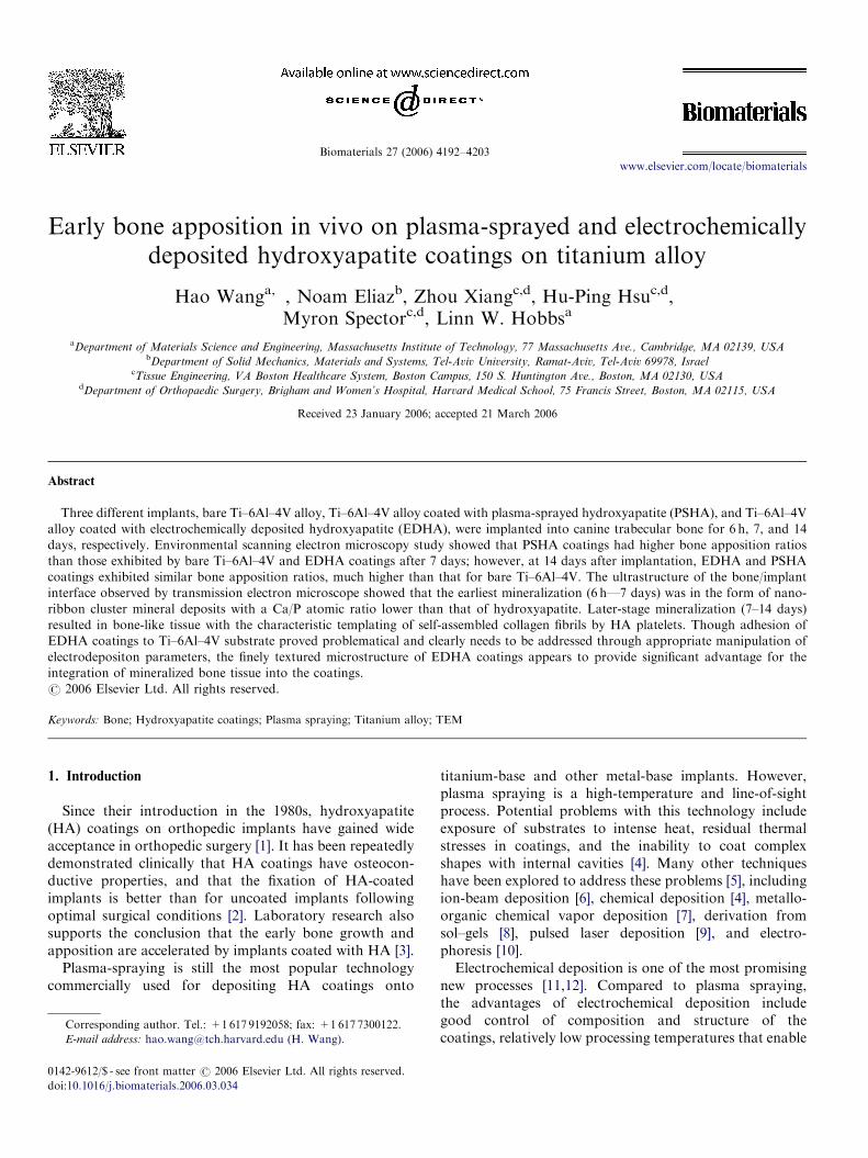
The surface morphologies of these two coatings werequite different, as shown in Fig. 2a and b, respectively. TheEDHA coating was characterized by individually resolva-ble HA crystallites with a plate-like shape growing normalto the substrate surface. Their typical width was severalmicrometers and their thickness less than 1 mm, similar tocrystals found in HA chemically deposited at roomtemperature [19]. The surface of PSHA exhibited adramatically different morphology: large globules ap-peared at the surface, and no individual HA crystallites
could be distinguished; this finding is consistent withmolten splats solidifying as individual units with at leastpartly (probably poly) crystalline character. The size of thesurface globules ranged from several micrometers down toabout 100 nm. The surface roughnesses (Ra), measured bysurface profilometry, were 4807120 nm for EDHA and13007400 nm for PSHA coatings, comparing to4657173 nm for the as-received Ti–6Al–4V rod.
3.2. ESEM measurements of bone apposition ratio
For all three types of implants, both trabecular bone andits debris from the surgery were observed to be presentaround the implants 6 h after implantation (Fig. 3a–c).Owing to the press-fit method of insertion of the surgicalimplant, direct contact between bone and implants wasalso, but infrequently, observed. The shortest implantationtime, 6 h, was far too short for a meaningful calculation ofnew bone apposition ratio for any implant and hence nonewas calculated.Seven days after implantation, new bone tissue was
observed apposing all three types of implants. The newlydeveloped bone tissue was different from trabecular bone,or its drillhole debris, in that the new tissue closelycontacted the implants and spread out over the implantsurface. Cellular lacunae were clearly visible in the newbone tissue (Fig. 4a–c).After 14 days, the surface apposition of new bone tissue
had increased on all three types of implants (Fig. 5a–c). Atthe same time, the new bone tissue became much thicker,denser and more closely conformed to the coating surfacetopography, especially apparent for PSHA-coated andEDHA-coated implants.The average surface bone apposition ratios (Fig. 6) for
each type of sample after 7 and 14 days implantation werecalculated from the ESEM images. Two-factor analysis ofvariance (ANOVA) revealed that there were significanteffects of time (po0:0001; power ¼ 1) and coating type(p ¼ 0:0002; power ¼ 0.99) on the apposition of bone tothe samples. Post-hoc testing using Fisher’s protected least-squares differences (PLSD) method showed that thedifference between the PSHA and Ti–6Al–4V groups wasstatistically significant (po0:0001), as was the differencebetween the EDHA and Ti–6Al–4V groups (p ¼ 0:002).There was, however, no statistically significant differencebetween the PSHA and EDHA groups when including alldata from the 7- and 14-day time periods. One-factorANOVA of the 7-day data also demonstrated that therewas a significant effect of the group on the percentage ofbone apposed to the surface (p ¼ 0:16; power ¼ 0.83). Inthis case, analyzing the 7-day data separately, there was astatistically significant difference in the apposition to thePSHA and Ti–6Al–4V groups (p ¼ 0:006), but there wasno statistically significant difference between the PSHAand EDHA groups, and between the EDHA andTi–6Al–4V groups.

ARTICLE IN PRESS
(b)
20 25 30 35 40 45 50 55 600
300
600
900
(313
)(3
22)
(311
)
(200
)
Ti(1
02)
+ (
004)
(402
)(4
10)(3
21)
(213
)(3
12)
(203
)(1
13)
(210
)(1
02) (2
22)
Ti(1
01)
+ (
301)
Ti(0
02)
Ti(1
00)
(202
)(3
00)
(112
)(2
11)
(002
)
(111
)
Diffraction angle, 2θ
Diffraction angle, 2θ (a)
20 25 30 35 40 45 50 55 600
1000
2000
3000
4000
(313
)
(311
)
(310
)
(200
)
Ti(1
02)
+ (
004)
(402
)(4
10)
(321
)(2
13)
(312
)
(222
)
Ti(1
01)
Ti(0
02)
Ti(1
00)
(202
)(3
00)
(112
)(2
11)
(210
)(1
02)
(002
)
(111
)
Inte
nsity
, c/s
Inte
nsity
, c/s
Fig. 1. X-ray diffraction spectra for: (a) EDHA, and (b) PSHA coatings (Cu Ka radiation).
H. Wang et al. / Biomaterials 27 (2006) 4192–4203 4195
3.3. TEM analysis of the bone/coating interface
TEM study was limited to implants coated with the twoforms of HA coatings only because the TEM samplepreparation procedure used in this study was not suited topreserving new bone apposing bare Ti–6Al–4V.
In the case of PSHA-coated samples implanted for 6 h(Fig. 7a), bone debris and blood cells were found near thecoating surface, but no evidence of mineralized tissue;trabecular bone tissue (from debris or the original bone)was observed only much farther away (not shown inFig. 7a). The Ca/P atom ratio for the coating, measured byXEDS, was 1.67, matching the stoichiometric value forHA. The image showed an undulating surface on the
coating, with possible indications of intergranular dissolu-tion. Small (o100 nm) HA crystalline platelets, likelyreprecipitating from dissolved HA coating [18], weresometimes found present within 1 mm of the HA coatingsurface.For EDHA-coated samples, 6 h after implantation, the
substrate side of the coating was smooth, suggesting thatthe whole coating had separated from the titaniumsubstrate due to poor bonding between the EDHA andthe substrate (Fig. 7b). The edges and corners of thecoating crystals opposing the drilled bone remained sharp,with little apparent sign of dissolution. XEDS analysisyielded a Ca/P atom ratio of 1.6, close to that ofstoichiometric HA.
ARTICLE IN PRESS
Fig. 2. ESEM images of the two coatings: (a) EDHA surface top view, and (b) PSHA surface top view.
H. Wang et al. / Biomaterials 27 (2006) 4192–42034196
After 7 days implantation, TEM analysis revealed a layerof dense mineralized tissue formed immediately adjacent tothe PSHA coating surface (Fig. 8a) and isolated fibrillarmineralized ‘‘clusters’’ further away from the coatinginterface. Higher resolution images (Fig. 8b and c) of theseclusters and the dense layer near the interface showed thatthey were composed of many fine nano-ribbons, whichwere several hundred nano-meters long but only severalnano-meters wide. Surrounding these clusters was anunmineralized collagenous matrix, whose fibers exhibitedthe characteristic banded structure visible after lead citrateand uranyl acetate staining. Differently oriented collagen
fibers were observed; however, their size was uniform, withdiameters consistently less than 50 nm and lengths as longas 1 mm. Collagen fibers were not, however, found withinthe clusters. XEDS analysis showed that the Ca/P atomicratio of the clusters was about 1.2, much lower than that ofHA but closer to the typical values for brushite (dicalciumphosphate dehydrate, DCPD, Ca/P ¼ 1.0), octacalciumphosphate (OCP, Ca/P ¼ 1.33), or even amorphouscalcium phosphate (ACP, Ca/P ¼ 1.5). By comparison,the Ca/P ratio of the HA coating was measured atabout 1.6, and that of mature bone tissue also close to1.6. The surrounding collagen matrix remained largely

ARTICLE IN PRESS
Fig. 3. ESEM images from cross-sections through implants and adjacent
bone tissue, 6 h implantation: (a) Ti–6Al–4V, (b) PSHA coating, and (c)
EDHA coating.
H. Wang et al. / Biomaterials 27 (2006) 4192–4203 4197
unmineralized at 7 days, the calcium content remainingvery low (less than 1 at%) and phosphorus almostundetectable.
By contrast, little tissue development was seen near theEDHA coating surface 7 days after implantation. Osteo-
blast-like cells were found in some regions, stationed closeto the coating surface, at a distance of approximately10–15 mm (Fig. 8d). The surface of the coating 1 week afterimplantation was still similar to that 6 h after implantation.By 14 days post-implantation, the mineralized tissue
layer on the PSHA coating had became much thicker andcovered almost the entire surface (Fig. 9a). The character-istic banded structure of stained collagen could be seen inthe new bone tissue layer. The Ca/P atom ratio of thistissue increased to nearly 1.7.Compared to PSHA, new mineralized tissue formation
on the EDHA coating increased much more substantiallybetween 7 and 14 days (Fig. 9b). Much new tissue wasfound on the EDHA surface and closely integrated with thecoating, interdigitating with the blade-like EDHA crystals.The Ca/P atomic ratio of the new tissue was close to 1.7.This dramatic increase in proximate mineralized tissueformation matches the increase in bone apposition ratiopreviously documented in the ESEM observations.
4. Discussion
Both EDHA and PSHA coatings used in this study werecomposed predominantly of HA and had very closenominal compositions. However, they were distinct insurface topographies, coating morphologies, microstruc-tures, and degree of crystallinity, one or all of which arelikely to have resulted in differing solubilities, i.e. theirability to initially release calcium and phosphorus into theenvironment. A confirmatory solubility test (Fig. 10)showed that PSHA dissolved much more readily thanEDHA. While the former reached saturation in distilledwater in 2 days, the latter occasioned a very low Caconcentration even after 10 days. It is presumed that thedifference in their solubilities is at least partially respon-sible for the different observed kinetics for early boneformation associated with these two coatings; and itcan certainly be stated that the solubility correlates withdifferent onset times for collagen release and itsmineralization.EDHA showed a low bone apposition ratio after 7 days,
intermediate between the corresponding values for bareTi–6Al–4V alloy and PSHA-coated samples. However,after 14 days, the EDHA bone apposition ratio increasedmarkedly, to that observed for PSHA, and much morethan that for bare Ti–6Al–4V. The initial low appositionratio may be attributable to the low solubility of EDHA.During the first 7 days, the coating made almost nocontribution to bone apposition via ion release andreprecipitation or by Ca signaling to osteoblasts [20,21];thus it exhibited almost the same apposition ratio as bareTi–6Al–4V. PSHA, with its partial amorphous content andconsequently higher solubility in vivo, contributed a muchhigher local concentration of calcium and phosphorus ions,which could assist in and accelerate local mineralization ofnew bone or be involved in cell signaling. Nevertheless, thediffering solubilities dictated only different short-term

ARTICLE IN PRESS
Fig. 4. ESEM images from cross-sections through implants and adjacent
bone tissue, 7 days implantation: (a) Ti–6Al–4V, (b) PSHA coating, and
(c) EDHA coating.
Fig. 5. ESEM images from cross-sections through implants and adjacent
bone tissue, 14 days implantation: (a) Ti–6Al–4V, (b) PSHA coating, and
(c) EDHA coating.
H. Wang et al. / Biomaterials 27 (2006) 4192–42034198
mineralization behaviors. By 14 days, the surface apposi-tion ratio of EDHA increased sharply and caught up withthat of PSHA, suggesting that the lower dissolution rate ofEDHA was already sufficient to catalyze the formation ofnew bone. A similar initial disparity and later catch-up has
been reported for annealed vs. non-annealed PSHA coat-ings [18].At 7 days post-implantation, new mineralized tissue—
mainly composed of clustered nano-ribbons—was ob-served on the PSHA coatings only. Collagen fibers, visible

ARTICLE IN PRESS
Fig. 6. Average bone apposition ratios after 7 and 14 days of implantation. The error bars are standard deviations.
Fig. 7. Cross-section TEM micrographs of samples implanted for 6 h: (a) PSHA, and (b) EDHA. Thin sections were stained with uranyl acetate and lead
citrate.
H. Wang et al. / Biomaterials 27 (2006) 4192–4203 4199
upon staining, were also present close to the coatingsurface, but were not yet mineralized. After 14 days, newbone-like tissue was already very well developed in thevicinity of both types of coatings, incorporating collagenfibers. Many proteins, among them osteonectin [22],
osteocalcin [23] and bone sialoprotein [24], have beenhypothesized to serve as nuclei or templates for initialdeposition of HA [25]. However, in this study the earliestmineral deposits observed were actually long nano-ribbons,of large aspect ratio, considerably different in morphology

ARTICLE IN PRESS
Fig. 8. Cross-section TEM micrographs of samples implanted for 7 days: (a) PSHA coating and mineralized clusters, (b) a detailed image of a mineralized
cluster close to PSHA coating surface, (c) detail of newly mineralized tissue at the interface with PSHA coating, and (d) EDHA coating. Cracks were
introduced by microtoming PSHA coatings. Thin sections were stained with uranyl acetate and lead citrate.
H. Wang et al. / Biomaterials 27 (2006) 4192–42034200
from the proteins cited. The clustered nano-ribbons wereonly about several nano-meters in width, but severalhundred nm in length. Thus, the potential role of shortconnective proteins is relegated to nucleation and nottemplating. The nano-ribbons are, however, very close insize to a single collagen molecule (1.5 nm diameter, 300 nmlong) or five-stranded fibrils (about 4 nm diameter) [26]. Ithas been generally believed that individual collagen
molecules do not by themselves have the ability to initiatemineral deposition [27]; however, minimally self-assembledprotofibrils (such as the five-strand fiber mentioned above)have been suggested to have the ability to nucleate mineralprecipitation along the fibril length on the evidence of arecent in vitro study [28]. Hence, the clustered mineralnano-ribbons could represent calcium phosphates nucleat-ing on individual collagen molecules or minimally stranded

ARTICLE IN PRESS
Fig. 9. Cross-section TEM micrographs of samples implanted for 14 day:
(a) PSHA, and (b) EDHA coatings. Thin sections were stained with uranyl
acetate and lead citrate.
H. Wang et al. / Biomaterials 27 (2006) 4192–4203 4201
fibrils. This explanation has not been advanced before andremains speculative, pending identification of the nucleat-ing agent. On the other hand, self-assembled collagen fibersin bone are not mineralized along their length, but insteadat inter-molecule junctions by crystallographically and
morphologically oriented platelets [29] that bear norelationship to collagen molecule length or morphology(except for the molecular self-assembly motif).A significant observation is the low Ca/P atomic ratio in
the early stage mineral nano-ribbon deposits. The valuestrongly suggests that HA is not the initial calciumphosphate phase involved in the earliest stage of miner-alization. It has been observed in vitro that ACP and OCP,which have more favorable formation kinetics than HA,are deposited before HA on NaOH-treated titaniumsurfaces [30]. The present study has shown that the samemay occur for bone formation in vivo. Theories of proteincontrol of bone mineralization have mainly focused on theshape and size match between proteins and the HA unit cell[31,32]. However, the present observations suggest that therole of proteins in nucleating calcium phosphates of otherstoichiometries, such as ACP, DCPD, and OCP, could beinitially more important.EDHA, as a potential substitute for PSHA, confers some
advantages, such as low processing temperature anddeposition on complicated shapes. The platey surfacemorphology of EDHA coatings may be particularlyefficacious in promoting integration of newly mineralizedtissue with the HA coating and in maximizing themechanical integrity of the coating-bone interface, How-ever, low bonding strength between coating and substratewas also observed. This drawback could be overcome bytailoring the substrate composition or topography beforeelectrodeposition—for example, by mechanically or che-mically texturing the substrate surface or by pre-forming ahydrogel surface layer [33] onto Ti alloy surfaces. Thoughthe EDHA coatings were substantially thinner than thePSHA coatings, it has been shown [34] that even thick(50 mm) PSHA coatings appear to be substantially remo-deled eventually, after months or years of implantation inhuman models. The much thinner EDHA coatings cannevertheless still prove efficacious over the critical weeks-to-months period for establishment of apposing miner-alized tissue, before substantial remodeling has occurred;and when substantial remodeling does begin, the initialcoating thickness may be irrelevant.
5. Conclusions
In agreement with a previous in vivo study [18], PSHAcoatings of implantation into canine trabecular bone werefound to accelerate early stage mineralization of bonetissue formation over that occurring for bare alloyimplants. Kinetic differences in the early stage (p7 days)of observed mineralization correlated closely to differencesin coating solubilities, though later-stage (14 days) miner-alization rates were relatively unaffected. The early stagemineral product had a morphology (nano-ribbon clusters)and composition (Ca/P�1.2) distinguishably different fromthat of mature bone, likely representing a calciumphosphate phase with lower Ca/P ratio (DCPD, ACP,OCP). Though smaller proteins may direct the peculiar

ARTICLE IN PRESS
0 50 100 150 200 250
0.00
0.05
0.10
0.15
0.20
0.25
0.30
0.35
Ti64
EDHA
PSHA
Cal
cium
con
cent
ratio
n (m
M)
Hours
Fig. 10. Aqueous solubilities of PSHA and EDHA in de-ionized water at room temperature. Bare Ti–6Al–4V alloy serves as a reference (n ¼ 1; error bars
are standard deviations from three measurements).
H. Wang et al. / Biomaterials 27 (2006) 4192–42034202
nano-ribbon growth morphology, mineralization could beoccurring along the length of single collagen molecules orminimally stranded collagen fibrils. EDHA coatings wereslower in inducing early stage mineralization (p7 days)than PSHA coatings but were more efficacious than bareTi–6Al–4V alloy, and were indistinguishable from PSHAcoatings in the later (14 days) mineralized tissue appositionratio and microstructure they induced in vivo. The surfacemorphology and higher surface area of EDHA coatingsappear to have resulted in better mechanical integration ofcoating and mineralized tissue. The low adhesion ofEDHA coatings to substrates will need to be addressedto better realize the promise inherent in electrodepositioncoating approaches.
Acknowledgments
This work was supported by the Division of Bioengi-neering and Environmental Systems of the US NationalScience Foundation under award DMR-9904046, theCambridge-MIT Institute, and the John F. Elliott chair(LWH). The authors are grateful to Dr. T.M. Sridhar forpreparing the EDHA coatings. The VA Boston HealthcareSystem provided the facility for animal surgeries at theirJamaica Plain campus.
References
[1] Overgaard S. Calcium phosphate coating for fixation of bone
implants. Acta Orthop Scand 2000;71:1–74.
[2] Soballe K, Hansen ES, Rasmussen HB, Jorgensen PH, Bunger C.
Tissue ingrowth into titanium and hydroxyapatite-coated implants
during stable and unstable mechanical conditions. J Orthop Res
1992;10:285–99.
[3] Thomas KA, Cook SD, Haddad Jr RJ, Kay JF, Jarcho M. Biologic
response to hydroxylapatite-coated titanium hips. A preliminary
study in dogs. J Arthroplasty 1989;4:43–53.
[4] Wen HB, de Wijn JR, Cui FZ, de Groot K. Preparation of calcium
phosphate coatings on titanium implant materials by simple
chemistry. J Biomed Mater Res 1998;41:227–36.
[5] Suchanek W, Yoshimura M. Processing and properties of hydro-
xyapatite-based biomaterials for use as hard tissue replacement
implants. J Mater Res 1998;13:94–117.
[6] Luo ZS, Cui FZ, Li WZ. Low-temperature crystallization of calcium
phosphate coatings synthesized by ion-beam-assisted deposition.
J Biomed Mater Res 1999;46:80–6.
[7] Spoto G, Ciliberto E, Allen GC. A new synthetic route to
hydroxyapatite coatings. J Mater Chem 1994;4:1849–50.
[8] Russell SW, Luptak KA, Suchicital CTA, Alford TL, Pizziconi VB.
Chemical and structural evolution of sol–gel-derived hydroxyapatite
thin films under rapid thermal processing. J Am Ceram Soc
1996;79:837–42.
[9] Tucker BE, Cottell CM, Auyeung RC, Spector M, Nancollas GH.
Pre-conditioning and dual constant composition dissolution kinetics
of pulsed laser deposited hydroxyapatite thin films on silicon
substrates. Biomaterials 1996;17:631–7.
[10] Ducheyne P, Radin S, Heughebaert M, Heughebaert JC. Calcium-
phosphate ceramic coatings on porous titanium—effect of structure
and composition on electrophoretic deposition, vacuum sintering and
in vitro dissolution. Biomaterials 1990;11:244–54.
[11] Shirkhanzadeh M. Calcium-phosphate coatings prepared by electro-
crystallization from aqueous-electrolytes. J Mater Sci-Mater Med
1995;6:90–3.
[12] Shirkhanzadeh M. Bioactive calcium-phosphate coatings prepared by
electrodeposition. J Mater Sci Lett 1991;10:1415–7.
[13] Redepenning J, McIsaac JP. Electrocrystallization of brushite coat-
ings on prosthetic alloys. Chem Mater 1990;2:625–7.
[14] Shirkhanzadeh M. Electrochemical preparation of bioactive calcium-
phosphate coatings on porous substrates by the periodic pulse
technique. J Mater Sci Lett 1993;12:16–9.
ARTICLE IN PRESSH. Wang et al. / Biomaterials 27 (2006) 4192–4203 4203
[15] Shirkhanzadeh M. Direct formation of nanophase hydroxyapatite on
cathodically polarized electrodes. J Mater Sci-Mater Med
1998;9:67–72.
[16] Vijayaraghavan TV, Bensalem A. Electrodeposition of apatite
coating on pure titanium and titanium-alloys. J Mater Sci Lett
1994;13:1782–5.
[17] Royer P, Rey C. Calcium-phosphate coatings for orthopedic
prosthesis. Surf Coat Technol 1991;45:171–7.
[18] Porter AE, Hobbs LW, Rosen VB, Spector M. The ultrastructure of
the plasma-sprayed hydroxyapatite-bone interface predisposing to
bone bonding. Biomaterials 2002;23:725–33.
[19] Feng QL, Wang H, Cui FZ, Kim TN. Controlled crystal growth of
calcium phosphate on titanium surface by NaOH-treatment. J Cryst
Growth 1999;200:550–7.
[20] Tfelt-Hansen J, Brown EM. The calcium-sensing receptor in normal
physiology and pathophysiology: a review. Crit Rev Clin Lab Sci
2005;42:35–70.
[21] Hofer AM. Another dimension to calcium signaling: a look at
extracellular calcium. J Cell Sci 2005;118:855–62.
[22] Termine JD, Kleinman HK, Whitson SW, Conn KM, McGarvey
ML, Martin GR. Osteonectin, a bone-specific protein linking mineral
to collagen. Cell 1981;26:99–105.
[23] Hauschka PV, Lian JB, Gallop PM. Direct identification of calcium-
binding amino-acid, gamma-carboxyglutamate, in mineralized tissue.
P Natl Acad Sci USA 1975;72:3925–9.
[24] Oldberg A, Franzen A, Heinegard D. The primary structure of a cell-
binding bone sialoprotein. J Biol Chem 1988;263:19430–2.
[25] Hunter GK, Hauschka PV, Poole AR, Rosenberg LC, Goldberg HA.
Nucleation and inhibition of hydroxyapatite formation by miner-
alized tissue proteins. Biochem J 1996;317:59–64.
[26] Smith JW. Molecular pattern in native collagen. Nature 1968;219:
157–8.
[27] Veis A. Mineralization in organic matrix frameworks. Rev Mineral
Geochem 2003;54:249–89.
[28] Zhang W, Liao SS, Cui FZ. Hierarchical self-assembly of nano-fibrils
in mineralized collagen. Chem Mater 2003;15:3221–6.
[29] Rosen VB, Hobbs LW, Spector M. The ultrastructure of anorganic
bovine bone and selected synthetic hydroxyapatites used as bone
graft substitute materials. Biomaterials 2002;23:921–8.
[30] Feng QL, Cui FZ, Wang H, Kim TN, Kim JO. Influence of solution
conditions on deposition of calcium phosphate on titanium by
NaOH-treatment. J Cryst Growth 2000;210:735–40.
[31] Hoang QQ, Sicheri F, Howard AJ, Yang DS. Bone recognition
mechanism of porcine osteocalcin from crystal structure. Nature
2003;425:977–80.
[32] Sarig S. Aspartic acid nucleates the apatite crystallites of bone: a
hypothesis. Bone 2004;35:108–13.
[33] Wen HB, de Wijn JR, Cui FZ, de Groot K. Preparation of bioactive
Ti6Al4V surfaces by a simple method. Biomaterials 1998;19:
215–21.
[34] Porter AE, Taak P, Hobbs LW, Coathup MJ, Blunn GW, Spector M.
Bone bonding to hydroxyapatite and titanium surfaces on femoral
stems retrieved from human subjects at autopsy. Biomaterials
2004;25:5199–208.
Related Documents