ORIGINAL ARTICLE Early acute depletion of lymphocytes in calicivirus-infected adult rabbits Raquel M. Marques & António Costa-e-Silva & Artur P. Águas & Luzia Teixeira & Paula G. Ferreira Accepted: 21 July 2010 / Published online: 11 August 2010 # Springer Science+Business Media B.V. 2010 Abstract Rabbit Haemorrhagic Disease (RHD) is a lethal infection caused by calicivirus that kills 90% of the infected adult rabbits within 3 days. The calicivirus replicates in the liver and causes a fulminant hepatitis. Most studies on the pathology of RHD have been focused on the fulminant liver disease. This may not be the only mechanism in the pathogenesis of RHD: calicivirus infection may also induce leukopenia in the infected adult rabbits. We show now by flow cytometry analysis that the calicivirus induces an early decrease in B and T cells, in both spleen and liver. The depletion of B and T cells was associated with apoptosis labelled by annexin V. These changes occurred in rabbits before they showed enzymatic evidence of liver damage and persisted after liver transaminase values were very high. We conclude that depletion of lymphocytes caused by the calicivirus infection precedes or attends liver damage. The relative contribution of this lymphocyte depletion for the pathogenesis of the fatal calicivirus infection of rabbits remains to be investigated. Keywords Haemorrhagic disease . Inflammation . Liver . Spleen . T lymphocyte . B lymphocyte . Macrophage . Apoptosis . Flow cytometry Introduction Rabbit Haemorrhagic Disease (RHD) is a “dengue-like” viral disorder that causes very high mortality of rabbits within a few days of infection. RHD kills more than 90% of adult rabbits 24–72 hours after their experimental inoculation with calicivirus (Ohlinger et al. 1990; Shien et al. 2000). The viral agent replicates in the liver, and induces a fulminant necrotic hepatitis that kills the animals (Marcato et al. 1991; Mitro and Krauss 1993; Ferreira et al. 2006a, b). Current views on the natural history of RHD are focused on the Vet Res Commun (2010) 34:659–668 DOI 10.1007/s11259-010-9437-7 R. M. Marques (*) : A. Costa-e-Silva : A. P. Águas : L. Teixeira : P. G. Ferreira Department of Anatomy, ICBAS (Abel Salazar Institute for Biomedical Science) and UMIB (Unit for Multidisciplinary Biomedical Research), University of Porto, Largo Professor Abel Salazar, 2, 4099-003 Porto, Portugal, European Union e-mail: [email protected]

Welcome message from author
This document is posted to help you gain knowledge. Please leave a comment to let me know what you think about it! Share it to your friends and learn new things together.
Transcript
ORIGINAL ARTICLE
Early acute depletion of lymphocytesin calicivirus-infected adult rabbits
Raquel M. Marques & António Costa-e-Silva &
Artur P. Águas & Luzia Teixeira & Paula G. Ferreira
Accepted: 21 July 2010 /Published online: 11 August 2010# Springer Science+Business Media B.V. 2010
Abstract Rabbit Haemorrhagic Disease (RHD) is a lethal infection caused by calicivirusthat kills 90% of the infected adult rabbits within 3 days. The calicivirus replicates in theliver and causes a fulminant hepatitis. Most studies on the pathology of RHD have beenfocused on the fulminant liver disease. This may not be the only mechanism in thepathogenesis of RHD: calicivirus infection may also induce leukopenia in the infected adultrabbits. We show now by flow cytometry analysis that the calicivirus induces an earlydecrease in B and T cells, in both spleen and liver. The depletion of B and T cells wasassociated with apoptosis labelled by annexin V. These changes occurred in rabbits beforethey showed enzymatic evidence of liver damage and persisted after liver transaminasevalues were very high. We conclude that depletion of lymphocytes caused by the calicivirusinfection precedes or attends liver damage. The relative contribution of this lymphocytedepletion for the pathogenesis of the fatal calicivirus infection of rabbits remains to beinvestigated.
Keywords Haemorrhagic disease . Inflammation . Liver . Spleen . T lymphocyte .
B lymphocyte . Macrophage . Apoptosis . Flow cytometry
Introduction
Rabbit Haemorrhagic Disease (RHD) is a “dengue-like” viral disorder that causes very highmortality of rabbits within a few days of infection. RHD kills more than 90% of adultrabbits 24–72 hours after their experimental inoculation with calicivirus (Ohlinger et al.1990; Shien et al. 2000). The viral agent replicates in the liver, and induces a fulminantnecrotic hepatitis that kills the animals (Marcato et al. 1991; Mitro and Krauss 1993;Ferreira et al. 2006a, b). Current views on the natural history of RHD are focused on the
Vet Res Commun (2010) 34:659–668DOI 10.1007/s11259-010-9437-7
R. M. Marques (*) : A. Costa-e-Silva : A. P. Águas : L. Teixeira : P. G. FerreiraDepartment of Anatomy, ICBAS (Abel Salazar Institute for Biomedical Science) and UMIB(Unit for Multidisciplinary Biomedical Research), University of Porto, Largo Professor Abel Salazar, 2,4099-003 Porto, Portugal, European Unione-mail: [email protected]
fulminant liver disease triggered by the virus (Ferreira et al. 2006a, b). Nevertheless, othermechanisms may also concur for the rapid course of the disease, namely the systemicleukocyte depletion that we have previously described in RHD (Ferreira et al. 2006a). Thisphenomenon is characteristic not only of the RHD but also of others haemorrhagic feverssuch as in Ebola virus infection that is associated with a severe apoptosis of lymphocytes(Leroy et al. 2000; Baize et al. 2002; Bradfute et al. 2007).
The fast progress of calicivirus infection into fulminant hepatitis in adult rabbits makes itdifficult to map the sequence of cellular changes that occurs as a result of calicivirusinoculation, since most of the animals are dead less than 48 hours after inoculation.Furthermore, values of biochemical markers of liver damage have shown that RHDprogresses at a different rate in individual (Ferreira et al. 2006a).
We have investigated here the early alterations in lymphocyte subpopulations that occurin spleen and liver of calicivirus-infected adult rabbits, and we have compared these datawith the level of hepatic damage caused by calicivirus at 24 hours of infection.
The present work extends information obtained by previous investigations on theimmunological response of rabbits to calicivirus, namely on the changes of B and Tlymphocytes present in the peripheral blood of infected animals (Tokarz-Deptula andDeptula 2004, 2009; Niedzwiedzka-Rystwej et al. 2009)
Materials and methods
Rabbits
Fourteen New Zealand White rabbits (Oryctolagus cuniculus) were used in this study whenthey were 8 weeks old. All animals were purchased from a Spanish breeder (Charles RiverLaboratories España, SA, Spain) and were pathogen free. The rabbits were kept understandard conditions of housing with unrestricted access to food and water; this was doneaccording to the European Union Directive no. 86/609/CEE.
Calicivirus infection of adult rabbits
The initial inoculum of caliciviruses used in our laboratory was a kind offer of ProfessorFrancisco Parra, University of Oviedo, Spain. Current inocula were produced fromhomogenates of livers from infected rabbits, and they were obtained as previouslydescribed (Ferreira et al. 2004). Seven rabbits were injected intramuscularly with a PBSsuspension of caliciviruses that had a 212 titre in haemagglutination units and then theywere sacrificed at 24 hours after inoculation in order to determine the percentage ofapoptosis and the total number of the major subpopulations of leukocytes (macrophages, Tand B lymphocytes) in the liver and spleen of calicivirus infected- rabbits. The kinetics ofleukocyte subpopulations was evaluated by flow cytometry. The other seven animals wereinoculated only with PBS (the vehicle of the calicivirus) and they were used as controls ofthe infection. Before the euthanasia, blood samples were collected by venepuncture forevaluation of the serum concentration of alanine aminotransferase (ALT) and aspartateaminotransferase (AST). The biochemical parameters were measured by spectophotometricmethods (Cobas Integra 800, Roche Diagnostics GmbH, Mannhein, Germany). All rabbitswere euthanized with an intravenous injection of an anaesthetic mixture of xylazine(Rompun®, Bayer Co., Portugal, 3 mg/kg) and ketamine (Imalgéne 1000®, Merial,Portugal, 10 mg/kg), 1 hour after the intramuscularly administration of morphine (Morfina
660 Vet Res Commun (2010) 34:659–668
Labesfal®, Labesfal, Portugal, 5 mg/kg). The vascular system was washed by perfusion,through a cannula inserted in aorta, with a saline solution (40°C) and then with HEPESsolution (0,14 M NaCl; 2,7 mM KCl; 0,28 mM Na2HPO4.12H2O; 10 mM HEPES) at 40°C.Liver and spleen were aseptically collected in order to be processed and analysed by flowcytometry.
Isolation of mononuclear cells from liver
A 6–7 g portions of the liver were cut aseptically and placed in a Potter-Elvehjemhomogenizer with 3 mL of a Balance Salt Solution (1,26 mM CaCl2; 5,4 mM KCl;0,44 mM KH2PO4; 0,49 mM MgCl2.6H2O; 0,40 mM MgSO4.7H2O; 0,14 M NaCl and0,63 mM Na2HPO4) where they were gently mechanically disrupted (the homogenizationwas performed at low rotation for minimize the cell rupture). The resulting cell suspensionwas divided into two equal aliquots and transferred to 50 mL falcon tubes; BSS was thenadded to complete a total volume of 100 mL. The cell suspension was filtered through a100 μm nylon mesh cell strainer to remove cellular aggregates and tissue debris. The filtratewas washed by centrifugation at 1500 rpm for 10 min at 4°C. Pellets were resuspended in10 mL of BSS each and then they were filtered twice through a 70 μm nylon mesh cellstrainer. For mononuclear cells isolation, 10 mL of the suspension was carefully layered on5 mL of Percoll (Histopaque1077—Sigma) and centrifuged at 3000 rpm for 15 min at roomtemperature (Centrifuge Hoefer Labofuge 300). The layer formed between the BSS and thePercoll layers was collected and transferred to a 15 mL Falcon Flask. The cell suspensionwas washed by centrifugation at 1000 rpm for 10 min and the pellet was resuspended in10 mL of BSS. Cells were stained with Turk’s reagent and counted in a Neubauer Chamber(dilution 1:10).
Isolation of mononuclear cells from spleen
Spleen cells were obtained by gentle smearing of the organ in a sterile plate with 10 mL ofBSS solution. Cells were later washed by centrifugation at 1000 rpm for 10 min at 4°C(Centrifuge Sigma 3–16 K). The pellet was resuspended in 5 mL of ACK solution (0,15 MNH4Cl, 1 mM KHCO3 and 0,1 mM EDTA, pH 7,2) and incubated for 2–3 min on ice forred blood cell lysis. Five mL of BSS were added to the suspension that was centrifuged at1000 rpm for 5 min. The pellet was resuspended in 15 mL of BSS. Cells were stained withTurk’s reagent and then counted in a Neubauer Chamber (dilution 1:20).
Flow cytometry analysis
Mononuclear cells isolated from liver and spleen were added to cytometry tubes(1×10⁶cells/tube); 400 μL of PBS supplemented with 1% BSA and sodium azide(10 mM) was added to each tube. The cells were washed by centrifugation at 1000 rpmfor 10 min at room temperature. Immunoflorescence cytometric analysis of cell surfaceantigens was performed in a FACSscan with CellQuest software, using the followingantibodies: FITC-conjugated goat polyclonal antibody anti-rabbit IgM (Serotec), for theanalysis of B cells (it recognizes B cells, IgM positive); FITC-conjugated mouse monoclonalantibody anti-rabbit T-lymphocytes (Serotec) that recognizes T cells and immunoprecipitates, a67 kD glycoprotein (Kotani et al. 1993); FITC conjugated monoclonal antibody anti-mouseCD11b Mac-1α (BD Pharmingen), for the analysis of macrophages. For flow cytometryanalysis of cells that are undergoing apoptosis, it was used an early apoptotic marker namely,
Vet Res Commun (2010) 34:659–668 661
annexin V, which has a high affinity to the phosphatidylserine exposed to the external cellularenvironment (Apoptotic cells present membrane phospholipid phosphatidylserine trans-located from inner to the outer leaflet of the plasma membrane). The late apoptotic/ necroticcells were labelled with propidium iodide. Both lymphocyte and macrophage populationswere gated on the basis of forward and side scatters and then were analyzed for the respectivemarkers. For the calculation of the total number of cells, we have used the following formula:(cellular concentration, cells/mL × volume of the cell suspension, mL) × percentage ofpositive cells.
Statistical analysis
Statistical comparison of data from all groups of rabbits was performed using the testMann-Witney, calculated with PASW Statistics 17.0 software. Differences were consideredsignificant at p<0.05.
Results
Liver biochemistry and lymphocyte subpopulations were characterized in adult rabbits24 hours after a lethal experimental infection of the animals with calicivirus.
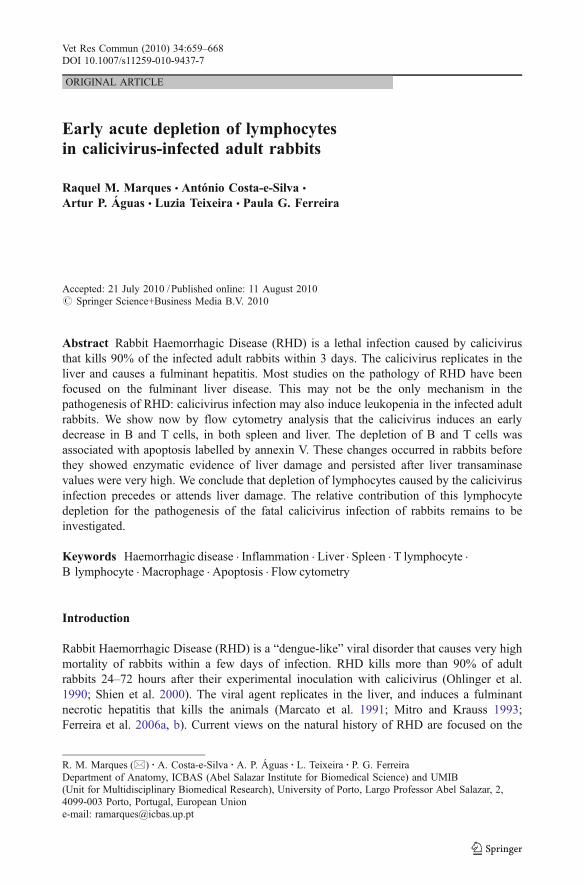
Fig. 1 Comparison of the serum concentration of AST and ALT between control and calicivirus-infectedadult rabbits. Twenty four hours after viral inoculation, calicivirus infection of adult rabbits induced a widevariation of the AST and ALT values
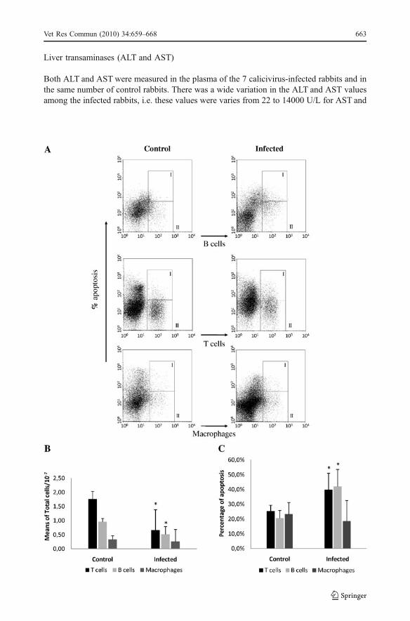
Fig. 2 a Representative flow cytometry dot-plots: the regions identified in the dot-plots as II represent thepercentage of positive B cells (IgM+), T cells and macrophages (CD11b+) present in the liver of control andinfected adult rabbits, after 24 hours of infection; the regions identified in the dot-plots as I represent thepercentage of apoptosis (annexinV+) for each cell population analysed, without excluding the cells labelledannexinV+/propidium iodide+. b Comparison of the number of leukocytes (macrophages, B and Tlymphocytes) present in the liver of control and calicivirus-infected adult rabbits, after 24 hours of infection;Mann-Witney test (* p<0.05). Calicivirus infection of adult rabbits is associated to a significant decrease ofthe liver lymphocytes (B and T cells). c Comparison of the percentage of apoptosis of liver leukocytes(macrophages, B and T lymphocytes) between control and calicivirus-infected adult rabbits, after 24 hours ofinfection; Mann-Witney test (* p<0.05). The significant decrease of B and T cells in the liver of infectedanimals was associated with an increase of the number of apoptotic cells
b
662 Vet Res Commun (2010) 34:659–668
Liver transaminases (ALT and AST)
Both ALT and AST were measured in the plasma of the 7 calicivirus-infected rabbits and inthe same number of control rabbits. There was a wide variation in the ALT and AST valuesamong the infected rabbits, i.e. these values were varies from 22 to 14000 U/L for AST and
Vet Res Commun (2010) 34:659–668 663
from 29 to 1749 U/L for ALT. In more detail: 2 of the rabbits (animals A, C in Fig. 1) hadlow values of transaminases (less than 100 U/L), 3 rabbits (animals D, E and G) presented amoderate value (342-1282 U/L for AST and 11-191U/L for ALT) and 2 other rabbits(animals B and F) showed high values (above 9000 U/L for AST and 1000 U/L for ALT).Values for controls were below 29 U/L for AST and 41 U/L for ALT (Fig. 1).
664 Vet Res Commun (2010) 34:659–668
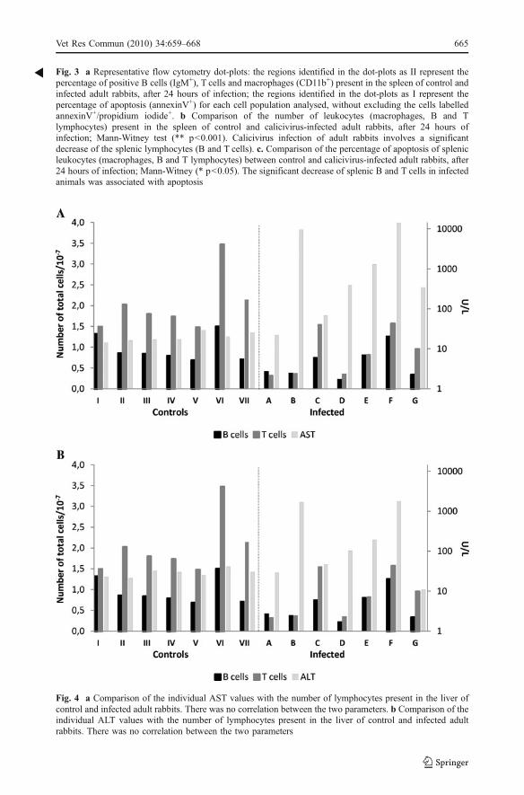
Fig. 4 a Comparison of the individual AST values with the number of lymphocytes present in the liver ofcontrol and infected adult rabbits. There was no correlation between the two parameters. b Comparison of theindividual ALT values with the number of lymphocytes present in the liver of control and infected adultrabbits. There was no correlation between the two parameters
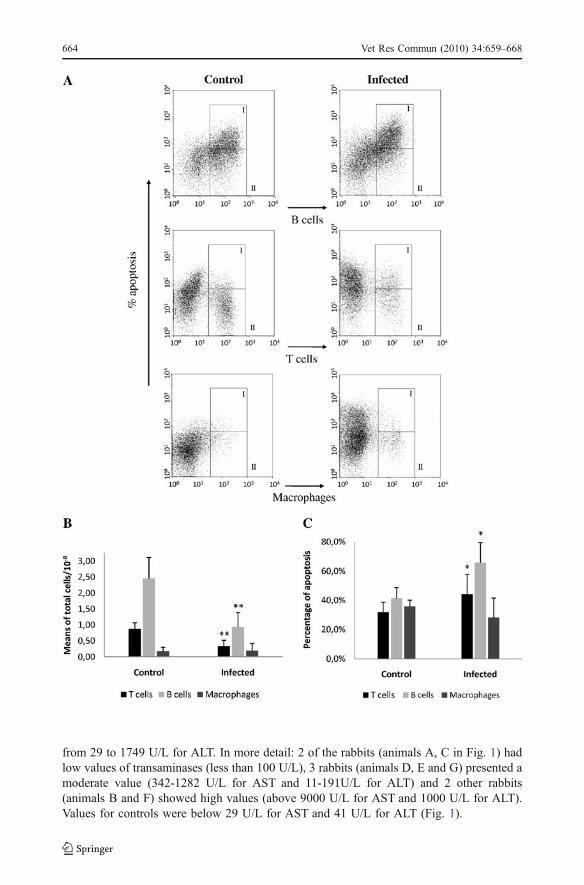
Fig. 3 a Representative flow cytometry dot-plots: the regions identified in the dot-plots as II represent thepercentage of positive B cells (IgM+), T cells and macrophages (CD11b+) present in the spleen of control andinfected adult rabbits, after 24 hours of infection; the regions identified in the dot-plots as I represent thepercentage of apoptosis (annexinV+) for each cell population analysed, without excluding the cells labelledannexinV+/propidium iodide+. b Comparison of the number of leukocytes (macrophages, B and Tlymphocytes) present in the spleen of control and calicivirus-infected adult rabbits, after 24 hours ofinfection; Mann-Witney test (** p<0.001). Calicivirus infection of adult rabbits involves a significantdecrease of the splenic lymphocytes (B and T cells). c. Comparison of the percentage of apoptosis of splenicleukocytes (macrophages, B and T lymphocytes) between control and calicivirus-infected adult rabbits, after24 hours of infection; Mann-Witney (* p<0.05). The significant decrease of splenic B and T cells in infectedanimals was associated with apoptosis
R
Vet Res Commun (2010) 34:659–668 665
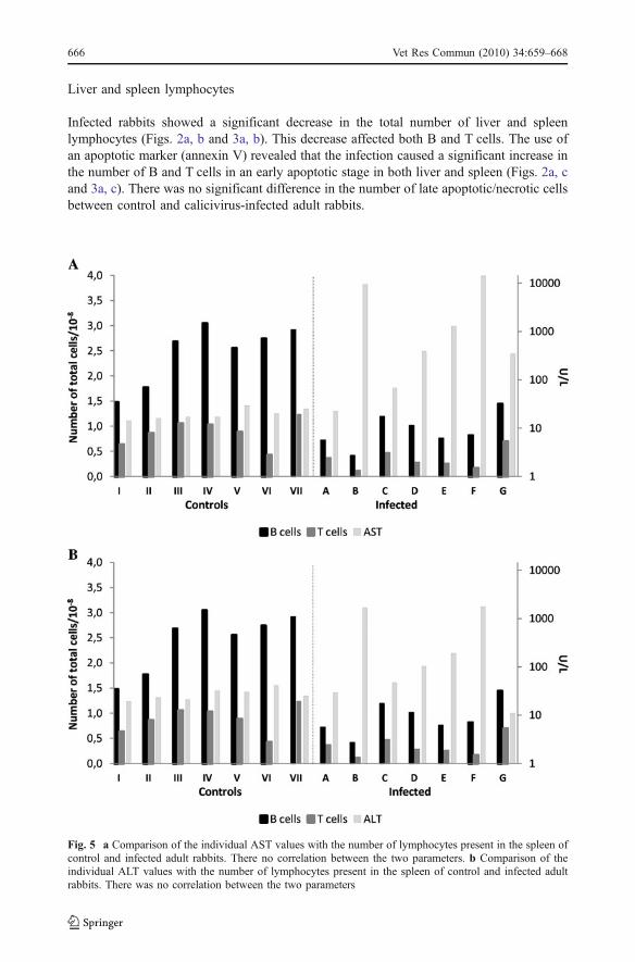
Liver and spleen lymphocytes
Infected rabbits showed a significant decrease in the total number of liver and spleenlymphocytes (Figs. 2a, b and 3a, b). This decrease affected both B and T cells. The use ofan apoptotic marker (annexin V) revealed that the infection caused a significant increase inthe number of B and T cells in an early apoptotic stage in both liver and spleen (Figs. 2a, cand 3a, c). There was no significant difference in the number of late apoptotic/necrotic cellsbetween control and calicivirus-infected adult rabbits.
Fig. 5 a Comparison of the individual AST values with the number of lymphocytes present in the spleen ofcontrol and infected adult rabbits. There no correlation between the two parameters. b Comparison of theindividual ALT values with the number of lymphocytes present in the spleen of control and infected adultrabbits. There was no correlation between the two parameters
666 Vet Res Commun (2010) 34:659–668
Correlation between lymphocyte depletion and liver damage
Comparison of the individual transaminase values with lymphocyte number in the liver andspleen of infected rabbits showed no correlation between the two parameters, i.e. the rabbitswith higher transaminase values were not the animals with the greatest decrease inlymphocyte number and vice versa (Figs. 4 and 5).
Discussion
We have described before that calicivirus infection causes a significant decrease in thegeneral pool circulating leukocytes of the adult rabbit. We describe now that this leukopeniainvolves both B and T lymphocytes, and is associated with apoptosis of the white bloodcells and precedes the fulminant liver disease that kills the animals.
Previous investigations of ours had indicated that the calicivirus infected-adult rabbitsdevelop severe leukopenia involving both circulating heterophils and lymphocytes (Ferreiraet al. 2006a). We show now that lymphocytes are decreased not only in the spleen but alsoin the target organ of viral replication, the liver. We found that the B and T cell loss wasassociated with apoptosis. Previous studies on the pathogenesis of RHD were not able todemonstrate the presence of the virus in lymphocytes (Ramiro-Ibáñez et al. 1999). Thisfinding suggests that the apoptosis of B and T-lymphocytes that we observed in both thespleen and the liver could be a bystander effect of the viral infection. It may contribute forthe rapid progression of the disease of immune response impairment.
The acute lymphocytes depletion that we have documented in RHD recalls similar dataobtain in Ebola hemorrhagic disease, where a significant loss of lymphocytes was reported inthe early stage of infection (Baize et al. 2002; Mahanty and Bray 2004; Bradfute et al. 2007).Patients who died from Ebola disease presented defective humoral responses and widespreadintravascular apoptosis of T cells. In contrast, patients who have survived Ebola infectionwere capable of produce an early and strong humoral response (Baize et al. 2002). RHD killsadult rabbits within 2–3 days; therefore the animals do not have enough time to mount aneffective immune response against the virus. If rabbits are allowed to produce specificantibodies, before calicivirus infection, they will become resistant to the viral infection(Parra and Prieto 1990; Laurent et al. 1994; Bertagnoli et al. 1996; Ferreira et al. 2008). Forinstance, it has been proposed that the minority (around 10%) of adult rabbits that surviveRHD achieve survival because they develop hepatitis at a relatively slow pace; afterrecovering from the calicivirus-induced liver disease these rabbits will depict long-termprotection from RHD and this is due to circulating anti-calicivirus antibodies (Cooke 2002).This investigation adds a new piece of information on pathogenesis of RHD: in addition toinduce the fulminant hepatitis, the virus depletes both B and T lymphocytes, thus impairingthe immune response of the host. These changes in leukocytes occurred in rabbits beforeshowing enzymatic evidence of liver damage and persisted after liver transaminases valueswere very high. We conclude that loss of immune cells precedes or attends liver damage inrabbits submitted to lethal calicivirus infection. The relative contribution of this lymphocytedepletion for the natural history of RHD requires further investigations.
Acknowledgements We are grateful to Professor Francisco Parra (University of Oviedo) for the offer of theinitial sample of calicivirus suspension that was used to infect the rabbits. We thank Dr. José Carlos Oliveira(Santo António Hospital), Mrs. Maria Júlia Reis (Santo António Hospital) and Mrs. Manuela Silva forexcellent technical assistance. This work was funded by grants from FCT, Portugal.
Vet Res Commun (2010) 34:659–668 667
References
Baize S, Leroy EM, Georges AJ, Georges-Courbot MC, Capron M, Bedjabaga I, Lansoud-Soukate J,Mavoungou E (2002) Inflammatory responses in Ebola virus-infected patients. Clin Exp Immunol 128:163–168.
Bertagnoli S, Gelfi J, Petit F, Vautherott JF, Rasschaert D, Laurentt S, Le Gallf G, Boilletot E, Chantal J,Boucraut-Baralon C (1996) Protection of rabbits against rabbit viral haemorrhagic disease with avaccinia-RHDV recombinant virus. Vaccine 14 (6): 506–510.
Bradfute SB, Braun DR, Shamblin JD, Geisbert JB, Paragas J, Garrison A, Hensley LE, Geisbert TW (2007)Lymphocyte Death in a Mouse Model of Ebola Virus Infection. J Infect Dis 196 (S2): 296-304.
Cooke BD (2002) Rabbit haemorrhagic disease: field epidemiology and the management of wild rabbitpopulations. Rev sci tech Off int Epiz 2: 347–358.
Ferreira PG, Costa-e-Silva A, Monteiro E, Oliveira MJR, Águas AP (2004) Transient decrease in bloodheterophils and sustained liver damage caused by calicivirus infection of young rabbits that are naturallyresistant to rabbit haemorrhagic disease. Res Vet Sci 76: 83–94.
Ferreira PG, Costa-e-Silva A, Oliveira MJR, Monteiro E, Águas AP (2006a) Severe leukopenia and liverbiochemistry changes in adult rabbits after calicivirus infection. Res Vet Sci 80: 218–225.
Ferreira PG, Costa-e-Silva A, Oliveira MJR, Monteiro E, Águas AP (2006b) Liver enzymes andultrastructure in rabbit haemorrhagic disease (RHD). Vet Res Comm 30: 393–401.
Ferreira PG, Dinís M, Costa-e-Silva A, Oliveira MJR, Águas AP (2008) Adult rabbits acquire resistance tolethal calicivirus infection by adoptive transfer of sera from infected young rabbits. Vet ImmunoImunopathol 121: 364–69.
Kotani M, Yamamura Y, Tamatani T, Kitamura F, Miyasaka M (1993) Generation and characterisation ofmonoclonal antibodies against rabbit CD4, CD5 and CD11a cell surface antigens. J Immunol Methods157: 241–252.
Laurent S, Vautherot JF, Madelaine MF, Gall G, Rasschaert D (1994) Recombinant Rabbit HemorrhagicDisease Virus Capsid Protein Expressed in Baculovirus Self-Assembles into Virus like Particles andInduces Protection. J Virol 68 (10): 6794–6798.
Leroy EM, Baize S, Volchkov VE, Fisher-Hoch SP, Georges-Courbot MC, Lansoud-Soukate J, Capro M,Debré P, McCormick JB, Georges AJ (2000) Human asymptomatic Ebola infection and stronginflammatory response. The Lancet 355: 2210–2215.
Mahanty S, Bray M (2004) Pathogenesis of filoviral haemorrhagic fevers. The Lancet, Infect Dis 4: 487–498.Marcato PS, Benazzi C, Vecchi G, Galleotti M, Della Salda L, Sarli G, Lucidi P (1991) Clinical and
pathological features of viral haemorrhagic disease of rabbits and the European brown hare syndrome.Rev Sci Tech Off Int Epiz 10: 371–392.
Mitro S, Krauss H (1993) Rabbit hemorrhagic disease: a review with special reference to its epizootiology.Eur J Epid 9: 70–78.
Niedzwiedzka-Rystwej P, Pawlikowska M, Hukowska-Szematowicz B, et al. (2009) Immunological andgenetic studies of RHD (rabbit haemorrhagic disease) virus strains. Central Eur J Immuno 34 (2):61-67.
Ohlinger VF, Haas B, Meyers G, Weiland F, Thiel H-J (1990) Identification and characterization of the viruscausing rabbit hemorrhagic disease. J Virol 64: 3331–3336.
Parra F, Prieto M (1990) Purification and characterization of a calicivirus as the causative agent of a lethalhemorrhagic disease in rabbits. J Virol 64: 4013–4015.
Ramiro-Ibáñez F, Martín-Alonso JM, Palencia PG, Parra F, Alonso C (1999) Macrophage tropism of rabbithemorrhagic disease virus is associated with vascular pathology. Virus Res 60: 21–28.
Shien JH, Shieh HK, Lee LH (2000) Experimental infections of rabbits with rabbit haemorrhagic diseasevirus monitored by polymerase chain reaction. Res Vet Sci 68: 255–259.
Tokarz-Deptula B, Deptula W (2004) T and B lymphocytes and their subpopulations in peripheral blood inrabbits experimentally infected with Fr-2 strain of viral haemorrhagic disease (VHD) virus. Bull Vet.InstPulawy 48 (4): 367–370.
Tokarz-Deptula B, Deptula T (2009) Selected immunological factors in rabbits infected with fourstrains (Fr-1, Fr-2, SGM, Mal) of the RHD virus (rabbit haemorrhagic disease). Central Eur J Immuno34 (4): 222-226.
668 Vet Res Commun (2010) 34:659–668
Related Documents