EAR PAIN

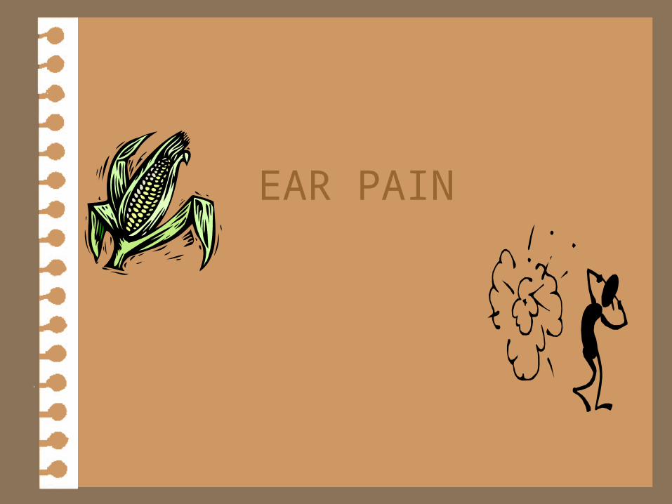
EAR PAIN. Auricle 4 Hematoma 4 Cellulitis 4 Relapsing Polychondritis.
Dec 23, 2015
Welcome message from author
This document is posted to help you gain knowledge. Please leave a comment to let me know what you think about it! Share it to your friends and learn new things together.
Transcript
EAR PAIN
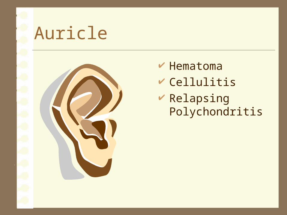
Auricle
Hematoma Cellulitis Relapsing
Polychondritis
Hematoma
A localized mass of extravasated blood within the auricle- “bruise”
Hematoma
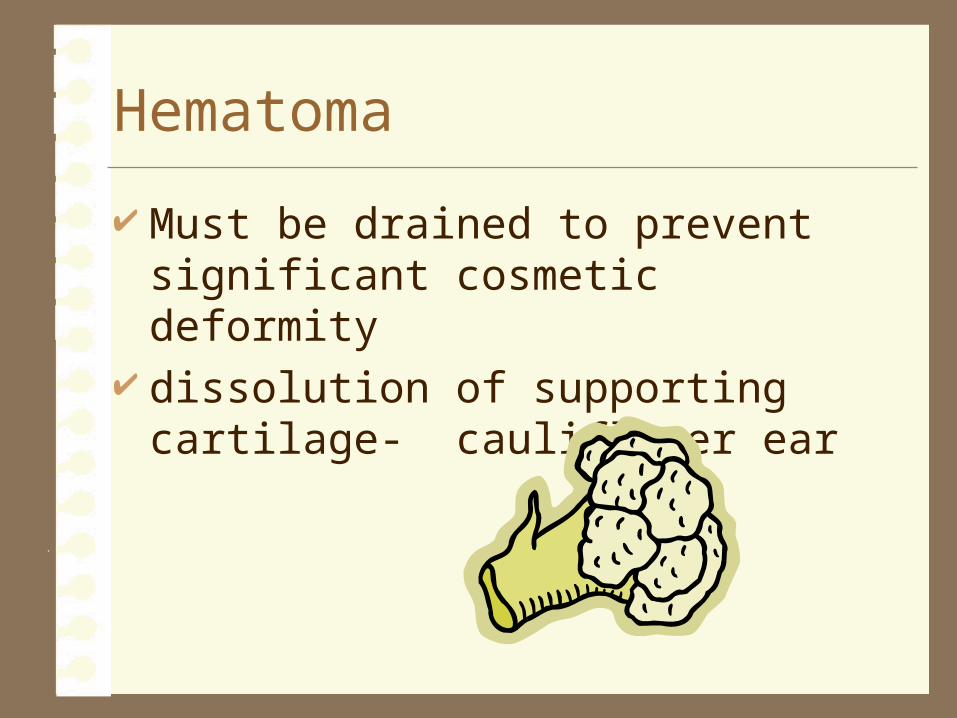
Must be drained to prevent significant cosmetic deformity
dissolution of supporting cartilage- cauliflower ear
Cellulitis
Inflammation of the cellular tissue May include lobule Treat with Augmentin or Keflex Complications- perichondritis and its
resultant deformity
Relapsing Polychondritis
Auricular erythema and edema Recurrent, frequently bilateral, painful Does not include lobule- no cartilage Systematic- may progress to involvement of
the tracheobronchial tree Treat- Corticosteroids might forestall
cartilage dissolution
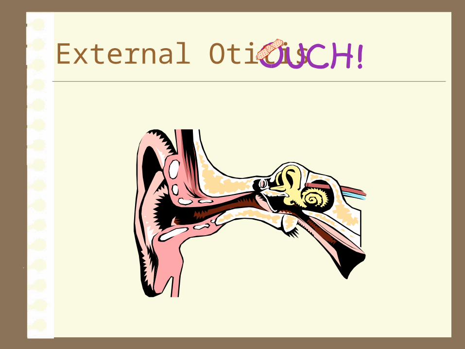
External Otitis
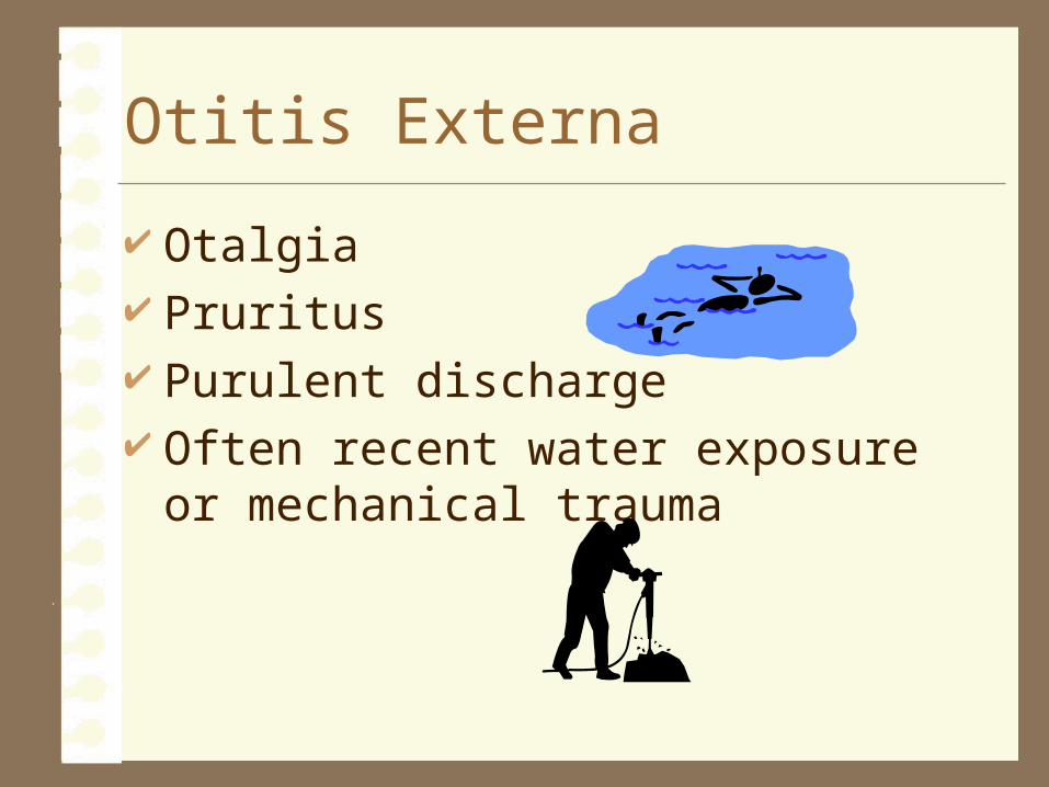
Otitis Externa
Otalgia Pruritus Purulent discharge Often recent water exposure or mechanical
trauma
Examination
Erythema Edema Purulent exudate Auricular pain with manipulation TM- moves normally with pneumatic
otoscopy
Treatment
Avoid moisture Otic drops containing aminoglycoside
antibiotic and anti-inflammatory corticosteroid--neomycin sulfate, polymyxin B sulfate, and hydrocortisone
Ear wick
Auricular Pruritis
Common site- meatus usually self induced
– excoriation
– overly zealous ear cleaning
Otitis Externa?? Dermatologic condition
– seborrheic dermatitis
– psoriasis
Treatment
Regeneration of Cerumen “blanket” Avoid drying agents- soap & water, swabs Mineral oil 0.1% Triamcinolone- topical corticosteroid Oral antihistamine Stop messing with it!!!!
Malignant External Otitis
Persistent external otitis Evolves into Osteomyelitis of the skull base
– Diabetic or Immunocompromised
Pseudomonas aeruginosa
Clinical Findings
Persistent foul aural discharge Granulation in the ear canal Deep otalgia Progressive cranial nerve palsies
– (VI, VII, IX, X, XI, XII)
Diagnosis confirmed with CT – osseous erosion
Treatment
Prolonged (antipseudomonal) ATB therapy– IV or Oral ciprofloxacin
Occasional surgical debridement
Serous Otitis Media
Caused by negative pressure– Blocked auditory tube– Transudation of fluid
• children- tubes more narrow, more horizontal
• common after URI
• adults- persistent--think cancer
Clinical Findings
Dull, hypomobile TM Air bubbles in middle ear Conductive hearing loss
Treatment
Autoinflation Oral corticosteroids Oral ATB All else fails, ventilating tubes
Barotrauma
Negative pressure tends to collapse and lock the auditory tube– Rapid altitudinal change
• Air travel
• Scuba diving
Treatment
Swallow, yawn, autoinflate Systemic or topical decongestants
– pseudoephedrine– phenylephrine nasal spray
If persists on ground after treatments listed above…– Myringotomy provides immediate relief – Ventilating tubes- frequent flyer
Acute Otitis Media
Bacterial infection of the mucosally lined air-containing spaces of the temporal bone.– Usually precipitated by viral URI which causes
auditory tube edema…accumulation of fluid that becomes secondarily infected with bacteria
– Streptococcus pneumoniae (49%), Haemophilus influenzae (14%), Moraxella catarrhalis (14%)
H&P Findings
Otalgia Aural pressure Decreased hearing Fever erythema Decreased mobility of TM TM bulge
– perforation eminent
Treatment
ATB– amoxicillin– erythromycin– sulfonamides
Decongestants Tympanocentesis Ventilating tubes ppx
– sulfamethoxazole– amoxicillin
Chronic Otitis Media
Chronic infection Perforation of TM usually present Mucosal changes P. aeruginosa, Proteus, Staphylococcus
aureus
Clinical Findings
Hallmark- purulent aural discharge Pain- on/off Conductive hearing loss
Treatment
Removal of debris earplugs to protect against water exposure ATB drops for exacerbations Definitive- surgical TM repair
– eliminate infection– reconstruction of TM
Cholesteatoma*
Special variety of chronic otitis media
Most common cause is prolonged auditory tube dysfunction, with resultant chronic negative middle ear pressure that draws inward the upper flaccid portion of the tympanic membrane.
*see picture
Cholesteatoma
Creates a squamous epithelium-lined sac Becomes obstructed and fills with
desquamated keratin and becomes chronically infected
Typically erodes bone, causes destruction of nerves, may spread intracranially
Cholesteatoma
Physical examination– epitympanic retraction pocket or marginal
tympanic membrane perforation that exudes keratin debris
Treatment– surgical marsupialization of the sac or its
complete removal
Mastoiditis- complication of OM
Postauricular pain and erythema Spiking fever X-ray reveals coalescence of the mastoid air
cells due to destruction of their bony septa IV ATB and myringotomy for culture and
drainage Mastoidectomy if other fails...
Petrous apicitis- complication of OM Medial portion of the petrous bone between
the inner ear and clivus may become a site of persistent infection
Foul discharge, deep ear and retro-orbital pain, and sixth nerve palsy
Prolonged ATB therapy and surgical drainage
Otogenic skull base osteomylitis- complication of OM Osteomyelitis of the skull base Usually due to P aeruginosa
Facial paralysis- complication of OM Acute-
– Results from inflammation of the nerve in its middle ear segment, perhaps through bacterially secreted neurotoxins
• Myringotomy for drainage and culture
• IV ATB
• prognosis excellent

Chronic– Evolves slowly due to chronic pressure on the
nerve in the middle ear or mastoid by cholesteatoma
– surgical correction of the underlying disease– prognosis less favorable
Sigmoid sinus thrombosis - complication of OM Trapped infection within the mastoid air cells
adjacent to the sigmoid sinus may cause septic thrombophlebitis
Systemic sepsis- spiking fevers, chills Increased intracranial pressure- HA, lethargy,
nausea and vomiting, papilledema Diagnosis- MR venography Tx- IV ATB, surgical drainage
Central Nervous System Infection - complication of OM Otogenic meningitis- most common
intracranial complication of ear infection
Non-auditory causes of earache
Temporomandibular joint dysfunction– chewing (soft foods, massage)– psychogenic– dental malocclusion (dental referral)
Glossopharyngeal neuralgia– refractory to medical management, may
respond to decompression of ninth nerve
Non-auditory causes of earache
Infections and neoplasia that involve the oropharynx, hypopharynx, and larynx– persistent earache demands specialty referral to
exclude cancer of the upper aerodigestive tract

Related Documents