911 Mycologia, 95(5), 2003, pp. 911–920. q 2003 by The Mycological Society of America, Lawrence, KS 66044-8897 Dyrithiopsis lakefuxianensis gen. et sp. nov. from Fuxian Lake, Yunnan, China, and notes on the taxonomic confusion surrounding Dyrithium R. Jeewon 1 School of Biological Sciences, King Henry Building, University of Portsmouth, Portsmouth, PO1 2DY, United Kingdom L. Cai Centre for Research in Fungal Diversity, Department of Ecology & Biodiversity, University of Hong Kong, Pokfulam Road, Hong Kong, SAR, PR China E. C. Y. Liew School of Land, Water & Crop Sciences, McMillan Building A05, University of Sydney, NSW 2006, Australia K. Q. Zhang Laboratory for conservation and Utilization of Bio- resource, Yunnan University, Kunming, Yunnan, PR China K. D. Hyde Centre for Research in Fungal Diversity, Department of Ecology & Biodiversity, University of Hong Kong, Pokfulam Road, Hong Kong, SAR, PR China Abstract: A new taxon with Dyrithium-like character- istics was collected from Lake Fuxian in China. The taxon is typical of the Amphisphaeriaceae in that it has relatively large, ostiolate, immersed ascomata, unitun- icate asci with a J1 subapical ring, and brown asco- spores. It is similar to Dyrithium in that it has muri- form ascospores, but considerable confusion sur- rounds this genus. In Dyrithium asci are bitunicate and lack a J1 subapical ring, while this was not true of our species. A new genus, Dyrithiopsis, therefore is established to accommodate this new taxon. Details of its anamorph also are provided, based on cultural studies. Parsimony analyses of part of the large-sub- unit rDNA provide further evidence to support the familial placement of this new genus in the Amphis- phaeriaceae. The taxonomic position of Dyrithium also is discussed. Key words: Amphisphaeriaceae, anamorph, Pes- talotiopsis, phylogeny, rDNA Accepted for publication March 13, 2003. 1 Corresponding author. E-mail: [email protected] INTRODUCTION Dyrithium M.E. Barr is a monotypic genus represent- ed by Sphaeria livida Fr., which was included in the Amphisphaeriaceae by Barr (1994). However, Kang et al (1999a) re-examined genera in the Amphis- phaeriaceae and excluded Dyrithium because the asci in the collection they examined were bitunicate. This collection had been listed as a collection of Dyrithium lividum (Fr.) M.E. Barr by Barr (1994). However, Kang et al (1999a) had not examined the holotype of Sphaeria livida. During a biodiversity survey of fungi on submerged wood in Lake Fuxian, Yunnan, China (Cai et al 2002), we collected a taxon with characteristics that keyed out to Dyrithium in the key provided by Barr (1994). Ascospores were brown and muriform, while asci were unitunicate and the ostiolate ascomata were relatively large and immersed. The taxon differed from Dyrithium, however, in that it clearly has unitun- icate asci with a J1 subapical ring. In this respect it was even more typical of the Amphisphaeriaceae. The objectives of this study are: (i) to establish Dy- rithiopsis as a new genus: (ii) to support its inclusion in the family Amphisphaeriaceae based on phyloge- netic analyses of part of the rDNA sequences; and (iii) to discuss its morphological affinities with Dyri- thium. MATERIALS AND METHODS Morphological and cultural studies. A fresh specimen of the new species collected from Yunnan, China, and the herbar- ium specimen of Dyrithium obtained from UPS were ex- amined morphologically. To establish anamorph-teleo- morph connections of the new species, single ascospores from ascomata were isolated by micromanipulation, follow- ing the procedures as outlined by Goh (1999) and Choi et al (1999), and allowed to grow on PDA agar under light at 24–26 C. For examination of morphological features of the teleomorph, cotton blue/lactic acid and phase-contrast mi- croscopy were used. Measurements of the conidia arising from the single ascospore culture were done in water. DNA extraction, amplification and sequencing. Single-spore cultures of the new species were grown on PDA medium for 3 wk. Sources, Genbank accession numbers, designation and geographical origin of species used in this study are listed in TABLE I. Mycelia were used as the starting material for DNA extraction, following a modified protocol of Doyle

Welcome message from author
This document is posted to help you gain knowledge. Please leave a comment to let me know what you think about it! Share it to your friends and learn new things together.
Transcript
911
Mycologia, 95(5), 2003, pp. 911–920.q 2003 by The Mycological Society of America, Lawrence, KS 66044-8897
Dyrithiopsis lakefuxianensis gen. et sp. nov. from Fuxian Lake, Yunnan, China,and notes on the taxonomic confusion surrounding Dyrithium
R. Jeewon1
School of Biological Sciences, King Henry Building,University of Portsmouth, Portsmouth, PO1 2DY,United Kingdom
L. CaiCentre for Research in Fungal Diversity, Department ofEcology & Biodiversity, University of Hong Kong,Pokfulam Road, Hong Kong, SAR, PR China
E. C. Y. LiewSchool of Land, Water & Crop Sciences, McMillanBuilding A05, University of Sydney, NSW 2006,Australia
K. Q. ZhangLaboratory for conservation and Utilization of Bio-resource, Yunnan University, Kunming, Yunnan, PRChina
K. D. HydeCentre for Research in Fungal Diversity, Department ofEcology & Biodiversity, University of Hong Kong,Pokfulam Road, Hong Kong, SAR, PR China
Abstract: A new taxon with Dyrithium-like character-istics was collected from Lake Fuxian in China. Thetaxon is typical of the Amphisphaeriaceae in that it hasrelatively large, ostiolate, immersed ascomata, unitun-icate asci with a J1 subapical ring, and brown asco-spores. It is similar to Dyrithium in that it has muri-form ascospores, but considerable confusion sur-rounds this genus. In Dyrithium asci are bitunicateand lack a J1 subapical ring, while this was not trueof our species. A new genus, Dyrithiopsis, therefore isestablished to accommodate this new taxon. Detailsof its anamorph also are provided, based on culturalstudies. Parsimony analyses of part of the large-sub-unit rDNA provide further evidence to support thefamilial placement of this new genus in the Amphis-phaeriaceae. The taxonomic position of Dyrithium alsois discussed.
Key words: Amphisphaeriaceae, anamorph, Pes-talotiopsis, phylogeny, rDNA
Accepted for publication March 13, 2003.1 Corresponding author. E-mail: [email protected]
INTRODUCTION
Dyrithium M.E. Barr is a monotypic genus represent-ed by Sphaeria livida Fr., which was included in theAmphisphaeriaceae by Barr (1994). However, Kanget al (1999a) re-examined genera in the Amphis-phaeriaceae and excluded Dyrithium because the asciin the collection they examined were bitunicate. Thiscollection had been listed as a collection of Dyrithiumlividum (Fr.) M.E. Barr by Barr (1994). However,Kang et al (1999a) had not examined the holotypeof Sphaeria livida.
During a biodiversity survey of fungi on submergedwood in Lake Fuxian, Yunnan, China (Cai et al2002), we collected a taxon with characteristics thatkeyed out to Dyrithium in the key provided by Barr(1994). Ascospores were brown and muriform, whileasci were unitunicate and the ostiolate ascomata wererelatively large and immersed. The taxon differedfrom Dyrithium, however, in that it clearly has unitun-icate asci with a J1 subapical ring. In this respect itwas even more typical of the Amphisphaeriaceae.
The objectives of this study are: (i) to establish Dy-rithiopsis as a new genus: (ii) to support its inclusionin the family Amphisphaeriaceae based on phyloge-netic analyses of part of the rDNA sequences; and(iii) to discuss its morphological affinities with Dyri-thium.
MATERIALS AND METHODS
Morphological and cultural studies. A fresh specimen of thenew species collected from Yunnan, China, and the herbar-ium specimen of Dyrithium obtained from UPS were ex-amined morphologically. To establish anamorph-teleo-morph connections of the new species, single ascosporesfrom ascomata were isolated by micromanipulation, follow-ing the procedures as outlined by Goh (1999) and Choi etal (1999), and allowed to grow on PDA agar under light at24–26 C. For examination of morphological features of theteleomorph, cotton blue/lactic acid and phase-contrast mi-croscopy were used. Measurements of the conidia arisingfrom the single ascospore culture were done in water.
DNA extraction, amplification and sequencing. Single-sporecultures of the new species were grown on PDA mediumfor 3 wk. Sources, Genbank accession numbers, designationand geographical origin of species used in this study arelisted in TABLE I. Mycelia were used as the starting materialfor DNA extraction, following a modified protocol of Doyle
912 MYCOLOGIA
TABLE I. Fungal strains used in the study, and their accession numbers, hosts and locality
SpeciesSource ofculturesa Host, geographic origin GenBank No.
IngroupAmphisphaeria umbrinaBartalinia robillardoidesDiscosia sp.Discostroma tostaDyrithiopsis lakefuxianensisLepteutypa cupressiMonochaetia monochaeta
HKUCC 994BRIP 14180HKUCC 6626HKUCC 1004HKUCC 7303IMI 052255CBS 199.82
Tilia sp., SwitzerlandMacrotyloma daltonii, AustraliaUnidentified dead leaf, Hong KongUnidentified terrestrial wood, Hong KongSubmerged wood, ChinaCupressus forbesii, KenyaQuercus pubescens, Italy
AF452029AF382366AF382381AF382380AF452047AF382379AF382370
Pestalotia palmarumPestalotia vacciniiPestalotiopsis maculansPestalotiopsis versicolorSeimatosporium vacciniiSeiridium cupressiTruncatella angustata
ATCC 10085ICMP 5446CBS 322.76BRIP 14534ICMP 7003FABI, CMW 5596ICMP 7062
Coconut Palm, IndiaVaccinium sp., New ZealandCamellia sp., FrancePsidium guajava, AustraliaVaccinium ashei Reade, New ZealandCupressus sempervirens, S. AfricaMalus domestica, New Zealand
AF382361AF382362AF382354AF382357AF382374AF382378AF382383
OutgroupsDiaporthe phaseolorumDothidea sambuciHypocrea schweinitziiOphiostoma africanum
NAb
CBS 198.58
NAb
NAb
Acer pseudoplatanus, SwitzerlandNAb
NAb
U47830AF382387U47833AF221015
Nectria pityrodesNectria sesquiciliXylaria curtaXylaria hypoxylonc
ATCC 208843ATCC 66880NAb
ATCC 42768
NAb
NAb
NAb
NAb
AF193240AF193241U47940U47841
a ATCC: American Type Culture Collection; BRIP: Queensland Department of Primary Industries Plant Pathology Herbar-ium; CBS: Centraalbureau voor Schimmelcultures; FABI: Forestry and Agricultural Biotechnology Institute; HKUCC: TheUniversity of Hong Kong Culture Collection; ICMP: International Collection of Microorganisms from Plants.
b NA: Information not available.
and Doyle (1987). Part of the LSU of the rDNA was ampli-fied by PCR with primers LROR and LR05. Amplified prod-ucts then were purified using minicolumns, purification res-in and buffer, according to the manufacturer’s protocol(Wizard PCR Preps DNA Purification System). Purifiedproducts were sequenced directly, using primers LROR,LR3R, LR5, LR3 and other reagents, following the manu-facturer’s protocol (Pharmacia Biotech). Sequences wereedited manually and assembled using the Alf software andSeqPup (Gilbert 1996).
Phylogenetic analyses. Phylogenetic analyses were per-formed in PAUP* 4.0b9 (Swofford 2002) on part of the LSUof the rDNA sequenced. Analyses included representativesof members from other orders (sequences available fromGenBank) to resolve phylogenetic relationships and to rootcladograms. Trees were inferred using the heuristic searchoption with 10, 100 and 1000 random-sequence additions.Gaps were treated as missing data, and characters were un-ordered and weighted equally and differentially. Clade sta-bility was assessed in a bootstrap analysis with 1000 repli-cates, random-sequence additions with maxtrees set to 5000and other default parameters as implemented in PAUP*.For maximum-likelihood (ML) analyses, a single tree gen-erated under the maximum-parsimony criterion was used
as a starting tree and transition-transversion ratios, base fre-quencies and shape parameter were estimated. Using theseinitial estimates of substitution rates and kinds, a heuristicsearch with TBR branch swapping was used to find a max-imum-likelihood tree. The gamma model of site-rate varia-tion was used with no enforcement of a molecular clock.Initial branch lengths were obtained with Rogers-Swoffordapproximation methods. Neighbor-joining (NJ) analyseswas conducted under different models of distance algo-rithms, including HKY85, JC, K2P and F81, and the supportfor individual clades within the tree was assessed by 1000replicates of bootstrapping. Dothidea sambuci was used asoutgroup.
RESULTS
Description of Dyrithiopsis lakefuxianensis (teleo-morph)
Dyrithiopsis L. Cai, R. Jeewon et K.D. Hyde, gen. nov.[Ascomata immersa vel semi-immersa, subglobosa, nigra,
coriacea, papillata, ostiolata, periphysata, paraphysata. Asciunitunicati, 8-spori, cylindrici, pedicellati, apparatus apicaleJ1 praediti. Ascosporae, fusiformes vel ellipsoid, pallidbrunneae, muriformes, euseptatae.]
913JEEWON ET AL: A NEW AMPHISPHAERIACEOUS ASCOMYCETE FROM CHINA
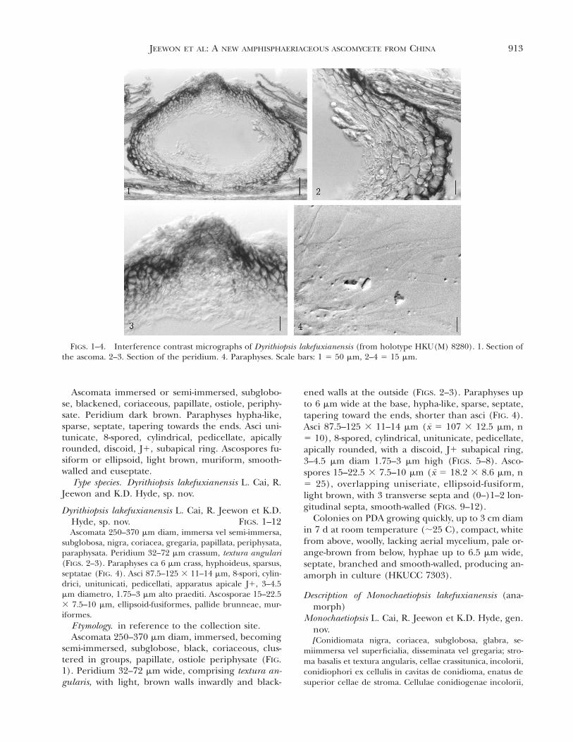
FIGS. 1–4. Interference contrast micrographs of Dyrithiopsis lakefuxianensis (from holotype HKU(M) 8280). 1. Section ofthe ascoma. 2–3. Section of the peridium. 4. Paraphyses. Scale bars: 1 5 50 mm, 2–4 5 15 mm.
Ascomata immersed or semi-immersed, subglobo-se, blackened, coriaceous, papillate, ostiole, periphy-sate. Peridium dark brown. Paraphyses hypha-like,sparse, septate, tapering towards the ends. Asci uni-tunicate, 8-spored, cylindrical, pedicellate, apicallyrounded, discoid, J1, subapical ring. Ascospores fu-siform or ellipsoid, light brown, muriform, smooth-walled and euseptate.
Type species. Dyrithiopsis lakefuxianensis L. Cai, R.Jeewon and K.D. Hyde, sp. nov.
Dyrithiopsis lakefuxianensis L. Cai, R. Jeewon et K.D.Hyde, sp. nov. FIGS. 1–12Ascomata 250–370 mm diam, immersa vel semi-immersa,
subglobosa, nigra, coriacea, gregaria, papillata, periphysata,paraphysata. Peridium 32–72 mm crassum, textura angulari(FIGS. 2–3). Paraphyses ca 6 mm crass, hyphoideus, sparsus,septatae (FIG. 4). Asci 87.5–125 3 11–14 mm, 8-spori, cylin-drici, unitunicati, pedicellati, apparatus apicale J1, 3–4.5mm diametro, 1.75–3 mm alto praediti. Ascosporae 15–22.53 7.5–10 mm, ellipsoid-fusiformes, pallide brunneae, mur-iformes.
Etymology. in reference to the collection site.Ascomata 250–370 mm diam, immersed, becoming
semi-immersed, subglobose, black, coriaceous, clus-tered in groups, papillate, ostiole periphysate (FIG.1). Peridium 32–72 mm wide, comprising textura an-gularis, with light, brown walls inwardly and black-
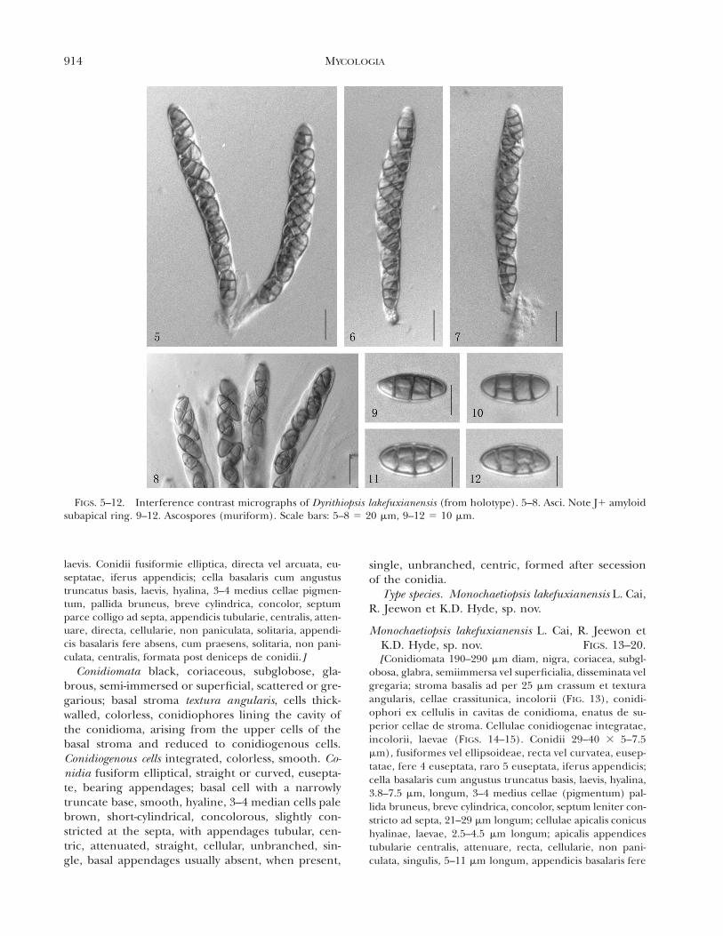
ened walls at the outside (FIGS. 2–3). Paraphyses upto 6 mm wide at the base, hypha-like, sparse, septate,tapering toward the ends, shorter than asci (FIG. 4).Asci 87.5–125 3 11–14 mm (x 5 107 3 12.5 mm, n5 10), 8-spored, cylindrical, unitunicate, pedicellate,apically rounded, with a discoid, J1 subapical ring,3–4.5 mm diam 1.75–3 mm high (FIGS. 5–8). Asco-spores 15–22.5 3 7.5–10 mm (x 5 18.2 3 8.6 mm, n5 25), overlapping uniseriate, ellipsoid-fusiform,light brown, with 3 transverse septa and (0–)1–2 lon-gitudinal septa, smooth-walled (FIGS. 9–12).
Colonies on PDA growing quickly, up to 3 cm diamin 7 d at room temperature (;25 C), compact, whitefrom above, woolly, lacking aerial mycelium, pale or-ange-brown from below, hyphae up to 6.5 mm wide,septate, branched and smooth-walled, producing an-amorph in culture (HKUCC 7303).
Description of Monochaetiopsis lakefuxianensis (ana-morph)
Monochaetiopsis L. Cai, R. Jeewon et K.D. Hyde, gen.nov.[Conidiomata nigra, coriacea, subglobosa, glabra, se-
miimmersa vel superficialia, disseminata vel gregaria; stro-ma basalis et textura angularis, cellae crassitunica, incolorii,conidiophori ex cellulis in cavitas de conidioma, enatus desuperior cellae de stroma. Cellulae conidiogenae incolorii,
914 MYCOLOGIA
FIGS. 5–12. Interference contrast micrographs of Dyrithiopsis lakefuxianensis (from holotype). 5–8. Asci. Note J1 amyloidsubapical ring. 9–12. Ascospores (muriform). Scale bars: 5–8 5 20 mm, 9–12 5 10 mm.
laevis. Conidii fusiformie elliptica, directa vel arcuata, eu-septatae, iferus appendicis; cella basalaris cum angustustruncatus basis, laevis, hyalina, 3–4 medius cellae pigmen-tum, pallida bruneus, breve cylindrica, concolor, septumparce colligo ad septa, appendicis tubularie, centralis, atten-uare, directa, cellularie, non paniculata, solitaria, appendi-cis basalaris fere absens, cum praesens, solitaria, non pani-culata, centralis, formata post deniceps de conidii.]
Conidiomata black, coriaceous, subglobose, gla-brous, semi-immersed or superficial, scattered or gre-garious; basal stroma textura angularis, cells thick-walled, colorless, conidiophores lining the cavity ofthe conidioma, arising from the upper cells of thebasal stroma and reduced to conidiogenous cells.Conidiogenous cells integrated, colorless, smooth. Co-nidia fusiform elliptical, straight or curved, eusepta-te, bearing appendages; basal cell with a narrowlytruncate base, smooth, hyaline, 3–4 median cells palebrown, short-cylindrical, concolorous, slightly con-stricted at the septa, with appendages tubular, cen-tric, attenuated, straight, cellular, unbranched, sin-gle, basal appendages usually absent, when present,
single, unbranched, centric, formed after secessionof the conidia.
Type species. Monochaetiopsis lakefuxianensis L. Cai,R. Jeewon et K.D. Hyde, sp. nov.
Monochaetiopsis lakefuxianensis L. Cai, R. Jeewon etK.D. Hyde, sp. nov. FIGS. 13–20.[Conidiomata 190–290 mm diam, nigra, coriacea, subgl-
obosa, glabra, semiimmersa vel superficialia, disseminata velgregaria; stroma basalis ad per 25 mm crassum et texturaangularis, cellae crassitunica, incolorii (FIG. 13), conidi-ophori ex cellulis in cavitas de conidioma, enatus de su-perior cellae de stroma. Cellulae conidiogenae integratae,incolorii, laevae (FIGS. 14–15). Conidii 29–40 3 5–7.5mm), fusiformes vel ellipsoideae, recta vel curvatea, eusep-tatae, fere 4 euseptata, raro 5 euseptata, iferus appendicis;cella basalaris cum angustus truncatus basis, laevis, hyalina,3.8–7.5 mm, longum, 3–4 medius cellae (pigmentum) pal-lida bruneus, breve cylindrica, concolor, septum leniter con-stricto ad septa, 21–29 mm longum; cellulae apicalis conicushyalinae, laevae, 2.5–4.5 mm longum; apicalis appendicestubularie centralis, attenuare, recta, cellularie, non pani-culata, singulis, 5–11 mm longum, appendicis basalaris fere
915JEEWON ET AL: A NEW AMPHISPHAERIACEOUS ASCOMYCETE FROM CHINA
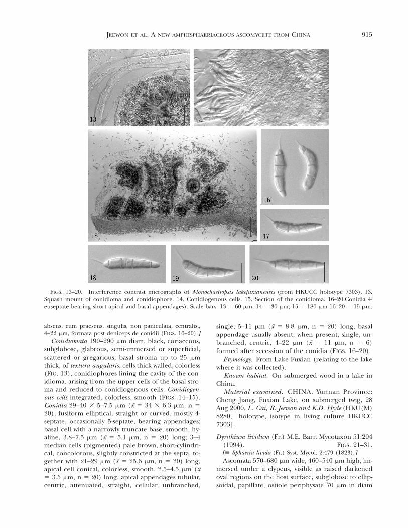
FIGS. 13–20. Interference contrast micrographs of Monochaetiopsis lakefuxianensis (from HKUCC holotype 7303). 13.Squash mount of conidioma and conidiophore. 14. Conidiogenous cells. 15. Section of the conidioma. 16–20.Conidia 4-euseptate bearing short apical and basal appendages). Scale bars: 13 5 60 mm, 14 5 30 mm, 15 5 180 mm 16–20 5 15 mm.
absens, cum praesens, singulis, non paniculata, centralis,,4–22 mm, formata post deniceps de conidii (FIGS. 16–20).]
Conidiomata 190–290 mm diam, black, coriaceous,subglobose, glabrous, semi-immersed or superficial,scattered or gregarious; basal stroma up to 25 mmthick, of textura angularis, cells thick-walled, colorless(FIG. 13), conidiophores lining the cavity of the con-idioma, arising from the upper cells of the basal stro-ma and reduced to conidiogenous cells. Conidiogen-ous cells integrated, colorless, smooth (FIGS. 14–15).Conidia 29–40 3 5–7.5 mm (x 5 34 3 6.3 mm, n 520), fusiform elliptical, straight or curved, mostly 4-septate, occasionally 5-septate, bearing appendages;basal cell with a narrowly truncate base, smooth, hy-aline, 3.8–7.5 mm (x 5 5.1 mm, n 5 20) long; 3–4median cells (pigmented) pale brown, short-cylindri-cal, concolorous, slightly constricted at the septa, to-gether with 21–29 mm (x 5 25.6 mm, n 5 20) long,apical cell conical, colorless, smooth, 2.5–4.5 mm (x5 3.5 mm, n 5 20) long, apical appendages tubular,centric, attenuated, straight, cellular, unbranched,
single, 5–11 mm (x 5 8.8 mm, n 5 20) long, basalappendage usually absent, when present, single, un-branched, centric, 4–22 mm (x 5 11 mm, n 5 6)formed after secession of the conidia (FIGS. 16–20).
Etymology. From Lake Fuxian (relating to the lakewhere it was collected).
Known habitat. On submerged wood in a lake inChina.
Material examined. CHINA. Yunnan Province:Cheng Jiang, Fuxian Lake, on submerged twig, 28Aug 2000, L. Cai, R. Jeewon and K.D. Hyde (HKU(M)8280, [holotype, isotype in living culture HKUCC7303].
Dyrithium lividum (Fr.) M.E. Barr, Mycotaxon 51:204(1994). FIGS. 21–31.[[ Sphaeria livida (Fr.) Syst. Mycol. 2:479 (1823).]Ascomata 570–680 mm wide, 460–540 mm high, im-
mersed under a clypeus, visible as raised darkenedoval regions on the host surface, subglobose to ellip-soidal, papillate, ostiole periphysate 70 mm in diam
916 MYCOLOGIA
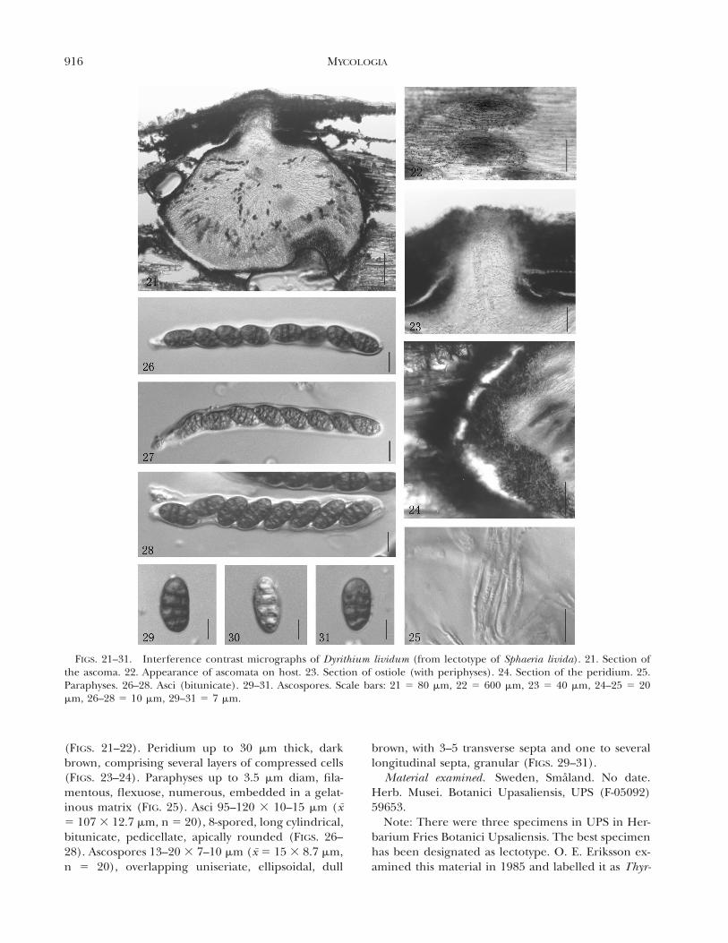
FIGS. 21–31. Interference contrast micrographs of Dyrithium lividum (from lectotype of Sphaeria livida). 21. Section ofthe ascoma. 22. Appearance of ascomata on host. 23. Section of ostiole (with periphyses). 24. Section of the peridium. 25.Paraphyses. 26–28. Asci (bitunicate). 29–31. Ascospores. Scale bars: 21 5 80 mm, 22 5 600 mm, 23 5 40 mm, 24–25 5 20mm, 26–28 5 10 mm, 29–31 5 7 mm.
(FIGS. 21–22). Peridium up to 30 mm thick, darkbrown, comprising several layers of compressed cells(FIGS. 23–24). Paraphyses up to 3.5 mm diam, fila-mentous, flexuose, numerous, embedded in a gelat-inous matrix (FIG. 25). Asci 95–120 3 10–15 mm (x5 107 3 12.7 mm, n 5 20), 8-spored, long cylindrical,bitunicate, pedicellate, apically rounded (FIGS. 26–28). Ascospores 13–20 3 7–10 mm (x 5 15 3 8.7 mm,n 5 20), overlapping uniseriate, ellipsoidal, dull
brown, with 3–5 transverse septa and one to severallongitudinal septa, granular (FIGS. 29–31).
Material examined. Sweden, Smaland. No date.Herb. Musei. Botanici Upasaliensis, UPS (F-05092)59653.
Note: There were three specimens in UPS in Her-barium Fries Botanici Upsaliensis. The best specimenhas been designated as lectotype. O. E. Eriksson ex-amined this material in 1985 and labelled it as Thyr-
917JEEWON ET AL: A NEW AMPHISPHAERIACEOUS ASCOMYCETE FROM CHINA
idium lividum (Pers. Fr.) Sacc. The other two Sphaer-ia livida are isotypes.
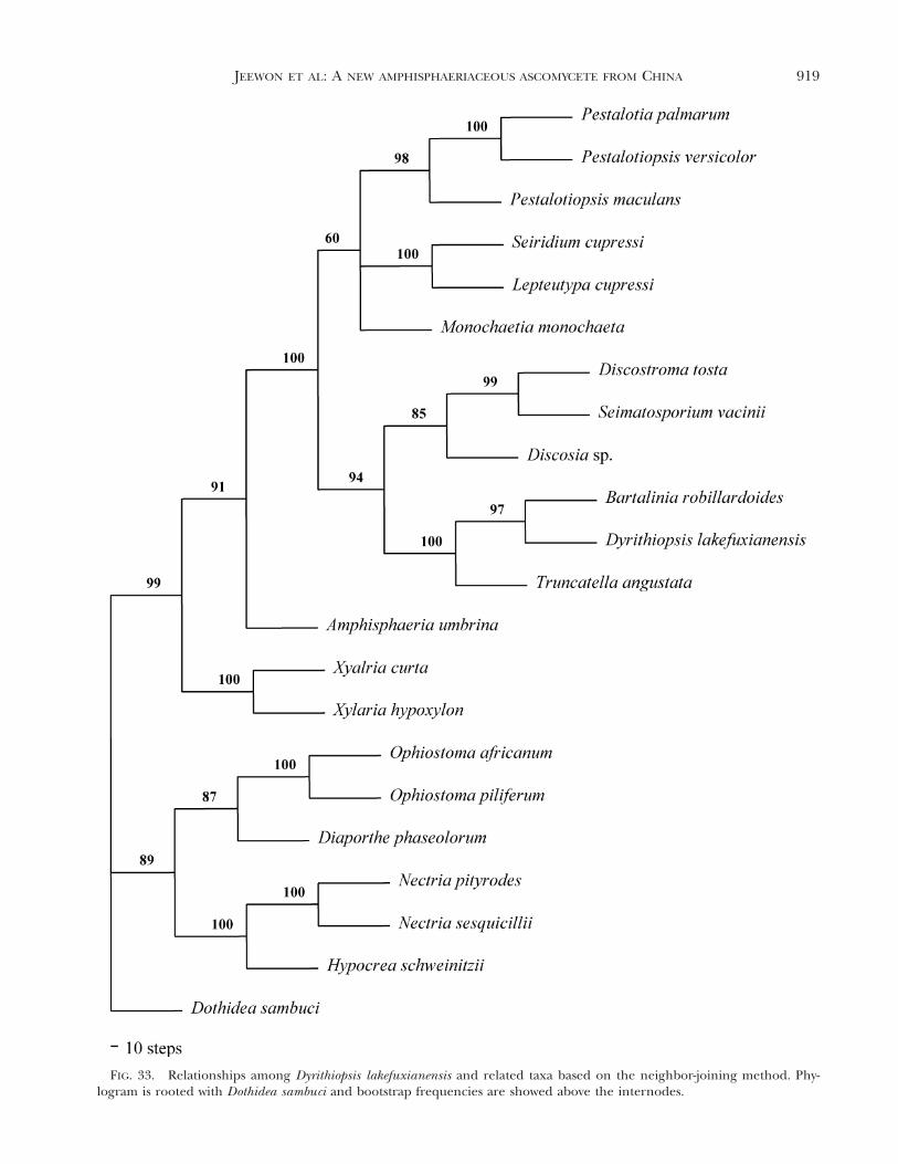
Phylogenetic analyses. The alignment of the datasetconsists of 900 bp, of which 189 were parsimony in-formative. Unweighted parsimony analyses and treat-ing gaps as missing data resulted in three trees onone island. Tree length was 636 steps, CI 5 0.654, RI5 0.695, RC 5 0.455 and HI 5 0.346. Unweightedparsimony analyses (transition-transversion ratio of1.5:1) of the same dataset resulted in one most-par-simonious tree of 765 steps, CI 5 0.654, RI 5 0.700,RC 5 0.457 and HI 5 0.346. Weighted parsimonyanalyses yielded trees with better resolution that weremore strongly supported by bootstrap values thantrees generated from unweighted parsimony analyses.The single parsimonious tree obtained by treatinggaps as missing data and with a transition-transver-sion ratio of 1.5:1 is shown in FIG. 32. Maximum-like-lihood (ML) analyses under the HKY model, with anestimated shape parameter of 0.2 and an estimatedtransition-transversion ratio of 1.7, yielded a singletree of log-likelihood of 4345.69818. The topology ofthe ML tree is identical in that of FIG. 32 (results notshown). The phylogram generated under the neigh-bor-joining method was similar to FIG. 32 with a treelength of 644 steps, CI 5 0.646, RI 5 0.684, RC 50.442 HI 5 0.352 and -log likelihood of 4646.14791(FIG. 33).
Clade A (FIG. 32) supports the monophyly of allamphisphaeriaceaous genera producing Pestalotiop-sis-like anamorphs and is supported by a high boot-strap confidence (99%). Dyrithiopsis lakefuxianensisappear to be closely related to the genus BartaliniaTassi and forms a sister group to the genus Trunca-tella Stey. These relationships also are supported byhigh bootstrap values.
DISCUSSION
Morphological studies coupled with phylogeneticanalyses of rDNA sequences reported here provideadditional insights regarding the systematic place-ment of the new genus Dyrithiopsis. On morpholog-ical grounds, Dyrithiopsis has close affinities to thegenus Dyrithium, which currently is accommodatedin the family Amphisphaeriaceae (Barr 1994). How-ever, considerable taxonomic confusion surroundsthe genus Dyrithium (Kang et al 1999a). The genericname Dyrithium, based on D. lividum, was introducedfor Sphaeria livida by Barr (1994). Kang et al (1999a)examined a collection of Sphaeria livida, which wascited as having unitunicate asci by Barr (1994), andfound that the asci were distinctly bitunicate (FIGS.21–31, Kang et al 1999a). We requested Fries typematerial of Sphaeria livida to resolve this confusion.
The material, despite its age, is in reasonable condi-tion, and ascomata contain numerous asci and asco-spores. Although we could find no evidence that theasci were fissitunicate, it was clear that they were bi-tunicate (FIGS. 26–28) and reminiscent of generasuch as Verruculina (Kohlm.) Kohlm. & Volkm.-Kohlm. (Hyde et al 2000). There was no J1 subapicalascal ring. Because of the ascal characters that aremore typical of the loculoascomycetes, we are con-vinced that Dyrithium is not amphisphaeriaceous.The loculoascomycetous nature of the taxon also wasevident in the pseudoparaphyses, which were longand straight between the asci, anastomosed above theasci, did not taper and were embedded in a gelati-nous matrix. This is typical of bitunicate genera, suchas Massarina Sacc., as compared to the sparse taper-ing paraphyses in the Amphisphaeriaceae (Poonythet al 2000).
Similar confusion surrounds genera such as Rous-sella Sacc. and Saccardoella Speg. (Hyde et al 1996a)because, although the asci in these genera are bitun-icate, they do not show fissitunicate dehiscence. Wetherefore describe a new genus Dyrithiopsis to accom-modate our new taxon, which clearly is amphisphaer-iaceous, based on its ascal characters. Characters per-taining to the Amphisphaeriaceae and related fami-lies already have been elaborated (Hyde 1996, Hydeet al 1996b, Kang et al 1999b).
The outcome of molecular analyses based on thelarge subunit rDNA sequences provide further evi-dence that Dyrithiopsis is phylogenetically related toother members of the family Amphisphaeriaceae(FIG. 32). In recent studies, it has been shown thatascomycetous genera producing Pestalotiopsis-like an-amorphs as well as coelomycetous genera, such asBartalinia, Discosia Libert, Pestalotiopsis Stey., Mono-chaetia (Sacc.) Allesh., Seimatosporium Corda, Seiri-dium Nees: Fries and Truncatella, are members of thefamily Amphisphaeriaceae (Kang et al 1998; 1999b;Jeewon et al 2002, 2003a, b). Based on morphologi-cal and molecular data, the taxonomic concept ofthis family has been revised to accommodate generathat produce Pestalotiopsis-like anamorphs (Kang etal 1998, 1999a, b). Results from our molecular studyindicate that Dyrithiopsis is strictly amphisphaeri-aceaous because it is nested between Discostroma Cle-ments and Lepteutypa Petrak, which are members ofthe Amphisphaeriaceae sensu stricto (Barr 1994,Hawksworth et al 1995, Kang et al 1998, 1999b). Inaddition, it possesses unitunicate asci with a J1 sub-apical ring and a Pestalotiopsis-like anamorph, char-acteristic of other members of this family.
The new genus Dyrithiopsis produced a coelomy-cetous anamorph with morphological affinities toPestalotiopsis, Monochaetia and Seimatosporium. It has

918 MYCOLOGIA
FIG. 32. Phylogram depicting the relationships of Dyrithiopsis lakefuxianensis with respect to others members of theAmphisphaeriaceae. Tree was generated by a weighted parsimony analysis (transition transversion ratio of 1.5:1) from partialLSU sequences of the rDNA gene. Dothidea sambuci was the designated outgroup. Bootstrap values (1000 replicates) areindicated on nodes. * Represents those clades that collapsed in the bootstrap analysis.
919JEEWON ET AL: A NEW AMPHISPHAERIACEOUS ASCOMYCETE FROM CHINA
FIG. 33. Relationships among Dyrithiopsis lakefuxianensis and related taxa based on the neighbor-joining method. Phy-logram is rooted with Dothidea sambuci and bootstrap frequencies are showed above the internodes.
920 MYCOLOGIA
four euseptate fusoid, brown conidia, which are con-stricted slightly at the septum, similar to those speciesin Bartalinia, Pestalotiopsis, Monochaetia, Seimatospor-ium and Truncatella. This anamorph, however, ismorphologically distinguishable from Monochaetiaand Pestalotiopsis and Seiridium, with respect to theappendages and conidial shape. Conidia are fusiformelliptical and characterized by three pigmented (hon-ey brown) median cells that are slightly constricted.End cells are hyaline, bear a single and short apicaland basal appendage that are unbranched and usu-ally curved and separated by a septum from the endcells. This anamorph also differs morphologicallyfrom Bartalinia and Truncatella (its closest relativesin the tree). Bartalinia is characterized by mediancells that are almost hyaline or pale brown with apicalappendages arising from a particular locus above theapical cell (unlike Monochaetiopsis, which is separatedby a septum). Truncatella, however, possesses mostlytwo median cells with highly branched apical ap-pendages. Further details regarding the morpholog-ical characters and phylogenetic relationships per-taining to Bartaliniam, Discosia, Pestalotiopsis, Mono-chaetia, Seimatosporium, Seiridium and Truncatellahave been discussed by Jeewon et al (2002, 2003b).A new coelomycetous taxon, Monochaetiopsis lakefux-ianensis therefore was erected to describe this fungus.
ACKNOWLEDGMENTS
This study was financially supported by the National NaturalScience Foundation of China (NSFC 3026002, 30230020).We would like to thank UPS for kindly providing materialfor examination. Helen Leung and Heidi Kong are thankedfor technical assistance.
LITERATURE CITED
Barr ME. 1994. Notes on the Amphisphaeriaceae and relatedfamilies. Mycotaxon 51:191–224.
Cai L, Tsui CKM, Zhang K, Hyde KD. 2002. Aquatic fungifrom Lake Fuxian, Yunnan, China. Fungal Diversity 9:57–70.
Choi YW, Hyde KD, Ho WH. 1999. Single spore isolation offungi. Fungal Diversity 3:29–38.
Doyle JJ, Doyle JL. 1987. A rapid DNA isolation procedurefor small quantities of fresh leaf tissues. PhytochemBull 19:11–15.
Gilbert DG. 1996. SeqPup, biosequence editor and analysissoftware for molecular biology Bionet. Software.
Goh TK. 1999. Single spore isolation using a hand-madeglass needle. Fungal Diversity 2:47–63.
Hawksworth DL, Kirk PM, Sutton BC, Pegler DN. 1995. Ain-sworth & Bisby’s dictionary of the fungi. 8th ed. Wal-lingford, UK: CAB International.
Hyde KD. 1996. Fungi from palms. XXIX. Arecophila gen.nov. Amphisphaeriaceae, Ascomycota, with five newspecies and two new combinations. Nova Hedwigia 63:81–100.
———, Eriksson OE, Yue JZ. 1996a. Roussoella, a genus ofuncertain relationships with a Cytoplea anamorph. My-col Res 100:1522–1528.
———, Kang JC, Kong RYC. 1996b. Fungi from palms.XXX. Notes on Amphisphaeria species described frompalms and a description of A. umbrina. Nova Hedwigia63:101–108.
———, Sarma VV, Jones EBG. 2000. Morphology and tax-onomy of higher marine fungi. In: Hyde KD, PointingSB, eds. Marine Mycology—a practical approach. HongKong: Fungal Diversity Press. p 172–204.
Kang JC, Kong RYC, Hyde KD. 1998. Studies on the Am-phisphaeriales I. Amphisphaeriaceae (sensu stricto) andits phylogenetic relationships inferred from 5.8S rDNAand ITS sequences. Fungal Diversity 1:147–157.
———, Hyde KD, Kong RYC. 1999a. Studies on Amphis-phaeriales: the genera excluded from the Amphis-phaeriaceae, Cainiaceae and Clypeosphaeriaceae. Fun-gal Diversity 2:135–151.
———, ———, ———. 1999b. Studies on Amphisphaeri-ales: The Amphisphaeriaceae (sensu stricto). Mycol Res103:53–64.
Jeewon R, Liew ECY, Hyde KD. 2002. Phylogenetic relation-ships of Pestalotiopsis and allied genera inferred fromribosomal DNA sequences and morphological charac-ters. Mol Phylogenet Evol 25:378–392.
———, ———, Simpson JA, Hodgkiss IJ, Hyde KD. 2003.Phylogenetic significance of morphological charactersin the taxonomy of Pestalotiopsis species. Mol Phylo-genet Evol 27:372–383.
Jeewon R, Liew ECY, Hyde KD. 2003b. Molecular systemat-ics of the Amphisphaeriaceae based on cladistic analy-ses of partial LSU rDNA gene sequences. MycologicalResearch (In press).
Poonyth AD, Hyde KD, Aptroot A, Peerally A. 2000. Maur-itiana rhizophorae gen. et sp. nov. (Ascomycetes Requi-enellaceae), with a list of terrestrial saprobic mangrovefungi. Fungal Diversity 4:101–116.
Swofford DL. 2002. PAUP*: phylogenetic analysis using par-simony and other methods. Version 4.0b8. Sunderland,Massachusetts U.S.A. Sinauer Associates.
Related Documents