REPORT DYNC2H1 Mutations Cause Asphyxiating Thoracic Dystrophy and Short Rib-Polydactyly Syndrome, Type III Nathalie Dagoneau, 1,7 Marie Goulet, 1,7 David Genevie `ve, 1 Yves Sznajer, 2 Jelena Martinovic, 1 Sarah Smithson, 3 Ce ´line Huber, 1 Genevie `ve Baujat, 1 Elisabeth Flori, 4 Laura Tecco, 5 Denise Cavalcanti, 1 Anne-Lise Delezoide, 6 Vale ´rie Serre, 1 Martine Le Merrer, 1 Arnold Munnich, 1 and Vale ´rie Cormier-Daire 1, * Jeune asphyxiating thoracic dystrophy (ATD) is an autosomal-recessive chondrodysplasia characterized by short ribs and a narrow thorax, short long bones, inconstant polydactyly, and trident acetabular roof. ATD is closely related to the short rib polydactyly syndrome (SRP) type III, which is a more severe condition characterized by early prenatal expression and lethality and variable malfor- mations. We first excluded IFT80 in a series of 26 fetuses and children belonging to 14 families diagnosed with either ATD or SRP type III. Studying a consanguineous family from Morocco, we mapped an ATD gene to chromosome 11q14.3-q23.1 in a 20.4 Mb region and iden- tified homozygous mutations in the cytoplasmic dynein 2 heavy chain 1 (DYNC2H1) gene in the affected children. Compound hetero- zygosity for DYNC2H1 mutations was also identified in four additional families. Among the five families, 3/5 were diagnosed with ATD and 2/5 included pregnancies terminated for SRP type III. DYNC2H1 is a component of a cytoplasmic dynein complex and is directly involved in the generation and maintenance of cilia. From this study, we conclude that ATD and SRP type III are variants of a single disorder belonging to the ciliopathy group. Jeune asphyxiating thoracic dystrophy (ATD [MIM 208500]) is an autosomal-recessive chondrodysplasia char- acterized by short ribs and respiratory insufficiency and is often fatal in the first year of life. Retinal degeneration, cystic renal disease, and liver involvement occasionally occur in the course of the disease. Mutations in the intra- flagellar transport 80 (IFT80 [MIM 611177]) gene have been recently identified in 3/39 families originating from Pakistan and Turkey, ascribing ATD to the ciliopathy group. 1 ATD is known to be genetically heterogeneous with another locus mapped on chromosome 15q13. 2 ATD is closely related to the short rib polydactyly group, especially to the type III (SRP type III, also called Verma- Naumoff, [MIM 263510]). Both conditions share the same radiological features (including the polydactyly), but SRP type III is more severe, characterized by an early prenatal expression and lethality, variable malformations (cleft lip and/or palate; polycystic kidneys; gastrointes- tinal, urogenital, brain, and/or cardiac malformations), and severely shortened tubular bones having round meta- physeal ends with lateral spikes. 3,4 Here, we report the mapping of a locus on chromosome 11q and the identifica- tion of cytoplasmic dynein 2 heavy chain 1 (DYNC2H1 [MIM 603297]) mutations in five families with ATD or SRP type III, supporting the view that ATD is an heteroge- neous disorder overlapping with SRP type III and belongs to the ciliopathy group. 5 Criteria for inclusion in the study were (1) short ribs and a constricted thoracic cage, (2) trident acetabular roof, (3) small hands and feet, and (4) shortening of the long bones. Included were a total of 15 children (ranging in age from 0 to 19 years of age) and 11 fetuses belonging to 14 fami- lies, diagnosed with either ATD (15 living children and four fetuses) or SRP type III (seven fetuses). Among the fetuses, the diagnosis of SRP (rather than ATD) was as- signed on the basis of the extreme severity of the thorax narrowness and the long bone shortness with the presence of metaphyseal spikes. Among all families with ATD or SRP type III, six originating from Tunisia, Turkey, Portugal, and Morocco were consanguineous. Blood samples from patients and unaffected relatives were obtained with the appropriate written consent, in accordance with the French ethical standards regarding human subjects. We first excluded IFT80 either by linkage analysis in consanguineous families with the use of microsatellite markers or by direct sequencing in isolated cases (data not shown). We then focused on a large consanguineous Moroccan family with two affected children (Figure 1). We performed a genome-wide search, using the GeneChip Human Mapping 250K NspI array (Affymetrix) on DNA samples of the two affected children and one parent. Array experi- ments were performed according to protocols provided by the manufacturer. The SNP genotypes were analyzed with 1 De ´partement de Ge ´ne ´tique, Unite ´ INSERM U781, Universite ´ Paris Descartes, Assistance Publique-Ho ˆpitaux de Paris (AP-HP), Ho ˆpital Necker-Enfants Malades, 75015 Paris, France; 2 Pediatric Clinical Genetics, Ho ˆpital Universitaire des Enfants Reine Fabiola and Center for Human Genetics, U.L.B., 1020 Brussels, Belgium; 3 Department of Clinical Genetics, St Michael’s Hospital, Bristol BS2 8EG, UK; 4 Service de Cytoge ´ne ´tique, Ho ˆpital de Hautepierre, 67091 Strasbourg, France; 5 Department of Gynaecology and Obstetrics, Brugmann University Hospital, CHU Brugmann, 1020 Brussels, Belgium; 6 Depart- ment of Developmental Biology, Universite ´ Paris Diderot, AP-HP, Ho ˆpital Robert Debre ´, 75935 Paris, France 7 These authors contributed equally to this work *Correspondence: [email protected] DOI 10.1016/j.ajhg.2009.04.016. ª2009 by The American Society of Human Genetics. All rights reserved. 706 The American Journal of Human Genetics 84, 706–711, May 15, 2009

Welcome message from author
This document is posted to help you gain knowledge. Please leave a comment to let me know what you think about it! Share it to your friends and learn new things together.
Transcript
REPORT
DYNC2H1 Mutations CauseAsphyxiating Thoracic Dystrophy andShort Rib-Polydactyly Syndrome, Type III
Nathalie Dagoneau,1,7 Marie Goulet,1,7 David Genevieve,1 Yves Sznajer,2 Jelena Martinovic,1
Sarah Smithson,3 Celine Huber,1 Genevieve Baujat,1 Elisabeth Flori,4 Laura Tecco,5
Denise Cavalcanti,1 Anne-Lise Delezoide,6 Valerie Serre,1 Martine Le Merrer,1 Arnold Munnich,1
and Valerie Cormier-Daire1,*
Jeune asphyxiating thoracic dystrophy (ATD) is an autosomal-recessive chondrodysplasia characterized by short ribs and a narrow
thorax, short long bones, inconstant polydactyly, and trident acetabular roof. ATD is closely related to the short rib polydactyly
syndrome (SRP) type III, which is a more severe condition characterized by early prenatal expression and lethality and variable malfor-
mations. We first excluded IFT80 in a series of 26 fetuses and children belonging to 14 families diagnosed with either ATD or SRP type III.
Studying a consanguineous family from Morocco, we mapped an ATD gene to chromosome 11q14.3-q23.1 in a 20.4 Mb region and iden-
tified homozygous mutations in the cytoplasmic dynein 2 heavy chain 1 (DYNC2H1) gene in the affected children. Compound hetero-
zygosity for DYNC2H1 mutations was also identified in four additional families. Among the five families, 3/5 were diagnosed with ATD
and 2/5 included pregnancies terminated for SRP type III. DYNC2H1 is a component of a cytoplasmic dynein complex and is directly
involved in the generation and maintenance of cilia. From this study, we conclude that ATD and SRP type III are variants of a single
disorder belonging to the ciliopathy group.
Jeune asphyxiating thoracic dystrophy (ATD [MIM
208500]) is an autosomal-recessive chondrodysplasia char-
acterized by short ribs and respiratory insufficiency and is
often fatal in the first year of life. Retinal degeneration,
cystic renal disease, and liver involvement occasionally
occur in the course of the disease. Mutations in the intra-
flagellar transport 80 (IFT80 [MIM 611177]) gene have
been recently identified in 3/39 families originating from
Pakistan and Turkey, ascribing ATD to the ciliopathy
group.1 ATD is known to be genetically heterogeneous
with another locus mapped on chromosome 15q13.2
ATD is closely related to the short rib polydactyly group,
especially to the type III (SRP type III, also called Verma-
Naumoff, [MIM 263510]). Both conditions share the
same radiological features (including the polydactyly),
but SRP type III is more severe, characterized by an early
prenatal expression and lethality, variable malformations
(cleft lip and/or palate; polycystic kidneys; gastrointes-
tinal, urogenital, brain, and/or cardiac malformations),
and severely shortened tubular bones having round meta-
physeal ends with lateral spikes.3,4 Here, we report the
mapping of a locus on chromosome 11q and the identifica-
tion of cytoplasmic dynein 2 heavy chain 1 (DYNC2H1
[MIM 603297]) mutations in five families with ATD or
SRP type III, supporting the view that ATD is an heteroge-
neous disorder overlapping with SRP type III and belongs
to the ciliopathy group.5
706 The American Journal of Human Genetics 84, 706–711, May 15
Criteria for inclusion in the study were (1) short ribs and
a constricted thoracic cage, (2) trident acetabular roof, (3)
small hands and feet, and (4) shortening of the long bones.
Included were a total of 15 children (ranging in age from
0 to 19 years of age) and 11 fetuses belonging to 14 fami-
lies, diagnosed with either ATD (15 living children and
four fetuses) or SRP type III (seven fetuses). Among the
fetuses, the diagnosis of SRP (rather than ATD) was as-
signed on the basis of the extreme severity of the thorax
narrowness and the long bone shortness with the presence
of metaphyseal spikes. Among all families with ATD or SRP
type III, six originating from Tunisia, Turkey, Portugal, and
Morocco were consanguineous. Blood samples from
patients and unaffected relatives were obtained with the
appropriate written consent, in accordance with the
French ethical standards regarding human subjects.
We first excluded IFT80 either by linkage analysis in
consanguineous families with the use of microsatellite
markers or by direct sequencing in isolated cases (data
not shown).
We then focused on a large consanguineous Moroccan
family with two affected children (Figure 1). We performed
a genome-wide search, using the GeneChip Human
Mapping 250K NspI array (Affymetrix) on DNA samples
of the two affected children and one parent. Array experi-
ments were performed according to protocols provided by
the manufacturer. The SNP genotypes were analyzed with
1Departement de Genetique, Unite INSERM U781, Universite Paris Descartes, Assistance Publique-Hopitaux de Paris (AP-HP), Hopital Necker-Enfants
Malades, 75015 Paris, France; 2Pediatric Clinical Genetics, Hopital Universitaire des Enfants Reine Fabiola and Center for Human Genetics, U.L.B., 1020
Brussels, Belgium; 3Department of Clinical Genetics, St Michael’s Hospital, Bristol BS2 8EG, UK; 4Service de Cytogenetique, Hopital de Hautepierre,
67091 Strasbourg, France; 5Department of Gynaecology and Obstetrics, Brugmann University Hospital, CHU Brugmann, 1020 Brussels, Belgium; 6Depart-
ment of Developmental Biology, Universite Paris Diderot, AP-HP, Hopital Robert Debre, 75935 Paris, France7These authors contributed equally to this work
*Correspondence: [email protected]
DOI 10.1016/j.ajhg.2009.04.016. ª2009 by The American Society of Human Genetics. All rights reserved.
, 2009
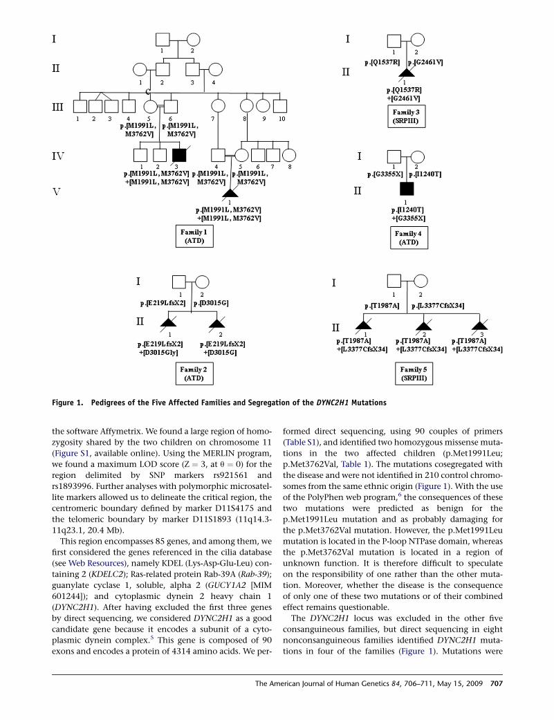
Figure 1. Pedigrees of the Five Affected Families and Segregation of the DYNC2H1 Mutations
the software Affymetrix. We found a large region of homo-
zygosity shared by the two children on chromosome 11
(Figure S1, available online). Using the MERLIN program,
we found a maximum LOD score (Z ¼ 3, at q ¼ 0) for the
region delimited by SNP markers rs921561 and
rs1893996. Further analyses with polymorphic microsatel-
lite markers allowed us to delineate the critical region, the
centromeric boundary defined by marker D11S4175 and
the telomeric boundary by marker D11S1893 (11q14.3-
11q23.1, 20.4 Mb).
This region encompasses 85 genes, and among them, we
first considered the genes referenced in the cilia database
(see Web Resources), namely KDEL (Lys-Asp-Glu-Leu) con-
taining 2 (KDELC2); Ras-related protein Rab-39A (Rab-39);
guanylate cyclase 1, soluble, alpha 2 (GUCY1A2 [MIM
601244]); and cytoplasmic dynein 2 heavy chain 1
(DYNC2H1). After having excluded the first three genes
by direct sequencing, we considered DYNC2H1 as a good
candidate gene because it encodes a subunit of a cyto-
plasmic dynein complex.5 This gene is composed of 90
exons and encodes a protein of 4314 amino acids. We per-
The Am
formed direct sequencing, using 90 couples of primers
(Table S1), and identified two homozygous missense muta-
tions in the two affected children (p.Met1991Leu;
p.Met3762Val, Table 1). The mutations cosegregated with
the disease and were not identified in 210 control chromo-
somes from the same ethnic origin (Figure 1). With the use
of the PolyPhen web program,6 the consequences of these
two mutations were predicted as benign for the
p.Met1991Leu mutation and as probably damaging for
the p.Met3762Val mutation. However, the p.Met1991Leu
mutation is located in the P-loop NTPase domain, whereas
the p.Met3762Val mutation is located in a region of
unknown function. It is therefore difficult to speculate
on the responsibility of one rather than the other muta-
tion. Moreover, whether the disease is the consequence
of only one of these two mutations or of their combined
effect remains questionable.
The DYNC2H1 locus was excluded in the other five
consanguineous families, but direct sequencing in eight
nonconsanguineous families identified DYNC2H1 muta-
tions in four of the families (Figure 1). Mutations were
erican Journal of Human Genetics 84, 706–711, May 15, 2009 707
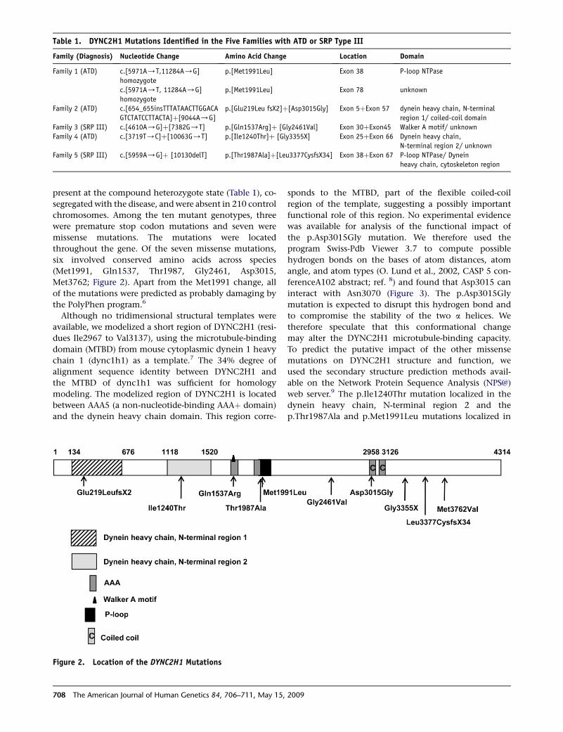
Table 1. DYNC2H1 Mutations Identified in the Five Families with ATD or SRP Type III
Family (Diagnosis) Nucleotide Change Amino Acid Change Location Domain
Family 1 (ATD) c.[5971A/T,11284A/G]
homozygote
p.[Met1991Leu] Exon 38 P-loop NTPase
c.[5971A/T, 11284A/G]
homozygote
p.[Met1991Leu] Exon 78 unknown
Family 2 (ATD) c.[654_655insTTTATAACTTGGACA
GTCTATCCTTACTA]þ[9044A/G]
p.[Glu219Leu fsX2]þ[Asp3015Gly] Exon 5þExon 57 dynein heavy chain, N-terminal
region 1/ coiled-coil domain
Family 3 (SRP III) c.[4610A/G]þ[7382G/T] p.[Gln1537Arg]þ [Gly2461Val] Exon 30þExon45 Walker A motif/ unknown
Family 4 (ATD) c.[3719T/C]þ[10063G/T] p.[Ile1240Thr]þ [Gly3355X] Exon 25þExon 66 Dynein heavy chain,
N-terminal region 2/ unknown
Family 5 (SRP III) c.[5959A/G]þ [10130delT] p.[Thr1987Ala]þ[Leu3377CysfsX34] Exon 38þExon 67 P-loop NTPase/ Dynein
heavy chain, cytoskeleton region
present at the compound heterozygote state (Table 1), co-
segregated with the disease, and were absent in 210 control
chromosomes. Among the ten mutant genotypes, three
were premature stop codon mutations and seven were
missense mutations. The mutations were located
throughout the gene. Of the seven missense mutations,
six involved conserved amino acids across species
(Met1991, Gln1537, Thr1987, Gly2461, Asp3015,
Met3762; Figure 2). Apart from the Met1991 change, all
of the mutations were predicted as probably damaging by
the PolyPhen program.6
Although no tridimensional structural templates were
available, we modelized a short region of DYNC2H1 (resi-
dues Ile2967 to Val3137), using the microtubule-binding
domain (MTBD) from mouse cytoplasmic dynein 1 heavy
chain 1 (dync1h1) as a template.7 The 34% degree of
alignment sequence identity between DYNC2H1 and
the MTBD of dync1h1 was sufficient for homology
modeling. The modelized region of DYNC2H1 is located
between AAA5 (a non-nucleotide-binding AAAþ domain)
and the dynein heavy chain domain. This region corre-
708 The American Journal of Human Genetics 84, 706–711, May 15,
sponds to the MTBD, part of the flexible coiled-coil
region of the template, suggesting a possibly important
functional role of this region. No experimental evidence
was available for analysis of the functional impact of
the p.Asp3015Gly mutation. We therefore used the
program Swiss-Pdb Viewer 3.7 to compute possible
hydrogen bonds on the bases of atom distances, atom
angle, and atom types (O. Lund et al., 2002, CASP 5 con-
ferenceA102 abstract; ref. 8) and found that Asp3015 can
interact with Asn3070 (Figure 3). The p.Asp3015Gly
mutation is expected to disrupt this hydrogen bond and
to compromise the stability of the two a helices. We
therefore speculate that this conformational change
may alter the DYNC2H1 microtubule-binding capacity.
To predict the putative impact of the other missense
mutations on DYNC2H1 structure and function, we
used the secondary structure prediction methods avail-
able on the Network Protein Sequence Analysis (NPS@)
web server.9 The p.Ile1240Thr mutation localized in the
dynein heavy chain, N-terminal region 2 and the
p.Thr1987Ala and p.Met1991Leu mutations localized in
Figure 2. Location of the DYNC2H1 Mutations
2009

the P loop NTPase are predicted to be in a helices. The
p.Gln1537Arg and p.Gly2461Val mutations are localized,
respectively, in the Walker A motif and in a nonconserved
domain, and both are predicted to be in a random coil.
Taken altogether, these data suggest that the six missense
mutations may induce local conformational changes
altering the function of DYNC2H1.
We report here the identification of DYNC2H1 muta-
tions in five distinct families with either ATD or SRP type
III (Table 2 and Figures 4 and 5). Interestingly, while the
Figure 3. Tridimensional Structure Model of a DYNC2H1Putative Microtubule-Binding Domain Via Swiss-Pdb Viewer3.7 RepresentationThe modeled protein is represented in orange and is superimposedwith the template (PDB code: 3err) shown in gray. BetweenAsp3015 (in red) and Asn3070 (in green), a putative hydrogenmay be computed. The mutation p.Asp3015Gly disrupts this stabi-lizing hydrogen bond, inducing a local conformational changealtering the function of this putative microtubule-bindingdomain.
The Am
submission of our manuscript was being processed,
a similar paper was published by Merrill et al., reporting
DYNC2H1 mutations in three families with SRP type III.10
Our findings further support the view that ATD and SRP
type III are allelic disorders and belong to the same spec-
trum. ATD and SRP type III have been previously reported
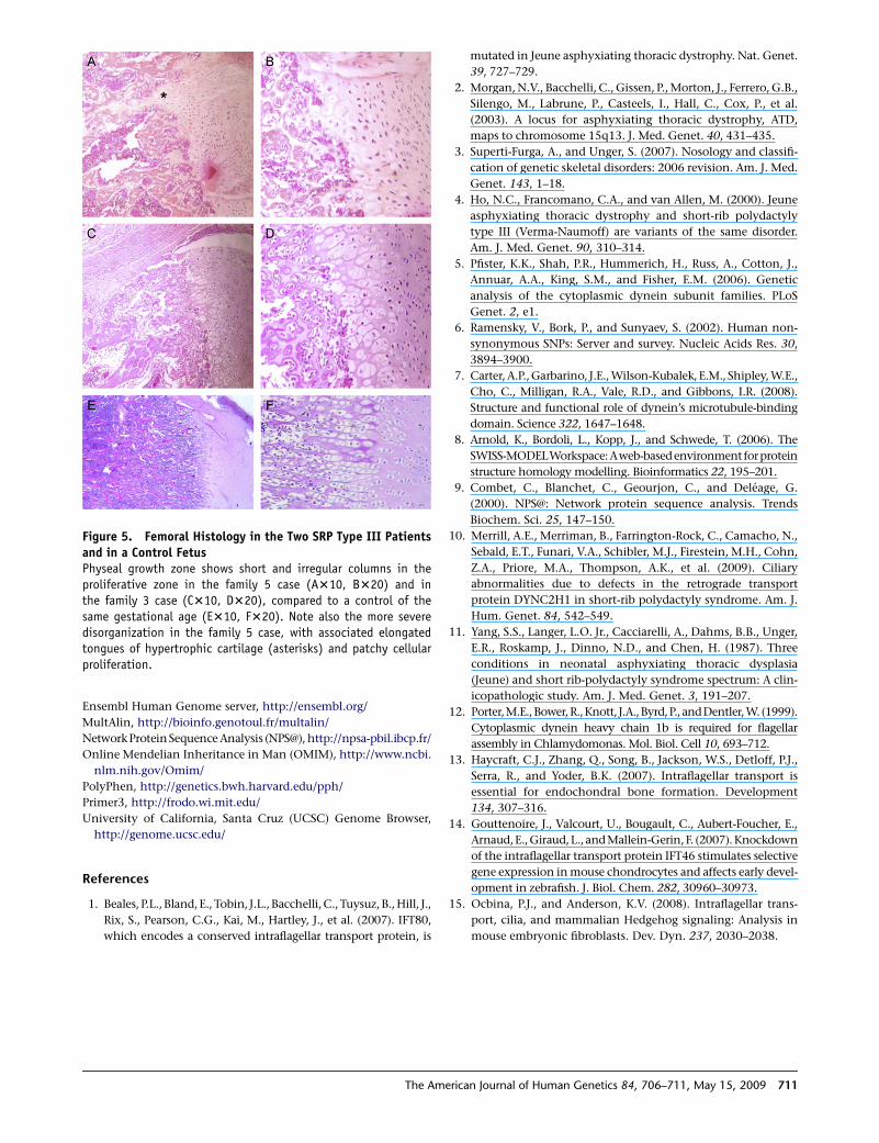
in the same family.4 Moreover, the same histological
anomalies at the growth plate level have been reported
in the two disorders, also suggesting that these conditions
are variants of a single disorder, with SRP type III being at
the more severe end of the spectrum (Figure 5).11 In the
SRP cases (four fetuses belonging to families 3 and 5), the
diagnosis was made antenatally (before 20 weeks of gesta-
tion [wg]). Postaxial polydactyly of the hands was present
in one case (family 3 case), and other malformations
included renal tubular microcysts, hepatic biliary hyper-
plasia (case 3), and unexplained ascites (case 1, family 5).
The three ATD families included a total of five affected
cases. In family 1, one child died of respiratory distress
and pregnancy of her aunt was terminated at 28 wg for
severe narrowing of the thorax. In family 2, two pregnan-
cies were terminated for severe narrowing of the thorax,
one at 28 wg and on at 26 wg. Finally, the family 4 case,
the only survivor, is now 19 years old, and no eye, liver,
or kidney manifestations have been hitherto detected.
This wide clinical variability prompted us to search for
genotype-phenotype correlation. We identified a majority
of missense mutations (7/10) in both ATD and SRP, as well
as three nonsense mutations, also occurring in both ATD
and SRP, with no obvious correlation between genotype
and phenotype. The absence of homozygous nonsense
mutations supports a partial loss of DYNC2H1 function.
The same findings have been observed in the three ATD
cases as a result of IFT80 mutations—two missense muta-
tions and one in-frame deletion mutation have been found
so far.1
The finding of DYNC2H1 mutations in ATD and SRP
type III gives strong support to the view that these condi-
tions belong to the ciliopathy group, as previously
Table 2. Clinical Features of the Five Families with DYNC2H1 Mutations
Family Origin CSAge of AffectedPatient(s) Diagnosis Polydactyly
KidneyAnomaly
Liver/PancreasMicroscopic Changes
OtherFeatures
1 Morocco Yes Case 1: Death at day 2 ATD No No No No
1 Morocco Yes Case 2 : TP at 28 wg ATD No No No No
2 France No Case 1: TP at 28 wg ATD No No No No
2 France No Case 2: TP at 24 wg ATD No No No No
3 France No Case 1: TP at 25 wg SRP type III Postaxial
and Bilateral
Tubular
microcysts
Hepatic biliary, hyperplasia,
periduodenal pancreas
Anal anteposition,
micropenis
4 France No Case 1: 19 years ATD No No No No
5 Madagascar No Case 1: TP at 24 wg SRP type III No No No Ascites
5 Madagascar No Case 2 : TP at 22 wg SRP type III No No No No
5 Madagascar No Case 3 : TP at 16 wg SRP type III No No No No
CS denotes consanguinity, TP denotes terminated pregnancy, wg denotes weeks of gestation.
erican Journal of Human Genetics 84, 706–711, May 15, 2009 709

suggested by the discovery of IFT80 mutations in ATD.
DYNC2H1 is a component of the cytoplasmic dynein
complex, DYNC2, in association with the light interme-
diate chain (DYNC2LI1) and is directly involved in the
contacts and translocation of the dynein complex along
microtubules via its large motor domain.12 Moreover, in
mammalian tissues, the colocalization of DYNC2H1,
DYNC2LI1, and IFT pathway homologs supports a specific
role in the generation and maintenance of mammalian
cilia. Primary cilia and IFT proteins have been shown to
be components of morphogenetic pathways (including
Hedgehog and Wnt pathways) essential for skeletal devel-
opment, and IFT proteins have been shown to be
involved in chondrocyte maturation through bone
morphogenetic protein (BMP) signaling.13,14 The findings
of DYNC2H1 mutations in ATD and SRP type III give
support to the role of cilia and IFT proteins in endochon-
dral bone formation. The presence of polydactyly also
suggests a requirement for DYNC2H1 in Sonic Hedgehog
signaling.15
Figure 4. Radiological Findings in theFamilies with DYNC2H1 Mutations(A) ATD cases. a and b: case 1 (day 2) fromfamily 1; c: case 1 (28 wg) from family 2;d and e: affected child (4 mo) from family4. Note the short bones, narrow thorax,and trident aspect of the acetabular roof(c, arrow). Note also the advanced prox-imal femoral ossification in the family 4case (e).(B) SRP cases. a–c: terminated pregnancyat 25 wg from family 3; d–f: case 1 fromfamily 5. Note the severe shortness of thelong bones, the narrow thorax, and thetrident aspect of the acetabular roof.Note also the round metaphyseal endswith lateral spikes (c, arrows), the tripli-cate calcaneum (d, arrow), and the post-axial polydactyly (a, arrow).Note the bowing of the femora observed inthe cases from families 1–3 and 5 (Ab, Ac,Bc, Bd).
In conclusion, this study demon-
strates that ATD and SRP type III
belong to the same heterogeneous
spectrum of conditions caused by
either IFT80 or DYNC2H1 mutations
and suggests a role for altered primary
cilium function in these disorders.
Yet, the presence of nine families
with ATD and/or SRP type III
unlinked to IFT80 and DYNC2H1
supports the genetic heterogeneity
of this condition. Ongoing studies
will hopefully lead to identification
of other disease gene(s) presumably also involved in
primary cilia function.
Supplemental Data
Supplemental Data include one figure and one table and can be
found with this article online at http://www.ajhg.org/.
Acknowledgments
Part of this work has been supported by a national grant from Pro-
gramme Hospitalier de Recherche Clinique (PHRC AOM06031).
Received: March 15, 2009
Revised: April 17, 2009
Accepted: April 22, 2009
Published online: May 14, 2009
Web Resources
The URLs for data presented herein are as follows:
Cilia database: ExPASy, http://www.expasy.ch/sprot/; Zebrafish
and comparative genomics blast, http://danio.mgh.harvard.
edu/blast/blast_grp.html
710 The American Journal of Human Genetics 84, 706–711, May 15, 2009
Ensembl Human Genome server, http://ensembl.org/
MultAlin, http://bioinfo.genotoul.fr/multalin/
Network Protein Sequence Analysis (NPS@), http://npsa-pbil.ibcp.fr/
Online Mendelian Inheritance in Man (OMIM), http://www.ncbi.
nlm.nih.gov/Omim/
PolyPhen, http://genetics.bwh.harvard.edu/pph/
Primer3, http://frodo.wi.mit.edu/
University of California, Santa Cruz (UCSC) Genome Browser,
http://genome.ucsc.edu/
References
1. Beales, P.L., Bland, E., Tobin, J.L., Bacchelli, C., Tuysuz,B., Hill, J.,
Rix, S., Pearson, C.G., Kai, M., Hartley, J., et al. (2007). IFT80,
which encodes a conserved intraflagellar transport protein, is
Figure 5. Femoral Histology in the Two SRP Type III Patientsand in a Control FetusPhyseal growth zone shows short and irregular columns in theproliferative zone in the family 5 case (A310, B320) and inthe family 3 case (C310, D320), compared to a control of thesame gestational age (E310, F320). Note also the more severedisorganization in the family 5 case, with associated elongatedtongues of hypertrophic cartilage (asterisks) and patchy cellularproliferation.
The Am
mutated in Jeune asphyxiating thoracic dystrophy. Nat. Genet.
39, 727–729.
2. Morgan, N.V., Bacchelli, C., Gissen, P., Morton, J., Ferrero, G.B.,
Silengo, M., Labrune, P., Casteels, I., Hall, C., Cox, P., et al.
(2003). A locus for asphyxiating thoracic dystrophy, ATD,
maps to chromosome 15q13. J. Med. Genet. 40, 431–435.
3. Superti-Furga, A., and Unger, S. (2007). Nosology and classifi-
cation of genetic skeletal disorders: 2006 revision. Am. J. Med.
Genet. 143, 1–18.
4. Ho, N.C., Francomano, C.A., and van Allen, M. (2000). Jeune
asphyxiating thoracic dystrophy and short-rib polydactyly
type III (Verma-Naumoff) are variants of the same disorder.
Am. J. Med. Genet. 90, 310–314.
5. Pfister, K.K., Shah, P.R., Hummerich, H., Russ, A., Cotton, J.,
Annuar, A.A., King, S.M., and Fisher, E.M. (2006). Genetic
analysis of the cytoplasmic dynein subunit families. PLoS
Genet. 2, e1.
6. Ramensky, V., Bork, P., and Sunyaev, S. (2002). Human non-
synonymous SNPs: Server and survey. Nucleic Acids Res. 30,
3894–3900.
7. Carter, A.P., Garbarino, J.E., Wilson-Kubalek, E.M., Shipley, W.E.,
Cho, C., Milligan, R.A., Vale, R.D., and Gibbons, I.R. (2008).
Structure and functional role of dynein’s microtubule-binding
domain. Science 322, 1647–1648.
8. Arnold, K., Bordoli, L., Kopp, J., and Schwede, T. (2006). The
SWISS-MODELWorkspace:Aweb-basedenvironment forprotein
structure homology modelling. Bioinformatics 22, 195–201.
9. Combet, C., Blanchet, C., Geourjon, C., and Deleage, G.
(2000). NPS@: Network protein sequence analysis. Trends
Biochem. Sci. 25, 147–150.
10. Merrill, A.E., Merriman, B., Farrington-Rock, C., Camacho, N.,
Sebald, E.T., Funari, V.A., Schibler, M.J., Firestein, M.H., Cohn,
Z.A., Priore, M.A., Thompson, A.K., et al. (2009). Ciliary
abnormalities due to defects in the retrograde transport
protein DYNC2H1 in short-rib polydactyly syndrome. Am. J.
Hum. Genet. 84, 542–549.
11. Yang, S.S., Langer, L.O. Jr., Cacciarelli, A., Dahms, B.B., Unger,
E.R., Roskamp, J., Dinno, N.D., and Chen, H. (1987). Three
conditions in neonatal asphyxiating thoracic dysplasia
(Jeune) and short rib-polydactyly syndrome spectrum: A clin-
icopathologic study. Am. J. Med. Genet. 3, 191–207.
12. Porter,M.E.,Bower,R.,Knott, J.A.,Byrd,P., andDentler,W. (1999).
Cytoplasmic dynein heavy chain 1b is required for flagellar
assembly in Chlamydomonas. Mol. Biol. Cell 10, 693–712.
13. Haycraft, C.J., Zhang, Q., Song, B., Jackson, W.S., Detloff, P.J.,
Serra, R., and Yoder, B.K. (2007). Intraflagellar transport is
essential for endochondral bone formation. Development
134, 307–316.
14. Gouttenoire, J., Valcourt, U., Bougault, C., Aubert-Foucher, E.,
Arnaud,E., Giraud, L., and Mallein-Gerin, F. (2007). Knockdown
of the intraflagellar transport protein IFT46 stimulates selective
gene expression in mouse chondrocytes and affects early devel-
opment in zebrafish. J. Biol. Chem. 282, 30960–30973.
15. Ocbina, P.J., and Anderson, K.V. (2008). Intraflagellar trans-
port, cilia, and mammalian Hedgehog signaling: Analysis in
mouse embryonic fibroblasts. Dev. Dyn. 237, 2030–2038.
erican Journal of Human Genetics 84, 706–711, May 15, 2009 711
Related Documents