The Plant Cell, Vol. 13, 1929–1943, August 2001, www.plantcell.org © 2001 American Society of Plant Biologists Dynamic Recruitment of Cdc2 to Specific Microtubule Structures during Mitosis Magdalena Weingartner, a Pavla Binarova, b Denisa Drykova, b Alois Schweighofer, a Jean-Pierre David, c Erwin Heberle-Bors, a John Doonan, d and László Bögre e,1 a Institute of Microbiology and Genetics, University of Vienna, Vienna Biocenter, Dr. Bohrgasse 9, A-1030 Vienna, Austria b Institute of Microbiology, Academy of Sciences of the Czech Republic, Víde ská 1083, 142 20 Prague 4, Czech Republic c Institute of Molecular Pathology, Dr. Bohrgasse 7, A-1030 Vienna, Austria d Cell Biology Department, John Innes Centre, Norwich NR4 7UH, United Kingdom e School of Biological Sciences, Royal Holloway, University of London, Egham TW20 OEX, United Kingdom A-type cyclin-dependent kinases (CDKs), also known as cdc2, are central to the orderly progression of the cell cycle. We made a functional Green Fluorescent Protein (GFP) fusion with CDK-A (Cdc2-GFP) and followed its subcellular lo- calization during the cell cycle in tobacco cells. During interphase, the Cdc2-GFP fusion protein was found in both the cytoplasm and the nucleus, where it was highly resistant to extraction. In premitotic cells, a bright and narrow equato- rial band appeared on the cell surface, resembling the late preprophase band, which disintegrated within 10 min as fol- lowed by time-lapse images. Cdc2-GFP was not found on prophase spindles but left the chromatin soon after this stage and associated progressively with the metaphase spindle in a microtubule-dependent manner. Arresting cells in mitosis through the stabilization of microtubules by taxol further enhanced the spindle-localized pool of Cdc2-GFP. To- ward the end of mitosis, Cdc2-GFP was found at the midzone of the anaphase spindle and phragmoplast; eventually, it became focused at the midline of these microtubule structures. In detergent-extracted cells, the Cdc2-GFP remained associated with mitotic structures. Retention on spindles was prevented by pretreatment with the CDK-specific inhibi- tor roscovitine and was enhanced by the protein phosphatase inhibitor okadaic acid. Furthermore, we demonstrate that both the endogenous CDK-A and Cdc2-GFP were cosedimented with taxol-stabilized plant microtubules from cell extracts and that Cdc2 activity was detected together with a fraction of polymerized tubulin. These data provide evi- dence that the A-type CDKs associate physically with mitotic structures in a microtubule-dependent manner and may be involved in regulating the behavior of specific microtubule arrays throughout mitosis. n ˇ INTRODUCTION The spatial and temporal coordination of cellular events contributes to the precise reproduction of cells at each divi- sion. The cyclin-dependent kinases (CDKs), which are con- served from yeast to animals to plants (Mironov et al., 1999; Pines, 1999), play a central role in coordinating these events. The sequential activation of CDKs by different cy- clins at different times in the cell cycle ensures orderly pro- gression through the cycle. In yeast, there is only a single CDK that performs multiple functions, whereas in animals and plants, there are multiple CDKs that are specialized. Based on structural similarities, plant CDKs have been classified as CDK-A, CDK-B, etc. (Joubes et al., 2000). Canonical plant CDK, or CDK-A, is highly conserved at the amino acid sequence level and is most similar to the yeast Cdc2/CDC28 or animal CDK1-3 proteins. All CDK-A proteins contain the conserved cyclin interac- tion or PSTAIRE motif and have been shown to functionally complement yeast cdc2/CDC28 mutations (Colasanti et al., 1991; Ferreira et al., 1991; Hirt et al., 1991, 1993; Fobert et al., 1996). CDK-A mRNAs are expressed in dividing and di- vision-competent cells throughout the cell cycle, and their protein kinase activity becomes activated at both the G1- to-S and G2-to-M transitions (Bögre et al., 1997; Magyar et al., 1997). The overexpression of a kinase-negative mutant form of the Arabidopsis CDK-A blocks or slows down cell cycle progression in both G1- and G2-phases, further indi- cating its multiple roles in the cell cycle (Hemerly et al., 1995). Similarly, roscovitine, a drug with high specificity to canonical CDKs, blocks both G1-to-S and G2-to-M transi- tions (Planchais et al., 1997; Binarova et al., 1998) and re- sults in disturbed spindle organization, such as monopolar spindle formation during mitosis (Binarova et al., 1998). 1 To whom correspondence should be addressed. E-mail l.bogre@ rhul.ac.uk; fax 44-1784-434326.

Welcome message from author
This document is posted to help you gain knowledge. Please leave a comment to let me know what you think about it! Share it to your friends and learn new things together.
Transcript

The Plant Cell, Vol. 13, 1929–1943, August 2001, www.plantcell.org © 2001 American Society of Plant Biologists
Dynamic Recruitment of Cdc2 to Specific Microtubule Structures during Mitosis
Magdalena Weingartner,
a
Pavla Binarova,
b
Denisa Drykova,
b
Alois Schweighofer,
a
Jean-Pierre David,
c
Erwin Heberle-Bors,
a
John Doonan,
d
and László Bögre
e,1
a
Institute of Microbiology and Genetics, University of Vienna, Vienna Biocenter, Dr. Bohrgasse 9, A-1030 Vienna, Austria
b
Institute of Microbiology, Academy of Sciences of the Czech Republic, Víde ská 1083, 142 20 Prague 4, Czech Republic
c
Institute of Molecular Pathology, Dr. Bohrgasse 7, A-1030 Vienna, Austria
d
Cell Biology Department, John Innes Centre, Norwich NR4 7UH, United Kingdom
e
School of Biological Sciences, Royal Holloway, University of London, Egham TW20 OEX, United Kingdom
A-type cyclin-dependent kinases (CDKs), also known as cdc2, are central to the orderly progression of the cell cycle.We made a functional Green Fluorescent Protein (GFP) fusion with CDK-A (Cdc2-GFP) and followed its subcellular lo-calization during the cell cycle in tobacco cells. During interphase, the Cdc2-GFP fusion protein was found in both thecytoplasm and the nucleus, where it was highly resistant to extraction. In premitotic cells, a bright and narrow equato-rial band appeared on the cell surface, resembling the late preprophase band, which disintegrated within 10 min as fol-lowed by time-lapse images. Cdc2-GFP was not found on prophase spindles but left the chromatin soon after thisstage and associated progressively with the metaphase spindle in a microtubule-dependent manner. Arresting cells inmitosis through the stabilization of microtubules by taxol further enhanced the spindle-localized pool of Cdc2-GFP. To-ward the end of mitosis, Cdc2-GFP was found at the midzone of the anaphase spindle and phragmoplast; eventually, itbecame focused at the midline of these microtubule structures. In detergent-extracted cells, the Cdc2-GFP remainedassociated with mitotic structures. Retention on spindles was prevented by pretreatment with the CDK-specific inhibi-tor roscovitine and was enhanced by the protein phosphatase inhibitor okadaic acid. Furthermore, we demonstratethat both the endogenous CDK-A and Cdc2-GFP were cosedimented with taxol-stabilized plant microtubules from cellextracts and that Cdc2 activity was detected together with a fraction of polymerized tubulin. These data provide evi-dence that the A-type CDKs associate physically with mitotic structures in a microtubule-dependent manner and maybe involved in regulating the behavior of specific microtubule arrays throughout mitosis.
n
INTRODUCTION
The spatial and temporal coordination of cellular eventscontributes to the precise reproduction of cells at each divi-sion. The cyclin-dependent kinases (CDKs), which are con-served from yeast to animals to plants (Mironov et al., 1999;Pines, 1999), play a central role in coordinating theseevents. The sequential activation of CDKs by different cy-clins at different times in the cell cycle ensures orderly pro-gression through the cycle.
In yeast, there is only a single CDK that performs multiplefunctions, whereas in animals and plants, there are multipleCDKs that are specialized. Based on structural similarities,plant CDKs have been classified as CDK-A, CDK-B, etc.(Joubes et al., 2000). Canonical plant CDK, or CDK-A, ishighly conserved at the amino acid sequence level and is
most similar to the yeast Cdc2/CDC28 or animal CDK1-3proteins.
All CDK-A proteins contain the conserved cyclin interac-tion or PSTAIRE motif and have been shown to functionallycomplement yeast cdc2/CDC28 mutations (Colasanti et al.,1991; Ferreira et al., 1991; Hirt et al., 1991, 1993; Fobert etal., 1996). CDK-A mRNAs are expressed in dividing and di-vision-competent cells throughout the cell cycle, and theirprotein kinase activity becomes activated at both the G1-to-S and G2-to-M transitions (Bögre et al., 1997; Magyar etal., 1997). The overexpression of a kinase-negative mutantform of the Arabidopsis CDK-A blocks or slows down cellcycle progression in both G1- and G2-phases, further indi-cating its multiple roles in the cell cycle (Hemerly et al.,1995). Similarly, roscovitine, a drug with high specificity tocanonical CDKs, blocks both G1-to-S and G2-to-M transi-tions (Planchais et al., 1997; Binarova et al., 1998) and re-sults in disturbed spindle organization, such as monopolarspindle formation during mitosis (Binarova et al., 1998).
1
To whom correspondence should be addressed. E-mail [email protected]; fax 44-1784-434326.
1930 The Plant Cell
CDK-A members were found to bind some of the D-type cy-clins (Healy et al., 2000), and in the yeast two-hybrid assay itinteracts with the A2-type cyclin (Roudier et al., 2000), but itis not known if CDK-A associates with B-type cyclins. Asecond class of CDKs is found only in plants (Doonan andFobert, 1997), with members containing the PPTALRE orPPTTLRE motifs; it is named the CDK-B class (Joubes et al.,2000). These CDKs appear to have specific functions in S-,G2-, and M-phases, as suggested by their phase-specificexpression and activity (Fobert et al., 1996; Magyar et al.,1997). However, their exact roles are not well understood.
CDK1 location changes dramatically during cell cycle pro-gression in both yeast (Alfa et al., 1990) and animals (Bailly etal., 1989). A proportion of CDK1 is located in the interphasenucleus, where it is bound tightly to chromatin (Riabowol etal., 1989). During mitosis, CDK1 localizes to the spindle polebody in yeast (Alfa et al., 1990) and to centrosomes in ani-mal cells (Bailly et al., 1989). During mitosis, CDK1 is knownto associate with spindle microtubules (Leiss et al., 1992),and it regulates their stability through the phosphorylation ofmicrotubule-associating proteins such as MAP4 or MAP215(Verde et al., 1990; Tournebize et al., 2000); it also affectsmicrotubule movements through the phosphorylation ofmotor proteins (Walczak et al., 1998). A recent report impli-cates CDK1 as having an important role in regulating the ori-entation of asymmetric divisions in
Drosophila melanogaster
(Tio et al., 2001).In plants, the orientation of cell division is thought to be
important for normal development, and in many tissues it isstringently controlled (Doonan, 2000). In most higher plantcells, the division plane is predicted by an enigmatic micro-tubule array called the preprophase band (PPB). Immunolo-calization of CDK-A proteins, using antibodies against theconserved PSTAIRE domain or against the C terminus(Mineyuki et al., 1991; Colasanti et al., 1993; Bögre et al.,1997; Mews et al., 1997; Stals et al., 1997), indicate thepresence of CDK-A at multiple locations: in the nucleus,on the mitotic chromosomes, and associated with severalmicrotubule arrays, including the PPB, the spindle, and thephragmoplast. Another CDK, Medsa;CDK-B2;1, also is foundon mitotic structures (Ayaydin et al., 2000). However, the re-ported localization patterns are complex and sometimescontradictory, depending on the source of the antibody andeven the fixation conditions used.
In this study, we used a Green Fluorescent Protein (GFP)fusion with a CDK-A protein, Medsa;CDK-A;2, to follow itslocalization in living cells. We give a detailed description ofits dynamic association with microtubules during mitosisand provide evidence that recruitment to the spindle ismicrotubule dependent. Furthermore, Cdc2 activity wasfound to be required for microtubule recruitment. Finally,we show that Medsa;CDK-A;2 and tubulin copurify with mi-crotubules from mitotic cell extracts. These results col-lectively suggest that, in plants, CDK-A proteins havea continuous role in regulating microtubule organizationthroughout mitosis.
RESULTS
The Cdc2-GFP Fusion Protein Remains Functional as Tested by Yeast Complementation and by Measuring Its Protein Kinase Activity in Plant Cells
To investigate the dynamic localization of CDK-A proteins inliving plant cells, we made a translational fusion betweenMedsa;CDK-A;2 and GFP, hereafter referred to as Cdc2-GFP. To establish that the Cdc2-GFP fusion product was afunctional cyclin-dependent protein kinase, we tested itsability to complement temperature-sensitive strains of bud-ding yeast carrying the CDC28-4 and CDC28-1N mutations(Surana et al., 1991). Figure 1A shows that expression of ei-ther Medsa;CDK-A;2 or the GFP fusion protein allowed theCDC28-1N temperature-sensitive mutant to grow at the re-strictive temperature (37
�
C). Similar results were obtainedfor the CDC28-4 strain (data not shown). We observed yeastcells expressing Cdc2-GFP with an epifluorescence micro-scope and found that the signal was confined mostly to thenucleus and the bud neck (Figure 1B).
Figure 1. Complementation Assay in Yeast.
(A) Complementation analysis of the Saccharomyces cerevisiaeCDC28-1N temperature-sensitive mutation containing the emptypYES yeast shuttle vector, pYES with Medsa;CDK-A;2 (pYES Cdc2),or the fusion of Medsa;CDK-A;2 with GFP (pYES Cdc2-GFP) andgrown at 28 and 37�C on induction medium containing galactose.(B) Localization of Cdc2-GFP expressed in yeast cells at differentphases of the cell cycle as shown by a fluorescence image (left) andby a differential interference contrast (DIC) image to visualize thecells (right). The arrowhead indicates a bud neck, where a spindle islocalized in yeast. Bar � 5 �m.
Cdc2-GFP 1931
To express the Cdc2-GFP fusion protein in plants, weplaced it under the control of a tetracycline-regulated pro-moter (Weinmann et al., 1994). This should repress Cdc2-GFP expression in tobacco lines containing the tetracyclinerepressor protein and thus avoid problems if the Cdc2-GFPexpression was deleterious. However, we found that trans-formation efficiencies were similar in tobacco lines with andwithout the tetracycline repressor and concluded that theconstitutive expression of Cdc2-GFP is not deleterious toplants. No phenotypic differences were observed in trans-formants from either line that were associated with thecdc2-GFP transgene, so we decided to continue these exper-iments only with plants constitutively expressing Cdc2-GFP.
Seed from five independent transgenic lines containingCdc2-GFP as well as from five lines expressing GFP alonewere germinated on hygromycin, and their root cells wereobserved by confocal laser scanning microscopy. Plantsexpressing the Cdc2-GFP fusion protein or GFP alonedisplayed a readily observable green fluorescence in the nu-cleus and the cytoplasm (data not shown). However, be-cause of the strong autofluorescence of tobacco root tipcells, high resolution images could not be obtained. There-fore, we generated cell suspensions from selected GFP andCdc2-GFP plants.
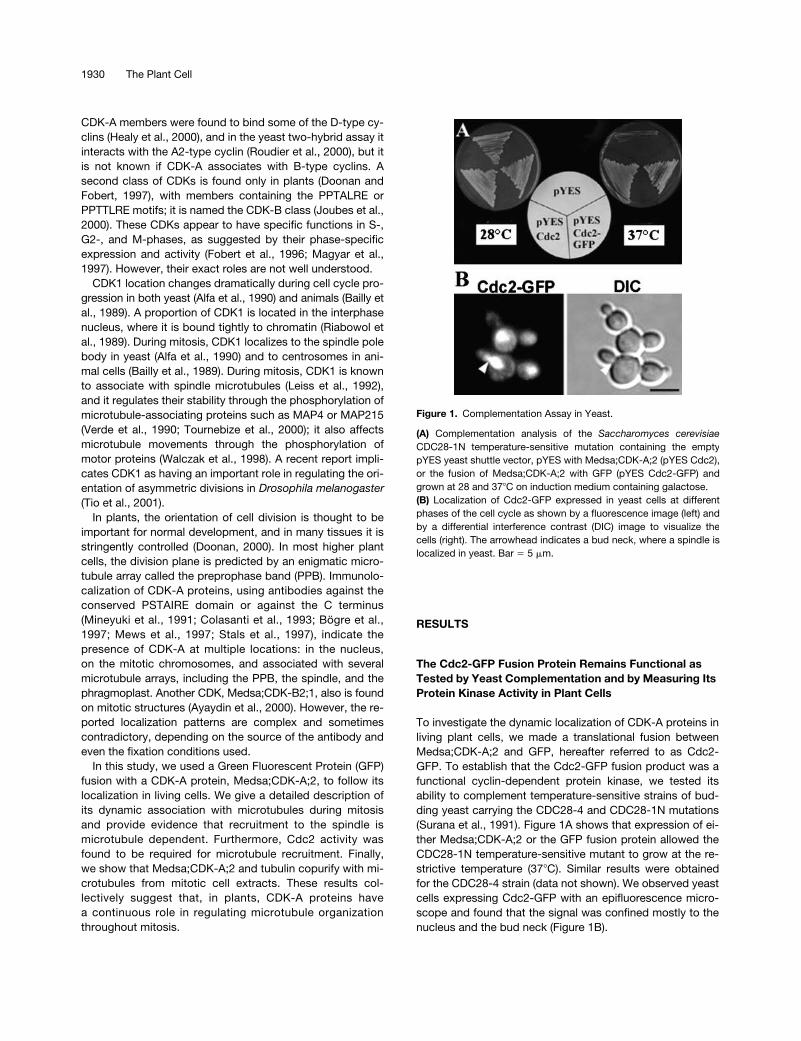
To establish that the fusion protein was being expressedand processed correctly, protein extracts from both GFPand Cdc2-GFP transgenic cell cultures were separated bySDS-PAGE, blotted, and probed with anti-PSTAIRE (datanot shown) or anti-GFP (Figure 2A) antibodies. As predicted,
the GFP transgenic lines contained a band of the expected27 kD and the cdc2-GFP transgenic lines contained a bandof 60 kD (Figure 2A). A weaker band with a reduced molecu-lar mass also was detected in the Cdc2-GFP–expressingplants. The different forms might be attributable to post-translational modifications. Cdc2-GFP accumulates to amuch lesser extent than GFP, and this was observed in sev-eral independent transgenic lines. The reduced levels ofCdc2-GFP may indicate either that the Cdc2-GFP fusionprotein is not translated efficiently or that the protein is un-stable compared with GFP. However, there is no evidencefor extensive proteolysis, and no free GFP was detected inthe cdc2-GFP lines.
To confirm that the Cdc2-GFP fusion protein is functionalin plant cells, we tested its binding to a conserved regulator,p13
suc1
, and measured its protein kinase activity. The Cdc2-GFP fusion protein, but not GFP alone, could bind to p13
suc1
(Figure 2B). When immunopurified from cell extracts using aGFP-specific antibody, the Cdc2-GFP fusion protein hadprotein kinase activity and readily phosphorylated histoneH1 (Figure 2C). These biochemical experiments indicate thatCdc2-GFP is functional in plant cells.
Cdc2-GFP Is Bound Tightly to Chromatin in Interphase
Several independent GFP and Cdc2-GFP transgenic lineswere observed using epifluorescence microscopy, but be-cause all lines for a given construct were essentially similar,we chose a single representative GFP line and Cdc2-GFPline for detailed analyses.
During interphase, Cdc2-GFP–derived fluorescence wasweaker than, but broadly similar to, that of GFP. Both werelocalized to the nucleus and the cytoplasmic strands (Figure
Figure 2. Cdc2-GFP Fusion Protein Is Expressed in a Correct Size,Binds to p13suc1, and Is Active in Tobacco Cells.
(A) Immunoblotting using extracts prepared from suspension cul-tured cells expressing GFP (lane 1) or Cdc2-GFP (lane 2) with aGFP-specific antibody.(B) Binding of GFP (lane 1) or Cdc2-GFP (lane 2) to p13suc1. Extractsprepared as in (A) were mixed with p13suc1 beads, and the boundfractions were immunoblotted with a GFP-specific antibody.(C) Histone H1 kinase activity of GFP (lane 1) and Cdc2-GFP (lane 2)immunopurified from extracts using a GFP-specific antibody. Thephosphorylated histone H1 is indicated by an arrow.Molecular mass markers are labeled at right.
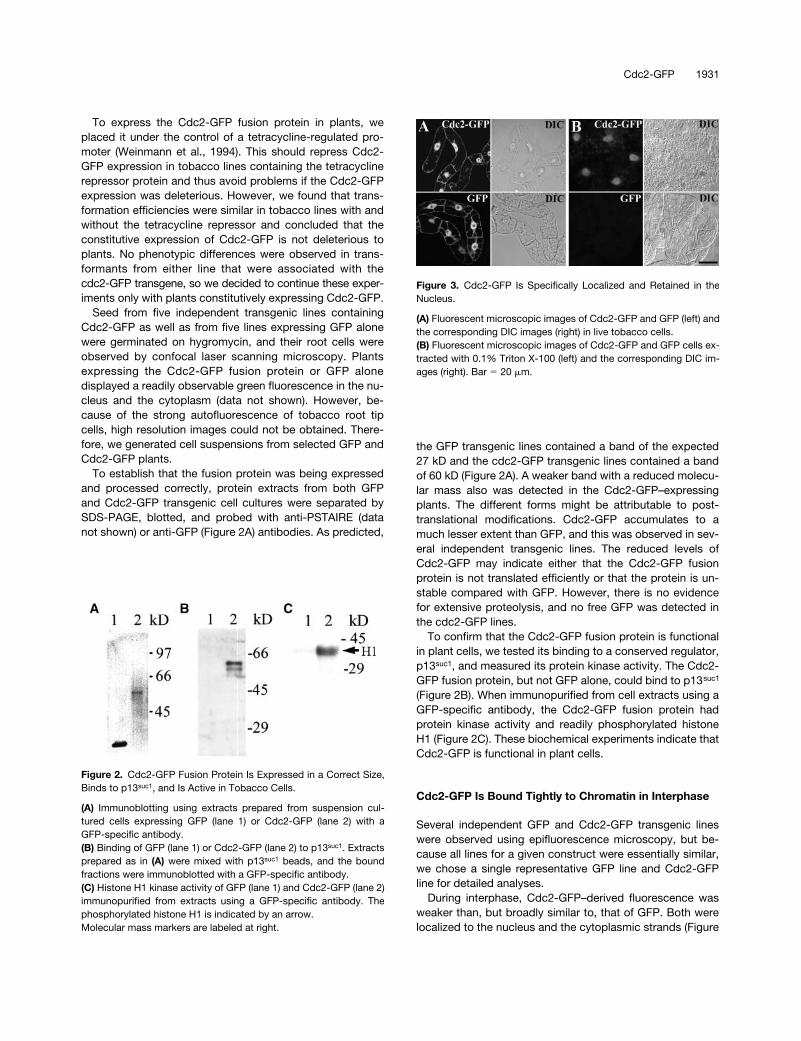
Figure 3. Cdc2-GFP Is Specifically Localized and Retained in theNucleus.
(A) Fluorescent microscopic images of Cdc2-GFP and GFP (left) andthe corresponding DIC images (right) in live tobacco cells.(B) Fluorescent microscopic images of Cdc2-GFP and GFP cells ex-tracted with 0.1% Triton X-100 (left) and the corresponding DIC im-ages (right). Bar � 20 �m.
1932 The Plant Cell
3A). The size of the free GFP molecule is somewhat smallerthan the reported size exclusion limit of the nuclear pores(Chytilova et al., 2000), and, consistent with this fact, GFPwas distributed freely in both the cytoplasm and the nu-cleus. However, the size of the Cdc2-GFP fusion protein isabove the size exclusion limit of the nuclear pores, suggest-ing that Cdc2-GFP is transported to the nucleus by an ac-tive mechanism.
To further characterize the association of Cdc2-GFP withnuclear structures, we extracted both GFP– and Cdc2-GFP–expressing cells with a mild nonionic detergent. Under
these conditions, membranes and the nuclear envelopewere dissolved and soluble proteins were extracted. As ex-pected, GFP was extracted completely by this treatment.However, most of the Cdc2-GFP fusion protein was re-tained in the nucleus (Figure 3B). A fuzzy halo around thenucleus within the extracted cell also was evident (Figure3B), indicating that Cdc2-GFP is tightly bound. We furtherextracted these cells with increased salt concentrations (upto 0.5 M NaCl) and increased levels of detergent (up to 1%Triton X-100). None of these treatments completely elimi-nated nucleus-bound Cdc2-GFP (data not shown). Weconclude that Cdc2-GFP is bound strongly and retainedspecifically in the nucleus.
Cdc2-GFP Accumulates in an Equatorial Band on the Cell Cortex during Late Preprophase
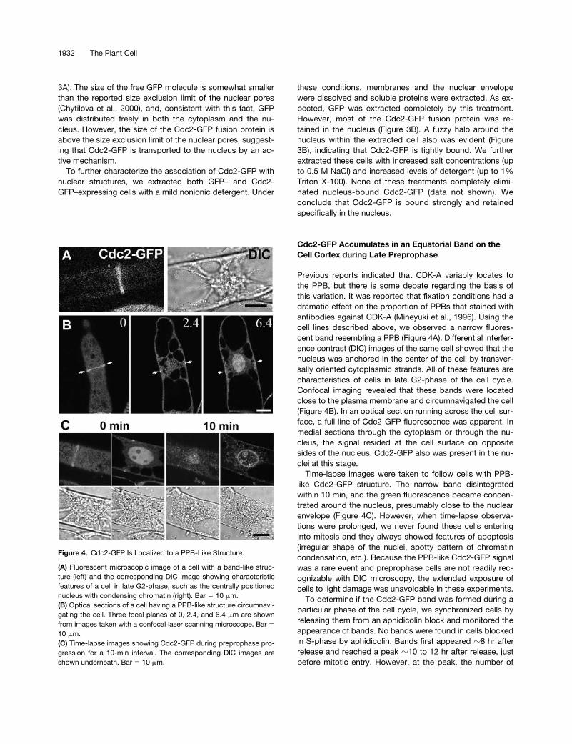
Previous reports indicated that CDK-A variably locates tothe PPB, but there is some debate regarding the basis ofthis variation. It was reported that fixation conditions had adramatic effect on the proportion of PPBs that stained withantibodies against CDK-A (Mineyuki et al., 1996). Using thecell lines described above, we observed a narrow fluores-cent band resembling a PPB (Figure 4A). Differential interfer-ence contrast (DIC) images of the same cell showed that thenucleus was anchored in the center of the cell by transver-sally oriented cytoplasmic strands. All of these features arecharacteristics of cells in late G2-phase of the cell cycle.Confocal imaging revealed that these bands were locatedclose to the plasma membrane and circumnavigated the cell(Figure 4B). In an optical section running across the cell sur-face, a full line of Cdc2-GFP fluorescence was apparent. Inmedial sections through the cytoplasm or through the nu-cleus, the signal resided at the cell surface on oppositesides of the nucleus. Cdc2-GFP also was present in the nu-clei at this stage.
Time-lapse images were taken to follow cells with PPB-like Cdc2-GFP structure. The narrow band disintegratedwithin 10 min, and the green fluorescence became concen-trated around the nucleus, presumably close to the nuclearenvelope (Figure 4C). However, when time-lapse observa-tions were prolonged, we never found these cells enteringinto mitosis and they always showed features of apoptosis(irregular shape of the nuclei, spotty pattern of chromatincondensation, etc.). Because the PPB-like Cdc2-GFP signalwas a rare event and preprophase cells are not readily rec-ognizable with DIC microscopy, the extended exposure ofcells to light damage was unavoidable in these experiments.
To determine if the Cdc2-GFP band was formed during aparticular phase of the cell cycle, we synchronized cells byreleasing them from an aphidicolin block and monitored theappearance of bands. No bands were found in cells blockedin S-phase by aphidicolin. Bands first appeared
�
8 hr afterrelease and reached a peak
�
10 to 12 hr after release, justbefore mitotic entry. However, at the peak, the number of
Figure 4. Cdc2-GFP Is Localized to a PPB-Like Structure.
(A) Fluorescent microscopic image of a cell with a band-like struc-ture (left) and the corresponding DIC image showing characteristicfeatures of a cell in late G2-phase, such as the centrally positionednucleus with condensing chromatin (right). Bar � 10 �m.(B) Optical sections of a cell having a PPB-like structure circumnavi-gating the cell. Three focal planes of 0, 2.4, and 6.4 �m are shownfrom images taken with a confocal laser scanning microscope. Bar �10 �m.(C) Time-lapse images showing Cdc2-GFP during preprophase pro-gression for a 10-min interval. The corresponding DIC images areshown underneath. Bar � 10 �m.
Cdc2-GFP 1933
the Cdc2-GFP bands was considerably lower (
�
1%) com-pared with the percentage of PPBs determined from immu-nofluorescence staining for microtubules with anti–
�
-tubulinantibodies (
�
10%). The whole range of microtubular PPBstages was visualized by immunofluorescence: from broadPPBs at the beginning of its formation to narrow bands atthe latest stage of their existence, not long before nuclearenvelope breakdown (data not shown). The extremely nar-row Cdc2-GFP–derived bands, and its low percentage com-pared with PPBs, further suggest that Cdc2-GFP associatesonly with a portion of the PPBs, probably only for a brief pe-riod during late G2-phase.
The broad spectrum kinase inhibitor K252-a accumulatescells with PPBs when added to synchronized cells in earlyG2-phase (Katsuta and Shibaoka, 1992). Using this drug,we could increase the number of PPBs in our culture (up to30%), but Cdc2-GFP bands were completely absent. Thismay indicate that the Cdc2-GFP band forms just after theK252-a arrest point or that protein kinase activity is requiredfor Cdc2 association with the PPB.
Cdc2-GFP Relocates from Chromatin to Microtubules in the Metaphase Spindle
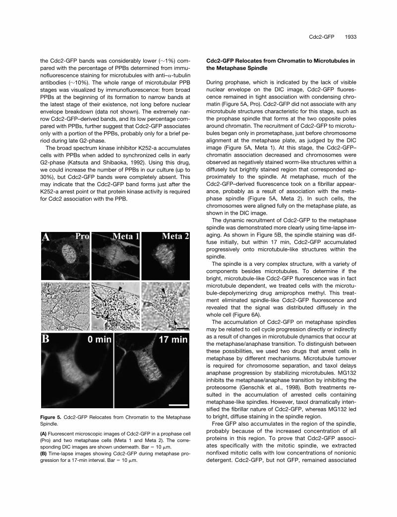
During prophase, which is indicated by the lack of visiblenuclear envelope on the DIC image, Cdc2-GFP fluores-cence remained in tight association with condensing chro-matin (Figure 5A, Pro). Cdc2-GFP did not associate with anymicrotubule structures characteristic for this stage, such asthe prophase spindle that forms at the two opposite polesaround chromatin. The recruitment of Cdc2-GFP to microtu-bules began only in prometaphase, just before chromosomealignment at the metaphase plate, as judged by the DICimage (Figure 5A, Meta 1). At this stage, the Cdc2-GFP–chromatin association decreased and chromosomes wereobserved as negatively stained worm-like structures within adiffusely but brightly stained region that corresponded ap-proximately to the spindle. At metaphase, much of theCdc2-GFP–derived fluorescence took on a fibrillar appear-ance, probably as a result of association with the meta-phase spindle (Figure 5A, Meta 2). In such cells, thechromosomes were aligned fully on the metaphase plate, asshown in the DIC image.
The dynamic recruitment of Cdc2-GFP to the metaphasespindle was demonstrated more clearly using time-lapse im-aging. As shown in Figure 5B, the spindle staining was dif-fuse initially, but within 17 min, Cdc2-GFP accumulatedprogressively onto microtubule-like structures within thespindle.
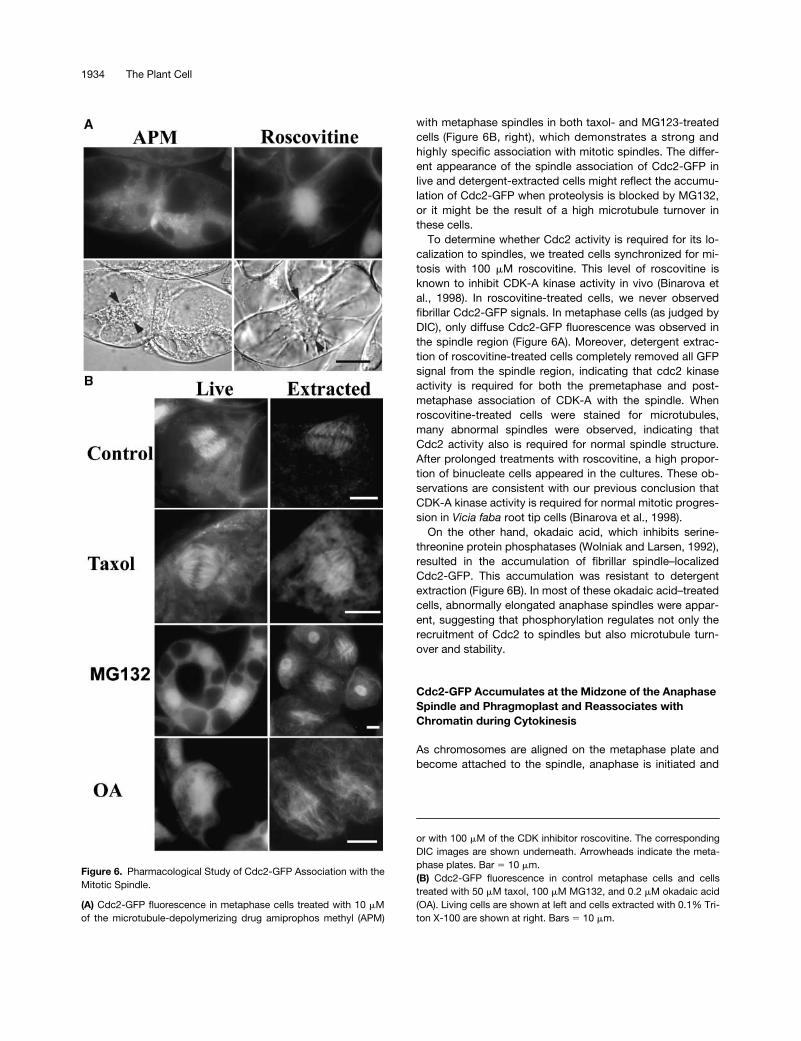
The spindle is a very complex structure, with a variety ofcomponents besides microtubules. To determine if thebright, microtubule-like Cdc2-GFP fluorescence was in factmicrotubule dependent, we treated cells with the microtu-bule-depolymerizing drug amiprophos methyl. This treat-ment eliminated spindle-like Cdc2-GFP fluorescence andrevealed that the signal was distributed diffusely in thewhole cell (Figure 6A).
The accumulation of Cdc2-GFP on metaphase spindlesmay be related to cell cycle progression directly or indirectlyas a result of changes in microtubule dynamics that occur atthe metaphase/anaphase transition. To distinguish betweenthese possibilities, we used two drugs that arrest cells inmetaphase by different mechanisms. Microtubule turnoveris required for chromosome separation, and taxol delaysanaphase progression by stabilizing microtubules. MG132inhibits the metaphase/anaphase transition by inhibiting theproteosome (Genschik et al., 1998). Both treatments re-sulted in the accumulation of arrested cells containingmetaphase-like spindles. However, taxol dramatically inten-sified the fibrillar nature of Cdc2-GFP, whereas MG132 ledto bright, diffuse staining in the spindle region.
Free GFP also accumulates in the region of the spindle,probably because of the increased concentration of allproteins in this region. To prove that Cdc2-GFP associ-ates specifically with the mitotic spindle, we extractednonfixed mitotic cells with low concentrations of nonionicdetergent. Cdc2-GFP, but not GFP, remained associated
Figure 5. Cdc2-GFP Relocates from Chromatin to the MetaphaseSpindle.
(A) Fluorescent microscopic images of Cdc2-GFP in a prophase cell(Pro) and two metaphase cells (Meta 1 and Meta 2). The corre-sponding DIC images are shown underneath. Bar � 10 �m.(B) Time-lapse images showing Cdc2-GFP during metaphase pro-gression for a 17-min interval. Bar � 10 �m.
1934 The Plant Cell
with metaphase spindles in both taxol- and MG123-treatedcells (Figure 6B, right), which demonstrates a strong andhighly specific association with mitotic spindles. The differ-ent appearance of the spindle association of Cdc2-GFP inlive and detergent-extracted cells might reflect the accumu-lation of Cdc2-GFP when proteolysis is blocked by MG132,or it might be the result of a high microtubule turnover inthese cells.
To determine whether Cdc2 activity is required for its lo-calization to spindles, we treated cells synchronized for mi-tosis with 100
�
M roscovitine. This level of roscovitine isknown to inhibit CDK-A kinase activity in vivo (Binarova etal., 1998). In roscovitine-treated cells, we never observedfibrillar Cdc2-GFP signals. In metaphase cells (as judged byDIC), only diffuse Cdc2-GFP fluorescence was observed inthe spindle region (Figure 6A). Moreover, detergent extrac-tion of roscovitine-treated cells completely removed all GFPsignal from the spindle region, indicating that cdc2 kinaseactivity is required for both the premetaphase and post-metaphase association of CDK-A with the spindle. Whenroscovitine-treated cells were stained for microtubules,many abnormal spindles were observed, indicating thatCdc2 activity also is required for normal spindle structure.After prolonged treatments with roscovitine, a high propor-tion of binucleate cells appeared in the cultures. These ob-servations are consistent with our previous conclusion thatCDK-A kinase activity is required for normal mitotic progres-sion in
Vicia faba
root tip cells (Binarova et al., 1998).On the other hand, okadaic acid, which inhibits serine-
threonine protein phosphatases (Wolniak and Larsen, 1992),resulted in the accumulation of fibrillar spindle–localizedCdc2-GFP. This accumulation was resistant to detergentextraction (Figure 6B). In most of these okadaic acid–treatedcells, abnormally elongated anaphase spindles were appar-ent, suggesting that phosphorylation regulates not only therecruitment of Cdc2 to spindles but also microtubule turn-over and stability.
Cdc2-GFP Accumulates at the Midzone of the Anaphase Spindle and Phragmoplast and Reassociates with Chromatin during Cytokinesis
As chromosomes are aligned on the metaphase plate andbecome attached to the spindle, anaphase is initiated and
Figure 6.
Pharmacological Study of Cdc2-GFP Association with theMitotic Spindle.
(A)
Cdc2-GFP fluorescence in metaphase cells treated with 10
�
Mof the microtubule-depolymerizing drug amiprophos methyl (APM)
or with 100
�
M of the CDK inhibitor roscovitine. The correspondingDIC images are shown underneath. Arrowheads indicate the meta-phase plates. Bar
�
10
�
m.
(B)
Cdc2-GFP fluorescence in control metaphase cells and cellstreated with 50
�
M taxol, 100
�
M MG132, and 0.2
�
M okadaic acid(OA). Living cells are shown at left and cells extracted with 0.1% Tri-ton X-100 are shown at right. Bars
�
10
�
m.
Cdc2-GFP 1935
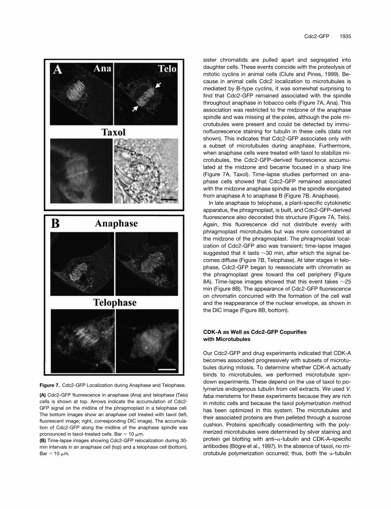
sister chromatids are pulled apart and segregated intodaughter cells. These events coincide with the proteolysis ofmitotic cyclins in animal cells (Clute and Pines, 1999). Be-cause in animal cells Cdc2 localization to microtubules ismediated by B-type cyclins, it was somewhat surprising tofind that Cdc2-GFP remained associated with the spindlethroughout anaphase in tobacco cells (Figure 7A, Ana). Thisassociation was restricted to the midzone of the anaphasespindle and was missing at the poles, although the pole mi-crotubules were present and could be detected by immu-nofluorescence staining for tubulin in these cells (data notshown). This indicates that Cdc2-GFP associates only witha subset of microtubules during anaphase. Furthermore,when anaphase cells were treated with taxol to stabilize mi-crotubules, the Cdc2-GFP–derived fluorescence accumu-lated at the midzone and became focused in a sharp line(Figure 7A, Taxol). Time-lapse studies performed on ana-phase cells showed that Cdc2-GFP remained associatedwith the midzone anaphase spindle as the spindle elongatedfrom anaphase A to anaphase B (Figure 7B, Anaphase).
In late anaphase to telophase, a plant-specific cytokineticapparatus, the phragmoplast, is built, and Cdc2-GFP–derivedfluorescence also decorated this structure (Figure 7A, Telo).Again, this fluorescence did not distribute evenly withphragmoplast microtubules but was more concentrated atthe midzone of the phragmoplast. The phragmoplast local-ization of Cdc2-GFP also was transient; time-lapse imagessuggested that it lasts
�
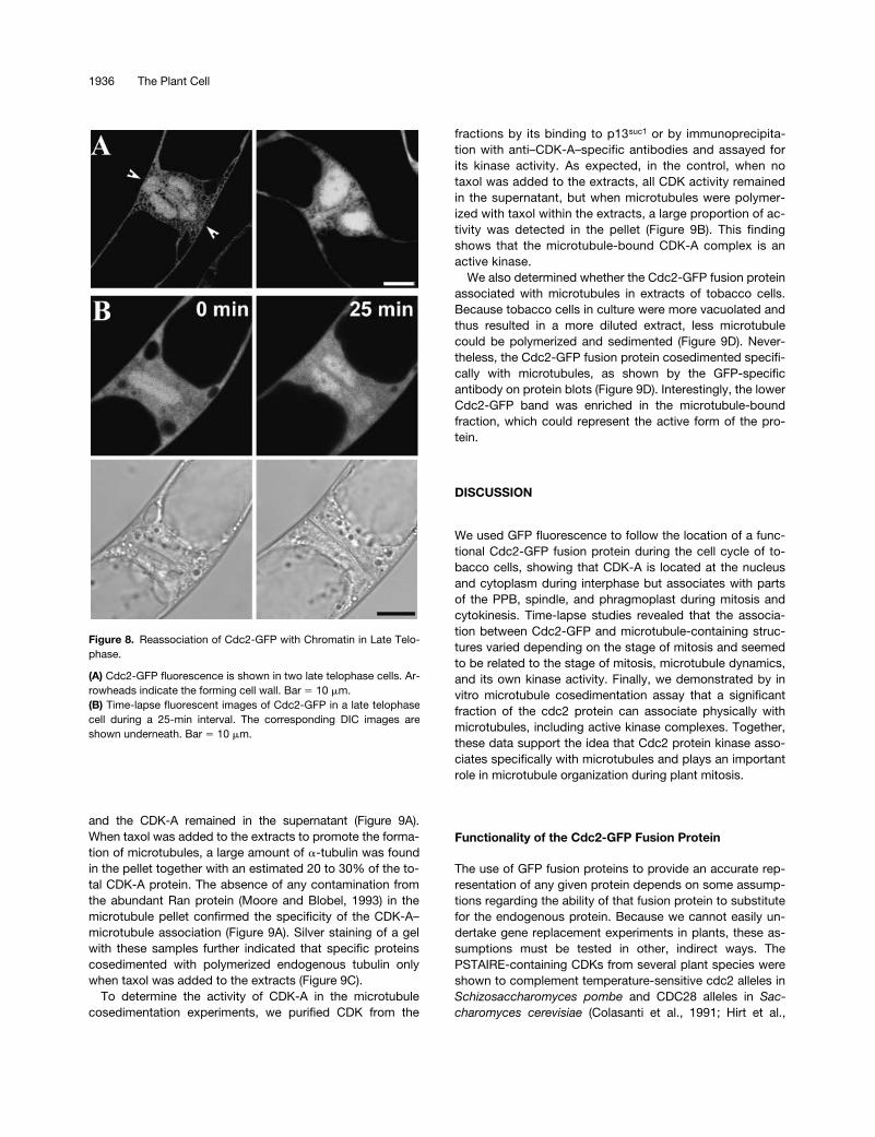
30 min, after which the signal be-comes diffuse (Figure 7B, Telophase). At later stages in telo-phase, Cdc2-GFP began to reassociate with chromatin asthe phragmoplast grew toward the cell periphery (Figure8A). Time-lapse images showed that this event takes
�
25min (Figure 8B). The appearance of Cdc2-GFP fluorescenceon chromatin concurred with the formation of the cell walland the reappearance of the nuclear envelope, as shown inthe DIC image (Figure 8B, bottom).
CDK-A as Well as Cdc2-GFP Copurifieswith Microtubules
Our Cdc2-GFP and drug experiments indicated that CDK-Abecomes associated progressively with subsets of microtu-bules during mitosis. To determine whether CDK-A actuallybinds to microtubules, we performed microtubule spin-down experiments. These depend on the use of taxol to po-lymerize endogenous tubulin from cell extracts. We used
V.faba
meristems for these experiments because they are richin mitotic cells and because the taxol polymerization methodhas been optimized in this system. The microtubules andtheir associated proteins are then pelleted through a sucrosecushion. Proteins specifically cosedimenting with the poly-merized microtubules were determined by silver staining andprotein gel blotting with anti–
�
-tubulin and CDK-A–specificantibodies (Bögre et al., 1997). In the absence of taxol, no mi-crotubule polymerization occurred; thus, both the
�
-tubulin
Figure 7. Cdc2-GFP Localization during Anaphase and Telophase.
(A) Cdc2-GFP fluorescence in anaphase (Ana) and telophase (Telo)cells is shown at top. Arrows indicate the accumulation of Cdc2-GFP signal on the midline of the phragmoplast in a telophase cell.The bottom images show an anaphase cell treated with taxol (left,fluorescent image; right, corresponding DIC image). The accumula-tion of Cdc2-GFP along the midline of the anaphase spindle waspronounced in taxol-treated cells. Bar � 10 �m.(B) Time-lapse images showing Cdc2-GFP relocalization during 30-min intervals in an anaphase cell (top) and a telophase cell (bottom).Bar � 10 �m.
1936 The Plant Cell
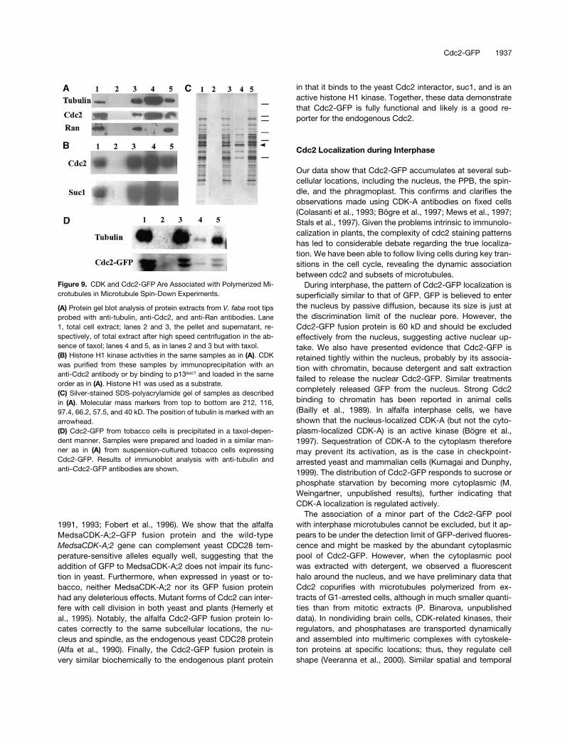
and the CDK-A remained in the supernatant (Figure 9A).When taxol was added to the extracts to promote the forma-tion of microtubules, a large amount of
�
-tubulin was foundin the pellet together with an estimated 20 to 30% of the to-tal CDK-A protein. The absence of any contamination fromthe abundant Ran protein (Moore and Blobel, 1993) in themicrotubule pellet confirmed the specificity of the CDK-A–microtubule association (Figure 9A). Silver staining of a gelwith these samples further indicated that specific proteinscosedimented with polymerized endogenous tubulin onlywhen taxol was added to the extracts (Figure 9C).
To determine the activity of CDK-A in the microtubulecosedimentation experiments, we purified CDK from the
fractions by its binding to p13
suc1
or by immunoprecipita-tion with anti–CDK-A–specific antibodies and assayed forits kinase activity. As expected, in the control, when notaxol was added to the extracts, all CDK activity remainedin the supernatant, but when microtubules were polymer-ized with taxol within the extracts, a large proportion of ac-tivity was detected in the pellet (Figure 9B). This findingshows that the microtubule-bound CDK-A complex is anactive kinase.
We also determined whether the Cdc2-GFP fusion proteinassociated with microtubules in extracts of tobacco cells.Because tobacco cells in culture were more vacuolated andthus resulted in a more diluted extract, less microtubulecould be polymerized and sedimented (Figure 9D). Never-theless, the Cdc2-GFP fusion protein cosedimented specifi-cally with microtubules, as shown by the GFP-specificantibody on protein blots (Figure 9D). Interestingly, the lowerCdc2-GFP band was enriched in the microtubule-boundfraction, which could represent the active form of the pro-tein.
DISCUSSION
We used GFP fluorescence to follow the location of a func-tional Cdc2-GFP fusion protein during the cell cycle of to-bacco cells, showing that CDK-A is located at the nucleusand cytoplasm during interphase but associates with partsof the PPB, spindle, and phragmoplast during mitosis andcytokinesis. Time-lapse studies revealed that the associa-tion between Cdc2-GFP and microtubule-containing struc-tures varied depending on the stage of mitosis and seemedto be related to the stage of mitosis, microtubule dynamics,and its own kinase activity. Finally, we demonstrated by invitro microtubule cosedimentation assay that a significantfraction of the cdc2 protein can associate physically withmicrotubules, including active kinase complexes. Together,these data support the idea that Cdc2 protein kinase asso-ciates specifically with microtubules and plays an importantrole in microtubule organization during plant mitosis.
Functionality of the Cdc2-GFP Fusion Protein
The use of GFP fusion proteins to provide an accurate rep-resentation of any given protein depends on some assump-tions regarding the ability of that fusion protein to substitutefor the endogenous protein. Because we cannot easily un-dertake gene replacement experiments in plants, these as-sumptions must be tested in other, indirect ways. ThePSTAIRE-containing CDKs from several plant species wereshown to complement temperature-sensitive cdc2 alleles in
Schizosaccharomyces
pombe
and CDC28 alleles in
Sac-charomyces
cerevisiae
(Colasanti et al., 1991; Hirt et al.,
Figure 8. Reassociation of Cdc2-GFP with Chromatin in Late Telo-phase.
(A) Cdc2-GFP fluorescence is shown in two late telophase cells. Ar-rowheads indicate the forming cell wall. Bar � 10 �m.(B) Time-lapse fluorescent images of Cdc2-GFP in a late telophasecell during a 25-min interval. The corresponding DIC images areshown underneath. Bar � 10 �m.
Cdc2-GFP 1937
1991, 1993; Fobert et al., 1996). We show that the alfalfaMedsaCDK-A;2–GFP fusion protein and the wild-type
MedsaCDK-A;2
gene can complement yeast CDC28 tem-perature-sensitive alleles equally well, suggesting that theaddition of GFP to MedsaCDK-A;2 does not impair its func-tion in yeast. Furthermore, when expressed in yeast or to-bacco, neither MedsaCDK-A;2 nor its GFP fusion proteinhad any deleterious effects. Mutant forms of Cdc2 can inter-fere with cell division in both yeast and plants (Hemerly etal., 1995). Notably, the alfalfa Cdc2-GFP fusion protein lo-cates correctly to the same subcellular locations, the nu-cleus and spindle, as the endogenous yeast CDC28 protein(Alfa et al., 1990). Finally, the Cdc2-GFP fusion protein isvery similar biochemically to the endogenous plant protein
in that it binds to the yeast Cdc2 interactor, suc1, and is anactive histone H1 kinase. Together, these data demonstratethat Cdc2-GFP is fully functional and likely is a good re-porter for the endogenous Cdc2.
Cdc2 Localization during Interphase
Our data show that Cdc2-GFP accumulates at several sub-cellular locations, including the nucleus, the PPB, the spin-dle, and the phragmoplast. This confirms and clarifies theobservations made using CDK-A antibodies on fixed cells(Colasanti et al., 1993; Bögre et al., 1997; Mews et al., 1997;Stals et al., 1997). Given the problems intrinsic to immunolo-calization in plants, the complexity of cdc2 staining patternshas led to considerable debate regarding the true localiza-tion. We have been able to follow living cells during key tran-sitions in the cell cycle, revealing the dynamic associationbetween cdc2 and subsets of microtubules.
During interphase, the pattern of Cdc2-GFP localization issuperficially similar to that of GFP. GFP is believed to enterthe nucleus by passive diffusion, because its size is just atthe discrimination limit of the nuclear pore. However, theCdc2-GFP fusion protein is 60 kD and should be excludedeffectively from the nucleus, suggesting active nuclear up-take. We also have presented evidence that Cdc2-GFP isretained tightly within the nucleus, probably by its associa-tion with chromatin, because detergent and salt extractionfailed to release the nuclear Cdc2-GFP. Similar treatmentscompletely released GFP from the nucleus. Strong Cdc2binding to chromatin has been reported in animal cells(Bailly et al., 1989). In alfalfa interphase cells, we haveshown that the nucleus-localized CDK-A (but not the cyto-plasm-localized CDK-A) is an active kinase (Bögre et al.,1997). Sequestration of CDK-A to the cytoplasm thereforemay prevent its activation, as is the case in checkpoint-arrested yeast and mammalian cells (Kumagai and Dunphy,1999). The distribution of Cdc2-GFP responds to sucrose orphosphate starvation by becoming more cytoplasmic (M.Weingartner, unpublished results), further indicating thatCDK-A localization is regulated actively.
The association of a minor part of the Cdc2-GFP poolwith interphase microtubules cannot be excluded, but it ap-pears to be under the detection limit of GFP-derived fluores-cence and might be masked by the abundant cytoplasmicpool of Cdc2-GFP. However, when the cytoplasmic poolwas extracted with detergent, we observed a fluorescenthalo around the nucleus, and we have preliminary data thatCdc2 copurifies with microtubules polymerized from ex-tracts of G1-arrested cells, although in much smaller quanti-ties than from mitotic extracts (P. Binarova, unpublisheddata). In nondividing brain cells, CDK-related kinases, theirregulators, and phosphatases are transported dynamicallyand assembled into multimeric complexes with cytoskele-ton proteins at specific locations; thus, they regulate cellshape (Veeranna et al., 2000). Similar spatial and temporal
Figure 9. CDK and Cdc2-GFP Are Associated with Polymerized Mi-crotubules in Microtubule Spin-Down Experiments.
(A) Protein gel blot analysis of protein extracts from V. faba root tipsprobed with anti-tubulin, anti-Cdc2, and anti-Ran antibodies. Lane1, total cell extract; lanes 2 and 3, the pellet and supernatant, re-spectively, of total extract after high speed centrifugation in the ab-sence of taxol; lanes 4 and 5, as in lanes 2 and 3 but with taxol.(B) Histone H1 kinase activities in the same samples as in (A). CDKwas purified from these samples by immunoprecipitation with ananti-Cdc2 antibody or by binding to p13suc1 and loaded in the sameorder as in (A). Histone H1 was used as a substrate.(C) Silver-stained SDS-polyacrylamide gel of samples as describedin (A). Molecular mass markers from top to bottom are 212, 116,97.4, 66.2, 57.5, and 40 kD. The position of tubulin is marked with anarrowhead.(D) Cdc2-GFP from tobacco cells is precipitated in a taxol-depen-dent manner. Samples were prepared and loaded in a similar man-ner as in (A) from suspension-cultured tobacco cells expressingCdc2-GFP. Results of immunoblot analysis with anti-tubulin andanti–Cdc2-GFP antibodies are shown.
1938 The Plant Cell
control of cytoplasm- and membrane-associated microtu-bule nucleation and organization might operate in largeacentrosomal plant cells in which various microtubular ar-rays change their positions during cell cycle progression.
Preprophase Band
The PPB, which is formed at the cell surface just before mi-tosis, predicts the plane of cell division but disassemblesjust before mitosis (Mineyuki, 1999). Recruitment of CDK-Ato the PPB seems to be a transient event just before the dis-assembly of the PPB. Cdc2-GFP, like CDK-A, is found withonly a small proportion of PPB (Colasanti et al., 1993; Mewset al., 1997). We determined when Cdc2-GFP localized to thePPB by cell cycle synchronization and arrest with the pro-tein kinase inhibitor K252-a. K252-a arrests cells in G2-phaseand accumulates cells with PPBs (Katsuta and Shibaoka,1992). However, we never observed Cdc2-GFP associatingwith PPB in K252-a–arrested cells, indicating that the CDK-Aassociation depends on the phosphorylation of some com-ponent in the PPB or that the association occurs after thearrest point of K252-a. The extremely narrow configurationof the Cdc2-GFP band and its disappearance within a shorttime interval also suggest that Cdc2-GFP is associated witha late PPB structure. Correspondingly, one of the maize mi-totic cyclins, Zeama;CycB1;2, associates transiently withthe late PPB, whereas Zeama;CycA1;1 is localized to thePPB over a longer period (Mews et al., 1997). Microinjectionof active CDK-A kinase to plant cells before division selec-tively accelerated the disassembly of the PPB but not ofother microtubule structures (Hush et al., 1996), whereasthe inhibition of CDKs by the drug roscovitine delayed thisprocess (Binarova et al., 1998). Together, these data sug-gest that CDK-A directly induces the disassembly of PPB inpreprophase.
Cdc2 Association with the Spindle
Cdc2-GFP did not accumulate on the prophase spindle butremained associated with condensing chromosomes. Atthis stage of the cell cycle, Cdc2 is involved in many impor-tant cellular processes, including nuclear envelope break-down and chromatin condensation. The microinjection ofactive Cdc2 induces premature condensation of chromatin(Hush et al., 1996), whereas roscovitine inhibition of CDKactivity delays it and prevents nuclear envelope breakdown(Binarova et al., 1998). As cells approach metaphase, Cdc2-GFP leaves the chromatin and is recruited progressively tothe spindle, which seems to peak at metaphase. The inter-action between CDK-A and plant spindle microtubules ap-peared to be strong, because it was resistant to detergentextraction and could be cosedimented with taxol-stabilizedmicrotubules from cell extracts. This unusually stable inter-action between cdc2 and microtubules might relate to
plant-specific spindle formation without the spatial cue ofcentrosomes, where the bipolar spindle configuration frompoles to kinetochores requires the selective stabilization andmovement of microtubules.
Cytokinesis
The CDK complex is inactivated at the metaphase/ana-phase transition by the degradation of the cyclin componentby the mitosis-specific protein degradation machinery, theanaphase-promoting complex (Genschik et al., 1998). Thedegradation of cyclins containing the mitotic destructionbox was confirmed using Cyclin B1–GFP, Cyclin B2–GFP,and Cyclin A2–GFP fusion proteins (M. Weingartner, unpub-lished results). However, a curious feature of some plantcyclins, such as Zeama;CycB1;1, Zeama;CycB2;1, andZeama;CycA1;1, is their prolonged presence through ana-phase and telophase in association with mitotic cytoskele-ton structures, as revealed by immunolocalization studies(Mews et al., 1997). The cyclin degradation machinery isthought to associate with chromatin, and chromatin-boundcyclin is located appropriately for degradation (Clute andPines, 1999). Thus, the recruitment of the CDK-A–cyclincomplex to spindle and phragmoplast microtubules mightprotect a pool of cyclins from degradation by sequesteringthem away from the chromatin-located proteolysis machin-ery. Alternatively, CDK-A might associate with cyclins inanaphase and telophase, which do not contain a mitotic de-struction box, and thus might not be targeted by the mitoticproteolysis machinery. Interestingly, it has been shown thatthe CycD2;1 and CycD3;1 transcripts accumulate during mi-tosis (Sorrell et al., 1999).
An important role for CDK-A in regulating cytoskeletonstructures is prompted by its specific locations in anaphaseand telophase. Cdc2-GFP is present at the midzone of theanaphase spindle, where new populations of microtubulesassemble, but it is missing at the poles on the shortening ki-netochore microtubules. Later, it accumulates at the mid-plane of cell division at the end of anaphase and along themidline of forming phragmoplast microtubules. Even thoughcytokinesis appears mechanistically different in plants andanimals, recent studies indicate that the midplane of cell di-vision might play a conserved role for signaling division sitesduring cytokinesis in plant, fungal, and animal cells, and anumber of regulatory proteins accumulate at the spindlemidzone during cytokinesis (Hales et al., 1999; Robinsonand Spudich, 2000). In plant cells, two related mitogen-acti-vated protein kinases (MAPKs), MMK3 from alfalfa andNTF6 from tobacco, were found transiently at the midplaneof cell division by immunofluorescence (Calderini et al.,1998; Bögre et al., 1999). A potential upstream regulator ofthese MAPKs is NPK1, which also localizes to the midplane(Nishihama et al., 2001). NPK1 has a potential Cdc2 phos-phorylation site and therefore is a putative Cdc2 kinase sub-strate. Another regulator of NPK1 is a mitosis-specific
Cdc2-GFP 1939
kinesin (Ito et al., 1998; Nishihama et al., 2001). The role andtargets of this cytokinesis signaling pathway are not wellunderstood, but it might regulate MAPs, such as MAP65(Smertenko et al., 2000), or molecular motors, such asTPRK125 (Asada et al., 1997; Barroso et al., 2000) andAtPAKRP1 (Lee and Liu, 2000), which are located similarlyat the midplane of cell division.
Modulation of Microtubule Dynamics and Movementsby CDKs
Microtubules are intrinsically unstable structures because ofa continuous flux of tubulin between “free” tubulin dimersand polymerized microtubules, and it is this dynamic insta-bility that makes the microtubule cytoskeleton such anadaptable feature of cellular organization. There are growingnumbers of microtubule-interacting proteins known in ani-mal cells that modulate microtubule polymerization and dy-namics and that are modified by cell cycle–dependentphosphorylation (Desai and Mitchison, 1997). Cyclin B1–CDK,when added to Xenopus laevis egg extracts, induced thedramatic shortening of microtubules, which was correlatedwith a severalfold increase in catastrophe frequency (Verdeet al., 1992). Similarly, interphase but not mitotic plant ex-tracts promoted the polymerization of long microtubules(Stoppin et al., 1996). In animal cells, the phosphorylation ofMAP4 by Cdc2 results in its reduced affinity for microtu-bules during mitosis, which leads to greater microtubuleinstability (Ookata et al., 1997). Correspondingly, the elimi-nation of cdc2 phosphorylation sites on MAP4 maintains itsaffinity for microtubules during mitosis and leads to defectsin anaphase A chromosome movement (Shiina and Tsukita,1999), similar to when plant Cdc2 kinase is inhibited withroscovitine (Binarova et al., 1998). XMAP215 is another MAPisolated from X. laevis eggs containing two conserved CDK1phosphorylation sites. Its human homolog, pTOG, has beenfound to interact with Cyclin B1, thus targeting the Cdc2 ki-nase to microtubules during mitosis (Charrasse et al., 2000).However, unlike other MAPs isolated thus far, mitotic phos-phorylation of XMAP215 by CDK1 does not reduce its micro-tubule binding but strongly decreases its activity to promotemicrotubule elongation (Vasquez et al., 1999). The stable in-teraction of Cdc2 with the spindle in our experiments couldbe attributable to a MAP with similar characteristics.
Plant microtubules are at least as dynamic as those in an-imals, are regulated similarly by MAPs, and are determinedby their composition (plants contain multiple copies of func-tional �- and �-tubulin genes) and by various post-transla-tional modifications (Lloyd and Hussey, 2001). StructuralMAPs are just being molecularly identified in plants, and it isapparent that plant MAPs are highly specialized to selec-tively bind and regulate subsets of microtubules at specificlocations. For example, NtMAP65-1 is bound selectively toa subset of interphase and certain mitotic microtubules(Smertenko et al., 2000). The apparent similarity in Cdc2 and
NtMAP65-1 localizations (e.g., at the PPB, at the midzone ofthe anaphase spindle, and at the midline of the phragmo-plast) might suggest a functional relationship.
Microtubule-associated motor proteins are essential inspindle assembly and function and localize to the kineto-chores, the spindle poles, and the spindle midzone in animaland fungal cells. The minus end–directed motor, dynein,which is important for chromatin-mediated spindle assem-bly, was shown to be phosphorylated in mitosis by the Cdc2kinase (Dell et al., 2000). HsEg5 from human cells and XLP2from X. laevis are plus end–directed kinesin-related proteins(KRPs), which are required for centrosome separation, spin-dle assembly, and maintenance and which are controlled bycdc2/p34 phosphorylation (Blangy et al., 1995). Phosphory-lation of HsEg5 increases during mitosis and promotes itsspindle association. Depleting Eg5 results in monopolar spin-dles very similar to those found in roscovitine-treated plantcells (Binarova et al., 1998).
Three genes that encode KRPs have been identified invarious plant species, and one of them, TKRP125, containsa conserved cdc2 phosphorylation site and belongs to thesame bimC subfamily of KRPs, like Eg5 (Asada et al., 1997;Barroso et al., 2000). This plus end–directed motor proteinlocalizes to the mitotic spindle at increasing levels towardthe equatorial plane, to the interzone of the anaphase spin-dle, and to the phragmoplast. One of the suggested func-tions of TKRP125 is the sliding of interzone microtubules,resulting in the elongation of the anaphase spindle. Record-ing time-lapse images, we observed a similarly stable colo-calization of Cdc2-GFP with interzone microtubules duringthe elongation of the anaphase spindle. Moreover, the stabi-lization of microtubules by taxol led to the focusing of Cdc2-GFP fluorescence to the midline, suggesting movement to-ward the plus end of the microtubules. Therefore, TKRP125might be another potential binding partner and substratethat targets the Cdc2 kinase to spindle microtubules.
The localization of Cdc2-GFP in living cells, its physicalinteraction with microtubules, and the phenotypes of cellswhen Cdc2 is inhibited by roscovitine provide insights intothe role of Cdc2 during mitosis. Our detailed pharmacologi-cal studies suggest that Cdc2 might regulate microtubulestability and movement. The Cdc2-GFP fusion protein andthe assays we developed, together with the molecularprobes becoming available, will allow further testing of Cdc2functions in the regulation of MAPs and motor proteins.
METHODS
Plasmid Construction and Transformation Techniques
The S65T mutant of the plant codon–optimized synthetic Green Fluo-rescent Protein (GFP; Chiu et al., 1996) with a unique NcoI site atthe initiation codon was obtained from Toni Schäffer (Ludwig-Maxi-milians-Universität, Munich, Germany). The GFP coding region was

1940 The Plant Cell
recloned as an EcoRI–XhoI fragment into the yeast shuttle vectorpYES2 (Stratagene, La Jolla, CA) and as a KpnI–SalI fragment intothe tetracycline-inducible binary plant expression vector pBinHygTx(Weinmann et al., 1994).
A chimeric Cdc2-GFP fusion was constructed by cloning the GFPgene as an NcoI–XhoI fragment into plasmid pBS-cdc2 carrying theMedsa;CDK-A;2 coding region with an inserted unique NcoI site atthe 3� terminus. The cdc2-GFP fusion gene was subcloned as aBstXI–XhoI fragment into the pYES2 yeast shuttle vector (pYES2-cdc2-GFP) and as a KpnI–SalI fragment into the tetracycline-induc-ible binary plant expression vector (pBinHygTx-cdc2-GFP). Yeasttransformation and complementation assays were performed asdescribed previously (Hirt et al., 1993). Transgenic plants wereobtained by Agrobacterium tumefaciens–mediated leaf disc trans-formation with the tobacco (Nicotiana tabacum) lines Petit HavanaSR1 and Samsung Tb-Tet (Weinmann et al., 1994). Five indepen-dent transformants for each construct were propagated, and seedwere obtained. The hygromycin-resistant progeny were used in theexperiments.
Cell Culture and Synchronization
Suspension cultures were initiated by placing stem segments oftransgenic Cdc2-GFP and GFP lines of T2 generation in 1 mg·L�2
2,4-D containing Murashige and Skoog (1962) solid medium, and theresulting calli were transferred after 1 month into liquid Linsmaier andSkoog–modified medium (Sano et al., 1999) supplemented with 40�g·mL�2 hygromycin B.
Synchronization started with a 1:5 dilution of 7-day-old culture.After 8 hr, 10 �g·L�2 aphidicoline was added to the medium. Theaphidicoline was removed after 16 hr by five changes of medium in 1hr. Drugs were added 7 hr after aphidicoline removal in G2-phase orat 10 hr in mitosis. The following drug concentrations were used inthese experiments: 10 �M amiprophos methyl, 50 �M taxol, 100 �Mroscovitine (a gift from M. Strnad, Olomouc, Czech Republic), 0.2 �Mokadaic acid, 2 �M K252a, and 100 �M MG132. Samples were col-lected at various intervals and used immediately for observation. Tofollow cell cycle progression, flow cytometric analysis was per-formed as described (Bögre et al., 1997) using a PAS2 flow cytome-ter (Partec, Münster, Germany).
Microscopy Techniques for GFP Detection
A drop of cell suspension was transferred on a slide, carefully cov-ered with a cover slip, and observed with an upright fluorescence mi-croscope (Axioplan 2; Zeiss, Jena, Germany) equipped with a GFPfilter (HQ480/20X, HQ510/20M; AF Analysentechnik, Jena, Germany).Images were taken using a cooled charge-coupled device black-and-white digital camera (SPOT-2; Diagnostic Instruments, Burroughs,MI) and Metaview imaging software (Diagnostic Instruments).
Confocal images were taken using a Leica (Wetzlar, Germany) TNTlaser scanning confocal microscope with argon laser illumination at488 nm and through a fluorescein isothiocyanate filter set. For trans-mission light images, differential interference contrast optics wereused with both the Zeiss and Leica microscopes. To reduce pho-tobleaching, the excitation light was turned on only during imagecapture. Images were contrast enhanced using image-processingsoftware (Photoshop; Adobe Systems, Mountain View, CA).
Detergent Extraction of Cells
Cells were extracted in detergent essentially as described previouslyfor the preparation of cytoskeletons (Chan et al., 1996). Cells were in-cubated in the enzyme solution (1% [w/v] cellulase R10, 0.2% [w/v]mazerozyme R10 [Yakult, Tokyo, Japan], and 0.45 M sorbitol) in PME(0.1 M 1,4-piperazinediethanesulfonic acid, 1 mM MgSO4, and 1 mMEGTA, pH 6.9) for 15 min. Cells with partially digested cell walls werewashed twice in PME with 0.45 M sorbitol and then incubated in ex-traction buffer (10% [w/v] DMSO, 0.05% [w/v] Nonidet P-40, and0.45 M sorbitol in PME) for 15 min. The resulting detergent-extractedcells were washed twice in wash 2 (10% DMSO in PME) and ob-served directly with the fluorescence microscope.
Protein Blotting, Immunoprecipitation, p13Suc1 Binding, and Histone H1 Kinase Assay
The samples were homogenized in 3 volumes of homogenizationbuffer containing 25 mM Tris-HCl, pH 7.5, 15 mM MgCl2, 15 mMEGTA, 75 mM NaCl, 1 mM DTT, 0.1% (w/v) Tween 20, 15 mM 4-nitro-phenylphosphatebis, 15 mM �-glycerophosphate, 0.5 mM Na3VO3, 1mM NaF, 0.5 mM phenylmethylsulfonyl fluoride, and 5 �g/mL leu-peptin and aprotinin. After centrifugation at 100,000g, the superna-tant was collected and the protein concentration was measured withthe Bradford reagent and adjusted to 2 mg·mL�2. SDS sample bufferwas added, and the samples were heated for 2 min and stored at�20�C. After electrophoresis, the gels were electroblotted onto poly-vinylidene difluoride (Millipore, Bedford, MA) membranes in 50 mMTris base and 50 mM boric acid buffer, pH 8.3, in a liquid electroblot-ting system (Hoefer, San Francisco, CA) overnight at 30 V with cool-ing. For immunodetection, an anti-GFP rabbit serum (MolecularProbes, Eugene, OR) was applied in blocking buffer at a dilution of1:1000 and alkaline phosphatase–conjugated goat anti–rabbit IgG(Sigma) was used as a second antibody in a dilution of 1:5000. Thereaction was visualized with nitroblue tetrazolium/5-bromo-4-chloro-3-indolyl phosphate substrates (CDP-Star; Amersham).
Cyclin-dependent kinase (CDK) activities were measured in thesamples after immunoprecipitation with the anti-Cdc2 antibody orthe anti-GFP antibody or after binding to p13suc1 as described previ-ously (Bögre et al., 1997). For GFP immunoprecipitation, 15 �L ofmouse anti-GFP hybridoma supernatant (a gift from Jan Elliott, Aus-tralian National University, Canberra) and 25 �L of anti-mouse Igagarose beads (Sigma) were added to the cell extracts and the tubeswere rotated for 3 to 4 hr. Washing conditions were the same as forCdc2 (Bögre et al., 1997).
Microtubule Cosedimentation Assay
Extracts of Vicia faba root meristem or tobacco-cultured cells werehomogenized in Hepes buffer containing 50 mM Hepes, pH 7.4, 1mM EGTA, 1 mM MgCl2, 75 mM NaCl, 1 mM DTT, 0.1% (w/v) Tween20, 15 mM 4-p-nitrophenylphosphate, 15 mM �-glycerophosphate,0.5 mM Na3VO3, 1 mM NaF, 0.5 mM phenylmethylsulfonyl fluoride,and 5 �g·mL�2 leupeptin and aprotinin. The samples were centri-fuged at 27,000g for 60 min. To polymerize microtubules in the ex-tract, GTP was added to 1 mM and taxol was added to 20 �M. After15 min of polymerization, the extracts were loaded on top of thecushion of Hepes buffer with 40% sucrose and centrifuged at20,000g for 40 min. After three washes with Hepes buffer, the pellet
Cdc2-GFP 1941
was resuspended in 50 �L of SDS sample buffer and loaded for elec-trophoresis and protein gel blotting. CDK activity measurementswere made by resuspending the pellets in the same volume as theoriginal extracts and using equal volumes for immunoprecipitationsand p13suc1 binding, followed by protein kinase assay as describedpreviously (Bögre et al., 1997). Monoclonal mouse anti–�-tubulin,DMA1 (Sigma), polyclonal rabbit anti–CDK-A (Bögre et al., 1997),polyclonal anti-Ran (Babco, Richmond, CA), and monoclonal mouseanti-GFP (provided by Jan Elliott) antibodies were used for immuno-detection.
ACKNOWLEDGMENTS
Thanks to J. Elliott for providing the GFP antibody and to F. Klein(Botany Institute, Vienna University) for providing the epifluorescencemicroscopy facility at the initial phase of the project. Moreover, wethank Gireg Weingartner for his incredible patience while finishingthis work. M.W. was supported by a Ph.D. fellowship from the Aus-trian Academy of Sciences. This work was supported by an AustrianFörderung der Wissenschaftlichen Forschung grant to E.H.-B., byEuropean Union Framework V project ECCO Grant QLRT-1999-00454 to E.H.-B., L.B., and P.B., by Grant A5020803/1998 from theGrant Agency of the Czech Academy of Sciences to P.B., and by aBiotechnology and Biological Science Research Council grant toL.B. and J.-P.D.
Received March 13, 2001; accepted May 30, 2001.
REFERENCES
Alfa, C.E., Ducommun, B., Beach, D., and Hyams, J.S. (1990).Distinct nuclear and spindle pole body population of cyclin-cdc2in fission yeast. Nature 347, 680–682.
Asada, T., Kuriyama, R., and Shibaoka, H. (1997). TKRP125, akinesin-related protein involved in the centrosome-independentorganization of the cytokinetic apparatus in tobacco BY-2 cells. J.Cell Sci. 110, 179–189.
Ayaydin, F., Vissi, E., Meszaros, T., Miskolczi, P., Kovacs, I.,Feher, A., Dombradi, V., Erdodi, F., Gergely, P., and Dudits, D.(2000). Inhibition of serine/threonine-specific protein phospha-tases causes premature activation of cdc2MsF kinase at G2/M tran-sition and early mitotic microtubule organisation in alfalfa. Plant J.23, 85–96.
Bailly, E., Doree, M., Nurse, P., and Bornens, M. (1989). p34cdc2is located in both nucleus and cytoplasm; part is centrosomallyassociated at G2/M and enters vesicles at anaphase. EMBO J. 8,3985–3995.
Barroso, C., Chan, J., Allan, V., Doonan, J., Hussey, P., andLloyd, C. (2000). Two kinesin-related proteins associated with thecold-stable cytoskeleton of carrot cells: Characterization of anovel kinesin, DcKRP120–2. Plant J. 24, 859–868.
Binarova, P., Dolezel, J., Draber, P., Heberle-Bors, E., Strnad,M., and Bogre, L. (1998). Treatment of Vicia faba root tip cellswith specific inhibitors to cyclin-dependent kinases leads toabnormal spindle formation. Plant J. 16, 697–707.
Blangy, A., Lane, H.A., d’Herin, P., Harper, M., Kress, M., andNigg, E.A. (1995). Phosphorylation by p34cdc2 regulates spindleassociation of human Eg5, a kinesin-related motor essential forbipolar spindle formation in vivo. Cell 83, 1159–1169.
Bögre, L., Zwerger, K., Meskiene, I., Binarova, P., Czizmadia, V.,Planck, C., Wagner, E., Hirt, H., and Heberle-Bors, E. (1997).The cdc2Ms kinase is differently regulated in the cytoplasm and inthe nucleus. Plant Physiol. 113, 841–852.
Bögre, L., Calderini, O., Binarova, P., Mattauch, M., Till, S., Kiegerl,S., Jonak, C., Pollaschek, C., Barker, P., Huskisson, N.S., Hirt,H., and Heberle-Bors, E. (1999). A MAP kinase is activated late inplant mitosis and becomes localized to the plane of cell division.Plant Cell 11, 101–114.
Calderini, O., Bogre, L., Vicente, O., Binarova, P., Heberle-Bors,E., and Wilson, C. (1998). A cell cycle regulated MAP kinase witha possible role in cytokinesis in tobacco cells. J. Cell Sci. 111,3091–3100.
Chan, J., Rutten, T., and Lloyd, C. (1996). Isolation of microtubule-associated proteins from carrot cytoskeletons: A 120 kDa mapdecorates all four microtubule arrays and the nucleus. Plant J. 10,251–259.
Charrasse, S., Lorca, T., Doree, M., and Larroque, C. (2000). TheXenopus XMAP215 and its human homologue TOG proteins inter-act with cyclin B1 to target p34cdc2 to microtubules during mito-sis. Exp. Cell Res. 254, 249–256.
Chiu, W., Niwa, Y., Zeng, W., Hirano, T., Kobayashi, H., andSheen, J. (1996). Engineered GFP as a vital reporter in plants.Curr. Biol. 6, 325–330.
Chytilova, E., Macas, J., Sliwinska, E., Rafelski, S.M., Lambert,G.M., and Galbraith, D.W. (2000). Nuclear dynamics in Arabidop-sis thaliana. Mol. Biol. Cell 11, 2733–2741.
Clute, P., and Pines, J. (1999). Temporal and spatial control ofcyclin B1 destruction in metaphase. Natl. Cell Biol. 1, 82–87.
Colasanti, J., Tyers, M., and Sundaresan, V. (1991). Isolation andcharacterization of cDNA clones encoding a functional p34cdc2homologue from Zea mays. Proc. Natl. Acad. Sci. USA 88, 3377–3381.
Colasanti, J., Cho, S.-O., Wick, S., and Sundaresan, V. (1993).Localisation of the functional p34cdc2 homolog of maize in theroot tip and stomatal complex cells: Association with predicteddivision sites. Plant Cell 5, 1101–1111.
Dell, K.R., Turck, C.W., and Vale, R.D. (2000). Mitotic phosphoryla-tion of the dynein light intermediate chain is mediated by cdc2kinase. Traffic 1, 38–44.
Desai, A., and Mitchison, T.J. (1997). Microtubule polymerizationdynamics. Annu. Rev. Cell Dev. Biol. 13, 83–117.
Doonan, J. (2000). Social controls on cell proliferation in plants.Curr. Opin. Plant Biol. 3, 482–487.
Doonan, J., and Fobert, P. (1997). Conserved and novel regulatorsof the plant cell cycle. Curr. Opin. Cell Biol. 9, 824–830.
Ferreira, P.C., Hemerly, A.S., Villarroel, R., Van Montagu, M., andInze, D. (1991). The Arabidopsis functional homolog of thep34cdc2 protein kinase. Plant Cell 3, 531–540.
Fobert, P.R., Gaudin, V., Lunness, P., Coen, E.S., and Doonan,J.H. (1996). Distinct classes of cdc2-related genes are differen-tially expressed during the cell division cycle in plants. Plant Cell8, 1465–1476.

1942 The Plant Cell
Genschik, P., Criqui, M.C., Parmentier, Y., Derevier, A., andFleck, J. (1998). Cell cycle-dependent proteolysis in plants: Iden-tification of the destruction box pathway and metaphase arrestproduced by the proteasome inhibitor mg132. Plant Cell 10,2063–2076.
Hales, K.G., Bi, E., Wu, J.Q., Adam, J.C., Yu, I.C., and Pringle,J.R. (1999). Cytokinesis: An emerging unified theory for eukary-otes? Curr. Opin. Cell Biol. 11, 717–725.
Healy, J.M., Menges, M., Doonan, J.H., and Murray, J.A. (2000).The Arabidopsis D-type cyclins CycD2 and CycD3 both interact invivo with the PSTAIRE cyclin-dependent kinase Cdc2a but aredifferentially controlled. J. Biol. Chem. 276, 7041–7047.
Hemerly, A., Engler, J.A., Bergounioux, C., Van Montagu, M.,Engler, G., Inze, D., and Ferreira, P. (1995). Dominant negativemutants of the Cdc2 kinase uncouple cell division from iterativeplant development. EMBO J. 14, 3925–3936.
Hirt, H., Pay, A., Gyorgyey, J., Bako, L., Nemeth, K., Bogre, L.,Schweyen, R.J., Heberle-Bors, E., and Dudits, D. (1991). Com-plementation of a yeast cell cycle mutant by an alfalfa cDNAencoding a protein kinase homologous to p34cdc2. Proc. Natl.Acad. Sci. USA 88, 1636–1640.
Hirt, H., Pay, A., Bogre, L., Meskiene, I., and Heberle-Bors, E.(1993). cdc2MsB, a cognate cdc2 gene from alfalfa, complementsthe G1/S but not the G2/M transition of budding yeast cdc28mutants. Plant J. 4, 61–69.
Hush, J., Wu, L., John, P.C., Hepler, L.H., and Hepler, P.K. (1996).Plant mitosis promoting factor disassembles the microtubule pre-prophase band and accelerates prophase progression in Trades-cantia. Cell Biol. Int. 20, 275–287.
Ito, M., Iwase, M., Kodama, H., Lavisse, P., Komamine, A.,Nishihama, R., Machida, Y., and Watanabe, A. (1998). A novelcis-acting element in promoters of plant B-type cyclin genes acti-vates M phase-specific transcription. Plant Cell 10, 331–341.
Joubes, J., Chevalier, C., Dudits, D., Heberle-Bors, E., Inze, D.,Umeda, M., and Renaudi, J.P. (2000). CDK-related proteinkinases in plants. Plant Mol. Biol. 43, 607–620.
Katsuta, J., and Shibaoka, H. (1992). Inhibition by kinase inhibitorsof the development and the disappearance of the preprophase bandof microtubules in tobacco BY-2 cells. J. Cell Sci. 103, 397–405.
Kumagai, A., and Dunphy, W.G. (1999). Binding of 14–3-3 proteinsand nuclear export control the intracellular localization of themitotic inducer Cdc25. Genes Dev. 13, 1067–1072.
Lee, Y.R., and Liu, B. (2000). Identification of a phragmoplast-associ-ated kinesin-related protein in higher plants. Curr. Biol. 10, 797–800.
Leiss, D., Felix, M.A., and Karsenti, E. (1992). Association ofcyclin-bound p34cdc2 with subcellular structures in Xenopuseggs. J. Cell Sci. 102, 285–297.
Lloyd, C., and Hussey, P. (2001). Microtubule-associated proteinsin plants: Why we need a map. Natl. Rev. Mol. Cell Biol. 2, 41–47.
Magyar, Z., et al. (1997). Cell cycle phase specificity of putativecyclin-dependent kinase variants in synchronized alfalfa cells.Plant Cell 9, 223–235.
Mews, M., Sek, F.J., Moore, R., Volkmann, D., Gunning, B.E.S.,and John, P.C.L. (1997). Mitotic cyclin distribution during maizecell division: Implications for the sequence diversity and functionof cyclins in plants. Protoplasma 200, 128–145.
Mineyuki, Y. (1999). The preprophase band of microtubules: Itsfunction as a cytokinetic apparatus in higher plants. Int. Rev.Cytol. 187, 1–50.
Mineyuki, Y., Yamashita, M., and Nagahama, Y. (1991). P34cdc2kinase homologue in the preprophase band. Protoplasma 162,182–186.
Mineyuki, Y., Aioi, H., Yamashita, M., and Nagahama, Y. (1996). Acomparative study on stainability of preprophase bands by thePSTAIR antibody. J. Plant Res. 109, 185–192.
Mironov, V., De Veylder, L., Van Montagu, M., and Inze, D. (1999).Cyclin-dependent kinases and cell division in plants: The nexus.Plant Cell 11, 509–521.
Moore, M.S., and Blobel, G. (1993). The GTP-binding protein Ran/TC4 is required for protein import into the nucleus. Nature 365,661–663.
Murashige, T., and Skoog, F. (1962). A revised medium for rapidgrowth and bioassays with tobacco tissue culture. Physiol. Plant.15, 473–497.
Nishihama, R., Ishikawa, M., Araki, S., Soyano, T., Asada, T., andMachida, Y. (2001). The NPK1 mitogen-activated protein kinasekinase kinase is a regulator of cell-plate formation in plant cytoki-nesis. Genes Dev. 15, 352–363.
Ookata, K., Hisanaga, S., Sugita, M., Okuyama, A., Murofushi,H., Kitazawa, H., Chari, S., Bulinski, J.C., and Kishimoto, T.(1997). MAP4 is the in vivo substrate for CDC2 kinase in HeLacells: Identification of an M-phase specific and a cell cycle-inde-pendent phosphorylation site in MAP4. Biochemistry 36, 15873–15883.
Pines, J. (1999). Four-dimensional control of the cell cycle. Natl. CellBiol. 1, 73–79.
Planchais, S., Glab, N., Trehin, C., Perennes, C. Bureau, J.M.,Meijer, L., and Bergounioux, C. (1997). Roscovitine, a novelcyclin-dependent kinase inhibitor, characterizes restriction pointand G2/M transition in tobacco BY-2 cell suspension. Plant J. 12,191–202.
Riabowol, K., Draetta, G., Brizuela, L., Vandre, D., and Beach, D.(1989). The cdc2 kinase is a nuclear protein that is essential formitosis in mammalian cells. Cell 57, 393–401.
Robinson, D.N., and Spudich, J.A. (2000). Towards a molecularunderstanding of cytokinesis. Trends Cell. Biol. 10, 228–237.
Roudier, F., Fedorova, E., Gyorgyey, J., Feher, A., Brown, S.,Kondorosi, A., and Kondorosi, E. (2000). Cell cycle function of aMedicago sativa A2-type cyclin interacting with a PSTAIRE-typecyclin-dependent kinase and a retinoblastoma protein. Plant J.23, 73–83.
Sano, T., Kuraya, Y., Amino, S., and Nagata, T. (1999). Phosphateas a limiting factor for the cell division of tobacco BY-2 cells. PlantCell Physiol. 40, 1–8.
Shiina, N., and Tsukita, S. (1999). Mutations at phosphorylationsites of Xenopus microtubule-associated protein 4 affect itsmicrotubule-binding ability and chromosome movement duringmitosis. Mol. Biol. Cell 10, 597–608.
Smertenko, A., Saleh, N., Igarashi, H., Mori, H., Hauser-Hahn, I.,Jiang, C.J., Sonobe, S., Lloyd, C.W., and Hussey, P.J. (2000). A

Cdc2-GFP 1943
new class of microtubule-associated proteins in plants. Natl. CellBiol. 2, 750–753.
Sorrell, D.A., Combettes, B., Chaubet-Gigot, N., Gigot, C., andMurray, J.A. (1999). Distinct cyclin D genes show mitotic accu-mulation or constant levels of transcripts in tobacco bright yellow-2 cells. Plant Physiol. 119, 343–352.
Stals, H., Bauwens, S., Traas, J., Van Montagu, M., Engler, G.,and Inze, D. (1997). Plant CDC2 is not only targeted to the pre-prophase band, but also co-localizes with the spindle, phragmo-plast, and chromosomes. FEBS Lett. 418, 229–234.
Stoppin, V., Lambert, A.M., and Vantard, M. (1996). Plant microtu-bule-associated proteins (MAPs) affect microtubule nucleationand growth at plant nuclei and mammalian centrosomes. Eur. J.Cell Biol. 69, 11–23.
Surana, U., Robitsch, H., Price, C., Schuster, T., Fitch, I.,Futcher, A.B., and Nasmyth, K. (1991). The role of CDC28 andcyclins during mitosis in the budding yeast S. cerevisiae. Cell 65,145–161.
Tio, M., Udolph, G., Yang, X., and Chia, W. (2001). cdc2 links theDrosophila cell cycle and asymmetric division machineries.Nature 409, 1063–1067.
Tournebize, R., Popov, A., Kinoshita, K., Ashford, A.J., Rybina,S., Pozniakovsky, A., Mayer, T.U., Walczak, C.E., Karsenti, E.,and Hyman, A.A. (2000). Control of microtubule dynamics by theantagonistic activities of XMAP215 and XKCM1 in Xenopus eggextracts. Natl. Cell Biol. 2, 13–19.
Vasquez, R.J., Gard, D.L., and Cassimeris, L. (1999). Phosphory-lation by CDK1 regulates XMAP215 function in vitro. Cell Motil.Cytoskeleton 43, 310–321.
Veeranna, G.J., Shetty, K.T., Takahashi, M., Grant, P., and Pant,H.C. (2000). Cdk5 and MAPK are associated with complexes ofcytoskeletal proteins in rat brain. Brain Res. Mol. Brain Res. 76,229–236.
Verde, F., Labbe, J.C., Doree, M., and Karsenti, E. (1990). Regula-tion of microtubule dynamics by cdc2 protein kinase in cell-freeextracts of Xenopus eggs. Nature 343, 233–238.
Verde, F., Dogterom, M., Stelzer, E., Karsenti, E., and Leibler, S.(1992). Control of microtubule dynamics and length by cyclin A-and cyclin B-dependent kinases in Xenopus egg extracts. J. CellBiol. 118, 1097–1108.
Walczak, C.E., Vernos, I., Mitchison, T.J., Karsenti, E., andHeald, R. (1998). A model for the proposed roles of differentmicrotubule-based motor proteins in establishing spindle bipolar-ity. Curr. Biol. 8, 903–913.
Weinmann, P., Gossen, M., Hillen, W., Bujard, H., and Gatz, C.(1994). A chimeric transactivator allows tetracycline-responsivegene expression in whole plants. Plant J. 5, 559–569.
Wolniak, S.M., and Larsen, P.M. (1992). Changes in the metaphasetransit times and the pattern of sister chromatid separation in sta-men hair cells of Tradescantia after treatment with protein phos-phatase inhibitors. J. Cell Sci. 102, 691–715.
Related Documents