2695 RESEARCH ARTICLE INTRODUCTION The directed migration of epithelial tissues can be considered to be the driving force of organogenesis (Friedl et al., 2004; Lecaudey and Gilmour, 2006). A common feature of migrating epithelia is that cells at the so-called leading edge adopt many features that are more typical of mesenchyme, including a loss of apicobasal polarity, reduced expression of epithelial markers, such as tight junction proteins, and increased numbers of dynamic filopodia. This change in cell state at leading regions has been termed a ‘pseudo’ epithelial-mesenchymal transition (PEMT), to take into account the fact that these cells remain in contact (Pastor-Pareja et al., 2004). This is a crucial step in epithelial morphogenesis, as these leading edge cells, in many contexts, are responsible for the detection of extrinsic cues and tissue guidance. Thus, one major aim has been to identify the signaling pathways that select and differentiate the leading cells from the rest of the group (Martin-Blanco and Knust, 2001). Key regulators of this decision include the c-Jun N-terminal kinases (JNKs) and other members of the family of mitogen-activated protein kinases (MAPKs), which are known to be required for the advancement of the leading edge of epithelia in a number of morphogenetic contexts (Xia and Karin, 2004). As MAPKs act as signaling hubs downstream of a number of cell surface receptors, the cellular responses that they elicit are highly dependent on extracellular signals. One large class of extracellular signaling molecules that can act through MAPKs is the fibroblast growth factors (Fgfs). Fgfs regulate many different types of developmental processes (Affolter and Weijer, 2005; Thisse and Thisse, 2005). For example, Fgf signaling has been shown to promote the migration of mesodermal cells during gastrulation in both invertebrates and vertebrates (Ciruna and Rossant, 2001; Gryzik and Muller, 2004; Yang et al., 2002). During the development of the Drosophila respiratory system, polarized epithelial sacs produce dynamic membrane projections characteristic of ‘leader’ cells in response to nearby patches of the Fgf-ligand branchless (Sutherland et al., 1996; Cabernard and Affolter, 2005; Ribeiro et al., 2002). Here, Fgf acts as both a chemoattractant to determine the direction in which branches migrate and as a morphogen to select leader ‘tip’ cells from an equivalent group of epithelial cells, with this role going to the cell with highest levels of Fgf receptor activation (Ghabrial and Krasnow, 2006). How cells become arranged and organized behind the leading edge, however, is less understood. The zebrafish lateral line primordium is a migrating epithelial placode whose function is to deposit a series of mechanosensory hair cell organ progenitors, termed proneuromasts, along a stripe of the chemokine Sdf1a (Cxcl12a – Zebrafish Information Network) that runs along each flank of the embryo (Ghysen and Dambly-Chaudiere, 2007). Interestingly, these organ progenitors, rather than being preassembled, form repeatedly during migration as multicellular rosette structures that appear behind the leading edge. Here, we exploit this interesting feature of lateral line morphogenesis to address how tissues become organized during migration, a poorly understood subject of general importance. Through the use of mutants and a reversible small-molecule inhibitor, we show that the activity of two Fgf ligands, Fgf3 and Fgf10, is required redundantly for both internal organization and tissue migration. By performing time-lapse imaging on embryos in which Fgf signaling is acutely inhibited or hyperactivated, we demonstrate that, surprisingly, the activation of Fgf signaling drives cells towards a ‘non-leader’ fate by increasing epithelial character, essentially the opposite effect from what has been described in other contexts. Finally, we show that the dynamic expression of point sources of Fgf ligands controls the spatiotemporal pattern of epithelialization underlying sensory organ formation in the lateral line. Dynamic Fgf signaling couples morphogenesis and migration in the zebrafish lateral line primordium Virginie Lecaudey 1, *, Gulcin Cakan-Akdogan 1, *, William H. J. Norton 2 and Darren Gilmour 1,† The collective migration of cells in the form of cohesive tissues is a hallmark of both morphogenesis and repair. The extrinsic cues that direct these complex migrations usually act by regulating the dynamics of a specific subset of cells, those at the leading edge. Given that normally the function of tissue migration is to lay down multicellular structures, such as branched epithelial networks or sensory organs, it is surprising how little is known about the mechanisms that organize cells behind the leading edge. Cells of the zebrafish lateral line primordium switch from mesenchyme-like leader cells to epithelial rosettes that develop into mechanosensory organs. Here, we show that this transition is regulated by an Fgf signaling circuit that is active within the migrating primordium. Point sources of Fgf ligand drive surrounding cells towards a ‘non-leader’ fate by increasing their epithelial character, a prerequisite for rosette formation. We demonstrate that the dynamic expression of Fgf ligands determines the spatiotemporal pattern of epithelialization underlying sensory organ formation in the lateral line. Furthermore, this work uncovers a surprising link between internal tissue organization and collective migration. KEY WORDS: Lateral line primordium, Zebrafish, Collective migration, Fgf, Rosette morphogenesis Development 135, 2695-2705 (2008) doi:10.1242/dev.025981 1 European Molecular Biology Laboratory, Meyerhofstrasse 1, Heidelberg, Germany. 2 Institute of Developmental Genetics, GSF-Research Center for Environment and Health, Ingolstaedter Landstrasse 1, Munich, Germany. *These authors contributed equally to this work † Author for correspondence (e-mail: [email protected]) Accepted 19 June 2008 DEVELOPMENT

Welcome message from author
This document is posted to help you gain knowledge. Please leave a comment to let me know what you think about it! Share it to your friends and learn new things together.
Transcript
2695RESEARCH ARTICLE
INTRODUCTIONThe directed migration of epithelial tissues can be considered to
be the driving force of organogenesis (Friedl et al., 2004;
Lecaudey and Gilmour, 2006). A common feature of migrating
epithelia is that cells at the so-called leading edge adopt many
features that are more typical of mesenchyme, including a loss of
apicobasal polarity, reduced expression of epithelial markers,
such as tight junction proteins, and increased numbers of dynamic
filopodia. This change in cell state at leading regions has been
termed a ‘pseudo’ epithelial-mesenchymal transition (PEMT), to
take into account the fact that these cells remain in contact
(Pastor-Pareja et al., 2004). This is a crucial step in epithelial
morphogenesis, as these leading edge cells, in many contexts, are
responsible for the detection of extrinsic cues and tissue guidance.
Thus, one major aim has been to identify the signaling pathways
that select and differentiate the leading cells from the rest of the
group (Martin-Blanco and Knust, 2001). Key regulators of this
decision include the c-Jun N-terminal kinases (JNKs) and other
members of the family of mitogen-activated protein kinases
(MAPKs), which are known to be required for the advancement
of the leading edge of epithelia in a number of morphogenetic
contexts (Xia and Karin, 2004). As MAPKs act as signaling hubs
downstream of a number of cell surface receptors, the cellular
responses that they elicit are highly dependent on extracellular
signals.
One large class of extracellular signaling molecules that can act
through MAPKs is the fibroblast growth factors (Fgfs). Fgfs regulate
many different types of developmental processes (Affolter and
Weijer, 2005; Thisse and Thisse, 2005). For example, Fgf signaling
has been shown to promote the migration of mesodermal cells
during gastrulation in both invertebrates and vertebrates (Ciruna and
Rossant, 2001; Gryzik and Muller, 2004; Yang et al., 2002). During
the development of the Drosophila respiratory system, polarized
epithelial sacs produce dynamic membrane projections
characteristic of ‘leader’ cells in response to nearby patches of the
Fgf-ligand branchless (Sutherland et al., 1996; Cabernard and
Affolter, 2005; Ribeiro et al., 2002). Here, Fgf acts as both a
chemoattractant to determine the direction in which branches
migrate and as a morphogen to select leader ‘tip’ cells from an
equivalent group of epithelial cells, with this role going to the cell
with highest levels of Fgf receptor activation (Ghabrial and
Krasnow, 2006). How cells become arranged and organized behind
the leading edge, however, is less understood.
The zebrafish lateral line primordium is a migrating epithelial
placode whose function is to deposit a series of mechanosensory
hair cell organ progenitors, termed proneuromasts, along a stripe
of the chemokine Sdf1a (Cxcl12a – Zebrafish Information
Network) that runs along each flank of the embryo (Ghysen and
Dambly-Chaudiere, 2007). Interestingly, these organ progenitors,
rather than being preassembled, form repeatedly during migration
as multicellular rosette structures that appear behind the leading
edge. Here, we exploit this interesting feature of lateral line
morphogenesis to address how tissues become organized during
migration, a poorly understood subject of general importance.
Through the use of mutants and a reversible small-molecule
inhibitor, we show that the activity of two Fgf ligands, Fgf3 and
Fgf10, is required redundantly for both internal organization and
tissue migration. By performing time-lapse imaging on embryos
in which Fgf signaling is acutely inhibited or hyperactivated, we
demonstrate that, surprisingly, the activation of Fgf signaling
drives cells towards a ‘non-leader’ fate by increasing epithelial
character, essentially the opposite effect from what has been
described in other contexts. Finally, we show that the dynamic
expression of point sources of Fgf ligands controls the
spatiotemporal pattern of epithelialization underlying sensory
organ formation in the lateral line.
Dynamic Fgf signaling couples morphogenesis and migrationin the zebrafish lateral line primordiumVirginie Lecaudey1,*, Gulcin Cakan-Akdogan1,*, William H. J. Norton2 and Darren Gilmour1,†
The collective migration of cells in the form of cohesive tissues is a hallmark of both morphogenesis and repair. The extrinsic cuesthat direct these complex migrations usually act by regulating the dynamics of a specific subset of cells, those at the leading edge.Given that normally the function of tissue migration is to lay down multicellular structures, such as branched epithelial networks orsensory organs, it is surprising how little is known about the mechanisms that organize cells behind the leading edge. Cells of thezebrafish lateral line primordium switch from mesenchyme-like leader cells to epithelial rosettes that develop into mechanosensoryorgans. Here, we show that this transition is regulated by an Fgf signaling circuit that is active within the migrating primordium.Point sources of Fgf ligand drive surrounding cells towards a ‘non-leader’ fate by increasing their epithelial character, a prerequisitefor rosette formation. We demonstrate that the dynamic expression of Fgf ligands determines the spatiotemporal pattern ofepithelialization underlying sensory organ formation in the lateral line. Furthermore, this work uncovers a surprising link betweeninternal tissue organization and collective migration.
KEY WORDS: Lateral line primordium, Zebrafish, Collective migration, Fgf, Rosette morphogenesis
Development 135, 2695-2705 (2008) doi:10.1242/dev.025981
1European Molecular Biology Laboratory, Meyerhofstrasse 1, Heidelberg, Germany.2Institute of Developmental Genetics, GSF-Research Center for Environment andHealth, Ingolstaedter Landstrasse 1, Munich, Germany.
*These authors contributed equally to this work†Author for correspondence (e-mail: [email protected])
Accepted 19 June 2008 DEVELO
PMENT
2696
MATERIALS AND METHODSFish stocksZebrafish (Danio rerio) were raised and staged as previously described
(Westerfield, 1994; Kimmel et al., 1995). The following mutant strains were
used: fgf3/limabsent (liat24149) (Herzog et al., 2004); fgf10/daedalus(daetbvbo) (Norton et al., 2005), cxcr4b/odyseus (odyJI049). The cldnb:gfptransgenic line (Haas and Gilmour, 2006) and the hsp70:dn-fgfr1 transgenic
line were described previously (Lee et al., 2005). The hsp70:fgf3-myc(hsp70:fgf3) transgenic line was generated by placing full-length fgf3-myc(Maves et al., 2002) under the control of the zebrafish hsp70 promoter
(Halloran et al., 2000), using the Tol2 Kit (Kwan et al., 2007).
Whole-mount in situ hybridization, immunohistochemistry andhair cell labelingIn situ hybridizations (ISH) and immunofluorescence (IF) stainings were
performed according to standard procedures. The atoh1a, fgfr3 and fgfr4cDNA probes were cloned by RT-PCR. Primers used were as follows:
atoh1a-F, 5�-TCCGTCCCTGTATCCATAGC-3�;atoh1a-R, 5�-GTTGAATGTTCCGTCCTCGT-3�;fgfr3-F, 5�-CGGCACGAGCTTTCACACAG-3�;fgfr3-R, 5�-AAACCCTAAAGCTCCCTGTTTTT-3�;fgfr4-F, 5�-TATAATGATGCTTGGCACTATGAAA-3�; and
fgfr4-R, 5�-TGGGGAGATCTACTTGTACTCATTC-3�.fgf10 (Ng et al., 2002), fgfr1, fgfr2 and pea3 (Roehl and Nusslein-Volhard,
2001) RNA probes were kind gifts from Carl Neumann. For IF, the
following antibodies were used: rabbit aPKC (1/100; Santa Cruz), mouse
ZO1 (1/200; Zymed), mouse acetylated-tubulin (1/1000; Sigma), rabbit GFP
(1/500; Torrey Pines Biolabs) and mouse GFP (1/500; JL8, Clontech). For
labelling the hair cells, 2- to 3-dpf embryos were incubated in FM4-64 (10
mg/ml; Molecular Probes) in embryo medium (E3) for 2 minutes and rinsed
in fresh E3.
Morpholino injectionThe morpholino targeting atoh1a (MoAtoh1a, 5�-ATCCATTCTGT -
TGGTTTGTGCTTTT-3�; Gene-Tools, Oregon, USA) was described
previously (Millimaki et al., 2007) and was injected at a concentration of
0.05 mM.
Heat-shock, SU5402 and DAPT treatmentshsp70:dn-fgfr1 and hsp70:fgf3 embryos (30 hpf) were heat-shocked for 20
minutes at 37°C. For SU5402 treatments, 24-hpf cldnb:gfp embryos were
dechorionated and incubated for 6 hours in 5 to 10 μM SU5402 in E3
(Calbiochem #572630). Control embryos were treated with E3 containing
the same amount of DMSO. For time-lapse imaging during drug treatment,
pretreated embryos were mounted with agarose and E3 each containing 60
μM SU5402. For washout experiments, embryos treated with SU5402 were
washed in E3 containing 0.5% DMSO for 2 hours and mounted for imaging.
For DAPT treatment cldnb:gfp embryos at 20ss were dechorionated and
incubated in 100 μM DAPT in E3 for 10 hours.
Electron microscopyElectron microscopy was performed as previously described (Pouthas et al.,
2008).
Mosaic analysisMosaic embryos were generated by transplantation as described previously
(Haas and Gilmour, 2006).
ImagingEmbryos were anesthetized in 0.01% Tricane and mounted on glass-bottom
petri dishes (Mattek) in 1% low melting point agarose. Time-lapse imaging
was performed on Perkin Elmer Spinning Disc (ERS, RS, LCI) and Olympus
Fluoview 1000 confocal microscopes using 10�/NA0.3, 20�/NA0.75, 40�W/NA1.2 or 60� W/NA1.2 objectives. Usually, z-stacks were captured at 2-
3 μm intervals and flattened by maximum projection in ImageJ.
Image processingImage processing and measurements were performed with ImageJ 1.38 X.
Overlays of visible ISH and GFP IF were obtained by inverting the NBT-
BCIP image with ImageJ.
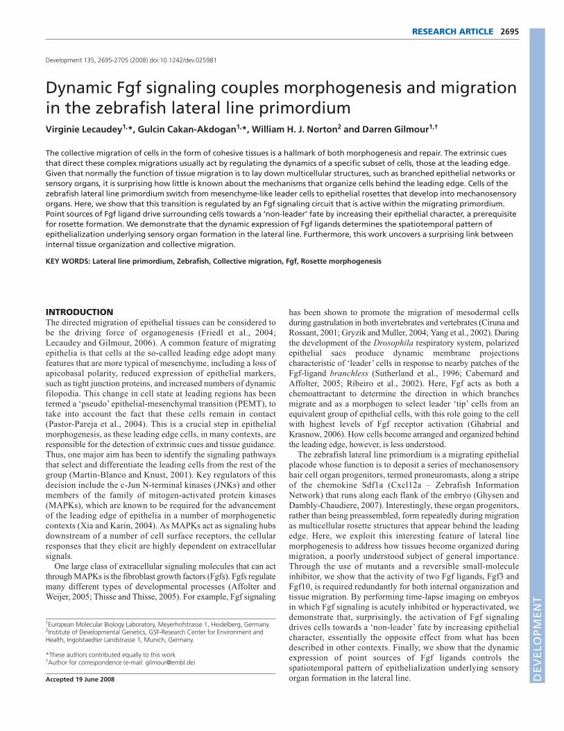
RESULTSFgf3 and Fgf10 are active within the migratingprimordiumPrevious work has shown that the Fgf-target gene pea3 is expressed
by the lateral line primordium as it migrates from head to tail along
either flank of the embryo (Munchberg et al., 1999; Roehl and
Nusslein-Volhard, 2001). We analyzed the expression of a number of
Fgf ligands and receptors in order to determine which are responsible
for pea3 activation in this context. Of the four characterized Fgf
receptors, only fgfr1 is clearly expressed in the migrating primordium
(Fig. 1A, data not shown). This approach also identified two Fgf
ligands, fgf3 and fgf10, as being candidates for mediating signaling in
this context. Interestingly, both genes are expressed within the
primordium, being upregulated in leading regions (Fig. 1B,C). In
addition, expression of fgf10 can be clearly seen in one or two cells at
the centre of rosette-like proneuromasts (Fig. 1C, arrows), whereas
fgf3 cannot be detected (Fig. 1B). Interestingly, the combined
expression of these genes is somewhat complementary to that of fgfr1and pea3. Receptor and target are downregulated at the leading edge
and at the centre of rosettes, both of which are sites of increased ligand
transcription (Fig. 1A,D, arrows).
We next tested whether the activity of these ligands can account
for the pea3 expression pattern, by crossing the previously identified
mutants limabsent (lia) and daedalus (dae), which encode mutations
in fgf3 (Herzog et al., 2004) and fgf10 (Norton et al., 2005),
respectively, to our previously described lateral line reporter,
cldnb:gfp (Haas and Gilmour, 2006). Although the expression of
pea3 in the respective mutants is indistinguishable from wild type
(wt; not shown), it is entirely absent from the migrating primordium
of fgf3;fgf10 double mutant embryos. Other embryonic expression
domains, such as the somite borders and the midbrain-hindbrain
boundary, however, remain unchanged (Fig. 1E-H). By comparison,
embryos treated with the Fgf receptor inhibitor SU5402 for five
hours show a complete absence of pea3 expression throughout the
embryo (Fig. 1I, J). We conclude that Fgf3 and Fgf10 mediate Fgf
signaling during lateral line primordium migration.
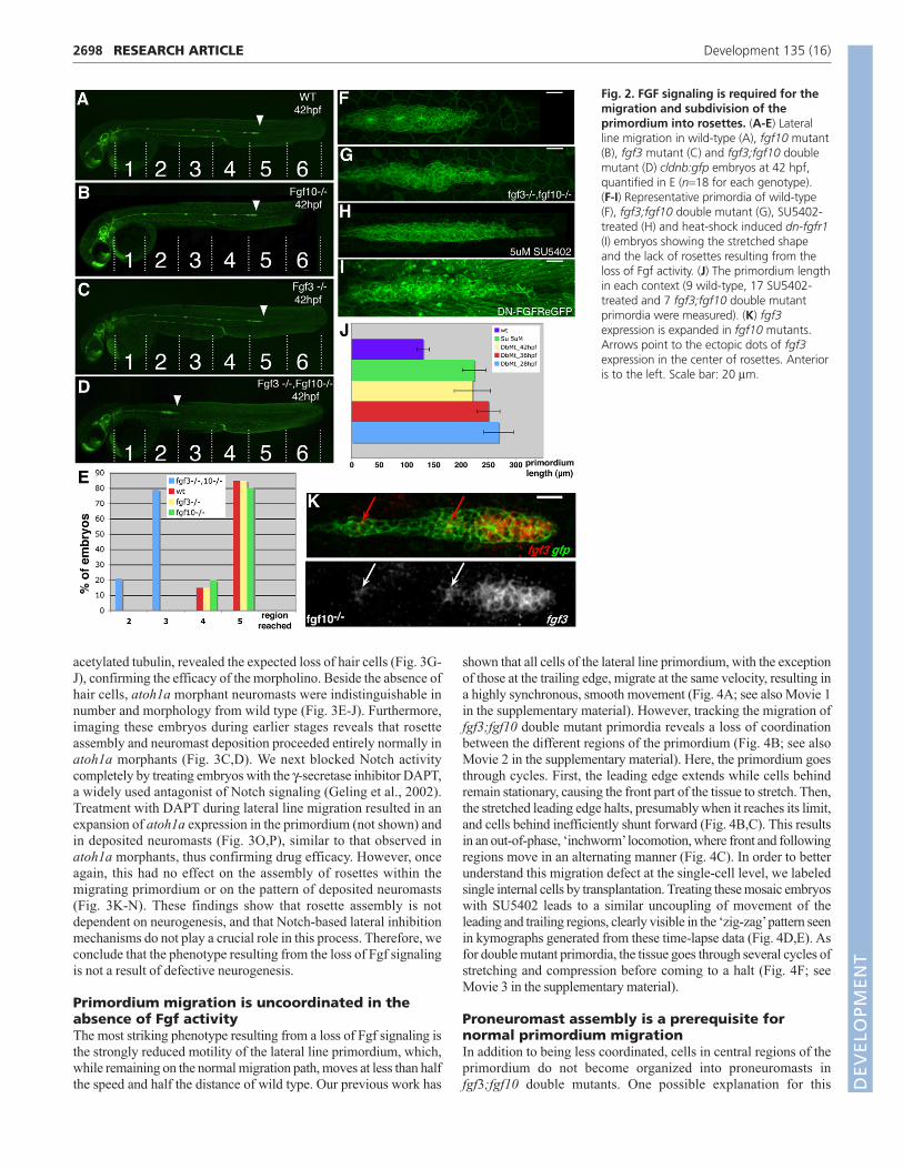
Fgf3 and Fgf10 are required for migration andsubdivision of the lateral lineWe next addressed the requirement for Fgf signaling in lateral line
morphogenesis. Single mutants for fgf3 and fgf10 showed no
detectable change in lateral line development (Fig. 2A-C), a result that
is consistent with normal pea3 expression within the primordium in
these embryos. By contrast, fgf3;fgf10 double mutant primordia
showed a number of striking defects when compared with wild-type
controls (Fig. 2A,D). The most obvious difference was that the speed
of migration was strongly reduced, with the primordium having
migrated less than half the distance of wild type by 42 hours post-
fertilization (hpf) (n=18; Fig. 2A,D,E). Furthermore, there was a
change in morphology: the migrating primordia adopted an extended
conformation, becoming twice as long as wild-type siblings (Fig.
2F,G,J). Similarly, blocking Fgf signaling with the Fgfr inhibitor
SU5402, or using a heat-shock inducible, dominant-negative form of
Fgfr1 (Lee et al., 2005), blocked migration and caused extended tissue
morphology (Fig. 2H-J).
Closer inspection of double mutant primordia revealed that the
assembly of neuromast progenitors was also strongly affected.
Whereas two to three rosette-like neuromast organs can be
morphologically identified within wild-type primordia (Fig. 2F), no
such organization was visible in double mutants between 28 and 36
hpf (Fig. 2G, see also Fig. S1 in the supplementary material).
Developmental timecourses revealed that, at later stages, organ
RESEARCH ARTICLE Development 135 (16)
DEVELO
PMENT
assembly defects are less pronounced, with 71% (n=14) of the double
mutant primordia showing a normal number of rosettes. This recovery
of rosettes could be due to the fact that the fgf3 allele used has some
residual activity (Herzog et al., 2004). Indeed, treating embryos with
5 μM SU5402 for 6 hours completely removes organ progenitors from
the primordium (Fig. 2H). Given that only fgf10 mRNA is detected in
the centre of rosettes, the finding that rosette assembly was not
affected in fgf10 single mutants was somewhat surprising. However,
expression analysis reveals that fgf3 assumes an ‘fgf10-like’ pattern in
fgf10 mutants, expanding posteriorly in the leading region and
increasing at rosette centres (compare Fig. 2K with Fig. 1B).
On the basis of this analysis, it appears that Fgf signaling is
required for several aspects of lateral line morphogenesis, including
rosette assembly, tissue morphology and efficient migration. We
therefore decided to address the relationship between these
processes and the role of Fgf signaling in each.
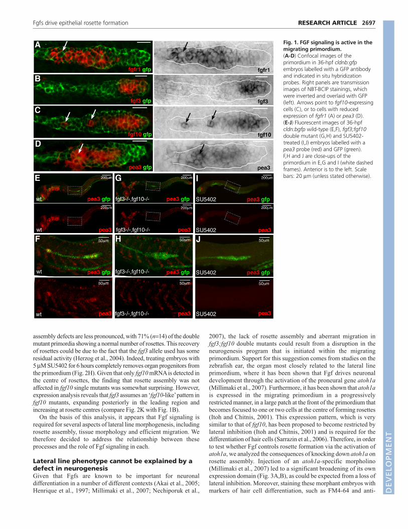
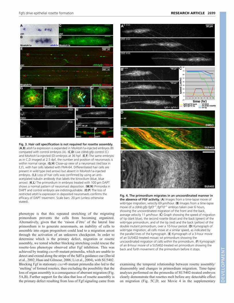
Lateral line phenotype cannot be explained by adefect in neurogenesisGiven that Fgfs are known to be important for neuronal
differentiation in a number of different contexts (Akai et al., 2005;
Henrique et al., 1997; Millimaki et al., 2007; Nechiporuk et al.,
2007), the lack of rosette assembly and aberrant migration in
fgf3;fgf10 double mutants could result from a disruption in the
neurogenesis program that is initiated within the migrating
primordium. Support for this suggestion comes from studies on the
zebrafish ear, the organ most closely related to the lateral line
primordium, where it has been shown that Fgf drives neuronal
development through the activation of the proneural gene atoh1a(Millimaki et al., 2007). Furthermore, it has been shown that atoh1ais expressed in the migrating primordium in a progressively
restricted manner, in a large patch at the front of the primordium that
becomes focused to one or two cells at the centre of forming rosettes
(Itoh and Chitnis, 2001). This expression pattern, which is very
similar to that of fgf10, has been proposed to become restricted by
lateral inhibition (Itoh and Chitnis, 2001) and is required for the
differentiation of hair cells (Sarrazin et al., 2006). Therefore, in order
to test whether Fgf controls rosette formation via the activation of
atoh1a, we analyzed the consequences of knocking down atoh1a on
rosette assembly. Injection of an atoh1a-specific morpholino
(Millimaki et al., 2007) led to a significant broadening of its own
expression domain (Fig. 3A,B), as could be expected from a loss of
lateral inhibition. Moreover, staining these morphant embryos with
markers of hair cell differentiation, such as FM4-64 and anti-
2697RESEARCH ARTICLEFgfs drive epithelial rosette formation
Fig. 1. FGF signaling is active in themigrating primordium.(A-D) Confocal images of theprimordium in 36-hpf cldnb:gfpembryos labelled with a GFP antibodyand indicated in situ hybridizationprobes. Right panels are transmissionimages of NBT-BCIP stainings, whichwere inverted and overlaid with GFP(left). Arrows point to fgf10-expressingcells (C), or to cells with reducedexpression of fgfr1 (A) or pea3 (D).(E-J) Fluorescent images of 36-hpfcldn:bgfp wild-type (E,F), fgf3;fgf10double mutant (G,H) and SU5402-treated (I,J) embryos labelled with apea3 probe (red) and GFP (green).F,H and J are close-ups of theprimordium in E,G and I (white dashedframes). Anterior is to the left. Scalebars: 20 μm (unless stated otherwise).
DEVELO
PMENT
2698
acetylated tubulin, revealed the expected loss of hair cells (Fig. 3G-
J), confirming the efficacy of the morpholino. Beside the absence of
hair cells, atoh1a morphant neuromasts were indistinguishable in
number and morphology from wild type (Fig. 3E-J). Furthermore,
imaging these embryos during earlier stages reveals that rosette
assembly and neuromast deposition proceeded entirely normally in
atoh1a morphants (Fig. 3C,D). We next blocked Notch activity
completely by treating embryos with the γ-secretase inhibitor DAPT,
a widely used antagonist of Notch signaling (Geling et al., 2002).
Treatment with DAPT during lateral line migration resulted in an
expansion of atoh1a expression in the primordium (not shown) and
in deposited neuromasts (Fig. 3O,P), similar to that observed in
atoh1a morphants, thus confirming drug efficacy. However, once
again, this had no effect on the assembly of rosettes within the
migrating primordium or on the pattern of deposited neuromasts
(Fig. 3K-N). These findings show that rosette assembly is not
dependent on neurogenesis, and that Notch-based lateral inhibition
mechanisms do not play a crucial role in this process. Therefore, we
conclude that the phenotype resulting from the loss of Fgf signaling
is not a result of defective neurogenesis.
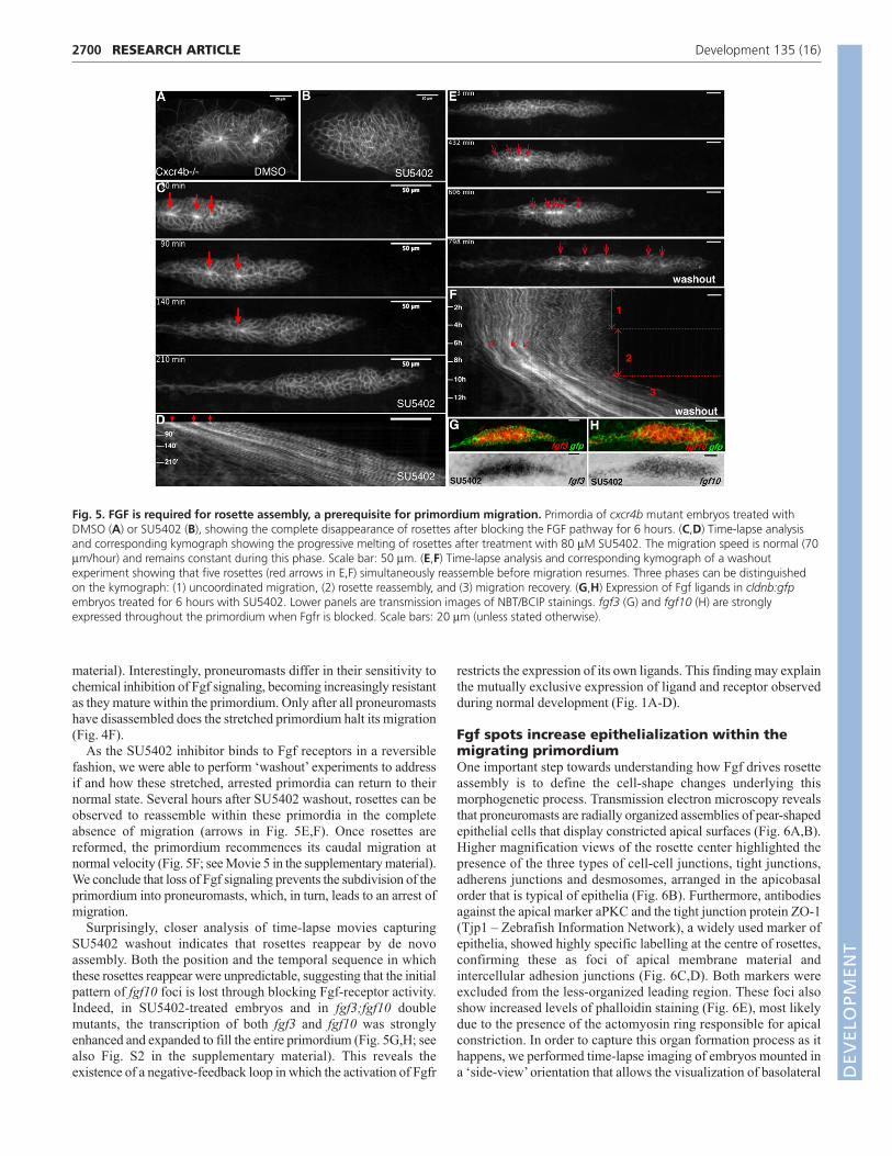
Primordium migration is uncoordinated in theabsence of Fgf activityThe most striking phenotype resulting from a loss of Fgf signaling is
the strongly reduced motility of the lateral line primordium, which,
while remaining on the normal migration path, moves at less than half
the speed and half the distance of wild type. Our previous work has
shown that all cells of the lateral line primordium, with the exception
of those at the trailing edge, migrate at the same velocity, resulting in
a highly synchronous, smooth movement (Fig. 4A; see also Movie 1
in the supplementary material). However, tracking the migration of
fgf3;fgf10 double mutant primordia reveals a loss of coordination
between the different regions of the primordium (Fig. 4B; see also
Movie 2 in the supplementary material). Here, the primordium goes
through cycles. First, the leading edge extends while cells behind
remain stationary, causing the front part of the tissue to stretch. Then,
the stretched leading edge halts, presumably when it reaches its limit,
and cells behind inefficiently shunt forward (Fig. 4B,C). This results
in an out-of-phase, ‘inchworm’ locomotion, where front and following
regions move in an alternating manner (Fig. 4C). In order to better
understand this migration defect at the single-cell level, we labeled
single internal cells by transplantation. Treating these mosaic embryos
with SU5402 leads to a similar uncoupling of movement of the
leading and trailing regions, clearly visible in the ‘zig-zag’ pattern seen
in kymographs generated from these time-lapse data (Fig. 4D,E). As
for double mutant primordia, the tissue goes through several cycles of
stretching and compression before coming to a halt (Fig. 4F; see
Movie 3 in the supplementary material).
Proneuromast assembly is a prerequisite fornormal primordium migrationIn addition to being less coordinated, cells in central regions of the
primordium do not become organized into proneuromasts in
fgf3;fgf10 double mutants. One possible explanation for this
RESEARCH ARTICLE Development 135 (16)
Fig. 2. FGF signaling is required for themigration and subdivision of theprimordium into rosettes. (A-E) Lateralline migration in wild-type (A), fgf10 mutant(B), fgf3 mutant (C) and fgf3;fgf10 doublemutant (D) cldnb:gfp embryos at 42 hpf,quantified in E (n=18 for each genotype).(F-I) Representative primordia of wild-type(F), fgf3;fgf10 double mutant (G), SU5402-treated (H) and heat-shock induced dn-fgfr1(I) embryos showing the stretched shapeand the lack of rosettes resulting from theloss of Fgf activity. (J) The primordium lengthin each context (9 wild-type, 17 SU5402-treated and 7 fgf3;fgf10 double mutantprimordia were measured). (K) fgf3expression is expanded in fgf10 mutants.Arrows point to the ectopic dots of fgf3expression in the center of rosettes. Anterioris to the left. Scale bar: 20 μm.
DEVELO
PMENT
phenotype is that this repeated stretching of the migrating
primordium prevents the cells from becoming organized.
Alternatively, given that the ‘raison d’être’ of the lateral line
primordium is to generate neuromasts, an inability of cells to
assemble into organ progenitors could lead to a migration arrest
through the activation of an unknown checkpoint. In order to
determine which is the primary defect, migration or rosette
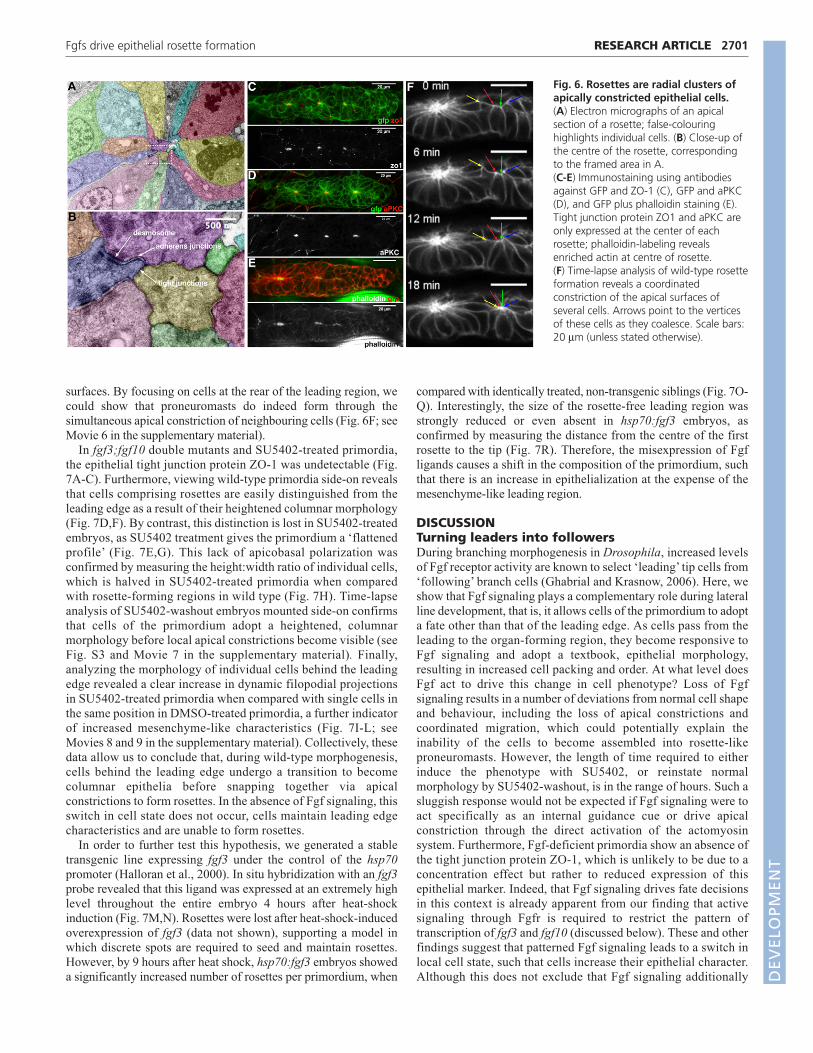
assembly, we tested whether blocking stretching could rescue the
rosette-loss phenotype observed after Fgf inhibition. This was
achieved by treating cxcr4b mutant primordia, which are unable to
detect and extend along the stripe of the Sdf1a guidance cue (David
et al., 2002; Haas and Gilmour, 2006; Li et al., 2004), with SU5402.
Blocking Fgf in stationary cxcr4b mutant primordia also led to the
‘melting’ of formed rosettes, thus excluding the possibility that the
loss of organ assembly is a consequence of aberrant migration (Fig.
5A,B). Further support for the idea that loss of rosette assembly is
the primary defect resulting from loss of Fgf signaling came from
examining the temporal relationship between rosette assembly/
disassembly and changes in primordium migration. Time-lapse
analyses performed on the primordia of SU5402-treated embryos
clearly demonstrate that rosettes melt before any detectable effect
on migration (Fig. 5C,D; see Movie 4 in the supplementary
2699RESEARCH ARTICLEFgfs drive epithelial rosette formation
Fig. 3. Hair cell specification is not required for rosette assembly.(A,B) atoh1a expression is expanded in MoAtoh1a-injected embryos (B)compared with control embryos (A). (C,D) Live cldnb:gfp control (C)and MoAtoh1a-injected (D) embryos at 36 hpf. (E,F) The same embryosas in C,D imaged at 2.5 dpf, the number and position of neuromasts iswithin normal range. (G,H) Close-up view of a neuromast (red box inE,F), with hair cells labeled with FM4-64. Differentiated hair cells arepresent in wild type (red arrow) but absent in MoAtoh1a-injectedembryos. (I,J) Loss of hair cells was confirmed by using an anti-acetylated tubulin antibody that labels the kinocilium (blue, bluearrow). (K,L) The primordium in embryos treated with 100 μm DAPTshows a normal pattern of neuromast deposition. (M,N) Primordia inDAPT and control embryos are indistinguishable. (O,P) The loss ofrestricted atoh1a expression in deposited neuromasts confirms theefficacy of DAPT treatment. Scale bars: 20 μm (unless otherwisestated).
Fig. 4. The primordium migrates in an uncoordinated manner inthe absence of FGF activity. (A) Images from a time-lapse movie ofwild-type migration, velocity 69 μm/hour. (B) Images from a time-lapsemovie of a cldnb:gfp fgf3–/–;fgf10–/– embryo taken over 6 hours,showing the uncoordinated migration of the front and the back,average velocity 11 μm/hour. (C) Graph showing the speed of migrationof tip (dark blue), the second rosette (blue) and the back (green) of thewild-type primordium, and of the tip (red) and the back (yellow) of thedouble mutant primordium, over a 10-hour period. (D) Kymograph ofwild-type migration; all cells move at a similar speed, as indicated bythe parallel lines of the kymograph. (E) Kymograph of a 3-hour movieof an SU5402-treated mosaic wt primordium showing theuncoordinated migration of cells within the primordium. (F) Kymographof an 8-hour movie of a SU5402-treated wt primordium showing theback and forth movement of the primordium before it stops.
DEVELO
PMENT
2700
material). Interestingly, proneuromasts differ in their sensitivity to
chemical inhibition of Fgf signaling, becoming increasingly resistant
as they mature within the primordium. Only after all proneuromasts
have disassembled does the stretched primordium halt its migration
(Fig. 4F).
As the SU5402 inhibitor binds to Fgf receptors in a reversible
fashion, we were able to perform ‘washout’ experiments to address
if and how these stretched, arrested primordia can return to their
normal state. Several hours after SU5402 washout, rosettes can be
observed to reassemble within these primordia in the complete
absence of migration (arrows in Fig. 5E,F). Once rosettes are
reformed, the primordium recommences its caudal migration at
normal velocity (Fig. 5F; see Movie 5 in the supplementary material).
We conclude that loss of Fgf signaling prevents the subdivision of the
primordium into proneuromasts, which, in turn, leads to an arrest of
migration.
Surprisingly, closer analysis of time-lapse movies capturing
SU5402 washout indicates that rosettes reappear by de novo
assembly. Both the position and the temporal sequence in which
these rosettes reappear were unpredictable, suggesting that the initial
pattern of fgf10 foci is lost through blocking Fgf-receptor activity.
Indeed, in SU5402-treated embryos and in fgf3;fgf10 double
mutants, the transcription of both fgf3 and fgf10 was strongly
enhanced and expanded to fill the entire primordium (Fig. 5G,H; see
also Fig. S2 in the supplementary material). This reveals the
existence of a negative-feedback loop in which the activation of Fgfr
restricts the expression of its own ligands. This finding may explain
the mutually exclusive expression of ligand and receptor observed
during normal development (Fig. 1A-D).
Fgf spots increase epithelialization within themigrating primordiumOne important step towards understanding how Fgf drives rosette
assembly is to define the cell-shape changes underlying this
morphogenetic process. Transmission electron microscopy reveals
that proneuromasts are radially organized assemblies of pear-shaped
epithelial cells that display constricted apical surfaces (Fig. 6A,B).
Higher magnification views of the rosette center highlighted the
presence of the three types of cell-cell junctions, tight junctions,
adherens junctions and desmosomes, arranged in the apicobasal
order that is typical of epithelia (Fig. 6B). Furthermore, antibodies
against the apical marker aPKC and the tight junction protein ZO-1
(Tjp1 – Zebrafish Information Network), a widely used marker of
epithelia, showed highly specific labelling at the centre of rosettes,
confirming these as foci of apical membrane material and
intercellular adhesion junctions (Fig. 6C,D). Both markers were
excluded from the less-organized leading region. These foci also
show increased levels of phalloidin staining (Fig. 6E), most likely
due to the presence of the actomyosin ring responsible for apical
constriction. In order to capture this organ formation process as it
happens, we performed time-lapse imaging of embryos mounted in
a ‘side-view’ orientation that allows the visualization of basolateral
RESEARCH ARTICLE Development 135 (16)
Fig. 5. FGF is required for rosette assembly, a prerequisite for primordium migration. Primordia of cxcr4b mutant embryos treated withDMSO (A) or SU5402 (B), showing the complete disappearance of rosettes after blocking the FGF pathway for 6 hours. (C,D) Time-lapse analysisand corresponding kymograph showing the progressive melting of rosettes after treatment with 80 μM SU5402. The migration speed is normal (70μm/hour) and remains constant during this phase. Scale bar: 50 μm. (E,F) Time-lapse analysis and corresponding kymograph of a washoutexperiment showing that five rosettes (red arrows in E,F) simultaneously reassemble before migration resumes. Three phases can be distinguishedon the kymograph: (1) uncoordinated migration, (2) rosette reassembly, and (3) migration recovery. (G,H) Expression of Fgf ligands in cldnb:gfpembryos treated for 6 hours with SU5402. Lower panels are transmission images of NBT/BCIP stainings. fgf3 (G) and fgf10 (H) are stronglyexpressed throughout the primordium when Fgfr is blocked. Scale bars: 20 μm (unless stated otherwise).
DEVELO
PMENT
surfaces. By focusing on cells at the rear of the leading region, we
could show that proneuromasts do indeed form through the
simultaneous apical constriction of neighbouring cells (Fig. 6F; see
Movie 6 in the supplementary material).
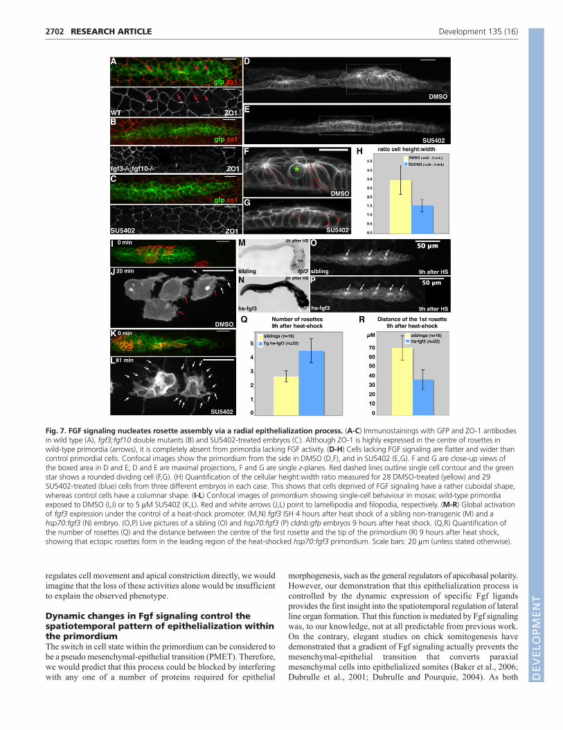
In fgf3;fgf10 double mutants and SU5402-treated primordia,
the epithelial tight junction protein ZO-1 was undetectable (Fig.
7A-C). Furthermore, viewing wild-type primordia side-on reveals
that cells comprising rosettes are easily distinguished from the
leading edge as a result of their heightened columnar morphology
(Fig. 7D,F). By contrast, this distinction is lost in SU5402-treated
embryos, as SU5402 treatment gives the primordium a ‘flattened
profile’ (Fig. 7E,G). This lack of apicobasal polarization was
confirmed by measuring the height:width ratio of individual cells,
which is halved in SU5402-treated primordia when compared
with rosette-forming regions in wild type (Fig. 7H). Time-lapse
analysis of SU5402-washout embryos mounted side-on confirms
that cells of the primordium adopt a heightened, columnar
morphology before local apical constrictions become visible (see
Fig. S3 and Movie 7 in the supplementary material). Finally,
analyzing the morphology of individual cells behind the leading
edge revealed a clear increase in dynamic filopodial projections
in SU5402-treated primordia when compared with single cells in
the same position in DMSO-treated primordia, a further indicator
of increased mesenchyme-like characteristics (Fig. 7I-L; see
Movies 8 and 9 in the supplementary material). Collectively, these
data allow us to conclude that, during wild-type morphogenesis,
cells behind the leading edge undergo a transition to become
columnar epithelia before snapping together via apical
constrictions to form rosettes. In the absence of Fgf signaling, this
switch in cell state does not occur, cells maintain leading edge
characteristics and are unable to form rosettes.
In order to further test this hypothesis, we generated a stable
transgenic line expressing fgf3 under the control of the hsp70promoter (Halloran et al., 2000). In situ hybridization with an fgf3probe revealed that this ligand was expressed at an extremely high
level throughout the entire embryo 4 hours after heat-shock
induction (Fig. 7M,N). Rosettes were lost after heat-shock-induced
overexpression of fgf3 (data not shown), supporting a model in
which discrete spots are required to seed and maintain rosettes.
However, by 9 hours after heat shock, hsp70:fgf3 embryos showed
a significantly increased number of rosettes per primordium, when
compared with identically treated, non-transgenic siblings (Fig. 7O-
Q). Interestingly, the size of the rosette-free leading region was
strongly reduced or even absent in hsp70:fgf3 embryos, as
confirmed by measuring the distance from the centre of the first
rosette to the tip (Fig. 7R). Therefore, the misexpression of Fgf
ligands causes a shift in the composition of the primordium, such
that there is an increase in epithelialization at the expense of the
mesenchyme-like leading region.
DISCUSSIONTurning leaders into followersDuring branching morphogenesis in Drosophila, increased levels
of Fgf receptor activity are known to select ‘leading’ tip cells from
‘following’ branch cells (Ghabrial and Krasnow, 2006). Here, we
show that Fgf signaling plays a complementary role during lateral
line development, that is, it allows cells of the primordium to adopt
a fate other than that of the leading edge. As cells pass from the
leading to the organ-forming region, they become responsive to
Fgf signaling and adopt a textbook, epithelial morphology,
resulting in increased cell packing and order. At what level does
Fgf act to drive this change in cell phenotype? Loss of Fgf
signaling results in a number of deviations from normal cell shape
and behaviour, including the loss of apical constrictions and
coordinated migration, which could potentially explain the
inability of the cells to become assembled into rosette-like
proneuromasts. However, the length of time required to either
induce the phenotype with SU5402, or reinstate normal
morphology by SU5402-washout, is in the range of hours. Such a
sluggish response would not be expected if Fgf signaling were to
act specifically as an internal guidance cue or drive apical
constriction through the direct activation of the actomyosin
system. Furthermore, Fgf-deficient primordia show an absence of
the tight junction protein ZO-1, which is unlikely to be due to a
concentration effect but rather to reduced expression of this
epithelial marker. Indeed, that Fgf signaling drives fate decisions
in this context is already apparent from our finding that active
signaling through Fgfr is required to restrict the pattern of
transcription of fgf3 and fgf10 (discussed below). These and other
findings suggest that patterned Fgf signaling leads to a switch in
local cell state, such that cells increase their epithelial character.
Although this does not exclude that Fgf signaling additionally
2701RESEARCH ARTICLEFgfs drive epithelial rosette formation
Fig. 6. Rosettes are radial clusters ofapically constricted epithelial cells.(A) Electron micrographs of an apicalsection of a rosette; false-colouringhighlights individual cells. (B) Close-up ofthe centre of the rosette, correspondingto the framed area in A.(C-E) Immunostaining using antibodiesagainst GFP and ZO-1 (C), GFP and aPKC(D), and GFP plus phalloidin staining (E).Tight junction protein ZO1 and aPKC areonly expressed at the center of eachrosette; phalloidin-labeling revealsenriched actin at centre of rosette.(F) Time-lapse analysis of wild-type rosetteformation reveals a coordinatedconstriction of the apical surfaces ofseveral cells. Arrows point to the verticesof these cells as they coalesce. Scale bars:20 μm (unless stated otherwise).
DEVELO
PMENT
2702
regulates cell movement and apical constriction directly, we would
imagine that the loss of these activities alone would be insufficient
to explain the observed phenotype.
Dynamic changes in Fgf signaling control thespatiotemporal pattern of epithelialization withinthe primordiumThe switch in cell state within the primordium can be considered to
be a pseudo mesenchymal-epithelial transition (PMET). Therefore,
we would predict that this process could be blocked by interfering
with any one of a number of proteins required for epithelial
morphogenesis, such as the general regulators of apicobasal polarity.
However, our demonstration that this epithelialization process is
controlled by the dynamic expression of specific Fgf ligands
provides the first insight into the spatiotemporal regulation of lateral
line organ formation. That this function is mediated by Fgf signaling
was, to our knowledge, not at all predictable from previous work.
On the contrary, elegant studies on chick somitogenesis have
demonstrated that a gradient of Fgf signaling actually prevents the
mesenchymal-epithelial transition that converts paraxial
mesenchymal cells into epithelialized somites (Baker et al., 2006;
Dubrulle et al., 2001; Dubrulle and Pourquie, 2004). As both
RESEARCH ARTICLE Development 135 (16)
Fig. 7. FGF signaling nucleates rosette assembly via a radial epithelialization process. (A-C) Immunostainings with GFP and ZO-1 antibodiesin wild type (A), fgf3;fgf10 double mutants (B) and SU5402-treated embryos (C). Although ZO-1 is highly expressed in the centre of rosettes inwild-type primordia (arrows), it is completely absent from primordia lacking FGF activity. (D-H) Cells lacking FGF signaling are flatter and wider thancontrol primordial cells. Confocal images show the primordium from the side in DMSO (D,F), and in SU5402 (E,G). F and G are close-up views ofthe boxed area in D and E; D and E are maximal projections, F and G are single z-planes. Red dashed lines outline single cell contour and the greenstar shows a rounded dividing cell (F,G). (H) Quantification of the cellular height:width ratio measured for 28 DMSO-treated (yellow) and 29SU5402-treated (blue) cells from three different embryos in each case. This shows that cells deprived of FGF signaling have a rather cuboidal shape,whereas control cells have a columnar shape. (I-L) Confocal images of primordium showing single-cell behaviour in mosaic wild-type primordiaexposed to DMSO (I,J) or to 5 μM SU5402 (K,L). Red and white arrows (J,L) point to lamellipodia and filopodia, respectively. (M-R) Global activationof fgf3 expression under the control of a heat-shock promoter. (M,N) fgf3 ISH 4 hours after heat shock of a sibling non-transgenic (M) and ahsp70:fgf3 (N) embryo. (O,P) Live pictures of a sibling (O) and hsp70:fgf3 (P) cldnb:gfp embryos 9 hours after heat shock. (Q,R) Quantification ofthe number of rosettes (Q) and the distance between the centre of the first rosette and the tip of the primordium (R) 9 hours after heat shock,showing that ectopic rosettes form in the leading region of the heat-shocked hsp70:fgf3 primordium. Scale bars: 20 μm (unless stated otherwise).DEVELO
PMENT
systems generate a series of repetitive epithelial packets from less-
ordered mesenchymal cells, the similarities are striking, and it is
therefore intriguing that Fgf signaling promotes EMT in one context
but MET in another.
A crucial step towards understanding the periodicity of this
system was to define how the dynamic pattern of fgf10 dots that
nucleate rosettes is generated. In situ hybridization revealed that
fgf10 is expressed in a broad patch at the front of the primordium that
apparently becomes focused to one or two cells in organ-forming
regions. This transcription pattern is highly reminiscent of that
described for the proneural gene atoh1a, where focused expression
is thought to be controlled by Notch-based lateral inhibition (Itoh
and Chitnis, 2001). It was therefore surprising that perturbing Notch-
based lateral inhibition, either by atoh1a knockdown or by treatment
with the Notch antagonist DAPT, had no clear effect on rosette
assembly or on the final pattern of neuromasts. This is consistent
with the finding that mindbomb mutants have reduced Notch activity
but a seemingly normal pattern of deposited neuromasts (Itoh and
Chitnis, 2001). So how does fgf10 expression get restricted to dots?
Interestingly, fgf10 and fgfr1 show mutually exclusive expression
during organ assembly, suggesting that fgf10 expression may
become restricted via a negative-feedback loop. Furthermore, the
expression of both ligands expands throughout the entire
primordium in fgf3;fgf10 double mutants and SU5402-treated
embryos, giving credence to the idea that Fgfr1 activity directly
restricts Fgf ligand expression, possibly through a lateral inhibition-
type mechanism. Interestingly, inactivating fgf10 alone, through the
use of fgf10 mutants, leads to the formation of rosettes that are
nucleated by dots of fgf3, a clear demonstration of the robustness of
this pattern forming system.
A simple mechanism for generating rosetteswithin epitheliaIn addition to determining the spatiotemporal pattern of
epithelialization within the migrating primordium, Fgf signaling
leads to the formation of rosette assemblies. The teleological reason
for rosettes in this context is obvious, as these are the forerunners of
neuromasts, a series of volcano-like organs that penetrate the skin to
project mechansensory hairs cells into the surrounding water.
Similar multicellular rosettes have recently been shown to arise in
other epithelial tissues, suggesting that they could represent a
common functional unit of epithelial morphogenesis (Blankenship
et al., 2006; Brown et al., 2006; Wagstaff et al., 2008). They have
been particularly well described in the context of Drosophilagermband extension, where these transient rosettes directly
participate in tissue elongation by forming and resolving in a
directional manner that is influenced by embryonic anteroposterior
patterning cues. Despite superficial similarities between these
rosettes and those in the primordium, there are fundamental
differences in their three-dimensional organization. The rosettes in
the Drosophila blastoderm form when two rows of cells that are in
contact with each other constrict their shared interface, that is, one
side of their apical surface, with the resultant rosette resembling a
pie where each slice is a cell (Blankenship et al., 2006; Zallen and
Blankenship, 2008). The rosettes of the lateral line primordium, by
contrast, assemble by bunching together cells whose entire apical
surface is tightly constricted, with the resultant rosette resembling a
garlic bulb where each clove is a cell. Another type of rosette that
forms through the constriction of interfacial surfaces, rather than the
entire apical surface, appears in the wake of the morphogenetic
furrow that patterns the Drosophila retina. These arc-like rosettes,
which are the forerunners of the ommatidia, are built around atonal-expressing R8 cells that ‘recruit’ surrounding cells in a stepwise
fashion through the activation of the Egf-like ligand spitz (Brown et
al., 2006). While the parallels with the lateral line system are clearly
more obvious in this case, our demonstration that Notch signaling
or Atoh1a is not required for rosette assembly suggests that there
may be differences in the gene regulatory networks involved.
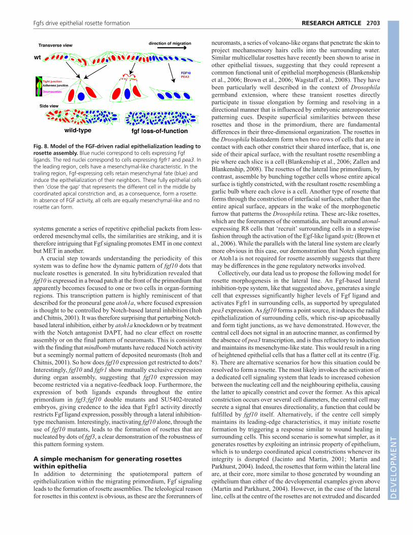
Collectively, our data lead us to propose the following model for
rosette morphogenesis in the lateral line. An Fgf-based lateral
inhibition-type system, like that suggested above, generates a single
cell that expresses significantly higher levels of Fgf ligand and
activates Fgfr1 in surrounding cells, as supported by upregulated
pea3 expression. As fgf10 forms a point source, it induces the radial
epithelialization of surrounding cells, which rise-up apicobasally
and form tight junctions, as we have demonstrated. However, the
central cell does not signal in an autocrine manner, as confirmed by
the absence of pea3 transcription, and is thus refractory to induction
and maintains its mesenchyme-like state. This would result in a ring
of heightened epithelial cells that has a flatter cell at its centre (Fig.
8). There are alternative scenarios for how this situation could be
resolved to form a rosette. The most likely invokes the activation of
a dedicated cell signaling system that leads to increased cohesion
between the nucleating cell and the neighbouring epithelia, causing
the latter to apically constrict and cover the former. As this apical
constriction occurs over several cell diameters, the central cell may
secrete a signal that ensures directionality, a function that could be
fulfilled by fgf10 itself. Alternatively, if the centre cell simply
maintains its leading-edge characteristics, it may initiate rosette
formation by triggering a response similar to wound healing in
surrounding cells. This second scenario is somewhat simpler, as it
generates rosettes by exploiting an intrinsic property of epithelium,
which is to undergo coordinated apical constrictions whenever its
integrity is disrupted (Jacinto and Martin, 2001; Martin and
Parkhurst, 2004). Indeed, the rosettes that form within the lateral line
are, at their core, more similar to those generated by wounding an
epithelium than either of the developmental examples given above
(Martin and Parkhurst, 2004). However, in the case of the lateral
line, cells at the centre of the rosettes are not extruded and discarded
2703RESEARCH ARTICLEFgfs drive epithelial rosette formation
Fig. 8. Model of the FGF-driven radial epithelialization leading torosette assembly. Blue nuclei correspond to cells expressing Fgfligands. The red nuclei correspond to cells expressing fgfr1 and pea3. Inthe leading region, cells have a mesenchymal-like characteristic. In thetrailing region, Fgf-expressing cells retain mesenchymal fate (blue) andinduce the epithelialization of their neighbors. These fully epithelial cellsthen ‘close the gap’ that represents the different cell in the middle bycoordinated apical constriction and, as a consequence, form a rosette.In absence of FGF activity, all cells are equally mesenchymal-like and norosette can form.
DEVELO
PMENT
2704
from the epithelium, as damaged cells are during wound healing, but
are rather encapsulated through this apical constriction process.
These are most likely the cells that give rise to sensory hair cell
progenitors within a few hours of this morphogenetic event.
Tissue compartmentalization is required forefficient migrationOne of the surprising findings of this work is that a defect in the
internal organization of the primordium has such a strong detrimental
effect on its migration. This finding is perhaps counterintuitive, as
increasing the relative proportion of cells with mesenchyme-like,
leading edge fate could be expected to improve migration efficiency
rather than decrease it. Given the numerous examples in which Fgf
ligands have been shown to act as chemoattractants during cell
migration (Affolter and Weijer, 2005), one alternative explanation for
this result is that Fgf3/Fgf10 act as diffusible guidance cues that
regulate directionality within the migrating primordium. However,
several findings presented here make it unlikely that this is the
primary role of internal Fgf signaling during primordium migration.
Not only does directional migration continue normally for several
hours after blocking Fgf signaling with SU5402, but a change in
rosette assembly always prefigures a change in migration behaviour.
Furthermore, ubiquitous, high-level expression of fgf3 does not lead
to a rapid change in tissue migration behaviour or direction, which
would be expected if it were to act as a chemoattractant. Rather, we
propose that the primordium possesses an internal checkpoint that
halts migration whenever there is a problem with rosette assembly.
Of course, a mechanism that couples migration behaviour to organ
assembly makes biological sense, as a migrating primordium that
does not deposit organs is of little use. As with rosette assembly, this
checkpoint could be mediated through the activation of a dedicated
signaling system or it may exploit intrinsic mechanical properties of
the tissue. Indeed, rosette formation may facilitate migration simply
by virtue of the fact that it converts a large number of migrating
mesenchyme-like cells into two or three packets that could be
controlled as single units, thus reducing the complexity of the system.
Answers to such questions will come from a deeper understanding of
the respective mechanical properties of epithelial and mesenchyme-
like domains within migrating tissues.
We are grateful to Petra Haas for contributions to early stages of this work, toKota Miura for expert feedback on image analysis, to Andreea Gruia for animalcare and to Andreas Kunze for help with generating transgenes using the Tol2Kit, which was a generous gift from Chi-Bin Chien. We thank Nga Ly-Hartig andClaude Anthony for assistance with EM, and the Advanced Light MicroscopyFacility at EMBL for imaging advice. We thank Eric Karsenti, Sebastian Streichan,Lars Hufnagel, Francesca Peri and the Gilmour lab for stimulating discussions,and Francesca Peri for critical reading of the manuscript. V.L. was supported byfellowships from EMBO and Marie Curie FP6.
Supplementary materialSupplementary material for this article is available athttp://dev.biologists.org/cgi/content/full/135/16/2695/DC1
ReferencesAffolter, M. and Weijer, C. J. (2005). Signaling to cytoskeletal dynamics during
chemotaxis. Dev. Cell 9, 19-34.Akai, J., Halley, P. A. and Storey, K. G. (2005). FGF-dependent Notch signaling
maintains the spinal cord stem zone. Genes Dev. 19, 2877-2887.Baker, R. E., Schnell, S. and Maini, P. K. (2006). A clock and wavefront
mechanism for somite formation. Dev. Biol. 293,116-126.Blankenship, J., Backovic, S., Sanny, J., Weitz, O. and Zallen, J. (2006).
Multicellular rosette formation links planar cell polarity to tissue morphogenesis.Dev. Cell 11, 459-470.
Brown, K., Baonza, A. and Freeman, M. (2006). Epithelial cell adhesion in thedeveloping Drosophila retina is regulated by Atonal and the EGF receptorpathway. Dev. Biol. 300, 710-721.
Cabernard, C. and Affolter, M. (2005). Distinct roles for two receptor tyrosinekinases in epithelial branching morphogenesis in Drosophila. Dev. Cell 9, 831-842.
Ciruna, B. and Rossant, J. (2001). FGF signaling regulates mesoderm cell fatespecification and morphogenetic movement at the primitive streak. Dev. Cell 1,37-49.
David, N. B., Sapede, D., Saint-Etienne, L., Thisse, C., Thisse, B., Dambly-Chaudiere, C., Rosa, F. M. and Ghysen, A. (2002). Molecular basis of cellmigration in the fish lateral line: role of the chemokine receptor CXCR4 and ofits ligand, SDF1. Proc. Natl. Acad. Sci. USA 99, 16297-16302.
Dubrulle, J. and Pourquie, O. (2004). fgf8 mRNA decay establishes a gradientthat couples axial elongation to patterning in the vertebrate embryo. Nature427, 419-422.
Dubrulle, J., McGrew, M. J. and Pourquie, O. (2001). FGF signaling controlssomite boundary position and regulates segmentation clock control ofspatiotemporal Hox gene activation. Cell 106, 219-232.
Friedl, P., Hegerfeldt, Y. and Tusch, M. (2004). Collective cell migration inmorphogenesis and cancer. Int. J. Dev. Biol. 48, 441-449.
Geling, A., Steiner, H., Willem, M., Bally-Cuif, L. and Haass, C. (2002). Agamma-secretase inhibitor blocks Notch signaling in vivo and causes a severeneurogenic phenotype in zebrafish. EMBO Rep. 3, 688-694.
Ghabrial, A. S. and Krasnow, M. A. (2006). Social interactions amongepithelial cells during tracheal branching morphogenesis. Nature 441, 746-749.
Ghysen, A. and Dambly-Chaudiere, C. (2007). The lateral line microcosmos.Genes Dev. 21, 2118-2130.
Gryzik, T. and Muller, H. A. (2004). FGF8-like1 and FGF8-like2 encode putativeligands of the FGF receptor Htl and are required for mesoderm migration in theDrosophila gastrula. Curr. Biol. 14, 659-667.
Haas, P. and Gilmour, D. (2006). Chemokine signaling mediates self-organizingtissue migration in the zebrafish lateral line. Dev. Cell 10, 673-680.
Halloran, M. C., Sato-Maeda, M., Warren, J. T., Su, F., Lele, Z., Krone, P. H.,Kuwada, J. Y. and Shoji, W. (2000). Laser-induced gene expression in specificcells of transgenic zebrafish. Development 127, 1953-1960.
Henrique, D., Tyler, D., Kintner, C., Heath, J. K., Lewis, J. H., Ish-Horowicz, D.and Storey, K. G. (1997). cash4, a novel achaete-scute homolog induced byHensen’s node during generation of the posterior nervous system. Genes Dev.11, 603-615.
Herzog, W., Sonntag, C., von der Hardt, S., Roehl, H. H., Varga, Z. M. andHammerschmidt, M. (2004). Fgf3 signaling from the ventral diencephalon isrequired for early specification and subsequent survival of the zebrafishadenohypophysis. Development 131, 3681-3692.
Itoh, M. and Chitnis, A. (2001). Expression of proneural and neurogenic genes inthe zebrafish lateral line primordium correlates with selection of hair cell fate inneuromasts. Mech. Dev. 102, 263-266.
Jacinto, A. and Martin, P. (2001). Morphogenesis: unravelling the cell biology ofhole closure. Curr. Biol. 11, R705-R707.
Kimmel, C. B., Ballard, W. W., Kimmel, S. R., Ullmann, B. and Schilling, T. F.(1995). Stages of embryonic development of the zebrafish. Dev. Dyn. 203, 253-310.
Kwan, K. M., Fujimoto, E., Grabher, C., Mangum, B. D., Hardy, M. E.,Campbell, D. S., Parant, J. M., Yost, H. J., Kanki, J. P. and Chien, C. B.(2007). The Tol2kit: a multisite gateway-based construction kit for Tol2transposon transgenesis constructs. Dev. Dyn. 236, 3088-3099.
Lecaudey, V. and Gilmour, D. (2006). Organizing moving groups duringmorphogenesis. Curr. Opin. Cell Biol. 18, 102-107.
Lee, Y., Grill, S., Sanchez, A., Murphy-Ryan, M. and Poss, K. D. (2005). Fgfsignaling instructs position-dependent growth rate during zebrafish finregeneration. Development 132, 5173-5183.
Li, Q., Shirabe, K. and Kuwada, J. Y. (2004). Chemokine signaling regulatessensory cell migration in zebrafish. Dev. Biol. 269, 123-136.
Martin, P. and Parkhurst, S. M. (2004). Parallels between tissue repair andembryo morphogenesis. Development 131, 3021-3034.
Martin-Blanco, E. and Knust, E. (2001). Epithelial morphogenesis: filopodia atwork. Curr. Biol. 11, R28-R31.
Maves, L., Jackman, W. and Kimmel, C. B. (2002). FGF3 and FGF8 mediate arhombomere 4 signaling activity in the zebrafish hindbrain. Development 129,3825-3837.
Millimaki, B. B., Sweet, E. M., Dhason, M. S. and Riley, B. B. (2007). Zebrafishatoh1 genes: classic proneural activity in the inner ear and regulation by Fgf andNotch. Development 134, 295-305.
Munchberg, S. R., Ober, E. A. and Steinbeisser, H. (1999). Expression of the Etstranscription factors erm and pea3 in early zebrafish development. Mech. Dev.88, 233-236.
Nechiporuk, A., Linbo, T., Poss, K. D. and Raible, D. W. (2007). Specification ofepibranchial placodes in zebrafish. Development 134, 611-623.
Ng, J. K., Kawakami, Y., Buscher, D., Raya, A., Itoh, T., Koth, C. M.,Rodriguez Esteban, C., Rodriguez-Leon, J., Garrity, D. M., Fishman, M. C.et al. (2002). The limb identity gene Tbx5 promotes limb initiation by interactingwith Wnt2b and Fgf10. Development 129, 5161-5170.
RESEARCH ARTICLE Development 135 (16)
DEVELO
PMENT
Norton, W. H., Ledin, J., Grandel, H. and Neumann, C. J. (2005). HSPGsynthesis by zebrafish Ext2 and Extl3 is required for Fgf10 signalling during limbdevelopment. Development 132, 4963-4973.
Pastor-Pareja, J. C., Grawe, F., Martin-Blanco, E. and Garcia-Bellido, A.(2004). Invasive cell behavior during Drosophila imaginal disc eversion ismediated by the JNK signaling cascade. Dev. Cell 7, 387-399.
Pouthas, F., Girard, P., Lecaudey, V., Ly, T. B., Gilmour, D., Boulin, C.,Pepperkok, R. and Reynaud, E. G. (2008). In migrating cells, the Golgicomplex and the position of the centrosome depend on geometrical constraintsof the substratum. J. Cell Sci. 121, 2406-2414.
Ribeiro, C., Ebner, A. and Affolter, M. (2002). In vivo imaging reveals differentcellular functions for FGF and Dpp signaling in tracheal branchingmorphogenesis. Dev. Cell 2, 677-683.
Roehl, H. and Nusslein-Volhard, C. (2001). Zebrafish pea3 and erm are generaltargets of FGF8 signaling. Curr. Biol. 11, 503-507.
Sarrazin, A., Villablanca, E., Nuñez, V., Sandoval, P., Ghysen, A. and Allende,M. (2006). Proneural gene requirement for hair cell differentiation in thezebrafish lateral line. Dev. Biol. 295, 534-545.
Sutherland, D., Samakovlis, C. and Krasnow, M. A. (1996). branchless encodesa Drosophila FGF homolog that controls tracheal cell migration and the patternof branching. Cell 87, 1091-1101.
Thisse, B. and Thisse, C. (2005). Functions and regulations of fibroblastgrowth factor signaling during embryonic development. Dev. Biol. 287, 390-402.
Wagstaff, L. J., Bellett, G., Mogensen, M. M. and Münsterberg, A. (2008).Multicellular rosette formation during cell ingression in the avian primitivestreak. Dev. Dyn. 237, 91-96.
Westerfield, M. (1995). The Zebrafish Book. A Guide for the Laboratory Use ofZebrafish (Danio rerio). Eugene, OR: University of Oregon Press.
Xia, Y. and Karin, M. (2004). The control of cell motility and epithelialmorphogenesis by Jun kinases. Trends Cell Biol. 14, 94-101.
Yang, X., Dormann, D., Munsterberg, A. E. and Weijer, C. J. (2002). Cellmovement patterns during gastrulation in the chick are controlled by positiveand negative chemotaxis mediated by FGF4 and FGF8. Dev. Cell 3, 425-437.
Zallen, J. A. and Blankenship, J. T. (2008). Multicellular dynamics duringepithelial elongation. Semin. Cell Dev. Biol. 19, 263-270.
2705RESEARCH ARTICLEFgfs drive epithelial rosette formation
DEVELO
PMENT
Related Documents


