Citation: Faridah, I.N.; Dania, H.; Chen, Y.-H.; Supadmi, W.; Purwanto, B.D.; Heriyanto, M.J.; Aufa, M.A.; Chang, W.-C.; Perwitasari, D.A. Dynamic Changes of Platelet and Factors Related Dengue Haemorrhagic Fever: A Retrospective Study in Indonesian. Diagnostics 2022, 12, 950. https:// doi.org/10.3390/diagnostics 12040950 Academic Editor: Laurent Bélec Received: 19 February 2022 Accepted: 31 March 2022 Published: 11 April 2022 Publisher’s Note: MDPI stays neutral with regard to jurisdictional claims in published maps and institutional affil- iations. Copyright: © 2022 by the authors. Licensee MDPI, Basel, Switzerland. This article is an open access article distributed under the terms and conditions of the Creative Commons Attribution (CC BY) license (https:// creativecommons.org/licenses/by/ 4.0/). diagnostics Article Dynamic Changes of Platelet and Factors Related Dengue Haemorrhagic Fever: A Retrospective Study in Indonesian Imaniar Noor Faridah 1,2 , Haafizah Dania 2 , Yen-Hsu Chen 3,4 , Woro Supadmi 2 , Barkah Djaka Purwanto 5,6 , Mochammad Junaidy Heriyanto 5,7 , Mahda Adil Aufa 8 , Wei-Chiao Chang 1,9,10, * and Dyah Aryani Perwitasari 2, * 1 Department of Clinical Pharmacy, School of Pharmacy, College of Pharmacy, Taipei Medical University, Taipei 11031, Taiwan; [email protected] 2 Department of Pharmacology and Clinical Pharmacy, Faculty of Pharmacy, University of Ahmad Dahlan, Yogyakarta 55164, Indonesia; fi[email protected] (H.D.); [email protected] (W.S.) 3 Center for Tropical Medicine and Infectious Disease, Kaohsiung Medical University, Kaohsiung 80708, Taiwan; [email protected] 4 College of Medicine, Kaohsiung Medical University, Kaohsiung 80708, Taiwan 5 Faculty of Medicine, University of Ahmad Dahlan, Yogyakarta 55191, Indonesia; [email protected] (B.D.P.); [email protected] (M.J.H.) 6 PKU Muhammadiyah Bantul Hospital, Bantul, Yogyakarta 55711, Indonesia 7 PKU Muhammadiyah Kota Yogyakarta Hospital, Yogyakarta 55122, Indonesia 8 PKU Muhammadiyah Gamping, Sleman, Yogyakarta 55294, Indonesia; [email protected] 9 Master Program for Clinical Pharmacogenomics and Pharmacoproteomics, School of Pharmacy, Taipei Medical University, Taipei 11031, Taiwan 10 Integrative Research Center for Critical Care, Wan Fang Hospital, Taipei Medical University, Taipei 11696, Taiwan * Correspondence: [email protected] (W.-C.C.); [email protected] (D.A.P.) Abstract: Dengue is a viral infection caused by the dengue virus (DENV). Dengue infection is a self-limited acute febrile illness caused by four serotypes of DENV (DENV-1~4). Early recognition of high-risk patients would be helpful to reduce mortality rates and prevent severe dengue. Our study aimed to identify factors related to dengue hemorrhagic fever (DHF) based on admission-day data, and further to understand the distribution of biochemical laboratory data in dengue patients. This retrospective study was conducted in hospitals in Yogyakarta city, Indonesia, and involved febrile patients who were admitted to the hospital with a diagnosis of dengue during 2018 and 2020. Logistic regression models were used to identify variables related to DHF. In this study, 1087 patients were included as suspected dengue patients, among them 468 had dengue fever (DF) and 619 had DHF. Over half of the DHF patients were males (55.9%) with an average age of 17.9 years, and with a secondary infection (71.3%). By a multivariate analysis, on-admission laboratory data of thrombocytopenia and hemoglobin showed significant association with DHF. Furthermore, DHF patients had significantly prolonged hospitalizations compared to DF patients. In conclusion, on- admission platelet counts and hemoglobin laboratory data are useful as predictors of DHF especially for suspected dengue patients with the limitations of diagnostic tests. Keywords: dengue hemorrhagic fever; platelet; risk factor 1. Introduction Dengue is a viral infection caused by the dengue virus (DENV) and transmitted to humans by the Aedes mosquito vector. This arboviral disease has spread throughout the world and poses a great public health burden, especially in tropical and subtropical countries [1]. The incidence of dengue reported to the World Health Organization (WHO) has grown significantly over eight-fold in the last two decades from 505,430 cases in 2000 to about 5.2 million in 2019 [2]. Indonesia is one of the dengue endemic countries and has been experiencing a high burden of dengue. Dengue infection has expanded to all regions Diagnostics 2022, 12, 950. https://doi.org/10.3390/diagnostics12040950 https://www.mdpi.com/journal/diagnostics

Welcome message from author
This document is posted to help you gain knowledge. Please leave a comment to let me know what you think about it! Share it to your friends and learn new things together.
Transcript
�����������������
Citation: Faridah, I.N.; Dania, H.;
Chen, Y.-H.; Supadmi, W.; Purwanto,
B.D.; Heriyanto, M.J.; Aufa, M.A.;
Chang, W.-C.; Perwitasari, D.A.
Dynamic Changes of Platelet and
Factors Related Dengue
Haemorrhagic Fever: A
Retrospective Study in Indonesian.
Diagnostics 2022, 12, 950. https://
doi.org/10.3390/diagnostics
12040950
Academic Editor: Laurent Bélec
Received: 19 February 2022
Accepted: 31 March 2022
Published: 11 April 2022
Publisher’s Note: MDPI stays neutral
with regard to jurisdictional claims in
published maps and institutional affil-
iations.
Copyright: © 2022 by the authors.
Licensee MDPI, Basel, Switzerland.
This article is an open access article
distributed under the terms and
conditions of the Creative Commons
Attribution (CC BY) license (https://
creativecommons.org/licenses/by/
4.0/).
diagnostics
Article
Dynamic Changes of Platelet and Factors Related DengueHaemorrhagic Fever: A Retrospective Study in IndonesianImaniar Noor Faridah 1,2 , Haafizah Dania 2, Yen-Hsu Chen 3,4 , Woro Supadmi 2, Barkah Djaka Purwanto 5,6,Mochammad Junaidy Heriyanto 5,7, Mahda Adil Aufa 8, Wei-Chiao Chang 1,9,10,* and Dyah Aryani Perwitasari 2,*
1 Department of Clinical Pharmacy, School of Pharmacy, College of Pharmacy, Taipei Medical University,Taipei 11031, Taiwan; [email protected]
2 Department of Pharmacology and Clinical Pharmacy, Faculty of Pharmacy, University of Ahmad Dahlan,Yogyakarta 55164, Indonesia; [email protected] (H.D.); [email protected] (W.S.)
3 Center for Tropical Medicine and Infectious Disease, Kaohsiung Medical University,Kaohsiung 80708, Taiwan; [email protected]
4 College of Medicine, Kaohsiung Medical University, Kaohsiung 80708, Taiwan5 Faculty of Medicine, University of Ahmad Dahlan, Yogyakarta 55191, Indonesia;
[email protected] (B.D.P.); [email protected] (M.J.H.)6 PKU Muhammadiyah Bantul Hospital, Bantul, Yogyakarta 55711, Indonesia7 PKU Muhammadiyah Kota Yogyakarta Hospital, Yogyakarta 55122, Indonesia8 PKU Muhammadiyah Gamping, Sleman, Yogyakarta 55294, Indonesia; [email protected] Master Program for Clinical Pharmacogenomics and Pharmacoproteomics, School of Pharmacy,
Taipei Medical University, Taipei 11031, Taiwan10 Integrative Research Center for Critical Care, Wan Fang Hospital, Taipei Medical University,
Taipei 11696, Taiwan* Correspondence: [email protected] (W.-C.C.); [email protected] (D.A.P.)
Abstract: Dengue is a viral infection caused by the dengue virus (DENV). Dengue infection is aself-limited acute febrile illness caused by four serotypes of DENV (DENV-1~4). Early recognitionof high-risk patients would be helpful to reduce mortality rates and prevent severe dengue. Ourstudy aimed to identify factors related to dengue hemorrhagic fever (DHF) based on admission-daydata, and further to understand the distribution of biochemical laboratory data in dengue patients.This retrospective study was conducted in hospitals in Yogyakarta city, Indonesia, and involvedfebrile patients who were admitted to the hospital with a diagnosis of dengue during 2018 and 2020.Logistic regression models were used to identify variables related to DHF. In this study, 1087 patientswere included as suspected dengue patients, among them 468 had dengue fever (DF) and 619 hadDHF. Over half of the DHF patients were males (55.9%) with an average age of 17.9 years, andwith a secondary infection (71.3%). By a multivariate analysis, on-admission laboratory data ofthrombocytopenia and hemoglobin showed significant association with DHF. Furthermore, DHFpatients had significantly prolonged hospitalizations compared to DF patients. In conclusion, on-admission platelet counts and hemoglobin laboratory data are useful as predictors of DHF especiallyfor suspected dengue patients with the limitations of diagnostic tests.
Keywords: dengue hemorrhagic fever; platelet; risk factor
1. Introduction
Dengue is a viral infection caused by the dengue virus (DENV) and transmittedto humans by the Aedes mosquito vector. This arboviral disease has spread throughoutthe world and poses a great public health burden, especially in tropical and subtropicalcountries [1]. The incidence of dengue reported to the World Health Organization (WHO)has grown significantly over eight-fold in the last two decades from 505,430 cases in 2000to about 5.2 million in 2019 [2]. Indonesia is one of the dengue endemic countries and hasbeen experiencing a high burden of dengue. Dengue infection has expanded to all regions
Diagnostics 2022, 12, 950. https://doi.org/10.3390/diagnostics12040950 https://www.mdpi.com/journal/diagnostics

Diagnostics 2022, 12, 950 2 of 12
of Indonesia. All four serotypes of dengue (DENV-1~4) were found around Indonesia [3],with reported dominance of DENV-3 in the Java and Bali regions [4] and DENV-2 in NorthKalimantan [5].
The presence of thrombocytopenia is a laboratory feature that is important to monitorin dengue infection. One of the hypotheses related to the mechanism of thrombocytopeniain dengue patients is due to excessive platelet destruction, followed by leakage or hemor-rhage during a dengue infection [6]. Thrombocytopenia has been considered as a factorrelated to the severity of dengue, such as DHF [7] and DSS [8]. Thus, the distributed plateletcounts might be used to predict disease progression in dengue patients.
In the current situation of being a country with high endemism, cases of suspected,probable, or confirmed dengue should be provided proper management as early as possible.Laboratory tests should still be used to confirm the clinical diagnosis. Diagnostic testsbased on the WHO recommendations include viral isolation and serotype identification,viral nucleic acid detection, and antigen or antibody detection; however, most of these testsare unavailable in every area [9]. Suspected cases of dengue based on the WHO criteriainclude patients who live in or have travelled to dengue endemic areas, who present with afever for more than three days, and who present with low white blood cells (WBCs) and/orlow platelets with or without a positive tourniquet test. Furthermore, suspected casesconfirmed by molecular diagnostic tests could be considered as probable or confirmeddengue [10].
Although the risk of mortality in dengue patients is low [11,12], the pattern of aware-ness related to dengue infection has changed to other possibilities of severe dengue, suchas risks of prolonged hospitalization [12], admission to the intensive care unit (ICU) [13],dengue shock syndrome (DSS) [14], or severe organ involvement [15]. Early recognitionof certain factors that contribute to disease severity would be very helpful for clinicalproviders in managing dengue patients. Thus, the aim in this study was to evaluate factorsassociated with DHF based on admission-day clinical features and biochemical laboratorydata in a population in Yogyakarta city, Indonesia.
2. Materials and Methods2.1. Population and Study Design
This was a retrospective study conducted in hospitals in Yogyakarta, Indonesia.Screening criteria consisted of febrile patients admitted to the hospital with a diagno-sis of dengue fever (DF) or DHF between 2018 and 2020. The exclusion criteria werepatients who had incomplete information for the criteria of suspected dengue or hadnegative results from the laboratory for dengue confirmation. The process of patients’selection, along with the inclusion and exclusion criteria, is described in Figure 1. Thisstudy was approved by the ethics committee of the PKU Muhammadiyah Yogyakartahospital (Ref: 00101/KT.7.4/III/2021) and health research ethics committee of Fakul-tas Kedokteran dan Ilmu Kesehatan Universitas Muhammadiyah Yogyakarta (UMY)(Ref: 063/EC-KEPK FKIK UMY/XII/2020).
Participants recruited in this study were suspected dengue patients, which weredefined as the presence of a fever with additionally a minimum of two or more of thefollowing manifestations: headache, nausea/vomiting, rash, any bleeding manifestationsuch as gum and gastric bleeding, leucopenia, thrombocytopenia, and an increase in thehematocrit level. Some of the suspected dengue patients (152 patients) had laboratoryconfirmation tests; rapid tests for immunoglobulin G (IgG), nonstructural protein 1 (NS1)or IgM antibody tests [10,16]. In this study, primary dengue infection was defined asdetectable of IgM or NS1 and negative IgG results, while secondary dengue infection wasdefined by positive IgM or NS1 and IgG results.

Diagnostics 2022, 12, 950 3 of 12
Figure 1. The flow chart for patients’ selection and research design.
Dengue is divided into DF and DHF based on the recommendations from the WHORegional Office for South East Asia 2011 [17] and the Ministry of Health Republic In-donesia [18]. DF is confirmed by the presence of either a mild febrile syndrome, or othersymptoms such as severe headache, muscle/bone pain, nausea/vomiting, and a rash.Moreover, DHF, is characterized by fever, hemorrhagic phenomena, and the presence ofthrombocytopenia with hemoconcentration as a sign of plasma leakage [19].
Data were retrospectively collected from patients’ medical records in each hospital.The following information was extracted for each patient: baseline characteristics (sex, age,and length of hospitalization), laboratory data (complete blood cell counts and laboratoryconfirmation of dengue), and clinical features. Patients’ characteristics and clinical featureswere recorded on the day of admission, while serological results and laboratory data werechecked each day during hospitalization.
2.2. Definitions of Serum Biochemical Parameters
Thrombocytopenia refers to a decreasing platelet count of ≤105/mm3 [10] and leucope-nia is defined as a decreasing number of leucocytes (<5000/mm3) [17]. Reference valuesof laboratory parameters were as follows: hematocrit, 36~40% for children, 40.7~50.3%for adult males, and 36.1~44.3% for adult females; serum glutamic pyruvic transaminase(SGPT) of 7~56 U/L and serum glutamic oxaloacetic transaminase (SGOT) were 5~40 U/L.
2.3. Statistical Analysis
DHF-related risk factors including basic characteristics, clinical features, and labora-tory data were identified using univariate and multivariate analysis between the DF andDHF groups. Basic characteristics and clinical features were summarized as frequencies and

Diagnostics 2022, 12, 950 4 of 12
percentages in each group. Descriptive statistics for continuous data are shown as the meanand standard deviation (SD). Analysis of the categorical variables between two groups wasperformed using X2 test or Fisher exact test. Moreover, a Student’s t-test or Mann–Whitneytest was used to analyze continuous data. Logistic regression models were used to identifyvariables related to DHF with and without adjusting age and gender. Significant variablesdetermined by the univariate analysis (p < 0.05) were included in the multivariate analysis.The significance level was set to p < 0.05. All analyses were performed using SAS software,vers. 9.4 (SAS, Cary, NC, USA) and R software, vers. 4.0.3 (RStudio, 250 Northern Ave,Boston, MA 02210).
3. Results
In total, 1688 subjects were admitted to the hospital between 2018 and 2020; however,1321 participants met WHO criteria as having suspected dengue. Among these participants,1087 participants were enrolled in a further analysis for which 152 of them were withserological tests (positive results of the IgM rapid test or NS1). Based on the serologicaltests, 45 patients were primary dengue infection which was defined as detectable of IgM orNS1 and negative IgG results, while 107 patients were secondary dengue infection whichwas defined by positive IgM or NS1 and IgG results. A flow chart of this study illustratedin Figure 1.
3.1. Demographics, Clinical Features, and Laboratory Data
The basic characteristics of patients with DF or DHF are presented in Table 1. Accord-ing to WHO criteria, 468 (43.05%) patients were classified as having DF, while 619 (56.95%)patients had DHF. Mean ages of the study population were 18.94 years in DF group and17.92 years in DHF group, which did not significantly differ. The highest percentage ofdengue patients was observed in adolescents (12~21 years), followed by young adults(21~45 years) and children (2~12 years). Over half of the patients were male (51.89%) witha higher proportion in the DHF group (55.9%) than in the DF group (46.58%). In termsof length of hospitalization, the mean hospitalization in the DHF group was significantlylonger at 4.85 days compared to 4.42 days for the DF group.
Table 1. Characteristics of Dengue Patients.
CharacteristicDF
(n = 468)DHF
(n = 619) Totalp-
Value cOR 95%CIp-
ValueAdjusted
OR95%CI
n % n %
Age (years),mean ± SD 18.94 ± 13.36 17.92 ± 12.16 0.1894 0.994 0.984–1.003 0.1266 0.993 0.983–1.002
Infant (<2years old) 13 2.78 12 1.94 25
(2.30) 1 1
Child (2–12years old) 140 29.91 196 31.66 336
(30.91) 0.3159 1.517 0.672–3.423 0.3133 1.523 0.672–3.451
Adolescent(12–21
years old)144 30.77 220 35.54 364
(33.49) 0.2241 1.655 0.735–3.729 0.2404 1.630 0.721–3.688
Young Adult(21–45
years old)142 30.34 166 26.82 308
(28.33) 0.5705 1.266 0.560–2.864 0.6005 1.245 0.548–2.826
Middle Adult(45–65
years old)26 5.56 23 3.72 49
(4.51) 0.9311 0.958 0.365–2.514 0.835 0.902 0.342–2.379
Geriatric (>65years old) 3 0.64 2 0.32 5 (0.46) 0.7441 0.722 0.102–5.095 0.6508 0.636 0.089–4.520
Male Gender 218 46.58 346 55.9 564(51.89) 0.0024 1.453 1.142–1.850 0.0017 1.473 1.156–1.877

Diagnostics 2022, 12, 950 5 of 12
Table 1. Cont.
CharacteristicDF
(n = 468)DHF
(n = 619) Totalp-
Value cOR 95%CIp-
ValueAdjusted
OR95%CI
n % n %
Length ofHospitalization
(days),mean ± SD
4.42 ± 1.35 4.85 ± 1.41 <0.0001 1.260 1.150–1.380 <0.0001 1.267 1.156–1.388
LaboratoryConfirmation
TestDengue withoutlaboratory test 431 92.09 504 81.42 935
(86.02)Dengue with
laboratory test 37 7.91 115 18.58 152(13.98)
PrimaryInfection 12 32.43 33 28.7 45
(29.61) 0.66521
0.77821
SecondaryInfection 25 67.57 82 71.3 107
(70.39) 1.193 0.537–2.650 1.125 0.496–2.549
DF: Dengue Fever; DHF: Dengue Hemorrhagic Fever; SD: standard deviation.
Among 1087 patients defined as having suspected dengue, 152 (13.98%) were identifiedas having a positive result of the IgM rapid test or NS1. The majority of patients, 70.39%from a total of 152 cases with laboratory test, were identified as having secondary dengue,and a higher percentage (76.63%) was confirmed among the DHF patients.
In addition to fever, common features of dengue patients during hospital admissionwere nausea/vomiting (66.97%), headache (35.79%), and abdominal pain (21.71%) (Table 2).Percentages of nausea/vomiting and abdominal pain were significantly higher in the DHFpatients than the DF patients. Fatigue was significantly more likely to be present in theDF group than the DHF group. High hematocrit (50.24%) and thrombocytopenia (66.94%)were also significantly higher in the DHF group. Furthermore, there was no difference inleucopenia between DF and DHF patients.
Table 2. Different Clinical Signs and Symptoms of Dengue Patients.
SymptomDF
(n = 468)DHF
(n = 619) Totalp-
Value cOR 95%CIp-
ValueAdjusted
OR95%CI
n % n %
Headache 168 35.9 221 35.7 389(35.79) 0.9471 0.992 0.772–1.274 0.8255 1.029 0.797–1.329
Rash 11 2.35 13 2.1 24(2.21) 0.7797 0.891 0.395–2.006 0.9279 0.963 0.425–2.180
Fatigue 28 5.98 21 3.39 49(4.51) 0.0442 0.552 0.309–0.985 0.0381 0.54 0.302–0.967
Diarrhea 32 6.84 36 5.82 68(6.26) 0.4914 0.841 0.514–1.376 0.3638 0.795 0.484–1.305
Gum Bleeding 13 2.78 14 2.26 27(2.48) 0.5889 0.81 0.377–1.740 0.6416 0.833 0.386–1.799
Nausea/Vomiting 295 63.03 433 69.95 728(66.97) 0.0165 1.365 1.058–1.761 0.0134 1.385 1.070–1.792
Abdominal Pain 90 19.23 146 23.59 236(21.71) 0.085 1.296 0.965–1.742 0.0452 1.358 1.007–1.832
Bone Pain 30 6.41 28 4.52 58(5.34) 0.1726 0.692 0.407–1.175 0.2021 0.706 0.414–1.205
Diagnostics 2022, 12, 950 6 of 12
Table 2. Cont.
SymptomDF
(n = 468)DHF
(n = 619) Totalp-
Value cOR 95%CIp-
ValueAdjusted
OR95%CI
n % n %
Hemoptysis 15 3.21 35 5.65 50 (4.6) 0.0596 1.809 0.976–3.354 0.0657 1.790 0.963–3.329Leucopenia
(WBC < 5000) 281 73.75 303 73.72 584(73.74) 0.9922 0.998 0.727–1.370 0.8093 0.961 0.695–1.329
High Hematocrit(HCT > 42) 190 41.13 311 50.24 501
(46.35) 0.003 1.446 1.133–1.844 0.0117 1.384 1.075–1.782
Thrombocytopenia(PLT < 105) 218 46.78 411 66.94 629
(58.24) <0.0001 2.303 1.798–2.951 <0.0001 2.412 1.873–3.106
HCT > 42 &PLT < 50 28 6.01 72 11.73 100
(9.26) 0.0016 2.077 1.319–3.271 0.0028 2.013 1.273–3.184
Elevated SGPT(SGPT > 56) 39 60.94 40 51.95 79
(56.03) 0.285 0.693 0.354–1.357 0.1835 0.625 0.313–1.249
Elevated SGOT(SGOT > 40) 35 54.69 53 68.83 88
(62.41) 0.0857 1.830 0.919–3.644 0.1545 1.666 0.825–3.364
DF: Dengue Fever; DHF: Dengue Hemorrhagic Fever; WBC: White Blood Cell; HCT: Hematocrit; PLT: Platelet;SGPT: Serum Glutamic Pyruvic Transaminase; SGOT: Serum Glutamic Oxaloacetic Transaminase.
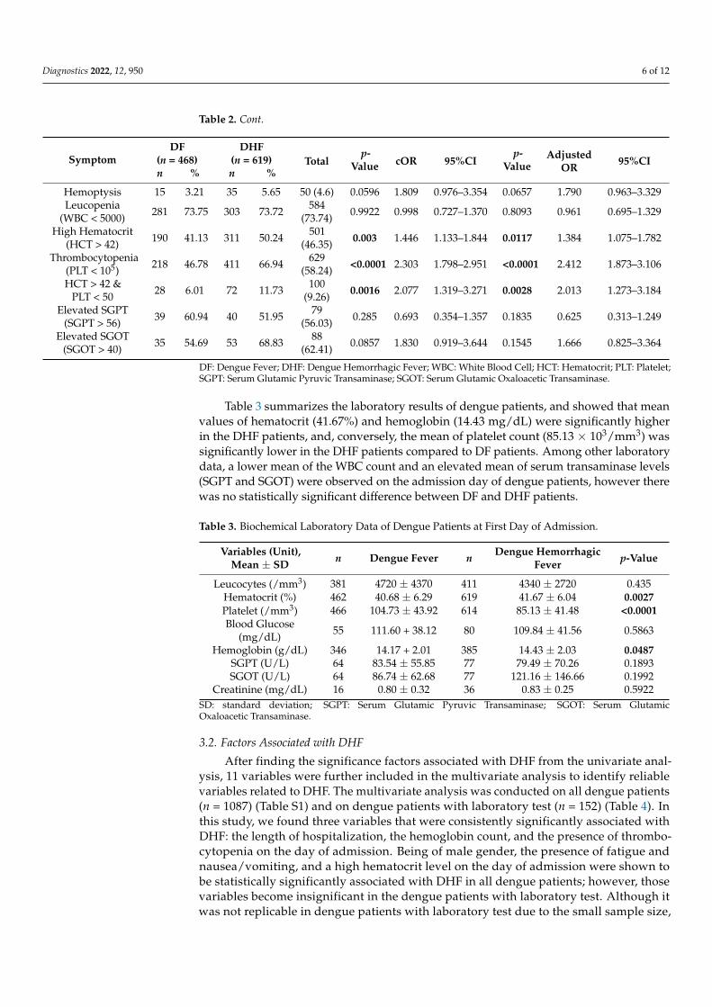
Table 3 summarizes the laboratory results of dengue patients, and showed that meanvalues of hematocrit (41.67%) and hemoglobin (14.43 mg/dL) were significantly higherin the DHF patients, and, conversely, the mean of platelet count (85.13 × 10³/mm3) wassignificantly lower in the DHF patients compared to DF patients. Among other laboratorydata, a lower mean of the WBC count and an elevated mean of serum transaminase levels(SGPT and SGOT) were observed on the admission day of dengue patients, however therewas no statistically significant difference between DF and DHF patients.
Table 3. Biochemical Laboratory Data of Dengue Patients at First Day of Admission.
Variables (Unit),Mean ± SD n Dengue Fever n Dengue Hemorrhagic
Fever p-Value
Leucocytes (/mm3) 381 4720 ± 4370 411 4340 ± 2720 0.435Hematocrit (%) 462 40.68 ± 6.29 619 41.67 ± 6.04 0.0027Platelet (/mm3) 466 104.73 ± 43.92 614 85.13 ± 41.48 <0.0001Blood Glucose
(mg/dL) 55 111.60 + 38.12 80 109.84 ± 41.56 0.5863
Hemoglobin (g/dL) 346 14.17 + 2.01 385 14.43 ± 2.03 0.0487SGPT (U/L) 64 83.54 ± 55.85 77 79.49 ± 70.26 0.1893SGOT (U/L) 64 86.74 ± 62.68 77 121.16 ± 146.66 0.1992
Creatinine (mg/dL) 16 0.80 ± 0.32 36 0.83 ± 0.25 0.5922SD: standard deviation; SGPT: Serum Glutamic Pyruvic Transaminase; SGOT: Serum GlutamicOxaloacetic Transaminase.
3.2. Factors Associated with DHF
After finding the significance factors associated with DHF from the univariate anal-ysis, 11 variables were further included in the multivariate analysis to identify reliablevariables related to DHF. The multivariate analysis was conducted on all dengue patients(n = 1087) (Table S1) and on dengue patients with laboratory test (n = 152) (Table 4). Inthis study, we found three variables that were consistently significantly associated withDHF: the length of hospitalization, the hemoglobin count, and the presence of thrombo-cytopenia on the day of admission. Being of male gender, the presence of fatigue andnausea/vomiting, and a high hematocrit level on the day of admission were shown tobe statistically significantly associated with DHF in all dengue patients; however, thosevariables become insignificant in the dengue patients with laboratory test. Although itwas not replicable in dengue patients with laboratory test due to the small sample size,

Diagnostics 2022, 12, 950 7 of 12
the trend indicated similarities of male gender, nausea/vomiting, and high hematocritlevels as possible risk factors of DHF.
Table 4. Multivariate Analysis to Evaluate Factors Related to Dengue Hemorrhagic Fever in DenguePatients with a Laboratory Test.
CharacteristicDengue Patients with Laboratory Test (n = 152)
p-Value cOR 95%CI
Young Adult (21–45 years old) 0.1449 7.378 0.502–108.349Male Gender 0.8436 1.113 0.385–3.216
Length of Hospitalization 0.0018 1.883 1.264–2.803Fatigue 0.8846 1.217 0.086–17.250
Gum Bleeding 0.0115 0.075 0.01–0.559Nausea/Vomiting 0.6159 1.338 0.429–4.171Abdominal Pain 0.6885 1.248 0.423–3.678
High Hematocrit (Hct > 42) 0.4428 1.574 0.494–5.016Thrombocytopenia (Plt < 100,000) 0.0029 6.058 1.848–19.862
Hct > 42 & Plt < 50 0.1746 0.3 0.053–1.707Hemoglobin 0.0206 0.919 0.855–0.987
HCT: Hematocrit; PLT: Platelet.
3.3. Evaluation of Platelet Profiles Related to DHF
The mean platelet count in DHF patients was significantly lower than that in DFpatients, and it was present on day 1, day 3, and the last day of hospitalization of allof the suspected dengue patients. However, on the last day of hospitalization for thepatients who had a laboratory test, the mean of platelet count did not significantly differbetween DF and DHF patients (Table 5). On day 1, the mean platelet count on DHF patients(85.13 × 10³/mm3) was below 100 × 103/mm3 but not in DF patients (104.73 × 10³/mm3).Furthermore, the mean platelet count declined on day 3 and had significantly increased bythe last day of hospitalization (Table 5).
Table 5. Mean of Platelet Count between DF and DHF patients on Day 1, Day 3 and Last Dayof Hospitalization.
Day ofHospitalization n Platelet Count of
Dengue Fever nPlatelet Count of
Dengue HemorrhagicFever
p-Value
Dengue Patients(n = 1087)
Day 1 466 104.73 ± 43.92 614 85.13 ± 41.48 <0.0001Day 3 418 84.97 ± 40.34 564 64.93 ± 34.21 <0.0001
Last Day 409 113.17 ± 43.26 571 103.12 ± 40.50 <0.0001
Dengue Patientswith Serological Test
(n = 152)
Day 1 37 125.32 ± 58.78 115 91.28 ± 47.45 0.0005Day 3 26 91.87 ± 35.36 86 74.57 ± 45.15 0.0151
Last Day 36 111.09 ± 46.83 113 112.74 ± 45.11 0.7478
On the first day of admission, a mean platelet count of <100 × 103/mm3 was observedin all age groups of DHF patients; however, it only occurred in young and middle adultsamong DF patients (Figure 2). Three age groups of children, adolescents, and young adultsdemonstrated significant differences in platelet counts in DF patients compared to DHFpatients. Regarding the distribution of thrombocytopenia in each age group, young adultpatients exhibited the lowest mean platelet count in both DF or DHF patients, at about90.85 × 103/mm3 and 77.72 × 10³/mm3, respectively. Thrombocytopenia was mostlyobserved on young-adult DF patients (39.91%); however, it mostly occurred in adolescentDHF patients (35.77%).

Diagnostics 2022, 12, 950 8 of 12
Figure 2. Distribution of Platelet (mean ± SD) in Dengue Patients based on: (A). Age; (B). Gender;(C). Serology Test; (D). Length of Hospitalization (ns = Non Significant; * p-value < 0.05;** p-value < 0.01; **** p-value < 0.0001; DF: Dengue Fever; DHF: Dengue Hemorrhagic Fever).
In terms of gender, results from the multivariate analysis of dengue patients withlaboratory test showed that gender was not associated with DHF (Table 4). Consistentwith those results, the mean platelet count was lower in DHF patients and significantlydiffered from that of DF patients; however, it occurred in both male and female patients. InDF, 55.05% of thrombocytopenic patients were female, and conversely, 56.93% of throm-bocytopenic DHF patients were male. In another subgroup analysis, the distribution ofplatelets based on serological tests provided information that patients with a primarydengue infection tended to have a higher mean platelet count than patients with secondarydengue infection (Figure 2). While there was no association between serological tests andDHF due to the small sample size of dengue cases with laboratory test, our results showedthat those with a secondary infection tended to have a lower mean platelet count comparedto those with a primary infection and tended to develop DHF.
The length of hospitalization was another parameter that showed a statistical dif-ference between DF and DHF patients in the multivariate analysis. After the subgroupanalysis, the results were consistent in two groups who were hospitalized three to five daysand more than five days. Figure 2 also indicated that patients with thrombocytopenia andwho developed DHF tended to have a prolonged hospitalization (>5 days) compared topatients with thrombocytopenia and DF.
4. Discussion
Dengue infection remains a serious health problem faced by tropical countries likeIndonesia. In terms of resource limitations, awareness of admission-day parameters wouldbe helpful in minimizing the possibility of severe dengue infection and mortality. In thisretrospective study, factors were identified to associate with DHF patients, for example, thepresence of low platelet counts and high hemoglobin levels on the day of admission. Inaddition, the length of hospitalization was found to be significantly correlated with DHF.
Demographic data revealed differences between DF and DHF patients of the malegender in this study. Previous reports have indicated the correlation between gender and

Diagnostics 2022, 12, 950 9 of 12
risk of dengue or severity of dengue [20,21]. It seems that male gender is a risk factor ofthrombocytopenia [21] and is also related to the development of severe dengue such asDSS [8]. A prolonged hospital stay was also identified to correlate with DHF. Patientswho suffer from dengue infection need more time in hospital, compared to patients withother acute febrile illnesses [22]. Other factors, such as multiple organ dysfunction(s),hypertension, and elevated liver enzymes, can contribute to the longer hospitalization ofDHF patients [12].
Dengue infection patients have similar features to other febrile illnesses. Therefore,specific variables need to be identified as predictors of DHF besides validation by laboratorydata to confirm dengue. Besides the symptoms of fever, our logistic regression model foundthat fatigue and nausea/vomiting were related to DHF in all dengue patients; however,results are insignificant in dengue patients with a laboratory test. Despite this finding, thesimilar trend was observed in both all dengue patients group and the dengue patientswith a laboratory test group. Importantly, symptoms of vomiting were also higher inpatients with DSS (p < 0.001); it occurred in DSS patients rather than in non-DSS patients [8].Even though this study did not identify significant symptoms as predictors of DHF, somefeatures demonstrated a trend that might associate with the progression to severe disease.Those features are nausea/vomiting and abdominal pain. Thus, patients with such clinicalfeatures should be monitored carefully to prevent shock [17].
Other presentations of DHF based on the WHO recommendations are hemorrhagicmanifestations such as a positive tourniquet test, petechiae, epistaxis, and gum or gastroin-testinal bleeding [17]. The hallmark of DHF, plasma leakage, occurs due to endothelialcell dysfunction caused by many cytokine mediators, mast cell products, or inflammatorylipid mediators [23]. Hemorrhagic manifestations representing abnormalities of hemostasis(blood coagulation) that can be detected by monitoring the prolonged activated partialthromboplastin time (APTT) or thrombin time [24]. In this study, we found that gumbleeding occurred in about 2% of either DF or DHF patients. Gum bleeding is one of thecommon mild hemorrhagic manifestations that could occur even in non-severe dengue [25].
Thrombocytopenia or a lower platelet count is one of the laboratory parametersthat is known to associate to the severity of dengue infection such as DHF [7], DSS [8],and severe organ involvement [15]. The underlying mechanisms of dengue infection –mediated thrombocytopenia are still unclear; however, there are some hypotheses includingbone marrow suppression and platelet destruction. Platelet destruction might be causedby DENV nonstructural protein 1 (NS1), a viral protein that is secreted into the bloodcirculation of dengue patients. NS1 is known to induce immune cell and platelet activationvia Toll-like receptor 4 (TLR4), followed by leakage or hemorrhaging during a dengueinfection [6]. Furthermore, NS1 antibody titers were significantly higher in DHF secondaryinfections compared to DF during the critical phase (days 6~7 of fever) [26]. Therefore,monitoring the incidence of thrombocytopenia is important to determine the patients whoare most likely to develop severe dengue.
Our study results revealed a significant association between thrombocytopenia(platelets < 100 × 103/mm3) and DHF. Platelets in this study demonstrated the same pat-tern as those of other studies in which a sudden significant drop in platelets in denguepatients occurred between days four and six of fever [27,28]. The platelet counts sig-nificantly dropped in DHF patients compared to DF patients on days one and three ofhospitalization. Some characteristics such as male gender and secondary dengue patientsreported higher percentages of thrombocytopenia on the day of admission and devel-oped DHF. Even though we did not find a significant association between male genderand secondary dengue as risk factors for DHF due to the limited sample size of denguepatients with a serological test, the pattern of these characteristics also reflected the corre-lation with thrombocytopenia in DHF patients The results were consistent with previousstudies [8,20,28].
Another laboratory feature that requires close monitoring is the hematocrit level [17].Our results showed a strong association between hematocrit and DHF in all dengue cases,

Diagnostics 2022, 12, 950 10 of 12
however, the significance was not observed in in dengue patients with a serological testdiagnosis. Even so, the same pattern of these results showed that a high hematocrit wascorrelated to DHF (cOR 1.45 in all dengue patients; cOR 1.57 in dengue patients withserological test). A rising hematocrit level is an indicator of plasma leakage that developsin DHF patients, whereas a decreasing hematocrit level is a sign of bleeding. Plasmaleakage in dengue infection is caused by extensive immune activation that results in acytokine storm. There is a hypothesis that in dengue patients with significant plasmaleakage, a rising hematocrit level occurs in the febrile phase to the critical phase, andit is associated with decreasing levels of some immune mediators that function in anti-inflammation (IL-1 receptor antagonist (IL-1RA)) and chemotaxis (interferon-γ-inducibleprotein 10 (IP-10)) [29].
The on-admission laboratory data revealed a relationship between DHF and hemoglobinlevels in the multivariate analysis. Hemoglobin levels are known to be significantly cor-related with hematocrit in dengue infection [30] In addition to that, hemoglobin andhematocrit levels in dengue patients continued to increase from day 3 of admission un-til day 10 compared to the non-dengue group (other acute febrile illnesses) [27]. Thoseresults were strengthened by other studies in Surabaya, Indonesia, in which the levelof hemoglobin in children and adult patients increased in DHF patients but not in DFpatients [31]. A significantly higher level of hemoglobin was also observed in DHF patientsduring acute febrile illness and on day five of the illness [32]. Both results highlight theimportance of recording hemoglobin data in dengue patients as a predictor of DHF andplasma leakage.
There were some limitations in this study. First, this was a retrospective study that useddata from available medical records. Therefore, some clinical information was incomplete,such as the virus type, patients’ comorbidities, and day-of-illness (or day-of-fever). Anotherlimitation is the sample size. The number of patients who had laboratory test is small(n = 152). In addition, this is a hospital-based study, which is related to severe disease;hence results cannot be generalized to all community-based dengue patients. However,these results should be helpful as supporting data for guiding management in some regionsthat do not have a well-equipped laboratory or trained staff.
5. Conclusions
In conclusion, on-admission platelet counts and hemoglobin laboratory data shouldbe considered as predictors of DHF especially for suspected dengue patients who lackserological or molecular diagnostic tests. Intensive monitoring of these parameters isimportant to prevent prolonged hospitalization and dengue severity. Future studies withlarger samples especially in the second population will be needed to confirm our findings.
Supplementary Materials: The following supporting information can be downloaded at: https://www.mdpi.com/article/10.3390/diagnostics12040950/s1, Table S1: Multivariate Analysis to EvaluateFactors Related Dengue Hemorrhagic Fever in All Dengue Patients; Table S2: Distribution of Plateleton Dengue Patients on the First Day of Admission.
Author Contributions: Conceptualization, I.N.F., H.D., W.-C.C., Y.-H.C. and D.A.P.; Formal analysis,I.N.F.; Resources, Y.-H.C., B.D.P., M.J.H. and M.A.A.; Data Curation, I.N.F., Y.-H.C., H.D. and W.S.;Writing—Original Draft Preparation, I.N.F.; Writing—Review and Editing, I.N.F., W.-C.C., Y.-H.C.and D.A.P.; Visualization, I.N.F.; Supervision, W.-C.C. and D.A.P.; Funding acquisition, W.-C.C. andD.A.P. All authors have read and agreed to the published version of the manuscript.
Funding: This research was supported by grants from the Institute for Research and CommunityService of University of Ahmad Dahlan (LPPM UAD), Research Team of Bigdata, Bioinformatic andPersonalized Medicine in Pharmaceutical Care (MABIF) in Faculty of Pharmacy University of AhmadDahlan, and grants from the Ministry of Science and Technology, Taiwan (MOST 110-2628-B-038-020;MOST 110-2314-B-038-161), and Taipei Medical University (12310-106079; 12310-106132).

Diagnostics 2022, 12, 950 11 of 12
Institutional Review Board Statement: This study was approved by the ethics committee of thePKU Muhammadiyah Yogyakarta hospital (Ref: 00101/KT.7.4/III/2021) and health research ethicscommittee of Fakultas Kedokteran dan Ilmu Kesehatan Universitas Muhammadiyah Yogyakarta(UMY) (Ref: 063/EC-KEPK FKIK UMY/XII/2020).
Informed Consent Statement: Not applicable.
Data Availability Statement: All datasets supporting the conclusions in this study are includedwithin the article.
Acknowledgments: We are grateful for the support of staff from PKU Muhammadiyah BantulHospital, PKU Muhammadiyah Yogyakarta Hospital, PKU Muhammadiyah Gamping Hospital,RSUD Wonosari, and RSUD Panembahan Senopati. We are also thankful for our team: Tuty LiaSyahfitri, Reza Aditya Nugroho, Nur Achmad Fauzi, Rizal Shidqi Ilyasa, Rafly Ramadhani for theirsupport in this study. Lastly, we would like to thank Henry Sung-Ching Wong, Che-Mai Chang, JafitTing, Wan-Hsuan Chou (Taipei Medical University) for their helpful comments during the studiesand thank to Hui-Ju Tsai (National Health Research Institutes, Taiwan) for manuscript revision.
Conflicts of Interest: The authors declare no conflict of interest.
References1. Messina, J.P.; Brady, O.J.; Golding, N.; Kraemer, M.U.G.; Wint, G.R.W.; Ray, S.E.; Pigott, D.M.; Shearer, F.M.; Johnson, K.;
Earl, L.; et al. The current and future global distribution and population at risk of dengue. Nat. Microbiol. 2019, 4, 1508–1515.[CrossRef] [PubMed]
2. World Health Organization. Dengue and Severe Dengue. Available online: https://www.who.int/news-room/fact-sheets/detail/dengue-and-severe-dengue (accessed on 14 December 2020).
3. Harapan, H.; Michie, A.; Mudatsir, M.; Sasmono, R.T.; Imrie, A. Epidemiology of dengue hemorrhagic fever in Indonesia:Analysis of five decades data from the National Disease Surveillance. BMC Res. Notes 2019, 12, 350. [CrossRef] [PubMed]
4. Utama, I.M.S.; Lukman, N.; Sukmawati, D.D.; Alisjahbana, B.; Alam, A.; Murniati, D.; Utama, I.; Puspitasari, D.; Kosasih, H.;Laksono, I.; et al. Dengue viral infection in Indonesia: Epidemiology, diagnostic challenges, and mutations from an observationalcohort study. PLoS Negl. Trop. Dis. 2019, 13, e0007785. [CrossRef] [PubMed]
5. Sasmono, R.T.; Sutjianto, A.; Santoso, M.S.; Sriwedari, K.; Yohan, B.; Mayasanti, E.; Hayati, R.F.; Denis, D. Molecular epidemiologyof dengue in North Kalimantan, a province with the highest incidence rates in Indonesia in 2019. Infect. Genet. Evol. 2021,95, 105036. [CrossRef] [PubMed]
6. Chao, C.H.; Wu, W.C.; Lai, Y.C.; Tsai, P.J.; Perng, G.C.; Lin, Y.S.; Yeh, T.M. Dengue virus nonstructural protein 1 activates plateletsvia Toll-like receptor 4, leading to thrombocytopenia and hemorrhage. PLoS Pathog. 2019, 15, e1007625. [CrossRef]
7. Kularatnam, G.A.M.; Jasinge, E.; Gunasena, S.; Samaranayake, D.; Senanayake, M.P.; Wickramasinghe, V.P. Evaluation ofbiochemical and haematological changes in dengue fever and dengue hemorrhagic fever in Sri Lankan children: A prospectivefollow up study. BMC Pediatrics 2019, 19, 87. [CrossRef]
8. Lam, P.K.; Ngoc, T.V.; Thu Thuy, T.T.; Hong Van, N.T.; Nhu Thuy, T.T.; Hoai Tam, D.T.; Dung, N.M.; Hanh Tien, N.T.; Thanh Kieu,N.T.; Simmons, C.; et al. The value of daily platelet counts for predicting dengue shock syndrome: Results from a prospectiveobservational study of 2301 Vietnamese children with dengue. PLoS Negl. Trop. Dis. 2017, 11, e0005498. [CrossRef]
9. World Health Organization. Handbook for Clinical Management of Dengue; World Health Organization: Geneva, Switzerland, 2012.10. World Health Organization. Dengue Guidelines for Diagnosis, Treatment, Prevention and Control: New Edition; World Health
Organization: Geneva, Switzerland, 2009.11. Huang, H.-S.; Hsu, C.-C.; Ye, J.-C.; Su, S.-B.; Huang, C.-C.; Lin, H.-J. Predicting the mortality in geriatric patients with dengue
fever. Medicine 2017, 96, e7878. [CrossRef]12. Mallhi, T.H.; Khan, A.H.; Sarriff, A.; Adnan, A.S.; Khan, Y.H. Determinants of mortality and prolonged hospital stay among
dengue patients attending tertiary care hospital: A cross-sectional retrospective analysis. BMJ Open 2017, 7, e016805. [CrossRef]13. Hsieh, C.-C.; Cia, C.-T.; Lee, J.-C.; Sung, J.-M.; Lee, N.-Y.; Chen, P.-L.; Kuo, T.-H.; Chao, J.-Y.; Ko, W.-C. A Cohort Study of Adult
Patients with Severe Dengue in Taiwanese Intensive Care Units: The Elderly and APTT Prolongation Matter for Prognosis. PLoSNegl. Trop. Dis. 2017, 11, e0005270. [CrossRef]
14. Lovera, D.; Martinez de Cuellar, C.; Araya, S.; Amarilla, S.; Gonzalez, N.; Aguiar, C.; Acuña, J.; Arbo, A. Clinical Characteristicsand Risk Factors of Dengue Shock Syndrome in Children. Pediatric Infect. Dis. J. 2016, 35, 1294–1299. [CrossRef] [PubMed]
15. Pang, J.; Hsu, J.P.; Yeo, T.W.; Leo, Y.S.; Lye, D.C. Diabetes, cardiac disorders and asthma as risk factors for severe organ involvementamong adult dengue patients: A matched case-control study. Sci. Rep. 2017, 7, 39872. [CrossRef] [PubMed]
16. Pan American Health Organization. Dengue: Guidelines for Patient Care in the Region of the Americas, 2nd ed.; World HealthOrganization, Regional Office for the Americas: Washington, DC, USA, 2016.
17. World Health Organization—Regional Office for South-East Asia. Comprehensive Guideline for Prevention and Control of Dengue andDengue Haemorrhagic Fever; Revised and Expanded Edition; WHO Regional Office for South-East Asia: New Delhi, India, 2011.
Diagnostics 2022, 12, 950 12 of 12
18. Ministry of Health Republic Indonesia. Guideline for Dengue Prevention and Control in Indonesia; Ministry of Health RepublicIndonesia: Jakarta, Republic of Indonesia, 2017.
19. World Health Organization. Dengue Haemorrhagic Fever: Diagnosis, Treatment, Prevention and Control, 2nd ed.; World HealthOrganization: Geneva, Switzerland, 1997.
20. Sangkaew, S.; Ming, D.; Boonyasiri, A.; Honeyford, K.; Kalayanarooj, S.; Yacoub, S.; Dorigatti, I.; Holmes, A. Risk predictors ofprogression to severe disease during the febrile phase of dengue: A systematic review and meta-analysis. Lancet Infect. Dis. 2021,21, 1014–1026. [CrossRef]
21. Castilho, B.M.; Silva, M.T.; Freitas, A.R.R.; Fulone, I.; Lopes, L.C. Factors associated with thrombocytopenia in patients withdengue fever: A retrospective cohort study. BMJ Open 2020, 10, e035120. [CrossRef] [PubMed]
22. Lim, J.K.; Chanthavanich, P.; Limkittikul, K.; Lee, J.-S.; Sirivichayakul, C.; Lee, K.S.; Lim, S.-K.; Yoon, I.-K.; Hattasingh, W. Clinicaland epidemiologic characteristics associated with dengue fever in 2011–2016 in Bang Phae district, Ratchaburi province, Thailand.PLoS Negl. Trop. Dis. 2021, 15, e0009513. [CrossRef] [PubMed]
23. Malavige, G.N.; Ogg, G.S. Pathogenesis of vascular leak in dengue virus infection. Immunology 2017, 151, 261–269. [CrossRef]24. Chuang, Y.-C.; Lin, Y.-S.; Liu, C.-C.; Liu, H.-S.; Liao, S.-H.; Shi, M.-D.; Lei, H.-Y.; Yeh, T.-M. Factors contributing to the disturbance
of coagulation and fibrinolysis in dengue virus infection. J. Formos. Med. Assoc. 2013, 112, 12–17. [CrossRef]25. Centers for Disease Control and Prevention (CDC). Dengue Clinical Case Management (DCCM) E-Learning: Bleeding Manifestations
in Patients with Dengue 2018; Centers for Disease Control and Prevention (CDC): Atlanta, GA, USA, 2018.26. Jayathilaka, D.; Gomes, L.; Jeewandara, C.; Jayarathna, G.S.B.; Herath, D.; Perera, P.A.; Fernando, S.; Wijewickrama, A.; Hardman,
C.S.; Ogg, G.S.; et al. Role of NS1 antibodies in the pathogenesis of acute secondary dengue infection. Nat. Commun. 2018, 9, 5242.[CrossRef]
27. Chaloemwong, J.; Tantiworawit, A.; Rattanathammethee, T.; Hantrakool, S.; Chai-Adisaksopha, C.; Rattarittamrong, E.; Noraset-thada, L. Useful clinical features and hematological parameters for the diagnosis of dengue infection in patients with acute febrileillness: A retrospective study. BMC Hematol. 2018, 18, 20. [CrossRef]
28. Ojha, A.; Nandi, D.; Batra, H.; Singhal, R.; Annarapu, G.K.; Bhattacharyya, S.; Seth, T.; Dar, L.; Medigeshi, G.R.; Vrati, S.; et al.Platelet activation determines the severity of thrombocytopenia in dengue infection. Sci. Rep. 2017, 7, 41697. [CrossRef]
29. Her, Z.; Kam, Y.-W.; Gan, V.C.; Lee, B.; Thein, T.-L.; Tan, J.J.L.; Platform, S.I.I.; Lee, L.K.; Fink, K.; Lye, D.C.; et al. Severityof Plasma Leakage Is Associated With High Levels of Interferon γ–Inducible Protein 10, Hepatocyte Growth Factor, MatrixMetalloproteinase 2 (MMP-2), and MMP-9 During Dengue Virus Infection. J. Infect. Dis. 2017, 215, 42–51. [CrossRef] [PubMed]
30. Sahassananda, D.; Thanachartwet, V.; Chonsawat, P.; Wongphan, B.; Chamnanchanunt, S.; Surabotsophon, M.; Desakorn, V.Evaluation of Hematocrit in Adults with Dengue by a Laboratory Information System. J. Trop. Med. 2021, 2021, 8852031.[CrossRef] [PubMed]
31. Wardhani, P.; Aryati, A.; Yohan, B.; Trimarsanto, H.; Setianingsih, T.Y.; Puspitasari, D.; Arfijanto, M.V.; Bramantono, B.; Suharto, S.;Sasmono, R.T. Clinical and virological characteristics of dengue in Surabaya, Indonesia. PLoS ONE 2017, 12, e0178443. [CrossRef][PubMed]
32. Ralapanawa, U.; Alawattegama, A.T.M.; Gunrathne, M.; Tennakoon, S.; Kularatne, S.A.M.; Jayalath, T. Value of peripheral bloodcount for dengue severity prediction. BMC Res. Notes 2018, 11, 400. [CrossRef]
Related Documents