Dynamic Causal Modeling of Load-Dependent Modulation of Effective Connectivity Within the Verbal Working Memory Network Danai Dima, 1,2 Jigar Jogia, 1 and Sophia Frangou 2 1 Department of Psychiatry, Icahn School of Medicine at Mount Sinai, New York, New York 2 Institute of Psychiatry, King’s College London, London, United Kingdom r r Abstract: Neuroimaging studies have consistently shown that working memory (WM) tasks engage a distributed neural network that primarily includes the dorsolateral prefrontal cortex, the parietal cor- tex, and the anterior cingulate cortex. The current challenge is to provide a mechanistic account of the changes observed in regional activity. To achieve this, we characterized neuroplastic responses in effec- tive connectivity between these regions at increasing WM loads using dynamic causal modeling of functional magnetic resonance imaging data obtained from healthy individuals during a verbal n-back task. Our data demonstrate that increasing memory load was associated with (a) right-hemisphere dominance, (b) increasing forward (i.e., posterior to anterior) effective connectivity within the WM net- work, and (c) reduction in individual variability in WM network architecture resulting in the right- hemisphere forward model reaching an exceedance probability of 99% in the most demanding condi- tion. Our results provide direct empirical support that task difficulty, in our case WM load, is a signifi- cant moderator of short-term plasticity, complementing existing theories of task-related reduction in variability in neural networks. Hum Brain Mapp 35:3025–3035, 2014. V C 2013 Wiley Periodicals, Inc. Key words: neuroimaging; fMRI; n-back task; dorsolateral prefrontal cortex; short-term plasticity; pari- etal; anterior cingulate r r INTRODUCTION Working memory (WM) refers to the ability to maintain, update, and manipulate goal-relevant information (Baddeley, 1992). WM tasks consistently recruit cortical regions within the dorsolateral prefrontal cortex (DLPFC, Brodmann area 9/46), the parietal cortex (PAR, BA7/40) and the dorsal ante- rior cingulate cortex (ACC, BA32; Fletcher and Henson, 2001; Nee et al., 2013; Wager and Smith, 2003). Within this network, there is evidence for relative functional specialization accord- ing to process; the DLPFC has been intimately linked to encoding, setting attentional priorities, and manipulating information (D’Esposito et al., 2000; Narayanan et al., 2005), the PAR has been associated with maintaining attentional focus and storing information (Guerin and Miller, 2011; Jonides et al., 1998), and the ACC has been implicated in error detection and performance adjustment (Carter et al., 1999). Regional activation within this network responds to WM load (i.e., the total demand on maintenance, updating, and manipulation processes) (Braver et al., 1997; Callicott et al., 1999; Owen et al., 1999; Volle et al., 2008) and may be sensi- tive to content being left-lateralized for verbal and right- lateralized for spatial and object information (Fletcher and Henson, 2001; Nee et al., 2013; Wager and Smith, 2003). Contract grant sponsor: NARSAD Independent Investigator Award 2008 (SF) *Correspondence to: Sophia Frangou, Icahn Medical Institute, 1425 Madison Ave, New York, NY 10128, USA. E-mail: [email protected] Received for publication 23 May 2013; Revised 17 July 2013; Accepted 18 July 2013. DOI 10.1002/hbm.22382 Published online 18 October 2013 in Wiley Online Library (wileyonlinelibrary.com). r Human Brain Mapping 35:3025–3035 (2014) r V C 2013 Wiley Periodicals, Inc.

Welcome message from author
This document is posted to help you gain knowledge. Please leave a comment to let me know what you think about it! Share it to your friends and learn new things together.
Transcript
Dynamic Causal Modeling of Load-DependentModulation of Effective Connectivity Within the
Verbal Working Memory Network
Danai Dima,1,2 Jigar Jogia,1 and Sophia Frangou2
1Department of Psychiatry, Icahn School of Medicine at Mount Sinai, New York, New York2Institute of Psychiatry, King’s College London, London, United Kingdom
r r
Abstract: Neuroimaging studies have consistently shown that working memory (WM) tasks engage adistributed neural network that primarily includes the dorsolateral prefrontal cortex, the parietal cor-tex, and the anterior cingulate cortex. The current challenge is to provide a mechanistic account of thechanges observed in regional activity. To achieve this, we characterized neuroplastic responses in effec-tive connectivity between these regions at increasing WM loads using dynamic causal modeling offunctional magnetic resonance imaging data obtained from healthy individuals during a verbal n-backtask. Our data demonstrate that increasing memory load was associated with (a) right-hemispheredominance, (b) increasing forward (i.e., posterior to anterior) effective connectivity within the WM net-work, and (c) reduction in individual variability in WM network architecture resulting in the right-hemisphere forward model reaching an exceedance probability of 99% in the most demanding condi-tion. Our results provide direct empirical support that task difficulty, in our case WM load, is a signifi-cant moderator of short-term plasticity, complementing existing theories of task-related reduction invariability in neural networks. Hum Brain Mapp 35:3025–3035, 2014. VC 2013 Wiley Periodicals, Inc.
Key words: neuroimaging; fMRI; n-back task; dorsolateral prefrontal cortex; short-term plasticity; pari-etal; anterior cingulate
r r
INTRODUCTION
Working memory (WM) refers to the ability to maintain,update, and manipulate goal-relevant information (Baddeley,1992). WM tasks consistently recruit cortical regions withinthe dorsolateral prefrontal cortex (DLPFC, Brodmann area
9/46), the parietal cortex (PAR, BA7/40) and the dorsal ante-rior cingulate cortex (ACC, BA32; Fletcher and Henson, 2001;Nee et al., 2013; Wager and Smith, 2003). Within this network,there is evidence for relative functional specialization accord-ing to process; the DLPFC has been intimately linked toencoding, setting attentional priorities, and manipulatinginformation (D’Esposito et al., 2000; Narayanan et al., 2005),the PAR has been associated with maintaining attentionalfocus and storing information (Guerin and Miller, 2011;Jonides et al., 1998), and the ACC has been implicated in errordetection and performance adjustment (Carter et al., 1999).Regional activation within this network responds to WMload (i.e., the total demand on maintenance, updating, andmanipulation processes) (Braver et al., 1997; Callicott et al.,1999; Owen et al., 1999; Volle et al., 2008) and may be sensi-tive to content being left-lateralized for verbal and right-lateralized for spatial and object information (Fletcher andHenson, 2001; Nee et al., 2013; Wager and Smith, 2003).
Contract grant sponsor: NARSAD Independent InvestigatorAward 2008 (SF)
*Correspondence to: Sophia Frangou, Icahn Medical Institute,1425 Madison Ave, New York, NY 10128, USA.E-mail: [email protected]
Received for publication 23 May 2013; Revised 17 July 2013;Accepted 18 July 2013.
DOI 10.1002/hbm.22382Published online 18 October 2013 in Wiley Online Library(wileyonlinelibrary.com).
r Human Brain Mapping 35:3025–3035 (2014) r
VC 2013 Wiley Periodicals, Inc.
However, measures of regional engagement are not suffi-cient to characterize the dynamic architecture of the WMnetwork. To date, attempts to model inter-regional relation-ships within the WM network have been based on minimiz-ing the discrepancy between observed and impliedcorrelations between hemodynamic activations (Axmacheret al., 2008; Esslinger et al., in press; Honey et al., 2002; Nar-ayanan et al., 2005). Although useful, this approach provideslimited information about specific mechanisms throughwhich neuronal circuits respond to WM demands (Kimet al., 2007; Ramnani et al., 2004). Of particular relevance isthe potential role of short-term plasticity given the conflu-ence of evidence that abnormalities in short-term plasticityunderlie WM impairment across a range of conditions fromnormal aging (Sala-Llonch et al., 2012) to the major mentaldisorders (Cramer et al., 2011; Stephan et al., 2006). Short-term plasticity is conceptualized in many ways, but as usedhere it refers to altered functional coupling within corticalcircuits as a function of experience and in response to exter-nal and internal cues (Salinas and Sejnowski, 2001; Stephanet al., 2009). Dynamic causal modeling (DCM; Friston et al.,2003) currently represents one of the most plausible meth-ods for estimating the effective strength of connectionsamong neuronal ensembles and their context-dependent(e.g., experimental) modulation (Stephan et al., 2010).
The aim of the current study was to determine the roleof WM load on short-term plasticity. To achieve this, wecombined Statistical Parametric Mapping (SPM; www.fi-l.ion.ucl.ac.uk/spm) with DCM analysis of functional mag-netic resonance imaging (fMRI) data derived from fortyhealthy adults performing the verbal n-back task. This taskinvolves maintenance and updating of information atincreasing levels of memory load and has been previouslyshown to engage robustly the DLPFC, PAR, and ACC(Owen et al., 2005; Wager and Smith, 2003). DCM analysisof individual responses to this task allowed us to estimatethe strength, laterality, and directionality of the effectivefunctional coupling between these key nodes of the WMnetwork and, crucially how these connection strengthswere modulated by memory load.
MATERIALS AND METHODS
Participants
Forty healthy right-handed adults were recruited viaadvertisement in the local press and were included if they(i) had no personal lifetime history of mental disorders orsubstance use as assessed after personal interview usingthe Structured Interview for DSM-IV-TR Axis I Disorders,non-patient edition (First et al., 2002), (ii) had no history ofhead injury or medical disorders, and (iii) did not takeany prescribed medication. An estimate of current intellec-tual function (IQ) was obtained using the Wechsler AdultIntelligence Scale-Revised (Wechsler, 1981). The sampledetails are shown in Table I. The study was approved by
the Ethics Committee of the Institute of Psychiatry and theSouth London and Maudsley National Health ServiceTrust. Written informed consent was obtained from allparticipants.
Experimental Design
The n-back task was used in a block design incorporatingalternating experimental and baseline conditions. A seriesof letters in yellow font were displayed on a blue screen for2 s each. The 0-back condition was used as baseline to con-trol for task engagement and vigilance. In the 0-back condi-tion participants were instructed to respond by buttonpress whenever the target letter “X” was displayed onscreen. In the experimental conditions (1, 2, and 3 back), thetarget letter was defined as any letter that was identical tothe one presented 1, 2, or 3 trials back. There were 18epochs in all, each lasting 30 s, comprising 14 letters with aratio of target to nontarget letters ranging from 2:12 to 4:10per epoch. The entire experiment lasted 9 min and includeda total of 49 target and 203 nontarget stimuli. To avoid anysystematic order effects, the conditions were pseudor-andomized. Performance was evaluated in terms of reactiontime to target letters and accuracy (% correct responses).
Image Acquisition
Gradient echo planar magnetic resonance (MR) imageswere acquired using a 1.5-T GE Neurooptimized Signa MRsystem (General Electric, Milwaukee, WI) fitted with 40mT/m high-speed gradients, at the Maudsley Hospital,London. Foam padding and a forehead strap were used tolimit head motion. A quadrature birdcage head coil wasused for radio frequency transmission and reception. Atotal of 180 T2*-weighted MR brain volumes depictingblood-oxygen-level-dependent (BOLD) contrast wereacquired at each of 36 near-axial, noncontiguous planes par-allel to the intercommissural (AC-PC) plane; repetition time(TR) 5 3,000 ms, echo time (TE) 5 40 ms, slice thickness 5 3mm, voxel dimensions 5 3.75 mm 3 3.75 mm 3 3.30 mm,
TABLE I. Demographic and behavioral data of study
participants (N 5 40)
Demographic dataSex (male:female) 20:20Age (years) 31.5 (10.4)WAIS-R IQ 115.5 (15.9)Behavioral performance on the n-back taskResponse time, 1 back (ms) 596 (210)Accuracy, 1 back (% correct) 100Response time, 2 back (ms) 659 (196)Accuracy, 2 back (% correct) 91.2 (13.85)Response time, 3 back (ms) 748 (224)Accuracy, 3 back (% correct) 72.8 (16.1)
Continuous variables are presented as mean and standard devia-tion; WAIS-R, Wechsler Adult Intelligence Scale-Revised.
r Dima et al. r
r 3026 r
matrix size 5 64 3 64, and flip angle 5 90�. Before eachacquisition sequence, four dummy data acquisition scanswere performed to allow the scanner to reach a steady statein T1 contrast.
During the same session, a high-resolution T1-weightedstructural image was acquired in the axial plane (inversionrecovery prepared, spoiled gradient-echo sequence;TR 5 18 ms, TE 5 5.1 ms, inversion time 5 450 ms, slicethickness 5 1.5 mm, voxel dimensions 5 0.9375 mm 3 0.9375mm 3 1.5 mm, matrix size 256 3 192, field of view 5 240 3
180 mm, flip angle 5 20�, and number of excitations 5 1) forsubsequent coregistration.
Image Processing
All analyses were implemented using StatisticalParametric Mapping software, version 8 (SPM8) (www.fil.ion.ucl.ac.uk/spm/software/spm8/). The BOLD imageswere realigned to the fifth volume to correct for interscanmovements by means of a rigid body transformation withthree rotation and three translation parameters. Subse-quently, the 180 fMRI images were spatially normalized tothe standard template of the Montreal Neurological Insti-tute (MNI) and resampled to a voxel size of 2 mm 3 2mm 3 2 mm. Finally, the images were smoothed using an8-mm full-width half maximum Gaussian kernel.
The smoothed single-participant images were analyzedvia multiple regressions using the linear convolutionmodel, with vectors of onset representing the experimentalconditions (1, 2, and 3 back) and the 0-back condition as abaseline. Six movement parameters were also entered asnuisance covariates. Serial correlations were removedusing an AR(1) model. A high pass filter (128s) wasapplied to remove low-frequency noise. Contrast imagesof each memory load condition versus baseline were pro-duced for each participant.
Conventional fMRI Analysis
Group-level analyses were based on random-effectsanalyses of the single-participant contrast images using thesummary statistic approach. Regions showing significanttask effect across all participants were identified usingone-sample t tests against zero. The statistical thresholdwas set to P< 0.05 with familywise error (FWE) correctionon a voxelwise basis and minimum cluster size 20 voxels.For all analyses, results are reported in MNI space.
Dynamic Causal Modeling
Selection of volumes of interest
Volumes of interest (VOIs) were defined bilaterally inthe PAR, ACC, and DLPFC based on evidence from pre-vious studies demonstrating robust and consistentinvolvement of these regions in WM (Glahn et al., 2005;Owen et al., 2005) and based on the results from the cur-
rent analyses that found significant effects of WM load inthese regions. The coordinates for the VOIs were basedon the group maxima from the contrast of 1-, 2-, and 3-back minus 0-back condition after conjunction analysis.The coordinates of the group maxima were DLPFC (left:x 5 248, y 5 36, z 5 30; right; x 5 48, y 5 38, z 5 30), PAR(left: x 5 238, y 5 256, z 5 42; right: x 5 36, y 5 252,z 5 44), and ACC (left: x 5 210, y 5 26, z 5 28; right:x 5 12, y 524, z 528). For each participant, VOIs of 5 mmradius were defined centered on participant-specific max-ima in these regions that were (i) within 4 mm from thegroup maxima, (ii) within the same anatomical area, asdefined by the PickAtlas toolbox (Maldjian et al., 2003),and (iii) adjusted using the effect of interest F-contrast.Regional time series were summarized with the firsteigenvariate of all activated (at P< 0.01) voxels withinthe participant-specific VOIs.
Specification of model architecture
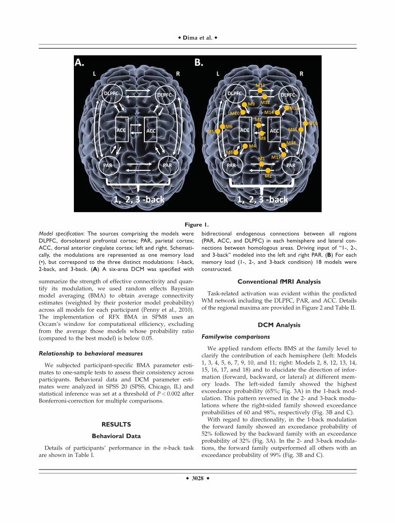
For each experimental condition (1, 2, and 3 back), we usedthe VOIs defined above (L-PAR and R-PAR, L-ACC and R-ACC, and L-DLPFC and R-DLPFC) to specify a six-areaDCM in all participants. Within each hemisphere, we definedbidirectional connections between these regions. Bidirec-tional connections were also specified between homologousregions in each hemisphere. For each experimental condition,18 endogenous connections were specified in total with themain effect of memory as the driving input entering the L-PAR and R-PAR (Fig. 1A). This architecture served as ourbase model which was then elaborated systematically to pro-duce 18 alternative variants for each experimental conditionto test how WM load could modulate the 18 connections (Fig.1B). In total, 54 models were constructed, fitted and com-pared in the 40 study participants.
Model comparison
Model comparison was implemented using random-effects (RFX) Bayesian model selection (BMS) in DCM10 tocompute exceedance and posterior probabilities at thegroup level (Stephan et al., 2009). The exceedance probabil-ity of a model denotes the probability that this model ismore likely than any other in a given dataset. In addition totesting individual models, we also made inferences aboutfamilies of models (Penny et al., 2010; Stephan et al., 2010).
Families were specified based on the laterality anddirection of WM information. To test for hemispheric later-ality, models were divided into a left-sided (Models 1, 3,4, 5, 6, 7, 9, 10, and 11) and a right-sided family (Models 2,8, 12, 13, 14, 15, 16, 17, and 18). To test for directionality ofWM information three families where created for eachexperimental condition, forward (Models 3, 5, 10, 14, 16,and 17), backwards (Models 4, 6, 9, 13, 15, and 18), andlateral (Models 1, 2, 7, 8, 11, and 12). All models wereincluded in the BMS procedure, both when comparingindividual models and model families. Finally, to
r Brain Connectivity in Increasing Memory Load r
r 3027 r
summarize the strength of effective connectivity and quan-tify its modulation, we used random effects Bayesianmodel averaging (BMA) to obtain average connectivityestimates (weighted by their posterior model probability)across all models for each participant (Penny et al., 2010).The implementation of RFX BMA in SPM8 uses anOccam’s window for computational efficiency, excludingfrom the average those models whose probability ratio(compared to the best model) is below 0.05.
Relationship to behavioral measures
We subjected participant-specific BMA parameter esti-mates to one-sample tests to assess their consistency acrossparticipants. Behavioral data and DCM parameter esti-mates were analyzed in SPSS 20 (SPSS, Chicago, IL) andstatistical inference was set at a threshold of P< 0.002 afterBonferroni-correction for multiple comparisons.
RESULTS
Behavioral Data
Details of participants’ performance in the n-back taskare shown in Table I.
Conventional fMRI Analysis
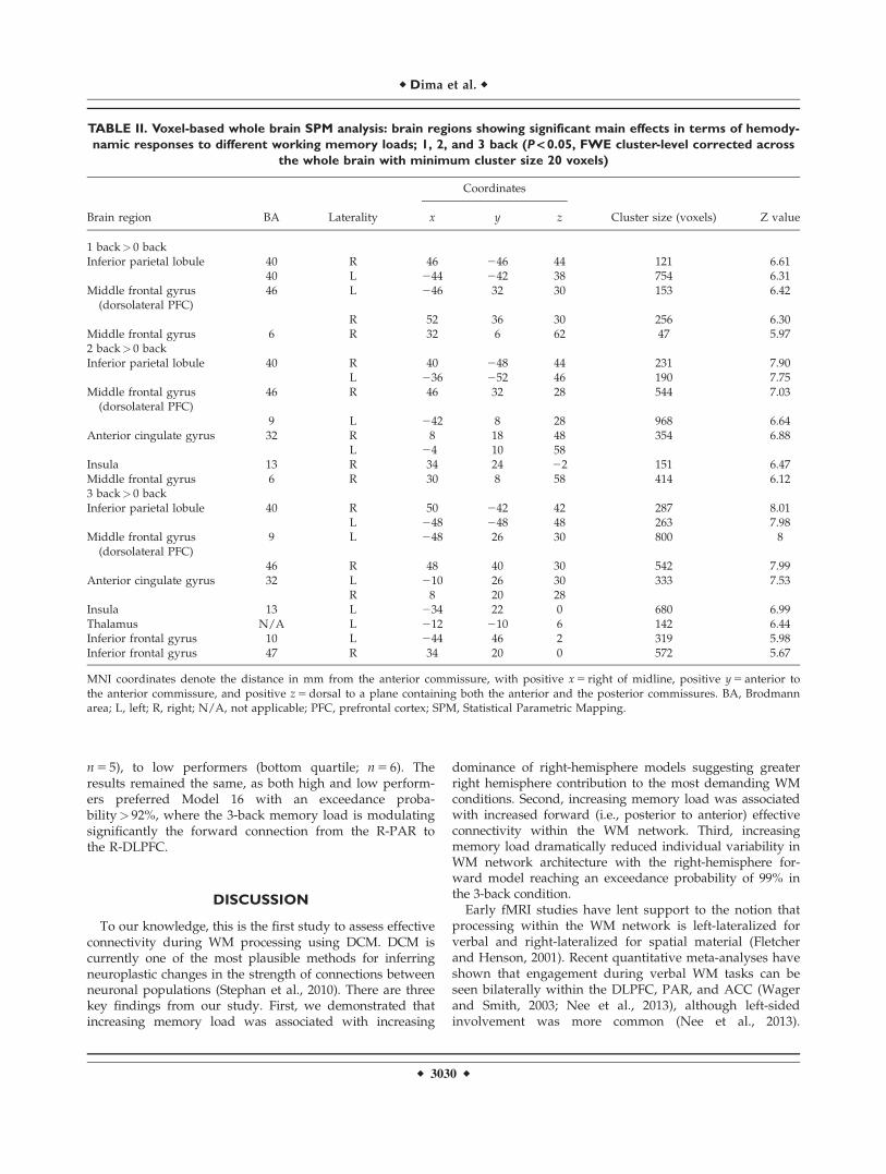
Task-related activation was evident within the predictedWM network including the DLPFC, PAR, and ACC. Detailsof the regional maxima are provided in Figure 2 and Table II.
DCM Analysis
Familywise comparisons
We applied random effects BMS at the family level toclarify the contribution of each hemisphere (left: Models1, 3, 4, 5, 6, 7, 9, 10, and 11; right: Models 2, 8, 12, 13, 14,15, 16, 17, and 18) and to elucidate the direction of infor-mation (forward, backward, or lateral) at different mem-ory loads. The left-sided family showed the highestexceedance probability (65%; Fig. 3A) in the 1-back mod-ulation. This pattern reversed in the 2- and 3-back modu-lations where the right-sided family showed exceedanceprobabilities of 60 and 98%, respectively (Fig. 3B and C).
With regard to directionality, in the 1-back modulationthe forward family showed an exceedance probability of52% followed by the backward family with an exceedanceprobability of 32% (Fig. 3A). In the 2- and 3-back modula-tions, the forward family outperformed all others with anexceedance probability of 99% (Fig. 3B and C).
Figure 1.
Model specification: The sources comprising the models were
DLPFC, dorsolateral prefrontal cortex; PAR, parietal cortex;
ACC, dorsal anterior cingulate cortex; left and right. Schemati-
cally, the modulations are represented as one memory load
(•), but correspond to the three distinct modulations: 1-back,
2-back, and 3-back. (A) A six-area DCM was specified with
bidirectional endogenous connections between all regions
(PAR, ACC, and DLPFC) in each hemisphere and lateral con-
nections between homologous areas. Driving input of “1-, 2-,
and 3-back” modeled into the left and right PAR. (B) For each
memory load (1-, 2-, and 3-back condition) 18 models were
constructed.
r Dima et al. r
r 3028 r
Comparing individual models
Comparing the individual models in each memory load,did not reveal an optimal model for the 1-back modulation(Fig. 4A). On the contrary, for the 2-back memory load,Model 16 was the best fitting model with exceedance prob-ability of 60%, where the modulation was placed from theR-PAR to the R-DLPFC (Fig. 4B). Model 5 was the secondbest model with exceedance probability of 40%. In Model5, 2-back modulated the forward connection from L-PARto L-DLPFC (Fig. 4B).
For the 3-back task, Model 16 outperformed all othermodels with an exceedance probability of 96%, where the3-back memory load is modulating significantly the forwardconnection from the R-PAR to the R-DLPFC (Fig. 4C).
Bayesian model averaging
The results from the BMA across all subjects andacross all fifty four models (18 models for each experi-
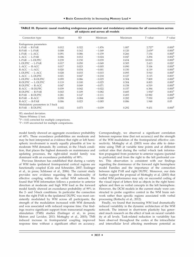
mental condition) are shown in Table III. All connectionsbetween the six areas of the WM network were signifi-cant. BMA parameter estimates of the endogenous con-nections were found to be significantly consistent acrossparticipants. The task condition significantly modulatedthe forward connection from the R-PAR to the R-DLPFC(Table III).
Behavioral correlations
Based on the robust modulation of the connection fromthe R-PAR to the R-DLPFC by increasing memory load,we performed correlations between the DCM parametersfor this connection in the 3-back condition with responsetime and accuracy. The WM modulation of this connectionnegatively correlated with response time (r 5 20.331,P 5 0.04) but not accuracy in the 3-back condition.
As expected, there were more errors in the 3-back thanthe 2-back condition. To examine the effect of perform-ance, we compared high performers (upper quartile;
Figure 2.
(A) Image showing task-related brain activation in the group
(N 5 40) during the working memory n-back task (FWE, cor-
rected at P< 0.05 on a voxelwise basis, minimum cluster size 20
voxels; image created with MRIcron http://www.mccauslandcen-
ter.sc.edu/mricro/mricron/index.html). (B) The effect of the
1-, 2-, and 3-back conditions on the left and right parietal cortex
(PAR), the left and right anterior cingulate cortex (ACC), and
left and right dorsolateral prefrontal cortex (DLPFC) in healthy
participants (N 5 40).
r Brain Connectivity in Increasing Memory Load r
r 3029 r
n 5 5), to low performers (bottom quartile; n 5 6). Theresults remained the same, as both high and low perform-ers preferred Model 16 with an exceedance proba-bility> 92%, where the 3-back memory load is modulatingsignificantly the forward connection from the R-PAR tothe R-DLPFC.
DISCUSSION
To our knowledge, this is the first study to assess effectiveconnectivity during WM processing using DCM. DCM iscurrently one of the most plausible methods for inferringneuroplastic changes in the strength of connections betweenneuronal populations (Stephan et al., 2010). There are threekey findings from our study. First, we demonstrated thatincreasing memory load was associated with increasing
dominance of right-hemisphere models suggesting greaterright hemisphere contribution to the most demanding WMconditions. Second, increasing memory load was associatedwith increased forward (i.e., posterior to anterior) effectiveconnectivity within the WM network. Third, increasingmemory load dramatically reduced individual variability inWM network architecture with the right-hemisphere for-ward model reaching an exceedance probability of 99% inthe 3-back condition.
Early fMRI studies have lent support to the notion thatprocessing within the WM network is left-lateralized forverbal and right-lateralized for spatial material (Fletcherand Henson, 2001). Recent quantitative meta-analyses haveshown that engagement during verbal WM tasks can beseen bilaterally within the DLPFC, PAR, and ACC (Wagerand Smith, 2003; Nee et al., 2013), although left-sidedinvolvement was more common (Nee et al., 2013).
TABLE II. Voxel-based whole brain SPM analysis: brain regions showing significant main effects in terms of hemody-
namic responses to different working memory loads; 1, 2, and 3 back (P < 0.05, FWE cluster-level corrected across
the whole brain with minimum cluster size 20 voxels)
Brain region BA Laterality
Coordinates
Cluster size (voxels) Z valuex y z
1 back> 0 backInferior parietal lobule 40 R 46 246 44 121 6.61
40 L 244 242 38 754 6.31Middle frontal gyrus
(dorsolateral PFC)46 L 246 32 30 153 6.42
R 52 36 30 256 6.30Middle frontal gyrus 6 R 32 6 62 47 5.972 back> 0 backInferior parietal lobule 40 R 40 248 44 231 7.90
L 236 252 46 190 7.75Middle frontal gyrus
(dorsolateral PFC)46 R 46 32 28 544 7.03
9 L 242 8 28 968 6.64Anterior cingulate gyrus 32 R 8 18 48 354 6.88
L 24 10 58Insula 13 R 34 24 22 151 6.47Middle frontal gyrus 6 R 30 8 58 414 6.123 back> 0 backInferior parietal lobule 40 R 50 242 42 287 8.01
L 248 248 48 263 7.98Middle frontal gyrus
(dorsolateral PFC)9 L 248 26 30 800 8
46 R 48 40 30 542 7.99Anterior cingulate gyrus 32 L 210 26 30 333 7.53
R 8 20 28Insula 13 L 234 22 0 680 6.99Thalamus N/A L 212 210 6 142 6.44Inferior frontal gyrus 10 L 244 46 2 319 5.98Inferior frontal gyrus 47 R 34 20 0 572 5.67
MNI coordinates denote the distance in mm from the anterior commissure, with positive x 5 right of midline, positive y 5 anterior tothe anterior commissure, and positive z 5 dorsal to a plane containing both the anterior and the posterior commissures. BA, Brodmannarea; L, left; R, right; N/A, not applicable; PFC, prefrontal cortex; SPM, Statistical Parametric Mapping.
r Dima et al. r
r 3030 r
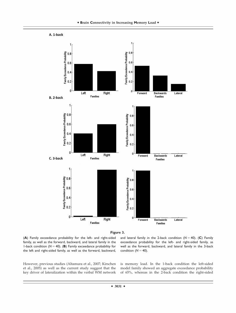
However, previous studies (Altamura et al., 2007; Kirschenet al., 2005) as well as the current study suggest that thekey driver of lateralization within the verbal WM network
is memory load. In the 1-back condition the left-sidedmodel family showed an aggregate exceedance probabilityof 65%, whereas in the 2-back condition the right-sided
Figure 3.
(A) Family exceedance probability for the left- and right-sided
family, as well as the forward, backward, and lateral family in the
1-back condition (N 5 40). (B) Family exceedance probability for
the left and right-sided family, as well as the forward, backward,
and lateral family in the 2-back condition (N 5 40). (C) Family
exceedance probability for the left- and right-sided family, as
well as the forward, backward, and lateral family in the 3-back
condition (N 5 40).
r Brain Connectivity in Increasing Memory Load r
r 3031 r
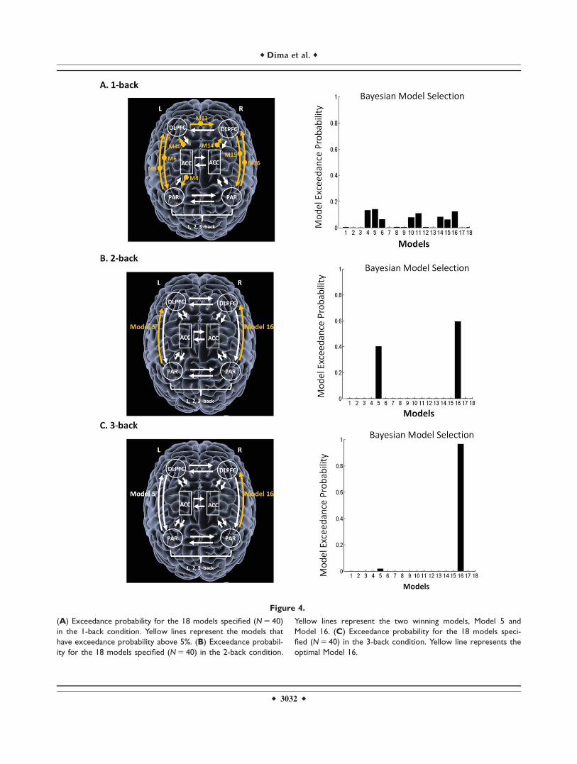
Figure 4.
(A) Exceedance probability for the 18 models specified (N 5 40)
in the 1-back condition. Yellow lines represent the models that
have exceedance probability above 5%. (B) Exceedance probabil-
ity for the 18 models specified (N 5 40) in the 2-back condition.
Yellow lines represent the two winning models, Model 5 and
Model 16. (C) Exceedance probability for the 18 models speci-
fied (N 5 40) in the 3-back condition. Yellow line represents the
optimal Model 16.
r Dima et al. r
r 3032 r
model family showed an aggregate exceedance probabilityof 60%. These exceedance probabilities are moderate andmay be best interpreted as evidence that right or left hemi-spheric involvement is nearly equally plausible at low tomoderate WM demands. By contrast, in the 3-back condi-tion, that places the highest demands on maintenance andupdating processes, the right-sided model family wasdominant with an exceedance probability of 98%.
Previous literature has established that during a varietyof WM tasks ipsilateral frontoparietal cortical regions arefunctionally coupled (Cole and Schneider, 2007; Esslingeret al., in press; Schlosser et al., 2006). The current studyprovides new evidence regarding the directionality ofeffective coupling within the verbal WM network. Wefound that WM information follows a posterior to anteriordirection at moderate and high WM load as the forwardmodel family showed an exceedance probability of 99% inthe 2- and 3-back conditions. In particular the connectionfrom the right PAR to the ipsilateral DLPFC was most con-sistently modulated by WM across all participants; thestrength of the modulation increased with WM demandsand was associated with reduced response time. Our find-ings receive significant support from transcranial magneticstimulation (TMS) studies (Esslinger et al., in press;Meiron and Lavidor, 2013; Mottaghy et al., 2003); TMSinduced increase in frontoparietal coupling improvedresponse time without a significant effect on accuracy.
Correspondingly, we observed a significant correlationbetween response time (but not accuracy) and the strengthof the WM modulation of the frontoparietal effective con-nectivity. Mottaghy et al. (2003) were also able to deter-mine using TMS at variable time points and at differentcortical sites that during the verbal n-back task informa-tion propagated from posterior to anterior regions (parietalto prefrontal) and from the right to the left prefrontal cor-tex. This observation is consistent with our findingsregarding the dominance of the forward right hemispheremodel Families and the importance of the connectionbetween right PAR and right DLPFC. Moreover, our datafurther support the proposal of Mottaghy et al. (2003) thatverbal WM performance may rely on successful coding ofthe visual input of letters first as objects in the right hemi-sphere and then as verbal concepts in the left hemisphere.However, the DCM models in the current study were con-structed to probe cognitive control in the WM brain net-work rather than specific regions associated with verbalprocessing (Rottschy et al., 2012).
Finally, we found that increasing WM load dramaticallyreduced variability in the dynamic architecture of the WMnetwork. The interest in short-term plasticity has gener-ated much research on the effect of task on neural variabil-ity at all levels. Task-related reduction in variability hasbeen observed throughout the cortex at the intracellularand intercellular level affecting membrane potential as
TABLE III. Dynamic causal modeling endogenous parameter and modulatory estimates for all connections across
all subjects and across all models
Connection type Mean SD Minimum Maximum T value P value
Endogenous parametersL-PAR ! R-PAR 0.012 0.322 21.876 1.007 2.727a 0.000b
R-PAR ! L-PAR 0.008 0.162 21.049 0.120 2.639a 0.000b
L-PAR ! L-ACC 0.091 0.086 20.159 0.266 7.154 0.000b
L-ACC ! L-PAR 0.006 0.012 20.024 0.047 3.294 0.002b
L-PAR ! L-DLPFC 0.239 0.150 20.039 0.654 10.818 0.000b
L-DLPFC ! L-PAR 0.017 0.050 20.049 0.305 2.413 0.020c
L-ACC ! R-ACC 0.017 0.023 20.005 0.090 5.106 0.000b
R-ACC ! L-ACC 0.020 0.034 20.001 0.150 4.119 0.000b
L-DLPFC ! L-ACC 0.028 0.033 20.015 0.095 5.910 0.000b
L-ACC ! L-DLPFC 0.021 0.047 20.218 0.127 3.115 0.003c
L-DLPFC ! R-DLPFC 0.099 0.086 20.025 0.304 7.809 0.000b
R-DLPFC ! L-DLPFC 0.119 0.100 20.025 0.304 8.003 0.000b
R-DLPFC ! R-ACC 0.047 0.049 20.071 0.158 6.519 0.000b
R-ACC ! R-DLPFC 0.039 0.042 20.022 0.157 6.384 0.000b
R-DLPFC ! R-PAR 0.043 0.109 20.082 0.685 1.936a 0.001b
R-PAR ! R-DLPFC 0.281 0.147 20.111 0.493 12.982 0.000b
R-PAR ! R-ACC 0.098 0.084 20.048 0.282 7.930 0.000b
R-ACC ! R-PAR 0.006 0.023 20.085 0.086 1.948 0.05Modulatory parameters in 3 backR-PAR ! R-DLPFC 0.102 0.073 20.039 0.292 9.431 0.000b
SD, standard deviation.aMann–Whitney U test.bP< 0.02 corrected for multiple comparisons.cP< 0.05 uncorrected for multiple comparisons.
r Brain Connectivity in Increasing Memory Load r
r 3033 r
well as individual and correlated neuronal firing (Church-land et al., 2010). Theoretical models have suggested simi-lar properties for large-scale neural networks (Rajan et al.,2010; Sussillo and Abbott, 2009). Our results provide directempirical support for these models with regard to WMand further suggest that task difficulty is a significantadditional moderator of short-term plasticity.
REFERENCES
Altamura M, Elvevag B, Blasi G, Bertolino A, Callicott JH,Weinberger DR, Mattay VS, Goldberg TE (2007): Dissociatingthe effects of Sternberg working memory demands in prefron-tal cortex. Psychiatry Res 154:103–114.
Axmacher N, Schmitz DP, Wagner T, Elger CE, Fell J (2008): Inter-actions between medial temporal lobe, prefrontal cortex andinferior temporal regions during visual working memory: Acombined intracranial EEG and functional magnetic resonanceimaging study. J Neurosci 28:7304–7312.
Baddeley A (1992): Working memory: The interface betweenmemory and cognition. J Cogn Neurosci 4:281–288.
Braver TS, Cohen JD, Nystrom LE, Jonides J, Smith EE, Noll DC(1997): A parametric study of prefrontal cortex involvement inhuman working memory. Neuroimage 5:49–62.
Callicott JH, Mattay VS, Bertolino A, Finn K, Coppola R, FrankJA, Goldberg TE, Weinberger DR (1999): Physiological charac-teristics of capacity constraints in working memory as revealedby functional MRI. Cereb Cortex 9:20–26.
Carter CS, Botvinick MM, Cohen JD (1999): The contribution ofthe anterior cingulate cortex to executive processes in cogni-tion. Rev Neurosci 10:49–57.
Churchland MM, Yu BM, Cunningham JP, Sugrue LP, Cohen MR,Corrado GS, Newsome WT, Clark AM, Hosseini P, Scott BB,Bradley DC, Smith MA, Kohn A, Movshon JA, ArmstrongKM, Moore T, Chang SW, Snyder LH, Lisberger SG, Priebe NJ,Finn IM, Ferster D, Ryu SI, Santhanam G, Sahani M, ShenoyKV (2010): Stimulus onset quenches neural variability: A wide-spread cortical phenomenon. Nat Neurosci 13:369–378.
Cole MW, Schneider W (2007): The cognitive control network:Integrated cortical regions with dissociable functions. Neuro-image 37:343–360.
Cramer SC, Sur M, Dobkin BH, O’Brien C, Sanger TD, TrojanowskiJQ, Rumsey JM, Hicks R, Cameron J, Chen D, Chen WG, CohenLG, deCharms C, Duffy CJ, Eden GF, Fetz EE, Filart R, FreundM, Grant SJ, Haber S, Kalivas PW, Kolb B, Kramer AF, LynchM, Mayberg HS, McQuillen PS, Nitkin R, Pascual-Leone A,Reuter-Lorenz P, Schiff N, Sharma A, Shekim L, Stryker M,Sullivan EV, Vinogradov S (2011): Harnessing neuroplasticity forclinical applications. Brain 134:1591–1609.
D’Esposito M, Postle BR, Rypma B (2000): Prefrontal cortical con-tributions to working memory: Evidence from event-relatedfMRI studies. Exp Brain Res 133:3–11.
Esslinger C, Sch€uler N, Sauer C, Gass D, Mier D, Braun U, OchsE, Schulze TG, Rietchel M, Kirsch P, Meyer-Lindenberg A:Induction and quantification of prefrontal cortical networkplasticity using 5 Hz rTMS and fMRI. Hum Brain Mapp (inpress).
First MB, Spitzer RL, Gibbon M, Williams JBW. 2002. Structured
Clinical Interview for DSM-IV Axis I Disorders, Research Ver-
sion, Non-patient Edition (SCID-I/NP). New York, NY: Bio-
metrics Research, New York State Psychiatric Institute.
Fletcher PC, Henson RN (2001): Frontal lobes and human mem-ory: Insights from functional neuroimaging. Brain 124: 849–881.
Friston KJ, Harrison L, Penny WD (2003): Dynamic causal model-ing. Neuroimage 19:1273–1302.
Glahn DC, Ragland JD, Abramoff A, Barrett J, Laird AR, BeardenCE, Velligan DI (2005): Beyond hypofrontality: A quantitative
meta-analysis of functional neuroimaging studies of working
memory in schizophrenia. Hum Brain Mapp 25:60–69.Guerin SA, Miller MB (2011): Parietal cortex tracks the amount of
information retrieved even when it is not the basis of a mem-ory decision. Neuroimage 55: 801–807.
Honey GD, Fu CH, Kim J, Brammer MJ, Croudace TJ, Suckling J,Pich EM, Williams SC, Bullmore ET (2002): Effects of verbalworking memory load on corticocortical connectivity modeledby path analysis of functional magnetic resonance imagingdata. Neuroimage 17:573–582.
Jonides J, Schumacher EH, Smith EE, Koeppe RA, Awh E, Reuter-
Lorenz PA, Marshuetz C, Willis CR (1998): The role of parietal
cortex in verbal working memory. J Neurosci 18:5026–5034.Kim J, Zhu W, Chang L, Bentler PM, Ernst T (2007): Unified struc-
tural equation modeling approach for the analysis of multisub-ject, multivariate functional MRI data. Hum Brain Map 28:85–93.
Kirschen MP, Chen ASH, Schraedley-Desmond P, Desmond JE
(2005): Load- and practice-dependent increases in cerebro-
cerebellar activation in verbal working memory: An fMRI
study. Neuroimage 24:462–472.
Maldjian JA, Laurienti PJ, Kraft RA, Burdette JH (2003): An auto-
mated method for neuroanatomic and cytoarchitectonic atlas-
based interrogation of fMRI data sets. Neuroimage 19:1233–
1239.Meiron O, Lavidor M (2013): Unilateral prefrontal direct current
stimulation effects are modulated by working memory load
and gender. Brain Stimul 6:440–447.Mottaghy FM, Gangitano M, Krause BJ, Pascual-Leone A (2003):
Chronometry of parietal and prefrontal activations in verbal
working memory revealed by transcranial magnetic stimula-
tion. Neuroimage 18:565–575.
Narayanan NS, Prabhakaran V, Bunge SA, Christoff K, Fine EM,
Gabrieli JDE (2005): The role of the prefrontal cortex in the
maintenance of verbal working memory: An event-related
fMRI analysis. Neuropsychology 19:223–232.Nee DE, Brown JW, Askren MK, Berman MG, Demiralp E,
Krawitz A, Jonides J (2013): A meta-analysis of executive com-ponents of working memory. Cereb Cortex 23:264–282.
Owen AM, Herrod NJ, Menon DK, Clark JC, Downey SPMJ,Carpenter TA, Minhas PS, Turkheimer FE, Williams EJ,Robbins TW, Sahakian BJ, Petrides M, Pickard JD (1999): Rede-fining the functional organization of working memory proc-esses within human lateral prefrontal cortex. Eur J Neurosci11:567–574.
Owen AM, McMillan KM, Laird AR, Bullmore ET (2005): N-back working memory paradigm: A meta-analysis of norma-tive functional neuroimaging studies. Hum Brain Mapp 25:46–59.
Penny WD, Stephan KE, Daunizeau J, Rosa MJ, Friston KJ,Schofield TM, Leff AP (2010): Comparing families of dynamiccausal models. PLoS Comput Biol 6:1–14.
Rajan K, Abbott LF, Sompolinsky H (2010): Stimulus-dependentsuppression of chaos in recurrent neural networks. Phys Rev EStat Nonlin Soft Matter Phys 82:011903.
r Dima et al. r
r 3034 r
Ramnani N, Behrens T, Penny WD, Matthews PM (2004): Newapproaches for exploring anatomical and functional connectiv-ity in the human brain. Biol Psychiatry 56:613–619.
Rottschy C, Langner R, Dogan I, Reetz K, Laird AR, Schulz JB,Fox PT, Eickhoff SB (2012): Modelling neural correlates ofworking memory: A coordinate-based meta-analysis. Neuro-image 60:830–846.
Salinas E, Sejnowski TJ (2001): Gain modulation in the centralnervous system: Where behavior, neurophysiology, and com-putation meet. Neuroscientist 7:430–440.
Sala-Llonch R, Pe~na-G�omez C, Arenaza-Urquijo EM, Vidal-Pi~neiroD, Bargall�o N, Junque Carme, Bartres-Faz D (2012): Brain con-nectivity during resting state and subsequent working mem-ory task predicts behavioural performance. Cortex 48:1187–1196.
Schlosser RG, Wagner G, Sauer H (2006): Assessing the workingmemory network: Studies with functional magnetic resonanceimaging and structural equation modeling. Neuroscience 139:91–103.
Stephan KE, Baldeweg T, Friston KJ (2006): Synaptic plasticity anddysconnection in schizophrenia. Biol Psychiatry 59:929–939.
Stephan KE, Penny WD, Daunizeau J, Moran RJ, Friston KJ (2009):Bayesian model selection for group studies. Neuroimage 15:1004–1017.
Stephan KE, Penny WD, Moran RJ, den Ouden HEM, DaunizeauJ, Friston KJ (2010): Ten simple rules for dynamic causal mod-eling. Neuroimage 49:3099–3109.
Sussillo D, Abbott LF (2009): Generating coherent patterns ofactivity from chaotic neural networks. Neuron 63:544–557.
Volle E, Kinkingn�ehun S, Pochon JB, Mondon K, Thiebaut deSchotten M, Seassau M, Duffau H, Samson Y, Dubois B, LevyR (2008): The functional architecture of the left posterior andlateral prefrontal cortex in humans. Cereb Cortex 18:2460–2469.
Wager TD, Smith EE (2003): Neuroimaging studies of workingmemory: A meta-analysis. Cogn Affect Behav Neurosci 3:255–274.
Wechsler D. 1981. Manual for the Wechsler Adult IntelligenceScale-Revised. New York, NY: Psychological Corporation.
r Brain Connectivity in Increasing Memory Load r
r 3035 r
Related Documents