This electronic thesis or dissertation has been downloaded from the King’s Research Portal at https://kclpure.kcl.ac.uk/portal/ Take down policy If you believe that this document breaches copyright please contact [email protected] providing details, and we will remove access to the work immediately and investigate your claim. END USER LICENCE AGREEMENT Unless another licence is stated on the immediately following page this work is licensed under a Creative Commons Attribution-NonCommercial-NoDerivatives 4.0 International licence. https://creativecommons.org/licenses/by-nc-nd/4.0/ You are free to copy, distribute and transmit the work Under the following conditions: Attribution: You must attribute the work in the manner specified by the author (but not in any way that suggests that they endorse you or your use of the work). Non Commercial: You may not use this work for commercial purposes. No Derivative Works - You may not alter, transform, or build upon this work. Any of these conditions can be waived if you receive permission from the author. Your fair dealings and other rights are in no way affected by the above. The copyright of this thesis rests with the author and no quotation from it or information derived from it may be published without proper acknowledgement. Biologically active constituents of chrysanthemum parthenium. Jessup, Deborah Margaret Download date: 26. Jul. 2022

Welcome message from author
This document is posted to help you gain knowledge. Please leave a comment to let me know what you think about it! Share it to your friends and learn new things together.
Transcript
This electronic thesis or dissertation has been
downloaded from the King’s Research Portal at
https://kclpure.kcl.ac.uk/portal/
Take down policy
If you believe that this document breaches copyright please contact [email protected] providing
details, and we will remove access to the work immediately and investigate your claim.
END USER LICENCE AGREEMENT
Unless another licence is stated on the immediately following page this work is licensed
under a Creative Commons Attribution-NonCommercial-NoDerivatives 4.0 International
licence. https://creativecommons.org/licenses/by-nc-nd/4.0/
You are free to copy, distribute and transmit the work
Under the following conditions:
Attribution: You must attribute the work in the manner specified by the author (but not in anyway that suggests that they endorse you or your use of the work).
Non Commercial: You may not use this work for commercial purposes.
No Derivative Works - You may not alter, transform, or build upon this work.
Any of these conditions can be waived if you receive permission from the author. Your fair dealings and
other rights are in no way affected by the above.
The copyright of this thesis rests with the author and no quotation from it or information derived from it
may be published without proper acknowledgement.
Biologically active constituents of chrysanthemum parthenium.
Jessup, Deborah Margaret
Download date: 26. Jul. 2022

BIOLOGICALLY ACTIVE CONSTITUENTS
of
CHRYSANTHEMUM PARTHENIUM
THESIS by DEBORAH MARGARET IESSUPfor the degree ofDoctor of Philosophyin the University of London
liily 1982
Pharniacognosy Research LaboratoriesDepsrtent of PharmacyChelsea CollegeUniversity of LondonLondon S'W3 6LX
ABSTRACT
The use of the leaves of Chrysanthemum parthenium in the prophylaxisof migraine has received much recent publicity. In view ofwidespread consumption of the plant with apparently efffectiveresults it was considered desirable to investigate this claim.
The present work thus involved successive extraction of freeze—driedleaves with light petroleum, chloroform, methanol and water. Each ofthese extracts was separately tested for spasmoltyic activity usingan j vitro preparation of guinea pig ileum. Agonists used in thetest were acetylcholine, 5—hydroxytryptamine and histamine. Thelight petroleum and chloroform extracts showed 100% inhibition of allthree agonists at a concentration of b 4 g/ml whereas the methanoland water extracts were devoid of activity. Successivechromatographic separation of the active extracts allowed isolationand purification of some active constituents. These all proved to besesquiterpene lactones, a class of secondary metabolite which iswidespread in the Compositae.
Some of these active materials were already known in the plant butothers, including the most active, are apparently new compounds. Thestructures of these materials have been elucidated by chemical andspectroscopic means, particularly hydrogen — i and carbon-13 nuclearmagnetic resonance spectroscopy. All the active substances may beconsidered as derivatives of parthenolide, the major germacranolideof the plant, but one (the most active) has a novel trimericstructure.
2

INDEX
Page
Abs tract
2
Index
3
List of Figures
6
List of Tables
8
Acknowledgements
11
Chrysanthemum parthenium
12
Foreward
13
PART I - INTRODUCTION
17
1 The family Conipositae
18
2 Secondary plant metabolites in the family Compositae
18
A Sesquiterpene lactones
18
3 Constituents found in Chrysanthemum parthenium
29
A Parthenolide
29
B Santamarine
38
C Chrysartemins A and B
41
4 Structural elucidation of sesquiterpene lactones
45
A NMR spectroscopy
45
B X—ray diffraction methods
57
5 Biological activity in the family Compositae
58
A Cytotoxic activity
58
B Spasmolytic activity
61
C Antiinflammatory activity
63
B Antibepatoxic and cholerectic activity
65
B Antimicrobial activity
67
3

F Antihyperlipidemic activity
69
G Insecticidal activity
70
6 Migraine
71
PART II - DISCUSSION
74
1 Screening procedure
75
2 Extraction procedure
79
3 mown compounds
83
A Parthenolido
83
B Chrysartemin A
85
4 Structural elucidation of D1156a
87
S Structural elucidation of D3177b
97
6 Structural elucidation of D1140a
107
7 Comments on D.1179a1 and D1179c
129
8 Comments on D1124a
130
9 The remaining fractions tested for spasmolytic activity
130
10 In vitro pharmacological studies on the efficacy of
132
Chrysanthemum parthenium in migraine
11 Preliminary toxicity studies
132
12 Clinical studies on the efficacy of Chrysanthemum
133
p arthenium in migraine
13 Relationship between structure and activity
136
14 Conclusion
137
PART III - EXPERIMENTAL
139
1 General details
140
2 Extraction of Chrysanthemum parthenium 'with light
142
petroleum (b.r. 40-60°C)
3 Spasmolytic activity of the light petroleum extract
142
4 Crude separation of the light petroleum extract
143
4
5 Spasmolytic activity of the combined fractions from the
143
light petroleum extract
6 Extraction of C. parthenium with chloroform
146
7 Spasmolytic activity of the chloroform extract
146
8 Crude separation of the chloroform extract
146
9 Spasmolytic activity of the combined fractions from the
146
chloroform extract
10 Separation of the chloroform extract on a larger scale
150
11 Extraction of C. parthenium with methanol
150
12 Spasmolytic activity of the methanol extract
150
13 Extraction of C. parthenium with water
154
14 Spasmolytic activity of the water extract
154
15 Studies on fractions 144-176 (A) from the light
154
petroleum extract
16 Studies on fractions 124-143 (B) from the light
165
petroleum extract
17 Studies on fractions 75-80 (C) from the light petroleum
171
extract
18 Studies on fractions 177-200 CD) from the light
171
petroleum extract
19 Studies on fractions 119-123 (E) from the light
177
petroleum extract
20 Studies on fractions 112-118 (F) from the light
180
petroleum extract
21 Studies on fractions 51-61 from the light petroleum
180
extract
22 Preparation of capsules for clinical studies
182
References
187
5

LIST OF FIGURES
Page
1 Four main types of sesquiterpene hydrocarbon
21
2 Simiplified sesquiterpene lactone biosynthesis
22
3 Possible sesquiterpene lactone biosynthesis. Proposal 1
22
4 Possible sesquiterpene lactone biosynthesis. Proposal 2
23
5 Possible biogenetic relationships of the different
25
skeletal types of sesquiterpene lactones
6 Cope rearrangement
26
7 Reactions used in the deductions about the positions of
32
the double bond and epoxide in
8 Summary of reactions leading to the structural
33
elucidation of 11
9 Conformation of parthenolide and costunolide
38
10 Summary of reactions leading to the structural
42
elucidation of santamarinc, 30
11 Partial structure of a sesquiterpene lactone containing
46
a C6 a,—unsaturated-7—lactone and an 8a—bydroxyl group
12 Conformations at C-6, C-7 and C-8 based on the distance
50
between the 8a—oxygen atom and K-13a as required to
account for the paramagnetic shifts in 6a—lactonised 8a-
hydroxy sesquiterpene lactones
13 Four major conformational forms of germacranolides
52
14 Conformations of dihydrotamaulipin A acetate
53
15 Two conformational forms of isabelin
56
16 Michael—type addition of an a—methylene— r—lactone with
61
cysteine
17 Rypothetical model of cytostatic action of sesquiterpene
62
lactones
18 Production of chamazulene during steam distillation of
64
chamomile oil
6
19 Spasmolytic activity of sub—fractions A59-65 tested at
77
io g/ml
20 Spasmolytic activity of sub —fractions A30-34 tested at
78
io g/ml
21 Extraction and isolation procedure of the light
80
petroleum extract of Chrysanthemum parthenium
22 Spasmolytic activity and percentage of weight of the
81
fractions obtained from column chromatography of the
light petroleum extract of Chrysanthemum parthenium
23 Possible biosynthetic route to D.1156a
94
24 Stereochemical possibilities for the new guianolide
95
DJ156a
25 Structure of chrysartemin A showing the epoxide
100
hydrogens and their chemical shift in the 'H NMR
spectrum (6, CDC13)
26 Structure of dehydrocostuslactone showing the chemical
101
shifts of the exomethylene hydrogens
27 Possible part structures for Dfl77b
102
28 Possible part structure for D1177b
104
29 Possible biosynthetic route to D1177b
108
30 Relactonisation of C-6 lactonised sesquiterpenes with a
112
C-8 ester or hydroxyl group in the presence of strong
base
31 Expanded 'H NMR spectrum of DI14OaH2 showing clear
114
geminal coupling of the C-13' hydrogens but no such
coupling of the C-13 hydrogens
32 Possible methods of linkage formation in DJ14OaR2
118
33 Proposed mechanism of the reaction resulting in DJ140aH2
119
34 Mass spectral behaviour of DJ14OaH2
122
35 Proposed stereochemistry of D1140aK2
123
36 NMR spectrum of Dfl4Oa (6, CDC13)
124
37 NMR spectrum of D.1140a112 (6, CDCl)
125
7

LIST OF TABLES
1 Distribution of sesquiterpene lactones in the plant
kingdom
2 Distribution of sesquiterpene lactones in the Compositae
3 Calculated H-13a paramagnetic shifts based on different
distances between H-13a and C-8 oxygen atoms
4 Observed H-13a paramagnetic shifts and measured
distances between H-13a and C-8 oxygen atoms using
probable conformations of some sesquiterpene lactones
5 m data for parthenolide (6, CDC13)
6 13 C NI4R data for D1156a (6, CDC13)
7 Selected H NMR signals of D1140a H2, D1156a, D1177b and
parthenolide (6, CDC13)
8 Selected 'H NMR signals of D1140a and DI14OaK2 (6,
CDC1 3)
9 Solvent elution of the column chromatography of the
light petroleum extract
10 Weights of combined fractions from the column
chromatography of the light petroleum extract
11 Spasmolytic activity of the combined fractions from the
light petroleum extract, tested at 1O 4 g/ml
12 Solvent elution of the column chromatography of the
chloroform extract
13 Weight of combined fractions of the column
chromatography of the chloroform extract
14 Spasmolytic activity of the combined fractions from the
chloroform extract, tested at ,O g/ml
15 Solvent elut ion of the column chromatography of the
chloroform extract
16 Weights of combined fractions of the column
chromatography of the chloroform extract
17 Solvent elution of the column chromatography of
fractions 144-176 (A) from the light petroleum extract
Page
20
28
47
48
86
93
113
128
144
145
147
148
149
151
152
153
155
8
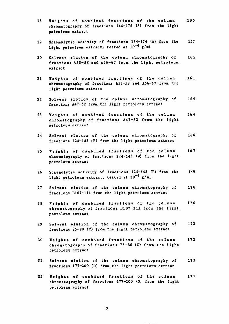
18 Weights of combined fractions of the column 155chromatography of fractions 144-176 (A) from the lightpetroleum extract
19 Spasmolytic activity of fractions 144-176 (A) from the 157light petroleum extract, tested at 1O • '4 g/ml
20 Solvent elution of the column chromatography of 161fractions A53-58 and A66-67 from the light petroleumextract
21 Weights of combined fractions of the column 161chromatography of fractions A53-58 and A66-67 from thelight petroleum extract
22 Solvent elution of the column chromatography of 164fractions A47-57 from the light petroleum extract
23 Weights of combined fractions of the column 164chromatography of fractions A47-52 from the lightpetroleum extract
24 Solvent elution of the column chromatography of 166fractions 124-143 (B) from the light petroleum extract
25 Weights of combined fractions of the column 167chromatography of fractions 124-143 (B) from the lightpetroleum extract
26 Spasmolytic activity of fractions 124-143 (B) from the 169light petroleum extract, tested at io g/ml
27 Solvent elution of the column chromatography of 170fractions B107-111 from the light petroleum extract
28 Weights of combined fractions of the column 170chromatography of fractions B107-111 from the lightpetroleum extract
29 Solvent elution of the column chromatography of 172fractions 75-80 (C) from the light petroleum extract
30 Weights of combined fractions of the column 172chromatography of fractions 75-80 (C) from the lightpetroleum extract
31 Solvent elution of the column chromatography of 173fractions 177-200 (D) from the light petroleum extract
32 Weights of combined fractions of the column 173chromatography of fractions 177-200 (D) from the lightpetroleum extract
9
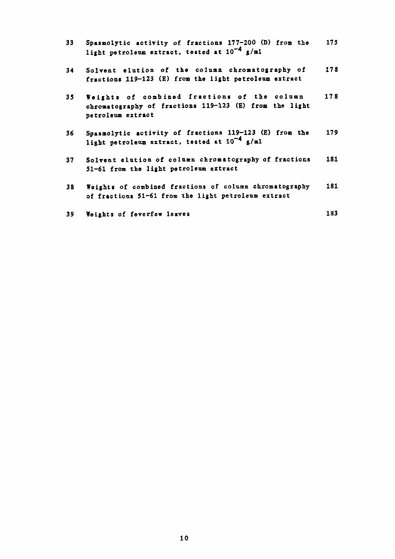
33 Spasmolytic activity of fractions 177-200 (D) from the 175
light petroleum extract, tested at io g/ml
34 Solvent elution of the column chromatography of 178
fractions 119-123 (E) from the light petroleum extract
35 Weights of combined fractions of the column 178
chromatography of fractions 119-123 (E) from the light
petroleum extract
36 Spasmolytic activity of fractions 119-123 (E) from the 179
light petroleum extract, tested at io g/ml
37 Solvent elution of column chromatography of fractions 181
51-61 from the light petroleum extract
38 Weights of combined fractions of column chromatography 181
of fractions 51-61 from the light petroleum extract
39 Weights of feverfew leaves 183
10

ACKNOWLEDGEMENTS
I am greatly indebted to the following people: —
Dr P 1 Hylands, Chelsea College, London, for his supervision,
advice and encouragement
Dr E S Johnson, Kings College, London, for help with the
pharmacological work, valuable discussion and the clinical work
The technical staff of the Pharmacognosy Department, Chelsea
College, London, for all their help
Dr G Hawles, Queen Mary College, London, for 400 MHz ½ NMR and
100 z ii spectra
Mr G McDonough, Chelsea College, London, for 200 MHz NMR
spectra
The staff of the mass spectrometry unit, Chelsea College, London,
and Mr D Carter of the London School of Pharmacy, London, for mass
spectra
Chelsea College for my research grant
The Curator, The Chelsea Physic Garden for the plant material
My parents and Peter for their constant encouragement.
11

FOREWORD
In 1978 and 1979 several articles appeared in the national and
provincial press about the efficacy of Chr ysanthemum parthenium in
migraine. 4 This was not a new discovery.
Chrysanthemum parthenium Bernh. belongs to the tribe Anthemideae of
the family Compositae. It is found throughout Europe both wild and
cultivated in and near gardens, walls and rivers. It is a perennial
plant growing to a height of 14 to 45 cm with strong—smelling,
greenish—yellow, bipinnate leaves. 5 The flower—heads are arranged in
a loose terminal coryab the central disc florets being yellow and the
outer ray florets white. A double variety is usually cultivated in
gardens for ornamental purposes.6&
Feverfew, to give the plant its common English name, is perhaps a
corruption of featherfew (relating to the form of the leaves) or more
attractively as far as the present study is concerned, of the word
febrifuge meaning that which dispels fever.516& One could expect
therefore to find folk lore use of the plant in cases of fever.
Richard Banckes, writing in his herbal of 1525 considered it
indispensable. He said it was,
'Good to assuage the access (ague or fever), quotidian (feverrecurring daily) or cramp'.7
Iohn Gerarde, in 1597, said of the plant,
'It is used both in drinks, and bound to the wrists with baysalt and the powder of glasse stamped together, as a mostsingular experiment against the ague'.
It is similarly referred to fifty years later by John Parkinson 9 and
later by Nicholas Culpeper)0
A very similar plant, however, Anthemis nobilis L., or Roman
13

chamomile, is mentioned more frequently as a febrifuge and,
particularly as far as this study is concerned, as an antispasmodic
as well as in migraine.6b,il Unlike feverfew, there is no record of
current use of this plant in migraine and so it could be that these
plants have been confused in the past. In fact, the 1934 British
Pharmaceutical Codex 2 states that feverfew could be and often was
substituted for the Roman chamomile. However, one can find
references in the literature which allude to the usefulness of
feverfew in headache and migraine.6'84°'13
John Gerarde says,
'Feverfew dried and made into powder, and two drams of it takenwith honie or sweet wine purgeth by siege melancholic andflegme; wherefore it is very good for them that are giddie inthe head, or which have the turning called vertigo, that is aswimming and turning in the head'.8
Parkinson 9 and Culpeper 10 only mention . p arthenium being used
externally for headache, for example,
'It is very effectual for all paines in the head, coming of acold, caufe, as Camerarius faith, the hearbe being bruised andapplied to the crowne of the head'.9
More recently feverfew is mentioned as being useful in hysterical
complaints and in allaying sensitiveness to pain in highly nervous
subjects.6al3 This may be of some relevance since migraine
sufferers are frequently of a nervous disposition.
The plant is quoted repeatedly as having some action on the female
reproductive systeis, 615 most frequently being said to expel the
placenta and still —born children and to induce abortion. Thus,
Gerarde says,
'it procureth womens sickness with speed; it bringeth forth theafterbirth, and the dead childe, whether it be drunke in thedecoction or boiled in a bath and the woman sit over it'.8
14

It is also reported to cause abortion in cows.16
As well as the current use of C. parthenium in migraine it has been
found to be beneficial to many sufferers of rheumatoid
arthritis. 1 ' 2 "7 Gerarde reports,
'Dioscorides also teacheth that it is profitable applied toSaint Antonies fire, to all inflamstion and hot swellings'.8
and Margaret Grieve6a that it gives relief to the face — ache or
earache of a rheumatic person.
In common with many medicinal plants feverfew was also used as a
tonic, 6a , 4 this probably being due to the presence of extremely
bitter sesquiterpene lactones.
Other uses are, as an expectorant, 8 '° for the prevention of insect
bites, 6a , fl for the removal of freckles 9 ' 1 ° and 'an especiall remedy
against opium, that is taken too liberally'.9'10
Interest in feverfew was suddenly reawakened in 1978 in Wales.4
Mrs Ann Jenkins of Cardiff, wife of the National Coal Board's Chief
Medical Officer, had suffered with severe migraine from her teens.
Conventional treatment was not successful in her case but in 1974 an
elderly Welsh miner heard of Mrs Jenkin's plight and sent her a clump
of the plant with instructions to eat some of the leaves every day.
After 6 months she did not have a headache for a whole month.
Previously she had had attacks every 10 days. After 14 months her
headaches had stopped and to date have not reappeared (8 years). Mrs
Jenkins was so impressed she told friends who suffered from migraine
about the plant. It is now believed that many thousands of people in
this country are taking feverfew for migraine and perhaps the same
number, if not more, are taking it for arthritis. 1 ' 2 " 7 Indeed Mrs
Jenkins, who is mainly responsible for the recent widespread use of
15
the plant found her rheumatic pains, which had previously made it
agonising for her to drive a car, had also disappeared. The case
history of Mrs Jenkins looks to be typical of those patients deriving
benefit from the plant. The effects take several months to appear.
Gradually the frequency and severity of the headaches diminish and
after some time may disappear altogether.
In view of the possible large scale consumption of feverfew in this
country a systematic investigation of the plant was thought to be
worthwhile on two counts. Firstly, it seems from patient reports
that feverfew is effective and therefore identification and isolation
of the active constituents of the plant is desirable. Secondly, it
was important to try to establish any possible toxic side-effects for
which the plant may be responsible. It has already been reported
that mouth ulcers occur 3 ' 17 '' 8 but long term kidney and liver
function and blood tests had not been performed on patients taking
the plant.
16

PART I
INTRODUCTION

1 THE FAMILY COMPOSITAE
The family Compositae contains nearly 1000 genera and about 15000
species. They are divided into two sub — families, Tubuliflorae and
Liguliflorae, and 13 tribes, as shown below:'9
A Tubuliflorae
1 Vernonieae
2 Eupatorieae
3 Astereae
4 Inuleae
5 Heliantheae
6 Helenieae
7 Anthemideae
8 Senecioneae
9 Calenduleae
10 Arctotideae
11 Cynareae
12 Mutiseae
B Liguliflorae
13 Cichorieae
2 SECONDARY PLANT METABOLITES IN THE FAMILY COMPOSITAE
The family Compositae is chemically extremely diverse. The combined
occurrence of sesquiterpene lactones, acetylenic compounds and
inulin—type fructans is almost as characteristic of the Compositae as
their capitula inflorescences. However, triterpenes and flavonoids
seem to be present in every member and seed oils sometimes contain
characteristic fatty acids. Large amounts of derivatives of caffeic
acid are known to occur as well as cyclitols, iridoid glycosides,
alkaloids, diterpenes, cyanogenic glycosides, essential oils,
coumarins and several types of phenolic constituents.2°
A SESQUITERPENE LACTONES
Two classes of secondary metabolites seem to have been selected
for special consideration namely the polyacetylenes, about which
18
much is written,2 ' and the sesquiterpene lactones which are of
more recent in t eres t.22a Increased appearance of this second
class is undoubtedly due to developments in instrumentation
leading to easier structural elucidation of the compounds. In
1960 barely a dozen naturally occurring sesquiterpenes had been
elucidated whereas today more than 600 compounds are known and
the pace of their discovery is quickening all the tiine.22b
To date the vast majority of sesquiterpene lactones belong to
the Compositae but this may stem from the intensity with which
this family and certain genera in particular such as Artemisia,
Ambrosia, Relenium and Vernonia have been examined.
Nevertheless the incidence of sesquiterpene lactones in the
Compositae is unusually high (Table 1) and their appearance in
other families may be attributed to parallelism. Moreover, the
distribution of sesquiterpene lactones within the Compositae
appears to harmonise, at least in part, with divisions laid down
by classical plant taxonomy especially when alterations of the
sesquiterpene carbon skeleton are considered.22b23
Biogenetic theory assumes that the biosynthesis of sesquiter-
penoids involves modification and/or cyclisation of the
pyrophosphate esters of trans,trans-farnesol, cis,trans-farnesol
or nerolidol.24 There is not much evidence for this in higher
plants but compared with the great variety of sesquiterpenoid
structures arising from such cyclisations, the number of
skeletal types so far encountered is quite low. There are four
main types of hydrocarbon skeletons, resulting from slightly
different cyclisation modes and subsequent rearrangements:
germacrane, guiane, elemane and eudesmane (Figure 1).
19

'iable 1
Distribution of sesquiterpene lactones in the plant kingdomZ2b
A Lactones formed by oxidation of 'head' methyl groups
Taxa
Compositae 450
Umbelliferae 12
Lauraceae 1
Bursereae 1
Magnoliaceae 5
Hepaticae 4
B Other Lactones
Aivaranthaceae
Aristolochiaceae 2
Cannellaccae 2
Lauraceae 2
By far the largest number, typical of the Compositac, are y-
lactones, the formation of which involves oxidation of one of
the two methyl groups in the isopropyl 'head' of the farnesol -
type precursor to a carboxyl group, oxidation of an adjacent
methylene group to a secondary alcohol and eventual ring closure
(Figure 2).2227
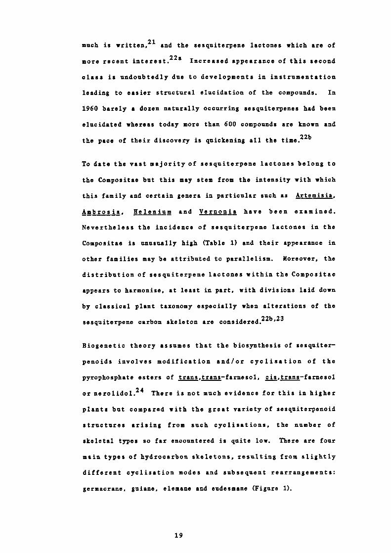
Costunolide, , is the most elementary cyclic sesquiterpene
lactone since it retains two of the three double bonds of
farnesyl pyprophosphate in the trans,trans configuration. It is
a germacranolide with a cyclodeca-1,5 —diene ring system. The
ending —olide is used to denote a compound possessing a lactone
20
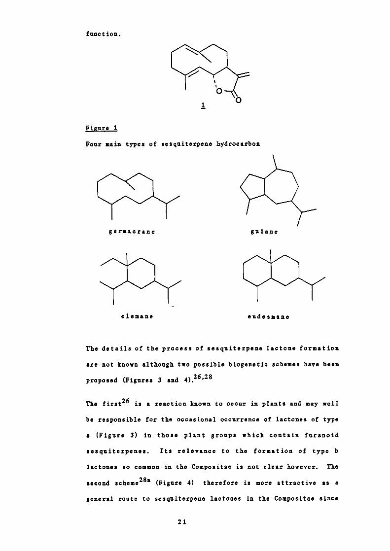
germacrane
e 1 emane
gui ane
eude smane
function.
1
Figure 1
Four main types of sesquiterpene hydrocarbon
The details of the process of sesquiterpene lactone formation
are not known although two possible biogenetic schemes have been
proposed (Figures 3 and 4)•2628
The first 26 is a reaction known to occur in plants and may well
be responsible for the occasional occurrence of lactones of type
a (Figure 3) in those plant groups which contain furanoid
sesquiterpenes. Its relevance to the formation of type b
lactones so common in the Compositae is not clear however. The
second scheme 28a (Figure 4) therefore is more attractive as a
general route to sesquiterpene lactones in the Conipositae since
21

----
some of the postulated intermediates occasionally accompany the
lactone end products. In addition it can be modified to lead to
the furanoid sesquiterpenes.
Figure 2
Simplified sesquiterpene lactone biosynthesis
trans, trans — fa me syi
g e rmac rano 1 ides
pyrophosphate
-ç
4%
eudesmanolides guianolides
Figure 3
Possible sequiterpene lactone biosynthesis. Proposal 1.
{0O$
A second, rarer type of y — lactone results from oxidation of a
non—terminal methyl group, for example a from Signbj,
hod g sonii, the only substance of this kind so far found in the
Compositae 29
22

KLI
H
2 0
Compounds embodying both types of lactone rings are more
frequently found, for example elephantopin 3•30
Figure 4
Possible sesquiterpene lactone biosynthesis. Proposal 2.
[l...rH; [1....H2OH
[OH[CHO H2OH {CHO
[ [:o
The germacranolides are probably the biogenetic precursors for
3
23
all the other types of sesquiterpenes (Figure 5)22c31a932 In
the figure, for simplicity, initial lactone ring closure at C-6
only is shown although that at C-8 is common.
Skeletons in the same vertical columns in Figure 5 are produced,
at least superficially, from the precursor farnesyl pyrophos-
phate by the same number of changes in the carbon skeleton and
thus may be said to exhibit the same degree of 'biogenetic
complexity' 22d
Individual members of a particular class however may differ
widely in oxidation state at various sites within the molecule,
for example hydroxyl or esterified hydroxyl groups at C —i, C-2,
C-3, C-5, C-6, C— S and C-9, either or both methyl groups
oxidised to hydroxymethyl, aldehyde, carboxyl or methylene
functions and either or both ring double bonds from farnesyl
pyrophosphate transformed into epoxide groups.281'
The eudesmanolides, cadinanolides and guianolides appear to be
derived by different methods of cyclisation of germacra—i(iO),4-
dienes or their epoxide derivatives presumably under the control
of different enzyme systems.281" 33 Methyl migrations in the
eudesmanolides give rise to the eremophilanolides and those in
the guianolides to the ambrosanolides and helenanolides.
Oxidative cleavage of the germacranolides, eudesmanolides,
ambrosanolides and helananolides is responsible for the
respective seco —derivatives. The formation of xanthanolides
involves a different method of ring fission from the
guianolides. Enzymatically induced ring contraction of the
guianolides gives rise to the chrymoranolides and that of the
eudesmanolides to the balkenolides, but so far these have been
found in only one species.
24

iiure 5
Possible biogenetic relationships of the different skeletal
types of sesquiterpene lactones
c±cio
/
e1eano1ide,,,,,,,,/eco—sudesano1id.s
/ ,,,/ eremophilanol ides bakkenol ides
/0
gerulacranoijdeg cadin*o1jds osanojides seco—ambrosanoljdes
0
zanthanol idis
o
helenanol ides 1..—he1enano1 ides
seco—ger.acranoljdes
Chrymoranol ides
c
1 1410
:R7Il3
15 0
25
C0
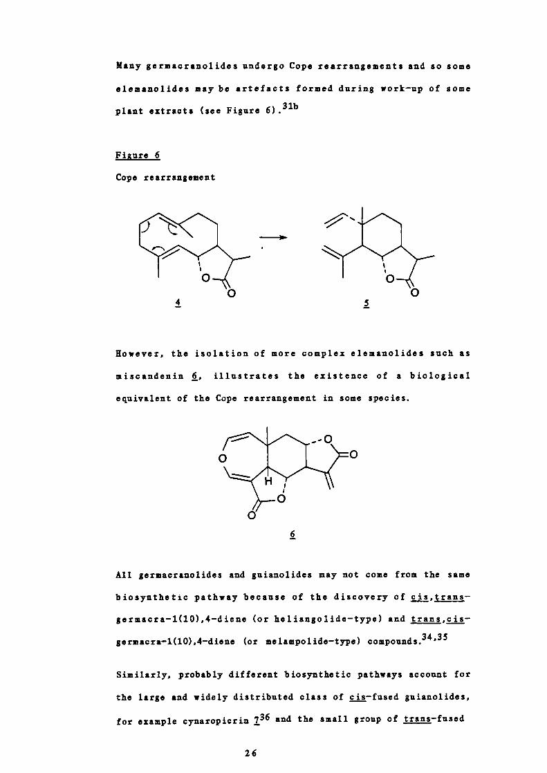
Many germacranolides undergo Cope rearrangements and so some
elemanolides maybe artefacts formed during work —up of some
plant extracts (see Figure 6)•31b
Figure 6
Cope rearrangement
Rowever, the isolation of more complex elemanolides such as
miscandenin , illustrates the existence of a biological
equivalent of the Cope rearrangement in some species.
6
All germacranolides and guianolides may not come from the same
biosynthetic pathway because of the discovery of cis,trans -
germacra-1(1O),4—diene (or heliangolide —type) and trans,cis -
germacra-1(lO),4—diene (or melampolide—type) compounds.34'35
Similarly, probably different biosynthetic pathways account for
the large and widely distributed class of cis—fused guianolides,
for example cynaropicrin 7 36 and the small group of trans—fused
26

o IiOAc' H\
o OH
HO'\H
0
guianolides such as gaillardin
08
7
(a) Anthemideae
flaying considered the sesquiterpene lactones in the family
Compositae, let us now be more specific and look at the
tribe Anthem ideae J . the tribe in which C. partheniurn is
placed. Table 2 gives the distribution of sesquiterpene
lactones in the Compositae.22e
The numbers in the columns represent the taxa from which the
lactones of a particular type have been isolated (since some
species produce more than one type of lactone, the sum of
numbers in a horizontal row generally exceeds the number of
taxa).
The Anthemideae, mainly Artentisia and Chrysanthemum, appear
to be fairly prolific producers of sesquiterpene lactones.
These lactones are mainly germacranolides, eudesmanolides
and guianolides, but the tribe also includes two unique
examples j. arteannin, 2 a cadinanolide from Artemisia
annua and chlorchrymorin, 10, a chrymoranolide (a rearranged
guianolide) from Chrysanthemum morifolium.22
27

V
V
-4
0p400UV
-I
V
0
0
-4
V
Vp414V
I
U
I
H
1.4V
V
V,0-4II
'-4
-4
- 00
ci 0 m
-4
,-I irs
in N'-4
0 - in i-I 00
'0 -1 00 00-4
-4
-4 -4
00 in
0 - - el-4 in
cn - in -
in
-4
C'l CO CO rl in i-Iin in in
in 0 -4 00 O r N e 0in ,-( 0 1-4
-4
%C - in -4 i1 - l N
e -4 -
V V V V VV C VC V V V V V VV V Co - V . C C.-4 V C o4 14 C V V 0 .4 V C -4
0 0 C - 0 V Iso 1.4 V C 0 V 0 1.4 C 0
C V - V V . V C 4 .
Ii Ps 4.5 4 - 0V C V V V P WC -4
. - . .0 U
Co C
V-4- -4 Co - 0P C 0 C 'C V P V 4C • C • -4
P -4 0Ii - V '-4 P0 0 - 0 Ca 14C C C 0I P I P0 V 0 .4C) - C) '• V V CC C C) C)
Ii II li U LI
CO U
CV
•0-4 C-4 C V
0 C V •P V CC ' V -4 -- •..4 '0 - 0-4 - '4 0 P
.P 0 COs PC C
0 0 P 00 .H C4) • -4 P14 C P CV.0 00 H C
ti II U II II
C
V C
'0 V
-4 '0
-4 -4
C 0 -
V P C 0
'0 C V P- 14 C '0 C-4 0 V -4
0 C 'O-4 A
P 00 VC 14 - p '14 V 0 C 0O 000EVC I C C I0 00 V 014 C) V '0 C)
o o P 000 C V 4) 4
U II II II
bI W CO
28
H0
CI--
H
09
10
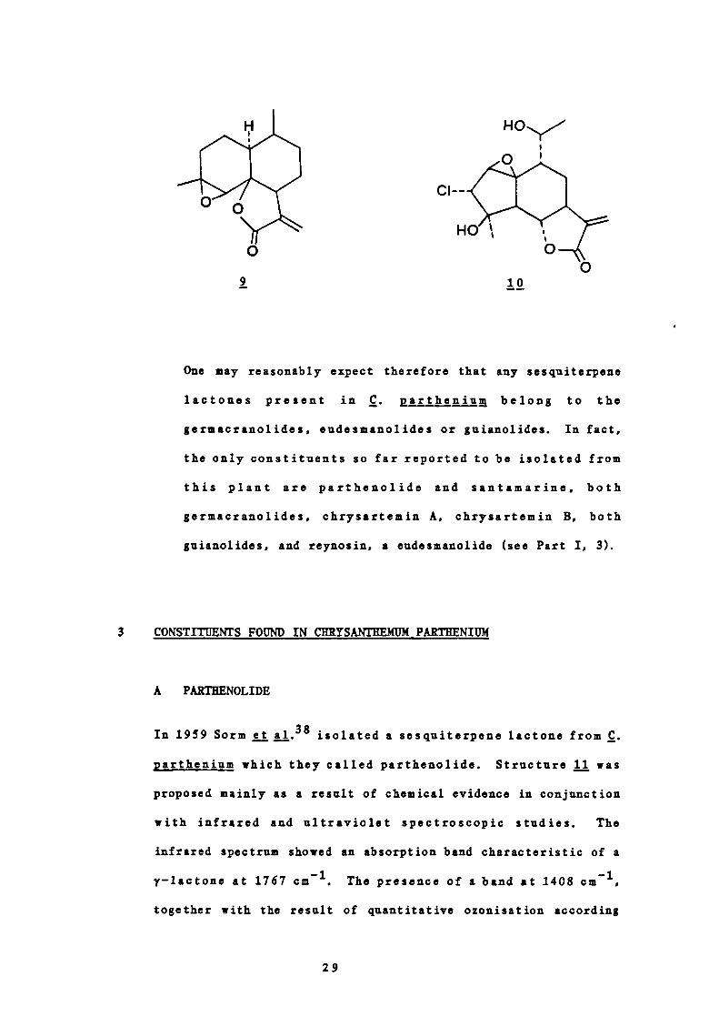
One may reasonably expect therefore that any sesquiterpene
lactones present in C. prtheniurn belong to the
germacranolides, eudesmanolides or guianolides. In fact,
the only constituents so far reported to be isolated from
this plant are parthenolide and santamarine, both
germacranolides, chrysartemin A, chrysartemin B, both
guianolides, and reynosin, a eudesmanolide (see Part I, 3).
3 CONSTITUENTS FOUND IN CHRYSANTHEMUM PARTHENItJM
A PARTHENOL IDE
In 1959 Sorm j.38 isolated a sesquiterpene lactone from C.
parthenium which they called parthenolide. Structure fl wasproposed mainly as a result of chemical evidence in conjunction
with infrared and ultraviolet spectroscopic studies. The
infrared spectrum showed an absorption band characteristic of a
y—lactone at 1767 cm'. The presence of a band at 1408
together with the result of quantitative ozonisation according
29
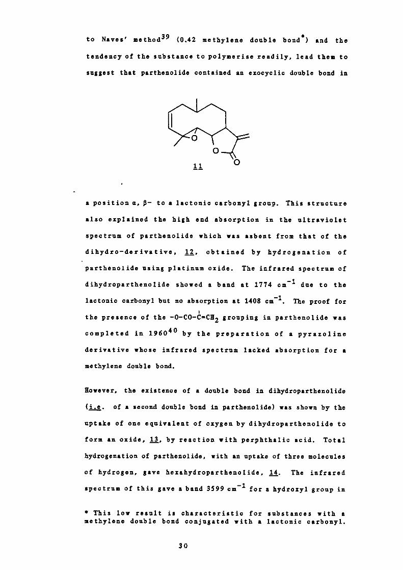
to Naves' method 39 (0.42 methylene double bond s ) and the
tendencyof the substance topolymerise readily, lead them to
suggest that parthenolide contained an exocyclic double bond in
II
a position a, - to a lactonic carbonyl group. This structure
also explained the high end absorption in the ultraviolet
spectrum of parthenolide which was asbent from that of the
dihydro — derivative, obtained by hydrogenation of
parthenolide using platinum oxide. The infrared spectrum of
dihydroparthenolide showed a band at 1774 cm 1 due to the
lactonic carbonyl but no absorption at 1408 cm. The proof for
the presence of the —0—CO—CCH2 grouping in parthenolide was
completed in 196040 by the preparation of a pyrazoline
derivative whose infrared spectrum lacked absorption for a
methylene double bond.
Kowever, the existence of a double bond in dihydroparthenolide
(i.e. of a second double bond in parthenolide) was shown by the
uptake of one equivalent of oxygen by dihydroparthenolide to
form an oxide, 13, by reaction with perphthalic acid. Total
hydrogenation of parthenolide, with an uptake of three molecules
of hydrogen, gave hexahydroparthenolide, 14. The infrared
spectrum of this gave a band 3599 cm for a hydroxyl group in
* This low result is characteristic for substances with amethylene double bond conjugated with a lactonic carbonyl.
30

addition to that for a lactonic carbonyl at 1750 cm 1 . From
this it was concluded that a third oxygen was present as an
oxide since neither a hydroxyl or ketone is present in the
natural compounds.
Hexahydroparthenolide, 14, gave rise to chamazulene, 15, on
selenium dehydrogenation thus leading Sormjj. to conclude
that parthenolide was a sesquiterpene lactone of the germacrane
type 41
Parthenolide and its dihydro —derivative, on oxidation with
nitric acid, gave a mixture of acids from which —methy1adipic
acid, was isolated so proving that at least four carbon
atoms of the presumed cyclodecane ring do not carry an oxidised
functional group and are substituted with one methyl group.
The character of the isolated double bond was partly explained
by the infrared spectra of parthenolide oxide, 13, and
dihydroparthenolide oxide, 17. Both these compounds gave bands
at 831 cm' characteristic of a disubstituted cis-1,2—oxide.
Hexahydroparthenolide, jj, on oxidation with chromic acid gave a
compound () with infrared absorption bands 1785 cm for a y-
lactone and 1717 cm for a ketone. Since reduction of
hexahydroparthenolide with lithium aluminium hydride gave a
triol, , which on oxidation with periodic acid consumed one
mole of reagent, the keto group in 19 must be adjacent to the
potential hydroxyl group in the lactone grouping.
This result thus shows that one of the C —O bonds of the oxide
ring of parthenolide is attached to carbon 5 of the 4,10 -
dimethyl-7—isopropylcyclodecane skeleton. As parthenolide gave
—methyladipic acid, 16, on oxidation with nitric acid the oxide
31

19
ring was presumed to be 3 —membered and the double bond so
located at position 2. This presumption was apparently verified
by isolation of formic acid from the volatile products of
ozonisation of dihydroparthenolide and acetic acid on subsequent
oxidation of the non—volatile material (Figure 7). The sequence
of reactions are summarised in Figure 8.
Figure 7
Reactions used in the deductions about the positions of the
double bond and epoxide in j
02 3 4/ \5
-HC = CH-C-CHCH3'
0-CH 'CH-C''CH
0II/ __________C-OH + OCH 4
CH3
0 0 0II II II /
-CH + CH+C-HOH CH3OH
In 1965 however Govindachari et al. isolated parthenolide from
32
I
HO2C
CO2H
16
15
L,rI
33
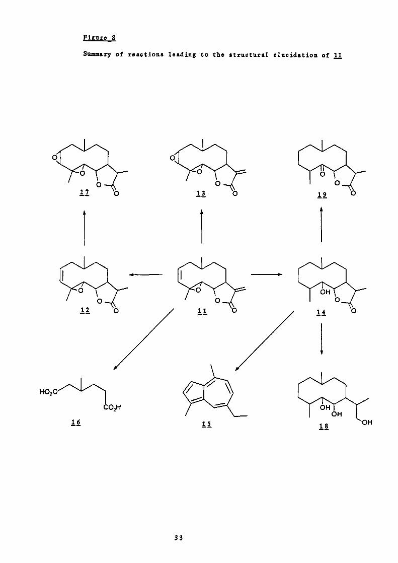
Figure 8
Summary of reactions leading to the structural elucidation of 11
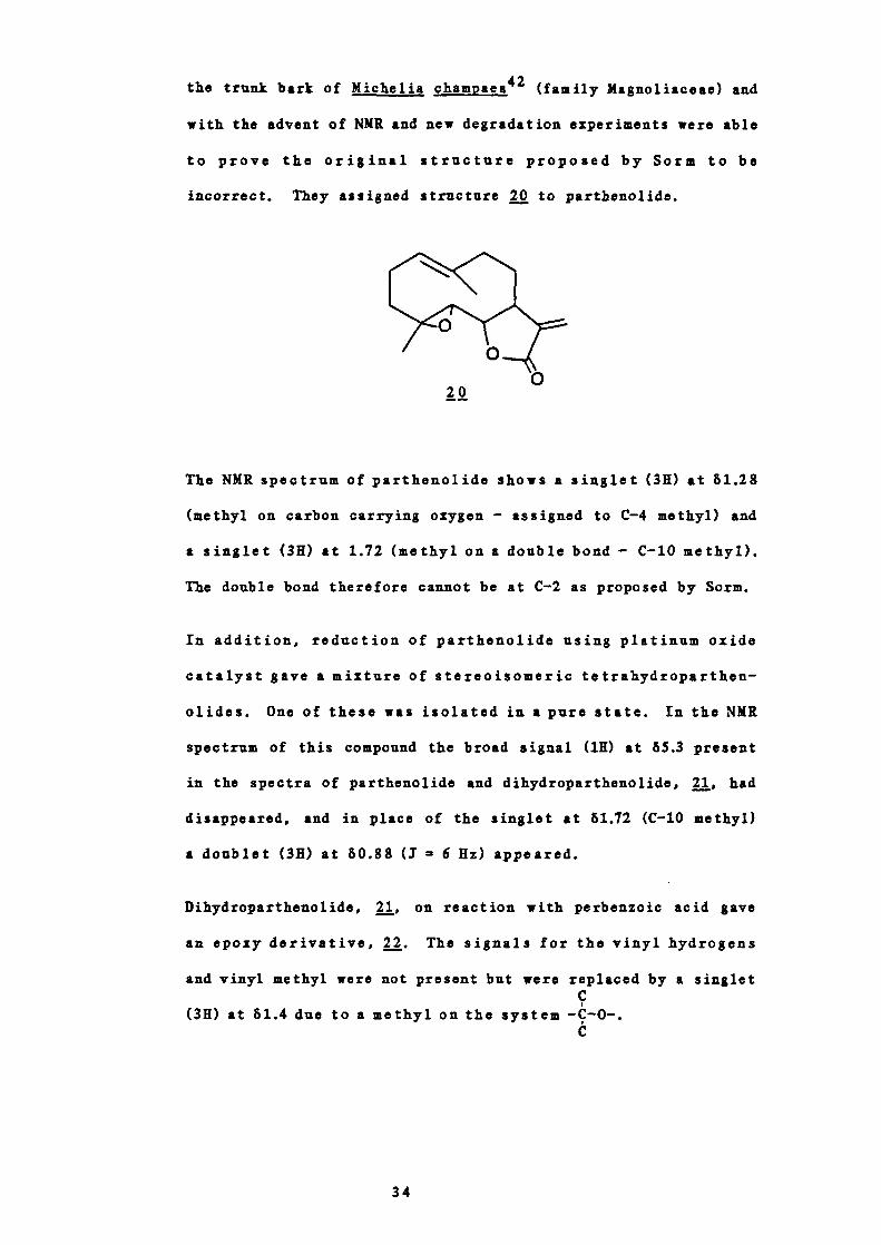
the trunk bark of Micheija cham paea42 (family Maguoliaceae) and
with the advent of NMR and new degradation experiments were able
to prove the original structure proposed by Sorm to be
incorrect. They assigned structure 20 to parthenolide.
20
The NMR spectrum of parthenolide shows a singlet (3K) at 61.28
(methyl on carbon carrying oxygen - assigned to C-4 methyl) and
a singlet (3K) at 1.72 (methyl on a double bond - C-1O methyl).
The double bond therefore cannot be at C-2 as proposed by Sorm.
In addition, reduction of parthenolide using platinum oxide
catalyst gave a mixture of stereoisomeric tetrahydroparthen -
olides. One of these was isolated in a pure state. In the NMR
spectrum of this compound the broad signal (1K) at 65.3 present
in the spectra of parthenolide and dihydroparthenolide, 21, had
disappeared, and in place of the singlet at 61.72 (C-10 methyl)
a doublet (3H) at 60.88 (J = 6 Hz) appeared.
Dihydroparthenolide, 21, on reaction with perbenzoic acid gave
an epoxy derivative, . The signals for the vinyl hydrogens
and vinyl methyl were not present but were replaced by a singletC
(3K) at 81.4 due to a methyl on the system —ç—o—.C
34
4i.
0
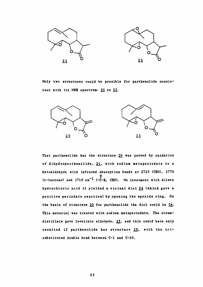
Only two structures could be possible for parthenolide consis-
tent with its NMR spectrum: or 23.
That parthenolide has the structure was proved by oxidation
of dihydroparthenolide, 21, with sodium metaperiodate to a
ketoaldehyde with infrared absorption bands at 2725 (CEO), 17700
(y—lactone) and 1710 cm (— —R, CEO). On treatment with dilute
hydrochloric acid it yielded a vicinal diol (which gave a
positive periodate reaction) by opening the epoxide ring. On
the basis of structure 20 for parthenolide the diol could be .
This material was treated with sodium metaperiodate. The steam—
distillate gave levulinic aldehyde, and this could have only
resulted if parthenolide has structure 20, with the tn -
substituted double bond between C—i and C—b.
35

OH
Hy'A25L4.
°
The absolute configuration of parthenolide was determined by
Bawdekar et al.43 in 1966 and shown to be as in 26.
Parthenolide has a close structural similarity to costunolide,
27, the stereochemistry of which is well established.44
Parthenolide can be considered as the 4,5 —monoepoxide of
costunolide and dihydroparthenolide the 4,5 —monoepoxide of
dihydrocostunolide. Dihydrocostunolide on treatment with excess
perbenzoic acid gave a diepoxide, 28, which was identical with
the epoxide of dihydroparthenolide with respect to infrared and
nuclear magnetic resonance spectra as well as mixed melting
36

point. The stereochemistry of parthenolide at C-6 and C-7 and
that of dihydroparthenolide at C-6, C-7 and C—li was therefore
established.
In 1976 Quick and Rogers45 examined the molecular structure of
parthenolide by X—ray crystallography. They found that the two
methyl groups are a —orientated, the 1(10) —double bond and the
equivalent of the 4—double bond are trans (as expected from
biosynthetic considerations) and the ring has a flattened
conformation (see Figure 9). The configuration of the
asymmetric atoms proved to be 4K, 5K, 6K, 7K.
The geometry of the epoxide showed the molecule to be directly
related to costunolide 46 so it follows that the diepoxide
described by Bawdekar as derivable from parthenolide
must be represented by .
This geometry agreed well with deductions made from nuclear
37
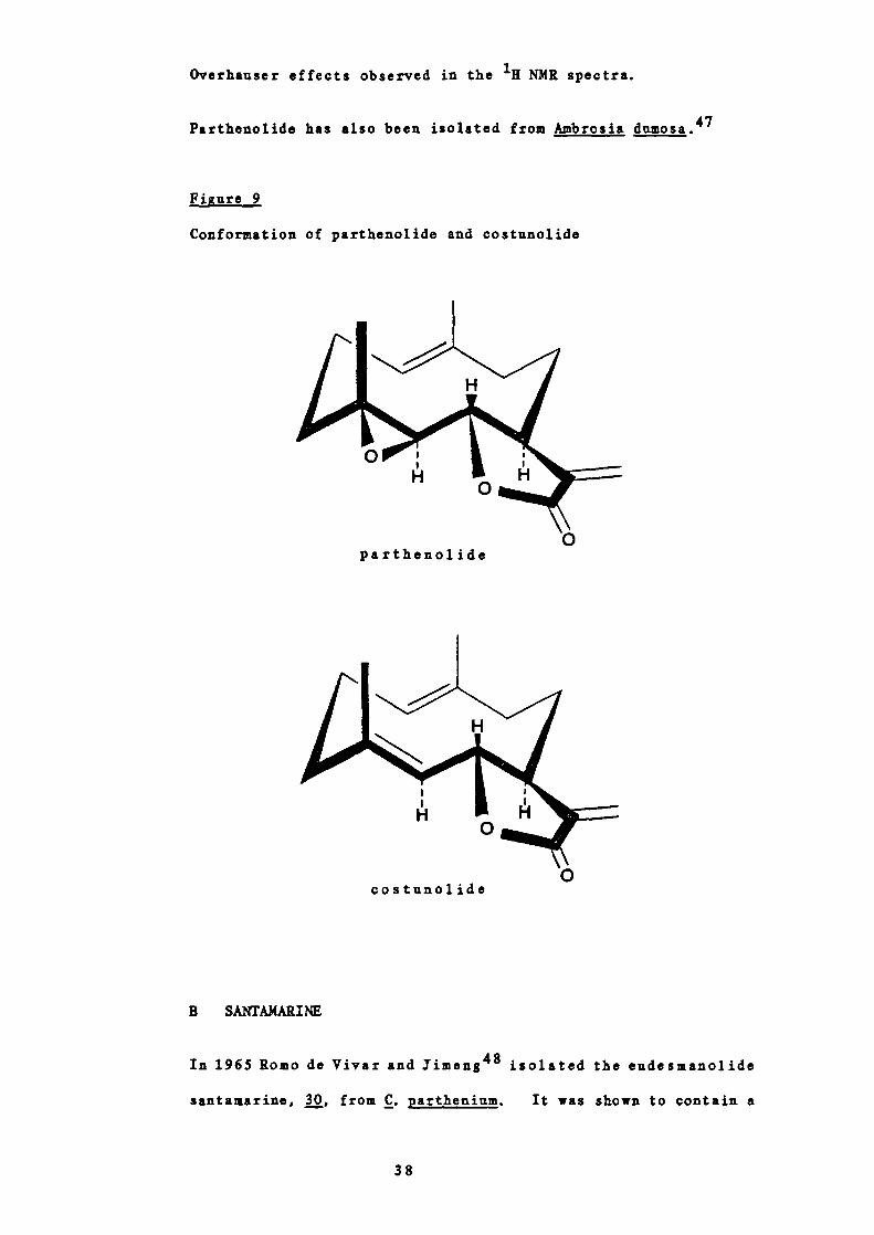
Overhauser effects observed in the 1 NMR spectra.
Parthenolide has also been isolated from Ambrosia dumosa.47
Fi gure 9
Conformation of parthenolide and costunolide
0partheno]. ide
0costunolide
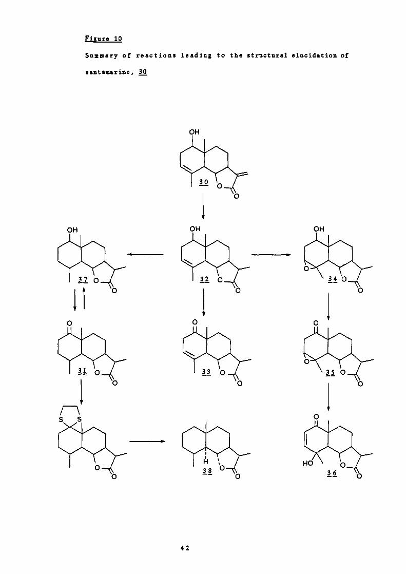
B SANTAMARINE
In 1965 Romo de Vi'var and Jimeng 48 isolated the eudesmanolide
santamarine, 30, from C. parthenium. It was shown to contain a
38

0
hydroxyl group by its infrared absorption at 3400 cm and the
formation of a monoacetate. It was proved to be a secondary
., '.,
alcohol because on oxidation of the dihydro compound with
chromic acid a keto derivative, 31, was formed, (1K at 1710
cm - six membered ring ketone). This compound gave a positive
Zimmermann test indicating that the ketone is flanked by at
least one methylene group. There were thus four possible
positions for the ketone: C-i, C-2, C-8 and C-9. On mild
alkaline treatment, however, a conjugated ketone was not
produced so C-8 was eliminated. Similarly, the dihydro deriva-
tive, 32, on oxidation gave a non-conjugated letone, 33, and
thus excluded position 2.
Of the remaining two positions C-i was favoured by the
ultraviolet absorption at 205-210 nm characteristic of , y -
unsaturated ketones. This was confirmed by chromic acid
39

OH 0
oxidation of the epoxide, j, to the keto epoxide, 35, which on
alkaline treatment gave an aj —unsaturated--y—hydrozyketone, 36
umax 215 nm, IR 3600, 1680 cm).
0
On hydrogenation of a good yield of the alcohol, 37, was
obtained. Therefore, attack of the C —i carbonyl by the
hydrogen was assumed to occur from the opposite side from C-9
which has a a —orientated methyl group, and the hydroxyl group
was therefore given the configuration $—equatorial.
On dehydrogenation santamarine did not produce an azulene so it
was assumed to be a eudesmanolide. The skeleton and
stereochemistry at C-5, C-6, C-7 and C—iO were established by
comparison with santanolide C, 38 (See Figure 10). The
structure of the remainder of the compound was elucidated in a
similar way to parthenolide (Part I, 3A) and the series of
40

reactions are summarised in Figure 10.
Santamarine has also been isolated from Ambrosia confertiflora
by Yoshioka
C CERYSARTEMINS A AND B
In 1969 Romo et .!J. 5 ° isolated two guianolides, chrysartemins A,
39, and B, 40, from C. p arthenium. The structures of these
compounds were proposed mainly on evidence from infrared,
nuclear magnetic resonance and mass spectrometry with additional
chemical proof.
Chrysartemin A, 39, was shown to contain a hydrozyl group and an
a—methylene——lactone from infrared absorption bands. The
hydroxyl group was assumed to be tertiary as it could not be
acetylated and was resistant to chromic acid oxidation.
The NMR spectrum of 39 in DMSO—d6 showed two doublets at 65.96
and 5.50 (1 3 Hz) for the exocyclic methylene hydrogens, a
doublet of doublets at 4.47 (1 11.5, 9.5 Hz) for the C-6
hydrogen, a doublet at 2.24 for the C—S hydrogen and doublets (I
= 1 Hz) at 3.42 and 3.25 for the hydrogens attached to the
carbon atoms bearing the epoxide. The methyl group signals.
41
-I
OH OHOH
HI!38
0
1
I-'
iiure 10
Summary of reactions leading to the structural elucidation of
santamarine, 30
OH
0
0
42

HO
appeared as two singlets at 61.38 (attached to a carbon atom
bearing an ether oxygen) and 0.90 (attached to a carbon atom
bearing a hydroxyl group). In CDC1 3 , a signal at 64.77
disappeared after equilibration with deuterium oxide and was
assigned to the hydroxyl hydrogen. Hydrogenation of
chrysartemin A gave the dihydro —derivative, the NMR spectrum of
which showed a doublet (1 7 Hz) in the methyl region.
Aromatisation of chrysartemin A gave chamazulene and so a guiane
structure was proposed with the lactone closed at C-6. The
signal in the NMR for H-6 at 64.47 is characteristic of a C-6
lactone in the guianolide series of sesquiterpene lactones.
On treatment with —toluenesulphonic acid chrysartemin A gave
the —to1uenesu1phonate, 4j, by opening of an epoxide the oxygen
atom of which was borne on secondary carbons. This was shown in
the NMR spectrum of the crude product by the appearance of two
doublets at 64.78 and 3.92. However, 41 also contained an
epozide formed between saturated carbons. Further spectroscopic
studies confirmed the latter's position and hence the complete
structure of chrysartemin A.
TsO
The structure of chrysartemin B, , was proposed by its very
similar spectral properties to chrysartemin A, 39. Chrysartemin
A has also been found in Artemisia mexicana and A. klotzchiana5°
43

while both chrysartemins A and B have been found in
Chrysanthemum morifoliu 5 ' where they have been shown to
stimulate root initiation.
Romo et al. also isolated santamarine, 30,° from C. partheniuzn
and after crystallisation of this were able to isolate a minor
constituent from the mother liquors. This was shown to be the
eudesmanolide, reynosin, .
The infrared spectrum of reynosin, 42, showed absorption bands
at 3520 and 3610 (hydroxyl), 1770 (y — lactone) and 1680 cm1
(conjugated double bond).
The NMR spectrum showed a pair of doublets (1 3 Hz) at 86.10
and 5.46 (exocyclic methylene conjugated with the 7—lactone),
two broad singlets with long —range coupling at 5.01 and 4.89
(C-4 exocyclic methylene), an apparent triplet (J 11 Hz) at
4.06 (R-6), a doublet of doublets at 3.53 (H — i), a sharp signal
at 2.04 which disappeared on addition of deuterium oxide
(hydroxyl hydrogen) and a methyl singlet at 0.85.
Reynosin has also been isolated from Ambrosia confertiflora.49
44

4 STRUCTURAL ELUCIDATION OF SESQUITERPENE LACTONES
A NMR SPECTROSCOPY
With the advent of NMR spectroscopy in the 1960352 the
structures of many sesquiterpenes have been elucidated and the
number of new structures appearing is increasing all the time.
The presence of certain structural groups gives rise to
characteristic spectral features which maybe of particular
help in the elucidation of these compounds such as an 11(13)-
double bond, 8a-hydroxyl or ester group, long range couplings
between H-6 and H-il and between the C-S methyl and the C-4
methylene hydrogens and the presence of C-6 or C-8 lactones.
(a) 11,(13)-donble bond
It is well established that allylic coupling occurs between
the two C-13 methylene hydrogens and H-7 for all sesquiter-
penes containing either a C-6 or C-8 aj-unsaturated y-
iactone.53 ' 54 The signals for the C-13 hydrogens thus
appear as two doublets (1 1-4 Hz) between 65.0 and 56.5
p.p.m. However, in some sesquiterpenes containing both a
C-6 aj-unsaturated-y-lactone and an 8a-hydroxyl group each
of the C-13 hydrogens gives rise to a doublet of doublets as
a result of geminal coupling in addition to the allylic
coupling 55 (see partial structure in Figure 11). In 11(13)-
unsaturated lactones not containing an 8a-oxygen function
the H-13 geminal coupling is 0.5 Hz or less and so is not
usually observed.53'54
Geminal coupling (I = 1 Hz or more) is thus only observed in
the NMR spectra when both an 8a-hydroxyl group and a C-6
a,-unsatursted-y-lsctones are present. Examination of
45

Hb
these spectra reveals that the signal for H-13a, j. the
hydrogen trans to the i—lactone carbonyl, always appears at
lower field than in the spectra of corresponding compounds
which differ only in that they do not contain the 8a —
hydroxyl group and thus do not show the geminal splitting
pattern.
Figure 11
Partial structure of a sesquiterpene lactone containing a
C-6 a, —unsaturated—y—lactone and an 8a—hydroxyl group.
flH
It has been proposed that the geminal coupling and the
paramagnetic shift for K-13a results mainly from the van der
Waals effect of the 8a—hydroxyl group upon the bonding
orbital of H-13a. 56 Zurcher56 calculated the ranges in ppm
for the van der Waals paramagnetic shifts and these are
compatible with those found experimentally (Tables 3 and 4).
This paramagnetic shift and the geminal coupling data can be
used for various stereochemical assignments. 55 A positive
shift in the range 0.4 - 0.7 ppm relative to the chemical
shift for E-13a in a compound without an 8 —hydroxyl group
together with a geminal coupling for H-13a denotes an a—
orientation for the 8—hydroxyl group and no such shift a
orientation.
46

When the absolute stereochemistry is known a positive
paramagnetic shift can be used for conformational analysis
for example a 0.5 - 0.7 ppm shift for H-13a in germacrano -
lides such as salonitenolide, j, requires a distance of 2.0
Table 3
Calculated H-13s paramagnetic shifts based on different
distances between —13a and C—S oxygen atoms
Distance Calculated shift in ppm
2.0 - 2.5 0.2 - 0.6
3.0 0.0 - 0.1
3.5 - 4.5 0.0
- 2.5 between the 8a —oxygen atom and H-13a (see Table 4)
and this distance is possible only when the conformation
with regard to C-6, C-7 and C-8 is as shown in Figure 12.
The assignment of the conformation at these positions
usually permits the assignment of the conformation of the
whole molecule.31C
(b) Long range couplings
(i) 11-6 - 11-11 couplings
The NMR spectra of 11,13—dihydro—eudesmanolides often show a
broadened triplet for the C-6 lactonic hydrogen e.g. in
colartin, 44,31d1 from Artemisia tripartita subsp. arbuscula
47

V-4'CV
00
-4V
Va0
V00
H0
00
-4
00
0
VV0
V
V
V
V14V
V
V V
-40V0V
00 VVaV -II .aV
a14
V 0
-1I 0= 0
0o -)' .014 Vo ,0V 0
,0 14o 04
0-4
Vco aI 1.4U 0
'4
3
a0404
-4
'4-4
V0
V
V-40
-4
V0
o.lV
V-40
,0
V N
'-4
V
044C,V
-4
0
'.40
-4 -4 -4V V V-4 -4 -414 14 14o 0 044 44 44V V V
0 0 V
* *• •
000
I I I
000
00
e*
cr1
(1 c'1 1
I I I
000
el c'i e1
- ei -
I I I
00
VV Vo '
-4I ,-4 V
i - 0 VH 00 V -14 V Ii -
a ooV V
. oI ' 14 ..4
000 V 00 00
.-I -4V V-4 -4H HV V
• ** ** *
-4
00
I I
00
00
0•0•
* I
0
cq
00
VV 0V '0'O-4-4 -4
0MO0 VII V 14
aV V
. o aI '014
000 V CO
00-4 V44V 0a o e14 0 1.4O -4 CCV'4 4.40 V 0O a0 II H 14
0 000- '4 14V 0 'o -
14 0 ,0 HO I 044 - 0V V 00 '00-C H '4 .0o V 0 I
00.00 000V V 0
-'40 0 .10-4 -4 V
+0a a o oO 0 0-
.14 .14 -4 .4.4V V 14 V
O 4400 0V 0 '-4 V00 00 - -4
-414H H 14 00 0 0I I 44I
00 00 V V0
'0 '0 H0 0 V VV V
V VV V 044 48
-4 Ii 14-4 4 .14 0 0I I V 0404
041400
0 0 0 V V0 V 40 0 .14 +4
48 44 0 -4VU .0.0.0,0 V V V
-40 0,0 '0 '00 0 4 V U00 VV V V 14 14.14 .14 0 V 0V V 04 V V-- a .c .0'0 '0-40 0
I I I I I
'4 c
V U0 00 0V V.4.4 .14V V *-4 - * *00.*.
48
43
0
0
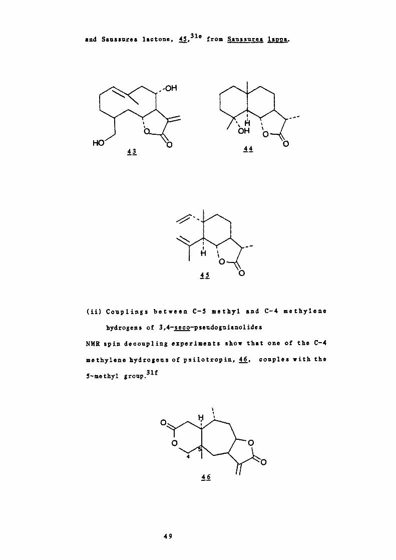
and Sauasures lactone, 45,310 from Saussurea lappa.
' H 'OH'
44
(ii) Couplings between C — S methyl and C'-4 methylene
hydrogens of 3,4—seco—pseudognianolides
NMR spin decoupling experiments show that one of the C-4
methylene hydrogens of psilotropin, j, couples with the
5—methyl gronp.3U
40
49

CC
C
U H
C
Figure 12
Conformation at C-6, C-7 and C-8 based on the distance between
the 8a—oxygen atom and H-13a as required to account for the
paramagne tic shifts in 6a— lactonised 8a—hydroxy sesquiterpene
lactones
germacranol ides guianoljdes
U
eudesmanolides
50
0
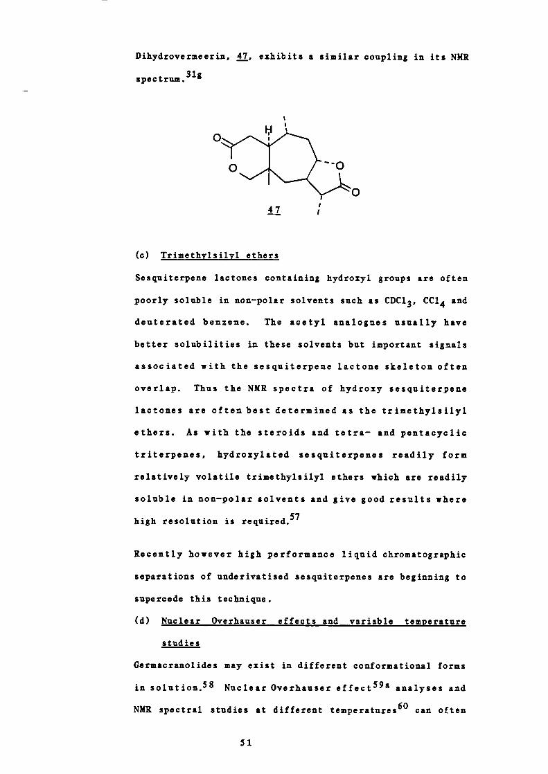
Dihydrovermeerin, , exhibits a similar coupling in its NMR
spec trum.3
..
ii
(c) Trimethylsil yl ethers
Sesquiterpene lactones containing hydroxyl groups are often
poorly soluble in non—polar solvents such as CDC1 3 , CC1 4 and
deuterated benzene. The acetyl analogues usually have
better solubilities in these solvents but important signals
associated with the sesquiterpene lactone skeleton often
overlap. Thus the NMR spectra of hydroxy sesquiterpene
lactones are often best determined as the trimethylsilyl
ethers. As with the steroids and tetra — and pentacyclic
triterpenes, hydroxylated sesquiterpenes readily form
relatively volatile trimethylsilyl ethers which are readily
soluble in non—polar solvents and give good results where
high resolution is required.57
Recently however high performance liquid chromatographic
separations of underivatised sesquiterpenes are beginning to
supercede this technique.
(d) Nuclear Overhauser effects and variable temperature
studies
Germacranolides may exist in different conformational forms
in solution.58 Nuclear Overhauser effect59a analyses and
NMR spectral studies at different temperatures 6° can often
51

A
B
C
D
7>
1 7>
distinguish conformers although no absolute methods are
available.
Germacranolides exist in four major conformational forms as
shown in Figure 1331h
Figure 13
Four major conformational forms of germacranolides
The compound dihydrotamaulipin A acetate, 43,61 was shown to
have the conformation j shown by NOE techniques (Figure
14).
With reference to j, an increase in the integrated
intensity of the H-2 and H-6 signals caused by irradiation
of the C-4 methyl signal, as well as enhancement of the H-2
signal by irradiation of the C-1O methyl signal, indicated
52

AcO
-T U
Figure 14
Conformations of dihydrotamaulipin A acetate. The figures
indicate percentage NOE enhancements.
1
0
49
that the lO—membered ring adopted the conformation shown
Li. in which the C-4 methyl, C-1O methyl, R-2 and H-6 arein the same direction. This corresponds with conformation C
in Figure 13.
The conformations of furanodjenone, 62 Linderalactone,63
bicyclogermacrene, 50, and of iso —bicyclogermacrene, Si,64
have been determined by similar techniques.
53

50 51
These latter two are of interest because it is postulated
that they are the biogenetic precursors of other
sesquiterpenes containing a fused 1,1'—dimethylcyclopropane
ring. 64 Bicyclogermacrene was shown to adopt conformation A
and iso—bicyclogermacrene C in Figure 13.
Isabelin, , is a naturally occurring germacranolide
isolated from Ambrosia p silostachy a. 6 ° NMR studies at
different temperatures established that isabelin existed in
52
solution at room temperature in a 10:7 ratio of two
conformers. The NM spectrum recorded in CDC1 3 at 250
showed two sets of signals which appeared to correspond with
two compounds in a 10:7 ratio. The material behaved as a
single compound chromatographically, during fractional
crystallisation experiments as well as during the
preparation of a number of derivatives and transformation
54

products. These results suggested that the NMR spectrum of
isabelin should be interpreted on the basis that isabelin
exists as two conformational isomers in solution at room
temperature. Conclusive evidence for this was provided by a
temperature controlled NMR study. Crystalline isabelin was
dissolved in CDC1 3 precooled to -50 and the NMR spectrum
recorded at this temperature within 40 minutes. The major
isomer appeared to correspond to the minor form at 250 and
this is probably the only conformer present in the crystals.
When the solution used for this —50°C NMR study was allowed
to warm to room temperature it again showed the 10:7 ratio
of conformers.
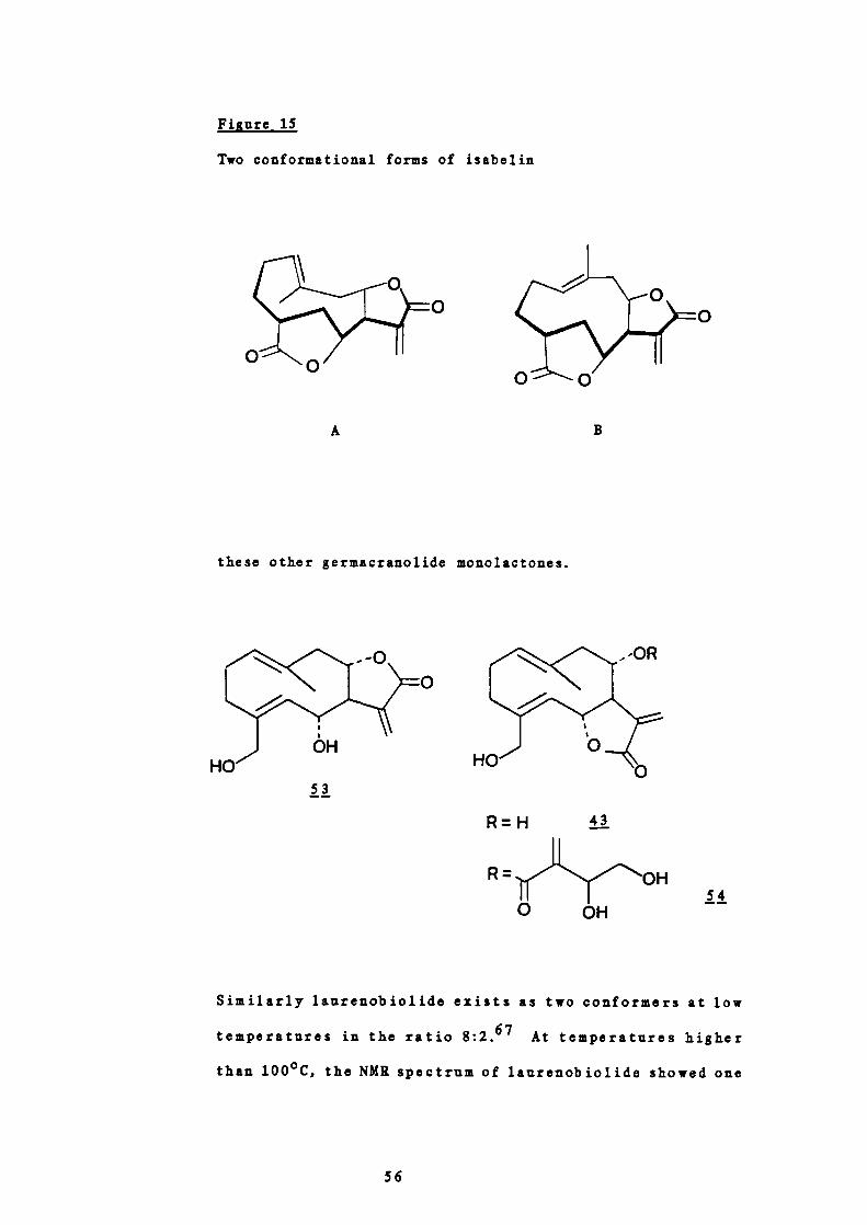
Two conformational forms A and B of isabelin were proposed
from consideration of molecular models and the different
values observed for their coupling constants (Figure 15).60
The bond angle between 11-7 and 11-6 in conformer A is
approximately 900, a value in accord with a small coupling
constant.65 The major form of isabelin in solution at room
temperature showed a 1 Hz coupling and was therefore
assigned structure A. The bond angle between 11-7 and 11-6 in
B is approximately 180° and thus a larger coupling constant
is expected. The minor conformer of isabelin exhibited a 7
Hz coupling constant and was therefore assigned structure B.
These two conformations were confirmed in 1971 by I. Tori
51.66 by nuclear Overhauser effect experiments. They also
proved the endocyclic double bond to have a trans
orientation, and since isabelin has been correlated with
artemisiifolin, 53, salonitenolide, 43 and cnicin, 54, the
results obtained also established a trans configuration for
55
A
B
I-,
0 0
Figure 15
Two conformational forms of isabelin
these other germacranolide monolactones.
- -OR
53
H
R=H ii
R "('OH .4.
OH
Similarly laurenobiolide exists as two conformers at low
temperatures in the ratio 8:2.67 At temperatures higher
than 100 0 C, the NMR spectrum of laurenobiolide showed one
56
set of sharp signals indicating a rapidly inverting ten—
membered ring while at temperatures lower than —20°C, two
sets of signals were observed, one for each isomer.
The conformations of neolinderalactone having cis—l(l0) and
trans-4(5) double bonds and sericenine having trans-1(l0)
and 2J1-4(5) double bonds were also studied by
intramolecular NOE and variable temperature studies.68
Two crystalline compounds urospermal A and B are
conformational isomers which have been shown to be
interconvertible in solution. 69 Each conformer is
considered to be stabilised by intramolecular hydrogen
bonding and the two forms are separable by chromatography.
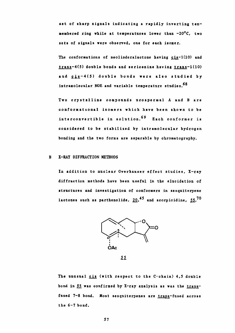
B X—RAY DIFFRACTION METhODS
In addition to nuclear Overhauser effect studies, X—ray
diffraction methods have been useful in the elucidation of
structures and investigation of conformers in sesquiterpene
lactones such as parthenolide, 20, and scorpioidine, 55•70
Ac
55
The unusual 2J.!. (with respect to the C—chain) 4,5 double
bond in 55 was confirmed by X—ray analysis as was the trans-
fused 7-8 bond. Most sesquiterpenes are trans —fused across
the 6-7 bond.
57

5 BIOLOGICAL ACTIVITY IN THE FAMILY COMPOSITAE
The Compositae is one of the largest families in the plant kingdom
comprising 1000 genera and 15000 species but, until recently, has
been the source of relatively few products of medicinal and economic
importance. Only about 30 species are used as crude drugs with just
16 drugs appearing in the pharmacopoeias. No more than 20 pure
substances are used therapeuticallyor available commercially.7
The current interest in the family stems largely from improved
structural elucidation techniques, which has led to the large number
of novel sesquiterpenes found together with the use of new screening
methods and of computer evaluation.
Seven main types of biological activity have been shown to be present
in the family namely, A cytotoxic, B spasmoloytic, C anti —
inflammatory, D antihepatoxic and cholerectic, E antimicrobial, F
antihyperlipidemic and 6 insecticidal.
A CYTOTOXIC ACTIVITY
The discovery of new compounds with cytotoxic activity has
received much publicity in recent years in view of their
possible use in cancer. The availability of modern, refined
methods of testing antitumour agents has encouraged a systematic
search for these agents from natural sources. Not surprisingly,
therefore, perhaps the greatest amount of work with regards to
biological activity in the Compositae has been directed towards
this aim. Many of the sesquiterpene lactones found in the
family, chiefly the germacranolides, guianolides and elemano-
lides, are especially active.7b The first positive results
were obtained with chamomile extracts and chamazulene 72 in the
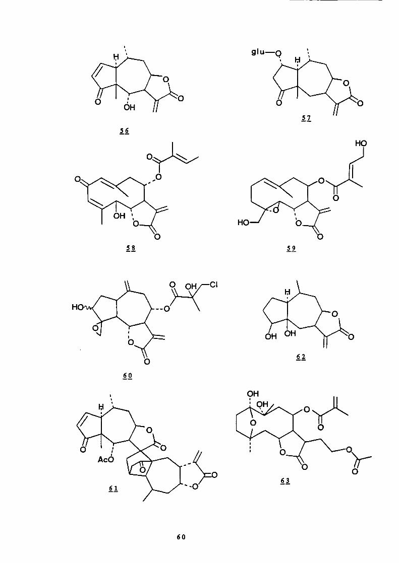
1950s but more recent examples include parthenolide,
58

helenalin, paucin, 57,73 molephantinin,
eupahyssopin, ,76 cnicin, chlorohyssopifolin C, 60,78
microlenin acetate, rudmollin, ,80 and piptocarphin A,
81
In 1943 Medawar et suggested that the cytotoxicity of
cardenolides and related compounds was associated with the
presence of the unsaturated lactone. Subsequent studies seem to
suggest that the antitumour activity of sesquiterpene lactones
is due to the presence of an a—methylene group on the y—lactone
ring as well as another functional group such as an epoxide,
chiorhydrin, unsaturated ester, unsaturated lactone or an
unsaturated ketone.73 ' 83 ' 84 Little is known however of the
relation between structure and activity in these compounds.
Nevertheless the demonstrated reactivity of unsaturated lactones
towards thiols and amines and the presence of other reactive
functional groups suggest that the cytotoxicity may result from
irreversible alkylation of nucleophilic centres in a biological
system73 ' 83 ' 84 such as the Michael —type addition of ci—methylene-
y—lactones with cysteine (Figure 16).
Hiadon and Twardowski 84 have investigated the mode of action of
some sesquiterpene lactones at a cellular level using HeLa
cells. At subtoxic concentrations they demonstrated arrest of
the HeLa cell in interphase (G1 and/or S, G2 ) and, at higher
concentrations complete and irreversible cytotoxic effects with
pylnosis (chromosome condensation leading to degeneration of
cell nuclei) and karyorrhexis (breaking of cell nuclei,
disintegration of chromatin into shapeless granularity). At the
molecular level they found inhibition of protein and RNA
synthesis and the translation process, but no significant
59
58
59
60
56
gIu Q
9H
63
0
LI
62
60

inhibition of DNA synthesis. These results led them to propose
a hypothetical model of the cytostatic action of sesquiterpene
lactones (Figure 17).
Figure 16
Michael—type addition of an a—methylene—y—lactone 'with cysteine.
H SH H
3 Yc02_ c02_
The therapeutic use of cytotoxic sesquiterpenes has been
prevented by their relatively high toxicity but chemical
modification may result in an increased therapeutic index.85'86
The isolation of new compounds with cytotoxicity to use as tools
to interpret the biochemical mechanisms involved in tumour
growth and control is also of great importance.
B SPASMOLYTIC ACTIvITY
As early as the Middle Ages, the leaves and roots of Petasites
hybridus were used for their anticonvulsive activity in asthma87
and in disturbances of the alimentary canal but it was not until
the late 1950s that the active principles were identified. They
were the compounds petasin, 64, iso —petasin, 65, S—petasin, 66,
and S—j—petasin, 7.
61

Figure 17
Hypothetical model of cytostatic action of sesquiterpene
lactones
CE
1 LL
_____ U_______ LA' SL A
GIJ
DC B
SL
DNA
_ I I..SL replication reverse transcription
transcription L_ E
DNA RNA C________ _______________ U
I LA
translation SL R
P R 01 E I Npolypeptideenzyme
SL sesquiterpene lactone
These compounds belong to the eremophilane class and are esters
of the 15 — alcohol petasol or —petasol with angelic acid or
methylmercaptoacrylic acid respectively.
Unfortunately Aebi et al. 87 did not specify the method used in
62

assessing spasmolytic activity. However, they did find that
chromatography on alumina destroyed activity probably by a ring
opening reaction under the basic conditions. The activity 'was
unchanged after chromatography on silica gel.
cIIIIIIIIrIr - ciIIiii'i1
R 64
65
R = 66
67
Matricaria chamomilla has also been shown to possess spasmolytic
activity but here the activity resides in the flavono glycosides
for example apigenin-7 — glycoside and cis—spiroether,
68 71a,88,89
H[o
H3C— (C C)2
68
C ANTIINFLAMMATORY ACTIVITY
The use of Matricaria chamomilla is well known for its anti-
63

69
H
inflammatory activity.7For a long time the only known active
principle was the blue azulene compound chamazulene, which is
produced from matricin, , a guianolide, during steam
distillation (Figure 18).
Figure 18
Production of chamazulene during steam distillation of chamomile
oil
More recently however other more active compounds have been
found such as a—bisabolol, fl, an unsaturated monocyclic
sesquiterpene alcohol.
71
Bisabolol ethers and esters have been prepared by semi—synthetic
routes to give compounds with enhanced antiphlogistic
activity.90 Spiroether, which is abundant in chamomile and
64
possesses spasmoiytic activity (Part I, SB) has also been shown
to have antiphiogistic activity.7
Rail et al. 9 ' have subsequently studied the mode of action of
sesquiterpene lactones as antiinflammatory agents and have
found, as with cytotoxic activity, the presence of an a -
methylene — y — lactone moiety to be of importance. Compounds
containing this grouping were shown to be potent inhibitors of
carrageenan — induced oedema and chronic adjuvant—induced
arthritis in rodents. Helenalin, 56, was found to be the most
potent antiinflammatory agent of the 22 compounds tested.
In the carrageenan— induced oedema screen, the presence of an a -
epoxycyclopentanone system in addition to the a —me thylene—y-
lactone contributed to activity whereas in the adjuvant—induced
arthritis, a third grouping, a —unsubstituted cyclopent-
enone ring, also instilled antiarthritic activity.
Hall 9l concluded that sesquiterpene lactones appeared to
be similar in activity to commercially available agents such as
indomethacin, due to their inhibition of neutrophil migration,
lysosomal rupture, enzymatic activity and prostaglandin
synthesis. In addition, only the germacranolides tested did not
elevate cyclic adenosine monophosphate levels.
D ANTIKEPATOXIC AND CHOLERECTIC ACTIVITY
The increased incidence of liver diseases in the western world
is due mainly to increased alcohol consumption and bad diet.
The development of liver protection agents or substances that
increase the ability of the liver to regenerate is therefore of
great importance.714'92
65

The fruits of Silybum marianum have been used as a liver remedy
since Dioscorides in A .D . 50 . 6C In 1949 Eichler and Hahn and
Mayer and Merge 71 ' reported that a tincture of the drug gave
protection to the liver against trinitrotoluene and carbon
tetrachloride and was successful against hepatitis. In 1968
Hahn et al. 93 found that the active principles were in the
flavonoid fraction of the drug. The flavonoids silybin,
silydianin and silychristin were isolated and shown to be the
active constituents.94
In these compounds the flavone molecule is attached to coniferyl
alcohol. They were given the collective name silymarins. One
site of action of silymarins is the outer cell membrane of the
liver where for example silybin can block the attachment of a
poison such as phalloidin to specific membrane receptors. It is
also the only known compound capable of displacing phalloidin
after it has become bound to a liver cell. Silybin also
stimulates the synthesis of ribosomal RNA in the nuclei of
hepatocytes 71f
Eelichrysum arenarium has also been used in folk medicine as an
anticholerectic and remedyof liverdiseases. The naringenin
glycosides helichrysin and salipurposide are thought to be
responsible for its action.71
Cynarin from the artichoke, C ynara scolymus, causes an increase
in bile secretion and this is thought to be primarily
responsible for its cholerectic and cholagogic activity. The
aqueous leaf extracts also cause an increase in the number of
binucleate hepatocytes and in the RNA concentration of liver
7 igcells.
66

E ANTIMICROBIAL ACTIVITY
Many polyacetylene compounds found in the family Compositae
possess bacteriostatic or fungistatic properties for example
Carlina oxide, 72, from Arlina acaulis, 95 capillin, j, from
Artemes ia capillus ,96 trideca-1-monoene-3,5,7,9,l1-pentayne, 74,
and trideca-1,11-diene-3,S,7,9-tetrayne, 75, from Arnica
montana 1 Arctium lappa and species of Echinacea and Pulicaria.96
1II1_CH2_ C C -1•)
C)2-CH3
0
CH3-(C C)5-CH= CH2CH3-CH=CH-(C C)- CHCH2
li II
The therapeutic use of these compounds, however, is limited by
their instability and high toxicity. Many synthetic analogues
were therefore prepared 97 in the hope of increasing stability
and decreasing toxicity. Systematic microbiological investiga-
tions of the natural and synthetic polyacetylenes provided much
information about structure activity relationships. If a non-
terminal triple bond is present then one substituent should be
an aromatic residue and the other should carry a functional
group such as an ester, thioamide, carbonyl, hydroxyl, aldehyde,
halogen, ethylene or acetylene adjacent to the triple bond. When
a terminal triple bond is present, the substituent should be an
67
aromatic acyl residue. Compounds of weaker activity are
obtained if this is replaced by an aliphatic residue.
Fungistatic activity was found to increase with the polarisation
of the triple bond and with lipid solubility whereas
bactericidal activity increased with the hydrophilic nature of
the compound.97
Antimicrobial activity in the family Compositae is not confined
to the polyacetylenes. In 1979 Blakeman and Atkinson 98 showed
that parthenolide, inhibited the growth of Gram—positive
bacteria, yeasts and filamentous fungi. On the basis of
chromatograms of extracts of Chrysanthemum parthenium they
suggested that parthenolide is located in the glands on the
surfaces of the leaves and seeds. This confirms the work of
Loomis and Croteau 99 who have stated that accumulation of
sesquiterpenes in large quantities in plants is almost always
associated with the presence of glandular structures. In
addition Blakeman and Atkinson 98 found at least four other
components present in the crude chloroform extracts of leaves
and seeds of C. parthenium to possess antimicrobial activity.
They also postulate that a possible function of the glands may
be protection of the plant against pathogens.
Bacteriostatic activity is also exhibited by the many phenolic
carboxylic acids that occur in the Compositae, such as caffeic
acid and chiorogenic acid. Echinacea preparations have been
used pharmaceutically as bacteriostats. 1 °° Part of this
activity resides in a complex depside consisting of
dihydroxyphenyl—ethanol, caffeic acid, rhamnose and 2 molecules
of glucose.101 Echinacea total extracts also show antiviral
activity' 02 although the active components have not yet been
68

LI
isolated.
F ANTIHYPERLIPIDEMIC ACTIVITY
Hall et al. 103 have reported that some naturally occurring
guianolid.s and germacranolides as well as synthetic related
compounds are antihyperlipidemic agents in mice. Several of the
compounds tested, for example helenalin, 56, tenulin, 76, 2,3 -
epoxytenulin, 77, deoxyelephantopin, Ji and eupahysopin, at
a daily dose of 20 mg/kg resulted in lowering of serum
cholesterol by more than 30% and serum triglycerides by 25%.
The commercially available compound Clofibrate had no effect at
20 mg/kg and required a daily dose of 300 mg/kg in rodents to
reduce serum cholesterol by 23%. The sesquiterpene lactones
therefore warrant further investigation as antihyperlipidemic
69

agents.
Certain features in the molecule appeared to be responsible for
lowering serum lipids including an a—methylene —y — lactoue, -
unsubstituted cyclopentenone ring and a—epozycyclopentanone
system. The compounds probably act by alkylating the thiol -
bearing enzymes of lipid synthesis such as acetyl —CoA, citrate-
lyase, acetyl CoA synthetase and —hydroxy——methylglntaryl—CoA
reductase by a Michael—type addition.83
G INSECTICIDAL ACTIVITY
The insecticidal properties of Chrysanthemum cinerarisefolium
have been known for a long time'° 4 and with the move against the
use of DOT are of renewed interest. The insecticidal activity
is due to the presence of six constituents: pyrethrins I and
II, cinerins I and II and jasmolins I and II. They are esters
of chrysanthemic acid (I series) or pyrethric acid (II series).
For activity the acid moiety must contain a cyclopropane ring
with gem—dimethyl groups. The free acids are inactive.7Th
Much work has been done on structure activity relationships7]h
and this has led to the preparation of synthetic compounds such
as allethrin which has about the same activity as pyrethrin I.
Other compounds with insecticidal activity have been isolated
from species of Echinacea, Chrysanthemum, Beliopsis and
Anacyclus . 7ih These are isobutylamides of long chain simple
unsaturated fatty acids or acetylene fatty acids. They probably
act as natural protective agents against insect attack in the
plant.
70
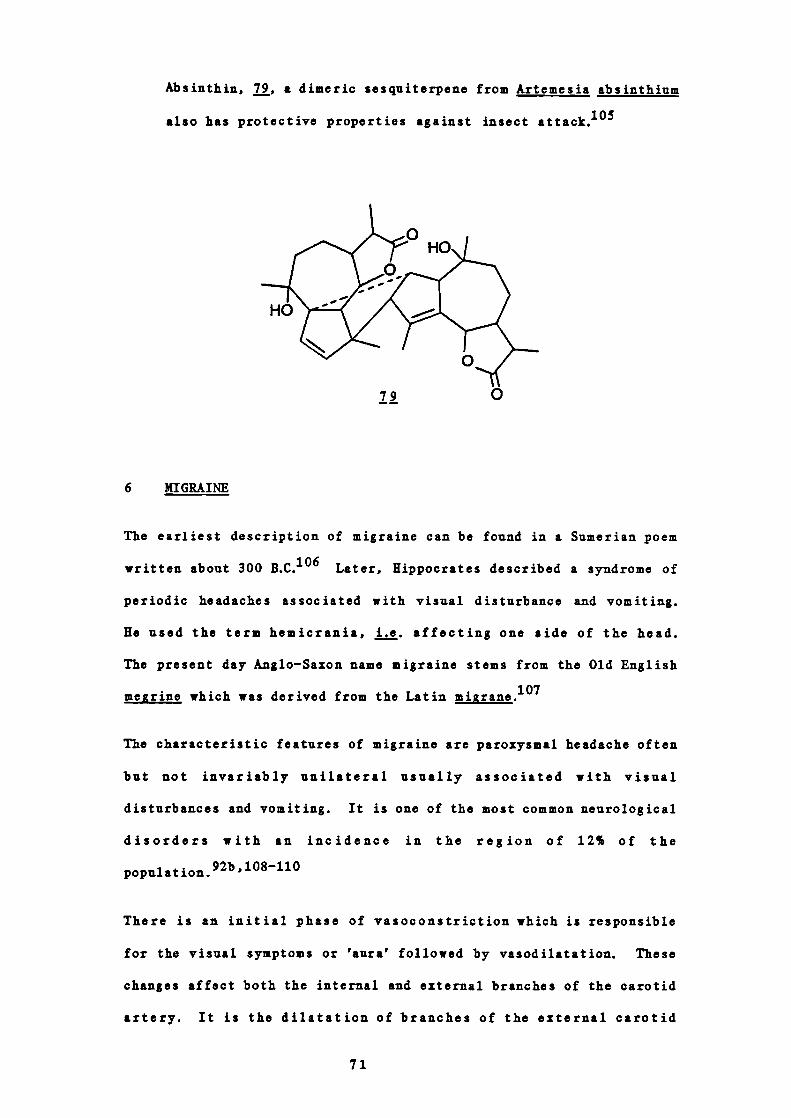
Absinthin, 79, a dimeric sesquiterpene from Artemesia absinthium
also has protective properties against insect attack.°5
6 MIGRAINE
The earliest description of migraine can be found in a Sumerian poem
written about 300 B.C. 106 Later, Hippocrates described a syndrome of
periodic headaches associated with visual disturbance and vomiting.
He used the term hemicrania, i.e. affecting one side of the head.
The present day Anglo—Saxon name migraine stems from the Old English
megrine which was derived from the Latin migrane.'°7
The characteristic features of migraine are paroxysmal headache often
but not invariably unilateral usually associated with visual
disturbances and vomiting. It is one of the most common neurological
disorders with an incidence in the region of 12% of the
population.92t' ,108-110
There is an initial phase of vasoconstriction which is responsible
for the visual symptoms or 'aura' followed by vasodilatation. These
changes affect both the internal and external branches of the carotid
artery. It is the dilatation of branches of the external carotid
71
artery and to a lesser extent the internal carotid artery which is
associated with the pulsatile headache. The vasoconstriction is
believed to be due to the release of amines such as noradrenaline
from neurones, 5-hydroxytryptamine from platelets and histamine from
mast celis.921'4°81
The cause (or causes) of migraine is unknown but there is a genetic
predisposition. Attacks are often related to some emotional
disturbance and sometimes precipitated by eating foods rich in
monoamines such as tyramine j. cheese.92bhlO8
In about one third of women migrainous symptoms are associated with
the menstrual cycle and this may account for the increased incidence
found in females.92bh]O9JHi Ovarian steroids and oral contracep-
tives may interfere with catecholamine metabolism. In addition, l7-
oestradiol and progesterone have been shown to potentiate the effects
of noradrenaline, adrenaline and tyramine.'' Prostaglandins may
also be involved in migraine since drugs inhibiting prostaglandin
synthesis such as aspirin are effective for both prophylactic and
acute treatment.
In the treatment of migraine, drugs with antinoradrenaline (e.g.
ergot alkaloids), anti-5-hydroxytryptamine (e.g. ergot alkaloids,
methylsergide, cyproheptidine) and antihistamine (e.g. prochlorpera-
zine) activities are all inconsistently efficacious. The action of
the ergot alkaloids probably depends upon the existing vascular tone.
Where there is low vascular tone the drug acts predominantly as a
constrictor agonist. This may be by action on adrenoreceptors or 5-
hydroxytryptamine receptors. En the presence of marked neurogenic
tone the a-adrenoreceptor blocking action of the drug is paramount
andvasodilatationresnits. The action of these drugs in migraine
72
therefore depends upon the stage at which they are administered. In
the constrictor stage the ergot alkaloids should cause dilation
thereby preventing the consequences of the constriction since it is
the initial vasoconstriction that appears to be responsible for the
subsequent features of the migraine attack. This may explain why
treatment with these drugs is more often successful when begun early
in the prodromal phase. The vasoconstrictor action of the alkaloids
can induce migraine if the vessels are not constricted at the time of
medication." The ergot alkaloids also have anti-5-hydroxytrypt-
amine actions and it may be that a successful migraine treatment
requires a combination of actions since the condition probably arises
from more than one cause.
In looking for new drugs with potential use in migraine it seems
reasonable therefore to test initially for spasmolytic activity j
vitro. An initial screen for activity must use a preparation that
responds well and gives rapid reproducible results. Such a prepara-
tion is the guinea pig ileum and appropriate agonists would be
vasoactive substances such as acetyicholine, bradykinin, histamine,
5-hydroxytryptamine and noradrenaline, all, at one time or another,
implicated in the pathogenesis of migraine."2
73
PART II
D IS CU S SI ON

Natural products have been and remain an important source of
biologically active compounds. The current misconception in the
medical profession as to the lack of value of these compounds has no
grounding. In 1973, the last year for which figures are available,
25.2% of the prescriptions dispensed in the United States contained
one or more constituents derived from higher plants. 113 The role of
these compounds in the development of new semi-synthetic or synthetic
drugs with enhanced efficacy or decreased toxicity cannot be under-
estimated. In addition the study of new compounds provides valuable
pharmacological information and, together with synthetic derivatives,
indications of possible structure-activity relationships. We should
not forget that virtually every pharmacological drug prototype
exhibiting theclassicaleffectsoftheclassconcernedisderived
from plant sources.
It is clear therefore that we must continue to look to plants as
sources of new drugs. These sources remain largely untapped. If the
main objective is to isolate constituents with pharmacological
activity the procedure undertaken must be directed precisely towards
this aim.
1 SCREENING PROCEDURE
Feverfew is a plant used for the alleviation of migraine. Since it
is not possible to produce migraine in experimental animals a
technique j vitro that could be related to the clinical situation
had to be selected. The cause of migraine is probably broadly based
so the initial screening for activity had to be such that false
negative results could be reduced to a minimum. This also
necessitated the use of a sensitive method since extracts prepared
may have contained only very small quantities of active compounds.
75
The use of large quantities of material required for a less sensitive
method would be impractical. The technique for evaluating biological
activity had also to provide rapid reproducible results.
Guinea pig ileum is a preparation that responds well and is readily
available. It was therefore selected for the primary screen. The
agonists acetyicholine, 5 —hydroxytryptamine and histamine were chosen
to give a broad guide to general spasmolytic activity. Such activity
would be useful in the treatment of migraine. The prostaglandin PGE2
was also used as an agonist in selected cases.
The procedure consisted of recording the log (dose) vs response
curves to acetylcholine, 5 —hydroxytryptamine and histamine in an
organ bath containing guinea pig ileum. ED 50 doses were taken and
given repeatedly until constant responses were achieved. The
antagonist i.e. the extract or fraction obtained from column
chromatography was then dissolved in the minimum of
dimethylsulphoxide and made up to a concentration of 1O 4 g/ml with
Irebs solution. This was added to the bath containing the ileum and
left for 30 minutes. The response of the tissue to the agonist was
then recorded and the percentage change taken. A control experiment
was performed in exactly the same way except that the antagonist was
omitted.
Figures 19 and 20 show typical results obtained. Figure 19 shows
sub—fractions A59-65 (see Part III, Table 19) caused 100% antagonism
to all three agonists at g/ml and the receptor sites of these
agonists remained blocked even after washing with Irebs solution.
The antagonism was therefore non—competitive. Figure 20 shows that
sub—fractions A30-34 (see Part III, Table 19) caused approximately
50% antagonism to the agonists but this blockade was reversible after
washing with rebs solution, i.e. competitive antagonism.
76

.1414
c
7C"4
.14
0
.14
0-I.14
14".4
I'.40
.14-4
-4.14U
U-4
"40E
-Ici
iI
sh
IT
La mine
agonist
SO control
IT
tamine
77

-4
14
U
01c1I
ci
-4a
70-4
U
U
0-I41C,
14
0
41-4
-441C,
U-441
-40a
N
N
ne
ne
ne
ilst
control
ne
78

2 EXTRACTION PROCEDuRE
Dried leaves of Chrysanthemum parthenium were exhaustively extracted
with solvents of increasing polarity: light petroleum, chloroform,
methanol and water. Each of these extracts was tested for
spasmolytic activity.
The methanol and water extracts showed no antagonism but the
petroleum and chloroform extracts did show antagonism of
acetyicholine, 5—hydroxytryptamine and histamine. In fact 100%
antagonism of the agonists was obtained using a concentration of 1O4
g/ml of the latter extracts. These two extracts were therefore
fractionated by column chromatography, and the fractions so obtained
tested for activity.
Since a great number of these fractions showed antagonism of the
agonists it was decided to proceed further only with the fractions
showing 90-100% activity at 1O 4 g/ml. These fractions were from the
light petroleum extract and were further sub —fractionated and tested.
Those sub—fractions still showing 100% antagonism were purified by
preparative TLC or column chromatography as appropriate and where
possible the active constituent isolated. In the majority of cases
the sub —fractions contained only one major compound. The light
petroleum extract possessed the greater activity so this extract was
investigated first. These results are summarised in Figures 21 and
22.
From Figure 22 it can be seen that the greatest activity resided in
fractions 5161, 75-80 (C), 119-123 (E), 124-143 (B), 144176 (A) and
177-200 (D). On examination of the TLC profiles of these combined
fractions it was observed that each contained at least one component
which gave a pink to purple colour after spraying with sulphuric acid
79
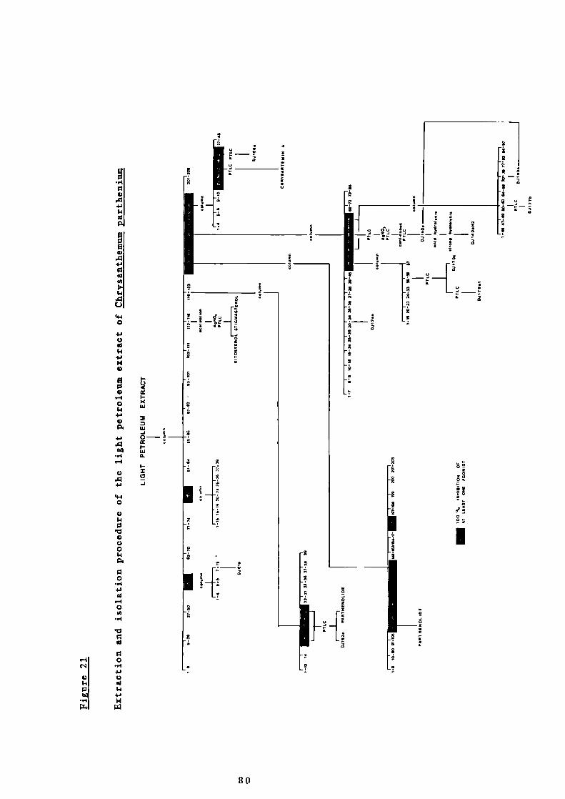
liii
z a
4 0
3
a
r41c'lI
4I
4
kU-
(4.4o
4'U
4' -
4)
4),-4 I-
o (Li
4'4)
_J 4"Li O-
(Lu_4 u.'-I
I-
4) I
'ii 9-j
'.40
4)110
VC)o
0
4'
—4
0
—4
,0
00
—4
C)
$1"a
H
ii-i4 o d'o o
- ía*9
94 9 a88 99 99 a99*9
9
0
0
I
HiH
80

NOI.LI9IHNI 0/0 IHOI]M %
E-4
C
441.4
F0a4).04)0
14,0U
44.40
4)0
'.444
C
I
C
a
14
a.4.I
aC1.4
U
E-40C,
C
.4.4
E014
44.4
C
-4
.4.4
0
0-44.4U
1.444.4
C
4.4
14.40
.4.4
-4C
C
4.4
CU1.4C
.4.4-4
-4.4.40
U-44.4
eliul 0141 E
;lI ri
81

followed by heating. Since such a reaction is often produced by
terpenes and moreover, an authentic specimen of parthenolide obtained
from Professor Sorm, gave a similar purple colour, it was considered
that these compounds were likely to be sesquiterpene lactones. In
addition from our knowledge of secondary plant metabolites found in
the family Compositse the sesquiterpene lactones are likely to be the
most biologically active group of substances.
In selecting the priority of the order of the work the weights of the
fractions, their complexity on TLC examination, as well as their
degree of inhibition of the three agonists were most important.
Nevertheless, in fractions where isolation of a particular component,
usually giving a pink to purple colour on spraying with sulphuric
acid, appeared particularly simple, purification was carried out.
This was done because even if the compound itself was found to be
inactive valuable information could possibly be gained as to the
nature of the hydrocarbon skeletons of the active substances. The
active compounds all proved to be sesquiterpene lactones however and
the non—active ones steroids, triterpenes or esters of long—chain
fatty acids and not, as hoped, sesquiterpene hydrocarbons.
The three most active fractions were 124-143 (B), 144-176 (A) and
177-200 (D). Fractions 124-143 (B) represented by far the largest
weight of the petroleum extract at 19% but by comparison with the
sample provided by Sorm the major component corresponded on TLC with
parthenolide. In addition, it showed only 91% inhibition of acetyl -
choline, but 1O activity against histamine and 5—hydroxytryptamine,
these latter two agonists probably being more important in the
pathogenesis of migraine. Similarly fractions 177-200 (D) showed
less activity against acetyicholine and moreover represented only 2!.
in weight of the original extract.
82

Fractions 144-176 (A) were therefore investigated first, 100%
inhibition being shown to all three agonists. In addition, at 5% of
the original extract weight, it was hoped enough active material
could be isolated to allow structure elucidation of the compounds.
However, since these compounds were to prove to be the most complex
of those isolated they will be discussed later. (Part II, 6)
3 INOWN COMPOUNDS
Five known sesquiterpene lactones have been reported to be present in
Chrysanthemum parthenium: parthenolide, chrysartemin A, chrysartemin
B, santamarine and reynosin. In the course of the present study only
the first two have been detected and isolated. This is because the
latter three compounds are probably located in the chloroform extract
of the plant which was not investigated here. It would be expected
that the latter three compounds would be responsible for some of the
activity shown by the fractionated chloroform extract. Furthermore,
as already stated the present work was governed by biological
activity, only those fractions having 100% activity to more than one
agonist being examined in detail. The chloroform fractions showed
less than 100% activity at the concentration tested so it may be
concluded that the compounds present were less active, or present in
lower concentrations than those in the light petroleum extract.
The possibility of the existence of more than chemotype cannot be
discounted at this stage but the time required to investigate this
suggestion was not available in the present study.
A PARTHENOLIDE
Parthenolide was first isolated from C. parthenium in 1959.38
83

It is the major secondary metabolite in the plant and is likely
to be formed biosynthetically by epoxidisation of the 4-double
bond of costunolide, the simplest sesquiterpene lactone.44
Bearing in mind the large quantity of parthenolide found in
feverfew it is reasonable to suggest that all subsequent
sesquiterpene lactones are derivatives of parthenolide.
Goviudachari al. revised the structure of parthenolide in
196542 and the absolute configuration was proved by Quick and
Rogers in 1976.
In the present work the parthenolide-containing fractions 124-
143 (B, in Figure 20) from chromatography of the petroleum
extract were further chromatographed on a column of silica gel
(see Figure 21). Parthenolide was located in sub-fractions B107-
111 and B112-115 and as suspected these fractions corresponded
with the major antagonist activity. It is interesting to note
however that sub-fractions B164-171 and B172-196 appeared to
show selective antihistamine activity and would therefore
warrant further investigation.
Parthenolide crystallised from chloroform and hexane as
colourless plates and was identical in all respects to an
authentic sample provided by Sorm. Since the original
structural work on this compound, the technique of NMR
spectroscopy has become routinely available and has been found
to be an extremely valuable tool in structural elucidation.59b
Accordingly the 13 C NMR spectrum of parthenolide was obtained in
the hope that its assignment would be of value in the structural
elucidation of the new compounds to be described below.
The decoupled 13C NMR spectrum of parthenolide in CDC1 3 solution
84

showed only fourteen clear signals. The position of the C-12
carbonyl signal could not be unequivocally located. It is not
unusual that carbonyl signals are often very weal and not easily
detected.59C The remaining signals were assigned with reference
to the fully coupled spectrum, use of tables of chemical
shiftsS9d and comparison with the data of known sesquiterpene
lactones. 8 ' 14 ' 7 Unequivocal assignments were not possible
in every case but the most likely are shown in Table 5.
B CURYSARTEMIN A
Chrysartemin A50 ' 5 ' was isolated from fractions 177-200 (D) from
the original light petroleum extract. These fractions showed
100% activity against 5 —hydroxytryptamine and histamine. The
combined fractions were further separated on a second silica gel
column and most of the activity was shown to reside in the three
sub— fractions Dl1-15, D16-20 and D21-26. The latter two sub—
fractions showed 100% activity against all three agonists.
Fractions D11-15 and D16-20 contained the same pink—reacting
component but other different components were present in each
set. In fractions D11-15 the pink—colouring component was not
the major one whereas it was in fractions D16-20. In addition
fractions D16-20 weighed 240 mg compared with 88 mg for
fractions D11-15. Fractions D16-20 were therefore further
purified by preparative TLC because of the greater likelihood of
isolating the active principle. The major component
crystallised from chloroform and hexane to give a compound the
data of which was identical with those reported in the
literature for chrysartemin A.50'5'
Previously however the NMR spectrum had only been recorded in
85

Table 5
NMR data for parthenolide (6, CDC13)
Multiplicity in off—Chemical shift
Carbon no.resonance spectrum
16.97 q 14 or 15
17.24 q 15 or 14
24.11 t 8or 3
30.64 t 3 or 8
36.35 t 9or 2
41.19 t 2or 9
47.65 d 7
61.45 d 5
66.36 s 4
82.38 d 6
121.08 d 1
125.24 t 13
134.51 s 10
139.21 $ 11
5The signal for the carbonyl carbon (C-12) was not observed
86

dimethyl suiphozide as solvent. With the increased sensitivity
of Fourier transform instruments it was now possible to obtain a
spectrum from a CDC1 3 solution and the data is reported in Part
III, 14C(a).
4 STRUCTURAL ELUCIDATION OF D1156a
The major component of sub—fractions D21-26 of fractions 177-200 (D)
(see Figure 21) was a component which gave a pink colour after
spraying with sulphuric acid and heating but had an R f value
different from that of chrysartemin A. This component also showed
100% inhibition of the three agonists. It was therefore isolated
after purification by preparative TLC using chloroform:methanol
(95:5) as developing solvent. The material (D1156a) crystallised
from chloroform and hexane to give 16 mg of white needles, m.p.
154°C.
The mass spectrum of this material did not show a clear molecular ion
but showed peaks at m/z 248 (6.0%), 246 (18.0%) and 244 (13.5%)
together with their appropriate isotope peaks. At first
therefore it was thought the material could be a mixture of
homologues. The 'K NMR spectrum however showed signals for only one
compound and the relatively high, sharp melting point also indicated
homogeneity. On more detailed examination of the mass spectrum it
was considered that the peaks at m/z 248-244 could really be fragment
ions since a cluster of low intensity ions was visible at m/z 262-
266. These could have given rise to the previous ones by loss of a
molecule of water.
Extra weight to this suggestion of ready dehydration was given by
examination of the infrared spectrum which showed a strong absorption
87

band at 3490 cm' for the presence of at least one hydroxyl group.
The 400 MHz NMR spectrum however did not show a signal for a
methine hydrogen (—Ca—OR) and therefore the hydroxyl group or groups
was likely to be tertiary. Tertiary alcohols are readily
dehydrated6SC and consequently rarely give a molecular ion peak in
the mass spectrum. It was likely then that the peaks at m/z 244-248
could have arisen by loss of water from those at m/z 262-266. In
addition however peaks at mlz 228 (19.8%) and 230 (10.0%) were
observed, these perhaps being due to loss of a second molecule of
water. The peaks at m/z 266 and 262 were of too low intensity to be
measured but accurate mass measurement at m/z 264 indicated a
molecular formula of C 15H2004 . This, together with the pink TLC
appearance of the material after spraying with sulphuric acid
followed by heating, indicated that the material was likely to be a
sesquiterpene lactone. Since parthenolide (C 15H2003 ) is the major
sesquiterpene lactone in Chr ysanthemum parthenium it is likely that
the new compound is an oxidised derivative thereof.
The infrared spectrum of this new compound showed absorption bands at
1750, 1660 and 1640 cm', characteristic of an a—methy1ene——lactone.
The presence of a conjugated lactone was confirmed by the strong
ultraviolet absorption at 208 nm (log a 3.95). In addition the 400
MBz 1R NMR spectrum showed two very sharp doublets at 65.54 and 6.25
(1 3.5 Hz) for the hydrogens of an ezocyclic double bond as in
parthenolide.42 These were assigned to R-13a and b. Such a coupling
in this class of compounds, i.e. sharp doublets, is due to an allylic
interaction with H-7. 53 ' 54 The smaller geminal coupling (I 1 Hz)
is not observed unless an 8a —oxygen substituent is also present55
(see Part I, 4A(a)).
In the present spectrum since these occurred as very sharp doublets
88

it was concluded that C-8 carried no a—oxygen function. This assumes
that the compound was lactonised at C-6 as in parthenolide.42
No other low field signals were observed in the NMR spectrum
showing that no additional vinyl hydrogens were present in the
molecule. The next lowest field signal in the spectrum was a clear
doublet of doublets at 64.24 (111, 1 = 12.3, 9.5 Hz) readily
assignable to 11-6 in sesquiterpene lactones. A spin—decoupling
experiment by irradiation at 64.24 caused another doublet of doublets
at 62.39 (1 = 12.3, 12.3 Hz) to collapse to a doublet and
simplification of the complex multiplet at 62.70 (1K) to reveal a
clear long range coupling of 3.5 Hz. The hydrogens giving rise to
these signals are therefore both coupled to 11-6 (but not to each
other) and thus may be readily assigned to H—S and 11-7 respectively.
Further, the signal at 62.70 showed a coupling of 3.5 Hz j. equal
to the coupling observed for H-13a and b. This is obviously due to
the allylic coupling with 11-7. The signal at 62.70 was thus
unequivocally assigned to 11-7. As extra evidence, on irradiation of
this signal the doublets at 65.54 and 6.25 did indeed collapse to
singlets, so confirming the signal at 62.70 to be due to K-7. From
the complexity of the signal at 62.70 and the appearance of the
exocyclic methylene signals, H-8 must carry two hydrogens.
Irradiation at 62.70 also caused considerable simplification of the
multiplets at 62.16 and 1.47 which were thus assigned to the C-8
hydrogens.
The doublet of doublets at 62.39 (1 12.3, 12.3 Hz) must thus be due
to H-5. On irradiation at 64.24 (11-6) the signal for 11-5 collapsed
to a doublet (I 12.3 Hz), implying that the carbon adjacent to H—S
carries only one hydrogen. Irradiation at 11-5 caused the 11-6 signal
(64.24) to collapse to a doublet (I 9.5 Hz) and also great simpli-
89

fication of the signal at 62.63. This latter signal was thus due to
the hydrogen on a carbon atom adjacent to C —S. The chemical shift of
H-5 (62.70) excludes the possibility of C-5 also carrying an oxygen
function.
As is likely from biosynthetic considerations the 'H NMR spectrum of
this material showed signals for two methyl groups. These were sharp
singlets integrating for three hydrogens each at 61.25 and 1.35.
Since they are sharp singlets they must be due to tertiary methyl
groups. Furthermore the chemical shifts of these indicate they are
attached to carbons carrying oxygen.
C— s carries no oxygen (from the chemical shift of H-5) on a methyl
group, the methyl groups being tertiary. The compound must therefore
be cyclised at C—S.
There are two possible modes of cyclisation of germacranolides i.e.
to eudesmanolides or guianolides (see Figure 5). A eudesmanolide
skeleton may be excluded by consideration of the chemical shift of
the methyls and the fact that their 'H NMR signals were singlets. In
addition an eremophilanolide structure, which could arise from a
methyl migration of a eudesinanolide (see Figure 5) was not admissable
either since C-5 was known to carry a hydrogen atom. This, together
with the low chemical shift of H-6, strongly indicated a guianolide
skeleton. As with the eremophilanolide, an ambrosanolide or
helenanolide structure, resulting from a methyl migration in a
guianolide, were also clearly inadmissable (see Figure 5).
Incidentally, these data also provide extra evidence that the new
compound is a C-6 lactonised sesquiterpene rather than being
lactonised at C-8. As has been previously mentioned, irradiation at
H-7 clearly located the hydrogen on the adjacent carbon atom to be at
90
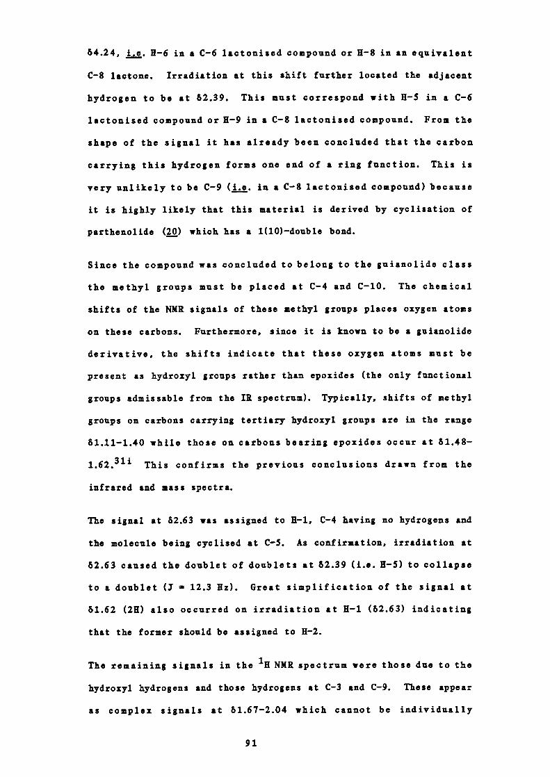
64.24, j. H-6 in a C-6 lactonised compound or H-8 in an equivalent
C-8 lactone. Irradiation at this shift further located the adjacent
hydrogen to be at 62.39. This must correspond with H —S in a C-6
lactonised compound or H-9 in a C-8 lactonised compound. From the
shape of the signal it has already been concluded that the carbon
carrying this hydrogen forms one end of a ring function. This is
very unlikely to be C-9 (j. in a C-8 lactonised compound) because
it is highly likely that this material is derived by cyclisation of
parthenolide (Q) which has a 1(10) —double bond.
Since the compound was concluded to belong to the gulanolide class
the methyl groups must be placed at C-4 and C-10. The chemical
shifts of the NMK signals of these methyl groups places oxygen atoms
on these carbons. Furthermore, since it is known to be a guianolide
derivative, the shifts indicate that these oxygen atoms must be
present as hydroxyl groups rather than epoxides (the only functional
groups admissable from the 1K spectrum). Typically, shifts of methyl
groups on carbons carrying tertiary hydroxyl groups are in the range
61.11-1.40 while those on carbons bearing epoxides occur at 61.48 —
This confirms the previous conclusions drawn from the
infrared and mass spectra.
The signal at 62.63 was assigned to H—i, C-4 having no hydrogeus and
the molecule being cyclised at C—S. As confirmation, irradiation at
62.63 caused the doublet of doublets at 62.39 (i.e. H-5) to collapse
to a doublet (1 12.3 Hz). Great simplification of the signal at
61.62 (2K) also occurred on irradiation at 11-1 (62.63) indicating
that the former should be assigned to 11-2.
The remaining signals in the ill NMR spectrum were those due to the
hydroxyl hydrogens and those hydrogens at C-3 and C-9. These appear
as complex signals at 61.67-2.04 which cannot be individually
91

assigned.
The only structure compatible with these data is 80, 4a, i0-
dihydroxy-i, 5a-gui-ii(13)en12, 6a-olactone. The compound is
given the trivial name partholide.
OH
This overall structure was confirmed by examination of the ' 3 C NMR
spectrum. Fifteen signals were present in the fully decoupled
spectrum. These were assigned with reference to the coupled INEPT
spectrum, consideration of the chemical shifts and comparison
with NMR data of parthenolide and other sesquiterpene
lactones. 8"147 The data is given in Table 6.
Possible biosynthetic routes to the new compound are given in Figure
23.
Assuming the new material is derived from parthenolide, the
stereochemistry at C-4, C-6 and C-7 is known. There are two
asymmetric centres at C-i and C-5 and therefore four possible isomers
with regard to the ring junction. In addition there are two further
possibilities since the stereochemistry at C-i0 is not yet proved.
Thus there are eight stereochemical possibilities which are
summarised in Figure 24.
The NMR spectrum showed that the coupling constant between H-5 and
92

Table 6
NMR data of D3156a, 80 (6, CDC13)
Multiplicity 3Chemical shift in INEPT Carbon no.
spectrum (Hz)
23.51 q 125 14 or 15
24.25 q 120 15 or 14
25.01 t 112 2 or 8
25.33 t 150 8 or 2
39.34 t 130 9 or 3
43.77 t 128 3 or 9
47.22 d 140 7
49.78 d 130 1
55.35 d 130 5
74.72 s - lOor 4
79.88 $ - 4orlO
82.74 d 150 6
120.37 t 163 13
138.45 $ - 11
169.37 s - 12
93

OH /..
figure 23
Possible biosynthetic route to D1156a
7
082
83
94
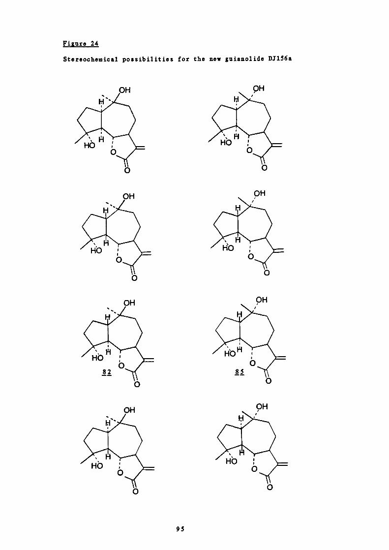
HO0
OH
OH
0
OH
Figure 24
Stereochemical possibilities for the new guianolide DJ156a
H
pH
HO
0
082
95
H6 6 is 12.3 Hz, is 12.3 Hz and 6 7 is 9.5 Hz. From5, , I
consideration of the Karplus relation, 65 such large coupling
constants could only be produced by a structure with large dihedral
angles between the appropriate vicinal hydrogens. Examination of
Dreiding models showed that such angles could only be produced by a
compound with a trans ring junction. Similarly, H-5 and R-6 must be
trans. By analogy with parthenolide (the stereochemistry of which
has been confirmed by X—ray crystallography45 ), since H-6 is , the
stereochemistry of H-5 is a and H —i, . Moreover, that i 6,7 is 9.5
Hz also indicates the presence of a trans lactone which, since fl-6 is
, fixes H-7 as a. This is of course expected if Q is derived from
parthenolide and has been assumed in the possibilities under
consideration. This leaves only two allowable structures J.2 .and
85.
The final question to be resolved is thus the stereochemistry at C-
10. This may best be postulated by a consideration of the likely
mode of biosynthesis of the compound.
With reference to Figure 23, two routes are possible. The simplest
involves the equivalent of acid catalysed opening of the 4,5—epoxide
followed by trans — annular attack by the it—electrons of the 1(10) —
double bond to form the guiane skeleton with a carbonium ion at C—iO
(81). In solution, such a cyclisation would be expected to proceed
in an anti fashion 8 and, since the C—O bond at C—S is , so produce
a 5p ring junction. From the NMR data however there is no doubt that
the hydrogen at C-5 is a so it may be concluded that the cyclisation
proceeds by an enzyme—mediated process such that the germacranolide
is adsorbed onto the surface of the appropriate enzyme which thus
directs the steric course of the reaction to yield exclusively the
5a—product. This would involve attack of the n—electrons of the
96
1(10) —bond from the top of the molecule j. from the same side as
the C(5)—O bond is broken. As the double bond is destroyed, rotation
of the 1(10) —bond occurs to yield the product with a 1 hydrogen and
the planar carbonium ion at C-10 as shown in . Finally, attack by
0H would take place from the less hindered face of the molecule
which is obviously also the top side so yielding which has an a
methyl group.
0
Similar considerations apply to the other possible route via the 9—
olefin (a). Epoxidation to yield would also take place from the
less hindered () face, opening of which by a cis addition of
hydrogen would also yield .
On examination of Dreiding models it is seen that the 7 —membered ring
may assume a conformation to minimise intramolecular interactions in
which such an a methyl group is equatorial. This is thus also the
thermodynamically favoured product. Incidentally, these deductions
may be taken to strongly indicate that is a true natural product
since if it were an artefact produced in solution during extraction
or purification, the material isomeric at C—S could be expected to be
formed.
S STRUCTURAL ELUCIDATION OF D1177b
Four components giving a pink to purple colour on spraying with
sulphuric acid and heating were present in fractions 144-176 (A) from
column chromatography of the light petroleum extract.
These fractions exhibited 100% activity to all three agonists and
were therefore further separated on a second silica gel column and
the sub—fractions tested for activity.
97

Sub—fractions A53-58 of the second column showed 100% inhibition of
all three agonists and sub — fractions A66-67 100% inhibition of 5 -
hdroxytryptamine and histamine. Both sets of sub —fractions contained
a different major pink—reacting component but several similar
components. It was therefore decided the best way to separate the
components, and to minimise losses, was to combine these two sub —
fractions and rechromatograph them on a third silica gel column.
From this third column fractions Aa50-63 contained only one major
component that appeared pink after spraying with sulphuric acid and
heating. The second pink — reacting substance was isolated
subsequently (fractions Aa70-76) and will be discussed later (part
II, 6). The former fractions (Aa50-63) were further purified by
preparative TLC and the major component crystallised from chloroform
and hexane to give 8 mg of D1177b as colourless needles, m.p. 116-
117°C.
The mass spectrum appeared to show a molecular ion peak at mlz 264
(40.8%) with a isotope peak at mlz 265 (16.8%). However, small
peaks were also observed at m/z 278 (0.2%), m/z 279 (2.6%) and m/z
280 (2.0%). Such a molecular weight together with the pink to purple
colour produced on spraying with sulphuric acid and heating indicated
a possible sesquiterpene lactone skeleton. This was confirmed by the
presence of various characteristic spectral features described below.
The infrared spectrum showed a strong absorption band at 3540 cm
indicating the presence of at least one hydroxyl group. In addition,
as in compounds such as parthenolide 42 absorption bands were observed
at 1760, 1665 and 1640 cm', these being characteristic of an a -
methylene — y —lactone grouping. The presence of such a conjugated
lactone was confirmed by the strong ultraviolet absorption at 215 nm
(log a, 4.10). Infrared absorption bands were also present at 1250,
98
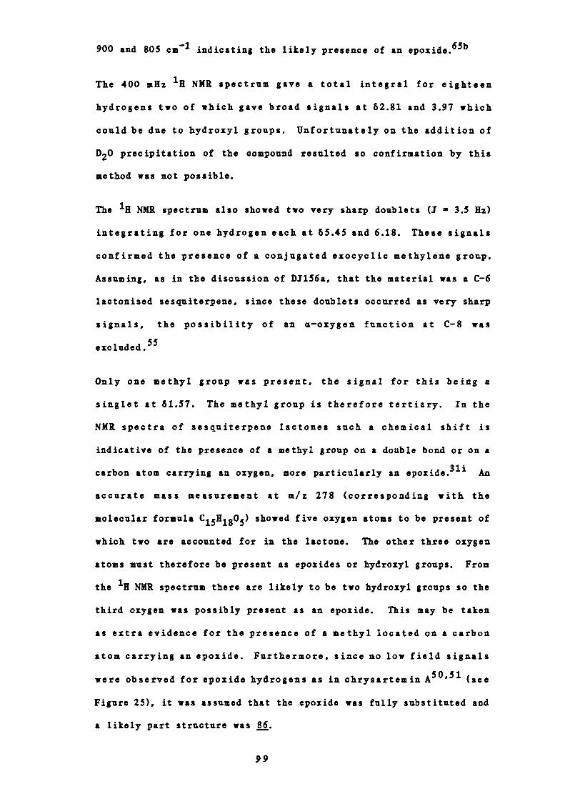
900 and 805 cm indicating the likely presence of an epoxide.65b
The 400 mHz 'H NMR spectrum gave a total integral for eighteen
hydrogens two of which gave broad signals at 62.81 and 3.97 which
could be due to hydroxyl groups. Unfortunately on the addition of
D20 precipitation of the compound resulted so confirmation by this
method was not possible.
The H NMR spectrum also showed two very sharp doublets (1 3.5 Hz)
integrating for one hydrogen each at 65.45 and 6.18. These signals
confirmed the presence of a conjugated exocyclic methylene group.
Assuming, as in the discussion of DJ156a, that the material was a C-6
lactonised sesquiterpene, since these doublets occurred as very sharp
signals, the possibility of an a—oxygen function at C-8 was
excluded
Only one methyl group was present, the signal for this being a
singlet at 61.57. The methyl group is therefore tertiary. In the
NMR spectra of sesquiterpene lactones such a chemical shift is
indicative of the presence of a methyl group on a double bond or on a
carbon atom carrying an oxygen, more particularly an epoxide.31 ' An
accurate mass measurement at m/z 278 (corresponding with the
molecular formula C15111805 ) showed five oxygen atoms to be present of
which two are accounted for in the lactone. The other three oxygen
atoms must therefore be present as epoxides or hydroxyl groups. From
the 'H NMR spectrum there are likely to be two hydroxyl groups so the
third oxygen was possibly present as an epoxide. This may be taken
as extra evidence for the presence of a methyl located on a carbon
atom carrying an epoxide. Furthermore, since no low field signals
were observed for epoxide hydrogens as in chrysartemin A 50 ' 5 ' (see
Figure 25), it was assumed that the epoxide was fully substituted and
a likely part structure was
99

63.32, d, 3 = 1.3 Hz
63.57, d, 3 = 1.3 HzH
The chemical shift of the methyl group (61.57) indicated a guianolide
structure as in chrysartemin A (C —b methyl at 61.5 8)50151 rather
than a less rigid germacranolide as in parthenolide (61.32).42
Further examination of the NMR spectrum showed that a second
exocyclic methylene group was present, the signals for which appeared
as broad singlets integrating for one hydrogen each at 64.88 and
5.21. (cf. reynosin, 64.90, 5.0349,50, dehydrocostuslactone, ,
65.08, 5 .27 119 - see Figure 26). This accounts for the second methyl
group usually present in sesquiterpenes which in this case has become
oxidised to give a methylene function.
Figure 25
Structure of chrysartemin A shoving the epozide hydrogens and their
chemical shifts in the NKR spectrum (6, CDC13)
100
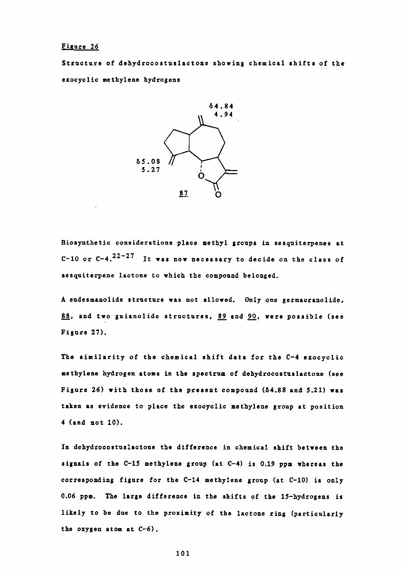
85.085.27
E.jgure 26
Structure of dehydrocostuslactone showing chemical shifts of the
exocyclic methylene hydrogens
84.84A OA
Biosynthetic considerations place methyl groups in sesquiterpenes at
C-10 or C-4.2227 It was now necessary to decide on the class of
sesquiterpene lactone to which the compound belonged.
A eudesmanolide structure was not allowed. Only one germacranolide,
88, and two guianolide structures, and 90, were possible (see
Figure 27).
The similarity of the chemical shift data for the C-4 exocyclic
methylene hydrogen atoms in the spectrum of dehydrocostuslactone (see
Figure 26) with those of the present compound (84.88 and 5.21) was
taken as evidence to place the exocyclic methylene group at position
4 (and not 10).
In dehydrocostuslactone the difference in chemical shift between the
signals of the C-iS methylene group (at C-4) is 0.19 ppm whereas the
corresponding figure for the C-14 methylene group (at C-b) is only
0.06 ppm. The large difference in the shifts of the 15-hydrogens is
likely to be due to the proximity of the lactone ring (particularly
the oxygen atom at C-6).
101

J gure 27
Possible part structures for D1177b
In the spectrum of D1177b, the difference is even larger at 0.33 ppm
which may not only be taken as evidence for the location of the
methylene function at C-4 but also the presence nearby of additional
functional groups causing greater non-equivalence of the hydrogen
nuclei.
Spin-decoupling NMR experiments assisted in subsequent structural
elucidation. Irradiation at 64.88 caused the signal at 65.21 to
collapse and vice versa thereby showing them to be coupled. On
irradiation at 84.88 however and thus removing the signal at 65.21, a
doublet at 85.23 (1K, 3 = 11.0 Hz) was more clearly seen. This was
only partially observed in the fully coupled spectrum. At first
sight this signal appeared to be due to the presence of a vinyl
102

(
hydrogen as in parthenolide 42 (measured in the present work at 65.23,
J 12.5 Hz, long range coupling 3.0 Hz) and the signal at 64.41 due
to H-6. R-6 however occurs as a characteristically sharp doublet of
doublets when there is a hydrogen at both C-5 and C-7 or as a clear
doublet when C-5 carries no hydrogen atoms. The signal at 64.41 in
the spectrum of this compound was rather broad however. It was
therefore concluded that the signal for E-6 was not in fact at 64.41
but was more likely to be the unresolved doublet at 65.23. This is a
very low chemical shift for the C-6 hydrogen in the spectra of these
compounds and thus indicated the presence of a grouping that was
causing considerable deshielding.
For example, in eupachloroxin 120 , 91, H-6 appears as a doublet at
65.02, and in pj—eupatoroxin120 , , a doublet at 65.08.
The epoxide cannot be present between C-3 and C-4 because of the
absence of low field epoxide hydrogeas but if the exocyclic methylene
group was at C-4 with a hydroxyl group at C—S the signal for the C-6
21.
hydrogen could be deshielded to 65.23 (see Figure 28). If this is
the case, the germacranolide structure must be excluded since as the
103

signal for 11-6 appears as a doublet there can be no hydrogens at C—S.
C-7 bears the lactone and is never di —substituted and therefore must
have one hydrogen.3
Figure 28
Possible part structure for Dfl77b
The placing of the exocyclic methylene group at C-4 is as earlier
stated consistent with the chemical shift analogies with the spectrum
of dehydrocostuslactone (Figure 26).
The coupling constant of 11.0 Hz for the signal at 65.23 is
consistent with a Dreiding model prediction for 11-6 in the usual
trans lactone. The signal at 62.31 is a complex multiplet
integrating for the hydrogen with coupling constants of 13.5 and 11.0
Hz. It was suspected therefore that this signal was due to 11-7.
Irradiation at this position caused a large reduction in the doublet
at 65.23. This, together with the coupling constant of 11.0 Hz
proved the signal at 65.23 to be due to 11-6 and that at 62.31 due to
11-7.
Irradiation at 62.31 also caused the low field doublets at 65.45 and
6.18 (i.e. the C-13 hydrogens) to collapse to singlets. This was
extra proof that the signal at 62.31 was due to 11-7, the coupling of
the C-13 hydrogens being due to allylic coupling with the C-7
104

hydrogen.53'54
In addition, on irradiation at H—i, the signal at 61.98 (integrating
for two hydrogens) was substantially reduced to a broad doublet with
a large coupling constant consistent with a geminal coupling. The
signal at 61.98 was thus assigned to the 8—hydrogens. Similarly, on
irradiation at 61.98 the signal at 61.73 (integrating for two
hydrogens) reduces to a broad doublet with a large coupling constant
and was thus assigned to the C-9 hydrogens.
From the chemical shift of the signal at 64.41 and its appearance it
was assigned to the hydrogen on a carbon atom carrying a hydroxyl
group. In pleniradin, the hydrogen at C-2 which also carries a
hydroxyl group resonates at &4.Z8.31 Similarly in j—eupatoroxin
it is at 64.50,311,120 in eupatoroxin at 64.30,311 and in eupatundin
at 64•2_4•5•31m In zaluzanin A, which has a methylene group at C-4,
the hydrogen at C-3 which also carries a bydroxyl group resonates at
54•53•31 Thus a signal for a methine hydrogen on a carbon carrying
a hydroxyl group at 64.41 is in the normal range for guianolides.
As C-8 bore no hydrozyl function it could only be placed at C-2 or C-
3, C-5 having already been shown to have no hydrogen. Irradiation at
the exocyclic metbylene signal at 64.88 caused the signal at 64.41 to
be simplified showing the corresponding hydrogen atoms to be coupled.
A likely explanation for this is that the signal at 64.41 due to a
methine hydrogen was allylically coupled with the 15 —hydrogens which,
of course, could only occur if the secondary hydroxyl group and thus
the methine hydrogen was present at C-3 (and not position 2). On
this basis therefore the hydroxyl group was placed at C-3.
Confirmation was to some extent provided by the attempted acetylation
of the material. Only a small amount of sample could be used for the
105
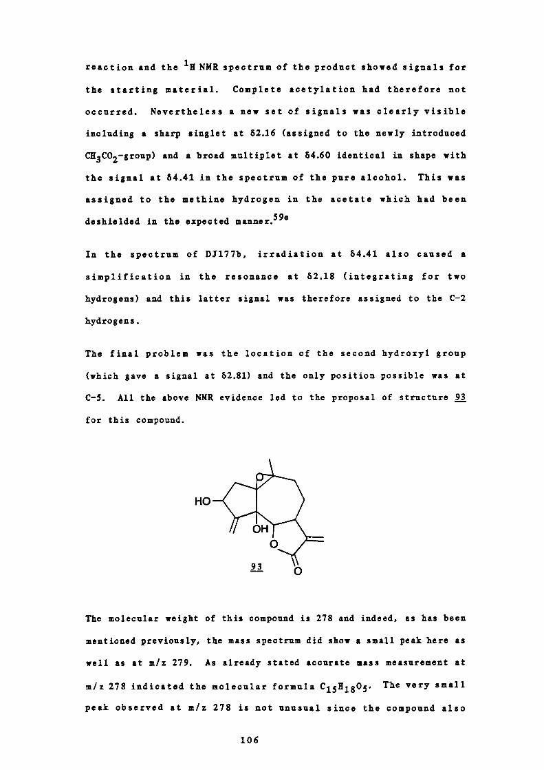
HO
reaction and the NMR spectrum of the product showed signals for
the starting material. Complete acetylation had therefore not
occurred. Nevertheless a new set of signals was clearly visible
including a sharp singlet at 82.16 (assigned to the newly introduced
C113 CO2—group) and a broad multiplet at 84.60 identical in shape with
the signal at 64.41 in the spectrum of the pure alcohol. This was
assigned to the methine hydrogen in the acetate which had been
deshielded in the expected manner.59e
In the spectrum of D1177b, irradiation at 64.41 also caused a
simplification in the resonance at 82.18 (integrating for two
hydrogens) and this latter signal was therefore assigned to the C-2
hydrogens.
The final problem was the location of the second hydroxyl group
(which gave a signal at 62.81) and the only position possible was at
C-5. All the above NMR evidence led to the proposal of structure 21
for this compound.
The molecular weight of this compound is 278 and indeed, as has been
mentioned previously, the mass spectrum did show a small peak here as
well as at m/z 279. As already stated accurate mass measurement at
m/z 278 indicated the molecular formula C 15 H 18 0 5 . The very small
peak observed at m/z 278 is not unusual since the compound also
106
contains a tertiary hydroxyl group which would readily dehydrate.65'
In fact, a much larger peal was observed at m/z 260 (8.0%),
corresponding with (M - 18).
The large peak at m/z 264 observed in the mass spectrum was concluded
to have arisen from the loss of a methyl radical from m/z 279, i.e.
(M+l).
A quantity of material sufficient to obtain a NMR spectrum to
confirm the above proposal was unfortunately not available.
The problem of the stereochemistry of D1177b is a difficult one. In
the proposed biosynthetic route (see Figure 29) the existing
stereochemistry is destroyed at both ring junction carbons.
Furthermore, since a hydroxylase enzyme is likely to be involved in
the introduction of the 3-hydrozyl group either epimer may result.
Examination of the coupling constants in the 'H NMR spectrum is of no
value since C-5 bears no hydrogen atom to interact with R-6 (with
fixed stereochemistry). Study of Dreiding models does not indicate
which of the sixteen possible isomers of D1177b is more or less
favoured than the others so unfortunately no stereochemical proposals
may be made other than to state that the C-6/C-7 junction is trans as
in parthenolide.42
The compound, 9, 10-epoxy-3, 5-dihydroxyguia-4(15), ll(13)-dien-12,
6a-olactone is a new sesquiterpene lactone and is given the trivial
name chrysanthemolide.
6 STRUCTURAL ELUCIDATION OF D5140a
Four components giving a pink to purple colour on spraying with
sulphuric acid and heating were present in the highly active light
107
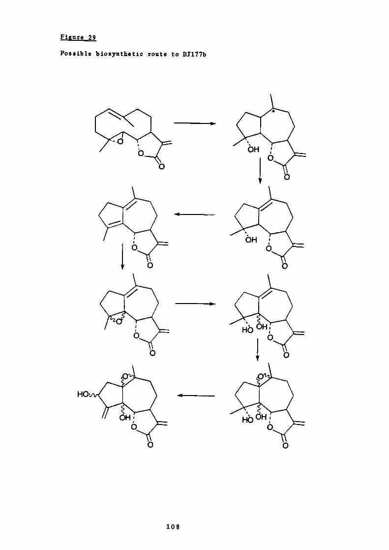
Figure
Possible biosynthetic route to Dfl77b
-4
-1
108
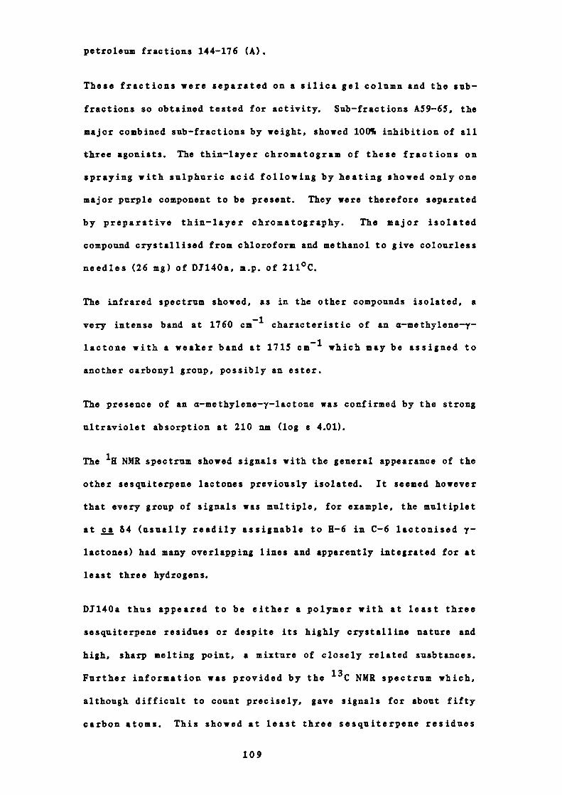
petroleum fractions 144-176 (A).
These fractions were separated on a silica gel column and the sub —
fractions so obtained tested for activity. Sub —fractions A59-65, the
major combined sub—fractions by weight, showed 100% inhibition of all
three agonists. The thin—layer chromatogram of these fractions on
spraying with sulphuric acid following by heating showed only one
major purple component to be present. They were therefore separated
by preparative thin— layer chromatography. The major isolated
compound crystallised from chloroform and methanol to give colourless
needles (26 mg) of DJ140a, m.p. of 211°C.
The infrared spectrum showed, as in the other compounds isolated, a
very intense band at 1760 cm characteristic of an a—methylene-7-
lactone with a weaker band at 1715 cm which may be assigned to
another carbonyl group, possibly an ester.
The presence of an a—methylene—y—lactone was confirmed by the strong
ultraviolet absorption at 210 nm (log a 4.01).
The NMR spectrum showed signals with the general appearance of the
other sesquiterpene lactones previously isolated. It seemed however
that every group of signals was multiple, for example, the multiplet
at ca 64 (usually readily assignable to H-6 in C-6 lactonised y -
lactones) had many overlapping lines and apparently integrated for at
least three hydrogens.
D1140a thus appeared to be either a polymer with at least three
sesquiterpene residues or despite its highly crystalline nature and
high, sharp melting point, a mixture of closely related susbtances.
Further information was provided by the ' 3 C NMR spectrum which,
although difficult to count precisely, gave signals for about fifty
carbon atoms. This showed at least three sesquiterpene residues
109

indeed to be present.
The mass spectral data on the other hand did not confirm this since,
either with the use of various voltages of electron impact or using a
chemical ionisation technique no corresponding molecular ion was
visible. Thin—layer chromatography in many different solvent systems
even using silver nitrate impregnated silica gel failed to show more
than a single discrete spot.
It was thus considered that the material could be a mixture of
conformational isomers. These are known to occur in the sesquiter -
penes particularly those with a large and thus conformationally
mobile ring such as the germacranolide isabelin (see Part I,
4A(d)). 6 ° It had been found before that recording the NMR spectrum
at low temperatures using pre —cooled solvent to dissolve the crystals
did allow signals for the predominate conformer of isabelin tobe
recorded. 6 ° Accordingly, a series of variable temperature u NMR
experiments was carried out on D1140a but no significant change in
the appearance of the signals could be detected.
Because of these results there was no option but to consider that the
material was a molecule with more than one sesquiterpene residue
which, for some reason, did not show a molecular ion on mass
spec trome try. Such materials are unusual in nature but there is
precedent for the existence of two types of such complex substances -
novel carbocyclic compounds such as absinthin, 79,121 and esters of a
sesquiterpene acid and a sesquiterpene alcohol such as 1,10-
dihydrolactucin-8—O--j—hypoglabrate, 94•122
With the nature of these materials in mind, and since the infrared
spectrum of DJ140a showed a band at 1715 cm which may be due to an
ester it was decided to try and hydrolyse the material in the hope of
110

0
simplifying the problem. If it were indeed an ester and so
hydrolysable with base the spectra of the products should be easier
to interpret. It is known however that on simple treatment with
alcoholic alkali C-6 lactonised sesquiterpene lactones are destroyed
and if an ester or hydroxyl function is present at C-8,
relactonisation occurs at C-8 during the work up, as in Figure 30.123
94
In an attempt to avoid this complication the material was first
treated under very mild conditions (dilute aqueous potassium
carbonate solution added to a dioxan solution of D1140a)) 23 No
change occurred however even after heating for several days. The
addition of further potassium carbonate solution caused no reaction
and so a few drops of potassium hydroxide solution was added.
Examination by thin-layer chromatography now did show the appearance
of more polar products but before the starting material had been
entirely consumed, the reaction was stopped and worked up in the
usual way. Recovered D1140a was recycled. Preparative thin-layer
chromatography allowed isolation of a more polar non-acidic product,
DI14OaH2, examination of the NMR spectrum of which was
illuminating.
This compound was much simpler than the natural product and most
signals were readily identifiable in terms of a dimeric sesquiterpene
111
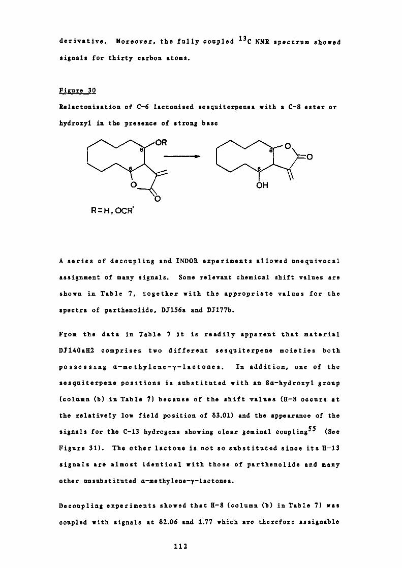
derivative. Moreover, the fully coupled NMR spectrum showed
signals for thirty carbon atoms.
Figure 30
Relactonisation of C-6 lactonised sesquiterpenes with a C-8 ester or
hydroxyl in the presence of strong base
RH, OCR
OH
A series of decoupling and INDOR experiments allowed unequivocal
assignment of many signals. Some relevant chemical shift values are
shown in Table 7, together with the appropriate values for the
spectra of parthenolide, D1156a and D317Th.
From the data in Table 7 it is readily apparent that material
DJ14OaR2 comprises two different sesquiterpene moieties both
possessing a—methylene — y — lactones. In addition, one of the
sesquiterpene positions is substituted with an 8a —hydroxyl group
(column (b) in Table 7) because of the shift values (U— S occurs at
the relatively low field position of 63.01) and the appearance of the
signals for the C—U hydrogens showing clear geminal coupling 55 (See
Figure 31). The other lactoue is not so substituted since its 11-13
signals are almost identical with those of parthenolide and many
other unsubstituted a—methylene—y—lactones.
Decoupling experiments showed that 11-8 (column (b) in Table 7) was
coupled with signals at 62.06 and 1.77 which are therefore assignable
112

Table 7
Selected 'K NMR signals of D1140aH2, D1156a, DJ17Th and parthenolide
(6, CDC13)
Hydrogen Dfl4OaH2
number a b
1 3.35 4.03 5.23 2.63 -
5 2.68 2.46 2.81 2.39 -
6 4.13 3.92 3.87 4.24 5.23
7 3.20 2.44 2.81 2.70 2.31
8
13a
13b
14
15
C=CH2
1.48,2.23 3.01
5.35 5.89
6.09 6.07
1.55 -
1.32 1.40
4.91,5.15
- 1.47,2.16 1.98,1.98
5.64 5.54 5.45
6.35 6.25 6.18
1.73
1.25 1.57
1.32
1.35 -
- 4.88,5.21
113

en
-I
'4'
U00
G0410
00
0'4
en
V.04.1
0
0-4
00U
-4
.5
0
'I
V-40
0-4
0.0
C-4
(140
a0'4410V
z
-4
V
0
0V
0'.4
.0en-4
C.0.14
f4.
0
0-4-4
0U
en-4
•.0en-4
.0en
114

to the two hydrogens at C-9. From the splitting of these 11-9 signals
it may be seen that they are not further coupled, i.e. C— l0 carries
no hydrogen atoms.
Only three methyl signals are visible in the 111 NMR spectrum of
D1140a112 (at 61.32, 1.40 and 1.55 and thus are likely to be on
carbons carrying oxygen) showing that one of the methyl groups in the
precursor sesquiterpene lactone had been removed. A clear set of
signals in the vinyl region however integrated for 2 hydrogens and
may thus be assigned to another exocyclic methylene fraction, as in
D1177b, so accounting for the lost methyl group. It is not readily
apparent at this stage however to which of the halves of the molecule
this methylene group belongs.
The shifts of the 11-6 signals at 64.13 and 3.92 strongly indicate the
presence of a germacrane skeleton since the corresponding signal in
the guianes and eudesmanes normally appears at lower field (64.4-4.8
or even lower depending on substituents cf. D3177b - 5.23).31
Furthermore, since in both cases the 11-5 signals are sharp doublets
and thus only coupled with the corresponding C-6 hydrogens, C-4
carries no hydrogen atoms in either case.
In summary then, at this stage, it may be stated that D1140a112
consists of a dimer compound of two non — identical sesquiterpene
lactones of the germacrane class, one of which bears an 8a—hydroxyl
group. Other substituents include an exocyclic methylene group,
three tertiary methyls and various oxygen residues. C-4 is fully
substituted in both cases and the linkage between the two residues is
stable to base, i.e. it is not an ester.
It is appropriate to mention at this stage that the signal at 53.01
in the (b) part of the molecule in D1140a112 was not present at this
115

shift in the 'H NMR spectrum of the parent natural substance DJ140a.
An identically shaped signal was however clearly visible at the much
lower shift of 65.19 characteristic of the methine hydrogen in
esters,'23"24 so proving that D1140a was indeed an ester of an acid
and an 8a—hydroxy sesquiterpene. It would therefore be expected that
such a material on hydrolysis would open and recyclise to the C-8
lactone as in Figure 30. There is no doubt however that the non—
acidic hydrolysis product D1140aK2 contains an 8a—hydroxy C-6
lactone. No explanation can be offered for this observation except
that the period of hydrolysis allowed, despite involving potassium
hydroxide, was short and the reaction stopped before completion to
allow isolation and recycling of the unconsumed starting material.
It may also be that, being a dimer, DI14OaH2 may be held in a stable
conformation perhaps by hydrogen bonding from the other half of the
molecule.
The linkage between the two halves is known not to be an ester and
thus can only be an ether or acetal if oxygen is involved, or
directly, 1j a carbon—carbon bond. For an acetal — type linkage an
hydrated aldehyde would be involved which would necessitate oxidation
of one of the methyl groups. This is clearly inadmissable since all
four non—ring carbons are accounted for in the three tertiary methyl
groups and one methylene function. Unless a novel carbocyclic
skeleton is involved it is thus likely that the linkage between the
two halves is via an ether.
From an examination of the 'H NMR spectrum of DJ14OaU2, all low field
resonances have been accounted for except for two signals. These are
a singlet at 63.35 (1K) characteristic of methine hydrogens in
epoxides (cf. 63.32 in the spectrum of chrysartemin A - Figure 25)
and a broad multiplet at 64.03, for a methine hydrogen of an ether or
116

secondary alcohol. The couplings of this latter signal are 9.5 Hz,
5.5 Hz and 1.5 Hz. Such complexity may only be explained by
postulating that the small coupling (1 1.5 Hz) is an allylic
interaction with the non— lactonic methylene group (64.91, 5.15, 1 =
1.5 Hz) similar to that observed 'with H-13a and b and H—I.
This gives a part structure of —CE(OR) —C=CK2 and locates the methine
hydrogen on a carbon adjacent to C-4 or C-10 in one of the germacrane
residues assuming no methyl migrations have occurred. Such a
rearrangement is unlikely to have taken place in the non—cyclised
compounds.
Because of the similarity of one set of the 'K NMR signals to those
of parthenolide and the co—occurrence of this material in .
parthenium it is not unreasonable to propose that D1140aK2 is derived
from parthenolide or a derivative. The newly formed linkage must not
involve the lactone in either half since the appropriate 'H NMR
signals are characteristic. Since no vinyl signals are present it is
likely that the corresponding epoxides are involved. Furthermore,
the two halves are likely to be identical except that one bears an
8a—hydroxyl group.
Possible methods of the linkage formation are shown in Figure 32.
Attack by the 4(5) —epoxide of one molecule could take place either at
the 1(10)— or 4(5)—epoxide of the other residue and similarly if the
1(10) — epoxide of the first 'was involved, this also could attack at
two places (see Figure 32). Only if the 4(5) —epoxide reacts with the
1(10)—epoxide of the other (route (a) in Figure 32) is a dimer formed
'which contains one epoxide 'with a methine hydrogen that would
resonate at 63.35 in the 1H NMR spectrum.
117
Figure 32
Possible methods of linkage formation in Dfl40aH2
The initial product would thus be a carbonium ion at C-1O 2 which
may readily lose a hydrogen atom from the 10 —methyl group to form an
exocyclic methylene group as in consistent with the part
structure deduced from the 1fi NMR spectrum.
This also shows that it must be the epoxide —O—C(1O) bond which
breaks since if the —O—C(1) bond were cleaved no low field methine
hydrogen would appear in the NMR spectrum. There are two possible
ways in which the 4(5) —epoxide could open j. to give either or
97 to which must be added (to one half only) an 8a—hydroxyl group.
Ignoring for the moment stereochemical considerations there are thus
four possible structures for D1140a112 - 98, 99, jQ and 1Q1.
118
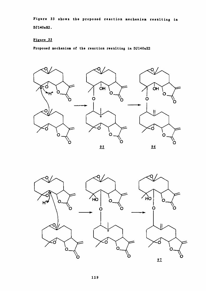
97
119
Figure 33 shows the proposed reaction mechanism resulting in
DJ14OaR2.
Figure 33
Proposed mechanism of the reaction resulting in D.T140aH2

These four structures differ only in the site of intermolecular
linkage and the point of attachment of the 8a —hydroxyl group. 98 and
100 have a secondary hydroxyl group at S in addition to the 8a —
hydroxyl. The NMR signals for H-5 occur at 62.68 and 2.46 whereas
K-8 in the portion which carries the 8 —hydroxyl group resonates at
the significantly lower field of 83.01. This strongly indicates
120

either structure 99 or 101 where C—S bears an ether or epoxide. The
final problem is thus the placement of the 8a—hydroxyl function.
Although it is clear from the decoupling experiments to which set of
H—S, H-6 and H—i signals the comparatively low field of H-8 signal
in the hydroxy—containing moiety belongs, the signals are not
'9 l0sufficiently distinctive to allow the two structures B and D to be
distinguished. Useful information was however obtained from study of
the mass spectrum.
Large peaks were visible at m/z 265 (7.1%) and 279 (12.9%) in the
mass spectrum of DJ14OaU2. If these arise as a result of simple C—O
cleavage then the sum (544) should represent the mass of the parent
molecule. Both structures 99 and 101 have formulae C 30H400 9 with
molecular weight 544 so confirming the overall proposals.
Furthermore, only if the lower half of the molecule carries the 8a —
hydroxyl group, 101, can the two fragments at m/z 265 and 279 be
produced. Accurate mass measurement at 265 and 279 indicated the
molecular formulae of the two halves of the molecule to be C15H2104
and C 15E1905 respectively, which confirms this proposal.
Further ions in the mass spectrum include peaks at m/z 281 (1.0%),
261 (1.3%), 247 (2.7%), 228 (5.0%), and 207 (8.9%). These may arise
as shown in Figure 34.
The NMR spectrum of DI14OaH2 was wholly consistent with the
proposed structure Qj.
With regard now to the stereochemistry of the molecule, from the
NMR data, it is clear that 11-5, 11-6 and H—i in both halves have a
trans orientation to each other as in parthenolide. There is no
reason to suppose that these centres are enantiomeric with those of
121

parthenolide so the most likely stereochemistry of the lactone
moieties in both halves of the molecule is as in parthenolide. This
also fixes the stereochemistry at C-4 and C-4'.
Fi gure 34
Mass spectral behaviour of DJ14OaR2
20743-\
250 247
15\\ ,,/48281 265
18_l/" \\_15261 264
\36
228
The only centres unaccounted for are C— i, C—iO and C— i'. From the
proposed mechanism of the biosynthesis of the molecule, the C(i')O-
i22
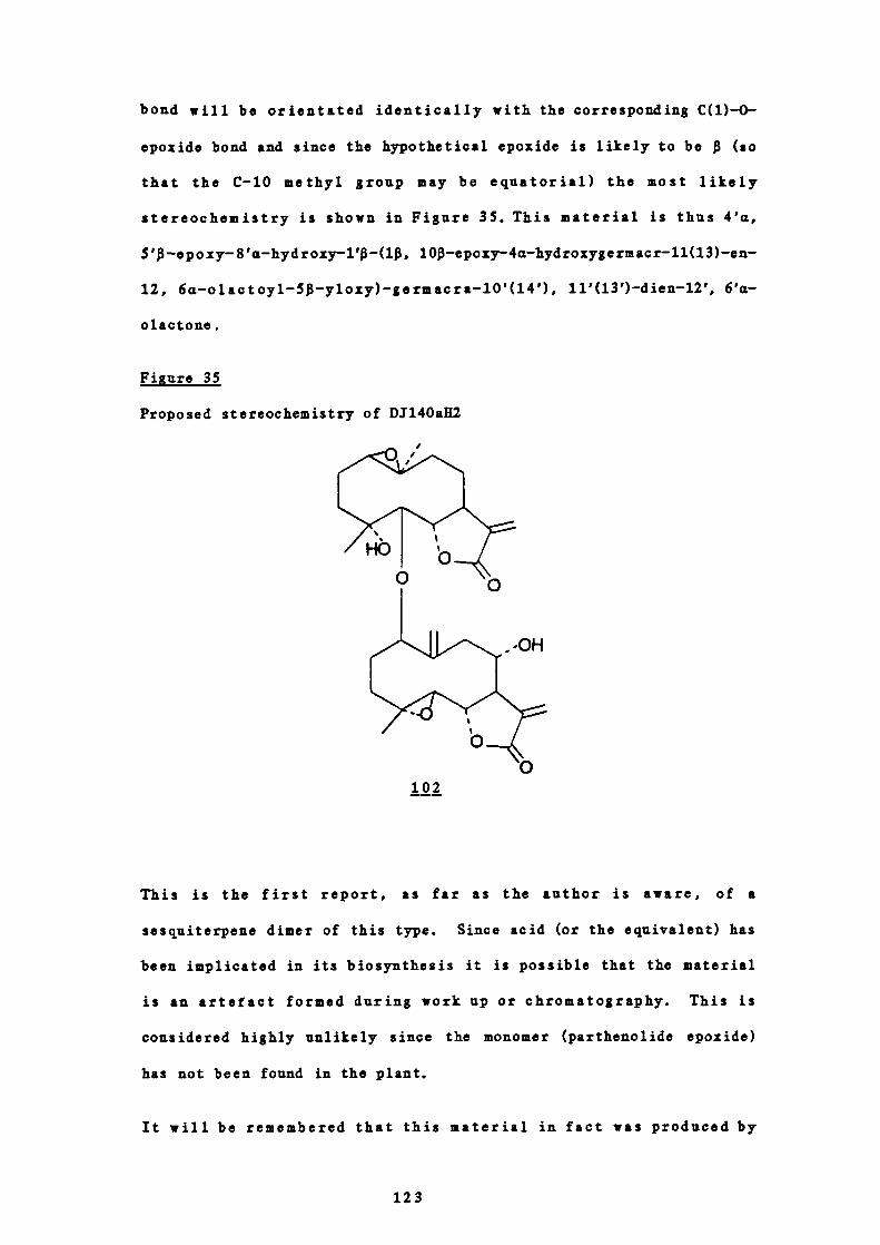
bond will be orientated identically with the corresponding C(1) —O-
epozide bond and since the hypothetical epoxide is likely to be (so
that the C-1O methyl group may be equatorial) the most likely
stereochemistry is shown in Figure 35. This material is thus 4'a,
5 ' —epoxy-8'a—hydroxy—l' —(1, 10 —epoxy-4a—hydroxygermacr-11(13) —en-
12, 6a—olactoyl-5 —y1oxy) —germacra-10'(14'), 11'(13') —dien-12', 6'a-
olactone.
Figure 35
Proposed stereochemistry of D1140ali2
1
102
This is the first report, as far as the author is aware, of a
sesquiterpene dimer of this type. Since acid (or the equivalent) has
been implicated in its biosynthesis it is possible that the material
is an artefact formed during work up or chromatography. This is
considered highly unlikely since the monomer (parthenolide epoxide)
has not been found in the plant.
It will be remembered that this material in fact was produced by
123

0
•1
'-4
U
IC
C
('I0
C)U
Ci
hi
zI -
a-0
124

U-
125

basic hydrolysis of the highly active DJ14Oa. If the 111 NMR spectra
of the two substances are compared (Figures 36 and 37) it is apparent
that a set of signals due to another sesquiterpene residue (together
with other signals) is missing from the spectrum of DJ140aH2 compared
with that of D1140a (Table 9). Close inspection allows assignment
of those signals in the spectrum of Di140a and it is considered
likely that they are caused by the presence of a molecule of a
sesquiterpene acid derived by opening of the lactone of the lower
half of D1140aH2. This would be similar to 1,10—dihydrolactucin-8 —O-
iso—hypoglabrate from Hypothocris oligocephala which is the ester
between 1,10—dihydrolactucin and isohypoglabric acid.'22
Of particular interest is a signal assignable to — 8 in the
esterifying sesquiterpene acid which resonates at the low field
position of 65.36. This shows that the sesquiterpene acid is itself
further esterified at C-8. Extra signals in the 'H NMR spectrum of
D3140a are assignable to the fragments (CH 3 ) 2—C, — c(CE3 ) —O— and
CK2 C(CO2— ) — from which it may be deduced that the structure of this
second esterifying acid is j9, 3—hydroxy-3, 4—dimethyl-2 -
methylenepentanoic acid.
CO2H
103
The proposed structure for D1140a is thus 104, 8'a — (4'a, 5' —epoxy-
1' — (1, 10—epoxy-4a—hydroxygermacr11(l3)en12, 6a—olactoyl-5 -
yloxy) —germacra-1O'(14'), 11'(13') —dien-12', 6a—olactoyl]-8"a— (3a-
hydroxy-3 a, 4a — d ime thyl-2a — me thylenepentanoyl)-4"a, 5 "—epoxy-1",
6"a—dihydroxygermacra-1O"(14"), 11"(13")—dien12"oate and is
126
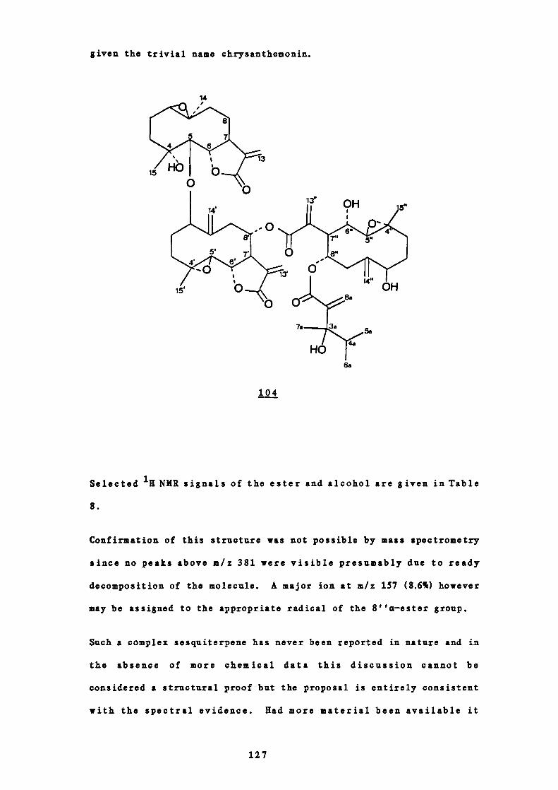
given the trivial name chrysanthemonin.
14/
/ \
1(H0 b0
6a
104
Selected 1H NMR signals of the ester and alcohol are given in Table
8.
Confirmation of this structure was not possible by mass spectrometry
since no peaks above m/z 381 were visible presumably due to ready
decomposition of the molecule. A major ion at m/z 157 (8.6%) however
may be assigned to the appropriate radical of the 8''a —ester group.
Such a complex sesquiterpene has never been reported in nature and in
the absence of more chemical data this discussion cannot be
considered a structural proof but the proposal is entirely consistent
with the spectral evidence. Had more material been available it
127
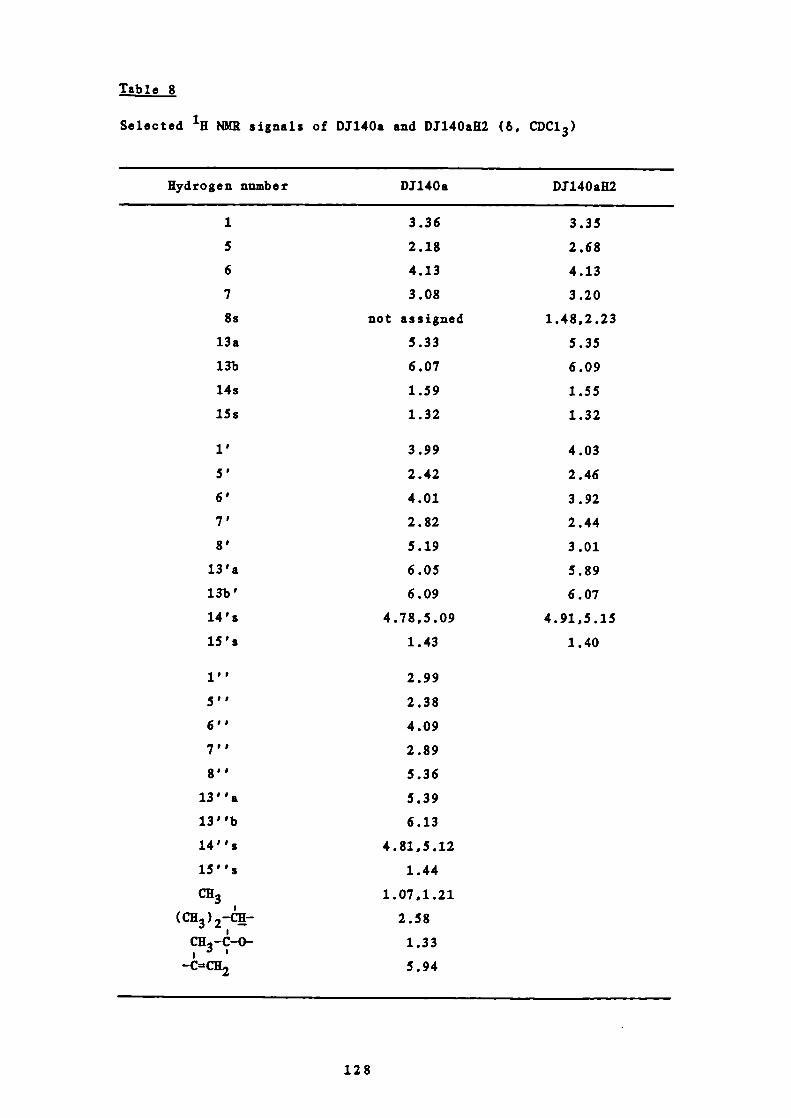
1'
5'
6'
7'
8'
13 'a
13b'
14's
15's
3.99
2.42
4.01
2.82
5.19
6.05
6.09
4 .78,5 .09
1.43
4.03
2.46
3.92
2.44
3.01
5.89
6 .07
4.91,5.15
1.40
Tble 8
Selected NMR signals of D1140a and D1140aH2 (6, CDC13)
Hydrogen number D1140a D1140afl2
1 3.36 3.35
5 2.18 2.68
6 4.13 4.13
7 3.08 3.20
8s not assigned 1.48,2.23
13a 5.33 5.35
13b 6.07 6.09
14s 1.59 1.55
lSs 1.32 1.32
1''
2.99
5,,
2.38
6''
4.09
7,,
2.89
8''
5.36
13''a 5.39
13''b
6.13
14''s 4.81,5.12
15' '5 1.44
CH3 1.07,1.21
2.58
CB,-C-O- 1.33
-C=CH2 5.94
128
would have been possible to isolate and attempt characterisation of
the acidic hydrolysis products. This would also have enabled
assignment of the NMR spectrum which was not possible in the
present study because of the presence of many similar signals.
It is hoped that future studies will be carried out to confirm this
novel structure, especially in view of the material's high non-
competitive spasmolytic activity (see Part III, Table 19).
D1140a was also isolated from sub — fractions Aa70-76 of the column
chromatography of fractions A53-58 and A66-67 (see Figure 21). This
material was combined with the D1140a isolated previously.
7 COMMENTS ON D,T179a1 AND D.T179c
The remaining sub —fractions from the separation of fractions 144-176
(A) to show 100% activity against all three agonists were sub —
fractions A47-52. These were further separated on a silica gel
column and fractions Ab34-36 contained two major pink components on
spraying with sulphuric acid and heating. Further separation by
preparative thin layer chromatography yielded the two compounds
DJ179a 1 and D3179c (see Figure 21).
D1179a1 is possibly a mixture of reynosin, chrysartemin A and a new
sesquiterpene lactone, dihydroreyuosin (which shows a methyl doublet
in the NMR spectrum at 61.40). There is also a possibility that
the three compounds are joined as in D1140a. D1179a1 was isolated
from the light petroleum extract in very low yield and a greater
quantity of this material is probably present in the chloroform
extract. Unfortunately the scarcity of material precluded further
examination.
129

D1179c was also a very complex sesquiterpene lactone containing an a -
methylene—y—lactone and, as with D1179a1, could also not be studied
in detail due to the very small amount isolated.
8 COM3ENTS ON D1124a
A mixture of fatty acid esters was isolated from sub —fractions A30-34
of the second column of fractions 144-176 (A) but was devoid of
spasmolytic activity and so was not examined further.
9 THE REMAINING FRACTIONS TESTED FOR SPASMOLYTIC ACTIVITY
Fractions 75-80 (C) showed 100% inhibition of the agonist s 5RT and
histamine as well as PGE 2 . The latter agonist was chosen because
substances which inhibit prostaglandin synthesis are used in the
treatment of migraine. After further separation ona second silica
gel column however the activity appeared to have been lost. A
similar situation was encountered with the remaining fractions from
the petroleum extract to show 100% activity j. fractions 51-61.
Only one major component was present in these and after further
separation by column chormatography was isolated from sub—fractions
7-15. All the sub —fractions from this second column however were
devoid of the significant activity shown by the original fractions.
The compound isolated from sub—fractions 7-15 proved to be of a long
chain fatty acid ester (D161a). Unfortunately, the increased purity
of the compound resulted in its being barely soluble in
dimethylsulphoxide. This solubility problem probably accounted for
the loss of activity in this case as well as in fractions 75-80 (C).
This finding also made one aware of possible erroneous results
130

however. It seems unlikely that fats common in all plants could
possess any significant activity even bearing in mind it being a
possible precursor for pros t aglandins.l2Sa Since prostaglandins have
not been isolated in any significant quantity 126 from plants to date
it was concluded that the activity apparently shown by fractions 51-
61 and fractions 75-80 (C) were false positive results.
The compounds, being fatty materials, were likely to have blocked
the agonist receptor sites on the guinea pig ileum purely by forming
a barrier. Since the membrane is fatty in nature the fractions under
test would have had a greater affinity for the membrane than for the
hydrophilic rebs solution. On addition of the agonists after the 30
minute equilibrium period access to the receptor sites would be
barred. Increasing the purity of the material however caused
precipitation of the substance and so no film could be produced. No
significant inhibition of the agonists was therefore recorded.
This problem showed some of the difficulties encountered with plant
extracts of greatly varying polarity and perhaps over—simplified test
procedure. Nevertheless it must be realised that the initial
screening method was chosen to be very sensitive in order to pick up
as much real activity as possible found in only small quantities.
Fractions 119-123 (E) showed good activity against 5 —HT and
histamine. These were further sub —fractionationed and tested for
spasmolytic activity. After further separation by preparative TLC an
unidentified triterpene and more parthenolide were isolated. It was
concluded that the activity of these fractions was probably due to
parthenolide.
The common plant sterols —sitosterol and stigmasterol were isolated
from fractions 112-118 (F). They did not show any significant
131

Spasmolytic activity.
10 IN VITRO PRARMACOLOGICAL STUDIES ON THE EFFICACY OF
CBRYSANTHEMIJM PARTHENIUM IN MIGRAINE
It has been demonstrated in the present work that a light petroleum
extract of . p arthenium showed very good spasmolytic activity in
vitro with a possible indication of highest activity against 5 -
hydroxytryptamine. This result is of significance in the clinical
application of the plant in migraine since 5 —hydroxytryptamine is
probably the most implicated of the agonists used in the pathogenesis
of the disease.'°8"'12
In addition the new compounds isolated showed inhibition of
prostaglandin E2.'251'
11 PRELIMINARY TOXICITY STUDIES
Miss Julia bce and Dr E S Johnson of Kings College, London have
carried out limited feverfew feeding experiments with guinea pigs.
10 mg/kg to 135 mg/kg of freeze—dried feverfew mixed with normal feed
(cf. human dose ca 1 mg/kg, based on a dose of three small leaves
each weighing 25mg after drying per person per day) was fed to guinea
pigs for periods of one to seven weeks. The ileum of each animal was
then exised and tested as in the in vitro studies using
acetylcholine, histamine, 5—hydroxytryptamine, nicotine, bradykinin
and prostaglandin E2 as agonists.
After feeding for one week the results showed a statistically
significant non—competitive inhibition to 5—hydroxytryptamine but no
effect on the response tobradykinin or nicotine was demonstrated.
132
The latter result implies that the plant was causing selective
antagonism to 5—hydroxytryptamine at the receptor and not the nerve
level.' 27 Furthermore, no significant inhibition of acetylcholine,
histamine or prostaglandin E2 was obtained.
These preliminary results confirm the suspicion from the j vitro
studies that the extracts and column fractions showed selective
activity against 5—hydroxytryptamine.
Feeding for longer than one week caused no further effect to the
response of the tissue. In addition, the tissues from the low—dose
and high—dose animals gave similar responses.
No adverse effect on the tissues or behaviour of the test animals was
noted even after administration of 135 times the equivalent human
dose for a period of seven weeks.
12 CLINICAL STUDIES ON THE EFFICACY OF CHRYSANTHEMUM PARTHENIIJM IN
MIGRAINE
The difficulties of extrapolating results obtained with isolated
tissue to clinical use in man are well known. This is particularly
so in this case since the disease cannot be studied in live animals.
The final test of efficacy of any drug must therefore rest in the
clinic.
The code of medical ethics precluded prescribing feverfew to patients
in the place of recognised treatments. In this case however, because
of the widespread publicity the plant has received many migraine
sufferers throughout Britain have been using feverfew. It was thus
possible to study a group of volunteers who had already been using
133
the remedy of their own accord. This procedure did not infringe
current legislation and permission was obtained from the Department
of Health and Social Security to set up a limited clinical trial at
the City of London Migraine Clinic, Charterhouse Square, London (DUSS
number MF/8000/2025).
Only those patients who had been taking feverfew continuously for at
least three months prior to the trial were eligible for enrolment.
Ten patients of either sex between 18 and 60 years suffering from
common, classical or mixed migraine were to be selected. They were
also required to have a migraine history of at least two years with
two to eight attacks a month before they started using feverfew. If
any of the patients were taking other drugs such as alpha — or beta-
blockers, tranquillisers, antidepressants, non —steroidal anti —
inflammatory agents used prophylactically or 'antimigraine drugs' for
example ergotamine, likely to interfere with the study, they were
excluded. Patients who were pregnant or had known mental illness
were also not allowed to participate.
The study was a double —blind, placebo controlled, crossover trial.
The patients were randomly allocated either feverfew capsules
containing their equivalent daily dose (on the basis that one 'normal
sized' leaf weighed 25 mg when dried) or placeo capsules. After
twelve weeks the patients crossed over to the other treatment.
Venous blood (20 ml) was taken on enrolment and four weekly
thereafter. The blood was analysed for liver and renal function and
general haematology. The patients were required to record each
migraine attack on diary cards.
Unfortunately, because of severe cramping experienced by one patient
the code had to be broken when only four patients had been enrolled.
This occurred in the fourth month when the patient was on placebo
134
capsules. There was no previous history of cramps and the
possibility of withdrawal symptoms from the feverfew are to be
investigated. A study of only four patients is obviously not
statistically valid but it is hoped that a larger study will soon be
carried out.
A much more valid clinical study is at present being undertaken by Dr
E S Johnson of hugs College, University of London. At the time of
writing, he has received 277 completed questionnaires from patients
who are taking or have taken feverfew for migraine. A typical case
history obtained from a questionnaire is outlined below.
A housewife now aged 75 began taking feverfew on the 27th of May 1978
after hearing of it from Mrs Jenkins of Cardiff. She takes one large
fresh leaf or two small ones in a sandwich every day at 11 a.m.
Consumption of the plant caused no change in bowel habit, appetite,
sleep, mood, breathing, heart beat or weight.
She did not suffer from hot flushes, palpitations, mouth ulcers,
indigestion, jaundice, bleeding gums or excessive thirst while taking
feverfew.
The only side effects she attributed to feverfew were a skin, rash on
her face, increased urine output and swollen ankles. These appeared
within a year of using the plant.
When she began taking feverfew her migraine headaches (diagnosed as
classical by her doctor) became much less severe and she experienced
less nausea with them. She has only taken Panadol for the headaches.
In April and May 1978, i.e. prior to taking feverfew, she suffered 8
migraine attacks. From the 27th May to the end of December 1978
7 months, she suffered 12 attacks. In 1979 she had 18 attacks but in
135

1980 she stopped taking the plant because of the skin rash and
suffered 32 attacks. In 1981 she began using the plant again and
experienced only 10 attacks.
There seems to be little doubt that feverfew is having a beneficial
effect in this case.
These questionnaires are being processed by computer and 60 patients
are to be tested for general haematology, urine, liver and renal
function.
13 RELATIONSHIP B1TwEEN STRUCtTRE AND ACTIVITY
It has recently been proposed by Collier al.128 and Makheja and
Bailey 29 that Chrysanthe parthenium acts in migraine by
inhibiting prostaglandin synthesis. Makheja and Bailey allege that a
phosphate buffer extract of feverfew inhibits platelet aggregation
and hence the release of arachiodonic acid and its conversion to the
prostaglandin thromboxane A 2. On repeating their experiment Dr E S
Johnson could get no such inhibition even at a much higher
concentration of plant extract.
All findings described in the present work point to the presence of
sesquiterpene lactones in the plant as being responsible for the
biological action. A phosphate buffer would not extract such
organic compounds. In addition no significant inhibition of
prostaglandin E2 was found in the ilea of guinea pigs who had been
eating the plant, this being more applicable to the human situation.
The evidence is heavily weighted in favour of the sesquiterpene
lactones as being responsible for the activity of feverfew.
The active compounds isolated in this study have certain structural
136

features in common. They all possess an a —methylene—y—lactone moiety
capable of undergoing a Michael —type addition reaction. (Part I,
Figure 16). The presence of this grouping has been shown to be
important in other activities shown by these compounds.73'84'91"°3
The most active compound is D1140a. This is a very large molecule
with three C—il methylene groups and three epox ides all capable of
binding irreversibly to an enzyme in much the same way as has been
proposed for the biosynthesis of the dimer DJ14OaH2. Such an
irreversible mechanism may account for the non —competitive antagonism
observed for these compounds. Undoubtedly the presence of other
active sites also enhances activity as has already been proved in the
case of ketones, esters and double bonds in cytotoxity studies.73
These compounds are highly reactive and it seems likely that much
semi—synthetic work is called for to produce a clinically effective
substance. It is well known that a —methylene — y — lactones cause
contact dermatitis 13° and the most frequently encountered side effect
in patients taking feverfew is mouth ulceration.
The presence of many similar sesquiterpene lactones in feverfew may
act in a synergistic manner lowering the toxic effects. Conversely,
administration of a pure compound could enhance such cytotoxic
actions and attempts to reduce the side effects could result in loss
of all activity.
14 CONCLUSION
The author has no doubt that Chrysanthemum p arthenium is an effective
prophylactic treatment for migraine. Discussions with patients
indicate that it may prove to be also of as great or even greater

value in the treatment of rheumatoid arthritis.
The present study has demonstrated that the plant possesses marked
spasmolytic activity which can be extrapolated to its clinical
efficacy in migraine. The active constituents isolated thus far
have all been sesquiterpene lactones. This class of metabolite, of
relatively recent interest, is known to show a wide range of
pharmacological activities. In the future they may rival the
alkaloids as the most important group of plant —derived biologically
active constituents.
The present work has resulted in the isolation of substances with
novel structures. These would be of basic phytochemical interest
even if they had no promising biological activity. They also provide
a basis from which the synthetic chemist could work either by
modification of the molecule to increase the therapeutic index, or,
as a template for similar totally synthetic compounds. Structure
activity relationship studies could result in a more thorough
understanding of the aetiology of migraine.
Clearly this process is extremely costly both in time and money.
Sadly, in these times of economic recession, the development of new
drugs is losing the battle against the reformulation of existing
drugs. Furthermore the unfounded scepticism of natural products
ensures that projects dedicated to the study of plants with a long
foJk lore history are drastically under—supported. Many potential
sources thus remain untapped.
It is the author's hope that the promising results of this study will
provide encouragement for similar projects so that with a rational
exploitation of the world's natural resources and an understanding of
the real relevance of the study of folk —lore the outcome may be safe
and effective treatment of disease.
138

PART III
E X P E R I M E N T A L

1 GENERAL DETAILS
A CHROMATOGRAPHY
(a) Adsorbents
Silica gel was the adsorbent used for all chromatography.
The silica gel used for column chromatography was lieselgel
60 (Merck) and for thin—layer chromatography (mc) lieselgel
0 (Merck) and lieselgel OF254 (Merck). TLC plates for
routine work, both analytical and preparative, were prepared
by mixing 15 g Kieselgel 0 and 15 g lieselgel OF254 with
60 ml distilled water. The slurry was spread in a layer
0.25 mm thick on 20 x 20 cm glass plates. The plates were
dried in the open air and then activated at 105°C for one
hour. Silver nitrate impregnated plates were prepared by
mixing 30 g Kieselgel G with 60 ml of a 10% w/v solution of
silver nitrate in distilled water. The slurry was spread in
a layer as before.
(b) Solvent systems
The solvents used for elation of columns and the development
of TLC plates are detailed in the appropriate sections.
Cc) Detection of components on mc plates
Short wave ultraviolet light (254 ma) and spraying with 20%
v/v sulphuric acid in distilled water followed by heating at
105°C for 10 minutes were used for the detection of
components.
B INFRARED SPECTRA
Obtained in solid films, Nujol mulls, chloroform solutions or
IBr discs using a Unicam SP 200 spectrometer.
140

C ULTRAVIOLET SPECTRA
Taken in spectroscopic ethanol at a pathlength of 1 cm using a
Perkin—Elmer Lambda 3 UV/VIS spectrophotometer.
D MELTING POINT
Taken with an Electrothermal melting point apparatus and are
uncorrected.
E RYDROGEN-1 NUCLEAR MAGNETIC RESONANCE SPECTRA
Recorded at 400 MHz using a Bruker WE 400 spectrometer or at 200
MHz on a Nicolet NT 200 spectrometer in deuterochioroform using
tetramethylsilane (TMS) as internal standard.
F CARBON—l3 NUCELAR MAGNETIC RESONANCE SPECTRA
Recorded at 100.6 MHz using a Bruker WH 400 spectrometer in
deuterochloroform using tetramethylsilane as internal standard.
Both and 13 C resonances are given in & (TMS & 0.0) and the
following abbreviations have been used: i singlet; d,
doublet; t, triplet; , quartet; rn, multiplet and 3, coupling
constant.
0 MASS SPECTRA
Recorded on an AEI MS 902 high resolution mass spectrometer or a
VG Micromass 16S spectrometer at 18 or 70 eV with direct inlet
temperatures of between 180°C and 240°C.
141

2 EXTRACTION OF CHRYSANThEMUM PARTHENIUM WITH LIGHT PETROLEUM
(b.r. 40-60°C)
5.8 kg of the leaves of Chrysanthemurn p artheniurn were collected in
January 1980 from the Chelsea Physic Garden, Royai Hospital Road,
London SW3 and freeze—dried using a Chemical Laboratory Instruments
SB4 freeze —drier. Avoucher specimen of the dried aerial parts of
the plant was placed in the museum at Chelsea College. The freeze —
dried leaves lost 82.8% on drying to give a final weight of 1 kg.
These were powdered and exhaustively extracted in a Soxhiet apparatus
with light petroleum b.r. 40-60°C for 7 days. The extract was
evaporated to dryness using a rotary evaporator under reduced
pressure to give a yellow oily residue weighing 44 g.
3 SPASMOLYIC ACTIVITY OF THE LIGHT PETROLEUM EXTRACT
50 mg of the petroleum extract was tested for spasmolytic activity
using acetyicholine (Ach), 5 —hydroxytry-ptamine (5—liT) and histamine
as agonists as described below.
Guinea pigs weighing 200-300 g were killed by a blow on the head.
The proximal ileum was excised and 2-3 cm lengths were cleaned and
suspended in Irebs solution (NaC1 118.4, KCl 4.7, CaC1 2 2.5, M8SO4
1.2, 1R2PO4 1.2, NaHCO3 25 and glucose 11.5 mMole/litre) at 37°C
through which a mixture of 95% O and 5% CO2 was constantly bubbled.
The lengths of ileum were set up to record longitudinal contractions
isometrically. Log (dose) vs response curves were recorded to
acetyleholine, 5 —hydroxytryptamine and histamine from which ED50
doses were taken and given repeatedly until constant responses were
achieved. The antagonist the petroleum extract, was then
dissolved in the minimum of dimethylsuiphoxide and made up to a
142
concentration of 1O 4 gIml with Irebs solution. This was added to
the bath containing the ileum and left for 30 minutes. After thorough
washing with Irebs solution to remove the residual antagonist the
responses of the tissue to the agonists were then recorded and the
percentage change calculated.
A control experiment was performed in exactly the same way except the
antagonist was omitted. (This was essential since leaving an undosed
tissue for 30 minutes often increases its sensitivity to exogenous
agonist). The petroleum extract showed 100% inhibition to the three
agonists.
4 CRUDE SEPARATION OF TEE LIGET PETROLEUM EXTRACT
1.25 g of the petroleum extract was taken and chromatographed on a
silica gel column (50 g) to investigate the components present and
the degree of separation in view of a larger scale examination. This
pilot study proved satisfactory and so 40 g of the petrol extract was
placed on a column containing 1.2 kg silica gel mixed to a slurry
with light petroleum b.r. 40-60°. 200 ml fractions were collected.
The fractions were examined by TLC and combined as appropriate.
Solvent elution of the column and weights of the combined fractions
are shown in Tables 9 and 10.
5 SPASMOLYTIC ACTIVITY OF TEE COMBINED FRACTIONS FROM THE
LIGHT PETROLEUM EXTRACT
The combined fractions from the petroleum extract were tested for
spasmolytic activity as described before (Part III, 3). The results
143

Table 9
Solvent elution of the column chromatography of the light petroleum
extract
Fraction numbers Solvent
1 - 24 light petroleum b.r. 400_600
25 - 36 1.0% v/v ethyl acetate in light petroleum
37 - 50 2.5% v/v ethyl acetate in light petroleum
51 - 65 5.0% v/v ethyl acetate in light petroleum
66 - 95 7.5% v/v ethyl acetate in light petroleum
96 - 112 10.0% v/v ethyl acetate in light petroleum
113 - 118 25.0% v/v ethyl acetate in light petroleum
119 - 166 50.0% v/v ethyl acetate in light petroleum
167 - 174
175 - 180
181 - 185
186 - 188
189 - 192
193 - 195
196 - 201
202 - 217
218 - 237
238
75.0% v/v ethyl acetate in light petroleum
ethyl acetate
1.0% v/v chloroform in ethyl acetate
5.0% v/v chloroform in ethyl acetate
10.0% v/v chloroform in ethyl acetate
25.0% v/v chloroform in ethyl acetate
50.0% v/v chloroform in ethyl acetate
chloroform
10.0% v/v methanol in chloroform
50.0% v/v methanol in chloroform
144
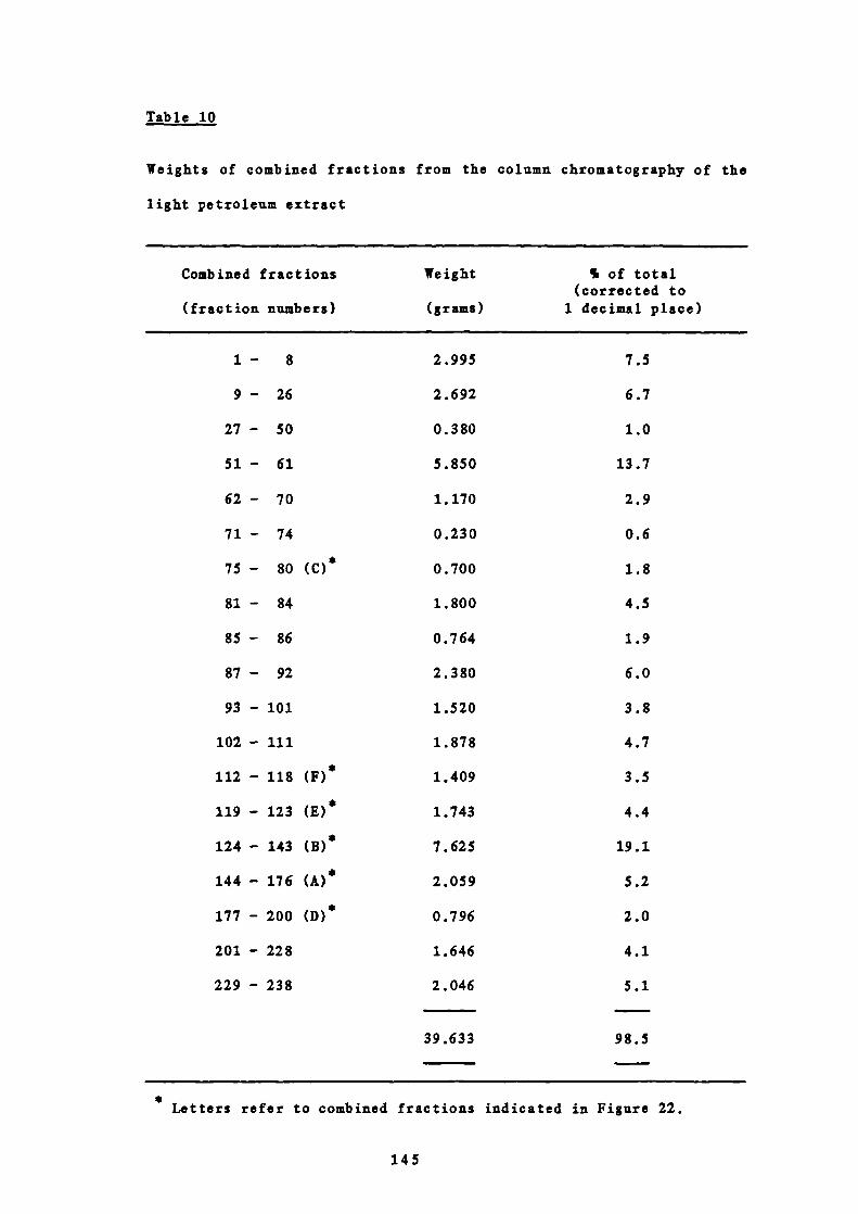
1.0
13.7
2.9
0.6
1.8
4.5
1.9
6.0
3.8
4.7
3.5
4.4
19.1
5.2
2.0
Table 10
Weights of combined fractions from the column chromatography of the
light petroleum extract
Combined fractions Weight % of total(corrected to
(fraction numbers) (grams) 1 decimal place)
1 - 8 2.995 7.5
9 - 26 2.692 6.7
27 - 50
51— 61
62 - 70
71 - 74
75 - 80 (C)
81 - 84
85— 86
87 — 92
93 - 101
102 - 111
112 - 118 (F)
119 - 123 (E)*
124 - 143 (B)*
144 - 176 (A)*
177 - 200 (D)*
0.380
5.850
1.170
0.23 0
0.700
1.800
0.764
2.3 80
1.5 20
1.878
1.409
1.743
7.625
2.059
0.796
201 - 228 1.646 4.1
229 - 238 2.046 5.1
39.633 98.5
* Letters refer to combined fractions indicated in Figure 22.
145

are shown in Table 11 and Figure 22.
6 EXTRACTION OF CHRYSANTHEMUM PARTHENI1JM WITH CHLOROFORM
The plant material, 950 g, previously extracted with light petroleum
b.r. 40-60°C was exhaustively extracted with chloroform in a Soxhiet
apparatus for 9 days. The extract was evaporated to dryness under
reduced pressure using a rotary evaporator under reduced pressure to
give a green oily residue weighing 46 g.
7 SPASMOLYTIC ACTIVITY OF TUE CHLOROFORM EXTRACT
50 mg of the chloroform extract was tested for spasmolytic activity
as before (Part III, 3). 100% inhibition of the three agonists was
obtained.
8 CRUDE SEPARATION OF THE CHLOROFORM EXTRACT
4.5 g of the chloroform extract was chromatographed on a silica gel
column (120 g) to investigate the components present and the degree
of separation in view of a larger scale examination. 100 ml
fractions were taken and combined as appropriate with reference to
TLC. Solvent elution of the column and weights of the combined
fractions are shown in Tables 12 and 13.
9 SPASMOL!TIC ACTIVITY OF THE COMBINED FRACTIONS FROM THE
CHLOROFORM EXTRACT
The combined fractions from the chloroform extract were tested for
146

Table 11
Spasmolytic activity of the combined fractions from the light
petroleum extract, tested at b 4 g/ml
% inhibition ofFraction numbers
Ach 5—fiT Histamine
1 — 8 0 0 0
9 — 26 0 0 0
27 — 50 20 31 33
51— 61 89 100 96
62 — 70 26 32 26
71— 74 55 87 50
75 - 80 (C) 91 100 100
81— 84 0 0 0
85 — 86 12 27 26
87 — 92 5 42 39
93-101 60 86 79
102 - 111 58 62 66
112 - 118 (F)* 20 42 35
119 - 123 (E) 84 97 97
124 - 143 (B) 91 100 100
144 - 176 (A)* 100 100 100
177 - 200 (D) 81 100 100
201 - 228 50 57 46
* Letters refer to combined fractions indicated in Figure 22.
147

Table 12
Solvent elution of the column chromatography of the chloroform
extract
Fraction numbers Solvent
1 - 12 50.O°k v/v ethyl acetate in light petroleum
13 - 16
17 - 27
28 - 32
33 - 36
37 - 40
41 - 45
46 - 54
$5 - 62
63 - 68
75.0% v/v ethyl acetate in light petroleum
ethyl acetate
10.0% v/v chloroform in ethyl acetate
20.0% v/v chloroform in ethyl acetate
50.0% v/v chloroform in ethyl acetate
chloroform
10.0% v/v methanol in chloroform
25.0% v/v methanol in chloroform
500% v/v methanol in chloroform
148
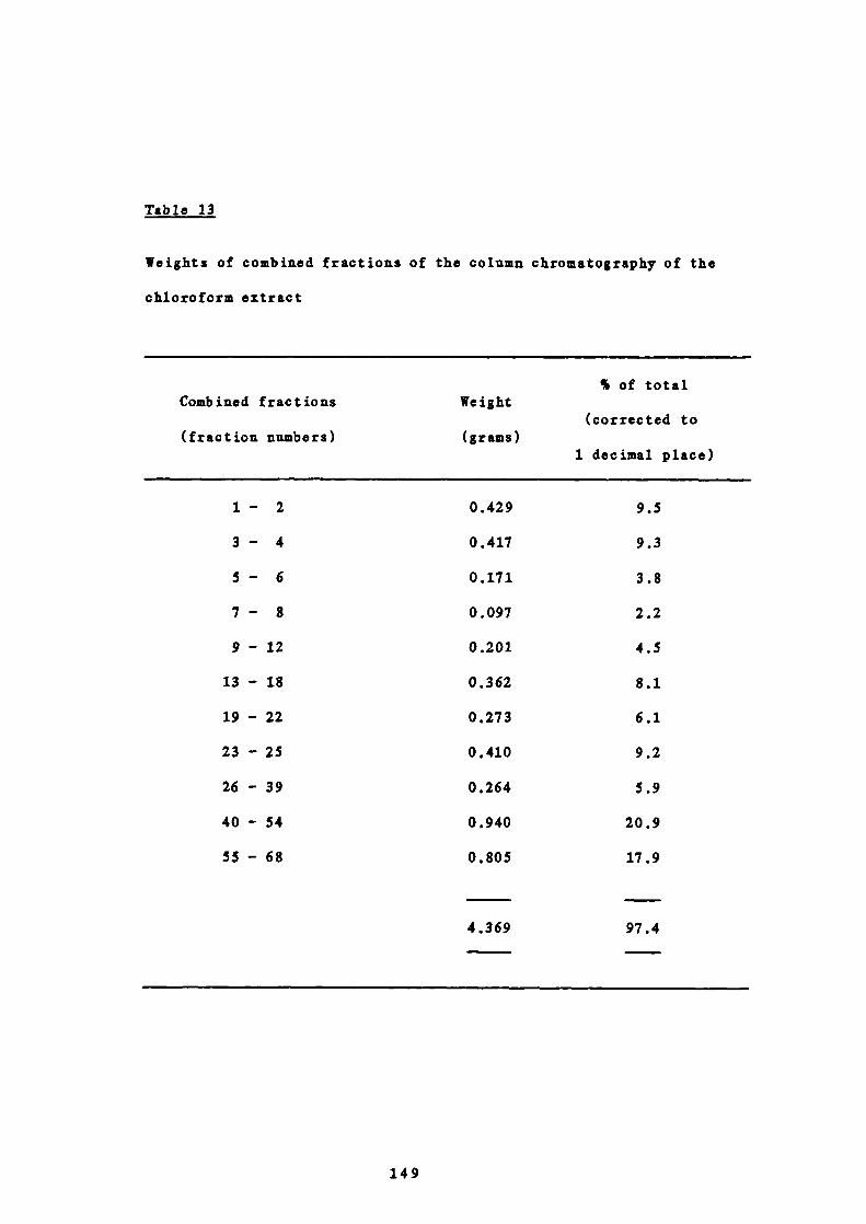
Table 13
Weights of combined fractions of the column chromatography of the
chloroform extract
% of totalCombined fractions Weight
(corrected to(fraction numbers) (grams)
1 decimal place)
1 - 2 0.429 9.5
3 - 4 0.417 9.3
5 - 6 0.171 3.8
7 - 8 0.097 2.2
9 - 12 0.201 4.5
13 - 18 0.362 8.1
19 - 22 0.273 6.1
23 - 25 0.410 9.2
26 - 39 0.264 5.9
40 - 54 0.940 20.9
55 - 68 0.805 17.9
4.369 97.4
149
spasmolytic activity as described before (Part III, 3). The results
are shown in Table 14. No fractions showed 100% antagonism to all
three agonists and so were not investigated further with respect to
pharmacological activity. The extract was fractionated however to
isolate new compounds in the hope that these would be of help in the
structural elucidation of the active ones from the light petroleum
extract.
10 SEPARATION OF THE CHLOROFORM EXTRACT ON A LARGER SCALE
39 g of the chloroform extract was chromatographed on a column of
silica gel (1 kg). 200 ml fractions were taken and combined as
appropriate with reference to TLC. Solvent elation of the columns
and weight of the combined fractions are shown in Tables 15 and 16.
11 EXTRACTION OF CHRYSANTHEMUM PARTHENIUM WITH METHANOL
A portion of the plant material, 25 g, previously extracted with
light petroleum and CUd 3 was extracted with methanol in a Soxhlet
for 8 days. After evaporation of the solvent a black residue (4.6 g)
was obtained.
12 SPASMOLYTIC ACTIVITY OF THE METHANOL EXTRACT
50 mg of the methanol extract was tested for spasmolytic activity as
before (Part III, 3). 0% inhibition of the three agonists was
obtained.
150
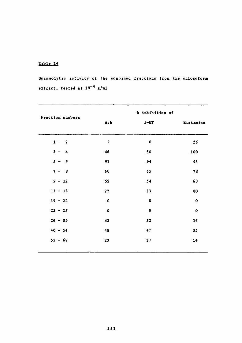
Table 14
Spasmolytic activity of the combined fractions from the chloroform
extract, tested at 1O 4 g/ml
% inMbition ofFraction numbers
Ach
5—UT
Uistamine
1— 2
9
0
26
3— 4
46
50
100
5— 6
91
94
93
7 — 8
60
65
78
9 - 12
52
54
63
13 - 18
22
33
80
19 - 22
0
0
0
23 - 25
0
0
0
26 - 39
43
32
16
40 - 54
48
47
35
55 - 68
23
37
14
151
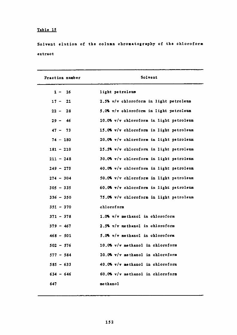
Table 15
Solvent elution of the column chromatography of the chloroform
extract
Fraction number Solvent
1 - 16 light petroleum
17 - 21 2.5% v/v chloroform in light petroleum
22 - 28 5.0% v/v chloroform in light petroleum
29 - 46 10.0% v/v chloroform in light petroleum
47 - 73 15.0% v/v chloroform in light petroleum
74 - 180 20.0% v/v chloroform in light petroleum
181 - 210 25.5% v/v chloroform in light petroleum
211 - 248 30.0% v/v chloroform in light petroleum
249 - 273 40.0% v/v chloroform in light petroleum
274 - 304 50.0% v/v chloroform in light petroleum
305 - 335 60.0% v/v chloroform in light petroleum
336 - 350 75.0% v/v chloroform in light petroleum
351 - 370 chloroform
371 - 378 1.0% v/v methanol in chloroform
379 - 467 2.5% v/v methanol in chloroform
468 - 501 5.0% v/v methanol in chloroform
502 - 576 10.0% v/v methanol in chloroform
577 - 584 20.0% v/v methanol in chloroform
585 - 633 40.0% v/v methanol in chloroform
634 - 646 60.0% v/v methanol in chloroform
647 methanol
152

Tble 16
Weights of combined fractions of the column chromatography of the
chloroform extract
% of totalCombined fractions Weight
(corrected to(fraction number) (grams)
1 decimal place)
1 - 55 0.209 0.5
56 - 130 0.746 1.9
131 - 153 2.161 5.5
154 - 158 0.266 0.7
159 - 160 0.183 0.5
161 - 199 1.540 3.9
200 - 247 3.105 8.0
248 - 267 1.211 3.1
268 - 278 0.672 1.7
279 - 300 2.396 6.1
301 - 307 0.410 1.1
308 - 328 1.745 4.5
329 - 339
0.469
1.2
340 - 358
1.646
4.2
359 - 382
1.475
3.8
383 - 384
1.482
3.8
385 - 387
1.172
3.0
388 - 393
1.970
5.1
394 - 397
1.259
3 .2
398 - 401
1.668
4.3
402 - 409
1.473
3.8
410 - 423
1.826
4.7
424 - 440
1.474
3.8
441 - 469
1.561
4.0
470 - 489
2.344
6.0
490 - 514
2.097
5.4
515 - 530
1.377
3.5
531 - 582
1.073
2.8
583 - 658
0.605
1.5
39.615 101.6
153

13 EXTRACTION OF CHRYSANTHEMUM PARTHENIUM WITH WATER
A portion of the plant material, 25 g, previously extracted with
light petroleum, chloroform and methanol was exhaustively extracted
with water in a Soxhiet for 7 days. After evaporation of the solvent
a black residue (3.9 g) was obtained.
14 SPASMOLYTIC ACTIVITY OF THE WATER EXTRACT
50 mg of the water extract was tested for spasmolytic activity as
before (Part III, 3). 0% inhibition of the three agonists was
obtained.
15 STUDIES ON FRACTIONS 144-176 (A) FROM THE LIGHT PETROLEUM
EXTRACT
Fractions 144-176 from the petroleum extract showed 100% inhibition
of the three agonists Ach, 5—fiT and histamine at 1O' 4 g/ml.
A FURTHER SEPARATION OF FRACTIONS 144-176 (A)
These fractions (1.959 g) were chromatographed on a silica gel
column (40 g). 100 ml fractions were taken and combined as
appropriate with reference to TLC. Solvent elution of the
column and weight of the combined fractions are shown in Tables
17 and 18.
B SPASMOL!TIC ACTIVITY OF FRACTIONS 144-17 6 (A)
Those fractions weighing more than 50 mg were tested for
spasmolytic activity as described before (Part III, 3). The
154
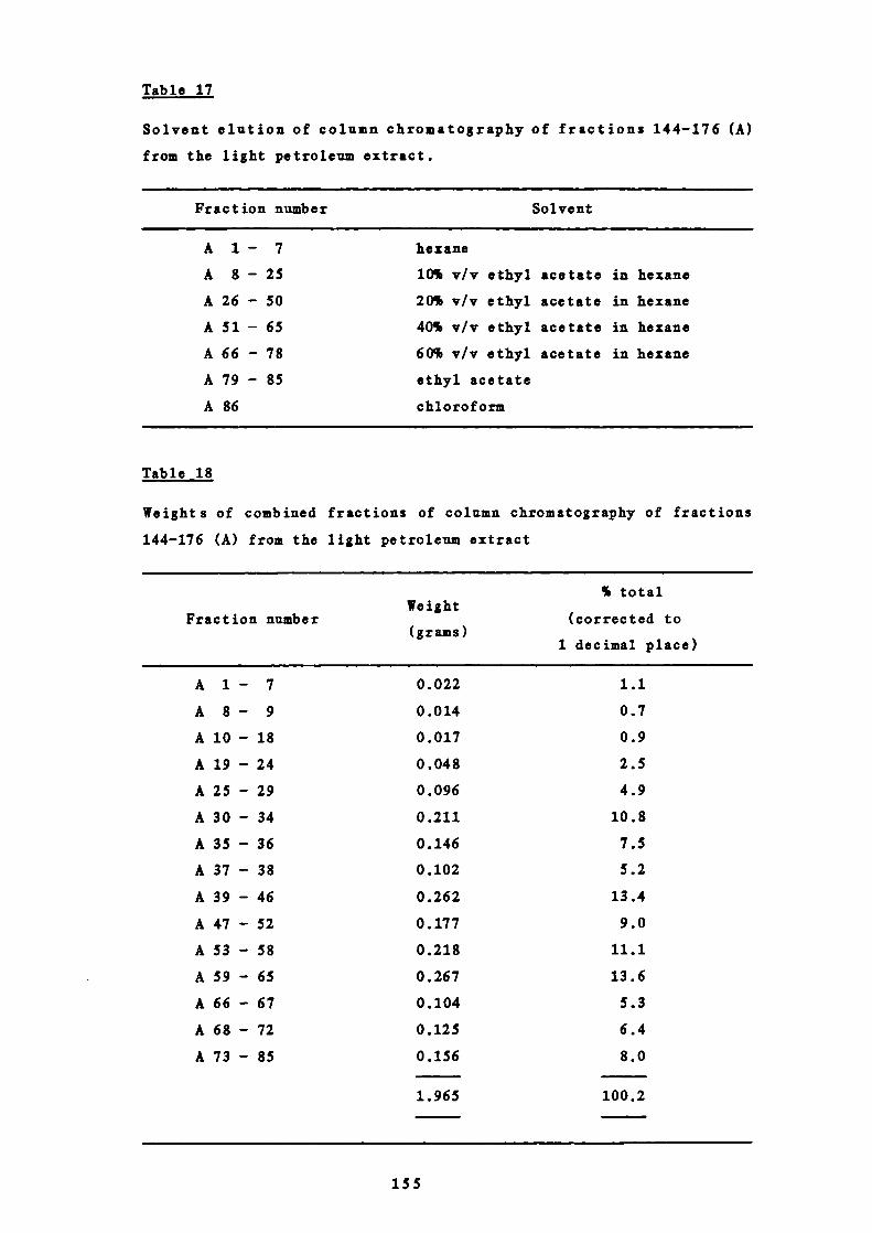
Table 17
Solvent elution of column chromatography of fractions 144-176 (A)
from the light petroleum extract.
Fraction number Solvent
A 1 — 7 hexane
A 8 - 25 10% v/v ethyl acetate in hexane
A 26 - 50 20% v/v ethyl acetate in hexane
A 51 - 65 40% v/v ethyl acetate in hexane
A 66 - 78 60% v/v ethyl acetate in hexane
A 79 - 85 ethyl acetate
A 86 chloroform
Table 18
Weights of combined fractions of column chromatography of fractions
144-176 (A) from the light petroleum extract
% totalWeight
Fraction number (corrected to(grams)
1 decimal place)
A 1— 7 0.022 1.1
A 8 — 9 0.014 0.7
A 10 - 18 0.017 0.9
A 19 - 24 0.048 2.5
A 25 - 29 0.096 4.9
A 30 - 34 0.211 10.8
A 35 - 36 0.146 7.5
A 37 - 38 0.102 5.2
A 39 - 46 0.262 13.4
A 47 - 52 0.177 9.0
A 53 - 58 0.218 11.1
A 59 - 65 0.267 13.6
A 66 - 67 0.104 5.3
A 68 - 72 0.125 6.4
A 73 - 85 0.156 8.0
1.965 100.2
155
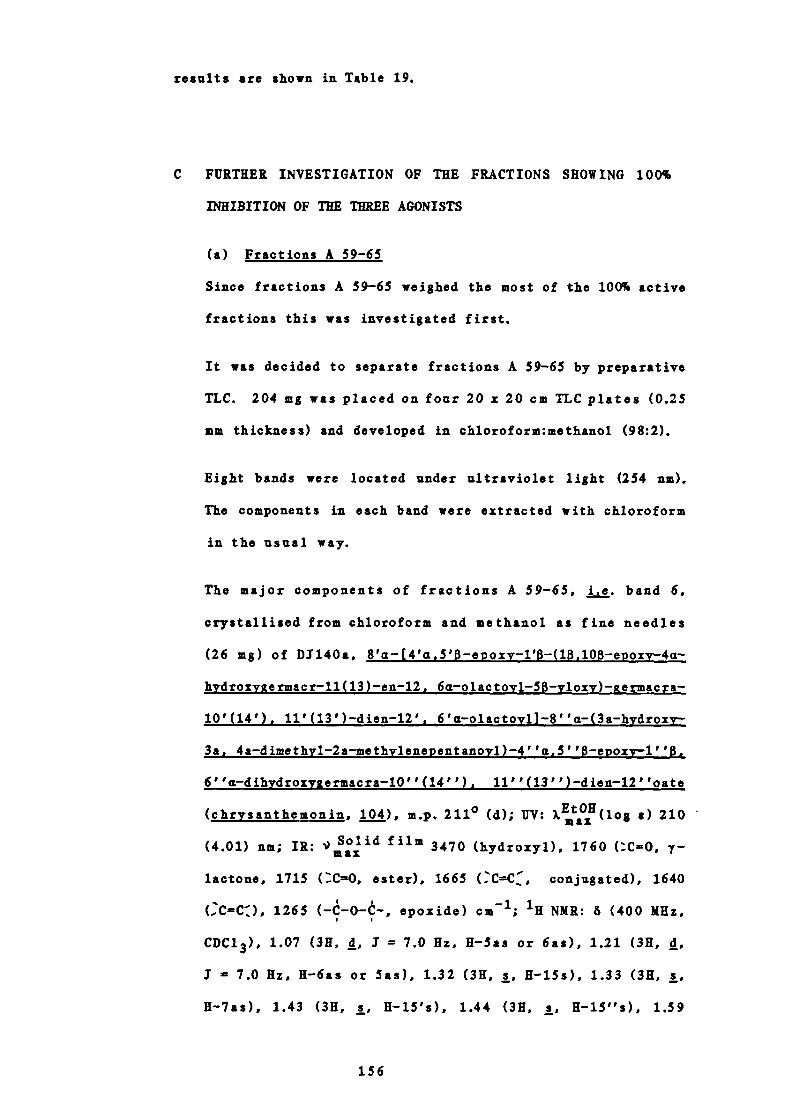
results are shown in Table 19.
C FURTHER INVESTIGATION OF THE FRACTIONS SHOWING 100%
INHIBITION OF THE THREE AGONISTS
(a) Fractions A 59-65
Since fractions A 59-65 weighed the most of the 100% active
fractions this was investigated first.
It was decided to separate fractions A 59-65 by preparative
TLC. 204 mg was placed on four 20 x 20 cm TLC plates (0.25
mm thickness) and developed in chloroform:methanol (98:2).
Eight bands were located under ultraviolet light (254 nm).
The components in each band were extracted with chloroform
in the usual way.
The major components of fractions A 59-65, i.e. band 6,
crystallised from chloroform and methanol as fine needles
(26 mg) of DJ140a, 8'a— [4'a,5'8 — e p oxy-1' —(1,1OB—epoxy-4a-
hydroxygermacr-11(13) —en-12, 6a—olactoyl-5 —y-loxy) —germacra-
10'(14'), 11'(13') —dien-12', 6'a—olactoyl]-8''a— (3a—hydroxy-
3a, 4a—dimethy-l-2a—methyleneentanoyl)-4' 'a,S' ' —epozy-1' '0.
6''a—dihydroxyg ermacra-10''(14''), 11''(13'')—dien-12''oate
(chrysanthemonin, ), m.p. 211° (d); UV: )(log a) 210
(4.01) nm; IR: 5oid film 3470 (hydroxyl), 1760 (CO 3 y-
lactone, 1715 (C—O, ester), 1665 CCC, conjugated), 1640
CCIICZ), 1265 ( —t —O—— , epoxide) cm'; 1K NMR: 6 (400 MHz,
CDC1 3 ),, 1.07 (3K, d, .7 = 7.0 Hz, K—Sas or 6as), 1.21 (3K, d,
I 7.0 Hz, H-6as or Sas), 1.32 (3K, s, H-15s), 1.33 (3K, s,
H-7as), 1.43 (3K, s, K— iS's), 1.44 (3K, s, K-15"s), 1.59
156

Table 19
Spasmolytic activity of fractions 144-176 (A) from the light
petroleum extract, tested at iO g/ml.
% inhibition ofFraction number
Ach 5—HT Histamine
A25-29 77 100 87
A30-34 60 48 55
A35-36 94 100 100
A37-38 97 98 97
A39-46 83 93 92
A 47 - 52 100 100 100
A 53 - 58 100 100 100
A 59 - 65 100 100 100
A66-67 90 100 100
A68-72 87 90 92
A73-85 75 80 70
157
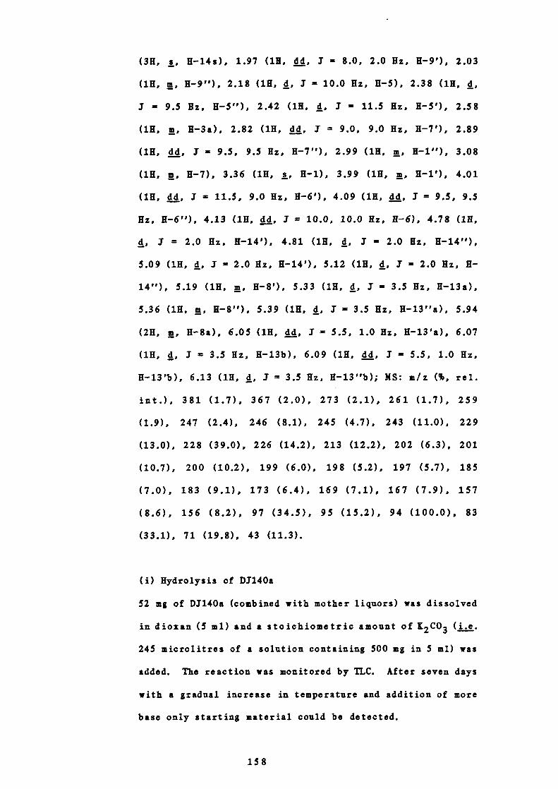
(3H, s, H-14s), 1.97 (111, dd, 1 8.0, 2.0 Hz, H-9'), 2.03
(111, m, 11-9"), 2.18 (111, d, I = 10.0 Hz, 11-5), 2.38 (1K, d,
1 9.5 Hz, H-5"), 2.42 (1K, d, 3 11.5 Hz, K 5'), 2.58
(111, rn, H-3a), 2.82 (111, dd, 3 = 9.0, 9.0 Hz, K-7'), 2.89
(111, dd, 3 9.5, 9.5 Hz, 11-7"), 2.99 (111, ii, H —i"), 3.08
(111, rn, 11-7), 3.36 (111, s, 11-1), 3.99 (1K, rn, K—i'), 4.01
(111, dd, 3 = 11.5, 9.0 Hz, H-6'), 4.09 (111, dd, I - 9.5, 9.5
Hz, 11-6"), 4.13 (1K, dd, 3 = 10.0, 10.0 Hz, H 6), 4.78 (1K,
3 - 2.0 Hz, 11-14'), 4.81 (111, d, I = 2.0 Hz, 11-14"),
5.09 (1K, d, 1 2.0 Hz, 11-14'), 5.12 (111, d, I - 2.0 Hz, K-
14"), 5.19 (111, rn, 11-8'), 5.33 (111, d, I = 3.5 Hz, H-13a),
5.36 (1K, rn, 11-8"), 5.39 (1K, d, 1 3.5 Hz, H-13"a), 5.94
(211, rn, H-8a), 6.05 (1K, dd, 1=5.5, 1.0 Hz, H 13'a), 6.07
(111, d, .1 = 3.5 Hz, K-13b), 6.09 (1K, dd, I = 5.5, 1.0 Hz,
H-13'b), 6.13 (111, d, 3 3.5 Hz, K-13"b), MS. m/z (%, rel.
mt.), 381 (1.7), 367 (2.0), 273 (2.1), 261 (1.7), 259
(1.9), 247 (2.4), 246 (8.1), 245 (4.7), 243 (11.0), 229
(13.0), 228 (39.0), 226 (14.2), 213 (12.2), 202 (6.3), 201
(10.7), 200 (10.2), 199 (6.0), 198 (5.2), 197 (5.7), 185
(7.0), 183 (9.1), 173 (6.4), 169 (7.1), 167 (7.9), 157
(8.6), 156 (8.2), 97 (34.5), 95 (15.2), 94 (100.0), 83
(33.1), 71 (19.8), 43 (11.3).
(i) Hydrolysis of DI14Oa
52 mg of DI14Oa (combined with mother liquors) was dissolved
in dioxan (5 ml) and a stoichiometric amount of 12 CO 3 (i.e.
245 microlitres of a solution containing 500 mg in 5 ml) was
added. The reaction was monitored by TLC. After seven days
with a gradual increase in temperature and addition of more
base only starting material could be detected.
158

2 ml of a 2% solution of KOH in H 20 was then added and the
solution refluxed for 30 minutes. After normal work up the
mixture was separated by preparative TLC using CUd 3 : MeOH
(49:2) as developing solvent. More than twenty bands were
located by ultraviolet light (254 nm) and spraying the edge
of the plate with 20% v/v sulphuric acid and heating. The
major component, located in band 2, was extracted with
CUd 3 . D1140a (j. the starting material) was located in
band 3 and this was extracted and hydrolysed again using 2%
w/v 100 solution and worked up as before. The organic
solvent—soluble portion of this and the extract from band 2
of the original hydrolysis were identical on TLC. They were
combined and purified by TLC using CUd 3 : MeOH (98:2) as
developing solvent. The plate was developed three times.
The major component was located in band 2 by spraying the
edge of the plate with sulphuric acid as before and heating.
This component was further purified by TLC using CBCL3
MoOR (98:2) as developing solvent. After extraction into
CUd 3 the material crystallised from COd 3 and hexane as
colourless plates (5 mg) of Di' 140a02, 4'a,5' — epoxy-8'a —
hydroxy—l' p— (l p ,l0p—epoxy--4a—hydroxygermacr—l1(13) —en-12,6a—
olactoyl-58—yloxy ) —g ermacra-10'(14'), 11'(13') —dien-12',6'a-
olactone (102), m.p. 159° (d); TTY: (log a), 209 (3.99)
nrn; 1K: 1d film, 3500 (OH), 1760 (X0, 7—lactone), 1665
(CC, conjugated), 1640 (C=C), 1260 ( — --O—— , epoxide)
cm"; H NMR: 6 (400 MHz, CDC1 3 ), 1.32 (3H, s, H-15s), 1.40
(311, s, 11-15's), 1.48 (111, rn, H-8), 1.55 (30, s, H-14s),
1.77 (111, rn, H-9'), 2.06 (111, rn, H 9'), 2.23 (111, rn, 11-8),
2.30 (111, d, 1 14.5 Hz), 2.44 (111, rn, 11-7'), 2.46 (111, d,
3' - 11.5 Hz, 11-5'), 2.59 (111, dd, 3' 14.5, 5.0 Hz), 2.68
'59

(111, d, 1 95 Hz, 11-5), 3.01 (111, rn, 11-8'), 3.20 (111, rn,
11-7), 3.35 (111, s, H-i), 3.92 (111, dd, 1 11.5, 8.0 Hz, K-
6'), 4.03 (1H, rn, H1'), 4.13 (111, , 1 9.5, 9.5 Hz, H-
6), 4.91 (1H, d, 1 1.5 Hz, 11-14'), 5.15 (111, d, 1 - 1.5
Hz, 11-14'), 5.35 (111, d, I - 3.5 Hz, K-13a), 5.89 (111, dd, I
5.5, 0.7 Hz, H-13'a), 6.07 (111, dd, I = 5.5, 0.7 Hz, H-
13'b), 6.09 (111, d, 1 3.5 Hz, H-13b); 3C NMR: 6 (100.6
MHz, CDC1 3 ), 15.10, 18.69 and 29.10 (C-14, C-is and C-15'),
23.40, 33.35, 34.64, 38.09, 38.77, 65.04, and 66.08 (C-2, C-
2', C-3, C-3', C-8, C-9 and C-9'), 43.24 and 44.31 (C-7 and
C-7'), 50.72 (C-8'), 55.30, 61.39 and 73.27 (C-4, C-b and
C-4'), 50.86, 62.90, 65.66 and 68.88 (C-i, C-i', C-5 and C-
5'), 75.35 and 79.95 (C-6 and C-6'), 118.00 and 118.64 (C-13
and C-13'), 135.69 (C-14'), 137.92 (C-b'), 140.98 (C-il),
170.60 and 178.64 (C-12 and C-12'). C-il' not seen; MS: m/z
(%, rel.int.), 430 (1.1), 356 (1.3), 342 (1.3), 281 (2.7),
281 (1.0), 280 (4.3), 279 (12.9), 266 (1.4), 265 (7.1), 261
(1.3), 247 (2.7), 228 (5.0), 207 (8.9), 167 (15.4), 150
(10.5), 149 (100), 113 (5.3), 99 (6.1), 98 (5.4), 97 (6.4),
94 (9.7), 87 (5.5), 85.1 (11.1), 85.0 (30.6), 83.1 (7.8),
83.0 (46.0), 71 (18.4), 70 (10.5), 69 (15.2), 60 (5.6), 57
(50.7), 56 (16.9), 55 (22.9); Accurate mass measurements:
Found: 265.1449; C 15 H2104 requires 265.1440; Found:
279.1241; C15111905 requires 279.1232.
(b) Fractions A 53-58 and A 66-67
These two sets of fractions were combined (0.236 g) and
chromatographed on a silica gel column (10 g). 100 ml
fractions were taken and combined as appropriate with
reference to TLC. Solvent elution of the column and weights
of the combined fractions are shown in Tables 20 and 21.
160

Table 20
Solvent elution of column chromatography of fractions A 53 - 58 and A
66-67 from the light petroleum extract
Fraction number Solvent
As 1 - 19 2.5% v/v chloroform in hexane
As 20 - 22 5% v/v chloroform in hexane
As 23 - 30 10% v/v chloroform in hexane
As 31 - 63 20% v/v chloroform in hexane
Aa 64 - 98 30% v/v chloroform in hexane
Table 21
Weights of combined fractions of column chromatography of fractions A
53-58 and A 66-67 from the light petroleum extract
Fraction number Weight % of total
As 1 - 46 0.007 3.0
Aa 47 - 49
0.019
8.1
Aa 50 - 63
0.039
16.5
Aa 64 - 69
0.048
20.3
As 70 - 76
0.030
12.7
Aa 77 - 93
0.056
23.7
As 94 - 98
0.028
11.9
0.227
96.2
161
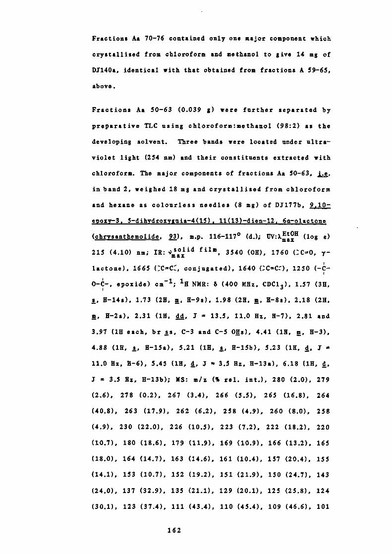
Fractions Aa 70-76 contained only one major component which
crystallised from chloroform and methanol to give 14 mg of
D1140a, identical with that obtained from fractions A 59-65,
above.
Fractions Aa 50-63 (0.039 g) were further separated by
preparative TLC using chloroform:methanol (98:2) as the
developing solvent. Three bands were located under ultra-
violet light (254 am) and their constituents extracted with
chloroform. The major components of fractions Aa 50-63, i.e.
in band 2, weighed 18 mg and crystallised from chloroform
and hexano as colourless needles (8 mg) of D1177b, 9,10-
epoxy-3, 5-dihydroxyguia-4(15), 11(13)-dien-12, 6a-olactone
(chrysanthemolide, ), m.p. 116-117° (d.); (log )
215 (4.10) nm; 1K: film, 3540 (OH), 1760 (C=0, -
lactone), 1665 (CCc, conjugated), 1640 (CC), 1250 (-C-
0-C-, epoxide) cm; 'H NMR: 6 (400 MHz, CDC1 3 ), 1.57 (3H,
s, H-14s), 1.73 (2K, rn, H-9s), 1.98 (2H, , R-8s), 2.18 (2K,
, H-2s), 2.31 (1K, dd, 3 = 13.5, 11.0 Hz, H-7), 2.81 and
3.97 (1H each, br ss, C-3 and C-S OHs), 4.41 (1K, rn,
4.88 (1K, s, H-15a), 5.21 (1H, s, K-lSb), 5.23 (1K, d, I
11.0 Hz, H-6), 5.45 (1K, d, 1 3.5 Hz, H-13a), 6.18 (111, d,
I = 3.5 Hz, H-13b); MS: m/z (% rel. mt.), 280 (2.0), 279
(2.6), 278 (0.2), 267 (3.4), 266 (5.5), 265 (16.8), 264
(40.8), 263 (17.9), 262 (6.2), 258 (4.9), 260 (8.0), 258
(4.9), 230 (22.0), 226 (10.5), 223 (7.2), 222 (18.2), 220
(10.7), 180 (18.6), 179 (11.9), 169 (10.9), 166 (13.2), 165
(18.0), 164 (14.7), 163 (14.6), 161 (10.4), 157 (20.4), 155
(14.1), 153 (10.7), 152 (19.2), 151 (21.9), 150 (24.7), 143
(24.0), 137 (32.9), 135 (21.1), 129 (20.1), 125 (25.8), 124
(30.1), 123 (37.4), 111 (43.4), 110 (45.4), 109 (46.6), 101
162
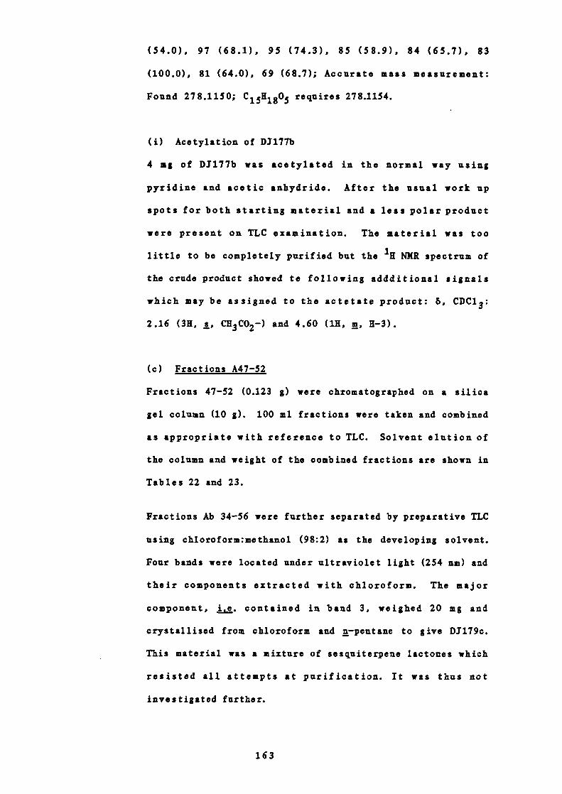
(54.0), 97 (68.1), 95 (74.3), 85 (58.9), 84 (65.7), 83
(100.0), 81 (64.0), 69 (68.7); Accurate mass measurement:
Found 278.1150; C 15H1805 requires 278.1154.
(i) Acetylation of DJ17Th
4 mg of D1177b was acetylated in the normal way using
pyridine and acetic anhydride. After the usual work up
spots for both starting material and a less polar product
were present on TLC examination. The material was too
little to be completely purified but the NMR spectrum of
the crude product showed te following addditional signals
which may be assigned to the actetate product: 6, CDC13:
2.16 (3K, s, CK3 CO2—) and 4.60 (1K, rn, H-3).
(c) Fractions A47-52
Fractions 47-52 (0.123 g) were chromatographed on a silica
gel column (10 g). 100 ml fractions were taken and combined
as appropriate with reference to TLC. Solvent elution of
the column and weight of the combined fractions are shown in
Tables 22 and 23.
Fractions Ab 34-56 were further separated by preparative TLC
using chloroform:methanol (98:2) as the developing solvent.
Four bands were located under ultraviolet light (254 nm) and
their components extracted with chloroform. The major
component, i.e. contained in band 3, weighed 20 mg and
crystallised from chloroform and n—peutsne to give D1179c.
This material was a mixture of sesquiterpene lactones which
resisted all attempts at purification. It was thus not
investigated further.
163
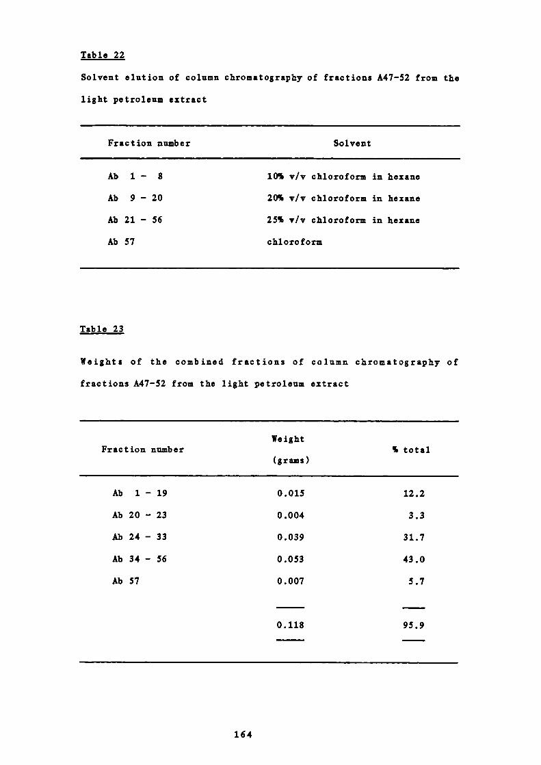
Table 22
Solvent elution of column chromatography of fractions A47-52 from the
light petroleum extract
Fraction number Solvent
Ab 1 - 8 10% v/v chloroform in hexane
Ab 9 - 20 20% v/v chloroform in hexane
Ab 21 - 56 25% v/v chloroform in hexan.e
Ab 57 chloroform
Table 23
Weights of the combined fractions of column chromatography of
fractions A47-52 from the light petroleum extract
WeightFraction number
% total(grams)
Ab 1 - 19 0.015 12.2
Ab 20 - 23 0.004 3.3
Ab 24 - 33 0.039 31.7
Ab 34 - 56 0.053 43.0
Ab 57 0.007 5.7
0.118 95.9
164

The components in band 1 (11 mg) were further purified by
preparative TLC. The plate was run seven times with
chloroform : methanol (98:2) as the developing solvent.
Four bands were located under ultraviolet light (254 urn) and
their components extracted with chloroform. The components
contained in band 1 of the latter plate weighed 6 mg and
crystallised from chloroform and a—pentane to give D1179a1.
This material was possibly also a mixture of sesquiterpene
lactones despite showing only one spot on TLC investigation.
Signals were seen in the 'H NMR spectrum which could be
assigned to reynosin, 5 ° chrysartemin B 50 ' 5 and perhaps a
dihydro —derivative of reynosin. The paucity of material
precluded further study at this stage but the further
possibility that of the material's being a trimeric compound
rather like D3140a cannot be discounted.
16 STUDIES ON FRACTIONS 124-143 (B) FROM THE LIGHT PETROLEUM
EXTRACT
Fractions 124-143 from the petroleum extract showed 10 inhibition
ofthe two agonists 5 —liT and histamine, and 91% inhibition of Ach.
A FURTHER SEPAIATION OF FRACTIONS 124-143 (B)
These fractions (7.525 g) were chromatographed on a silica gel
column (150 g). 100 ml fractions were taken and combined as
appropriate with reference to TLC. Solvent elution of the
column and weight of the combined fractions are shown in Tables
24 and 25.
165

Table 24
Solvent elution of column chromatography of fractions 124-143 (B)from the light petroleum extract
Fraction number Solvent
B 1 - 27 hexane
B 28 - 45 1% v/v chloroform in hexane
B 46 - 51 2% v/v chloroform in hexane
B 52 - 60 5% v/v chloroform in hexane
B 61 - 67 10% v/v chloroform in. hexane
B 68 - 80 20% v/v chloroform in hezane
B 81 - 136 40% v/v chloroform in hexane
B 137 - 149 60% v/v chloroform in hexane
B 150 - 170 75% v/v chloroform in hexane
B 171 - 194 chloroform
B 195 - 203 90% v/v chloroform in methanol
B 204 50% v/v chloroform in methanol
166

2.484
1.537
0.490
0.316
0.252
0.454
0.200
0.810
0.682
33.0
20.4
6.5
4.2
3.3
6.0
2.6
10.8
9.1
Table 25
Weights of combined fractions of column chromatography of fractions
124-143 (B) from the light petroleum extract.
WeightFraction number
% of total(grams)
B 1— 9 0.011 0.1
B 10 - 90 0.040 0.5
B 91 - 106 0.025 0.3
B 107 - 111
B 112 - 115
B 116 - 122
B 123 - 130
B 131 - 147
B 148 - 163
B 164 - 171
B 172 - 196
B 197 - 198
B 199 0.078 1.0
B 200 0.083 1.1
B 201 - 204 0.048 0.6
7.510 99.6
167
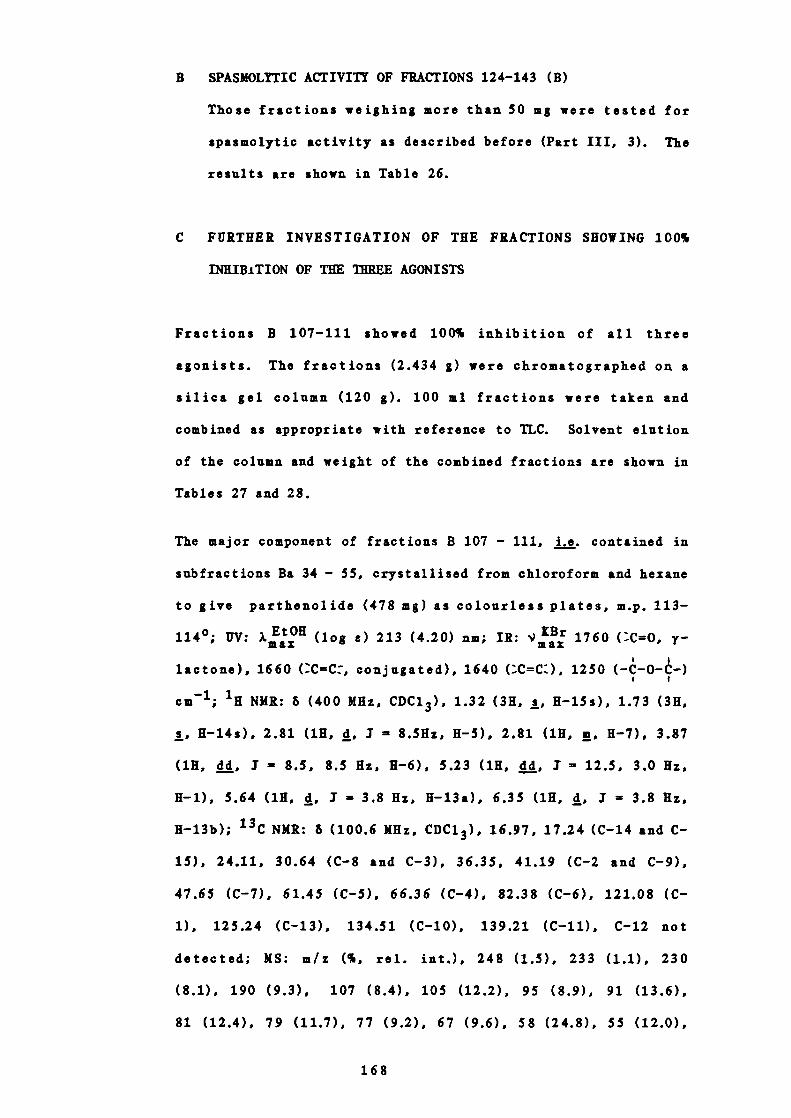
B SPASMOLITIC ACTIVITY OF FRACTIONS 124-143 (B)
Those fractions weighing more than 50 mg were tested for
spasmolytic activity as described before (Part III, 3). The
results are shown in Table 26.
C FURTHER INVESTIGATION OF THE FRACTIONS SHOWING 100%
INHIBiTION OF THE THREE AGONISTS
Fractions B 107-111 showed 100% inhibition of all three
agonists. The fractions (2.434 g) were chromatographed on a
silica gel column (120 g). 100 ml fractions were taken and
combined as appropriate with reference to TLC. Solvent elution
of the column and weight of the combined fractions are shown in
Tables 27 and 28.
The major component of fractions B 107 - 111, j , . contained in
subfractions Ba 34 - 55, crystallised from chloroform and hezane
to give parthenolide (478 mg) as colourless plates, m.p. 113-
114°; UV: XEtOH (log s) 213 (4.20) urn; 111: 1760 (C=0, y-
lactone), 1660 (C=C:, conjugated), 1640 (;C=C:), 1250 (-C-O--)
cm 1 ; 111 NMR: & (400 MHz, CDC1 3 ), 1.32 (311, s, 11-iSs), 1.73 (311,
s, H-14s), 2.81 (111, d, I = 8.5Hz, 11-5), 2.81 (111, rn, 11-7), 3.87
(111, dd, 3 8.5, 8.5 Hz, 11-6), 5.23 (111, dd, 1 12.5, 3.0 Hz,
11-1), 5.64 (111, 4, 1 3.8 Hz, H-13a), 6.35 (111, d, 1 3.8 Hz,
H-13b); 13 C NMR: 6 (100.6 MHz, CDC1 3 ), 16.97, 17.24 (C-14 and C-
15), 24.11, 30.64 (C-8 and C-3), 36.35, 41.19 (C-2 and C-9),
47.65 (C-7), 61.45 (C-5), 66.36 (C-4), 82.38 (C-6), 121.08 (C-
i), 125.24 (C-13), 134.51 (C-b), 139.21 (C-li), C-12 not
detected; MS: rn/i (%, rel. mt.), 248 (1.5), 233 (1.1), 230
(8.1), 190 (9.3), 107 (8.4), 105 (12.2), 95 (8.9), 91 (13.6),
81 (12.4), 79 (11.7), 77 (9.2), 67 (9.6), 58 (24.8), 55 (12.0),
168
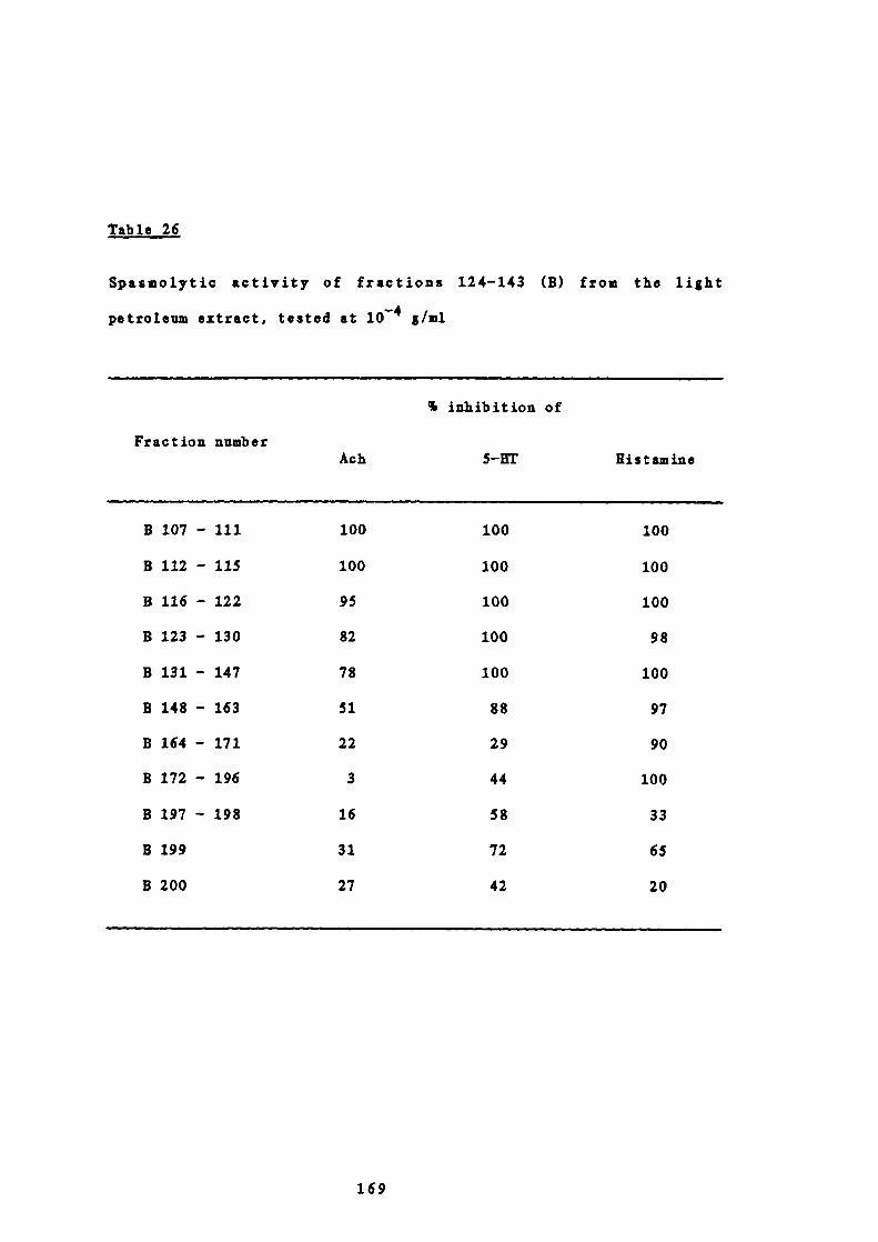
Table 26
Spasmolytic activity of fractions 124-143 (B) from the light
petroleum extract 1 tested at 1O 4 gIml
% inhibition of
Fraction numberAch 5—fiT flistainine
B 107 - 111 100 100 100
B 112 - 115 100 100 100
B 116 - 122 95 100 100
B 123 - 130 82 100 98
B 131 - 147 78 100 100
B148-163 51 88 97
B164-171 22 29 90
B172-196 3 44 100
B197-198 16 58 33
B199 31 72 65
B200 27 42 20
169
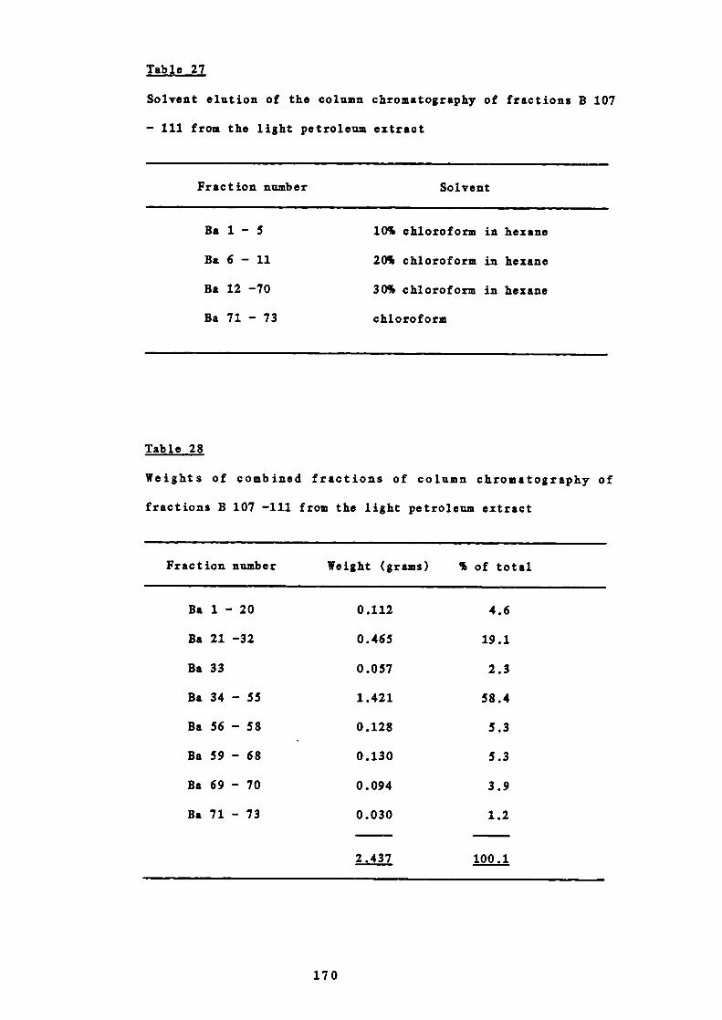
Table 27
Solvent elution of the column chromatography of fractions B 107
- 111 from the light petroleum extract
Fraction number Solvent
Ba 1 - 5 10% chloroform in hexane
Ba 6 - 11 20% chloroform in hexane
Ba 12 —70 30% chloroform in hezane
Ba 71 - 73 chloroform
Table 28
Weights of combined fractions of column chromatography of
fractions B 107 —111 from the light petroleum extract
Fraction number Weight (grams) % of total
Ba 1 - 20 0.112 4.6
Ba 21 —32 0.465 19.1
Ba 33 0.057 2.3
Ba 34 - 55 1.421 58.4
Ba 56 - 58 0.128 5.3
Ba 59 - 68 0.130 5.3
Ba 69 - 70 0.094 3.9
Ba 71 - 73 0.030 1.2
2.437 100.1
170

53 (11.3), 43 (100.0). Lit. 4 ° m.p. 107-111°. Lit. 42 m.p. 115°
This material was identical in all respects with an authentic
sample supplied by F.Sorm.
17 STUDIES ON FRACTIONS 75-80 (C) FROM THE LIGHT PETROLEUM EXTRACT
Fractions 75-80 (C) from the petroleum extract showed 100 %
inhibition of the two agonists, 5—UT and histamine, and 91%
inhibition of Ach.
A FURTHER SEPARATION OF FRACTIONS 75-80 (C)
0.65 g were chromatographed on a column of silica gel (60 g).
100 ml fractions were collected and combined as appropriate with
reference to TLC. The weights of the combined fractions and
solvent elution of the column are given in Tables 29 and 30.
18 STUDIES ON FRACTIONS 177-200 (D) FROM THE PETROLEUM EXTRACT
Fractions 177-200 from the petroleum extract showed 10Yo inhibition
of the two agonists 5—UT and histamine, and 81% inhibition of Ach.
A FURTHER SEPARATION OF FRACTIONS 177-200 (D)
These fractions (0.729 a) were chromatographed on a silica gel
column (30 g). 100 ml fractions were taken and combined as
appropriate with reference to TLC. Solvent elution of the
column and weights of the combined fractions are shown in Tables
31 and 32.
B SPASMOLYTIC ACTIVITY OF FRACTIONS 177-200 (D)
Those fractions weighing more than 50 mg were tested for
171
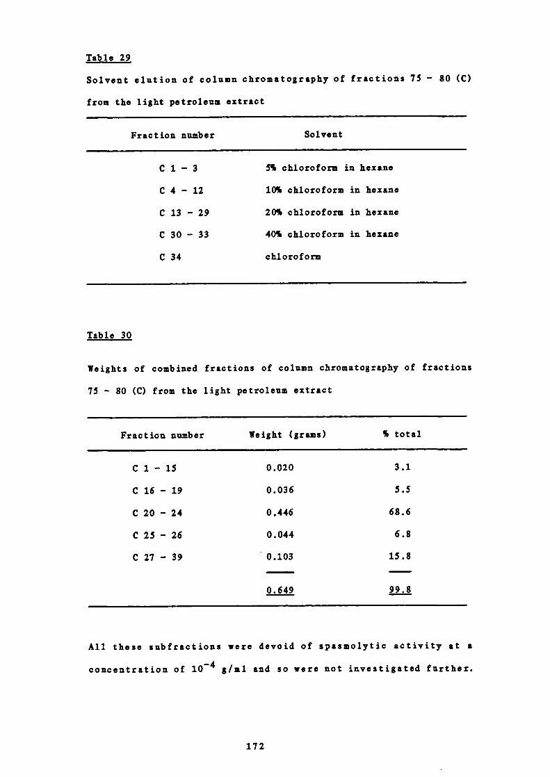
Table 29
Solvent elution of column chromatography of fractions 75 - 80 (C)
from the light petroleum extract
Fraction number Solvent
C 1 - 3 5% chloroform in hexane
C 4 - 12 10% chloroform in hexane
C 13 - 29 20% chloroform in hexane
C 30 - 33 40% chloroform in hexane
C 34 chloroform
Table 30
Weights of combined fractions of column chromatography of fractions
75 - 80 (C) from the light petroleum extract
Fraction number Weight (grams) % total
C 1 - 15 0.020 3.1
C 16 - 19 0.036 5.5
C 20 - 24 0.446 68.6
C 25 - 26 0.044 6.8
C 27 - 39 0.103 15.8
0.649 99.8
All these subfractions were devoid of spasmolytic activity at a
concentration of 1O 4 g/ml and so were not investigated further.
172

Table 31
Solvent elut ion of column chromatography of fractions 177-200 (D)
from the light petroleum extract
Fraction number Solvent
D 1 - 12 25% v/v ethyl acetate in hexane
D 13 - 17 40% v/v ethyl acetate in hexane
D 18 - 24 60% v/v ethyl acetate in hexane
D 25 - 31 80% v/v ethyl acetate in hexane
D 32 - 36 ethyl acetate
D 37 - 40 10% v/v chloroform in ethyl acetate
D 41 - 45 30% v/v chloroform in ethyl acetate
Table 32
Weights of combined fractions of column chromatography of fractions
177-200 (D) from the light petroleum extract.
WeightFraction number
% of total(grams)
D 1— 3 0.037 5.1
D 4 - 8 0.090 12.3
D 9 - 10 0.049 6.7
D 11 - 15
0.088
12.1
D 16 - 20
0.240
32.9
D 21 - 26 0.160 21.9
D 27 - 45 0.070 9.6
Total 0.734 100.6
173

spasmolytic activity as described before (Part III, 3). The
results are shown in Table 33.
C FURTHER INVESTIGATION OF THE FRACTIONS SHOWING 10O
INHIBITION OF THE AGONISTS
(a) Fractions D 16-20
Fractions D 16-20 (0.175 g) were further separated by
preparative TLC using chloroform:methanol (95:5) as
developing solvent. The plates were run three times. Three
bands were located under ultraviolet light (254 nm) and
their components extracted with chloroform. The major
component, i.e. in band 1, weighed 59 mg and crystallised
from chloroform and methanol as colourless plates (26 mg) of
DJ 154a, m.p. 248°; UV: a11 (log e) 208 (3.98) nm; IR:
?mIaBx1 3420 (OH), 1760 (C=O, y—lactone), 1675 (C=C,
conjugated), 1260 ( —C—O-4— , epoxide) cm; 'H NMR: 6 (400
MHz, CDC1 3 ), 1.15 (3K, s, H-15s), 1.58 (3H, s, H-14s), 2.87
(1K, d, 1 11.0 Hz, H-5), 3.30 (1K, rn, H-7), 3.32 (1K, d, I
= 1.3 Hz, H-3), 3.57 (1K, d, 3 1.3 Hz, H-2), 4.09 (1K, dd,
1 11.0, 10.0 Hz, H-6), 5.54 (1H, d, I = 3.5 Hz, H-13a),
6.19 (1K, d, 3 3.5 Hz, H-13b); MS: m/z (%, rel.int.) 279
(0.7), 278 (2.1), 263 (2.3), 262 (4.7), 260 (3.1), 249
(3.1), 231 (6.3), 217 (8.6), 203 (9.4), 189 (12.5), 175
(15.6), 165 (26.6), 151 (40.6), 149 (23.4), 147 (28.9), 123
(26.6), 121 (33.6), 112 (88.3), 109 (42.2), 97 (35.9), 95
(93.8), 91 (41.4), 85 (47.7), 83 (44.5), 81 (36.7), 79
(38.3), 77 (35.9), 71 (78.1), 69 (52.3), 67 (49.2), 57
(93.8), 55 (71.1), 53 (75.8), 43 (100.0). Lit. 5 ° m.p. 250°
This material was identical in all respects with literature
data on chrysartemin A.50'51
174
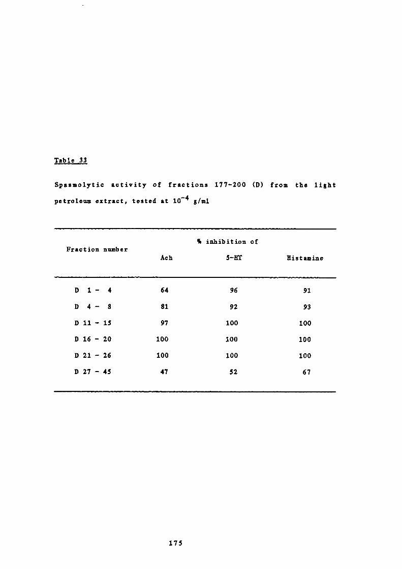
Table 33
Spasmolytic activity of fractions 177-200 (D) from the light
petroleum extract, tested at iO g/ml
% inhibition ofFraction number
Ach 5—HT Histamine
D 1 — 4 64 96 91
D 4 — 8 81 92 93
Dil—iS 97 100 100
D 16 - 20 100 100 100
D 21 - 26 100 100 100
D27-45 47 52 67
175

(b) Fractions D 21-26
Fractions D21-26 (0.110 g) were further separated by
preparative TLC using chloroform:rnethanol (95:5) as
developing solvent. The plates were run three times. Three
bands were located under ultraviolet light (254 urn) and by
spraying the edge of the plate with H 2 SO4 and heating.
Their components were extracted with chloroform.
The major component of fractions D 21-26, i.e. in band 2,
crystallised from chloroform and hexane to give colourless
needles (16 mg) of D1156a, 4a,l0—dihydroxy-1,5a—gui-
11(13) — en-12,6a — olactone ( p artholide, 82),rn.p. 1540; UV:
EtOH (log a) 208 (3.95) urn; IR: Solid film, 3490 (OH),
1750 (C=O, y—lactone), 1660 (;C = C, conjugated), 1640
(C=C); 1 fl NMR: 6 (400 MHz, CDC 3 ), 1.25 (311, s, H-14s),
1.35 (311, s, HiSs), 1.47 (111, rn, 11-8), 1.62 (211, rn, H-2s),
1.67 - 2.04 (611, br in, H-3s, fl-9s, 2 x OKs), 2.16 (111, rn, H-
8), 2.39 (111, dd, 1 12.3, 12.3 Hz, 11-5), 2.63 (111, rn, K-
1), 2.70 (111, rn, 11-7), 4.24 (1H, dd, 3 12.3, 9.5 Hz, H-6),
5.54 (111, d, 3 3.5 Hz, H-13a), 6.25 (111, d, 1 3.5 Hz, H-
13b); ' 3 C NMR: & (100.6 MHz, CDC1 3 ), 23.51, 24.25 (C-14 and
C — iS), 25.01, 25.33 (C-2 and C-8), 39.34, 43.77 (C-3 and C-
9), 47.22 (C-7), 49.78 (C-i), 55.35 (C-5), 74.72 (C-4),
79.88 (C— b), 82.74 (C-6), 120.37 (C-13), 138.45 (C—il),
169.37 (C-12); MS: rn/z (% rel.int.), 266 (0.4), 265 (0.9),
264 (1.8), 263 (1.8), 262 (3.6), 261 (2.7), 260 (4.5), 249
(3.0), 248 (6.0), 247 (7.2), 246 (18.0), 245 (6.3), 244
(13.5), 232 (4.5), 231 (15.3), 230 (10.0), 229 (9.0), 228
(19.8), 215 (12.6), 213 (18.9), 203 (22.5), 191 (22.5), 190
(27.0), 188 (24.3), 175 (24.3), 173 (22.5), 159 ( 24.3), 105
(45.0), 95 (44.6), 93 (44.1), 91 (59.5), 83 (85.6), 81
176
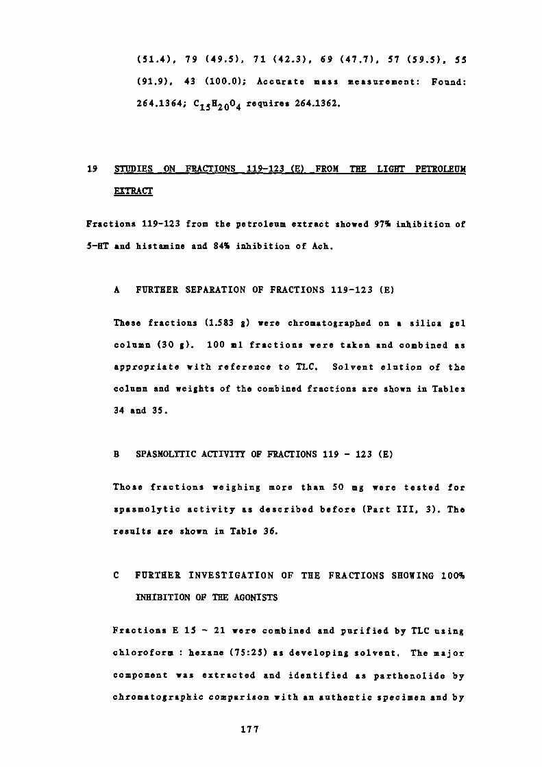
(51.4), 79 (49.5), 71 (42.3), 69 (47.7), 57 (59.5), 55
(91.9), 43 (100.0); Accurate mass measurement: Found:
264.1364; C 15 H2004 requires 264.1362.
19 STUDIES ON FRACTIONS 119-123 (E) FROM THE LIGHT PETROLEUM
EXTRACT
Fractions 119-123 from the petroleum extract showed 97% inhibition of
5—HT and histamine and 84% inhibition of Ach.
A FURTHER SEPARATION OF FRACTIONS 119-123 (E)
These fractions (1.583 g) were chromatographed on a silica gel
column (30 g). 100 ml fractions were taken and combined as
appropriate with reference to TLC. Solvent elution of the
column and weights of the combined fractions are shown in Tables
34 and 35.
B SPASMOL!TIC ACTIVITY OF FRACTIONS 119 - 123 (E)
Those fractions weighing more than 50 mg were tested for
spasmolytic activity as described before (Part III, 3). The
results are shown in Table 36.
C FURTHER INVESTIGATION OF THE FRACTIONS SHOWING 100%
INHIBITION OF THE AGONISTS
Fractions E 15 - 21 were combined and purified by TLC using
chloroform : hexane (75:25) as developing solvent. The major
component was extracted and identified as parthenolide by
chromatographic comparison with an authentic specimen and by
177

Table 34
Solvent elution of column chromatography of fractions 119-123 CE)
from the light petroleum extract
Fraction number Solvent
E 1 — 2 hexane
E 3 - 5 10% v/v chloroform in hexane
E 6 - 31 20% v/v chloroform in hexane
E 32 chloroform
Table 35
Weights of combined fractions of column chromatography of fractions
119-123 CE) from the light petroleum extract
WeightFraction number
% of total(grams)
E 1 - 13 0.010 0.6
E 14 0.007 0.4
E 15 0.058 3.7
E 16 - 18 0.450 28.4
E 19 - 21 0.201 12.7
E 22 - 31 0.148 9.3
E 32 - 36 0.251 15.9
E 37 - 38 0.064 4.0
E 39 0.395 25.0
1.584 100.0
178
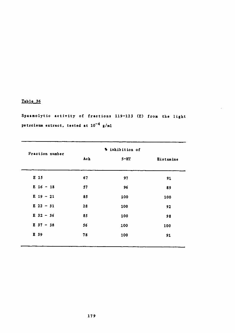
Table 36
Spasmolytic activity of fractions 119-123 (E) from the light
petroleum extract, tested at iO g/ml
% inhibition ofFraction number
Ach 5—UT Histamine
E15 67 97 91
E16-18 57 96 89
E19-21 85 100 100
E22-31 28 100 92
E32-36 85 100 98
E37-38 56 100 100
E39 78 100 91
179

spectroscopic means.
20 STUDIES ON FRACTIONS 112-118 (F) FROM THE LIGHT PETROLEUM EXTRACT
The major component of fractions 112-18 crystallised from chloroform
to give DJ52A. The mass spectrum of this shoved two compounds to be
present with molecular ions at m/z 414 and 412. The mixture was
acetylated with acetic anhydride in pyridine. The crude acetate
mixture, obtained after the normal work up, was separated using
argentation TLC with n—hexane:chloroform (90:10) as developing
solvent. The plates were run five times. The two compounds were
located by spraying the edge of the plate with 20% H 2 SO4 and
extracted with chloroform to yield 196 mg and 157 mg of materials
with melting points 126° and 141°. These were identical in all
respects with authentic specimens of —sitosterol and stigmasterol.
21 STUDIES ON FRACTIONS 5 1-61 FROM THE LIGHT PETROLEUM EXTRACT
Fractions 51-61 from the light petroleum extract showed 100%
inhibition of 5—HT, 96% inhibition of histamine and 89% of Ach.
A FURTHER SEPARATION OF FRACTIONS 51-61
S.8g were chromatographed on a column of silica gel (lOOg).
lOOmi fractions were taken and combined as appropriate with
reference to TLC. The solvent elution and weights of the
combined fractions are shown in Tables 37 and 38.
180
Table 37
Solvent elution of column chromatography of fractions 51-61
from the light petroleum extract
Fraction number Solvent
1-2 light petroleum
3-15 toluene
16-17 50% ethyl acetate in toluene
18 chloroform
19 methanol
Table 38
Weights of combined fractions of column chromatography of
fractions 51-61 from the light petroleum extract
Fraction Weight% total
number (grams)
1-4 3.091 53.3
5-6 1.291 22.3
7-15 1.291 22.3
16-19 0.064 1.1
5.737 99.0
All these subfractions were devoid of spasmolytic activity
at a concentration of iO g/ml. From subfractions 7-15,
DJ61a was isolated which proved to be mixture of long chain
fatty acid esters.
181

22 PREPARATION OF CAPSULES FOR CLINICAL STUDIES
A CALCULATION OF DOSE
(a) Weights of fresh and freeze —dried leaves
52 fresh feverfew leaves approximately 1.5 inches long and
1.2 inches wide each containing 5 leaflets were weighed
immediately after picking, freeze—dried and then re—weighed
to constant weight. These weights are given in Table 39.
Weights of fresh leaf
Mean 0.14908 g
Mean ± 2S 0.20588 - 0.09228 g
The weights of all fresh leaves lay within two standard
deviations of the mean.
Weights of dry leaf
Mean = 0.02566 g
Mean ± 2S = 0.036339 - 0.014981 g
The weights of all dry leaves lay within two standard
deviations of the mean.
(b) The dose of dry leaf equivalent to that of fresh leaf
Patients using feverfew for migraine take between one and
three fresh leaves daily in a bread and butter sandwich.
One fresh leaf weighs approximately 150 mg which is
equivalent to approximately 25 mg of freeze —dried leaf.
Therefore, capules containing 25, 50 and 75 mg of freeze —
dried feverfew, j. equivalent to 1, 2 or 3 fresh leaves,
were formulated.
182

Table 39
Weights of feverfew leaves
Weight of Weight of
Leaf Dry weightfresh leaf dry leaf
number Fresh weight(grams) (grams)
1 0.17348 0.02821 16.26124
2 0.14703 0.02687 18.27518
3 0.14980 0.02425 16.18825
4 0.19009 0.03040 15.99242
5 0.17416 0.03131 17.97772
6 0.16650 0.03090 18.55855
7 0.12151 0.02255 18.55814
8 0.13768 0.02223 16.14613
9 0.12153 0.02344 19.28741
10 0.13678 0.02321 16.96885
11 0.14708 0.02310 15.70573
12 0.13092 0.02155 16.46043
13 0.11424 0.01905 16.67542
14 0.11441 0.02244 19.61367
15 0.14299 0.02625 18.35792
16 0.13021 0.02357 18.10152
17 0.11370 0.01743 15.32981
18 0.12300 0.02333 18.96747
19 0.13763 0.02860 20.78035
20 0.11712 0.02372 20.25273
21 0.14862 0.03149 21.18826
22 0.18080 0.03515 19.44137
23 0.12394 0.02576 20.78425
24 0.19835 0.03591 18.10436
25 0.11916 0.02133 17.90030
26 0.12021 0.02219 18.45936
27 0.19378 0.03341 17.24120
continued on next page
183

Table 39 (continued)
Weight of Weight ofLeaf Dry weight
fresh leaf dry leafnumber Fresh weight
(grams) (grams)
28 0.11090 0.02027 18.27772
29 0.14977 0.02440 16.29164
30 0.12515 0.01875 14.98202
31 0.19099 0.03302 17.28886
32 0.13261 0.01838 13.86019
33 0.16754 0.02740 16.35430
34 0.20072 0.03397 16.92407
35 0.12833 0.01807 14.08088
36 0.19996 0.03227 16.13822
37 0.16540 0.02867 17.33373
38 0.14967 0.02257 15.07984
39 0.12571 0.01774 14.11184
40 0.20463 0.03629 17.56279
41 0.10423 0.01499 13.97828
42 0.16440 0.02614 15.90024
43 0.14735 0.02220 15.06616
44 0.14813 0.02344 15.82393
45 0.17356 0.02929 16.87600
46 0.12831 0.02247 17.51227
47 0.13057 0.02228 17.06364
48 0.17139 0.03173 18.51333
49 0.20492 0.03352 16.35760
50 0.15486 0.02604 17.26720
51 0.12881 0.02455 19.05907
52 0.16966 0.02823 16.63916
Total 7.75229 1.33433
Mean 0.14908 0.02566
S.D. 0.02840 0.00534
184

B FORMULATION OF CAPSULES
Ingredients 25 mg 50 mg 75 mg placebocapsules capsules capsules
Freezedried 25 50 75 -
feverfew
Lactose 145 130 105 q.s.
Chlorophyll - - - q.s.
Total 170 mg 180 mg 180 mg
The slight end —weight differences were governed by the
characteristics of the capsule filling machine. The capsules
used were Eli Lilly, size 2, opaque, No. VI 2116 BAS. The
average weight of the empty capsules was 60 mg.The ingredients
of the capsules were granulated in the usual way with a 20 mesh
sieve using 70% ethanol. Fines were removed by sieving with a
60 mesh sieve. Placebo capsules were prepared using lactose and
chlorophyll to give granules identical in colour with those
containing feverfew. The bottles containing the placebo
capsules were sprinkled with a small amount of feverfew powder
so that , on opening the bottles, both gave identical smell.
185

C BP TESTS ON TEE cApsuus 131a,b
Capsule Uniformity of weight 3Disintegration test3Th
25 mg Average 'wt. 20 caps. Disintegration time
171.8 mg. 6.5 mins.
Greatest deviation
from average = 9%
50 mg Average wt. 20 caps. Disintegration time
184.4 mg. 5.3 mins.
Greatest deviation
from average = 5%
75 mg Average vt. 20 caps. Disintegration time
183.9 mg. - 5.9 mins.
Greatest deviation
from average = 7%
Placebo Average wt. 20 caps. Disintegration time
243.2 mg. = 6.0 mins.
Greatest deviation
from average 10%
All the capsules complied with both requirements.
186

REFERENCES
1 R. Chapman, Sunday Express, 21st May 1978
2 G. Radcliffe, Glamorgan Gazette, 3rd-7th Aug. 1978
3 1. Watney, Prevention, 21, 62, 1978
4 P. Lees, Farmers Weekl y, p. 114, 23rd Nov. 1979
5 A.R. Clapham, T.G. Tutin and E.F. Warburg, Flora of the British
Isles, Reprint ed., Cambridge University Press, p. 1082, 1958
6 M. Grieve, A Modern Herbal, Ed. C. Leyel, Penguin Books,
Earmondsworth, 1976: (a) p. 309; (b) p. 185; (c) p. 797
7 M. Ireig, Green Medicine, Rand McNally and Co., Chicago, NewYork and San Francisco, p. 168, 1964
8 1. Gerarde, The Herbali or Gene rail Historie of Plantes, John
Norton, p. 526, 1597
9 1. Parkinson, Theatrum Botanicum: The Theater of Plantes or, An
Herball of a Large Extent, Tho. Coxes, London, p. 83, 1640
10 N. Culpeper, The Complete Herbal, A New Ed., p. 102, 1953
11 P. Schauenberg and F. Paris, Guide to Medicinal Plants,
Butterworth Press, London, p. 288, 1977
12 British Pharmaceutical Codex, The Pharmaceutical Press, London,
p. 132, 1934
13 1. de Bairacli Levy, The Illustrated Herbal Handbook, Faber and
Faber Ltd., London, p. 66, 1974
14 D. Law, Herbs for Cooking and for Healing, W. Foulshane and Co.
Ltd., London, p. 75, 1970
15 W.H. Lewis and M.P.F. Elvin —Lewis, Medical Botany, PlantsAffecting Man's Health, John Wiley, New York, p. 323, 1977
16 P. Perry, Amateur Gardenin g , p. 16, 19th Dec 1981
17 E.S. Johnson, Personal Communication
188

18 B. Jones and 0. Gillie, Sunday Times Magazine, p. 78, 12th April1981.
19 R. Regnauer, Chemotaxonomie der Pflanzen, Band 3, BjrkhauserVerlag, Basel und Stuttgart, p. 448, 1964
20 R. Hegnauer, The Biology and Chemistry of the Compositae, Vol.1, Eds. V.R. Heywood, LB. Harborne and B.L. Turner, AcademicPress, London, New York and San Francisco, p. 284, 1977
21 F. Bohlmann, C. Burkhardt and C. Zolero, Naturally OccurringAcetylenes, Academic Press, London and New York, 1973
22 W. Herz, The Biology and Chemistry of the Compositae, Vol. 1,Eds V.H. Heywood, LB. Harborne and B.L. Turner, Academic Press,London, New York and San Francisco, 1977: (a) p. 337; (b) p.338; (c) p. 343; (d) p. 341; (e) p. 346; (f) p. 349
23 V. Herout, Pharmacognosy and Phytochemistry, Ed. K. Wagner andL. Horhammer, Springer—Verlag, Berlin, Heidelberg and New York,
p. 93, 1971
24 W. Parker, J.S. Roberts and K. Ramage, p . Rev. Chem. Soc., 21,331, 1967
25 K. Greger, The Biology and Chemistry of the Compositac, Vol. 1,Eds. V.H. Keywood, .J.B. Harborne and B.L. Turner, AcademicPress, London, New York and San Francisco, p. 904, 1977
26 H. Hikino, Y. Kikino and T. Yosioka, Chem. Pharm. Bull., 10,641, 1962
27 W. Herz, Pharmacognosy and Phytochemistry, Eds. H. Wagner and L.Horhammer, Springer—Verlag, Berlin, Heidelberg and New York, p.64, 1971
28 T.A. Geissman, Recent Advances in Phytochemistry, Vol. 6,Academic Press, New York and London, 1973: (a) p. 70; (b) p. 79
29 Y. Tshizaki, Y. Tanahashi, T. Takahashi and I. Tori, Chem.Comm., 551, 1969
30 S.M. Kupchan, I.E. Kelsey and G.A. Sim, Tetr. Lett., 2863, 1967
31 H. Yoshioka, T.I. Mabry and B.N. Timaermann, SesquiterpeneLactones, Chemistry, NMR and Plant Distribution, University ofTokyo Press, 1973: (a) p.7; (b) p. 15; (c) p. 82; (d) p.211; (e) p. 412; (f) p. 331; (g) p. 330; (h) p. 20; (i) p.
189

142; (j) p . 375; (k) p. 371; (1) p. 370; (m) p. 364; (n)p.388
32 F. Sorm, I. Ag r. Food Chem., 19, 1081, 1971
33 W. Herz, Nobel Symposium, 25, 153, 1973
34 M. Nishikawa, K. Iamiya, A. Takabatake and H. Oshio,Tetrahedron, 22, 3061, 1966
35 S. Neidle and D. Rodgers, Chem. Comm., 140, 1970
36 A. Corbella, P. Garibaldi, G. Iommi, Z. Santek, M. Holub,B.Drozdz and E. Bloszyk, ibid., 386, 1972
37 T.A. Duliforce, G.A. Sirn, D.N.I. White, I.E. Keisey, and S.M.Kupchan, Tetr. Lett., 973, 1969
38 V. Herout, N. Soucek and F. Sorm, Chem. and md., 1069, 1959
39 Y.R. Naves, Heiv. Chim. Acta, 32, 1151, 1949
40 N. Soucek, V. Herout and F. Sorm, Coil. Czech. Chem. Comm., 26,803, 1961
41 P. do Mayo, Mono— and Sesquiterpenoids, Vol. 2, IntersciencePublishers, Inc., New York and London, p. 184, 1959
42 T.R. Govindachari, B.S loshi and V.N. Kamat, Tetrahedron, 21,1509, 1965
43 A.S. Bawdekar, G.R. Keikar and S.C. Bhattacharya, Tetr. Lett.,1225, 1966
44 A.S. Rao, G.R. Kelkar and S.C. Bhattacharya, Tetrahedron, 9,275, 1960
45 A. Quick and D. Rogers, I,C.S. Perkin II, 465, 1976
46 F. Sorm, N. Suchy, N. Holub, A. Linek, I. Hadinec and C. Novak,Tetr, Lett., 1893, 1970
47 T.A. Geissman and S. Matsueda, Phytochemistry, 7, 1613, 1968
190
48 A. Romo de Vivar and H. J'imenez, Tetrahedron, 21, 1741, 1965
49 H. Yoshioka, W. Renold, N.H. Fischer, A. Higo and T.J. Mabry,Phytochemistry, 9, 823, 1970
50 J. Romo, A. Romo de Vivar, R. Trevino, P. Joseph—Nathan and E.Diaz, ibid, 9, 1615, 1970
51 T. Osawa, A. Suzuki and S. Tamura, A g r. Biol. Chem., 35, 1966,1971
52 L.M. Jackman and S. Sternhell, Applications of NMR Spectroscopyin Organic Chemistry, Pergamon Press, Oxford, 1969
53 Z. Samek, Tetr. Lett., 671, 1970
54 S. Sternhell, Quart. Rev., 23, 236, 1969
55 H. Yoshioka, T.J. Mabry, M.A. Irwin, T.A. Geissman and Z. Samek,Tetrahedron, 27, 3317, 1971
56 R.F. Zurcher, Progress in NMR Spectroscopy in Organic Chemistry,Vol. 2, Holden—Day, San Francisco, p. 218, 1967
57 U. Yoshioka, N. Dennis, W. Kerz and T.J. Mabry, 1. Or g . Chern.,35, 627, 1970
58 Natural Products Chemistry, Vol. 1, Eds. L Nakanishi, T. Goto,S. Ito, S. Natori and S. Nozoe, Academic Press, Inc., New York,London, p. 98, 1974
59 R.I. Abraham and P. Loftus, Proton and Carbon-13 NMRSpectroscopy, An Integrated Approach, Hey-den and Sons Ltd.,London, Philadelphia and Rheine, 1978: (a) p. 107; (b) p. 142;(c) p.119; (d) p. 26; (e) p. 148
60 H. Yoshioka and T.J. Mabry, Tetrahedron, 25, 4767, 1969
61 N.S. Bhacca and N.H. Fischer, Chem. Comm., 68, 1969
62 H. Hikino, C. Konno and T. Takemoto, ibid, 662, 1969
63 1. Tori, I. Horibe, K. Kuriyama and I. Takeda, ibid, 957, 1970
191

64 I. Horibe, K. Tori and K. Nishimura, Tetrahedron, 29, 271, 1973
65 KM. Silverstein, G. Clayton Bassler and T.C. Morrill,Spectrometric Identification of Organic Compounds, 3rd ed., JohnWiley and Sons, Inc., New York, Sydney, London and Toronto,1974: (a) p. 191; (b) p. 96; (c) p. 21
66 K.Tori, I. Horibe, H. Yoshioka and T.J. Mabry, 1. Chem. Soc. B.,1084, 1971
67 1. Tori, I. Horibe, K. Kuriyama, H. Tada and I. Takeda, Chern.Comm., 1393, 1971
68 K. Tori, I. Horibe, H. Mirato and K. Takeda, Tetr. Lett., 4355,1971
69 LX. Bentley, J.G.St. C. Buchanan, T.G. Halsall and V. Thaller,Chem. Comm., 435, 1970
70 M.G.B. Drew, S.P. flitman and 3. Mann, I.C.S. Chem. Comm., 802,1980
71 H. Wagner, The Biology and Chemistry of the Compositae, Vol. 1,Ed. V.K. Heywood, ).B. Barborne and B.L. Turner, Academic Press,London, New York, San Fransisco, 1977: (a) p. 412; (b) p. 418;(c) p. 413; (d) p. 414; (e) p. 415; (f) p. 416; (g) p. 417;(h) p. 424
72 M.A. Kraul and F. Schmidt, Arch. Pharmaz., 290, 66, 1957
73 LB. Lee, E.S. Huang, C. Piantadozi, I.S. Pagano and T.A.Geissman, Cancer Rca., 31, 1649, 1971
74 K.H. Lee, T. Ibuka, H.C. Huang and D.L. Harris, I. Pharm. Sci.,64, 1077, 1975
75 LB. Lee, T. Ibuka, H. Furukawa, M. lozuka, LY. Wu, I.E. Halland H.C. Huang, ibid, 68, 1050, 1980
76 LH. Lee, T. Kimura, M. Okamoto and C.M. Cowherd, Tetr. Lett.,14, 1051, 1976
77 A.G. Gonzalez, V. Darias, J.N. Boada and M. Feria, Arch. deFarmacol. y Toxicol., 3, 241, 1977
78 A.G. Gonzalez, V. Darias, G. Alonso and E. Estevcz, Planta

Medica, 40, 179, 1980
79 Y. Jmakura, LII. Lee, D. Sims, R.Y. Yu, 1.11. Ball, H. Furukava,M. Itoigawa and I. Yonaha, I. Pharm. Sd., 69, 1044, 1980.
80 W. Herz, N. umar and J.F. Blount, 2. Or g . Chern., 46, 1356,
1981
81 P.L. Cowall, J.M. Cassady, C. Chang and 2.1. Kozlowski, ibid,
46, 1108, 1981
82 N.R. Farnsworth, I. Pharm. Sci., 55, 231, 1966
83 S.M. Iupchan, D.C. Fessler, M.A. Eakin and TJ. Giacobbe,Science, 168, 376, 1970
84 B. Uladon and T. Twandowski, Pol. I. Pharmacol, Pharm., 31, 35,1979
85 G.R. Pettit, J.C. Budzinski, G.M. Cragg, P. Brown, L.A. Johnstonand D. Rca, I. Med. Chem., 17, 1013, 1974
86 GA. Cordell, New Natural Products and Plant Drugs withPharmacological, Biological or Therapeutic Activity, Eds. H.Wagner and P. Wolff, Springer —Verlag, Berlin, Heidelberg and
New York, p. 54, 1977
87 A. Aebi, T. Waaler and I. Buchi, PharmWeekb lad, 93, 397, 1958
88 W. Lang and 1. Schwandt, Deutch. Apoth. —Zeitg, 150, 1957
89 1. Horhammer, H. Wagner and B. Salfner, Arzneim. Forsch., 13,33, 1963.
90 1. Thiele, V. Jako y le y , 0. Isaac and W.A. Schuller, ibid, 19,1878, 1969
91 I.H. Hall, C.O. Starnes, K.H. Lee and T.G. Waddell,I. Pharm. Sci., 69, 537, 1980
92 Davidson's Principles and Practice of Medicine, Ed. 1. Macleod,12th ed., Churchill Livingstone, Edinburgh, London and New York,1977: (a) p. 447; (b) p. 698
93 G. Hahn, H.D. Lehmann, M. lurten, K. Uebel and G. Vogel,
193
Arzneim. Forsch., 18, 698, 1968
94 H. Wagner, 0. Seligmann, L. Horhammer, M. Sejtz and 1.Sonnenbich].er, Tetr. Lett., 22, 1895
95 1. Scbmidt—Thome, Z. Naturf., Sb, 409, 1950
96 I.E. Schulte, G. Ruder and 2. Perlick, Arzneim. Forsch.,17,825, 1967
97 3. Reisch, W. Spitzner and I.E. Schulte, ibid., 17, 816, 1967
98 J.P. Blakeman and P. Atkinson, Physiol. Plant Pathol,, 15, 183,1979
99 W.D. Loomis and K. Croteau, Recent Advances in Phytochemistry,Vol. 6. Eds. V.C. Runeckles and TI. Mabry, Academic Press, NewYork and London, p. 147, 1973
100 The Dispensatory of the United States of America, 25th ed., T.B.Lippincott Company, Philadelphia, p. 1674, 1955
101 A. Stoll, I. Renz and A. Brack, Kelv. Chim. Acta, 33, 1877, 1950
102 D. Orinda, I. Diederich and A. Wacker, Arzneim. Forsch., 23,1119, 1973
103 I.E. Hall, I.E. Lee, C.O. Starnes, 0. Muraoka, Y. Sumida andT.G. Waddell, 1. Pharm. Sci., 69, 694, 1980
104 G.E. Trease and W.C. Evans, Pharmacognosy, 10th ed., BailliereTindall, London, p. 605, 1972.
105 1. Wada and I. Munakata, Ag r. Biol. Chem., 35, 115, 1971
106 W.C. Alvarez, Gastroenterology, 6, 524, 1945
107 P.). Congdon and W.I. Forsythe, Clin. Ped., 18, 353, 1979
108 Brains Clinical Neurology, 5th ed., revised K. Bannister, OxfordUniversity Press, Oxford, New York and Toronto, p. 190, 1978.
109 .1. C. Houston, C.L. Yoiner and T.R. Trounce, A Short Textbook ofMedicine, 5th ed., Hodder and Stoughton, London, Sydney,
194
Auckland and Toronto, p. 269, 1977
110 Biochemical Aspects of Nervous Disease, Ed. .TN. Cumings, PlenumPress, London and New York, p. 196, 1972
111 E.S. Johnson, Post g rad. Med. 1., 54, 231, 1978
112 F. Sicuteri, Research and Clinical Studies in Headache, 1, 6,1967
113 N.R. Farnswoth and A.S. Bingel, New Natural Products and PlantDrugs with Pharmacological, Biological or Therapeutic Activity,Eds. H. Wagner and P. Wolff, Springer—Verlag, Berlin, Heidelbergand New York, p.2, 1977
114 W. Herz, N. Kumar and .1. Blount, .1. Org . Chem., 46, 1356, 1981
115 Y—L. Lin and T.J. Mabry, I. Nat. Prod., 44, 722, 1981
116 M.J. Begley, L. Crombie, W.M. Crombie, A.K. Gatuma and A.Maradufu, I.C,S. Perkin I, 2702, 1981
117 W. Herz, S.V. Govindan and J.F. Blount, I. Or g . Chem., 45,3163, 1980
118 P. Sykes, A Guidebook to Mechanism in Organic Chemistry, 4thed., Longman Group Ltd., London, p. 177, 1975
119 S.B. Mathur, S.V. Hiremath, G.H. ulkarni, G.R. Kelkar and S.C.Bhattacharyya, Tetrahedron, 21, 3575, 1965
120 S.M. Iupchan, I.E. lelsey, M. Muruyama, J.M. Cassady, IC.Hemingway and J.R. Knox, I. Or g . Chein., 34, 3876, 1969
121 L. Novotny and V. Rerout, Coil. Czech. Chem. Comm., 27, 1508,1962
122 F. Bohlmann, R.K. Gupta and I. Jakupovic, Phytochemistry, 21,460, 1982
123 H. Yoshioka, W. Renold and T.I. Mabry, Chem. Comm., 148, 1970
124 A. Rnstaiyan, L. Nazarians and F. Bohlmann, Phytochemistry, 20,1152, 1981
195
125 D.Ranganathan and S. Ranganathan, Art in Biosynthesis, TheSynthetic Chemist's Challenge, Vol. 1, Academic Press, New York,San Fransisco and London, 1976: (a) p. 125; (b) p. 128
126 K.A. Attrep, WP. Bellman, M. Attrep, LB. Lee and W.E.Braselton, Lip ids, 15, 292, 1980
127 Goodman and Oilman's The Pharmacological Basis of Therapeutics,6th ed., Eds. A.G. Gilman, L.S. Goodman and A. Gilman, BailliereTindall, London, p. 638, 1980
128 H.O.J. Collier, N.M. Butt, W.J. McDonald—Gibson and S.A. Saed,The Lancet, 922, 1980
129 A.N. Makheja and J.M. Bailey, ibid., 1054, 1981
130 E. Bleumiak, J.C. Mitchell, T.A. Geissman and G.R.N. Towers,Contact Dermatitis, 2, 81, 1976
131 British Pharmacopoeia, The Pharmaceutical Press, London, 1973:(a) p. 459; (b) p. A131
196
Related Documents