Citation: Saef, J.; Montgomery, R.; Cedars, A.; Tang, W.H.W.; Rossano, J.W.; Maeda, K.; Kim, Y.Y.; Vaikunth, S.S. Durable Mechanical Circulatory Support in Adult Congenital Heart Disease: Reviewing Clinical Considerations and Experience. J. Clin. Med. 2022, 11, 3200. https:// doi.org/10.3390/jcm11113200 Academic Editor: Juan F. Delgado Jiménez Received: 1 April 2022 Accepted: 1 June 2022 Published: 3 June 2022 Publisher’s Note: MDPI stays neutral with regard to jurisdictional claims in published maps and institutional affil- iations. Copyright: © 2022 by the authors. Licensee MDPI, Basel, Switzerland. This article is an open access article distributed under the terms and conditions of the Creative Commons Attribution (CC BY) license (https:// creativecommons.org/licenses/by/ 4.0/). Journal of Clinical Medicine Review Durable Mechanical Circulatory Support in Adult Congenital Heart Disease: Reviewing Clinical Considerations and Experience Joshua Saef 1,2 , Robert Montgomery 3 , Ari Cedars 4 , Wai H. Wilson Tang 3 , Joseph W. Rossano 2 , Katsuhide Maeda 5 , Yuli Y. Kim 1,2 and Sumeet S. Vaikunth 1,2, * 1 Division of Cardiovascular Medicine, Department of Medicine, Perelman School of Medicine, University of Pennsylvania, Philadelphia, PA 19104, USA; [email protected] (J.S.); [email protected] (Y.Y.K.) 2 Division of Cardiology, Children’s Hospital of Philadelphia, 3401 Civic Center Boulevard, Philadelphia, PA 19104, USA; [email protected] 3 Robert and Suzanne Tomsich Department of Cardiovascular Medicine, Sydell and Arnold Miller Family Heart, Vascular and Thoracic Institute, Cleveland Clinic Foundation, Cleveland, OH 44195, USA; [email protected] (R.M.); [email protected] (W.H.W.T.) 4 Division of Cardiology, Johns Hopkins Medicine, Baltimore, MD 21287, USA; [email protected] 5 Division of Cardiothoracic Surgery, Children’s Hospital of Philadelphia, 3401 Civic Center Boulevard, Philadelphia, PA 19104, USA; [email protected] * Correspondence: [email protected]; Tel.: +215-615-3388 Abstract: Adults with congenital heart disease (ACHD) patients are one of the fastest growing populations in cardiology, and heart failure (HF) is the most common cause of morbidity and mortality amongst them. The need for advanced HF therapies in ACHD patients stands to grow substantially. The anatomic considerations for placing durable mechanical circulatory support (MCS) devices in ACHD patients often require specialized approaches. Despite this, increasing evidence suggests that durable MCS can be implanted safely with favorable outcomes in ACHD patients. Expansion of MCS use in ACHD patients is imperative to improve their clinical outcomes. Knowledge of ACHD-specific anatomic and physiologic considerations is crucial to HF programs’ success as they work to provide care to this growing population. Keywords: congenital heart disease; mechanical circulatory support 1. Introduction Successes in managing complex congenital heart disease (CHD) have driven a surge in prevalence [1]. Among CHD patients, heart failure (HF) causes substantial morbidity [2,3] and is the most common cause of mortality in adults with congenital heart disease (ACHD) [4]. Management of ACHD-related HF is complicated by the fact that anatomic complexity in the ACHD population has increased over time [5]. These factors confront HF and ACHD specialists with novel challenges as they work to allocate advanced HF therapies, including durable mechanical circulatory support (MCS) to ACHD patients. Current United Network for Organ Sharing (UNOS) heart transplant allocation criteria prioritize MCS to qualify for higher priority listing status [6]. The anatomic considerations for placing temporary and durable MCS in ACHD patients are complex and require focused expertise. Consequently, a lower proportion of ACHD patients are listed as high- priority transplant waitlist statuses compared to patients with other cardiomyopathies. This discrepancy has resulted in ACHD patients experiencing longer waitlist times and higher waitlist mortality rates and highlights the need to further define the potential for durable MCS in ACHD patients [7,8]. J. Clin. Med. 2022, 11, 3200. https://doi.org/10.3390/jcm11113200 https://www.mdpi.com/journal/jcm

Welcome message from author
This document is posted to help you gain knowledge. Please leave a comment to let me know what you think about it! Share it to your friends and learn new things together.
Transcript

Citation: Saef, J.; Montgomery, R.;
Cedars, A.; Tang, W.H.W.; Rossano,
J.W.; Maeda, K.; Kim, Y.Y.; Vaikunth,
S.S. Durable Mechanical Circulatory
Support in Adult Congenital Heart
Disease: Reviewing Clinical
Considerations and Experience. J.
Clin. Med. 2022, 11, 3200. https://
doi.org/10.3390/jcm11113200
Academic Editor: Juan F. Delgado
Jiménez
Received: 1 April 2022
Accepted: 1 June 2022
Published: 3 June 2022
Publisher’s Note: MDPI stays neutral
with regard to jurisdictional claims in
published maps and institutional affil-
iations.
Copyright: © 2022 by the authors.
Licensee MDPI, Basel, Switzerland.
This article is an open access article
distributed under the terms and
conditions of the Creative Commons
Attribution (CC BY) license (https://
creativecommons.org/licenses/by/
4.0/).
Journal of
Clinical Medicine
Review
Durable Mechanical Circulatory Support in Adult CongenitalHeart Disease: Reviewing Clinical Considerations and ExperienceJoshua Saef 1,2, Robert Montgomery 3 , Ari Cedars 4, Wai H. Wilson Tang 3 , Joseph W. Rossano 2,Katsuhide Maeda 5, Yuli Y. Kim 1,2 and Sumeet S. Vaikunth 1,2,*
1 Division of Cardiovascular Medicine, Department of Medicine, Perelman School of Medicine,University of Pennsylvania, Philadelphia, PA 19104, USA; [email protected] (J.S.);[email protected] (Y.Y.K.)
2 Division of Cardiology, Children’s Hospital of Philadelphia, 3401 Civic Center Boulevard,Philadelphia, PA 19104, USA; [email protected]
3 Robert and Suzanne Tomsich Department of Cardiovascular Medicine, Sydell and Arnold Miller FamilyHeart, Vascular and Thoracic Institute, Cleveland Clinic Foundation, Cleveland, OH 44195, USA;[email protected] (R.M.); [email protected] (W.H.W.T.)
4 Division of Cardiology, Johns Hopkins Medicine, Baltimore, MD 21287, USA; [email protected] Division of Cardiothoracic Surgery, Children’s Hospital of Philadelphia, 3401 Civic Center Boulevard,
Philadelphia, PA 19104, USA; [email protected]* Correspondence: [email protected]; Tel.: +215-615-3388
Abstract: Adults with congenital heart disease (ACHD) patients are one of the fastest growingpopulations in cardiology, and heart failure (HF) is the most common cause of morbidity andmortality amongst them. The need for advanced HF therapies in ACHD patients stands to growsubstantially. The anatomic considerations for placing durable mechanical circulatory support (MCS)devices in ACHD patients often require specialized approaches. Despite this, increasing evidencesuggests that durable MCS can be implanted safely with favorable outcomes in ACHD patients.Expansion of MCS use in ACHD patients is imperative to improve their clinical outcomes. Knowledgeof ACHD-specific anatomic and physiologic considerations is crucial to HF programs’ success as theywork to provide care to this growing population.
Keywords: congenital heart disease; mechanical circulatory support
1. Introduction
Successes in managing complex congenital heart disease (CHD) have driven a surge inprevalence [1]. Among CHD patients, heart failure (HF) causes substantial morbidity [2,3]and is the most common cause of mortality in adults with congenital heart disease (ACHD) [4].Management of ACHD-related HF is complicated by the fact that anatomic complexity inthe ACHD population has increased over time [5]. These factors confront HF and ACHDspecialists with novel challenges as they work to allocate advanced HF therapies, includingdurable mechanical circulatory support (MCS) to ACHD patients.
Current United Network for Organ Sharing (UNOS) heart transplant allocation criteriaprioritize MCS to qualify for higher priority listing status [6]. The anatomic considerationsfor placing temporary and durable MCS in ACHD patients are complex and requirefocused expertise. Consequently, a lower proportion of ACHD patients are listed as high-priority transplant waitlist statuses compared to patients with other cardiomyopathies.This discrepancy has resulted in ACHD patients experiencing longer waitlist times andhigher waitlist mortality rates and highlights the need to further define the potential fordurable MCS in ACHD patients [7,8].
J. Clin. Med. 2022, 11, 3200. https://doi.org/10.3390/jcm11113200 https://www.mdpi.com/journal/jcm

J. Clin. Med. 2022, 11, 3200 2 of 13
General Considerations
Assessing the severity of congenital heart disease requires several considerations,including patients’ native cardiac anatomy, surgical history, current physiology, and co-morbidities. Consequently, many centers have developed dedicated multidisciplinaryprograms. This approach has shown clinical benefit to ACHD patients and is necessarywhen considering patients for MCS [9].
The 2018 American College of Cardiology/American Heart Association guidelinesfor the management of ACHD advocate for an anatomic and physiologic classificationscheme meant to categorize disease severity using factors known to have prognosticvalue [10]. This new classification system represents a major advance in ACHD becauseanatomic complexity and clinical status frequently correlate poorly, and the natural historyof many lesions is less clear with newer advances in care. The physiologic considerationswithin this classification include arrhythmia, concomitant valvular heart disease, exercisecapacity, pulmonary hypertension, hypoxemia or cyanosis, vascular stenosis and NYHAfunctional classification.
While this is an unsurprising list, the manner in which ACHD patients present withthe aforementioned issues is often quite different from those with acquired heart disease.Hosseinpour et al. classify patients with HF and congenital heart disease into three cate-gories: those with uncorrected defects (e.g., a patient with an ostium secundum atrial septaldefect that goes undiagnosed into adulthood), those with previous reparative surgerybut with deteriorating ventricular function (e.g., a patient with dextro-transposition ofthe great arteries (d-TGA) who underwent an atrial switch procedure in his infancy whopresents in adulthood with systemic right ventricular failure), and those with previouspalliative surgery which is now failing (e.g., a patient with complex congenital heart diseasepalliated to a single ventricle circulation) [11]. When considering the adult population,these all speak to processes that drive clinical deterioration insidiously from birth. Thus,many ACHD patients adapt to their exertional limitations, such that they may not reportsymptoms until complications of HF are extreme and perhaps even irreversible [12,13].
The initial approach to care for ACHD patients with HF shares several principleswith that of care for adult HF patients without CHD. Patients should be assessed regularlyfor clinical signs of congestion and functional decline, and providers should have lowthresholds to refer patients to an ACHD provider for assessment and diagnostic studiestailored to the patient’s specific congenital heart disease.
While HF, both with and without CHD, is a state of neurohormonal perturbation,there is limited data to support elements of the guideline-directed medical therapy inCHD patients. Anatomic limitations often preclude contemporary measures for cardiacresynchronization therapy. Nevertheless, care is often focused on providing maximallytolerated medical therapy based on extrapolation from data in non-CHD patients andon treating CHD-specific contributors to poor cardiac output, such as arrhythmia orvalvular dysfunction.
Exact clinical thresholds to trigger consideration for advanced HF therapies are stillbeing defined, though current guidelines recommend consultation with ACHD and HFspecialists when ACHD patients experience clinical HF or severe ventricular dysfunc-tion [10,14]. Purported criteria have included multiple HF hospitalizations within oneyear, severe objective deterioration in exercise tolerance, persistent NYHA functional classIII-IV symptoms, intolerance to further guideline-directed pharmacotherapy, refractoryarrhythmias, and cardiac cachexia [15]. ACHD patients may experience these signs ofdeterioration in a less predictable fashion than those without congenital heart disease, andcare should be taken for prompt recognition and evaluation should these concerns arise.
2. Experience with Durable Mechanical Circulatory Support in ACHD Patients
While experience with implanting durable MCS devices into adult HF patients withoutcongenital heart disease has expanded substantially, experience is more limited in ACHDpatients. A recent analysis of the Interagency Registry for Mechanically Assisted Circulatory

J. Clin. Med. 2022, 11, 3200 3 of 13
Support (INTERMACS) database showed that of 16,182 patients who underwent durableMCS between 23 June 2006 and 31 December 2015, 126 (<0.1%) were ACHD patients [16].Amongst them, 36% had a systemic right ventricle (morphologic right ventricle supportingthe systemic circulation), and 13% had single ventricle physiology.
Similarly noteworthy is that of the 158 centers contributing to the INTERMACSregistry, there were only 59 implanted durable MCSs in ACHD patients. The substantialmajority (>70%) of those centers only implanted devices in one or two ACHD patients.The volume of ACHD patients on MCS tended to be higher (five or six patients) in centerswith higher volumes of durable MCS in non-ACHD patients, indicating that experiencewith advanced HF therapies in the broader adult population is an important contributor tocomfort in providing them to ACHD patients.
ACHD patients supported with MCS were younger, more often had depressed rightventricular function, and less often had depressed left ventricular function or mitral re-gurgitation than other adult HF patients. They were also, unfortunately, more likely to beallo-sensitized and have unfavorable chest anatomy for heart transplantation. Nonetheless,ACHD patients were more likely to have undergone device implantation with a plan fora bridge to transplant rather than as destination therapy compared to patients withoutcongenital heart disease.
Unsurprisingly, device preferences have evolved as technology has advanced. Before2010, HeartMate and HeartMate II were the most frequently implanted devices. The Heart-Ware HVAD was the most commonly reported ventricular assist device (VAD) implantedin ACHD patients between 2010 and 2017 in a recent systematic review (though morerecent experience with the HeartMate 3 device was not included) [17]. Data on the useof the HeartMate 3 in ACHD is of great interest, as the device’s profile is more forgivingwith complex anatomy and chest closure. Although numbers remain small, there hasbeen a trend toward a higher proportion of biventricular assist devices and total artificialheart (TAH) being used in ACHD patients compared to non-ACHD patients. A higherproportion of ACHD patients with a systemic left ventricle underwent TAH placement(12.7%) than with a systemic right ventricle (4.4%) or a single ventricular circulation (5.9%),likely representing a higher prevalence of subpulmonic right ventricular failure in thesepatients [16].
There are several important considerations in ACHD patients which are essential toperioperative management, the foremost being a thorough assessment of cardiac anatomy.Moreover, many patients with congenital heart disease have abnormal pulmonary hemo-dynamics due to compensatory remodeling or, by design, in the context of single ventriclecirculations. A thorough right heart catheterization with an assessment of pulmonaryvascular anatomy and resistance offers crucial anticipatory guidance for postoperativehemodynamics. ACHD patients, particularly patients with single ventricle circulation, arealso at increased risk of liver disease, as they often suffer chronically elevated systemicvenous pressures from cavo-pulmonary connections [12]. Liver cirrhosis is associated withincreased mortality in patients undergoing cardiac surgery [18].
Data on the postoperative management experience with durable MCS in ACHD pa-tients are limited. A case series of six ACHD patients (among the highest volumes describedin the INTERMACS registry) outlined an institutional strategy for guidance in the postop-erative period. All patients received initial inotropic support for subpulmonic circulatoryfailure. Hypoxemia was tolerated in patients who had known right-to-left shunting orveno-venous collaterals. Patients were given pulmonary vasodilator therapy with inhaledand intravenous agents until extubation, followed by oral therapy. Fluid congestion wascommon in the postoperative period and postoperative nephrology consultation was re-quested for all patients. All patients required diuresis and two-thirds required temporaryultrafiltration or hemodialysis. Five patients survived to discharge, all of whom achievedNYHA functional class III status before discharge, and the average length of stay was over80 days [19].

J. Clin. Med. 2022, 11, 3200 4 of 13
The most robust data on long term outcomes with durable MCS comes from a compar-ison of ACHD patients within the INTERMACS registry to propensity-matched controls.Cedars et al. evaluated early and late adverse event rates after MCS implantation, aswell as changes in functional status and quality of life. Compared to matched controls,ACHD patients were more likely to experience early renal dysfunction, early and latehepatic dysfunction, early and late respiratory failure, late cardiac arrhythmia, and infec-tion. Many of these adverse events were found to be severe, with 60% of ACHD patientswith renal dysfunction requiring dialysis and 47% with arrhythmia experiencing a sus-tained ventricular arrhythmia requiring cardioversion. ACHD patients also experienceda higher overall mortality after MCS implant, largely driven by differences seen in thefirst five months post-procedurally. Multivariate analysis showed that factors associatedwith mortality hazards were an age greater than 50 years and a biventricular assist device orTAH implantation. Notably, the presence of a systemic right, left, or single ventricle did notaffect mortality after durable MCS implantation. Nonetheless, surviving ACHD patientsexperienced similar improvements in all functional status and quality of life parameters totheir matched non-ACHD controls [20].
Though they are associated with worse outcomes, biventricular assist devices andTAH are an important consideration in ACHD as many types of congenital heart diseaseleave patients with subpulmonic ventricular dysfunction and pulmonary hypertension.Thus, the proportion of ACHD patients who were implanted with these devices was threetimes higher than in non-ACHD patients [16]. Whether the increased mortality hazard isa consequence of the complications related to the pulmonic circulatory system is unclear.Next-generation durable biventricular or hybrid devices create unique opportunities forACHD patients.
3. ACHD-Specific Anatomic Considerations
While there is great interest in offering durable MCS therapies to ACHD patients, thereare practical limitations that prevent wide adoption. Most devices have been designed tosupport a morphologic left ventricle in patients with levocardia and biventricular phys-iology, and there are many congenital heart lesions with a systemic left ventricle wherepatients are at risk of heart failure. Examples include tetralogy of Fallot, atrio-ventricularseptal defects, and Ebstein anomaly, among others. Nevertheless, the cardiac anatomic het-erogeneity is immense within the ACHD population, even before considering the varioussurgical palliations and thoracic vascular anomalies. These factors, compounded by theneed for adhesiolysis in many patients with multiple prior sternotomies and comorbidities,create technical challenges when planning MCS implantation even in the above lesions,which are most analogous to typical biventricular circulations.
Two anatomic categories that are specific to congenital heart disease and pose particu-lar challenges when considering MCS therapies are lesions with systemic right ventriclesand single ventricle circulations, which will be explored further below.
3.1. Congenital Heart Disease Associated with Systemic Right Ventricle
In d-TGA, there is atrio-ventriculo concordance but ventriculo-arterial discordance,with the most common van Praagh segmental anatomical classification being {S, D, D}. Sys-temic venous return flows through the right atrium, right ventricle, and then into the aortawithout traversing the pulmonary circulation. Pulmonary venous return flows throughthe left atrium, left ventricle, and into the pulmonary arteries. The physiology of parallelcirculations is incompatible with life without native shunting or surgical remedy [21].Earlier surgical techniques used atrial baffles to redirect venous inflow to the oppositeventricle, allowing for a deoxygenated systemic venous return to traverse the pulmonarycircuit and eject into the systemic circulation thereafter [22]. While this facilitates physio-logic oxygenation, a morphologic right ventricle is left to generate systemic pressures farbeyond its intended design. Though not all mortality experienced after this atrial switchprocedure is due to systemic right ventricular dysfunction, a recent retrospective study

J. Clin. Med. 2022, 11, 3200 5 of 13
showed only 60% of patients who had undergone this procedure were alive after 30 yearsof follow-up [23].
In congenitally corrected or levo-transposition of the great arteries (l-TGA), thereis both atrio-ventricular and ventriculo-arterial discordance, with the most commonvan Praagh segmental anatomical classification being {S, L, L}. The result is systemicvenous return entering the morphologic left ventricle and the pulmonary artery with pul-monary venous return entering the morphologic right ventricle and the aorta. Patientswithout comorbid cardiac malformations (a minority) can tolerate this physiology intoadulthood before detection. Nonetheless, the right ventricle is similarly vulnerable todysfunction as above given the inherent mismatch in oxygen supply and demand andpredominately circumferential instead of longitudinal shortening [24,25]. Patients withl-TGA most commonly have L-ventricular looping, which adds anatomic complexity, as thesystemic right ventricle sits on the left side, and the subpulmonic morphologic left ventriclesits on the right side.
Anatomically, the right ventricle is less concave, has a greater degree of trabeculation,possesses a more variable, diffuse subvalvular apparatus and has a thinner ventricularwall than the morphologic left ventricle. Each of these factors presents a challenge tothe placement of durable VADs. These anatomic challenges are ameliorated as the rightventricle dilates to assume a more globular morphology in the setting of chronic exposureto systemic afterload.
3.2. Durable Mechanical Circulatory Support Considerations in Systemic Right Ventricles
The optimal location for the inflow portion of an MCS device is variable in systemicright ventricles, depending on several factors, including the angle of the patients’ atrioven-tricular valve inflow, their ventricular arrangement, and mediastinal anatomy. Avoidingthe tricuspid subvalvular apparatus and the ventricular septum is paramount. Resectionof the trabeculations may be necessary to prevent inflow cannula obstruction [26]. Trans-esophageal echocardiography is often used to assist with identifying an appropriate inflowcannula location, followed by intraoperative epicardial echocardiography to verify that theinflow cannula position will be free of trabeculations, chordae, and papillary muscles.
Significant tricuspid regurgitation may complicate the MCS placement in the systemicright ventricle, and there are differing opinions among centers as to the need for tricuspidvalve repair or replacement at the time of VAD implantation. Where some centers routinelyperform bioprosthetic tricuspid valve replacement at the time of VAD implantation, othersdisregard the tricuspid regurgitation to minimize operative duration [27].
The systemic right ventricle’s position in the anterior chest in d-TGA post atrialswitch can create geometric challenges for VAD implantation, with the sewing ring for theinflow cannula typically needing to be on the diaphragmatic surface. Simultaneously, thesurgeon must attempt to find a location such that there is an adequate distance from theinterventricular septum. Consideration must also be taken such that sternotomy closuredoes not cause compression of intrathoracic or epigastric structures [28]. A summary of thekey considerations is highlighted in Table 1 below.
Despite significant anatomic and physiologic variability in patients presenting withsystemic right ventricular failure, case series and registry data demonstrate the feasibilityand benefit of durable mechanical support with HeartMate II (Abbott, Chicago, IL, USA)and Heartware devices (Medtronic, Minneapolis, MN, USA) [16,27,29]. However, the onlycurrently FDA approved durable VAD is the HeartMate 3 (Abbott, Chicago, IL, USA).The advantage of the HeartMate 3 over prior generations of devices in the particularcircumstance of supporting systemic right ventricles is that the inflow cannula is lowerprofile and thus less likely to obstruct a heavily trabeculated right ventricle. Additionally,the outflow cannula is larger and theoretically less likely to obstruct when accommodatingunique positioning. INTERMACS data show that VADs placed to support a systemic rightventricle have comparable survival to patients with other ACHD phenotypes with a 60%two-year survival, which is comparable to non-ACHD patients supported with durable

J. Clin. Med. 2022, 11, 3200 6 of 13
LVADs in the same time period [16]. Figure 1 below shows an example of HeartMate 3 in acongenitally corrected transposition anatomy.
Table 1. Summary of Key Considerations for Durable Mechanical Circulatory Support in the SystemicRight and Single Ventricle.
Systemic Right Ventricle Single Ventricle
Cardiac ConditionsDextro-Transposition of the Great Arteries
or Congenitally corrected Transpositionof the Great Arteries
Complex Congenital Heart Disease notamenable to biventricular repair
(e.g., Hypoplastic Left Heart Syndrome,Tricuspid Atresia, Double Inlet Left Ventricle,Unbalanced Atrioventricular Septal Defect)
Technical Challenges
Inflow Cannula Placement(e.g., trabeculations, papillary muscles)
Concomitant Need for AtrioventricularValve Surgery or Baffle RevisionAbnormal Ventricular LoopingCreating Geometric Challenges
Scarring and Fibrosis from Prior SternotomiesAchieving Adequate Separation
from Ventricular SeptumAvoiding Compression of Intrathoracic or
Epigastric Structures with Sternotomy Closure
Inflow Cannula Placement(e.g., trabeculations, papillary muscles)
Concomitant Need for AtrioventricularValve Surgery or Fenestration
Bleeding Risk (e.g., aortopulmonary orveno-venous collaterals)
Scarring and Fibrosis fromMultiple Prior Sternotomies
Neo-Aortic PathologyDevice Fit
Management Challenges Potential for Inflow Obstruction due to RightVentricular Anatomic and Geometric Features
Preload Dependency due toCavopulmonary Flow
Comorbidities(e.g., Fontan-associated liver disease)
Device Potential Ventricular Assist Devices Ventricular Assist Devices andCavopulmonary Assist Devices
J. Clin. Med. 2022, 11, x FOR PEER REVIEW 7 of 15
Figure 1. Illustration of HeartMate 3 placement in congenitally corrected transposition of the great
arteries.
3.3. Single Ventricle Circulations
Patients with a single ventricle palliation with Fontan circulations represent one of
the fastest growing populations within ACHD, many of whom experience signs or symp-
toms of Fontan failure as early as their second or third decades of life [30]. The single
ventricle circulation is surgical palliation used in those cases of complex CHD in which
dysgenesis of the cardiac mass leaves patients with a single effective ventricle of right,
left, or, in many cases, indeterminate morphology. In these cases, complex intercameral
communications, valvular disease and/or cardiomyopathy preclude surgical separation
of the traditional pulmonary and systemic circulations.
In a series of surgeries, systemic venous return is redirected to the pulmonary arterial
circulation without an interposed subpulmonic ventricle. Thus, systemic venous return
flows passively into the pulmonary arterial circulation. Pulmonary venous return flows
back into the heart, across the atrio-ventricular valve(s), and into the single ventricle to
supply blood to the systemic circulation. In this physiology, the transpulmonary flow is
driven solely by the gradient between the systemic venous pressure and pulmonary ve-
nous pressure [31].
This circulatory circuit is the quintessential preload-dependent physiology, so much
so, that iatrogenic shunts are created in the last stage of the palliation to ensure adequate
ventricular filling [32–34]. Diminished preload may be a consequence of venous insuffi-
ciency, obstruction of the cavo-pulmonary anastomosis, increases in pulmonary vascular
resistance, or diminution of the gradient between the systemic venous and pulmonary
venous pressure with hypovolemia or alternate causes.
3.4. Durable Mechanical Circulatory Support Considerations in Single Ventricle Circulations
Figure 1. Illustration of HeartMate 3 placement in congenitally corrected transposition of the great arteries.

J. Clin. Med. 2022, 11, 3200 7 of 13
3.3. Single Ventricle Circulations
Patients with a single ventricle palliation with Fontan circulations represent one of thefastest growing populations within ACHD, many of whom experience signs or symptomsof Fontan failure as early as their second or third decades of life [30]. The single ventriclecirculation is surgical palliation used in those cases of complex CHD in which dysgenesisof the cardiac mass leaves patients with a single effective ventricle of right, left, or, in manycases, indeterminate morphology. In these cases, complex intercameral communications,valvular disease and/or cardiomyopathy preclude surgical separation of the traditionalpulmonary and systemic circulations.
In a series of surgeries, systemic venous return is redirected to the pulmonary arterialcirculation without an interposed subpulmonic ventricle. Thus, systemic venous returnflows passively into the pulmonary arterial circulation. Pulmonary venous return flowsback into the heart, across the atrio-ventricular valve(s), and into the single ventricle tosupply blood to the systemic circulation. In this physiology, the transpulmonary flow isdriven solely by the gradient between the systemic venous pressure and pulmonary venouspressure [31].
This circulatory circuit is the quintessential preload-dependent physiology, so muchso, that iatrogenic shunts are created in the last stage of the palliation to ensure adequateventricular filling [32–34]. Diminished preload may be a consequence of venous insuffi-ciency, obstruction of the cavo-pulmonary anastomosis, increases in pulmonary vascularresistance, or diminution of the gradient between the systemic venous and pulmonaryvenous pressure with hypovolemia or alternate causes.
3.4. Durable Mechanical Circulatory Support Considerations in Single Ventricle Circulations
The decision to pursue durable MCS implantation for patients with single ventriclephysiology should only be made after a thorough diagnostic evaluation. In addition todelineating medical and surgical risk factors, the goal of such an evaluation should beto elucidate the predominant mechanism underlying Fontan circulatory failure. Issues,such as symptomatic arrhythmia, baffle obstruction, or significant veno-venous collateralshunting may cause severe symptoms that will not be alleviated by placing a traditionalVAD. Indeed, implanting a VAD into a single ventricle circulation with cavo-pulmonaryobstruction may lead to preload insufficiency and inflow cannula obstruction due to freewall suck-down.
Patient selection is paramount when considering MCS implantation in the singleventricle circulation. Fontan failure with reduced ejection fraction and Fontan failure withincreased end-diastolic pressure are the most likely to benefit, as the VAD can compensatefor the circulatory dysfunction. Fontan failure with normal hemodynamics and Fontanfailure with abnormal lymphatics are unlikely to benefit from the typical systemic VADtherapies [35,36].
Factors, such as repeated sternotomies, device fit, need for concurrent valve repair,baffle revision, and aortopulmonary and veno-venous collaterals should be carefully con-sidered (See Table 1 above). Three-dimensional imaging technologies have made majorstrides forward and have received interest as an important tool in surgical planning [37,38].Virtual reality platforms and 3D printing have been used to simulate MCS implantation inreplicas of patient-specific anatomy and can help surgeons better understand the optimalangle for implanting the inflow and outflow portions of MCS devices in patients withwidely variable anatomy [36,39].
A unique consideration in Fontan patients as compared to non-ACHD patients isthe presence of aortopulmonary collaterals, which can divert 15–30% of the aortic bloodflow in Fontan patients and represent a volume load on the ventricle [40]. Two recentcase reports of HeartMate 3 implantation had differing conclusions, as one report showedpreviously small aortopulmonary collaterals that underwent rapid progression with hemo-dynamic compromise and required coil embolization post-VAD implant while anotherreport showed near resolution of collateral burden after three years of VAD support [41,42].
J. Clin. Med. 2022, 11, 3200 8 of 13
Both cases, however, highlight the need for higher ventricular assist flows to achieveventricular unloading.
Patients with single ventricle circulations are also prone to protein losing enteropa-thy and plastic bronchitis—forms of lymphatic dysfunction felt to be a consequence ofchronically elevated systemic venous pressure. Clinical data on tolerance of durable MCSwith lymphatic dysfunction is sparse, though there is reason to believe it may increasethe risk of adverse events after implantation, given the known associations with systemicinflammation, cytopenias, thrombosis risk, frailty, infection, and tissue edema [43,44].
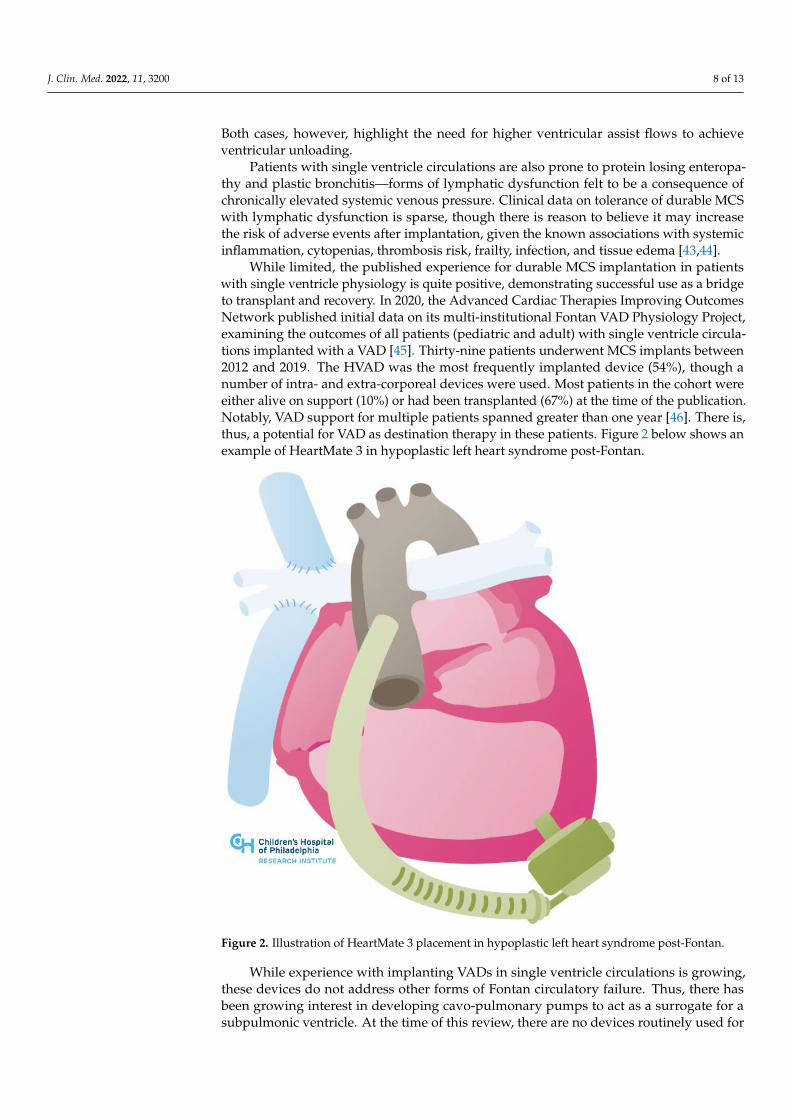
While limited, the published experience for durable MCS implantation in patientswith single ventricle physiology is quite positive, demonstrating successful use as a bridgeto transplant and recovery. In 2020, the Advanced Cardiac Therapies Improving OutcomesNetwork published initial data on its multi-institutional Fontan VAD Physiology Project,examining the outcomes of all patients (pediatric and adult) with single ventricle circula-tions implanted with a VAD [45]. Thirty-nine patients underwent MCS implants between2012 and 2019. The HVAD was the most frequently implanted device (54%), though anumber of intra- and extra-corporeal devices were used. Most patients in the cohort wereeither alive on support (10%) or had been transplanted (67%) at the time of the publication.Notably, VAD support for multiple patients spanned greater than one year [46]. There is,thus, a potential for VAD as destination therapy in these patients. Figure 2 below shows anexample of HeartMate 3 in hypoplastic left heart syndrome post-Fontan.
J. Clin. Med. 2022, 11, x FOR PEER REVIEW 9 of 15
Figure 2. Illustration of HeartMate 3 placement in hypoplastic left heart syndrome post-Fontan.
While experience with implanting VADs in single ventricle circulations is growing,
these devices do not address other forms of Fontan circulatory failure. Thus, there has
been growing interest in developing cavo-pulmonary pumps to act as a surrogate for a
subpulmonic ventricle. At the time of this review, there are no devices routinely used for
this purpose. Intravascular axial and rotary devices in the cavo-pulmonary circuit are un-
der development, as are bio-engineered reservoir pumps [47–49] (see Figure 3 below).
Berlin Heart has designed an implantable sub-pulmonary support system for patients
with Fontan failure as a means to improve end-organ function while awaiting transplant
[50]. These devices hold promise for averting untoward sequelae inherent to the Fontan
physiology in failing systems, and improving patients’ natural history of the disease.
Figure 2. Illustration of HeartMate 3 placement in hypoplastic left heart syndrome post-Fontan.
While experience with implanting VADs in single ventricle circulations is growing,these devices do not address other forms of Fontan circulatory failure. Thus, there hasbeen growing interest in developing cavo-pulmonary pumps to act as a surrogate for asubpulmonic ventricle. At the time of this review, there are no devices routinely used for

J. Clin. Med. 2022, 11, 3200 9 of 13
this purpose. Intravascular axial and rotary devices in the cavo-pulmonary circuit are underdevelopment, as are bio-engineered reservoir pumps [47–49] (see Figure 3 below). BerlinHeart has designed an implantable sub-pulmonary support system for patients with Fontanfailure as a means to improve end-organ function while awaiting transplant [50]. Thesedevices hold promise for averting untoward sequelae inherent to the Fontan physiology infailing systems, and improving patients’ natural history of the disease.
J. Clin. Med. 2022, 11, x FOR PEER REVIEW 10 of 15
Figure 3. Illustration of cavo-pulmonary assist device in hypoplastic left heart syndrome post-Fon-
tan.
4. Temporary Mechanical Circulatory Support
Experience with temporary MCS with ECMO in ACHD has been published [51],
though there are no large studies evaluating protocols to transition patients from tempo-
rary to durable MCS. There is growing experience with Impella devices (Abiomed Inc,
Danvers, MA). These devices are among the more commonly used in non-ACHD patients
in the setting of cardiogenic shock as a bridge to durable MCS/transplant or for peripro-
cedural support during complex procedures. Case reports document their use in various
congenital heart lesions, including the biventricular circulation with a systemic left ven-
tricle, the systemic right ventricle and the Fontan circulation (see Figures 4 and 5 below)
[52–57].
Figure 3. Illustration of cavo-pulmonary assist device in hypoplastic left heart syndrome post-Fontan.
4. Temporary Mechanical Circulatory Support
Experience with temporary MCS with ECMO in ACHD has been published [51],though there are no large studies evaluating protocols to transition patients from temporaryto durable MCS. There is growing experience with Impella devices (Abiomed Inc., Danvers,MA, USA). These devices are among the more commonly used in non-ACHD patients in thesetting of cardiogenic shock as a bridge to durable MCS/transplant or for periproceduralsupport during complex procedures. Case reports document their use in various congenitalheart lesions, including the biventricular circulation with a systemic left ventricle, thesystemic right ventricle and the Fontan circulation (see Figures 4 and 5 below) [52–57].
Clinicians must be mindful of the issues that ACHD patients may have with largebore peripheral access given from congenital vascular anomalies and scar tissue fromprior surgeries and catheterizations. Furthermore, attention must be paid to specificanatomic complexities such as the reconstructed neo-aorta in patients who have undergonea Damus–Kaye–Stansel anastomosis of the aorta and pulmonary artery or the tricuspidsubvalvar apparatus of the systemic right ventricle. Most recently, there has been a reportof an Impella RP device used to support the sub-pulmonary circulation as a bridge totransplant in a patient with Ebstein anomaly [58]. The use of advanced imaging and/or 3Dmodeling with virtual reality simulation has similarly been employed to facilitate thesenovel interventions.

J. Clin. Med. 2022, 11, 3200 10 of 13J. Clin. Med. 2022, 11, x FOR PEER REVIEW 11 of 15
Figure 4. Illustration of Impella in congenitally corrected transposition of the great arteries.
Figure 4. Illustration of Impella in congenitally corrected transposition of the great arteries.
J. Clin. Med. 2022, 11, x FOR PEER REVIEW 11 of 15
Figure 4. Illustration of Impella in congenitally corrected transposition of the great arteries.
Figure 5. Illustration of Impella in hypoplastic left heart syndrome post-Fontan.

J. Clin. Med. 2022, 11, 3200 11 of 13
Given the recent update to the UNOS heart transplant allocation criteria, the use oftemporary MCS may help shorten the waitlist time for transplants in ACHD patients. Innon-ACHD patients, Impella use has been found to decrease end-organ dysfunction andwaitlist times [59,60]. Anecdotally, Impella has been used for multiple months as a bridgeto transplant, especially in cases where an operation for a durable VAD implant is thoughtto be high risk.
5. Conclusions
The need for advanced HF therapies in ACHD patients stands to grow substantiallywithin the next decade. Though limitations exist, there is increasing evidence to suggestthat durable MCS can be implanted safely and with favorable outcomes in this population.Expansion of durable MCS use in ACHD patients may improve their clinical outcomesas centers gain experience, though each case requires thorough diagnostic evaluation andmultidisciplinary discussion between HF, ACHD, and surgical specialists.
Author Contributions: Conceptualization, J.S. and S.S.V.; writing—original draft preparation, J.S.;writing—review and editing, J.S., R.M., A.C., W.H.W.T., J.W.R., K.M., Y.Y.K. and S.S.V.; visualization,J.S. and S.S.V.; supervision, S.S.V. All authors have read and agreed to the published version ofthe manuscript.
Funding: This review received no external funding.
Conflicts of Interest: Saef is a recipient of the Razavi Fellowship for Clinical Research, whichsupports training in clinical research methods. Tang is supported by grants from the NationalInstitutes of Health (R01HL146754). Rossano has consulting relationships with Abiomed, Novartis,Bayer, and Amgen.
References1. Marelli, A.J.; Ionescu-Ittu, R.; Mackie, A.S.; Guo, L.; Dendukuri, N.; Kaouache, M. Lifetime prevalence of congenital heart disease
in the general population from 2000 to 2010. Circulation 2014, 130, 749–756. [CrossRef] [PubMed]2. Zomer, A.C.; Vaartjes, I.; van der Velde, E.T.; de Jong, H.M.; Konings, T.C.; Wagenaar, L.J.; Heesen, W.F.; Eerens, F.; Baur, L.H.;
Grobbee, D.; et al. Heart failure admissions in adults with congenital heart disease; risk factors and prognosis. Int. J. Cardiol. 2013,168, 2487–2493. [CrossRef] [PubMed]
3. Cedars, A.; Benjamin, L.; Vyhmeister, R.; Harris, K.; Bradley, E.A.; Wadia, S.; Awad, A.J.; Novak, E. Contemporary HospitalizationRate Among Adults with Complex Congenital Heart Disease. World J. Pediatric Congenit. Heart Surg. 2016, 7, 334–343. [CrossRef][PubMed]
4. Verheugt, C.L.; Uiterwaal, C.S.; van der Velde, E.T.; Meijboom, F.J.; Pieper, P.G.; van Dijk, A.P.; Vliegen, H.W.; Grobbee, D.E.;Mulder, B.J. Mortality in adult congenital heart disease. Eur. Heart J. 2010, 31, 1220–1229. [CrossRef]
5. Marelli, A.J.; Mackie, A.S.; Ionescu-Ittu, R.; Rahme, E.; Pilote, L. Congenital heart disease in the general population: Changingprevalence and age distribution. Circulation 2007, 115, 163–172. [CrossRef] [PubMed]
6. Parker, W.F.; Chung, K.; Anderson, A.S.; Siegler, M.; Huang, E.S.; Churpek, M.M. Practice Changes at U.S. Transplant CentersAfter the New Adult Heart Allocation Policy. J. Am. Coll. Cardiol. 2020, 75, 2906–2916. [CrossRef]
7. Alshawabkeh, L.I.; Hu, N.; Carter, K.; Opotowsky, A.R.; Light-McGroary, K.; Cavanaugh, J.E.; Bartlett, H.L. Wait-List Outcomesfor Adults With Congenital Heart Disease Listed for Heart Transplantation in the U.S. J. Am. Coll. Cardiol. 2016, 68, 908–917.[CrossRef]
8. Karamlou, T.; Hirsch, J.; Welke, K.; Ohye, R.G.; Bove, E.L.; Devaney, E.J.; Gajarski, R.J. A United Network for Organ Sharinganalysis of heart transplantation in adults with congenital heart disease: Outcomes and factors associated with mortality andretrans-plantation. J. Thorac. Cardiovasc. Surg. 2010, 140, 161–168. [CrossRef]
9. Mylotte, D.; Pilote, L.; Ionescu-Ittu, R.; Abrahamowicz, M.; Khairy, P.; Therrien, J.; Mackie, A.S.; Marelli, A. Specialized adultcongenital heart disease care: The impact of policy on mortality. Circulation 2014, 129, 1804–1812. [CrossRef]
10. Stout, K.K.; Daniels, C.J.; Aboulhosn, J.A.; Bozkurt, B.; Broberg, C.S.; Colman, J.M.; Crumb, S.R.; Dearani, J.A.; Fuller, S.;Gurvitz, M.; et al. 2018 AHA/ACC guideline for the management of adults with congenital heart disease: A report of theAmerican College of Cardiology/American Heart Association Task Force on Clinical Practice Guidelines. J. Am. Coll. Cardiol.2019, 73, e81–e192. [CrossRef]
11. Hosseinpour, A.R.; Cullen, S.; Tsang, V.T. Transplantation for adults with congenital heart disease. Eur. J. Cardio-Thorac. Surg.2006, 30, 508–514. [CrossRef] [PubMed]
12. Ross, H.J.; Law, Y.; Book, W.M.; Broberg, C.S.; Burchill, L.; Cecchin, F.; Chen, J.M.; Delgado, D.; Dimopoulos, K.; Everitt, M.D.; et al.Transplantation and mechanical circulatory support in congenital heart disease: A scientific statement from the American HeartAssociation. Circulation 2016, 133, 802–820. [CrossRef] [PubMed]
J. Clin. Med. 2022, 11, 3200 12 of 13
13. Warnes, C.A. The Adult with Congenital Heart Disease: Born to Be Bad? J. Am. Coll. Cardiol. 2005, 46, 1–8. [CrossRef] [PubMed]14. Baumgartner, H.; De Backer, J.; Babu-Narayan, S.V.; Budts, W.; Chessa, M.; Diller, G.P.; Lung, B.; Kluin, J.; Lang, I.M.;
Meijboom, F.; et al. ESC Scientific Document Group. 2020 ESC Guidelines for the management of adult congenital heart disease.Eur. Heart J. 2021, 42, 563–645. [CrossRef]
15. Givertz, M.M.; DeFilippis, E.M.; Landzberg, M.J.; Pinney, S.P.; Woods, R.K.; Valente, A.M. Advanced heart failure therapies foradults with congenital heart disease: JACC state-of-the-art review. J. Am. Coll. Cardiol. 2019, 74, 2295–22312. [CrossRef]
16. VanderPluym, C.J.; Cedars, A.; Eghtesady, P.; Maxwell, B.G.; Gelow, J.M.; Burchill, L.J.; Maltais, S.; Koehl, D.A.; Cantor, R.S.;Blume, E.D. Outcomes following implantation of mechanical circulatory support in adults with congenital heart disease: Ananalysis of the Interagency Registry for Mechanically Assisted Circulatory Support (INTERMACS). J. Heart Lung Transplant. 2018,37, 89–99. [CrossRef]
17. Steiner, J.M.; Krieger, E.V.; Stout, K.K.; Stempien-Otero, A.; Mahr, C.; Mokadam, N.A.; Hermsen, J.L. Durable mechanicalcirculatory support in teenagers and adults with congenital heart disease: A systematic review. Int. J. Cardiol. 2017, 245, 135–140.[CrossRef]
18. Jacob, K.A.; Hjortnaes, J.; Kranenburg, G.; de Heer, F.; Kluin, J. Mortality after cardiac surgery in patients with liver cirrhosisclas-sified by the Child-Pugh score. Interact. Cardiovasc. Thorac. Surg. 2015, 20, 520–530. [CrossRef]
19. Shah, N.R.; Lam, W.W.; Rodriguez, F.H., III; Ermis, P.R.; Simpson, L.; Frazier, O.H.; Franklin, W.J.; Parekh, D.R. Clinical outcomesafter ventricular assist device implantation in adults with complex congenital heart disease. J. Heart Lung Transplant. 2013,32, 615–620. [CrossRef]
20. Cedars, A.; Vanderpluym, C.; Koehl, D.; Cantor, R.; Kutty, S.; Kirklin, J.K. An Interagency Registry for Mechanically AssistedCirculatory Support (INTERMACS) analysis of hospitalization, functional status, and mortality after mechanical circulatorysupport in adults with congenital heart disease. J. Heart Lung Transplant. 2018, 37, 619–630. [CrossRef]
21. Brickner, M.E.; Hillis, L.D.; Lange, R.A. Congenital heart disease in adults. N. Engl. J. Med. 2000, 342, 334–342. [CrossRef][PubMed]
22. Mustard, W.T.; Keith, J.D.; Trusler, G.A.; Fowler, R.; Kidd, L. The surgical management of transposition of the great vessels.J. Thorac. Cardiovasc. Surg. 1964, 48, 953–958. [CrossRef]
23. Vejlstrup, N.; Sørensen, K.; Mattsson, E.; Thilén, U.; Kvidal, P.; Johansson, B.; Iversen, K.; Søndergaard, L.; Dellborg, M.; Eriksson, P.Long-Term Outcome of Mustard/Senning Correction for Transposition of the Great Arteries in Sweden and Denmark. Circulation2015, 132, 633–638. [CrossRef] [PubMed]
24. Brida, M.; Diller, G.-P.; Gatzoulis, M.A. Response by Brida et al to Letter Regarding Article, “Systemic Right Ventricle in Adultswith Congenital Heart Disease: Anatomic and Phenotypic Spectrum and Current Approach to Management”. Circulation 2018,138, 326–327. [CrossRef] [PubMed]
25. Filippov, A.A.; Del Nido, P.J.; Vasilyev, N.V. Management of systemic right ventricular failure in patients with congenitallycor-rected transposition of the great arteries. Circulation 2016, 134, 1293–1302. [CrossRef]
26. Joyce, D.L.; Crow, S.S.; John, R.; Louis, J.D.; Braunlin, E.A.; Pyles, L.A.; Kofflin, P.; Joyce, L.D. Mechanical circulatory support inpatients with heart failure secondary to transposition of the great arteries. J. Heart Lung Transplant. 2010, 29, 1302–1305. [CrossRef]
27. Roche, S.L.; Crossland, D.S.; Adachi, I.; Broda, C.; Jansen, K.; Hickey, E. Mechanical Circulatory Support for the Failing Sub-AorticRight Ventricle in Adults. Semin. Thorac. Cardiovasc. Surg. Pediatric Card. Surg. Annu. 2021, 24, 2–9. [CrossRef]
28. Serfas, J.D.; Patel, P.A.; Krasuski, R.A. Heart Transplantation and Mechanical Circulatory Support in Adults with CongenitalHeart Disease. Curr. Cardiol. Rep. 2018, 20, 81. [CrossRef]
29. O’Connor, M.J.; Lorts, A.; Davies, R.R.; Fynn-Thompson, F.; Joong, A.; Maeda, K.; Mascio, C.E.; McConnell, P.I.; Mongé, M.C.;Nandi, D.; et al. Early experience with the HeartMate 3 continuous-flow ventricular assist device in pediatric patients andpatients with congenital heart disease: A multicenter registry analysis. J. Heart Lung Transplant. 2020, 39, 573–579. [CrossRef]
30. Dennis, M.; Zannino, D.; Du Plessis, K.; Bullock, A.; Disney, P.J.; Radford, D.J.; Hornung, T.; Grigg, L.; Cordina, R.;D’Udekem, Y.; et al. Clinical Outcomes in Adolescents and Adults After the Fontan Procedure. J. Am. Coll. Cardiol. 2018,71, 1009–1017. [CrossRef]
31. Frescura, C.; Thiene, G. The new concept of univentricular heart. Front. Pediatrics 2014, 2, 62. [CrossRef] [PubMed]32. Redington, A. The physiology of the Fontan circulation. Prog. Pediatric Cardiol. 2006, 22, 179–186. [CrossRef]33. Gewillig, M.; Eyskens, B.; Heying, R.; Ganame, J.; La Gerche, A.; Brown, S.C.; Budts, W.; Gorenflo, M. The Fontan circulation:
Who controls cardiac output? Interact. Cardiovasc. Thorac. Surg. 2010, 10, 428–433. [CrossRef] [PubMed]34. Lemler, M.S.; Scott, W.A.; Leonard, S.R.; Stromberg, D.; Ramaciotti, C. Fenestration improves clinical outcome of the Fontan
proce-dure: A prospective, randomized study. Circulation 2002, 105, 207–212. [CrossRef]35. Book, W.M.; Gerardin, J.; Saraf, A.; Marie Valente, A.; Rodriguez, F., III. Clinical Phenotypes of Fontan Failure: Implications for
Management. Congenit. Heart Dis. 2016, 11, 296–308. [CrossRef]36. Villa, C.R.; Lorts, A.; Morales, D.L. Ventricular assist device therapy in the Fontan circulation. In Seminars in Thoracic and
Cardio-Vascular Surgery: Pediatric Cardiac Surgery Annual; WB Saunders: London, UK, 2021; Volume 24, pp. 19–25.37. Moore, R.A.; Madueme, P.C.; Lorts, A.; Morales, D.L.; Taylor, M.D. Virtual implantation evaluation of the total artificial heart and
compatibility: Beyond standard fit criteria. J. Heart Lung Transplant. 2014, 33, 1180–1183. [CrossRef]38. Moore, R.A.; Lorts, A.; Madueme, P.C.; Taylor, M.D.; Morales, D.L. Virtual implantation of the 50cc SynCardia total artificial heart.
J. Heart Lung Transplant. 2016, 35, 824–827. [CrossRef]

J. Clin. Med. 2022, 11, 3200 13 of 13
39. Farooqi, K.M.; Saeed, O.; Zaidi, A.; Sanz, J.; Nielsen, J.; Hsu, D.T.; Jorde, U.P. 3D Printing to Guide Ventricular Assist DevicePlacement in Adults with Congenital Heart Disease and Heart Failure. JACC Heart Fail. 2016, 4, 301–311. [CrossRef]
40. Ali, L.A.; Cadoni, A.; Rossi, G.; Keilberg, P.; Passino, C.; Festa, P. Effective Cardiac Index and Systemic-Pulmonary CollateralsEvaluated by Cardiac Magnetic Resonance Late After Fontan Palliation. Am. J. Cardiol. 2017, 119, 2069–2072. [CrossRef]
41. Smith, M.; El-Said, H.; Pretorius, V.; Mendenhall, M.; Thomas, T.; Reeves, R.R.; Enciso, J.S.; Alshawabkeh, L.; Nigro, J.;Adler, E.D.; et al. Significance of Aortopulmonary Collaterals in a Single-Ventricle Patient Supported with a HeartMate 3. Circ.Heart Fail. 2020, 13, e006473. [CrossRef]
42. Wells, D.A.; Coghill, M.; Szugye, N.; Moore, R.; Lorts, A.; Tweddell, J.S.; Morales, D.L. Transplantation and Arch Repair in Fontan3 Years after HeartMate 3: Technical Considerations. Ann. Thorac. Surg. 2021, in press. [CrossRef] [PubMed]
43. Rychik, J. Protein-losing enteropathy after Fontan operation. Congenit. Heart Dis. 2007, 2, 288–300. [CrossRef] [PubMed]44. Rodriguez de Santiago, E.; Téllez, L.; Garrido-Lestache Rodriguez-Monte, E.; Garrido-Gómez, E.; Aguilera-Castro, L.; Álvarez-
Fuente, M.; Del Cerro, M.J.; Albillos, A.; Romera, R.; Olavarria, A.; et al. Fontan protein-losing enteropathy is associated withadvanced liver disease and a proinflammatory intestinal and systemic state. Liver Int. 2020, 40, 638–645. [CrossRef] [PubMed]
45. Cedars, A.M.; Schumacher, K.; Kindel, S.; Lorts, A.; Rosenthal, D.; Chen, S.; Morales, D.; Peng, D.; O’Connor, M.;Simpson, K.E.; et al. The Fontan VAD Physiology Project (FVPP). J. Heart Lung Transplant. 2020, 39, S12–S13. [CrossRef]
46. Cedars, A.; Kutty, S.; Danford, D.; Schumacher, K.; Auerbach, S.R.; Bearl, D.; Chen, S.; Conway, J.; Dykes, J.C.; Jaworski, N.; et al.Systemic ventricular assist device support in Fontan patients: A report by ACTION. J. Heart Lung Transplant. 2021, 40, 368–376.[CrossRef]
47. Broda, C.R.; Taylor, D.A.; Adachi, I. Progress in experimental and clinical subpulmonary assistance for Fontan circulation.J. Thorac. Cardiovasc. Surg. 2018, 156, 1949–1956. [CrossRef]
48. Prêtre, R.; Häussler, A.; Bettex, D.; Genoni, M. Right-Sided Univentricular Cardiac Assistance in a Failing Fontan Circulation.Ann. Thorac. Surg. 2008, 86, 1018–1020. [CrossRef]
49. Rodefeld, M.D.; Marsden, A.; Figliola, R.; Jonas, T.; Neary, M.; Giridharan, G.A. Cavopulmonary Assist: Long-Term Reversal ofthe Fontan Paradox. J. Thorac. Cardiovasc. Surg. 2019, 158, 1627–1636. [CrossRef]
50. Berlin Heart [Internet]. Medical Professionals EXCOR Venous Cannula. Available online: https://www.berlinheart.de/en/medical-professionals/excorr-venous-cannula/ (accessed on 22 March 2022).
51. Broda, C.; Smith, P.A.; Wang, Y.; Sampaio, L.C.; Adachi, I.; Taylor, D.A. Construction and Evaluation of a Bio-Engineered Pump toEnable Subpulmonary Support of the Fontan Circulation: A Proof-of-Concept Study. J. Heart Lung Transplant. 2020, 39, S176.[CrossRef]
52. Acheampong, B.; Johnson, J.N.; Stulak, J.M.; Dearani, J.A.; Kushwaha, S.S.; Daly, R.C.; Haile, D.T.; Schears, G.J. PostcardiotomyECMO Support after High-risk Operations in Adult Congenital Heart Disease. Congenit. Heart Dis. 2016, 11, 751–755. [CrossRef]
53. Morray, B.H.; Dimas, V.V.; Lim, S.; Balzer, D.T.; Parekh, D.R.; Van Mieghem, N.M.; Ewert, P.; Kim, D.W.; Justino, H.;McElhinney, D.B.; et al. Circulatory support using the impella device in fontan patients with systemic ventricular dysfunc-tion: A multicenter ex-perience. Catheter. Cardiovasc. Interv. 2017, 90, 118–123. [CrossRef] [PubMed]
54. Fishberger, S.B.; Asnes, J.D.; Rollinson, N.L.; Cleman, M.W. Percutaneous right ventricular support during catheter ablationof intraatrial reentrant tachycardia in an adult with a mustard baffle—A novel use of the Impella device. J. Interv. Card.Electro.-Physiol. 2010, 29, 69–72. [CrossRef] [PubMed]
55. Adler, A.C.; Kodavatiganti, R. Mechanical Support with Impella During Malignant Arrhythmia Ablation: A Case Report on theGrowing Trend in the Electrophysiology Laboratory. A&A Case Rep. 2017, 8, 282–285.
56. White, C.W.; Ganapathi, A.; Schroder, J. A Minimally Invasive Approach to HeartMate 3 Implantation for Systemic VentricularFailure Following the Mustard Procedure for Transposition of the Great Arteries. ASAIO J. 2020, 66, e62–e63. [CrossRef] [PubMed]
57. Kim, J.L.; Vaikunth, S.S.; Haeffele, C.; MacArthur, J.W. Extracorporeal membrane oxygenator as a bridge to heart–liver en bloctransplant in a patient with Fontan circulation. JTCVS Tech. 2022, 12, 171–174. [CrossRef] [PubMed]
58. García Gómez, M.; Uribarri, A.; San Román Calvar, J.A.; Stepanenko, A. Clinical use of percutaneous mechanical circulatoryas-sistance in a patient with end-stage right-sided heart failure and massive tricuspid insufficiency due to congenital heart disease:First-in-the-world case report. Eur. Heart J. Case Rep. 2021, 5, ytab269. [CrossRef] [PubMed]
59. Upadhyaya, V.D.; Alshami, A.; Patel, I.; Douedi, S.; Quinlan, A.; Thomas, T.; Prentice, J.; Calderon, D.; Asif, A.; Sen, S.; et al.Outcomes of Renal Function in Cardiogenic Shock Patients with or Without Mechanical Circulatory Support. J. Clin. Med. Res.2021, 13, 283–292. [CrossRef]
60. Kingsford, P.; Onwuzurike, J.; Li, J.; Bradley, C.; Vaidya, A.; Wolfson, A.; DePasquale, E. Impact of the UNOS Policy AllocationChange on Waitlist Outcomes in Patients Bridged to Heart Transplantation on Impella. J. Heart Lung Transplant. 2021, 40, S268.[CrossRef]
Related Documents


