The Rockefeller University Press, 0021-9525/99/12/1153/14 $5.00 The Journal of Cell Biology, Volume 147, Number 6, December 13, 1999 1153–1166 http://www.jcb.org 1153 Duplication and Maintenance of Heterochromatin Domains Angela Taddei,* Danièle Roche,* Jean-Baptiste Sibarita,* Bryan M. Turner, ‡ and Geneviève Almouzni* *Institut Curie, Research section, UMR 144 et 218 du Centre National de la Recherche Scientifique (CNRS), 75248 Paris cedex 05, France; and ‡ Anatomy Department, University of Birmingham Medical School, Edgbaston, Birmingham, B152TT United Kingdom Abstract. To investigate the mechanisms that assure the maintenance of heterochromatin regions, we took advantage of the fact that clusters of heterochromatin DNA replicate late in S phase and are processed in dis- crete foci with a characteristic nuclear distribution. At the light microscopy level, within these entities, we fol- lowed DNA synthesis, histone H4 acetylation, hetero- chromatin protein 1 (Hp1a and -b), and chromatin as- sembly factor 1 (CAF-1). During replication, Hp1a and -b domains of concentration are stably maintained, whereas heterochromatin regions are enriched in both CAF-1 and replication-specific acetylated isoforms of histone H4 (H4Ac 5 and 12). We defined a time window of 20 min for the maintenance of this state. Further- more, treatment with Trichostatin A (TSA), during and after replication, sustains the H4Ac 5 and 12 state in heterochromatin excluding H4Ac 8 and 16. In compari- son, early replication foci, at the same level, did not dis- play any specific enrichment in H4Ac 5 and 12. These data emphasize the specific importance for heterochro- matin of the replication-associated H4 isoforms. We propose that perpetuation of heterochromatin involves self-maintenance factors, including local concentration of Hp1a and -b, and that a degree of plasticity is pro- vided by the cycle of H4 acetylation/deacetylation as- sisted by CAF-1. Key words: heterochromatin • nuclear organization • histone H4 acetylation • chromatin assembly factor 1 • heterochromatin protein 1 I N eucaryotic cells, the genome is packaged into large scale chromatin structures giving rise to distinct func- tional domains (Gasser and Laemmli, 1987; Cook, 1995; Strouboulis and Wolffe, 1996; Marshall et al., 1997; Bridger and Bickmore, 1998; Lamond and Earnshaw, 1998). The way in which these domains are established and then propagated at each cell division is still an open ques- tion (Lewin, 1998). To gain insight into this important issue, domains of heterochromatin make an attractive model due to their defined properties. Historically, based on cytological observations, a distinction was made be- tween euchromatin and heterochromatin (Heitz, 1928). By definition, heterochromatin corresponds to regions of the genome that remain intensely stained throughout the cell cycle and appear condensed. During interphase, hetero- chromatin domains are mainly located at the nuclear pe- riphery or surrounding the nucleoli. The largest regions of heterochromatin are found on the inactivated X chromo- some (in female mammalian cells) and near centromeres and telomeres. They generally replicate late in S phase (Goldman et al., 1984; Hatton et al., 1988) and are in- volved in various nuclear functions, including nuclear ar- chitecture, chromosome segregation, and gene silencing (Wallrath, 1998). Among heterochromatin markers, the best known pro- tein to date, heterochromatin protein 1 (HP1) 1 was identi- fied in Drosophila as an antigen localized to pericentric heterochromatin (James and Elgin, 1986; James et al., 1989). Genetic studies in Drosophila involved HP1 in a specific gene silencing process called position effect varie- gation (PEV; Henikoff, 1990; Elgin, 1996; Wakimoto, 1998). This effect is thought to be driven by heterochroma- tin proteins which, by propagating the organization of densely packed chromatin fibers, would help to form tran- scriptionally inert domains (Henikoff, 1990; Elgin, 1996; Cavalli and Paro, 1998). Several homologues of HP1 have now been identified in vertebrates: HP1a, M31, and M32 in mice; and HP1a, -b, and -g in humans and Xenopus (Singh et al., 1991; Saunders et al., 1993; Le Douarin et al., 1996; Ye and Worman, 1996; Pak et al., 1997). HP1b (M31, also called MOD1; Singh et al., 1991) is generally consid- ered as the true homologue of the Drosophila protein Address correspondence to G. Almouzni, Institut Curie, Research sec- tion, UMR 144 et 218 du CNRS, 75248 Paris cedex 05, France. Tel.: 133 (0)1 42 34 64 10. Fax: 133 (0)1 42 34 64 21. E-mail: [email protected] 1. Abbreviations used in this paper: BiodU, Biotin-16-deoxyuridine; BrdU, 5-bromo-29-deoxyuridine; CAF-1, chromatin assembly factor 1; H4Ac n, histone H4 acetylated at lysine n; HAT, histone acetyl transferase; HDAC, histone deacetylase; HP1, heterochromatin protein 1; pAb, poly- clonal antibody; TSA, Trichostatin A. on February 17, 2016 jcb.rupress.org Downloaded from Published December 13, 1999

Welcome message from author
This document is posted to help you gain knowledge. Please leave a comment to let me know what you think about it! Share it to your friends and learn new things together.
Transcript

The Rockefeller University Press, 0021-9525/99/12/1153/14 $5.00The Journal of Cell Biology, Volume 147, Number 6, December 13, 1999 1153–1166http://www.jcb.org 1153
Duplication and Maintenance of Heterochromatin Domains
Angela Taddei,* Danièle Roche,* Jean-Baptiste Sibarita,* Bryan M. Turner,
‡
and Geneviève Almouzni*
*Institut Curie, Research section, UMR 144 et 218 du Centre National de la Recherche Scientifique (CNRS), 75248 Paris cedex 05, France; and
‡
Anatomy Department, University of Birmingham Medical School, Edgbaston, Birmingham, B152TT United Kingdom
Abstract.
To investigate the mechanisms that assure the maintenance of heterochromatin regions, we took advantage of the fact that clusters of heterochromatin DNA replicate late in S phase and are processed in dis-crete foci with a characteristic nuclear distribution. At the light microscopy level, within these entities, we fol-lowed DNA synthesis, histone H4 acetylation, hetero-chromatin protein 1 (Hp1
a
and -
b
), and chromatin as-sembly factor 1 (CAF-1). During replication, Hp1
a
and -
b
domains of concentration are stably maintained, whereas heterochromatin regions are enriched in both CAF-1 and replication-specific acetylated isoforms of histone H4 (H4Ac 5 and 12). We defined a time window of 20 min for the maintenance of this state. Further-more, treatment with Trichostatin A (TSA), during and
after replication, sustains the H4Ac 5 and 12 state in heterochromatin excluding H4Ac 8 and 16. In compari-son, early replication foci, at the same level, did not dis-play any specific enrichment in H4Ac 5 and 12. These data emphasize the specific importance for heterochro-matin of the replication-associated H4 isoforms. We propose that perpetuation of heterochromatin involves self-maintenance factors, including local concentration of Hp1
a
and -
b
, and that a degree of plasticity is pro-vided by the cycle of H4 acetylation/deacetylation as-sisted by CAF-1.
Key words: heterochromatin • nuclear organization • histone H4 acetylation • chromatin assembly factor 1 • heterochromatin protein 1
I
N
eucaryotic cells, the genome is packaged into largescale chromatin structures giving rise to distinct func-tional domains (Gasser and Laemmli, 1987; Cook,
1995; Strouboulis and Wolffe, 1996; Marshall et al., 1997;Bridger and Bickmore, 1998; Lamond and Earnshaw,1998). The way in which these domains are established andthen propagated at each cell division is still an open ques-tion (Lewin, 1998). To gain insight into this importantissue, domains of heterochromatin make an attractivemodel due to their defined properties. Historically, basedon cytological observations, a distinction was made be-tween euchromatin and heterochromatin (Heitz, 1928). Bydefinition, heterochromatin corresponds to regions of the
genome that remain intensely stained
throughout the cellcycle and appear condensed. During interphase, hetero-chromatin domains are mainly located at the nuclear pe-riphery or surrounding the nucleoli. The largest regions ofheterochromatin are found on the inactivated X chromo-some (in female mammalian cells) and near centromeresand telomeres. They generally replicate late in S phase(Goldman et al., 1984; Hatton et al., 1988) and are in-volved in various nuclear functions, including nuclear ar-
chitecture, chromosome segregation, and gene silencing(Wallrath, 1998).
Among heterochromatin markers, the best known pro-tein to date, heterochromatin protein 1 (HP1)
1
was identi-fied in
Drosophila
as an antigen localized to pericentricheterochromatin (James and Elgin, 1986; James et al.,1989). Genetic studies in
Drosophila
involved HP1 in aspecific gene silencing process called position effect varie-gation (PEV; Henikoff, 1990; Elgin, 1996; Wakimoto,1998). This effect is thought to be driven by heterochroma-tin proteins which, by propagating the organization ofdensely packed chromatin fibers, would help to form tran-scriptionally inert domains (Henikoff, 1990; Elgin, 1996;Cavalli and Paro, 1998). Several homologues of HP1 havenow been identified in vertebrates: HP1
a
, M31, and M32in mice; and HP1
a
, -
b
, and -
g
in humans and
Xenopus
(Singh et al., 1991; Saunders et al., 1993; Le Douarin et al.,1996; Ye and Worman, 1996; Pak et al., 1997). HP1
b
(M31,also called MOD1; Singh et al., 1991) is generally consid-ered as the true homologue of the
Drosophila
protein
Address correspondence to G. Almouzni, Institut Curie, Research sec-tion, UMR 144 et 218 du CNRS, 75248 Paris cedex 05, France. Tel.:
1
33(0)1 42 34 64 10. Fax:
1
33 (0)1 42 34 64 21. E-mail: [email protected]
1.
Abbreviations used in this paper:
BiodU, Biotin-16-deoxyuridine; BrdU,5-bromo-2
9
-deoxyuridine; CAF-1, chromatin assembly factor 1; H4Ac n,histone H4 acetylated at lysine n; HAT, histone acetyl transferase;HDAC, histone deacetylase; HP1, heterochromatin protein 1; pAb, poly-clonal antibody; TSA, Trichostatin A.
on February 17, 2016
jcb.rupress.orgD
ownloaded from
Published December 13, 1999

The Journal of Cell Biology, Volume 147, 1999 1154
(Aagaard et al., 1999), however, both Hp1
a
and -
b
sharecommon properties and are associated with heterochro-matin (Horsley et al., 1996; Wallrath, 1998).
Another standard marker of heterochromatin domainsfrom mammals to yeast is the presence of under-acety-lated histone H4 isoforms (Jeppesen and Turner 1993;O’Neill and Turner, 1995; Belyaev et al., 1996; Braunsteinet al., 1996). This highly conserved protein, together withthe other histones H3, H2A, and H2B, form a histone oc-tamer around which DNA can wrap to achieve the firstlevel of chromatin organization, the core nucleosome (vanHolde, 1988; Wolffe, 1995). The NH
2
-terminal tail of allcore histones can be acetylated in vivo and steady-statelevels of acetylation depend on the balance between twoenzymatic activities, the histone acetyltransferases (HATs)and deacetylases (HDACs; Tsukiyama and Wu, 1997; Kuoand Allis, 1998). H4 can be acetylated in vivo on fourlysine residues (K5, K8, K12, and K16). Newly synthesizedhistone H4 is acetylated at lysines 5 and 12 (H4Ac 5 and12), a pattern of acetylation that is highly conservedamong eucaryotes (Sobel et al., 1995). Thus, it is generallyassumed that histone H4 is incorporated in such a formduring DNA replication (Annunziato, 1995), although nei-ther of these acetylated sites is strictly required for chro-matin assembly associated with replication in vivo and invitro (Ma et al., 1998b; Zhang et al., 1998). Therefore, acrucial question with respect to heterochromatin is how itsunderacetylated state is established and controlled (Grun-stein, 1998) since this is a critical parameter for both themaintenance of a transcriptional repressed state and forchromosome segregation in yeast (Ekwall et al., 1997;Grewal et al., 1998). In principle, one could imagine thatunderacetylated forms of histone H4 might be used forchromatin assembly in late S phase, thus ensuring a securemaintenance of the underacetylated state of heterochro-matin. Alternatively, the deposition of acetylated formscould be followed by their rapid deacetylation, therebyproviding a window of opportunity during which alterna-tive states could be established.
Newly synthesized histones have been proposed to re-quire cellular factors for their regulated deposition (An-nunziato, 1995; Sogo and Laskey, 1995). To date, one ofthe best candidates for facilitating histone deposition dur-ing DNA synthesis is chromatin assembly factor 1 (CAF-1).This protein complex contains three polypeptides, p150,p60, and p48, and is associated with newly synthesized andacetylated histones, H3 and H4, in nuclear extracts (Kauf-man et al., 1995; Verreault et al., 1996). CAF-1 colocalizes,in general, with sites of DNA replication (Krude, 1995).Functional homologues have been identified in differentspecies, including
Xenopus
,
Drosophila
, and yeast (Gail-lard et al., 1996; Kamakaka et al., 1996; Enomoto et al.,1997; Kaufman et al., 1997). Surprisingly, in yeast cellsnone of the genes corresponding to the individual CAF-1subunits (CAC1, 2, and 3), are essential. However, CACmutants were found to be moderately sensitive to UV irra-diation and exhibited gene silencing defects (Enomotoet al., 1997; Kaufman et al., 1997; Monson et al., 1997;Enomoto and Berman, 1998). Thus, whether CAF-1 is in-volved in assembling appropriately acetylated histones atheterochromatin and elsewhere remains to be determined(Grunstein, 1998).
The spatio-temporal organization of the S phase inhigher eucaryotes provides an opportunity to addressthese issues in defined regions of the nucleus. Indeed, inhigher eucaryotes, DNA synthesis takes place in discretegranular sites that correspond to several replication forksclustered in foci. These foci, with an average size of 500 nm(Nakamura et al., 1986; Nakayasu and Berezney, 1989),vary in number, location, and size at different times duringS phase (Nakayasu and Berezney, 1989; van Dierendoncket al., 1989; Fox et al., 1991; O’Keefe et al., 1992; Hozaket al., 1993). The largest foci are observed in late S phase,at which time a characteristic pattern for replication hasbeen defined (O’Keefe et al., 1992).
These properties of heterochromatin in vertebratesmake them particularly amenable to direct studies usingimmunofluorescence. We have used specific antibodies tofollow and relate the dynamics of DNA synthesis, histoneH4 acetylation, and the presence of HP1
a
, HP1
b
, andCAF-1 within heterochromatin domains. This was com-bined with a novel method for spatio-temporal analysis.Local concentrations of HP1 (
a
and
b
) were stably main-tained in these domains during replication. During thattime, acetylated H4Ac 5 and 12 (replication-associatedforms) and CAF-1 were found transiently in heterochro-matin domains. Furthermore, treatment with a histonedeacetylase inhibitor showed that the dynamics of acetyla-tion of histone H4 in heterochromatin is restricted to K5and K12, and to the time of DNA replication. Signif-icantly, CAF-1 was also found at early replication foci, al-though no enrichment of specific H4 Ac isoforms wasdetected. We integrate these data into a model for dupli-cation of heterochromatin, highlighting the importance ofreplication-associated acetylation dynamics.
Materials and Methods
Cell Culture, Synchronization, and Specific Treatments
HeLa and L929 cells were grown in 9-cm diam glass petri dishes (Falcon)in DME supplemented with 10% FCS, 10
m
g/ml antibiotics (penicillin andstreptomycin), and 2 mM
L
-glutamine (GIBCO BRL) at 37
8
C in an atmo-sphere of 5% CO
2
. For synchronization, HeLa cells grown to
z
50% con-fluence were arrested in early S phase with 2 mM hydroxyurea (SigmaChemical Co.) for 16 h, then released by several washes in PBS before a fi-nal wash in fresh medium. FACS analysis showed that these cells re-mained in S phase for 10 h before entering G2 (3 h), and finally mitosis(1 h). For late S phase samples, cells were routinely collected 7.5–8 h afterrelease. For immunofluorescence, cells were grown on glass coverslips inculture dishes. For Trichostatin A (TSA; Sigma Chemical Co.) treatment,exponentially growing cells were incubated in the presence of TSA (50ng/ml, a dose compatible with cell growth) in complete medium for a cho-sen time.
In Situ Replication Assays
5-bromo-2
9
-deoxyuridine (BrdU; Sigma Chemical Co.) incorporation wasperformed by incubating exponentially growing cells in the presence of40
m
M BrdU for 10 min. Cells were then washed in PBS and fixed with2% paraformaldehyde in PBS for 15 min at room temperature. For pulse–chase experiments, cells were washed twice with prewarmed PBS andonce in prewarmed medium after the BrdU pulse, and then cultured fordifferent time periods. The cells were further processed for isolation ofnuclei or fixed with 2% paraformaldehyde in PBS.
Biotin-16-deoxyuridine (BiodU) incorporation on isolated nuclei wasperformed essentially as described by Krude et al. (1997). In brief, afternuclei isolation in vitro run-on was initiated by adjusting the nuclear prep-aration to final concentrations: 40 mM K-Hepes, pH 7.8; 7 mM MgCl
2
; 3 mM
on February 17, 2016
jcb.rupress.orgD
ownloaded from
Published December 13, 1999

Taddei et al.
Duplication of Heterochromatin Domains
1155
ATP; 0.1 mM each of GTP, CTP, and UTP; 0.1 mM each of dATP, dGTP,and dCTP; 40
m
M bio-dUTP; 20 mM creatine phosphate; 0.5 mM DTT;and 2.5
m
g phosphocreatine kinase (all Boehringer Mannheim Corp.). Af-ter a 10-min incubation at 37
8
C, reactions were stopped by a 25
3
dilutionin PBS and immediately fixed in 2% paraformaldehyde. Nuclei were thendeposited onto polylysine-coated coverslips by gentle centrifugationthrough a 30% cushion of glycerol in PBS, rinsed twice with PBS, and per-meabilized in 0.2% Triton X-100 in PBS for 5 min. For the BiodU detec-tion FITC-conjugated streptavidin (Enzo) was used, and fluorescenceanalysis carried out as described.
Immunolabeling
Paraformaldehyde-fixed cells or nuclei were washed twice in PBS andthen incubated in 0.2% Triton X-100 in PBS for 5 min and blocked in 5%BSA, 0.1% Tween 20 in PBS (blocking buffer) for 10 min. Specific anti-bodies were then added at the appropriate dilution in blocking buffer. In-cubation was carried out for 45 min at room temperature. To visualize thestaining, FITC or Texas red-conjugated goat anti–rabbit or anti–mouseIgG (Jackson ImmunoResearch Laboratories, Inc.) were used. During thefinal washing steps with 0.1% Tween 20 in PBS, 0.25
m
g/ml, 4,6-diamino-2-phenylindole (DAPI; Sigma Chemical Co.) was added for DNAstaining.
In experiments involving BrdU immunodetection, a denaturation stepin 4 N HCl for 10 min at room temperature was incorporated, after thepermeabilization treatment, to render BrdU incorporated into DNA ac-cessible. Neutralization was achieved by washes in PBS, and the slideswere further processed as described above. Since detection of the nuclearproteins studied here is sensitive to HCl extraction, a specific protocol wasused for double staining with BrdU. Proteins were first revealed (first andsecond antibodies) on paraformaldehyde-fixed cells as described. After 3washes in PBS and 0.1% Tween 20, a second fixation step was included us-ing either 2% paraformaldehyde in PBS for 15 min or 5 mM ethylene gly-col-bis(succinimidylsuccinate; Pierce Chemical Co.) in PBS for 30 min at37
8
C, followed by several washes in 0.3 M glycine in PBS. After this step,DNA was denatured with 4 N HCl, before BrdU revelation. The cover-slips were mounted in Vectashield (Vector Laboratories Inc.). Despitepostfixation, some loss of signal could still occur. Incorporation of BiodU(on isolated nuclei) into DNA was used instead of BrdU as an alternative(not shown). This allowed us to avoid the HCl denaturation since thebiotinyl residue is accessible in the double helix of DNA.
Antibodies
Rabbit polyclonal antibodies (pAbs) against the different acetylated iso-forms of histones H4 and H2A have been characterized in detail (Turneret al., 1989). They recognize H4 isoforms acetylated on one of the fourlysines (K) acetylated in vivo or H2A acetylated at lysine 5 (H2A Ac 5).They were used at final dilutions of 1:500 for R101/12 (K12), R14/16(K16), and R123 (H2A Ac 5), and 1:1,000 for R232/8 (K8) and R41/5(K5). mAbs against HP1
a
(2HP1H5) and HP1
b
(IMOD-1A97), kindlyprovided by Dr. R. Losson and Dr. P. Chambon (CNRS, Strasbourg,France), were used at final dilutions of 1/400. The mAb against humanCAF-1 p150 (mAb1) was kindly provided by Dr. B. Stillman (Cold SpringHarbor Laboratory, Cold Spring Harbor, NY; Smith and Stillman, 1991).The rabbit pAb1, directed against the human CAF-1 p60, was kindly pro-vided by Dr. Marheineke (Ecole Normale Superieure, Ulm, France; Mar-heineke and Krude, 1998; Martini et al., 1998). The specificities of the an-tibodies were tested by Western analysis on all sources of materialstudied. Monoclonal rat anti-BrdU was purchased from Harlan; Sera-Labo, Texas red goat anti–rabbit immunoglobulin (Ig), FITC goat anti–rabbit Ig, and Texas red goat anti–rabbit Ig antibodies were purchasedfrom Jackson ImmunoResearch Laboratories, Inc.
Image Acquisition and Quantitative Analysis
Image acquisition was performed using a Leica TCS-4D confocal scanningmicroscope, equipped with an Acousto-Optical Tuneable Filter (AOTF),with a 100
3
NA 1.4 plan-apochromat oil immersion objective. Single opti-cal sections are presented. FITC and Texas red were excited by the argon-krypton laser at 488 and 568 nm, respectively. Red and green fluorescencewere separated by a 580-nm long-pass dichroic beam-splitter. A 520-nmband-pass filter and a 590-nm long-pass filter were used to minimize cross-talk and stop laser scattered light. DAPI staining, shown in Fig. 1, was ac-quired with the UV laser. Before acquiring a double staining z series, theintensity of excitation wavelengths and the power of photodetectors were
adjusted to avoid cross-talk. The fluorescence signals from both fluoro-chromes were recorded simultaneously in one scan, and saved separatelyon two channels to be processed independently.
Three-dimensional, two color images of doubly stained nuclei were re-corded using the same equipment. The voxel dimensions (x, y, z) of eachrecorded stack was adjusted to 0.05
3
0.05
3
0.25
m
m to verify the Shan-non sampling criteria (Goodman, 1968). The averaging parameter was ad-justed to have the best signal/noise ratio without bleaching the sampleduring the acquisition process. For H4Ac and H2AAc staining we couldnot reach this criteria without bleaching, leading us to perform the quanti-tative analysis on single optical sections (2D analysis). The same setup wasused for all sample acquisition to facilitate comparison between data. Nu-merical data were stored in 8-bit tiff format series for each color and trans-ferred for processing and analysis. Noise reduction and image quantifica-tion was developed in house. Metamorph software (Universal Imaging)was used as a support for executing our own Dynamic Link Libraries(DLL) written in Visual Basic and C languages for displaying results andbasic treatments. A nonlinear rank-adaptive denoising filter (Vila and Bo-lon, 1993) was applied to better distinguish fluorescent structures frombackground fluorescence without loss of resolution in transition regions.Red and green volumes were thresholded separately to avoid backgroundcontributions in the quantitative analysis. To make the measurements re-producible and comparable, red and green thresholds, designated Tr andTg, respectively, remained unchanged for each volume corresponding toeach series.
Voxels are sorted in four categories: C, colocalized; R, red only; G,green only; and N, noise. In N, voxels displaying green and red signal in-tensities below both thresholds Tr and Tg were counted. In C, voxels dis-playing green and red signal intensities above both thresholds Tr and Tgwere counted. In G, voxels displaying green signal intensity above Tg andred signal intensity below Tr were counted. In R, voxels displaying red sig-nal intensity above Tr and green signal intensity below Tg were counted.Data presented in Figs. 2 and 6 were computed similarly in 2D, using pix-els instead of voxels.
A control experiment, in which replication sites pulse-labeled withBrdU were revealed by a balanced mix of Texas red and FITC secondaryantibodies, gave 90% of colocalizing BrdU within the total population ofBrdU [(C) / (C
1
R) ratio], using the same parameters as in Fig. 4. This90% approaches the 100% normally anticipated. The difference is due tothe definition of our thresholds, which eliminates noise, but also leads tosystematic underestimation of our values. In addition, we find that thesevalues are dependent on the shape of the objects revealed in each colorand this has to be taken into account for interpretation of the data. In allthe graphs, for each point several experimental measurements were col-lected and the average value has been plotted in arbitrary units. For eachcouple of markers, the plots represent the red signals colocalizing with thegreen signals in arbitrary units [(C) / (C
1
R) ratio] normalized to themaximal value obtained in all the experiments involving this couple ofmarkers. For comparison of the relative enrichment of the various acety-lated isoforms of histones at either CAF-1 (see Fig. 3) or BrdU (see Figs. 2and 6) sites, all histone signals (in green, which were similar in shape)were considered as one marker. Normalization was thus achieved usingthe common maximal value obtained for each set of experiments involv-ing either CAF-1 or BrdU independently.
Results
There Is No Visible Disruption in Hp1
a
and -
b
Domains at Late Replication Foci
To investigate possible disorganization on a large scalethat could occur during replication of heterochromatin do-mains, we followed the distribution of HP1
a
and HP1
b
,two heterochromatin variants of HP1 (Le Douarin et al.,1996; Singh et al., 1991). Using an mAb, we examined therelationship between HP1
a
or -
b
and replication foci byimmunofluorescence in mouse (L929) and human (HeLa)cells. The fluorescence signals from both fluorochromeswere recorded simultaneously in a single scan under con-ditions that ensure that the illumination and detectionwere free of lateral shifts, a precondition for secure recog-nition of colocalization in the specimen. Different stages
on February 17, 2016
jcb.rupress.orgD
ownloaded from
Published December 13, 1999
The Journal of Cell Biology, Volume 147, 1999 1156
of S phase were identified by the distinct BrdU incorpora-tion patterns characteristic of early, mid, or late replicatingfoci (Nakayasu and Berezney, 1989; van Dierendonck et al.,1989; Fox et al., 1991; O’Keefe et al., 1992). Typical resultsobtained with early or late replication profiles are pre-sented in Fig. 1. Our confocal analysis revealed, for bothHP1
a
and HP1
b
, intensely labeled areas in dots corre-sponding to large blocks of pericentric heterochromatinidentified by dense DAPI staining in mouse cells at allstages of S phase. In addition, we reproducibly observed ageneral diffuse nuclear staining, the significance of whichremains to be established. Hp1
a
and -
b
dots were neverassociated with replication foci in early S phase. Remark-ably, in cells characterized by a late replication pattern, asignificant proportion of BrdU incorporation sites couldbe found within HP1 (
a
or
b
) dots. We further confirmedthese observations in HeLa cells (Fig. 1). We concludethat, for two different species, the existence of Hp1
a
and-
b
domains can coincide at this scale, with regions of DNAundergoing replication.
Enrichment of H4Ac 5 and 12 and Exclusion of H4Ac 8 and 16 Replication Foci Is Visible only in Late S Phase
From mammals to yeast, it has been demonstrated that H4underacetylation is a characteristic feature of heterochro-matin (Jeppesen and Turner, 1993; O’Neill and Turner,1995; Belyaev et al., 1996). To examine the state of H4acetylation at both early and late replication foci in HeLacells, we combined pulse labeling with BrdU and immuno-detection using antibodies specific for H4 acetylated at lys5, 8, 12, or 16 (Turner et al., 1989). An antibody to acety-lated H2A was used as a control for an acetylation groupunrelated to de novo chromatin assembly. Confocal analy-
sis showed that the different H4 and H2A antibodies dis-played a general punctate nuclear staining (enriched insome regions and excluded from the nucleolus). In early Scells, replication foci appeared equivalently associatedwith the four acetylated isoforms of H4. In contrast, in lateS cells, we distinguished two different staining patterns(Fig. 2 A): H4 isoforms acetylated at lys 5 or 12 were spe-cifically enriched at late replication foci, whereas isoformsacetylated at lys 8 or 16 were excluded from these particu-lar regions as is acetylated H2A. Similar results were ob-tained using mouse L929 or
Xenopus
A6 cells (not shown).This was further confirmed using a less destructive method(not shown, see Materials and Methods) that reveals thereplication foci by in vitro run-on in the presence of Bio-16-dUTP (Krude, 1995). To quantify these observations,we developed a program to estimate the degree of colocal-ization between two markers in sets of confocal opticalsections. This program and experimental controls to vali-date its application are described in Materials and Meth-ods. Thus, we could analyze the average behavior of sev-eral hundred foci, providing a more accurate estimation ofthe statistical significance of our observations (Fig. 2 B).From these data, we conclude that H4 acetylated at lys5 or 12 is specifically enriched at late replicating foci,whereas forms acetylated at lys 8 or 16 are excluded. Sinceacetylation at lys 5 and 12 is specific to newly synthesizedhistone H4, this enrichment probably corresponds to thedeposition of H4 on newly replicated heterochromatin re-gions. It is remarkable, however, that this specific enrich-ment could only be detected at late replication foci. Theabsence of specific enrichment of H4Ac 5 and 12 at earlyreplication foci could be due to the multiple acetylation/deacetylation events occurring concurrently in euchroma-tin regions that may mask changes associated specifically
Figure 1. Domains rich in Hp1a and -b can be found at late replicating foci. Visualization of DNA replication sites in asynchronouspopulations of mouse (L929) and human (HeLa) cells was performed by in vivo BrdU pulse labeling and detected by immunofluores-cence (red). Hp1a and -b were localized using specific mAbs (green). DNA was visualized by DAPI counterstaining (blue). Images ofcells harboring replication foci patterns characteristic of either early (top) or late (bottom) S phase are shown. Bars, 10 mm.
on February 17, 2016
jcb.rupress.orgD
ownloaded from
Published December 13, 1999

Taddei et al.
Duplication of Heterochromatin Domains
1157
with replication. Alternatively, the difference between theearly and late replication profile could reflect a differentmechanism for H4 deposition.
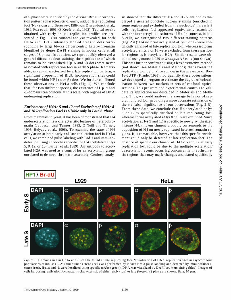
Although Equally Associated at Early and Late Replication Foci, CAF-1 Colocalizes with H4Ac 5 and 12 While Excluding H4Ac 8 and 16 only at Late Replication Foci
CAF-1 previously had been reported to be generally lo-cated at replication foci (Krude, 1995). However, a strictcomparison between early and late replication foci was notavailable. Using an mAb directed against p150-CAF-1(Smith and Stillman, 1989), a general localization of thissubunit at both late and early replication foci was ob-served by confocal analysis (Fig. 3 A). Quantification ofthese data in sets of confocal sections, demonstrated thatnewly replicated regions are equally associated with CAF-1in either early or late replication foci (Fig. 3 B). Impor-tantly, we observed that not all the p150-CAF-1 foci wereassociated with replication foci (green spots), promptingus to analyze the temporal association of CAF-1 with
DNA synthesis. In early S phase cells (Fig. 3 A), doublestaining patterns revealing each specific acetylated iso-form of H4, together with the p150-CAF-1 subunit, weresimilar. In contrast, in late S phase p150-CAF-1 could befound associated with H4Ac 5 or 12, but never with H4Ac16 or 8. Since H4Ac 8, in addition to H4Ac 5 and 12, werefound in a complex with CAF-1 in human nuclear extracts(Verreault et al., 1996), these findings suggest that theCAF-1 complex including H4Ac 8 is not involved at latereplication foci. The association of CAF-1 with early andlate replication foci argues for a general chromatin assem-bly mechanism throughout S phase. Remarkably, H4Ac 5and 12 association with CAF-1 reproduces the pattern ofH4 acetylation associated with DNA replication in hetero-chromatin regions (Fig. 2).
The Temporal Relationships between Two DNA Synthesis Events Can Be Reflected by theirSpatial Overlap
We used the BrdU/BiodU double labeling approach to re-late a temporal link between two DNA synthesis events to
Figure 2. Specific enrichment of H4 acetylated at lys 5 and 12can be detected by immunofluorescence at late, but not atearly replication foci in HeLa cells. A, DNA replication sites inan asynchronous population of HeLa cells were labeled in vivoby BrdU pulse (red). Different acetylated isoforms of H4 andH2AAc 5 were localized using pAbs specific for each acety-lated residue (K5, K8, K12, and K16, green). BrdU incorpora-tion site detection (red) was achieved after a second fixationand DNA denaturation by HCl (Materials and Methods).Early or late replicating cells were identified by their BrdUstaining pattern. Digital merge of both signals, correspondingto single optical sections, are shown in each case. Bars, 10 mm.B, Confocal sections from experiments similar to those pre-sented in A were collected for a series of nuclei. For each ofthese sections, colocalization was determined according to theprocedure described in Materials and Methods. The plot repre-sents the BrdU signals colocalizing with H2AAc 5 and each of
the acetylated isoforms in arbitrary units [(C) / (C 1 R), ratio normalized as described in Materials and Methods]. Error bars corre-spond to the maximum variance experimentally obtained.
on February 17, 2016
jcb.rupress.orgD
ownloaded from
Published December 13, 1999
The Journal of Cell Biology, Volume 147, 1999 1158
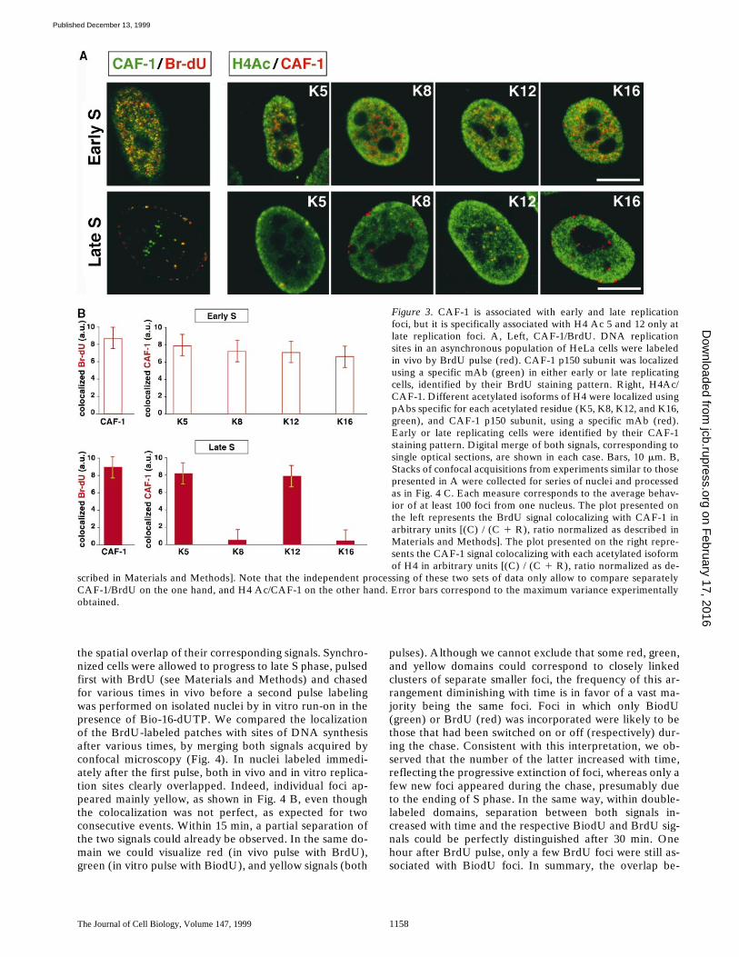
the spatial overlap of their corresponding signals. Synchro-nized cells were allowed to progress to late S phase, pulsedfirst with BrdU (see Materials and Methods) and chasedfor various times in vivo before a second pulse labelingwas performed on isolated nuclei by in vitro run-on in thepresence of Bio-16-dUTP. We compared the localizationof the BrdU-labeled patches with sites of DNA synthesisafter various times, by merging both signals acquired byconfocal microscopy (Fig. 4). In nuclei labeled immedi-ately after the first pulse, both in vivo and in vitro replica-tion sites clearly overlapped. Indeed, individual foci ap-peared mainly yellow, as shown in Fig. 4 B, even thoughthe colocalization was not perfect, as expected for twoconsecutive events. Within 15 min, a partial separation ofthe two signals could already be observed. In the same do-main we could visualize red (in vivo pulse with BrdU),green (in vitro pulse with BiodU), and yellow signals (both
pulses). Although we cannot exclude that some red, green,and yellow domains could correspond to closely linkedclusters of separate smaller foci, the frequency of this ar-rangement diminishing with time is in favor of a vast ma-jority being the same foci. Foci in which only BiodU(green) or BrdU (red) was incorporated were likely to bethose that had been switched on or off (respectively) dur-ing the chase. Consistent with this interpretation, we ob-served that the number of the latter increased with time,reflecting the progressive extinction of foci, whereas only afew new foci appeared during the chase, presumably dueto the ending of S phase. In the same way, within double-labeled domains, separation between both signals in-creased with time and the respective BiodU and BrdU sig-nals could be perfectly distinguished after 30 min. Onehour after BrdU pulse, only a few BrdU foci were still as-sociated with BiodU foci. In summary, the overlap be-
Figure 3. CAF-1 is associated with early and late replicationfoci, but it is specifically associated with H4 Ac 5 and 12 only atlate replication foci. A, Left, CAF-1/BrdU. DNA replicationsites in an asynchronous population of HeLa cells were labeledin vivo by BrdU pulse (red). CAF-1 p150 subunit was localizedusing a specific mAb (green) in either early or late replicatingcells, identified by their BrdU staining pattern. Right, H4Ac/CAF-1. Different acetylated isoforms of H4 were localized usingpAbs specific for each acetylated residue (K5, K8, K12, and K16,green), and CAF-1 p150 subunit, using a specific mAb (red).Early or late replicating cells were identified by their CAF-1staining pattern. Digital merge of both signals, corresponding tosingle optical sections, are shown in each case. Bars, 10 mm. B,Stacks of confocal acquisitions from experiments similar to thosepresented in A were collected for series of nuclei and processedas in Fig. 4 C. Each measure corresponds to the average behav-ior of at least 100 foci from one nucleus. The plot presented onthe left represents the BrdU signal colocalizing with CAF-1 inarbitrary units [(C) / (C 1 R), ratio normalized as described inMaterials and Methods]. The plot presented on the right repre-sents the CAF-1 signal colocalizing with each acetylated isoformof H4 in arbitrary units [(C) / (C 1 R), ratio normalized as de-
scribed in Materials and Methods]. Note that the independent processing of these two sets of data only allow to compare separatelyCAF-1/BrdU on the one hand, and H4 Ac/CAF-1 on the other hand. Error bars correspond to the maximum variance experimentallyobtained.
on February 17, 2016
jcb.rupress.orgD
ownloaded from
Published December 13, 1999
Taddei et al.
Duplication of Heterochromatin Domains
1159
tween the two signals decreased as a function of time, dueto both extinction and firing of replicating foci during thechase and to the progressive separation of clusters corre-sponding to DNA synthesized during the two pulseswithin one focus.
Quantification of BrdU-labeled regions associated withBiodU-labeled regions in sets of confocal sections showeda decrease in their spatial association, consistent with the
visual observation (Fig. 4 C). These results are in agree-ment with previous in vivo studies in rodent cells usingdouble pulse labeling in vivo and different methods ofquantification (Manders et al., 1996; Ma et al., 1998a). Theconsistency of these data further supports the validity ofour method. In this analysis, patches of synthesis occurringat 15 min intervals (corresponding approximately to thesynthesis of 50 Kb between both pulses) can be distin-
Figure 4. Within late replication foci, a defined delay betweentwo DNA synthesis events can be visualized by the degree ofoverlap between signals corresponding to these events. Sites ofDNA synthesis in a synchronous population of HeLa cells in lateS phase were first labeled in vivo by BrdU pulse (red). Then, nu-clei were isolated, immediately or after different chase times, andsites of DNA synthesis active after the chase were labeled by invitro run-on in the presence of Bio-16-dUTP (green). A, A digi-tal merge of single optical sections corresponding to both labels isshown for each chase time. B, Magnification of selected focishowing both labels (from windows in A) are shown. C, Stacks ofconfocal acquisitions from pulse–chase experiments, similar tothose presented in A, were collected for series of nuclei. A quan-titative analysis was then carried out on these samples accordingto the procedure described in Materials and Methods (3D analy-
sis). Each measurement corresponds to the average behavior of at least 100 foci from one nucleus. The plot represents the BrdU signalscolocalizing with BiodU in arbitrary units [(C) / (C 1 R), ratio normalized as described in Materials and Methods] as a function of chasetime. Error bars correspond to the maximum variance experimentally obtained.
on February 17, 2016
jcb.rupress.orgD
ownloaded from
Published December 13, 1999
The Journal of Cell Biology, Volume 147, 1999 1160
guished. Thus, this analysis could be used as a reference tostudy the large scale relationships between factors of inter-est and DNA synthesis.
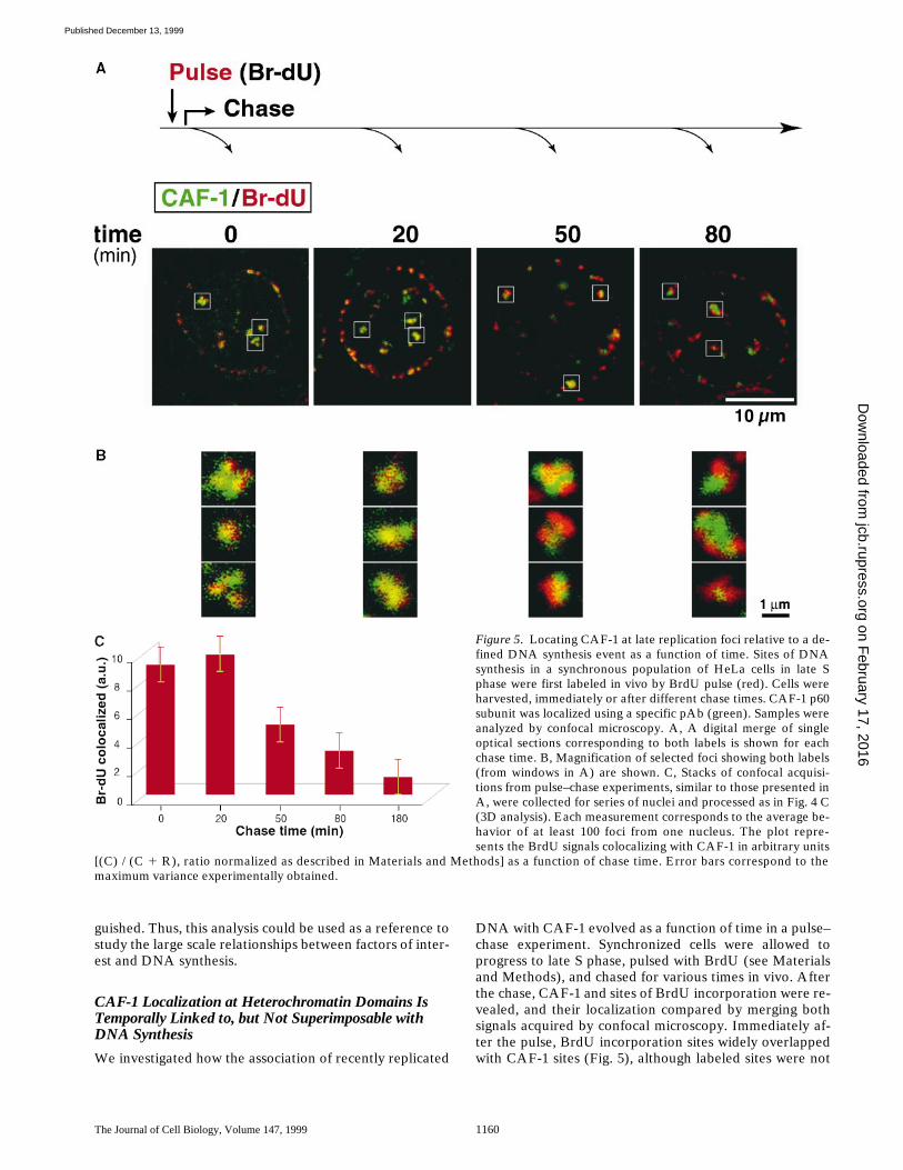
CAF-1 Localization at Heterochromatin Domains Is Temporally Linked to, but Not Superimposable with DNA Synthesis
We investigated how the association of recently replicated
DNA with CAF-1 evolved as a function of time in a pulse–chase experiment. Synchronized cells were allowed toprogress to late S phase, pulsed with BrdU (see Materialsand Methods), and chased for various times in vivo. Afterthe chase, CAF-1 and sites of BrdU incorporation were re-vealed, and their localization compared by merging bothsignals acquired by confocal microscopy. Immediately af-ter the pulse, BrdU incorporation sites widely overlappedwith CAF-1 sites (Fig. 5), although labeled sites were not
Figure 5. Locating CAF-1 at late replication foci relative to a de-fined DNA synthesis event as a function of time. Sites of DNAsynthesis in a synchronous population of HeLa cells in late Sphase were first labeled in vivo by BrdU pulse (red). Cells wereharvested, immediately or after different chase times. CAF-1 p60subunit was localized using a specific pAb (green). Samples wereanalyzed by confocal microscopy. A, A digital merge of singleoptical sections corresponding to both labels is shown for eachchase time. B, Magnification of selected foci showing both labels(from windows in A) are shown. C, Stacks of confocal acquisi-tions from pulse–chase experiments, similar to those presented inA, were collected for series of nuclei and processed as in Fig. 4 C(3D analysis). Each measurement corresponds to the average be-havior of at least 100 foci from one nucleus. The plot repre-sents the BrdU signals colocalizing with CAF-1 in arbitrary units
[(C) / (C 1 R), ratio normalized as described in Materials and Methods] as a function of chase time. Error bars correspond to themaximum variance experimentally obtained.
on February 17, 2016
jcb.rupress.orgD
ownloaded from
Published December 13, 1999
Taddei et al.
Duplication of Heterochromatin Domains
1161
exclusively yellow, as shown in Fig. 5 B. The signals corre-sponding to CAF-1 and BrdU had not separated within 20min. This contrasts with the signals corresponding to twosuccessive DNA synthesis events, which were separated at15 min after BrdU pulse (Fig. 4). After 50 min, CAF-1 andBrdU signals had begun to separate, and reached com-plete separation at 80 min after pulse. As in the previousexperiment, few foci with a single color appeared as chasetime increased. Hence, CAF-1 association with BrdU in-corporation sites appears related to DNA synthesis, butdoes not strictly follow the same dynamics.
Considering the association of BrdU signal with CAF-1,our quantitative analysis showed no significant changewithin the first 20 min after BrdU pulse (Fig. 5 C). Afterthis time, following the extinction of replication foci, thepercentage of colocalization decreased as a function oftime. Furthermore, comparing the quantitative analysis ofBrdU/CAF-1 and BrdU/BiodU pulse–chase experiments,a time difference of 20 min could be seen between releaseof CAF-1 and cessation of DNA synthesis. It is remark-able that after 80 min post-BrdU pulse, whereas replica-tion had stopped within foci labeled at the time of thepulse, CAF-1 could still be found associated with BrdU in-corporation sites. Further supporting these findings, wefound that the two largest subunits of CAF-1, p60 andp150, detected respectively using a pAb and an mAb, be-haved similarly in this assay. A similar analysis performedon early replicating foci gave comparable results (notshown), further suggesting a general behavior of CAF-1throughout S phase. This behavior of CAF-1 is consistentwith the rapid association of this assembly factor withnewly replicated regions of DNA and its retention for atleast 20 min at these locations.
The Association of H4Ac 5 and 12 With Heterochromatin Domains Is Transient
We then investigated how the association of H4Ac 5 withrecently replicated regions of the genome evolved as afunction of time in a pulse–chase experiment carried outas described above. Considering data in Figs. 2 and 3showing that replication specific acetylated isoform of H4(H4Ac 5 and 12) could not be detected enriched at earlyreplication foci, we concentrated our study on late replica-tion foci. Immediately after the pulse, BrdU incorporationsites were widely stained with H4Ac 5 antibody (Fig. 6),although labeled sites were not exclusively yellow, asshown in Fig. 6. The signals corresponding to BrdU werestill associated with H4Ac 5 after a 20-min chase. Thus,clusters of newly synthesized DNA can been found associ-ated with H4Ac 5 for at least 20 min. After 50 min, BrdUincorporation sites appeared partially dissociated fromH4Ac 5 and both signals were completely distinct in G2phase (after a 3-h chase). BrdU signal associated withH4Ac 5 was determined after different chase times on sev-eral optical sections from different nuclei to establish thestatistical significance of our observations (Fig. 6 C). Simi-lar data were obtained for H4Ac 12 in both human (Fig. 7)and
Xenopus
(not shown). Thus, H4 acetylation at lys 5and 12 appeared to be transiently associated with late rep-licated sequences in both mammals and amphibians. Thisresult contrasts with the maintenance of lys 12 acetylation
reported for heterochromatin from both yeast and
Dro-sophila
(Turner et al., 1992; Braunstein et al., 1996). The lossof H4Ac 5 and 12 enrichment at late replicated sequencesis likely to result from an enzyme catalyzed deacetylationor a loss of epitope accessibility. Preliminary immunola-beling experiments have failed to detect any specific en-richment of deacetylase catalytic subunits at late replica-tion foci (not shown). This may be because the specificenzymes involved still have to be identified, though wecannot exclude the possibility that the deacetylase activityat replication foci is locally regulated by a cofactor or amodification, rather than by varying the amount of en-zyme present. Alternatively, without any specific enrich-ment of HDAC, the lack of HAT in these regions could besufficient to account for H4 deacetylation following repli-cation as supported by Fig. 7.
Most importantly, these data strongly support the exist-ence of a time window of
z
20 min, during which hetero-chromatin regions are enriched in acetylated H4, followedby a deacetylation event.
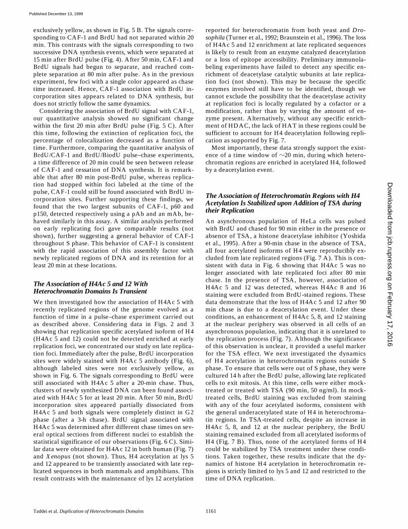
The Association of Heterochromatin Regions with H4 Acetylation Is Stabilized upon Addition of TSA during their Replication
An asynchronous population of HeLa cells was pulsedwith BrdU and chased for 90 min either in the presence orabsence of TSA, a histone deacetylase inhibitor (Yoshidaet al., 1995). After a 90-min chase in the absence of TSA,all four acetylated isoforms of H4 were reproducibly ex-cluded from late replicated regions (Fig. 7 A). This is con-sistent with data in Fig. 6 showing that H4Ac 5 was nolonger associated with late replicated foci after 80 minchase. In the presence of TSA, however, association ofH4Ac 5 and 12 was detected, whereas H4Ac 8 and 16staining were excluded from BrdU-stained regions. Thesedata demonstrate that the loss of H4Ac 5 and 12 after 90min chase is due to a deacetylation event. Under theseconditions, an enhancement of H4Ac 5, 8, and 12 stainingat the nuclear periphery was observed in all cells of anasynchronous population, indicating that it is unrelated tothe replication process (Fig. 7). Although the significanceof this observation is unclear, it provided a useful markerfor the TSA effect. We next investigated the dynamicsof H4 acetylation in heterochromatin regions outside Sphase. To ensure that cells were out of S phase, they werecultured 14 h after the BrdU pulse, allowing late replicatedcells to exit mitosis. At this time, cells were either mock-treated or treated with TSA (90 min, 50 ng/ml). In mock-treated cells, BrdU staining was excluded from stainingwith any of the four acetylated isoforms, consistent withthe general underacetylated state of H4 in heterochroma-tin regions. In TSA-treated cells, despite an increase inH4Ac 5, 8, and 12 at the nuclear periphery, the BrdUstaining remained excluded from all acetylated isoforms ofH4 (Fig. 7 B). Thus, none of the acetylated forms of H4could be stabilized by TSA treatment under these condi-tions. Taken together, these results indicate that the dy-namics of histone H4 acetylation in heterochromatin re-gions is strictly limited to lys 5 and 12 and restricted to thetime of DNA replication.
on February 17, 2016
jcb.rupress.orgD
ownloaded from
Published December 13, 1999

The Journal of Cell Biology, Volume 147, 1999 1162
Discussion
Immunofluorescence analysis of heterochromatin do-mains at late replicating foci shows that these domainscontain specific acetylated forms of histone H4, togetherwith the largest subunit of CAF-1. The enrichment of thehistone H4 acetylated at lys 5 and/or 12 was in strikingcontrast with the exclusion of H4Ac 8 and 16. We followedthe large scale dynamics of this domain through the tem-
poral analysis of DNA synthesis, the presence of CAF-1,acetylated H4, Hp1
a
, and Hp1
b
. Thus, we can distinguishproperties of heterochromatin domains that are either sta-ble (Hp1
a
and -
b
concentration) or transient (H4Ac 5 and12, and CAF-1). Comparison with early replicating foci/euchromatin regions shows that CAF-1 is also associatedwith DNA synthesis in these regions. In contrast, we couldnot reveal any specific enrichment in H4Ac 5 and 12, com-pared with 8 and 16.
Figure 6. Locating H4Ac 5 at late replication foci relative to adefined DNA synthesis event as a function of time. Sites of DNAsynthesis in a synchronous population of HeLa cells in late Sphase were first labeled in vivo by BrdU pulse (red). Cells wereharvested, immediately or after different chase times. H4Ac 5was localized using a specific pAb (green). Samples were ana-lyzed by confocal microscopy. A, A digital merge of single opticalsections corresponding to both labels is shown for each chasetime. B, Magnification of selected foci showing both labels (fromwindows in A) are shown. C, Confocal sections from pulse–chaseexperiments, similar to those presented in A, were collected for aseries of nuclei. For each of these sections, colocalization wasdetermined according to the procedure described in Materialsand Methods (2D analysis). The plot represents the BrdUsignals colocalizing with H4Ac 5 in arbitrary units [(C) / (C 1
R), ratio normalized as described in Materials and Methods] as a function of chase time. Error bars correspond to the maximum vari-ance experimentally obtained.
on February 17, 2016
jcb.rupress.orgD
ownloaded from
Published December 13, 1999
Taddei et al.
Duplication of Heterochromatin Domains 1163
Figure 7. Dynamics of H4 acetylation in heterochromatin regions is restricted to the time of their DNA replication. A, Sites of DNAsynthesis in an asynchronous population of HeLa cells were first labeled in vivo by BrdU pulse (red), and chased for 90 min either in thepresence or absence of TSA (50 ng/ml). Different acetylated isoforms of H4 were localized using specific pAbs (K5, K8, K12, and K16,green). B, Sites of DNA synthesis in an asynchronous population of HeLa cells were first labeled in vivo by BrdU pulse (red), andchased for 14 h to allow late replicating cells, labeled during the pulse, to exit mitosis, before prolonged incubation in the absence orpresence of TSA (50 ng/ml) for 90 min. Different acetylated isoforms of H4 were localized using specific pAbs (green). Digital merge ofboth signals, corresponding to single optical sections, are shown in each case. Bars, 10 mm. Note that these images were processed differ-ently to permit visualization, because the intensities recorded were very different 1 or 2 TSA. Enlarged inserts (top left of each im-ages) correspond to selected foci.
on February 17, 2016
jcb.rupress.orgD
ownloaded from
Published December 13, 1999
The Journal of Cell Biology, Volume 147, 1999 1164
We discuss how the replication-associated cycle ofacetylation/deacetylation may have a specific importancein heterochromatin regions.
Hp1a and -b Are Maintained through DNA Replication
The existence of local concentrations of specific proteinshas been proposed to compartmentalize chromatin re-gions, creating an index system for the nucleus (Gasserand Laemmli, 1987; Stone and Pillus, 1998). The local con-centration of specific proteins, such as Hp1a and -b, couldalso be critical for defining domains that should be re-pressed. In our study, no major changes in the distributionof Hp1a and -b were observed during DNA replication ofheterochromatin regions (Fig. 1; and not shown). This wassurprising at first, since the presence of Hp1a and -b mayreflect a high level of DNA condensation (Cavalli andParo, 1998) that may not necessarily be compatible withthe replication process. We thus conclude that, if the repli-cation fork is destabilizing the nucleosome (Sogo and Las-key, 1995), this is a local phenomenon that does not affectthe stability of the larger scale organization in a detectablemanner. The maintenance of the local concentration ofHp1a and -b as part of nucleoprotein complexes, either di-rectly interacting or simply in close proximity with DNA,may ensure the rapid reassociation of the replicated re-gions into heterochromatin. Furthermore, the ability ofHP1 proteins to oligomerize has led to the hypothesis thatthey can cross-link chromatin domains by the formation ofconcatenated multiprotein complexes (Cowell and Austin,1997). Thus, Hp1a and -b appear to be components thatfavor the maintenance of a heterochromatin region. Inter-estingly, in mouse cells, association of some genes with anHP1b domain has been shown to correlate with their tran-scriptional repression (Brown et al., 1997). One wouldthus predict that in vertebrates, overexpression of HP1bcould result in a spreading or expansion of these domains,possibly producing phenotypes resembling PEV (Festen-stein et al., 1996; Milot et al., 1996). Additional modifica-tion adding to stability in heterochromatin, such as meth-ylation of DNA in vertebrates (Bird, 1992), should also beincorporated into the overall picture. Future experimentswill provide insights into these issues.
The Replication-associated Acetylated Isoforms of Histone H4, Together with CAF-1, Provide a Window of Opportunity in Heterochromatin
Analysis of the distribution of CAF-1 and H4Ac 5, com-pared with DNA synthesis at heterochromatin regions,demonstrated a dynamic behavior that was not strictlyparallel to DNA synthesis (Figs. 4, 5, and 6). No major dif-ferences were observed between the location of CAF-1and patches of DNA corresponding to individual replica-tion foci observed within 20 min after their labeling inHeLa cells. For this chase time, we established that twoDNA synthesis events are already clearly separated. Themaintenance of CAF-1 association with these patches ofDNA after their synthesis is further emphasized by the de-tection of CAF-1 foci beyond the DNA replication time.This CAF-1 enrichment at the location of newly synthe-sized DNA soon after its synthesis, and its persistence, ex-ceeds the time involved in synthesizing the DNA length
corresponding to a nucleosome and depositing one set ofH3 and H4 at the replication fork (less than five minutes,according to Smith et al., 1984). This extended time periodis comparable to the postreplicative phase required forproper chromatin maturation (Levy and Jakob, 1978;Worcel et al., 1978; Smith et al., 1984). An attractive hy-pothesis is that it could define the time needed to process aunit of a size between 50 and 200 Kb, reminiscent of thechromatin loops. This would be compatible with the recentreport on CAF-1–coupled inheritance of chromatin actingpostreplicatively in vitro (Shibahara and Stillman, 1999).Similarly, we determined that newly synthesized DNA se-quences were associated with H4Ac 5 for z20 min in latereplication foci. The existence of a pool of acetylated his-tone H4 concentrated in the region of heterochromatin atthe time of replication constitutes a strong argument forbelieving that these histones are used to form the new nu-cleosomes at these foci. It is striking though, that, at alarge scale level, this replication-associated acetylated iso-form could not be specifically detected at early replicationfoci although CAF-1 was still found (Figs. 2 and 3). Sincethe biochemical approach has indicated a general usage ofH4Ac 5 and 12 (Annunziato, 1995) throughout the ge-nome, however, multiple acetylation/deacetylation eventsoccurring concurrently in these regions may mask changesassociated specifically with replication. In heterochroma-tin, remarkably, the acetylation level of H4 associated withhistone deposition at the replication fork appears as adominant event. The parallel between CAF-1 and H4Ac 5and 12 (Figs. 3, 5, and 6) in heterochromatin raises the pos-sibility that CAF-1 association with acetylated histoneshelps to coordinate loading of acetylated histone H4 witha deacetylation event. This latter event could be promotedthrough the interaction of the smallest subunit of CAF-1with a histone deacetylase previously detected in humancell extracts (Taunton et al., 1996). The heterochromatinspecific effect of CAC deletions in yeast strains leading tothe redistribution of the telomeric protein Rap1p, togetherwith a loss of silencing (Enomoto et al., 1997; Monson et al.,1997; Enomoto and Berman, 1998), could be explained bydefects in such a coordination when replicating hetero-chromatin regions. Future experiments should addresswhether perturbation of CAF-1 in higher eucaryotes simi-larly affects heterochromatin function. This may also re-late to the possible role of CAF-1 associated with DNA re-pair (Gaillard et al., 1996; Kaufman et al., 1997; Martiniet al., 1998). By facilitating the deacetylation step duringthe chromatin dynamics associated with DNA repair,CAF-1 may help to avoid the persistence of inappropriatechromatin organization.
Our data are consistent with the existence of a generalmechanism for nucleosome assembly employed through-out the genome. In this pathway, histone deposition wouldmake use of acetylated histones chaperoned by CAF-1that would facilitate deacetylation. Depending on the localenvironment, particularly the presence of HATs/HDACsand their regulators, it may be possible to generate distinctacetylated chromatin configurations at a steady state. Inhi-bition of histone deacetylase by TSA showed that the dy-namics of acetylation of histone H4 in heterochromatin isrestricted to K5 and K12, and to the time of the DNA rep-lication (Fig. 7). Prolonged exposure to the drug led to the
on February 17, 2016
jcb.rupress.orgD
ownloaded from
Published December 13, 1999

Taddei et al. Duplication of Heterochromatin Domains 1165
disruption of heterochromatin domains (HP1 staining, notshown, Taddei, A., and G. Almouzni unpublished results).These observations are reminiscent of those obtained inSaccharomyces pombe in which a TSA treatment dis-rupted centromeric organization and delocalized the swi6protein (Ekwall et al., 1997). Further investigations arenecessary to determine the exact basis of this phenome-non. It is intriguing that H4 acetylation seems to not be re-quired for general nucleosome formation, per se, either invitro or in vivo (Ma et al., 1998b; Zhang et al., 1998). It hasbeen proposed that the so-called futile cycles of enzymaticmetabolism may be needed for rapid response to changesin the environment and for evolution (Gerhart and Kirsch-ner, 1997). It is interesting to consider the possibility thatthe acetylation/deacetylation cycle chaperoned by CAF-1serves the same purpose. In that respect, it is noteworthythat mutations in histone tails affecting all the acetylatablesites in histone H4 gave rise in yeast to a checkpoint phe-notype (Megee et al., 1995; Hartzog and Winston, 1997).The importance of the alternative states generated by thefutile cycle could be envisaged at two different levels. At acellular level, this could help to monitor S phase progres-sion, DNA damage processing, and perhaps determinehow S phase events are coordinated. At the level of a pop-ulation, a certain degree of adaptability can be advanta-geous during evolution, as was discussed for the epigeneticinheritance of centromeres (Murphy and Karpen, 1998).This is exemplified in yeast with the switching mechanismat mating type loci that depends on replication (Laurensonand Rine, 1992).
We studied here how a preestablished structure couldbe propagated, the next challenge will be to understandhow these structures can be formed de novo, which will becritical during the development of an organism.
We thank Dr. B. Stillman, Dr. R. Losson, Dr. P. Chambon, and Dr. K.Marheinecke for antibodies. We thank Drs. G. Cavalli, P. Grandi, S.Holmes, C. Maison, J.G. Moggs, and P. Ridgway for critical reading. Weare grateful to the anonymous reviewers for their useful suggestions.
A. Taddei was initially a Ministère de l’Education Nationale del’Enseignement Supérieur de la Recherche fellow and then supported bythe Association pour la Recherche sur le Cancer (ARC). This work wassupported by a Training Mobility Research Network grant from the Euro-pean Community (G. Almouzni and B. Turner) and funding from Fonda-tion pour la Recherche Médicale, ARC, and Ligue contre le cancer.
Submitted: 24 June 1999Revised: 28 October 1999Accepted: 2 November 1999
References
Aagaard, L., G. Laible, P. Selenko, M. Schmid, R. Dorn, G. Schotta, S. Kuhfit-tig, A. Wolf, A. Lebersorger, P.B. Singh, et al. 1999. Functional mammalianhomologues of the Drosophila PEV-modifier Su(var)3-9 encode cen-tromere-associated proteins which complex with the heterochromatin com-ponent M31. EMBO (Eur. Mol. Biol. Organ.) J. 18:1923–1938.
Annunziato, A.T. 1995. Histone acetylation during chromatin replication andnucleosome assembly. In Nucleus. Vol. 1. A. Wolffe, editor. JAI Press,Greenwich, CT. 31–56.
Belyaev, N.D., A.M. Keohane, and B.M. Turner. 1996. Histone H4 acetylationand replication timing in Chinese hamster chromosomes. Exp. Cell Res. 225:277–285.
Bird, A. 1992. The essentials of DNA methylation. Cell. 70:5–8.Braunstein, M., R.E. Sobel, C.D. Allis, B.M. Turner, and J.R. Broach. 1996. Ef-
ficient transcriptional silencing in Saccharomyces cerevisiae requires a het-erochromatin histone acetylation pattern. Mol. Cell. Biol. 16:4349–4356.
Bridger, J.M., and W.A. Bickmore. 1998. Putting the genome on the map.Trends Genet. 14:403–409.
Brown, K.E., S.S. Guest, S.T. Smale, K. Hahm, M. Merkenschlager, and A.G.Fisher. 1997. Association of transcriptionally silent genes with Ikaros com-plexes at centromeric heterochromatin. Cell. 91:845–854.
Cavalli, G., and R. Paro. 1998. Chromo-domain proteins: linking chromatinstructure to epigenetic regulation. Curr. Opin. Cell Biol. 10:354–360.
Cook, P.R. 1995. A chromomeric model for nuclear and chromosome structure.J. Cell Sci. 108:2927–2935.
Cowell, I.G., and C.A. Austin. 1997. Self-association of chromo domain pep-tides. Biochem. Biophys. Acta. 1337:198–206.
Ekwall, K., T. Olsson, B.M. Turner, G. Cranston, and R.C. Allshire. 1997.Transient inhibition of histone deacetylation alters the structural and func-tional imprint at fission yeast centromeres. Cell. 91:1021–1032.
Elgin, S.C. 1996. Heterochromatin and gene regulation in Drosophila. Curr.Opin. Genet. Dev. 6:193–202.
Enomoto, S., and J. Berman. 1998. Chromatin assembly factor I contributes tothe maintenance, but not the re-establishment, of silencing at the yeast silentmating loci. Genes Dev. 12:219–232.
Enomoto, S., P. McCune-Zierath, M. Geraminejad, M. Sanders, and J. Berman.1997. Rfl2, a subunit of yeast chromatin assembly factor-I, is required fortelomeric chromatin function in vivo. Genes Dev. 11:358–363.
Festenstein, R., M. Tolaini, P. Corbella, C. Mamalaki, J. Parrington, M. Fox, A.Miliou, M. Jones, and D. Kioussis. 1996. Locus control region function andheterochromatin-induced position effect variegation. Science. 271:1123–1125.
Fox, M.H., D.J. Arndt-Jovin, T.M. Jovin, P.H. Baumann, and M. Robert-Nicoud. 1991. Spatial and temporal distribution of DNA replication sites lo-calized by immunofluorescence and confocal microscopy in mouse fibro-blasts. J. Cell Sci. 99:247–253.
Gaillard, P.H., E.M. Martini, P.D. Kaufman, B. Stillman, E. Moustacchi, and G.Almouzni. 1996. Chromatin assembly coupled to DNA repair: a new role forchromatin assembly factor I. Cell. 86:887–896.
Gasser, S.M., and U.K. Laemmli. 1987. A glimpse at chromosomal order.Trends Genet. 3:16–22.
Gerhart, J., and M. Kirschner. 1997. Contingency. In Cells, Embryos, and Evo-lution. Blackwell Science, Malden, MA. 45–89.
Goldman, M.A., G.P. Holmquist, M.C. Gray, L.A. Caston, and A. Nag. 1984.Replication timing of genes and middle repetitive sequences. Science. 224:686–692.
Goodman, J.W. 1968. Introduction to Fourier Optics. McGraw-Hill, New York.4–29.
Grewal, S.I., M.J. Bonaduce, and A.J. Klar. 1998. Histone deacetylase ho-mologs regulate epigenetic inheritance of transcriptional silencing and chro-mosome segregation in fission yeast. Genetics. 150:563–576.
Grunstein, M. 1998. Yeast heterochromatin: regulation of its assembly and in-heritance by histones. Cell. 93:325–328.
Hartzog, G.A., and F. Winston. 1997. Nucleosomes and transcription: recentlessons from genetics. Curr. Opin. Genet. Dev. 7:192–198.
Hatton, K.S., V. Dhar, E.H. Brown, M.A. Iqbal, S. Stuart, V.T. Didamo, andC.L. Schildkraut. 1988. Replication program of active and inactive multigenefamilies in mammalian cells. Mol. Cell. Biol. 8:2149–2158.
Heitz, E. 1928. Das heterochromatin der Moose. Jb. Wiss. Bot. 69:728.Henikoff, S. 1990. Position-effect variegation after 60 years. Trends Genet.
6:422–426.Horsley, D., A. Hutchings, G.W. Butcher, and P.B. Singh. 1996. M32, a murine
homologue of Drosophila heterochromatin protein 1 (HP1), localises to eu-chromatin within interphase nuclei and is largely excluded from constitutiveheterochromatin. Cytogenet. Cell Genet. 73:308–311.
Hozak, P., A.B. Hassan, D.A. Jackson, and P.R. Cook. 1993. Visualization ofreplication factories attached to a nucleoskeleton. Cell. 73:361–373.
James, T.C., and S.C. Elgin. 1986. Identification of a nonhistone chromosomalprotein associated with heterochromatin in Drosophila melanogaster and itsgene. Mol. Cell. Biol. 6:3862–3872.
James, T.C., J.C. Eissenberg, C. Craig, V. Dietrich, A. Hobson, and S.C. Elgin.1989. Distribution patterns of HP1, a heterochromatin-associated nonhis-tone chromosomal protein of Drosophila. Eur. J. Cell Biol. 50:170–180.
Jeppesen, P., and B.M. Turner. 1993. The inactive X chromosome in femalemammals is distinguished by a lack of histone H4 acetylation, a cytogeneticmarker for gene expression. Cell. 74:281–289.
Kamakaka, R.T., M. Bulger, P.D. Kaufman, B. Stillman, and J.T. Kadonaga.1996. Postreplicative chromatin assembly by Drosophila and human chro-matin assembly factor I. Mol. Cell. Biol. 16:810–817.
Kaufman, P.D., R. Kobayashi, N. Kessler, and B. Stillman. 1995. The p150 andp60 subunits of chromatin assembly factor I: a molecular link between newlysynthesized histones and DNA replication. Cell. 81:1105–1114.
Kaufman, P.D., R. Kobayashi, and B. Stillman. 1997. Ultraviolet radiation sen-sitivity and reduction of telomeric silencing in Saccharomyces cerevisiae cellslacking chromatin assembly factor I. Genes Dev. 11:345–357.
Krude, T. 1995. Chromatin assembly factor 1 (CAF-1) colocalizes with replica-tion foci in HeLa cell nuclei. Exp. Cell Res. 220:304–311.
Krude, T., M. Jackman, P.J. Pines, and R.A. Laskey. 1997. Cyclin/Cdk-depen-dent initiation of DNA replication in a human cell-free system. Cell. 88:109–119.
Kuo, M.H., and C.D. Allis. 1998. Roles of histone acetyltransferases anddeacetylases in gene regulation. Bioessays. 20:615–626.
Lamond, A.I., and W.C. Earnshaw. 1998. Structure and function in the nucleus.
on February 17, 2016
jcb.rupress.orgD
ownloaded from
Published December 13, 1999

The Journal of Cell Biology, Volume 147, 1999 1166
Science. 280:547–553.Laurenson, P., and J. Rine. 1992. Silencers, silencing, and heritable transcrip-
tional states. Microbiol. Rev. 56:543–560.Le Douarin, B., A.L. Nielsen, J.M. Garnier, H. Ichinose, F. Jeanmougin, R.
Losson, and P. Chambon. 1996. A possible involvement of TIF1 alpha andTIF1 beta in the epigenetic control of transcription by nuclear receptors.EMBO (Eur. Mol. Biol. Organ.) J. 15:6701–6715.
Levy, A., and K.M. Jakob. 1978. Nascent DNA in nucleosome like structuresfrom chromatin. Cell. 14:259–267.
Lewin, B. 1998. The mystique of epigenetics. Cell. 93:301–303.Ma, H., J. Samarabandou, R. Devdhar, R. Acharya, P.C. Cheng, C. Meng, and
R. Berezney. 1998a. Spatial and temporal dynamics of DNA replication sitesin mammalian cells. J. Cell Biol. 143:1415–1425.
Ma, X.J., J. Wu, B.A. Altheim, M.C. Schultz, and M. Grunstein. 1998b. Deposi-tion-related sites K5/K12 in histone H4 are not required for nucleosomedeposition in yeast. Proc. Natl. Acad. Sci. USA. 95:6693–6698.
Manders, E.M., J. Stap, J. Strackee, R. van Driel, and J.A. Aten. 1996. Dynamicbehavior of DNA replication domains. Exp. Cell Res. 226:328–335.
Marheineke, K., and T. Krude. 1998. Nucleosome assembly activity and intra-cellular localization of human CAF-1 changes during the cell division cycle.J. Biol. Chem. 273:15279–15286.
Marshall, W.F., J.C. Fung, and J.W. Sedat. 1997. Deconstructing the nucleus:global architecture from local interactions. Curr. Opin. Genet. Dev. 7:259–263.
Martini, E., D.M.J. Roche, K. Marheineke, A. Verreault, and G. Almouzni.1998. Recruitment of phosphorylated chromatin assembly factor 1 to chro-matin following UV irradiation of human cells. J. Cell Biol. 3:563–575.
Megee, P.C., B.A. Morgan, and M.M. Smith. 1995. Histone H4 and the mainte-nance of genome integrity. Genes Dev. 9:1716–1727.
Milot, E., J. Strouboulis, T. Trimborn, M. Wijgerde, E. de Boer, A. Langeveld,K. Tan-Un, W. Vergeer, N. Yannoutsos, F. Grosveld, et al. 1996. Hetero-chromatin effects on the frequency and duration of LCR-mediated genetranscription. Cell. 87:105–114.
Monson, E.K., D. de Bruin, and V.A. Zakian. 1997. The yeast Cac1 protein isrequired for the stable inheritance of transcriptionally repressed chromatinat telomeres. Proc. Natl. Acad. Sci. USA. 94:13081–13086.
Murphy, T.D., and G.H. Karpen. 1998. Centromeres take flight: alpha satelliteand the quest for the human centromere. Cell. 93:317–320.
Nakamura, H., T. Morita, and C. Sato. 1986. Structural organizations of repli-con domains during DNA synthetic phase in the mammalian nucleus. Exp.Cell Res. 165:291–297.
Nakayasu, H., and R. Berezney. 1989. Mapping replicational sites in the eucary-otic cell nucleus. J. Cell Biol. 108:1–11.
O’Keefe, R.T., S.C. Henderson, and D.L. Spector. 1992. Dynamic organizationof DNA replication in mammalian cell nuclei: spatially and temporally de-fined replication of chromosome-specific alpha-satellite DNA sequences. J.Cell Biol. 116:1095–1110.
O’Neill, L.P., and B.M. Turner. 1995. Histone H4 acetylation distinguishes cod-ing regions of the human genome from heterochromatin in a differentiation-dependent but transcription-independent manner. EMBO (Eur. Mol. Biol.Organ.) J. 14:3946–3957.
Pak, D.T., M. Pflumm, I. Chesnokov, D.W. Huang, R. Kellum, J. Marr, P. Ro-manowski, and M.R. Botchan. 1997. Association of the origin recognitioncomplex with heterochromatin and HP1 in higher eukaryotes. Cell. 91:311–323.
Saunders, W.S., C. Chue, M. Goebl, C. Craig, R.F. Clark, J.A. Powers, J.C. Eis-senberg, S.C. Elgin, N.F. Rothfield, and W.C. Earnshaw. 1993. Molecularcloning of a human homologue of Drosophila heterochromatin protein HP1using anti-centromere autoantibodies with anti-chromo specificity. J. CellSci. 104:573–582.
Shibahara, K.I., and B. Stillman. 1999. Replication-dependent marking of DNAby PCNA facilitates CAF-1-coupled inheritance of chromatin. Cell. 96:575–585.
Singh, P.B., J.R. Miller, J. Pearce, R. Kothary, R.D. Burton, R. Paro, T.C.James, and S.J. Gaunt. 1991. A sequence motif found in a Drosophila het-erochromatin protein is conserved in animals and plants. Nucleic Acids Res.19:789–794.
Smith, P.A., V. Jackson, and R. Chalkley. 1984. Two-stage maturation processfor newly replicated chromatin. Biochemistry. 23:1576–1581.
Smith, S., and B. Stillman. 1989. Purification and characterization of CAF-I, ahuman cell factor required for chromatin assembly during DNA replicationin vitro. Cell. 58:15–25.
Smith, S., and B. Stillman. 1991. Immunological characterization of chromatinassembly factor I, a human cell factor required for chromatin assembly dur-ing DNA replication in vitro. J. Biol. Chem. 266:12041–12047.
Sobel, R.E., R.G. Cook, C.A. Perry, A.T. Annunziato, and C.D. Allis. 1995.Conservation of deposition-related acetylation sites in newly synthesizedhistones H3 and H4. Proc. Natl. Acad. Sci. USA. 92:1237–1241.
Sogo, J.M., and R.A. Laskey. 1995. Chromatin replication and assembly. InChromatin Structure and Gene Expression. S.C.R. Elgin, editor. OxfordUniversity Press, New York. 49–71.
Stone, E.M., and L. Pillus. 1998. Silent chromatin in yeast: an orchestrated med-ley featuring Sir3p. Bioessays. 20:30–40.
Strouboulis, J., and A.P. Wolffe. 1996. Functional compartmentalization of thenucleus. J. Cell Sci. 109:1991–2000.
Taunton, J., C.A. Hassig, and S.L. Schreiber. 1996. A mammalian histonedeacetylase related to the yeast transcriptional regulator Rpd3p. Science.272:408–411.
Tsukiyama, T., and C. Wu. 1997. Chromatin remodeling and transcription.Curr. Opin. Genet. Dev. 7:182–191.
Turner, B.M., A.J. Birley, and J. Lavender. 1992. Histone H4 isoforms acety-lated at specific lysine residues define individual chromosomes and chroma-tin domains in Drosophila polytene nuclei. Cell. 69:375–384.
Turner, B.M., L.P. O’Neill, and I.M. Allan. 1989. Histone H4 acetylation in hu-man cells. Frequency of acetylation at different sites defined by immunola-beling with site-specific antibodies. FEBS Letts. 253:141–145.
van Dierendonck, J.H., R. Keyzer, C.J. van de Velde, and C.J. Cornelisse. 1989.Subdivision of S-phase by analysis of nuclear 5-bromodeoxyuridine stainingpatterns. Cytometry. 10:143–150.
van Holde, K.E. 1988. Chromatin. Springer-Verlag, New York.Verreault, A., P.D. Kaufman, R. Kobayashi, and B. Stillman. 1996. Nucleosome
assembly by a complex of CAF-1 and acetylated histones H3/H4. Cell. 87:95–104.
Vila, J.-L., and P. Bolon. 1993. Filtrage d’ordre adaptatif pour le prétraitementd’images naturelles. In 14ème Colloque Gretsi. 563–566.
Wakimoto, B.T. 1998. Beyond the nucleosome: epigenetic aspects of position-effect variegation in Drosophila. Cell. 93:321–324.
Wallrath, L.L. 1998. Unfolding the mysteries of heterochromatin. Curr. Opin.Genet. Dev. 8:147–153.
Wolffe, A. 1995. Chromatin: structure and function. Academic Press, London.Worcel, A., S. Han, and M.L. Wong. 1978. Assembly of newly replicated chro-
matin. Cell. 15:969–977.Ye, Q., and H.J. Worman. 1996. Interaction between an integral protein of the
nuclear envelope inner membrane and human chromodomain proteins ho-mologous to Drosophila HP1. J. Biol. Chem. 271:14653–14656.
Yoshida, M., S. Horinouchi, and T. Beppu. 1995. Trichostatin A and trapoxin:novel chemical probes for the role of histone acetylation in chromatin struc-ture and function. Bioessays. 17:423–430.
Zhang, W., J.R. Bone, D.G. Edmondson, B.M. Turner, and S.Y. Roth. 1998.Essential and redundant functions of histone acetylation revealed by muta-tion of target lysines and loss of the Gcn5p acetyltransferase. EMBO (Eur.Mol. Biol. Organ.) J. 17:3155–3167.
on February 17, 2016
jcb.rupress.orgD
ownloaded from
Published December 13, 1999
Related Documents