Duloxetine hydrochloride Mohan Bhadbhade, a * James Hook, a Chris Marjo, a Anne Rich a and Qinghong Lin b a UNSW Analytical Centre, The University of New South Wales, Sydney, NSW 2052, Australia, and b Arrow Laboratories Ltd, Croydon South, Victoria 3136, Australia Correspondence e-mail: [email protected] Received 31 July 2009; accepted 25 August 2009 Key indicators: single-crystal X-ray study; T = 150 K; mean (C–C) = 0.006 A ˚ ; disorder in main residue; R factor = 0.044; wR factor = 0.131; data-to-parameter ratio = 12.4. The title compound [systematic name: N-methyl-3- (1-naphthyloxy)-3-(2-thienyl)propan-1-aminium chloride], C 18 H 20 NOS + Cl , was crystallized from 1,4-dioxane. Twofold rotational disorder exhibited by the thiophene ring in a 0.580 (5):0.420 (5) ratio represents two different conforma- tions of the molecule that exist in the same crystal form. The crystal structure contains strong N—HCl hydrogen bonds. Related literature For therapeutic properties of duloxetine hydrochloride, see Waitekus & Kirkpatrick (2004). For related structures, see: Brenna et al. (2007); Tao et al. (2008). The title compound is reported to have different polymorphs on the basis of X-ray powder diffraction data, see: Ini et al. (2006). Experimental Crystal data C 18 H 20 NOS + Cl M r = 333.86 Monoclinic, P2 1 a = 9.7453 (10) A ˚ b = 6.9227 (7) A ˚ c = 13.4247 (16) A ˚ = 109.432 (4) V = 854.09 (16) A ˚ 3 Z =2 Mo K radiation = 0.35 mm 1 T = 150 K 0.38 0.08 0.03 mm Data collection Bruker Kappa APEXII CCD area- detector diffractometer Absorption correction: multi-scan (SADABS; Sheldrick, 2003) T min = 0.879, T max = 0.990 6386 measured reflections 2947 independent reflections 2255 reflections with I >2(I) R int = 0.058 Refinement R[F 2 >2(F 2 )] = 0.044 wR(F 2 ) = 0.131 S = 0.79 2947 reflections 237 parameters 110 restraints H-atom parameters constrained max = 0.17 e A ˚ 3 min = 0.21 e A ˚ 3 Absolute structure: Flack (1983), 1309 Friedel pairs Flack parameter: 0.05 (10) Table 1 Hydrogen-bond geometry (A ˚ , ). D—HA D—H HA DA D—HA N1—H1ACl1 i 0.92 2.23 3.113 (3) 161 N1—H1BCl1 0.92 2.18 3.087 (3) 170 Symmetry code: (i) x; y þ 1 2 ; z. Data collection: APEX2 (Bruker, 2007); cell refinement: SAINT (Bruker, 2007); data reduction: SAINT; program(s) used to solve structure: SHELXS97 (Sheldrick, 2008); program(s) used to refine structure: SHELXL97 (Sheldrick, 2008); molecular graphics: SHELXTL-Plus (Sheldrick, 2008); software used to prepare material for publication: SHELXL97. We are grateful to Professor Grainne Moran for her encouragement and interest in this work. Supplementary data and figures for this paper are available from the IUCr electronic archives (Reference: BG2289). References Brenna, E., Frigoli, S., Fronza, G., Fuganti, C. & Malpezzi, L. (2007). J. Pharm. Biomed. Anal. 43, 1573–1575. Bruker (2007). APEX2 and SAINT. Bruker AXS Inc., Madison, Wisconsin, USA. Flack, H. D. (1983). Acta Cryst. A39, 876–881. Ini, S., Shmueli, Y., Koltai, T. & Gold, A. (2006). Duloxetine. HCl Polymorphs WO/2006/081515, International Application No. PCT/US2006/003126. Publication Date: 03.08.2006. Sheldrick, G. M. (2003). SADABS. University of Go ¨ttingen, Germany. Sheldrick, G. M. (2008). Acta Cryst. A64, 112–122. Tao, X., Zhang, X.-Q., Yuan, L. & Wang, J.-T. (2008). Acta Cryst. E64, o553. Waitekus, A. B. & Kirkpatrick, P. (2004). Nat. Rev. Drug Discov. 3, 907–908. organic compounds o2294 Bhadbhade et al. doi:10.1107/S1600536809033996 Acta Cryst. (2009). E65, o2294 Acta Crystallographica Section E Structure Reports Online ISSN 1600-5368

Welcome message from author
This document is posted to help you gain knowledge. Please leave a comment to let me know what you think about it! Share it to your friends and learn new things together.
Transcript

Duloxetine hydrochloride
Mohan Bhadbhade,a* James Hook,a Chris Marjo,a Anne
Richa and Qinghong Linb
aUNSW Analytical Centre, The University of New South Wales, Sydney, NSW 2052,
Australia, and bArrow Laboratories Ltd, Croydon South, Victoria 3136, Australia
Correspondence e-mail: [email protected]
Received 31 July 2009; accepted 25 August 2009
Key indicators: single-crystal X-ray study; T = 150 K; mean �(C–C) = 0.006 A;
disorder in main residue; R factor = 0.044; wR factor = 0.131; data-to-parameter
ratio = 12.4.
The title compound [systematic name: N-methyl-3-
(1-naphthyloxy)-3-(2-thienyl)propan-1-aminium chloride],
C18H20NOS+�Cl�, was crystallized from 1,4-dioxane. Twofold
rotational disorder exhibited by the thiophene ring in a
0.580 (5):0.420 (5) ratio represents two different conforma-
tions of the molecule that exist in the same crystal form. The
crystal structure contains strong N—H� � �Cl hydrogen bonds.
Related literature
For therapeutic properties of duloxetine hydrochloride, see
Waitekus & Kirkpatrick (2004). For related structures, see:
Brenna et al. (2007); Tao et al. (2008). The title compound is
reported to have different polymorphs on the basis of X-ray
powder diffraction data, see: Ini et al. (2006).
Experimental
Crystal data
C18H20NOS+�Cl� Mr = 333.86
Monoclinic, P21
a = 9.7453 (10) Ab = 6.9227 (7) Ac = 13.4247 (16) A� = 109.432 (4)�
V = 854.09 (16) A3
Z = 2Mo K� radiation� = 0.35 mm�1
T = 150 K0.38 � 0.08 � 0.03 mm
Data collection
Bruker Kappa APEXII CCD area-detector diffractometer
Absorption correction: multi-scan(SADABS; Sheldrick, 2003)Tmin = 0.879, Tmax = 0.990
6386 measured reflections2947 independent reflections2255 reflections with I > 2�(I)Rint = 0.058
Refinement
R[F 2 > 2�(F 2)] = 0.044wR(F 2) = 0.131S = 0.792947 reflections237 parameters110 restraints
H-atom parameters constrained��max = 0.17 e A�3
��min = �0.21 e A�3
Absolute structure: Flack (1983),1309 Friedel pairs
Flack parameter: �0.05 (10)
Table 1Hydrogen-bond geometry (A, �).
D—H� � �A D—H H� � �A D� � �A D—H� � �A
N1—H1A� � �Cl1i 0.92 2.23 3.113 (3) 161N1—H1B� � �Cl1 0.92 2.18 3.087 (3) 170
Symmetry code: (i) �x; yþ 12;�z.
Data collection: APEX2 (Bruker, 2007); cell refinement: SAINT
(Bruker, 2007); data reduction: SAINT; program(s) used to solve
structure: SHELXS97 (Sheldrick, 2008); program(s) used to refine
structure: SHELXL97 (Sheldrick, 2008); molecular graphics:
SHELXTL-Plus (Sheldrick, 2008); software used to prepare material
for publication: SHELXL97.
We are grateful to Professor Grainne Moran for her
encouragement and interest in this work.
Supplementary data and figures for this paper are available from theIUCr electronic archives (Reference: BG2289).
References
Brenna, E., Frigoli, S., Fronza, G., Fuganti, C. & Malpezzi, L. (2007). J. Pharm.Biomed. Anal. 43, 1573–1575.
Bruker (2007). APEX2 and SAINT. Bruker AXS Inc., Madison, Wisconsin,USA.
Flack, H. D. (1983). Acta Cryst. A39, 876–881.Ini, S., Shmueli, Y., Koltai, T. & Gold, A. (2006). Duloxetine. HCl Polymorphs
WO/2006/081515, International Application No. PCT/US2006/003126.Publication Date: 03.08.2006.
Sheldrick, G. M. (2003). SADABS. University of Gottingen, Germany.Sheldrick, G. M. (2008). Acta Cryst. A64, 112–122.Tao, X., Zhang, X.-Q., Yuan, L. & Wang, J.-T. (2008). Acta Cryst. E64, o553.Waitekus, A. B. & Kirkpatrick, P. (2004). Nat. Rev. Drug Discov. 3, 907–908.
organic compounds
o2294 Bhadbhade et al. doi:10.1107/S1600536809033996 Acta Cryst. (2009). E65, o2294
Acta Crystallographica Section E
Structure ReportsOnline
ISSN 1600-5368

supplementary materials

supplementary materials
sup-1
Acta Cryst. (2009). E65, o2294 [ doi:10.1107/S1600536809033996 ]
Duloxetine hydrochloride
M. Bhadbhade, J. Hook, C. Marjo, A. Rich and Q. Lin
Comment
Duloxetine hydrochloride (1) is a new generation drug indicated for the management of major depressive disorders as wellas for neuropathic pain (Waitekus, et al., 2004). The compound 1 is reported to have different polymorphs on the basis ofX-ray powder diffraction data (Ini et al., 2006), but no single-crystal structure has as yet been presented. The only structuresreported are that of a related racemic precursor (Tao, et al., 2008) and of a regioisomer (Brenna et al., 2007). Herein wereport the structure of the drug itself (Fig. 1).
In the crystal structure, the thiophene ring is disordered over two positions obtained by 180 degree rotation aboutC11—C12 bond in a 0.580/0.420 (5) ratio. The same disorder with similar occupancies was also observed in the structureof an impurity (Brenna et al., 2007). These two orientations represent two different molecular conformations that exist inthe same crystal structure; in one of them (minor occupancy) the S atom makes a short intramolecular contact with theoxygen atom (S···O = 2.957 Å). The thiophene and naphthyl units are almost perpendicular to each other (angle betweentheir mean planes 87.9 (1) °).
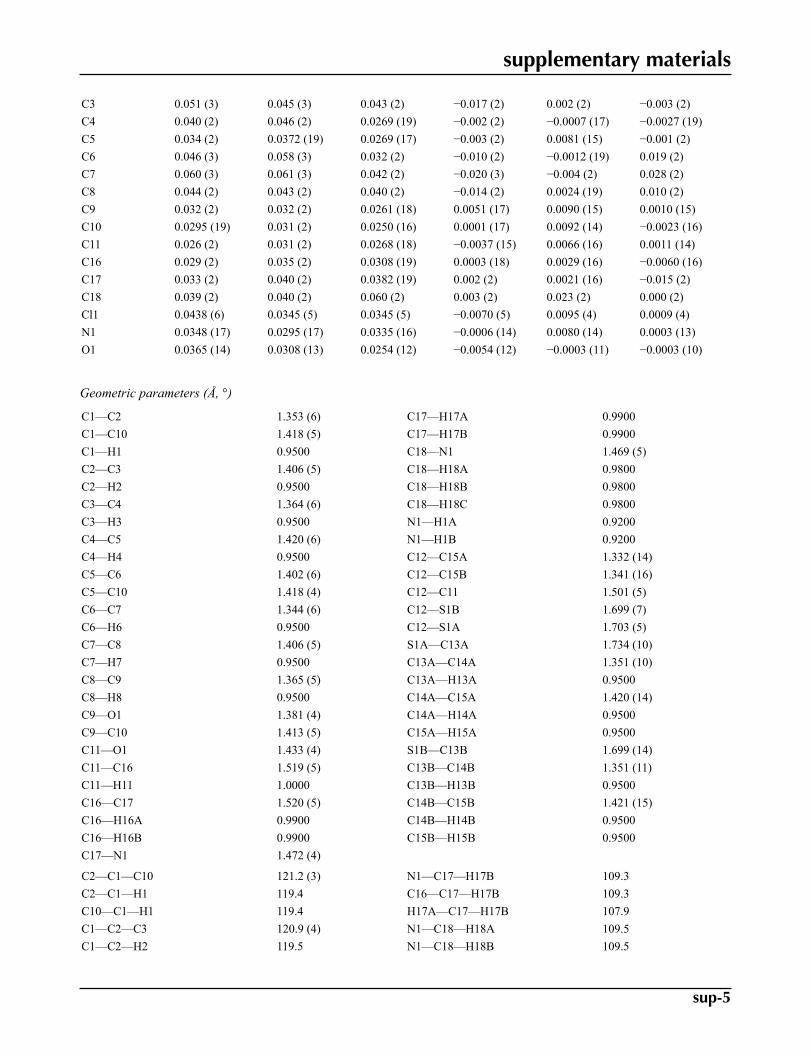
The crystal packing (Fig. 2) shows that both the H-atoms attached to the N atom of the side chain make strong almostlinear H-bonding contacts with the chloride ion.
Experimental
Microcrystalline powder of (1) was supplied by Arrow Laboratories Ltd.,Croydon, Australia. Recrystallization of thispowder by slow evaporation was attempted in acetonitrile, 1,4-dioxane,chlorobenzene and 2-propanol. Suitable single crys-tals in the form of thin plates were grown from the first three solvents, crystals from chlorobenzene were very thin silkyfibres unsuitable for single-crystal analysis. Crystals from the first three solvents yielded the same monoclinic P2(1) formhaving unit-cell parameters as given in Table 1. Amongst these, better quality crystals were obtained from 1,4-dioxane,which were used for further structural analysis.
Refinement
The twofold disorder of the thiophene ring noted first in the E-map at the structure solution stage (two strong peaks andtwo long bonds instead of one), was confirmed subsequently in the full-matrix least-squares refinement. The moleculargeometry for this ring was refined with restrained bond and angles. H atoms were idealized at their expected positions andallowed to ride both in coordinates (C—H = 0.96–0.99, N–H = 0.92 Å), as well as in their isotropic displacement factors(Uĩso~(H) = 1.2/1.5× U~equiv~(host).

supplementary materials
sup-2
Figures
Fig. 1. Molecular view of 1. Displacement ellipsoids drawn at 50% level.
Fig. 2. Packing view of1 showing N—H···Cl interactions.
N-methyl-3-(1-naphthyloxy)-3-(2-thienyl)propan-1-aminium chloride
Crystal data
C18H20NOS+·Cl– F000 = 352
Mr = 333.86 Dx = 1.298 Mg m−3
Monoclinic, P21 Mo Kα radiation, λ = 0.71073 ÅHall symbol: P 2yb Cell parameters from 1635 reflectionsa = 9.7453 (10) Å θ = 2.3–22.1ºb = 6.9227 (7) Å µ = 0.35 mm−1
c = 13.4247 (16) Å T = 150 Kβ = 109.432 (4)º Thin Plates, colourless
V = 854.09 (16) Å3 0.38 × 0.08 × 0.03 mmZ = 2
Data collection
Bruker Kappa APEXII CCD area-detectordiffractometer 2947 independent reflections
Radiation source: fine-focus sealed tube 2255 reflections with I > 2σ(I)Monochromator: graphite Rint = 0.058
T = 150 K θmax = 25.0º
φ scans, and ω scans with κ offsets θmin = 1.6ºAbsorption correction: Multi-scan(SADABS; Sheldrick, 2003) h = −10→11
Tmin = 0.879, Tmax = 0.990 k = −8→86386 measured reflections l = −15→15

supplementary materials
sup-3
Refinement
Refinement on F2 Hydrogen site location: inferred from neighbouringsites
Least-squares matrix: full H-atom parameters constrained
R[F2 > 2σ(F2)] = 0.044 w = 1/[σ2(Fo
2) + (0.1P)2 + 0.0292P]where P = (Fo
2 + 2Fc2)/3
wR(F2) = 0.131 (Δ/σ)max = 0.005
S = 0.79 Δρmax = 0.17 e Å−3
2947 reflections Δρmin = −0.21 e Å−3
237 parameters Extinction correction: none110 restraints Absolute structure: Flack (1983), 1309 Friedel pairsPrimary atom site location: structure-invariant directmethods Flack parameter: −0.05 (10)
Secondary atom site location: difference Fourier map
Special details
Geometry. All e.s.d.'s (except the e.s.d. in the dihedral angle between two l.s. planes) are estimated using the full covariance mat-rix. The cell e.s.d.'s are taken into account individually in the estimation of e.s.d.'s in distances, angles and torsion angles; correlationsbetween e.s.d.'s in cell parameters are only used when they are defined by crystal symmetry. An approximate (isotropic) treatment ofcell e.s.d.'s is used for estimating e.s.d.'s involving l.s. planes.
Refinement. Refinement of F2 against ALL reflections. The weighted R-factor wR and goodness of fit S are based on F2, convention-
al R-factors R are based on F, with F set to zero for negative F2. The threshold expression of F2 > σ(F2) is used only for calculating R-
factors(gt) etc. and is not relevant to the choice of reflections for refinement. R-factors based on F2 are statistically about twice as largeas those based on F, and R- factors based on ALL data will be even larger.
Fractional atomic coordinates and isotropic or equivalent isotropic displacement parameters (Å2)
x y z Uiso*/Ueq Occ. (<1)C1 0.7076 (5) 1.0388 (6) 0.3732 (3) 0.0414 (10)H1 0.6473 1.0546 0.3018 0.050*C2 0.8113 (5) 1.1721 (7) 0.4181 (3) 0.0559 (13)H2 0.8222 1.2804 0.3779 0.067*C3 0.9031 (5) 1.1525 (7) 0.5234 (3) 0.0498 (12)H3 0.9769 1.2456 0.5538 0.060*C4 0.8856 (4) 0.9990 (6) 0.5818 (3) 0.0404 (10)H4 0.9465 0.9877 0.6534 0.048*C5 0.7784 (4) 0.8557 (6) 0.5376 (2) 0.0333 (8)C6 0.7619 (5) 0.6944 (7) 0.5957 (3) 0.0488 (12)H6 0.8217 0.6804 0.6674 0.059*C7 0.6613 (5) 0.5596 (7) 0.5500 (3) 0.0594 (14)H7 0.6518 0.4508 0.5904 0.071*C8 0.5696 (5) 0.5748 (7) 0.4444 (3) 0.0453 (11)H8 0.5007 0.4764 0.4134 0.054*C9 0.5811 (4) 0.7331 (5) 0.3870 (3) 0.0302 (9)

supplementary materials
sup-4
C10 0.6874 (4) 0.8761 (5) 0.4309 (3) 0.0285 (8)C11 0.3962 (4) 0.6244 (5) 0.2248 (3) 0.0287 (8)H11 0.3506 0.5543 0.2709 0.034*C16 0.2800 (4) 0.7340 (5) 0.1395 (3) 0.0334 (9)H16A 0.2233 0.6416 0.0851 0.040*H16B 0.3277 0.8263 0.1051 0.040*C17 0.1769 (4) 0.8439 (7) 0.1826 (3) 0.0395 (9)H17A 0.1988 0.8111 0.2581 0.047*H17B 0.1925 0.9844 0.1776 0.047*C18 −0.0810 (4) 0.8802 (6) 0.1698 (3) 0.0449 (10)H18A −0.0634 0.8265 0.2405 0.067*H18B −0.1802 0.8483 0.1246 0.067*H18C −0.0694 1.0209 0.1746 0.067*Cl1 0.02776 (10) 0.35210 (15) 0.11567 (7) 0.0385 (3)N1 0.0237 (3) 0.7976 (4) 0.1242 (2) 0.0334 (7)H1A 0.0015 0.8416 0.0560 0.040*H1B 0.0131 0.6655 0.1214 0.040*O1 0.4920 (3) 0.7711 (3) 0.28494 (18) 0.0335 (6)C12 0.4783 (4) 0.4840 (6) 0.1806 (3) 0.0304 (9)S1A 0.4000 (4) 0.2836 (6) 0.1112 (4) 0.0390 (8) 0.580 (5)C13A 0.5587 (15) 0.220 (3) 0.088 (2) 0.043 (2) 0.580 (5)H13A 0.5703 0.1075 0.0508 0.051* 0.58C14A 0.6634 (16) 0.353 (2) 0.1292 (18) 0.045 (2) 0.580 (5)H14A 0.7575 0.3491 0.1227 0.054* 0.58C15A 0.6158 (17) 0.500 (3) 0.184 (2) 0.042 (4) 0.580 (5)H15A 0.6774 0.6027 0.2199 0.051* 0.58S1B 0.6515 (6) 0.5208 (10) 0.1826 (7) 0.0410 (14) 0.420 (5)C13B 0.656 (2) 0.310 (3) 0.119 (2) 0.042 (3) 0.420 (5)H13B 0.7390 0.2624 0.1043 0.051* 0.42C14B 0.526 (2) 0.221 (4) 0.091 (3) 0.045 (3) 0.420 (5)H14B 0.5030 0.1062 0.0498 0.054* 0.42C15B 0.427 (2) 0.318 (3) 0.131 (2) 0.053 (5) 0.420 (5)H15B 0.3325 0.2691 0.1237 0.063* 0.42
Atomic displacement parameters (Å2)
U11 U22 U33 U12 U13 U23
C12 0.030 (2) 0.034 (2) 0.0272 (18) −0.0013 (18) 0.0086 (16) 0.0036 (16)S1A 0.0360 (14) 0.0354 (13) 0.0453 (18) 0.0021 (11) 0.0130 (14) −0.0108 (10)C13A 0.046 (6) 0.048 (5) 0.038 (5) 0.018 (5) 0.019 (6) −0.002 (4)C14A 0.039 (4) 0.060 (7) 0.042 (5) 0.009 (4) 0.021 (4) −0.001 (5)C15A 0.044 (9) 0.049 (6) 0.042 (5) −0.005 (6) 0.025 (6) −0.001 (4)S1B 0.024 (2) 0.056 (2) 0.042 (2) −0.0013 (18) 0.0099 (19) 0.0093 (17)C13B 0.042 (5) 0.051 (6) 0.037 (7) 0.009 (5) 0.017 (5) 0.015 (5)C14B 0.045 (7) 0.050 (6) 0.036 (6) 0.005 (6) 0.010 (6) 0.003 (5)C15B 0.043 (7) 0.062 (12) 0.055 (11) 0.005 (7) 0.018 (8) 0.003 (8)C1 0.047 (2) 0.038 (2) 0.0303 (19) −0.007 (2) 0.0003 (18) 0.0082 (18)C2 0.062 (3) 0.048 (3) 0.043 (2) −0.020 (3) −0.002 (2) 0.010 (2)
supplementary materials
sup-5
C3 0.051 (3) 0.045 (3) 0.043 (2) −0.017 (2) 0.002 (2) −0.003 (2)C4 0.040 (2) 0.046 (2) 0.0269 (19) −0.002 (2) −0.0007 (17) −0.0027 (19)C5 0.034 (2) 0.0372 (19) 0.0269 (17) −0.003 (2) 0.0081 (15) −0.001 (2)C6 0.046 (3) 0.058 (3) 0.032 (2) −0.010 (2) −0.0012 (19) 0.019 (2)C7 0.060 (3) 0.061 (3) 0.042 (2) −0.020 (3) −0.004 (2) 0.028 (2)C8 0.044 (2) 0.043 (2) 0.040 (2) −0.014 (2) 0.0024 (19) 0.010 (2)C9 0.032 (2) 0.032 (2) 0.0261 (18) 0.0051 (17) 0.0090 (15) 0.0010 (15)C10 0.0295 (19) 0.031 (2) 0.0250 (16) 0.0001 (17) 0.0092 (14) −0.0023 (16)C11 0.026 (2) 0.031 (2) 0.0268 (18) −0.0037 (15) 0.0066 (16) 0.0011 (14)C16 0.029 (2) 0.035 (2) 0.0308 (19) 0.0003 (18) 0.0029 (16) −0.0060 (16)C17 0.033 (2) 0.040 (2) 0.0382 (19) 0.002 (2) 0.0021 (16) −0.015 (2)C18 0.039 (2) 0.040 (2) 0.060 (2) 0.003 (2) 0.023 (2) 0.000 (2)Cl1 0.0438 (6) 0.0345 (5) 0.0345 (5) −0.0070 (5) 0.0095 (4) 0.0009 (4)N1 0.0348 (17) 0.0295 (17) 0.0335 (16) −0.0006 (14) 0.0080 (14) 0.0003 (13)O1 0.0365 (14) 0.0308 (13) 0.0254 (12) −0.0054 (12) −0.0003 (11) −0.0003 (10)
Geometric parameters (Å, °)
C1—C2 1.353 (6) C17—H17A 0.9900C1—C10 1.418 (5) C17—H17B 0.9900C1—H1 0.9500 C18—N1 1.469 (5)C2—C3 1.406 (5) C18—H18A 0.9800C2—H2 0.9500 C18—H18B 0.9800C3—C4 1.364 (6) C18—H18C 0.9800C3—H3 0.9500 N1—H1A 0.9200C4—C5 1.420 (6) N1—H1B 0.9200C4—H4 0.9500 C12—C15A 1.332 (14)C5—C6 1.402 (6) C12—C15B 1.341 (16)C5—C10 1.418 (4) C12—C11 1.501 (5)C6—C7 1.344 (6) C12—S1B 1.699 (7)C6—H6 0.9500 C12—S1A 1.703 (5)C7—C8 1.406 (5) S1A—C13A 1.734 (10)C7—H7 0.9500 C13A—C14A 1.351 (10)C8—C9 1.365 (5) C13A—H13A 0.9500C8—H8 0.9500 C14A—C15A 1.420 (14)C9—O1 1.381 (4) C14A—H14A 0.9500C9—C10 1.413 (5) C15A—H15A 0.9500C11—O1 1.433 (4) S1B—C13B 1.699 (14)C11—C16 1.519 (5) C13B—C14B 1.351 (11)C11—H11 1.0000 C13B—H13B 0.9500C16—C17 1.520 (5) C14B—C15B 1.421 (15)C16—H16A 0.9900 C14B—H14B 0.9500C16—H16B 0.9900 C15B—H15B 0.9500C17—N1 1.472 (4)
C2—C1—C10 121.2 (3) N1—C17—H17B 109.3C2—C1—H1 119.4 C16—C17—H17B 109.3C10—C1—H1 119.4 H17A—C17—H17B 107.9C1—C2—C3 120.9 (4) N1—C18—H18A 109.5C1—C2—H2 119.5 N1—C18—H18B 109.5

supplementary materials
sup-6
C3—C2—H2 119.5 H18A—C18—H18B 109.5C4—C3—C2 119.5 (4) N1—C18—H18C 109.5C4—C3—H3 120.2 H18A—C18—H18C 109.5C2—C3—H3 120.2 H18B—C18—H18C 109.5C3—C4—C5 121.3 (3) C18—N1—C17 114.6 (3)C3—C4—H4 119.3 C18—N1—H1A 108.6C5—C4—H4 119.3 C17—N1—H1A 108.6C6—C5—C10 119.5 (4) C18—N1—H1B 108.6C6—C5—C4 121.9 (3) C17—N1—H1B 108.6C10—C5—C4 118.6 (3) H1A—N1—H1B 107.6C7—C6—C5 120.0 (4) C9—O1—C11 120.0 (3)C7—C6—H6 120.0 C15A—C12—C15B 107.3 (10)C5—C6—H6 120.0 C15A—C12—C11 126.5 (7)C6—C7—C8 122.0 (4) C15B—C12—C11 126.2 (8)C6—C7—H7 119.0 C15B—C12—S1B 110.1 (8)C8—C7—H7 119.0 C11—C12—S1B 123.7 (3)C9—C8—C7 119.0 (4) C15A—C12—S1A 110.5 (7)C9—C8—H8 120.5 C11—C12—S1A 122.9 (3)C7—C8—H8 120.5 S1B—C12—S1A 113.1 (3)C8—C9—O1 124.8 (3) C12—S1A—C13A 92.4 (4)C8—C9—C10 120.9 (3) C14A—C13A—S1A 110.4 (8)O1—C9—C10 114.4 (3) C14A—C13A—H13A 124.8C9—C10—C1 123.0 (3) S1A—C13A—H13A 124.8C9—C10—C5 118.5 (3) C13A—C14A—C15A 111.8 (10)C1—C10—C5 118.5 (3) C13A—C14A—H14A 124.1O1—C11—C12 110.4 (3) C15A—C14A—H14A 124.1O1—C11—C16 104.7 (3) C12—C15A—C14A 114.8 (11)C12—C11—C16 112.8 (3) C12—C15A—H15A 122.6O1—C11—H11 109.6 C14A—C15A—H15A 122.6C12—C11—H11 109.6 C13B—S1B—C12 93.3 (7)C16—C11—H11 109.6 C14B—C13B—S1B 110.6 (11)C11—C16—C17 112.6 (3) C14B—C13B—H13B 124.7C11—C16—H16A 109.1 S1B—C13B—H13B 124.7C17—C16—H16A 109.1 C13B—C14B—C15B 112.1 (12)C11—C16—H16B 109.1 C13B—C14B—H14B 124.0C17—C16—H16B 109.1 C15B—C14B—H14B 124.0H16A—C16—H16B 107.8 C12—C15B—C14B 113.7 (12)N1—C17—C16 111.7 (3) C12—C15B—H15B 123.2N1—C17—H17A 109.3 C14B—C15B—H15B 123.2C16—C17—H17A 109.3
Hydrogen-bond geometry (Å, °)
D—H···A D—H H···A D···A D—H···A
N1—H1A···Cl1i 0.92 2.23 3.113 (3) 161N1—H1B···Cl1 0.92 2.18 3.087 (3) 170Symmetry codes: (i) −x, y+1/2, −z.

supplementary materials
sup-7
Fig. 1

supplementary materials
sup-8
Fig. 2
Related Documents