Duke University School of Medicine Immunology Quality Assessment Center Tania Garrelts / Raul Louzao ACTG Meeting June 23,2009

Duke University School of Medicine Immunology Quality Assessment Center Tania Garrelts / Raul Louzao ACTG Meeting June 23,2009.
Dec 25, 2015
Welcome message from author
This document is posted to help you gain knowledge. Please leave a comment to let me know what you think about it! Share it to your friends and learn new things together.
Transcript
Duke University School of Medicine
Immunology Quality Assessment Center
Tania Garrelts / Raul Louzao
ACTG Meeting
June 23,2009
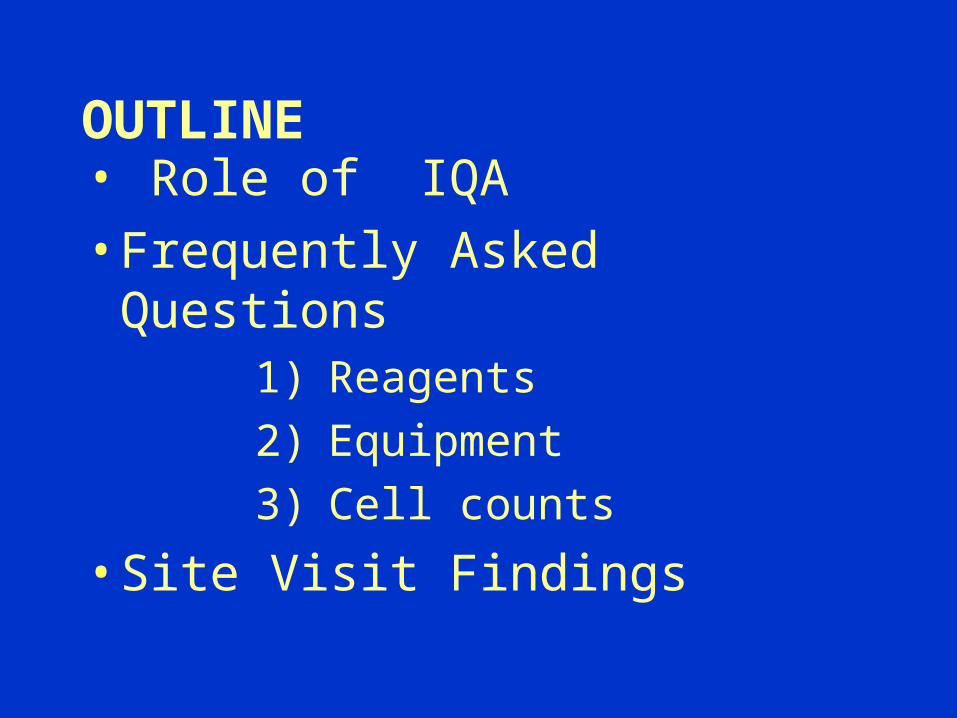
OUTLINE
• Role of IQA
• Frequently Asked Questions1) Reagents
2) Equipment
3) Cell counts
• Site Visit Findings
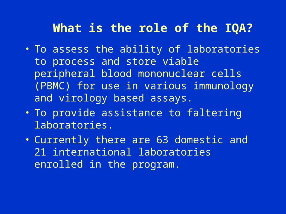
What is the role of the IQA?
• To assess the ability of laboratories to process and store viable peripheral blood mononuclear cells (PBMC) for use in various immunology and virology based assays.
• To provide assistance to faltering laboratories.
• Currently there are 63 domestic and 21 international laboratories enrolled in the program.
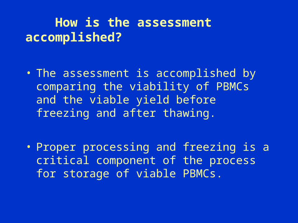
How is the assessment accomplished?
• The assessment is accomplished by comparing the viability of PBMCs and the viable yield before freezing and after thawing.
• Proper processing and freezing is a critical component of the process for storage of viable PBMCs.
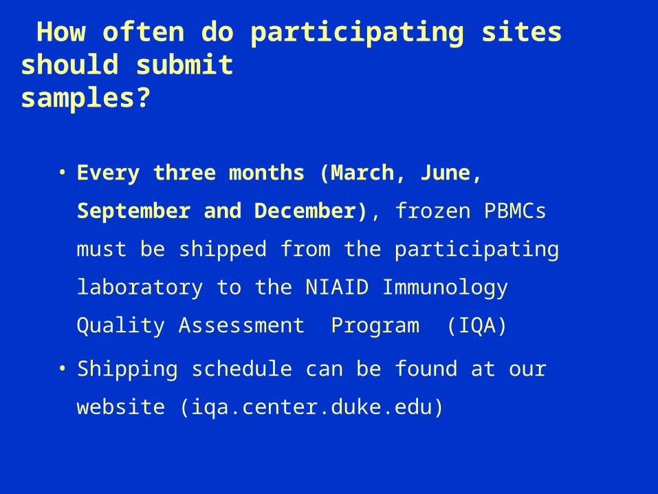
How often do participating sites should submit samples?
• Every three months (March, June, September
and December), frozen PBMCs must be shipped
from the participating laboratory to the NIAID
Immunology Quality Assessment Program (IQA)
• Shipping schedule can be found at our website
(iqa.center.duke.edu)
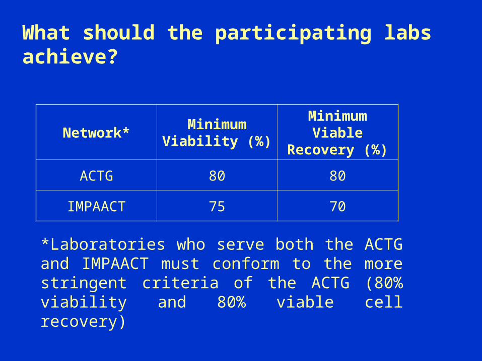
What should the participating labs achieve?
Network*Minimum
Viability (%)Minimum Viable
Recovery (%)
ACTG 80 80
IMPAACT 75 70
*Laboratories who serve both the ACTG and IMPAACT must conform to the more stringent criteria of the ACTG (80% viability and 80% viable cell recovery)
Frequently Asked Questions About
The Cryopreservation Of
Peripheral Blood Mononuclear Cells

Are Ficoll–Hypaque and Histopaque-1077 the same?
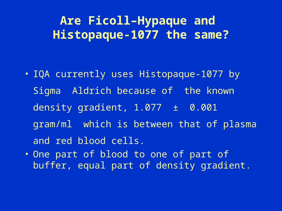
Are Ficoll–Hypaque and Histopaque-1077 the same?
• IQA currently uses Histopaque-1077 by
Sigma Aldrich because of the known density
gradient, 1.077 ± 0.001 gram/ml which is
between that of plasma and red blood cells. • One part of blood to one of part of buffer,
equal part of density gradient.
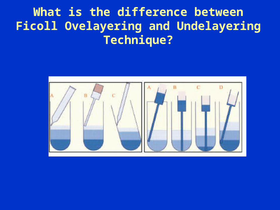
What is the difference between Ficoll Ovelayering and Undelayering
Technique?

Before and After Density Gradient

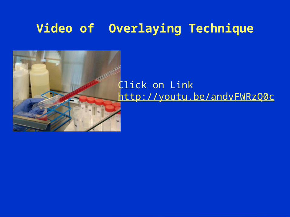
Video of Overlaying Technique
Manual Ficoll using Overlay and Underlay Methods
Click on the link bellow
http://youtu.be/-SqlIMLUfcI

Insufficient amount of density gradient
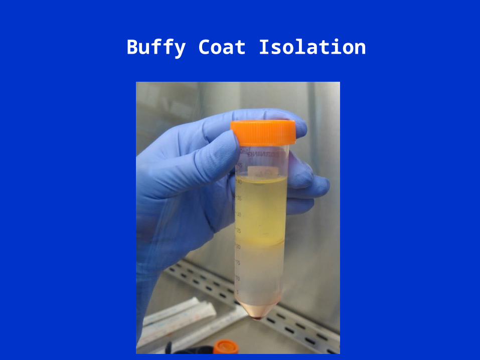
Buffy Coat Isolation

Good versus Poor Mononuclear cell isolation
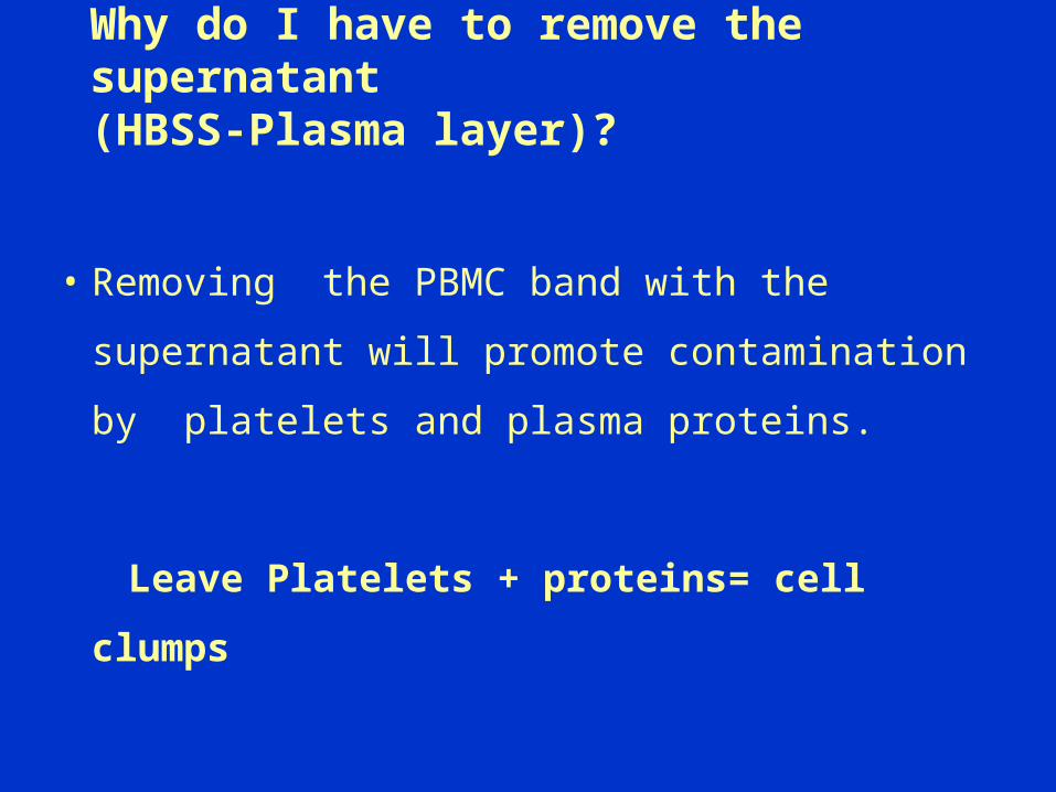
Why do I have to remove the supernatant (HBSS-Plasma layer)?
• Removing the PBMC band with the
supernatant will promote contamination by
platelets and plasma proteins.
Leave Platelets + proteins= cell clumps
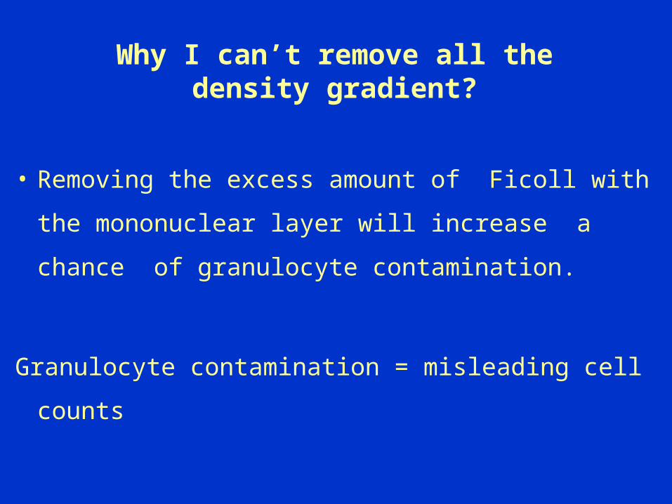
Why I can’t remove all the density gradient?
• Removing the excess amount of Ficoll with the
mononuclear layer will increase a chance of
granulocyte contamination.
Granulocyte contamination = misleading cell counts
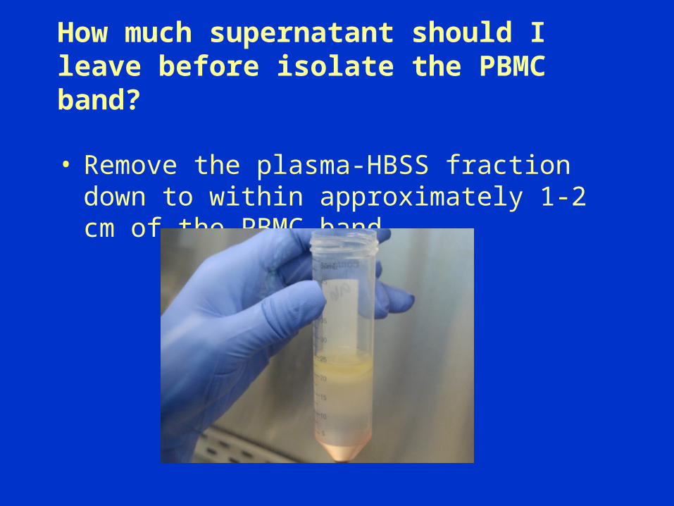
How much supernatant should I leave before isolate the PBMC band?
• Remove the plasma-HBSS fraction down to within approximately 1-2 cm of the PBMC band.

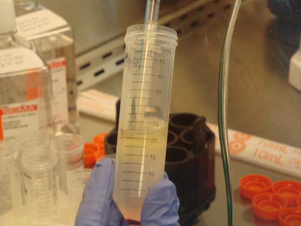
Common used techniques to isolate theMononuclear Layers.
Should I remove the cells stuck to the side of the test tube?
• From previous experience the cells attached
to the inner walls of the tubes are
erythrocytes, cell debri and platelets.
• The density gradient will separate the viable
lymphocytes and monocytes.

What should I do if I accidently mix the layered blood with ficoll?
• Don’t Panic.
• Finish Mixing the blood with ficoll. (New mixture)
• Set up new ficoll tubes and overlay or underlay them with the new mixture.
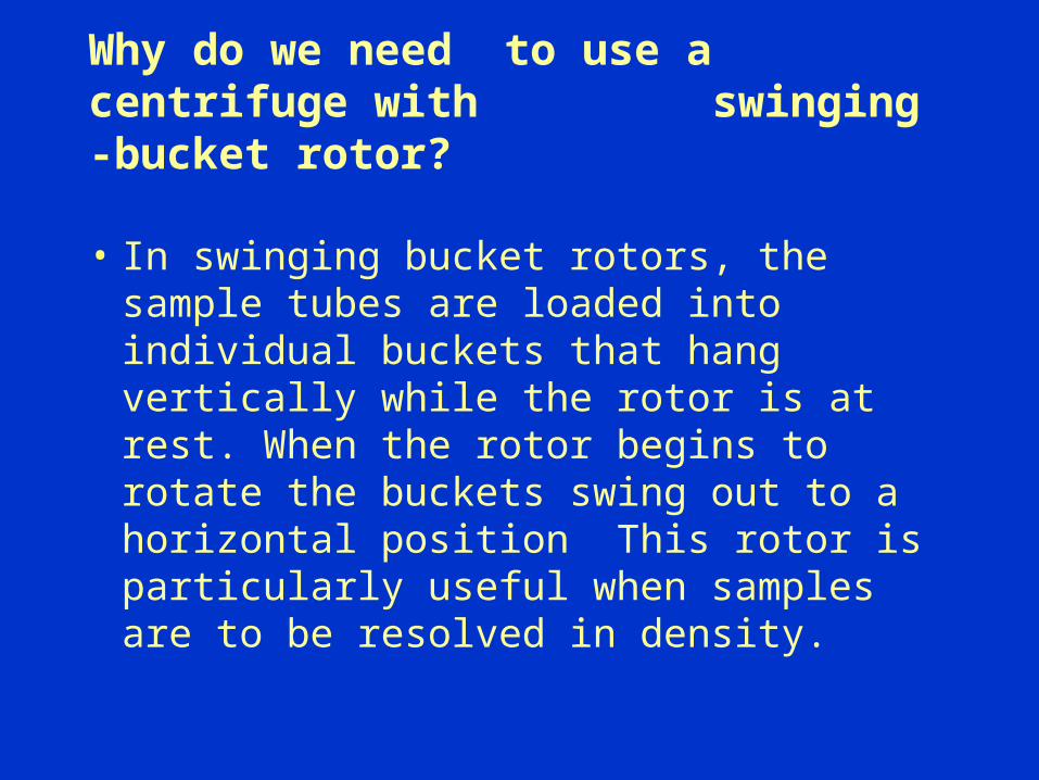
Why do we need to use a centrifuge with swinging -bucket rotor?
• In swinging bucket rotors, the sample tubes are loaded into individual buckets that hang vertically while the rotor is at rest. When the rotor begins to rotate the buckets swing out to a horizontal position This rotor is particularly useful when samples are to be resolved in density.
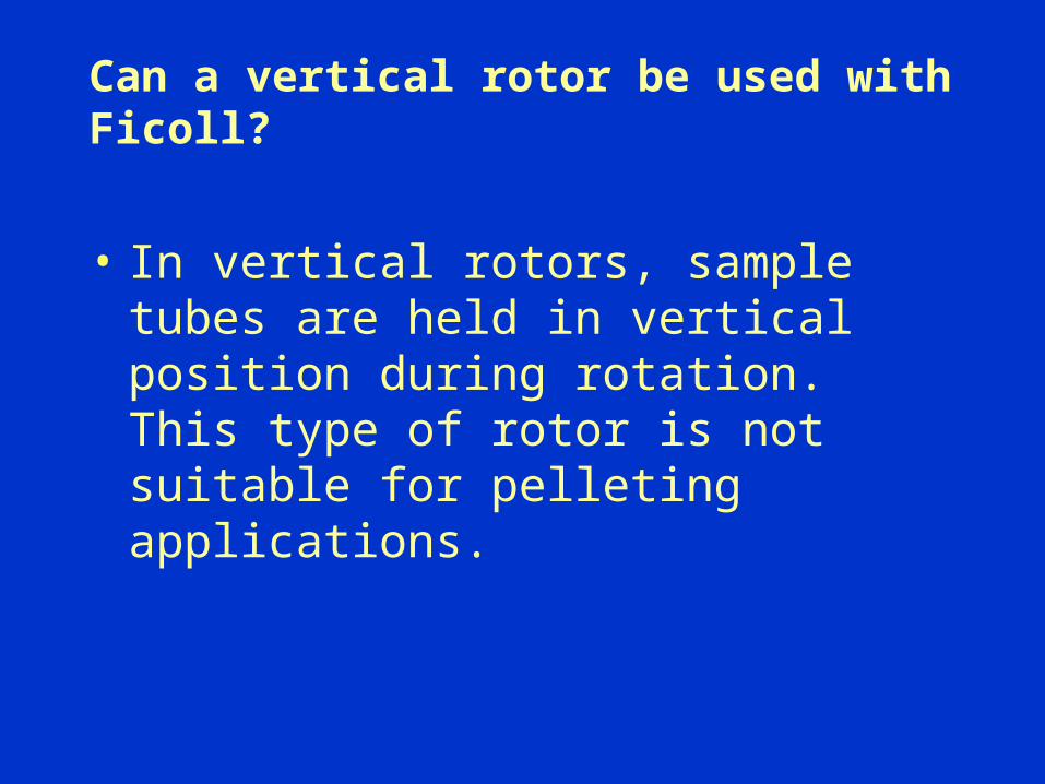
Can a vertical rotor be used with Ficoll?
• In vertical rotors, sample tubes are held in vertical position during rotation. This type of rotor is not suitable for pelleting applications.
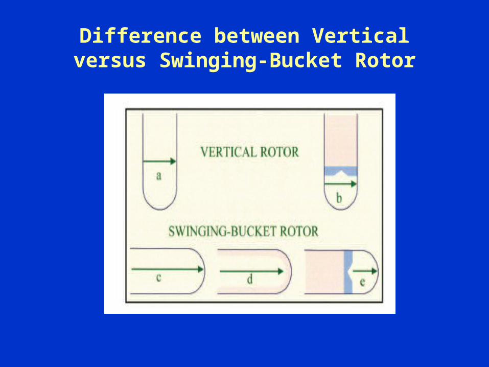
Difference between Vertical versus Swinging-Bucket Rotor

What temperature should I use during centrifugation?
• During separation the centrifuge temperature
should be control between 18-25 °C because the
density components changes with temperature.
For Example: Centrifugation at 4 °C may result in
poor cell recovery due to cell clumping.
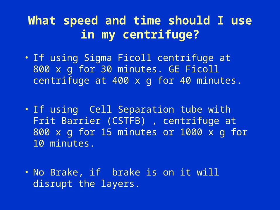
What speed and time should I use in my centrifuge?
• If using Sigma Ficoll centrifuge at 800 x g for 30 minutes. GE Ficoll centrifuge at 400 x g for 40 minutes.
• If using Cell Separation tube with Frit Barrier (CSTFB) , centrifuge at 800 x g for 15 minutes or 1000 x g for 10 minutes.
• No Brake, if brake is on it will disrupt the layers.
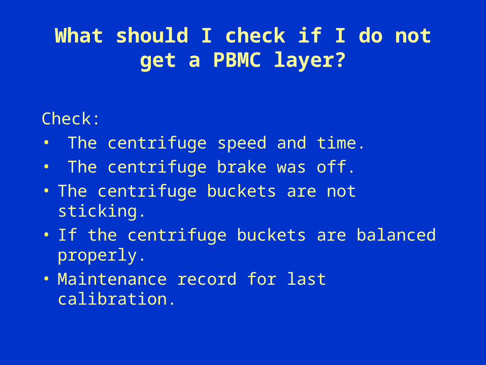
What should I check if I do not get a PBMC layer?
Check:• The centrifuge speed and time. • The centrifuge brake was off.• The centrifuge buckets are not sticking.• If the centrifuge buckets are balanced
properly.• Maintenance record for last calibration.
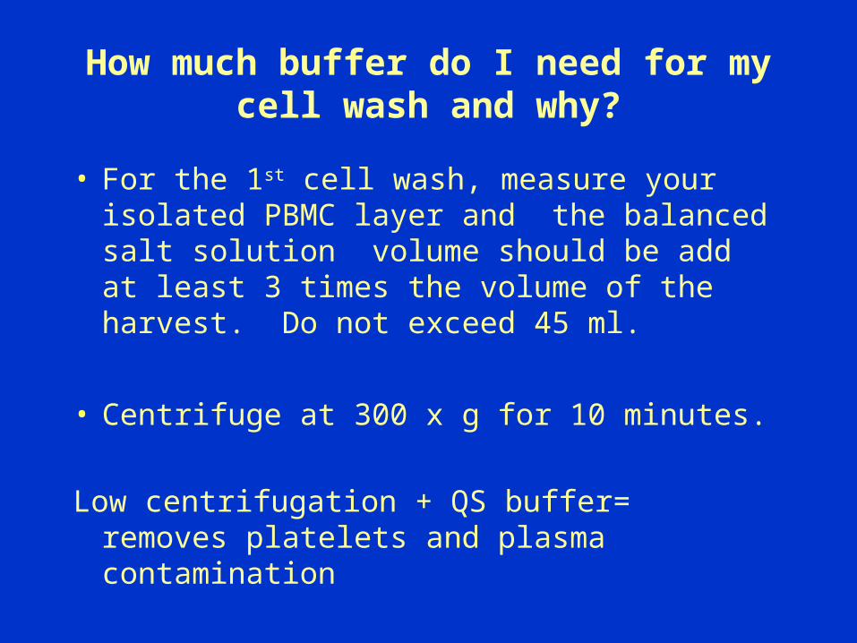
How much buffer do I need for my cell wash and why?
• For the 1st cell wash, measure your isolated PBMC layer and the balanced salt solution volume should be add at least 3 times the volume of the harvest. Do not exceed 45 ml.
• Centrifuge at 300 x g for 10 minutes.
Low centrifugation + QS buffer= removes platelets and plasma contamination
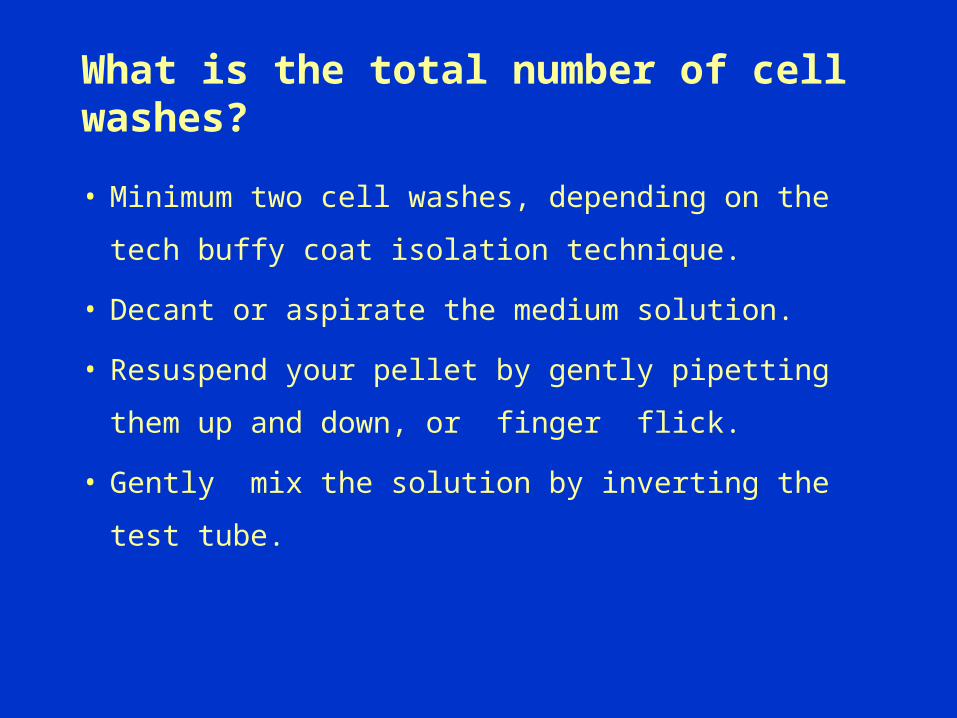
What is the total number of cell washes?
• Minimum two cell washes, depending on the tech
buffy coat isolation technique.
• Decant or aspirate the medium solution.
• Resuspend your pellet by gently pipetting them
up and down, or finger flick.
• Gently mix the solution by inverting the test tube.
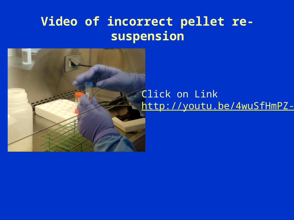
Video of incorrect pellet re-suspension
Click on Linkhttp://youtu.be/4wuSfHmPZ-k
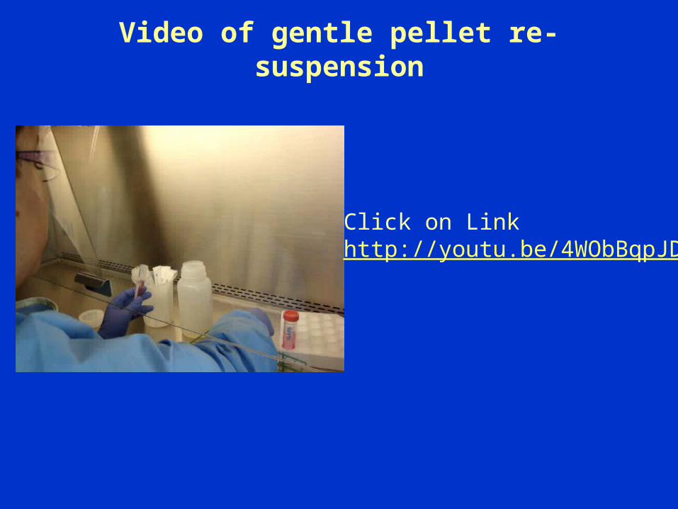
Video of gentle pellet re-suspension
Click on Linkhttp://youtu.be/4WObBqpJD0I

How much buffer is left in my test tube after I decant the supernatant?
• The amount of balanced buffer left over with
the pellet after the HBSS is decanted is
approximately 200 to 400uls.
• You have enough buffer to gently finger flick your
cells.
What volume of buffer should I use to re-suspend my pellets?
• If you have multiple tubes combine the pellets
by adding 1ml of buffer on each tube.
• Depending on the size of the cell pellet, the
re-suspension volume would range from 10%
to 50% of the usable whole blood.• For example:
Starting volume of whole blood is 17 mls. IQA would re-suspend cells in 5 mls ~30%
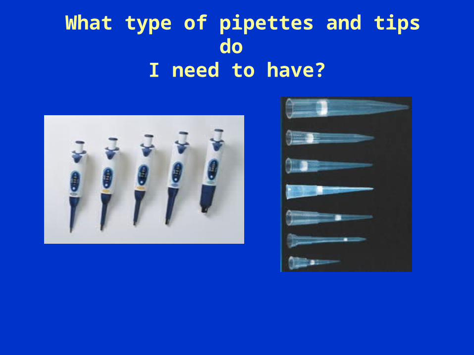
What type of pipettes and tips do I need to have?
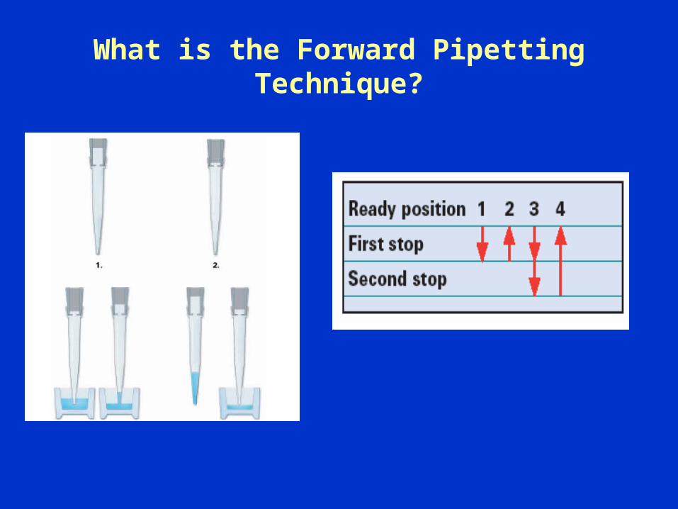
What is the Forward Pipetting Technique?
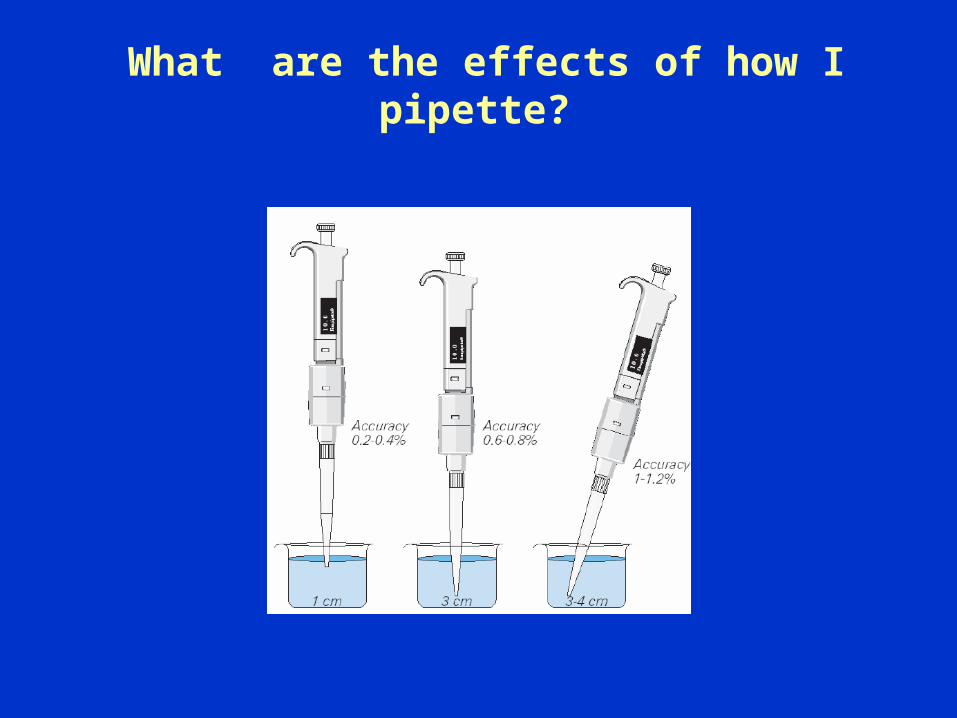
What are the effects of how I pipette?

What are some common pipetting technique errors?
— Human Factor, working too quickly.
— Removing the pipette tip before sample aspiration is
complete.
— Angle pipettes takes up too much liquid. Keep
vertical.
— Releasing the plunger too rapidly.
— Not pre-wetting a new tip.

What type of pipette aid should I use?
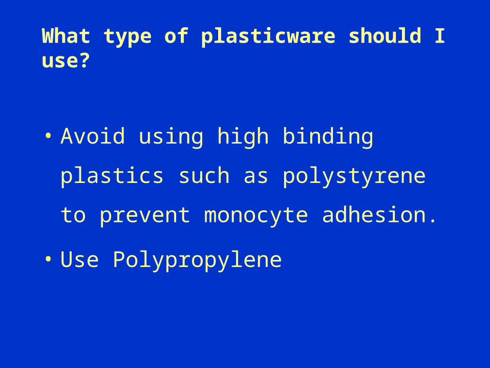
What type of plasticware should I use?
• Avoid using high binding plastics such
as polystyrene to prevent monocyte
adhesion.
• Use Polypropylene
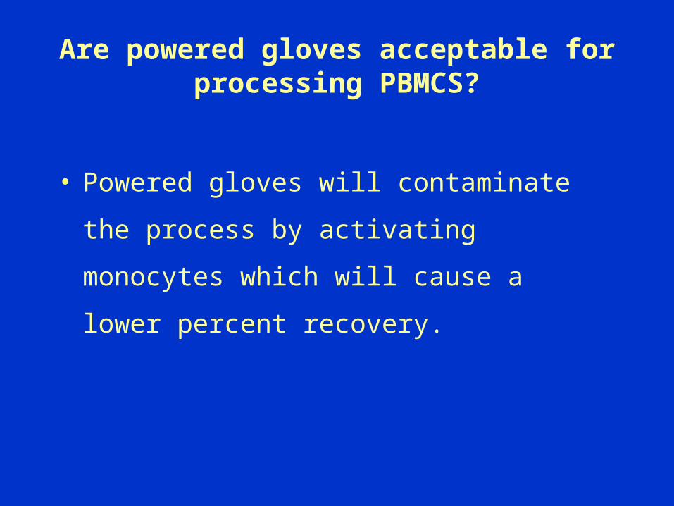
Are powered gloves acceptable for processing PBMCS?
• Powered gloves will contaminate the process
by activating monocytes which will cause a
lower percent recovery.
What is the pH, solution storage requirement, and length of time that cells can stay in
trypan blue?
• pH: 7.0 – 7.4
• Storage requirement: 15-30 °C. Some of vendors recommend that it be protected from light and filter after prolonged storage.
• Cells should be count within 3-10 minutes of mixing with trypan blue, longer incubation will result in cell death and low viability counts.
What are some common problems with a Hemacytometer?
1. Dirty hemacytometer or cover slip, clean with 70% alcohol and lens paper.
2. The chamber may have been loaded incorrectly.
3. The cover slip may have been bumped.
4. Insufficient mixing of sample.
5. Using a wrong cover slip.
Definition for Dilution
• Dilutions are expressed as the ratio of the quantity of a desired solute (serum, urine, chemical solution, etc.) contained in a solvent (diluent).
For example:
A 1:10 dilution of serum was made by adding one part serum to nine parts diluent to make a total of ten parts.
volume of serum/volume of solution = [1.0 mL serum ]/[1.0 ml serum + 9.0 mL H20]
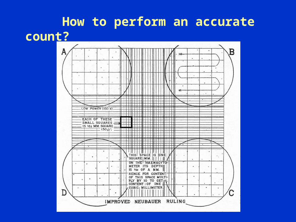
How to perform an accurate count?
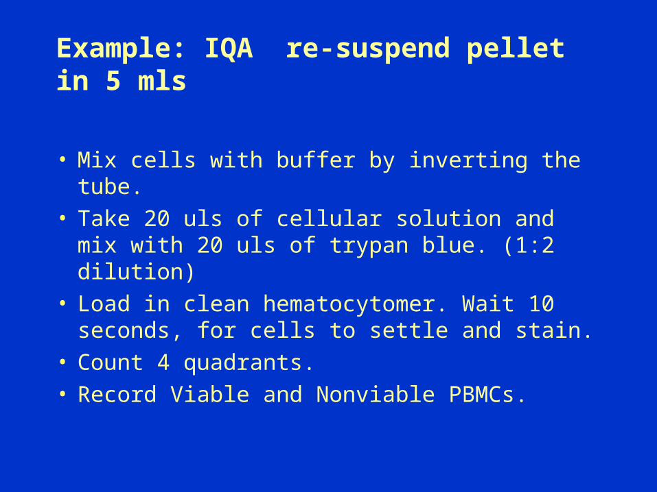
Example: IQA re-suspend pellet in 5 mls
• Mix cells with buffer by inverting the tube.• Take 20 uls of cellular solution and mix with
20 uls of trypan blue. (1:2 dilution)• Load in clean hematocytomer. Wait 10
seconds, for cells to settle and stain.• Count 4 quadrants. • Record Viable and Nonviable PBMCs.

What are the formulas for cell counts?

Example
Live (L) Dead (D) Dilution Factor Final Volume
488 20 1:2 5 ml
Answer: 488/4x2x5x10^4=12.2 x10^6 (total cell concentration)
Cell concentration per ml=488/4x2x10^4=2.44 cells/ml
Percent Viability= L/L+Dx 100 488/488+20=488/508 =0.96x100=96%
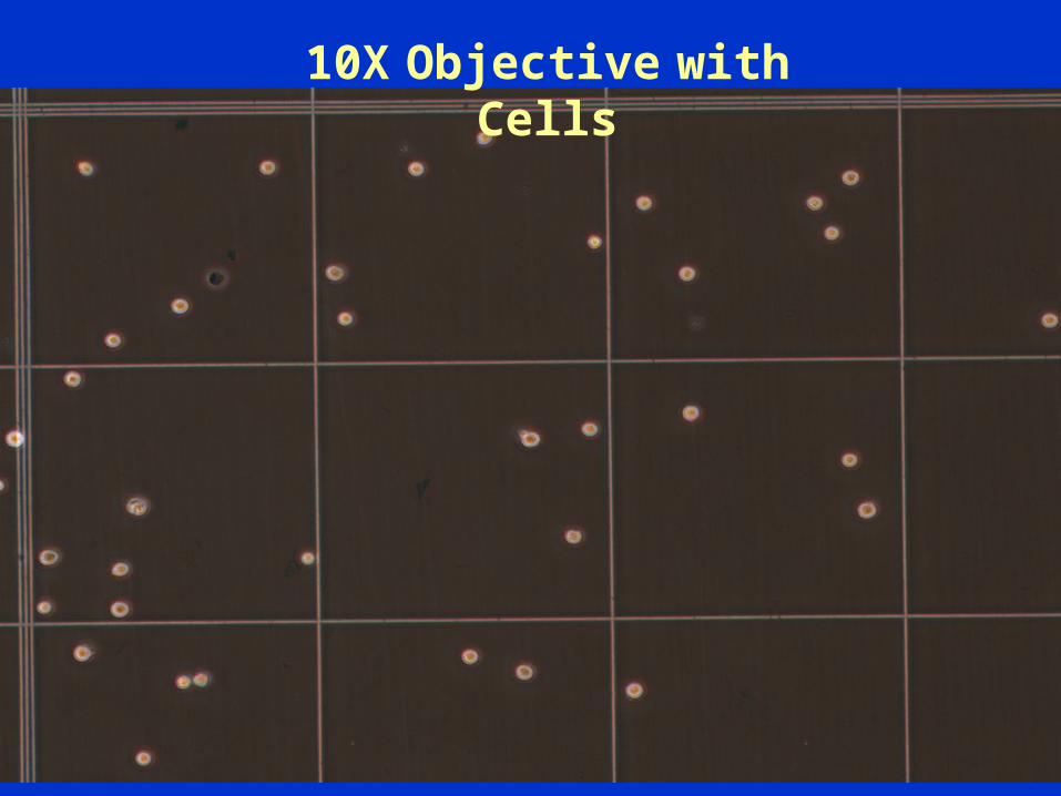
10X Objective with Cells

What is purpose of using DMSO?
Serves as a cryoprotective agent, it helps dehydrate the cells prior to intracellular freezing.
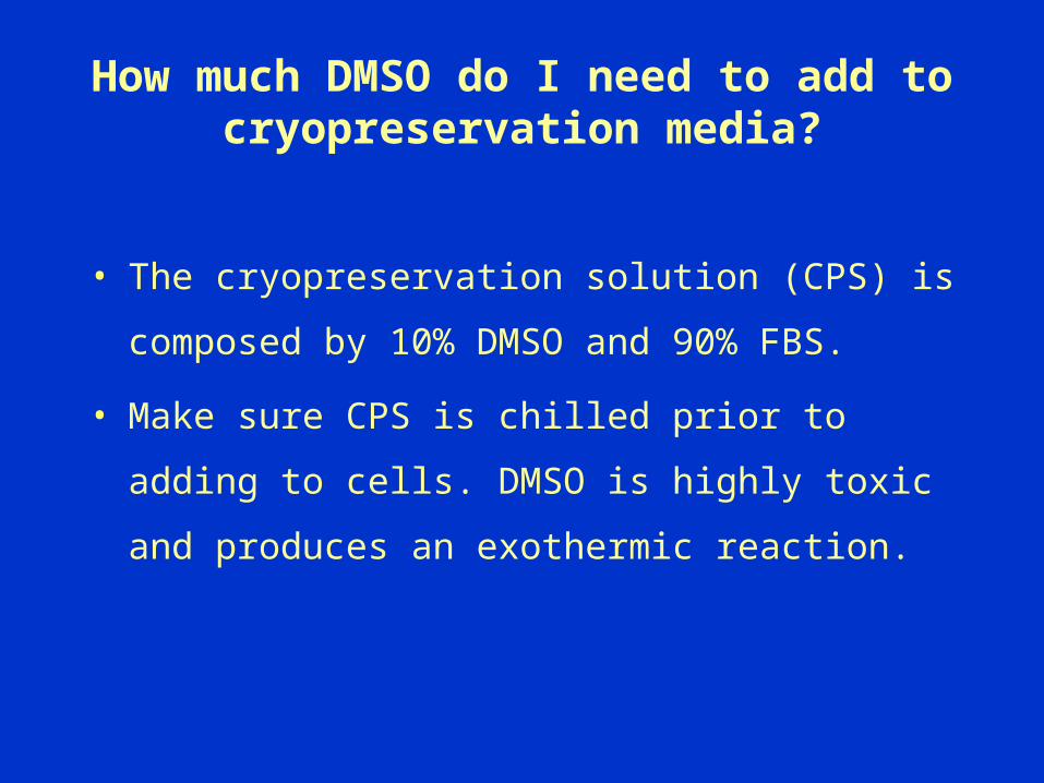
How much DMSO do I need to add to cryopreservation media?
• The cryopreservation solution (CPS) is composed
by 10% DMSO and 90% FBS.
• Make sure CPS is chilled prior to adding to cells.
DMSO is highly toxic and produces an exothermic
reaction.
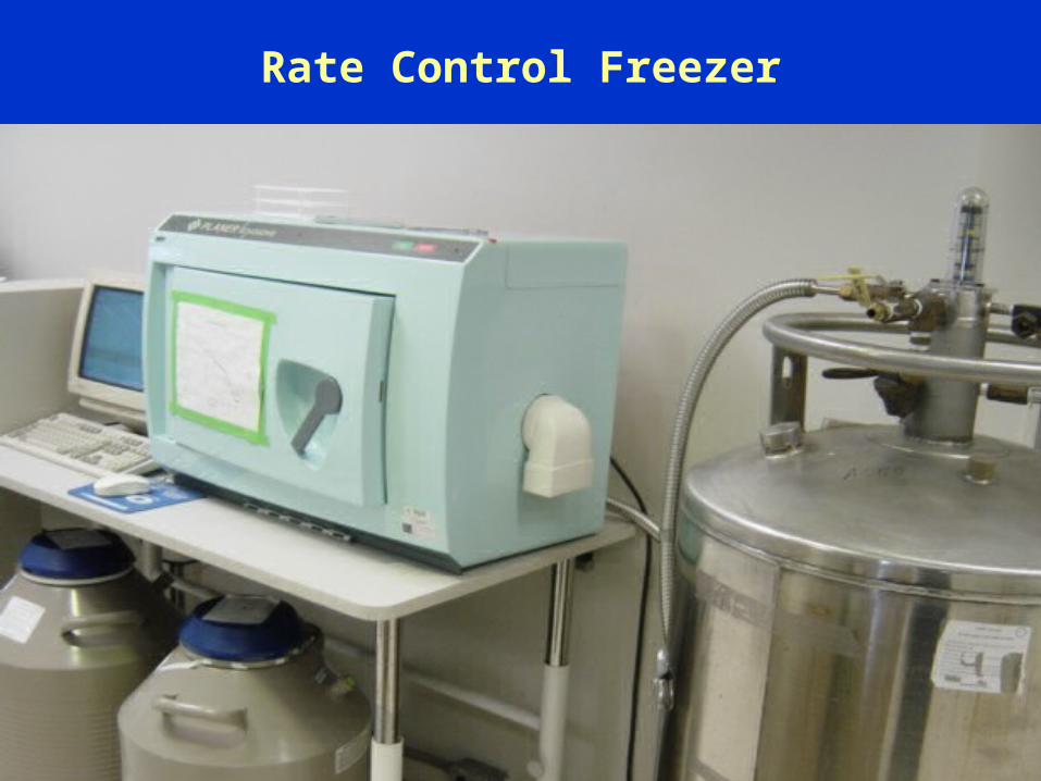
Rate Control Freezer
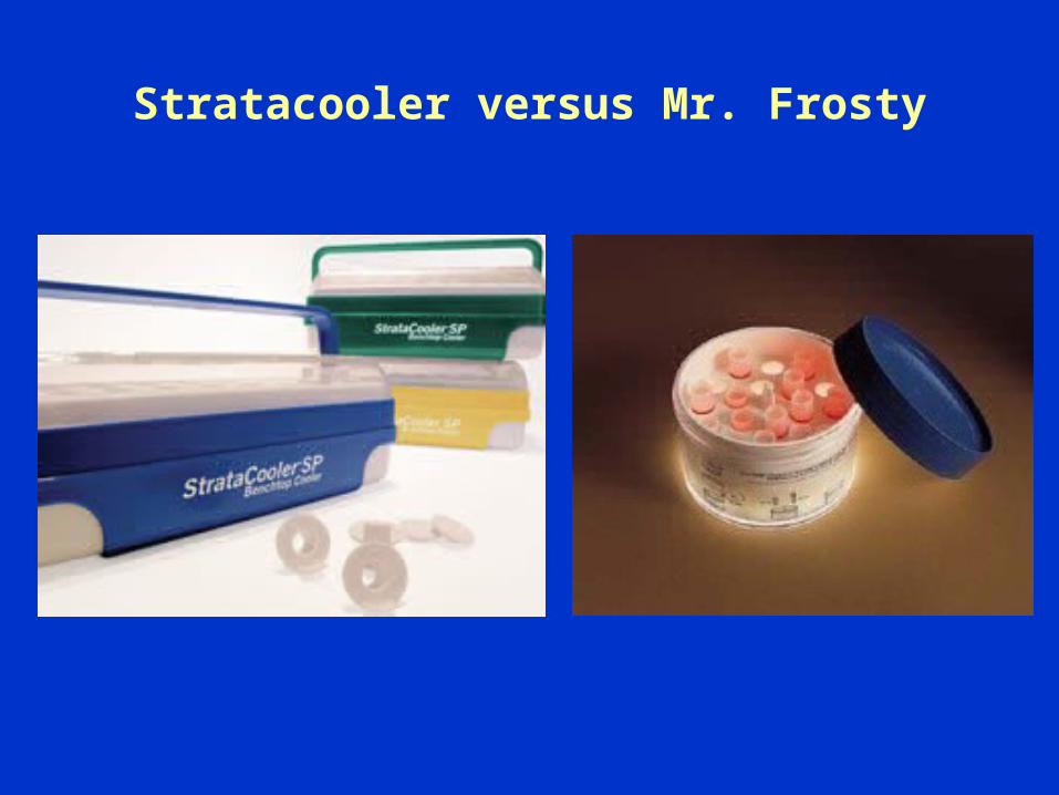
Stratacooler versus Mr. Frosty

Cooling Rate for Mr. Frosty and Rate Control Freezer
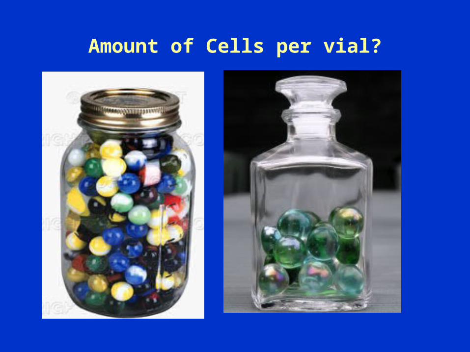
Amount of Cells per vial?
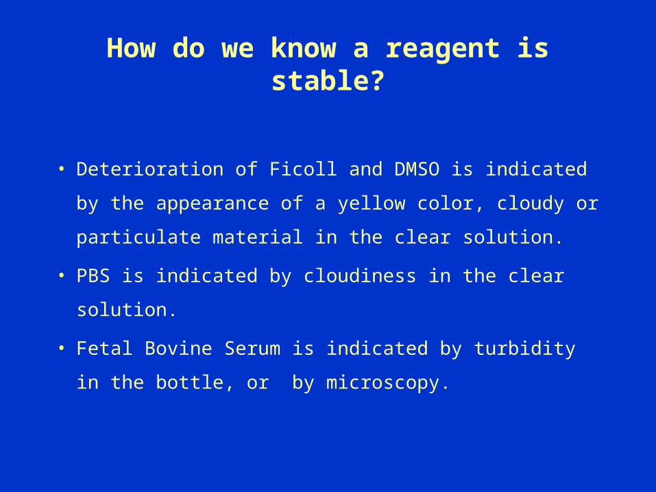
How do we know a reagent is stable?
• Deterioration of Ficoll and DMSO is indicated by
the appearance of a yellow color, cloudy or
particulate material in the clear solution.
• PBS is indicated by cloudiness in the clear solution.
• Fetal Bovine Serum is indicated by turbidity in the
bottle, or by microscopy.
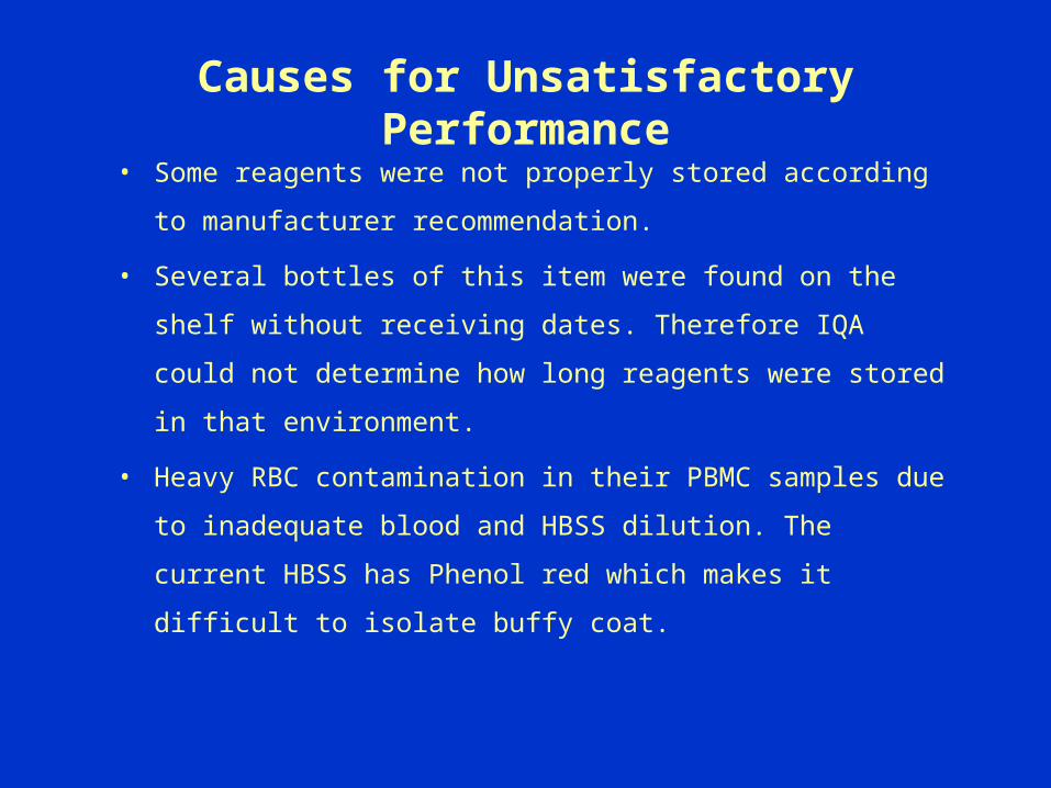
Causes for Unsatisfactory Performance
• Some reagents were not properly stored according to
manufacturer recommendation.
• Several bottles of this item were found on the shelf without
receiving dates. Therefore IQA could not determine how
long reagents were stored in that environment.
• Heavy RBC contamination in their PBMC samples due to
inadequate blood and HBSS dilution. The current HBSS has
Phenol red which makes it difficult to isolate buffy coat.
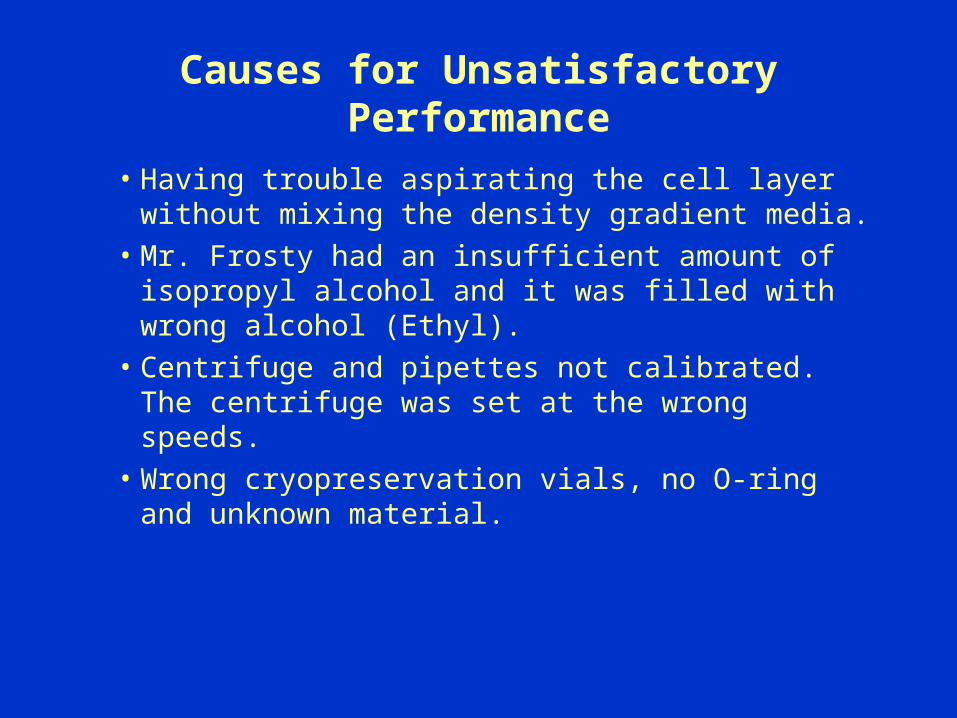
Causes for Unsatisfactory Performance
• Having trouble aspirating the cell layer without mixing the density gradient media.
• Mr. Frosty had an insufficient amount of isopropyl alcohol and it was filled with wrong alcohol (Ethyl).
• Centrifuge and pipettes not calibrated. The centrifuge was set at the wrong speeds.
• Wrong cryopreservation vials, no O-ring and unknown material.
Work Cited
• Cross- Network PBMC Processing Procedure
• Duke University Standard Operating Procedures.
• Cryopreservation Technical Manual by Nalgene,
Nunc
• Reagent Manufacturer inserts.
• Beckman Coulter
• Guide to Pipetting by AccuTek Laboratories
Acknowledgments
IQA Center LaboratoryTom DennyAmbrosia Garcia-LouzaoRaul LouzaoJohn WongEugene UrrutiaTania GarreltsEstelle Berengier
DHVI
Tony Moody
Josh Eudley
Dongning WangKyle LieblBrooke ParkerSherry LeonardSara BrownChristie BrinkleyJamilia Davis
Related Documents