Ductal Carcinoma In-Situ Prashant Gabani, MSIV Talha Shaikh, MD Faculty Advisor: Shelly Hayes, MD Fox Chase Cancer Center Philadelphia, PA

Welcome message from author
This document is posted to help you gain knowledge. Please leave a comment to let me know what you think about it! Share it to your friends and learn new things together.
Transcript
Ductal Carcinoma In-Situ
Prashant Gabani, MSIV Talha Shaikh, MD
Faculty Advisor: Shelly Hayes, MD
Fox Chase Cancer Center Philadelphia, PA

Case Presentation
• 53-year-old female underwent routine bilateral screening mammogram
– Findings: architectural distortion and coarse, clumped calcifications in the retroareolar left breast. Right breast normal.
• PMH – Otherwise healthy
Case Presentation
• OB/Gyn History – G0P0
– Age at menarche: 13
– Menopause at 46
– No history of oral contraceptives
– No history of hormone replacement therapy
• Family History – Mother with ovarian cancer at 79
– No family history of breast cancer
• Social History – Non-smoker
– 2-3 alcoholic drinks/week

Physical Exam • General: Well appearing Caucasian female in no acute distress
• HEENT: PERRLA, EOMI. Sclerae anicteric. No thyromegaly
• Lymphatic: No palpable cervical, supraclavicular, infraclavicular or axillary lymphadenopathy
• CV: Regular rate and rhythm. No murmurs, rubs or gallops
• Lungs: Clear to auscultation. No wheezes, rhonchi or rales
• Abdomen: Soft, non-tender and non-distended
• Breast: Inspection and palpation of the bilateral breasts demonstrates no erythema, edema, peau d’orange, nipple inversion, nipple discharge, or palpable masses. No axillary or supraclavicular lymphadenopathy.
• Extremities: No clubbing, edema or cyanosis
• Neurological Exam: CN II-XII grossly intact. Motor strength 5/5 in the upper and lower extremities. Sensation grossly intact. No focal neurologic deficits.
Workup
• Diagnostic bilateral mammogram
– Magnification views of left breast show clustered pleomorphic calcification in the retroareolar region
• Bilateral breast ultrasound
– 1.9 x 1.8 x 1.8 cm irregular, hypoechoic mass in the left retroareolar region at the 1:00 position

Left Breast Mammogram
Magnification views show clustered pleomorphic calcification in the retroareolar region

Left Breast Ultrasound
1.9 x 1.8 x 1.8 cm irregular, hypoechoic mass in the left retroareolar region at the 1:00 position
Workup
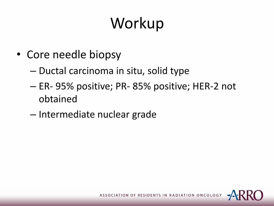
• Core needle biopsy
– Ductal carcinoma in situ, solid type
– ER- 95% positive; PR- 85% positive; HER-2 not obtained
– Intermediate nuclear grade
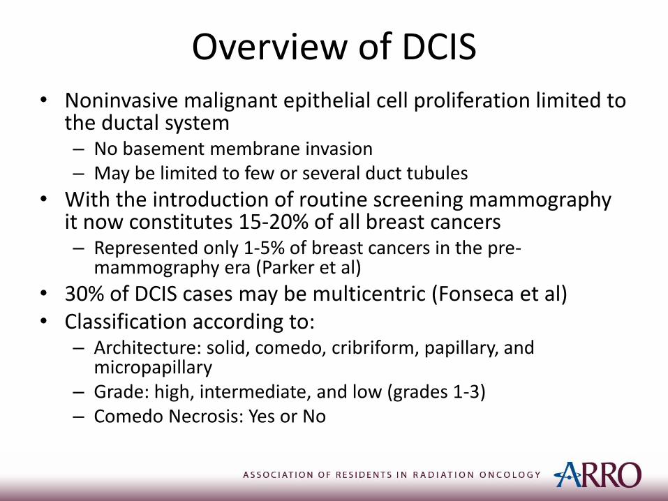
Overview of DCIS • Noninvasive malignant epithelial cell proliferation limited to
the ductal system – No basement membrane invasion – May be limited to few or several duct tubules
• With the introduction of routine screening mammography it now constitutes 15-20% of all breast cancers – Represented only 1-5% of breast cancers in the pre-
mammography era (Parker et al)
• 30% of DCIS cases may be multicentric (Fonseca et al) • Classification according to:
– Architecture: solid, comedo, cribriform, papillary, and micropapillary
– Grade: high, intermediate, and low (grades 1-3) – Comedo Necrosis: Yes or No
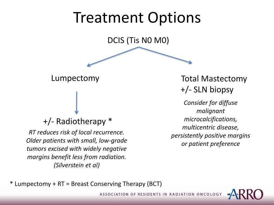
Treatment Options DCIS (Tis N0 M0)
Lumpectomy Total Mastectomy +/- SLN biopsy
Consider for diffuse malignant
microcalcifications, multicentric disease,
persistently positive margins or patient preference
+/- Radiotherapy * RT reduces risk of local recurrence.
Older patients with small, low-grade tumors excised with widely negative margins benefit less from radiation.
(Silverstein et al)
* Lumpectomy + RT = Breast Conserving Therapy (BCT)

Role of Radiotherapy after BCS
• No randomized trials compare BCT to mastectomy for DCIS, but comparisons of BCT to historic mastectomy controls suggest no OS difference
• 4 published randomized trials demonstrate benefit in local control with addition of whole breast RT compared to lumpectomy alone in DCIS:
– NSABP B-17
– EORTC 10853
– UK/Australia/New Zealand cooperative trial (UK/ANZ)
– Swedish Trial
Role of Radiotherapy after BCS
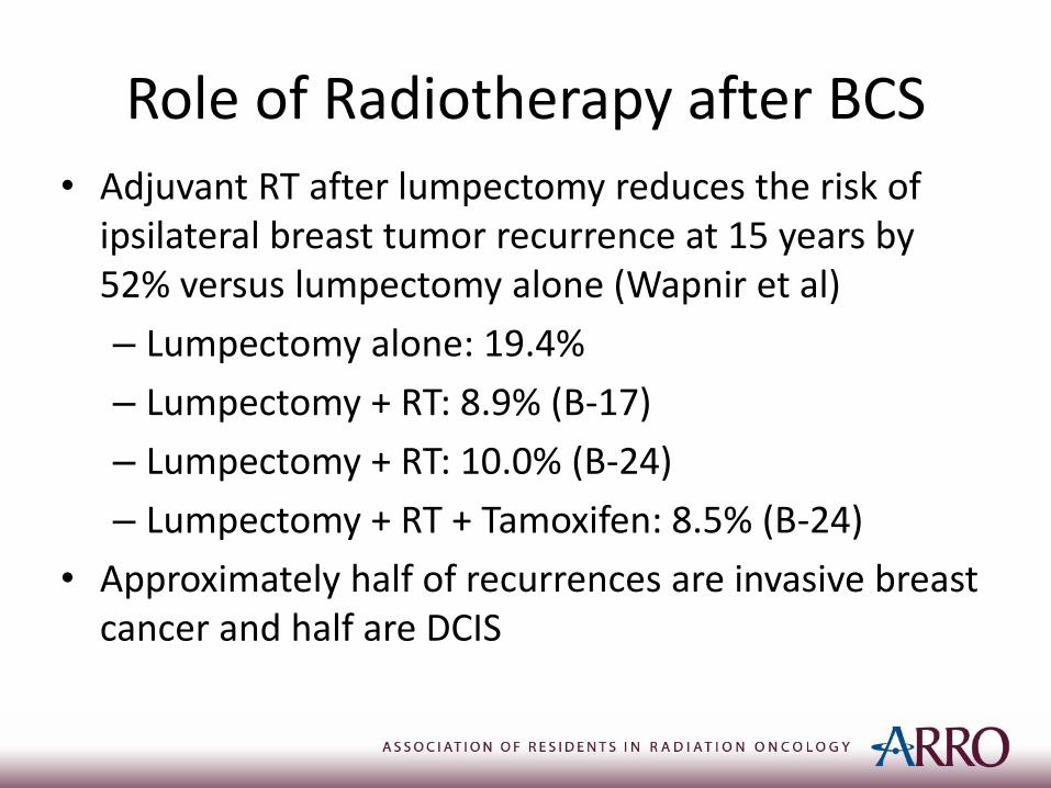
• Adjuvant RT after lumpectomy reduces the risk of ipsilateral breast tumor recurrence at 15 years by 52% versus lumpectomy alone (Wapnir et al)
– Lumpectomy alone: 19.4%
– Lumpectomy + RT: 8.9% (B-17)
– Lumpectomy + RT: 10.0% (B-24)
– Lumpectomy + RT + Tamoxifen: 8.5% (B-24)
• Approximately half of recurrences are invasive breast cancer and half are DCIS
Role of Tamoxifen after Lumpectomy
• NSABP B-24: The addition of tamoxifen to RT reduces overall cancer events at 5 years (Fisher et al. Lancet 1999)
– Decreased breast cancer events from 13.4% to 8.2%
– Ipsilateral 9.5% vs. 6.0%
– Contralateral 3.4 vs. 2.0%
– No difference in regional or distant mets
• In ER positive as opposed to ER negative tumors, the benefit of Tamoxifen is greater (Allred et al)
– ER positive 59% reduction of all breast cancer events
– ER-negative no significant benefit was observed

Margin Status in DCIS
• The definition of a negative margin is controversial
• Margins of 10 mm are accepted as negative
• Margins <1 mm are considered inadequate and re-excision should be performed
• Close margins (<1 mm) at the chest wall or skin do not mandate surgical re-excision, but may warrant higher doses of radiation (i.e. a boost)
NCCN version 3.2014
Case Treatment • Lumpectomy
– Pathology showed a 2.0 cm focus of DCIS, solid type, nuclear grade 2
– All margins were negative with the closest margin being 2.2 mm superiorly.
– ER (95%), PR (85%)
• Post-lumpectomy mammogram showed no residual calcifications
• Whole breast radiation therapy was delivered in the supine position (typically delivered 4-8 weeks after surgery)
– Prescribed dose was 5000 cGy in 25 fractions to the whole breast using IMRT and 6 MV photons
– Tumor bed received an additional 1000 cGy in 5 fractions using mini-tangents and 6 MV photons
• Systemic therapy
– Aromatase inhibitor was started after completion of radiation
Boost for DCIS
• No prospective randomized trials examining a boost for DCIS – Institutional preference – Retrospective, institutional experiences demonstrate varied outcomes
• EORTC 22881/10882 demonstrated reduction in local recurrence in patients with invasive breast cancer receiving a 16 Gy tumor cavity boost after BCS – Greatest benefit in women < 50 years old, however all patients
benefitted – Data often extrapolated to DCIS
• DCIS Collaborative Group Study – One of the largest, landmark trials showing a reduction in
local recurrence with radiation for DCIS – 72% of patients on this trial received a boost

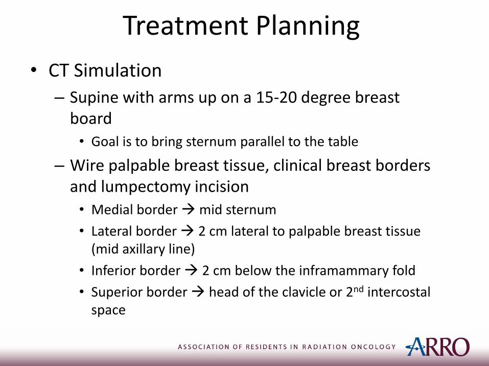
Treatment Planning
• 2D Treatment Planning – Uses plain x-rays for generating the plan – Assessment of treatment plan done by evaluating dose distribution at
midplane of breast – Wedges used to compensate for differences in tissue thickness – Significant dose heterogeneity on off axis regions (IM fold, axilla)
• 3D/IMRT Treatment Planning – Uses CT scan for generating the plan
• allows for better evaluation of target coverage, hot spots and dose to normal tissues
– IMRT improves dose homogeneity • decreases acute and chronic skin toxicity • Improves dose conformality
– better sparing of heart for left-sided cancers and lung
– Many different techniques utilized • field in field AKA fluence planning AKA forward planned IMRT • inverse planning AKA traditional IMRT
3D/IMRT
• Field in Field (Forward Planned) – Open medial and lateral tangents + segmental fields added
manually to attenuate beam in higher dose areas – MLCs used to improve homogeneity and to shield critical
structures
• IMRT (Inverse Planned) – Computerized algorithm used to reduce hot spots – Multiple weighted segments and beam angles can be used
to achieve optimal conformality – May result in more low dose spread
• Minimized by restricting beam angles to normal tangential arrangement
Treatment Planning
• CT Simulation
– Supine with arms up on a 15-20 degree breast board
• Goal is to bring sternum parallel to the table
– Wire palpable breast tissue, clinical breast borders and lumpectomy incision
• Medial border mid sternum
• Lateral border 2 cm lateral to palpable breast tissue (mid axillary line)
• Inferior border 2 cm below the inframammary fold
• Superior border head of the clavicle or 2nd intercostal space
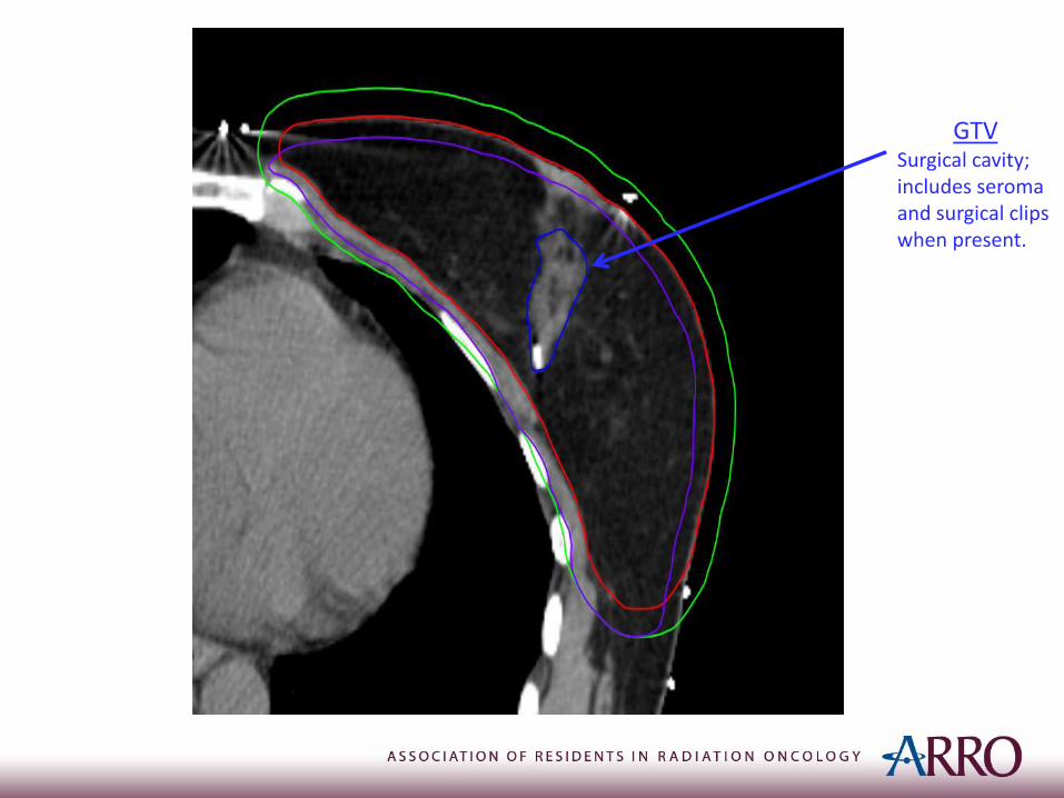
GTV Surgical cavity; includes seroma and surgical clips when present.
CTV: Posteriorly - Excludes pec major/minor Anterior - Skin Cranial/Caudal - Per clinical breast borders Medial - Sternal/rib junction Lateral - Mid axilla per clincial
reference
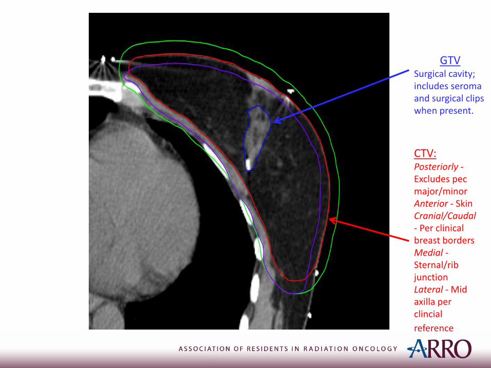
GTV Surgical cavity; includes seroma and surgical clips when present.
CTV: Posteriorly - Excludes pec major/minor Anterior - Skin Cranial/Caudal - Per clinical breast borders Medial - Sternal/rib junction Lateral - Mid axilla per clincial
reference
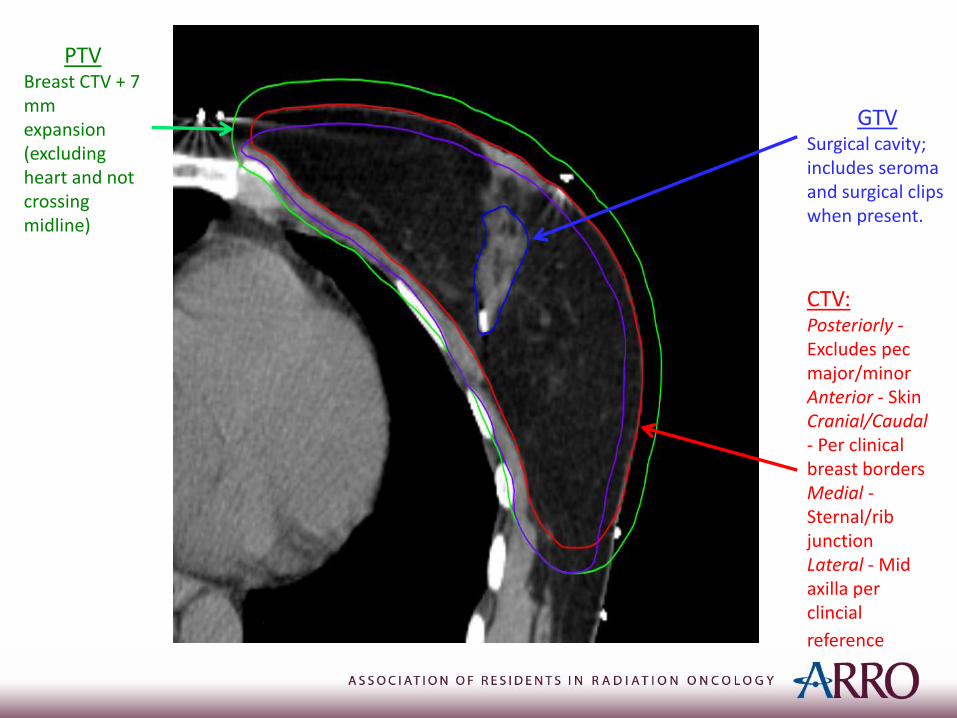
PTV Breast CTV + 7 mm expansion (excluding heart and not crossing midline)
GTV Surgical cavity; includes seroma and surgical clips when present.

CTV: Posteriorly - Excludes pec major/minor; Anterior – Skin; Cranial/Caudal - Per clinical breast borders; Medial - Sternal/rib junction; Lateral - Mid axilla per clinical
reference
PTV Breast CTV + 7 mm expansion (excluding heart and not crossing midline)
PTV-EVAL Excludes chest wall & pectoralis muscles; Extends to 5 mm from skin
GTV Surgical cavity; includes seroma and surgical clips when present.
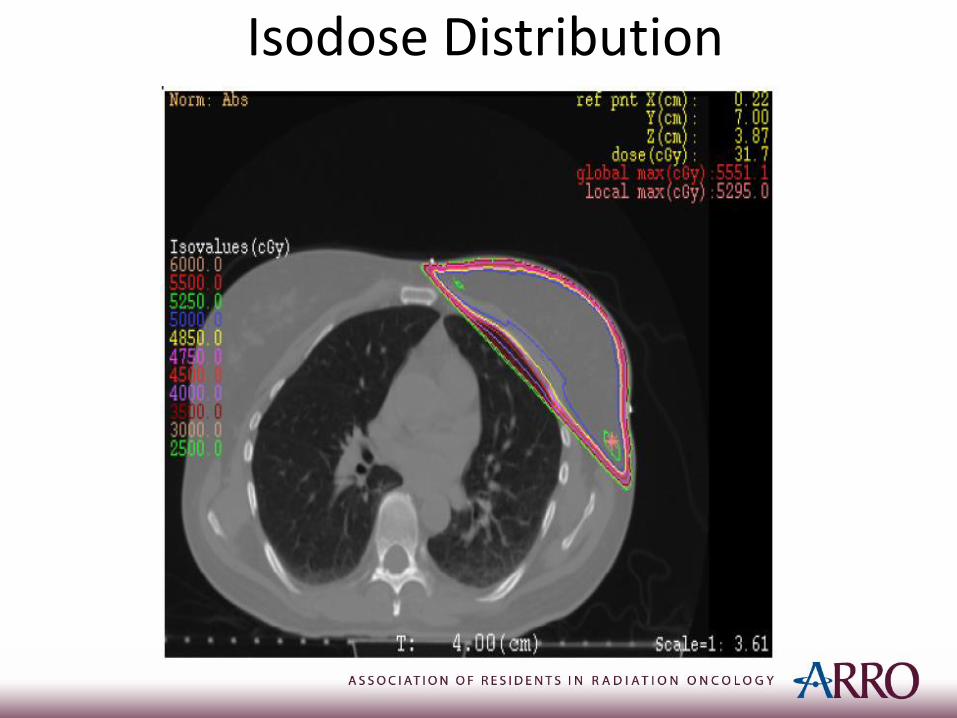
Isodose Distribution
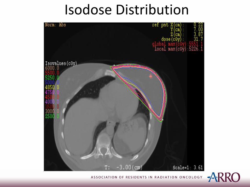
Isodose Distribution
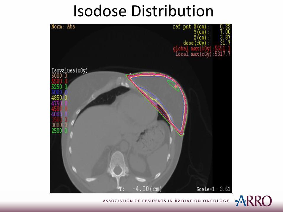
Isodose Distribution

Dose Volume Histogram

Dose-Volume Constraints (Per RTOG 1005)
Structure Constraint Percent
PTV 47.5 Gy >95%
PTV Max Dose 57.5 Gy
Heart 20 Gy <5%
Ipsilateral Lung 20 Gy <15%
Contralateral Lung 5 Gy <10%
Contralateral Breast Dmax 3.10 Gy
1.86 Gy
<5%
Thyroid Max point dose does not
exceed 2% of prescribed dose
Surveillance and Follow-up
• History and physical exam every 6-12 months for 5 years, then annually
• Mammogram every year
– 6-12 months post-radiation therapy if breast conserved

Teaching Points • Multidisciplinary management is critical in the treatment of
patients with DCIS
• The use of radiotherapy after lumpectomy in patients with DCIS decreases the risk of ipsilateral breast tumor recurrence in all patients but does not improve overall survival
– This risk reduction becomes increasingly small in patients with favorable features such as age > 60, small, unifocal low grade tumors excised with widely negative margins (> 1 cm).
– Thus, lumpectomy alone or lumpectomy followed by Tamoxifen can be considered in these patients
• The use of Tamoxifen in patients with ER+ DCIS reduces ipsilateral and contralateral breast tumor recurrence
• Patients undergoing mastectomy generally do not require adjuvant radiation
References • Allred D et al. (2002) Estrogen receptor expression as a predictive marker of
effectiveness of tamoxifen in the treatment of DCIS: findings from the NSABP Protocol B-24. Breast Cancer Res Treat 76:S36, (abstract 30)
• Fisher B et al. Tamoxifen in treatment of intraductal breast cancer: national surgical adjuvant breast and bowel project B-24 randomized controlled trial. Lancet 1999;353:1993-2000.
• Fonseca R et al. Ductal carcinoma in situ of the breast. Ann Intern Med 127 (11): 1013-22, 1997
• Parker SL et al., “Cancer statistics.” Ca-A Cancer Journal for Clinicians, vol. 46, no. 1, pp. 5–27, 1996
• Silverstein MJ et al. Choosing treatment for patients with ductal carcinoma in situ: fine tuning the University of Southern California/Van Nuys Prognostic Index. J Natl Cancer Inst Monogr. 2010;2010(41):193-6.
• Wapnir et al. “Long-term outcomes of invasive ipsilateral breast tumor recurrences after lumpectomy in NSABP B-17 and B-24 randomized clinical trials for DCIS.” J Natl Cancer Inst. 2011 Mar 16;103(6):478-88
• NCCN version 3.2014
Related Documents