Dual Language Use in Sign-Speech Bimodal Bilinguals: fNIRS Brain-Imaging Evidence Ioulia Kovelman 2 , Mark H. Shalinsky 1 , Katherine S. White 3 , Shawn N. Schmitt 4 , Melody S. Berens 1 , Nora Paymer 4 , and Laura-Ann Petitto 1,* 1 Department of Psychology, University of Toronto Scarborough, Canada 2 Department of Brain and Cognitive Sciences, Massachusetts Institute of Technology, MA, USA 3 Department of Brain and Cognitive Sciences, University of Rochester, NY, USA 4 Department of Psychological & Brain Sciences, Dartmouth College, Hanover, NH, USA Abstract The brain basis of bilinguals’ ability to use two languages at the same time has been a hotly debated topic. On the one hand, behavioral research has suggested that bilingual dual language use involves complex and highly principled linguistic processes. On the other hand, brain-imaging research has revealed that bilingual language switching involves neural activations in brain areas dedicated to general executive functions not specific to language processing, such as general task maintenance. Here we address the involvement of language-specific versus cognitive-general brain mechanisms for bilingual language processing by studying a unique population and using an innovative brain- imaging technology: bimodal bilinguals proficient in signed and spoken languages and functional Near-Infrared Spectroscopy (fNIRS; Hitachi ETG-4000), which, like fMRI, measures hemodynamic change, but which is also advanced in permitting movement for unconstrained speech and sign production. Participant groups included (i) hearing ASL-English bilinguals, (ii) ASL monolinguals, and (iii) English monolinguals. Imaging tasks included picture naming in “Monolingual mode” (using one language at a time) and in “Bilingual mode” (using both languages either simultaneously or in rapid alternation). Behavioral results revealed that accuracy was similar among groups and conditions. By contrast, neuroimaging results revealed that bilinguals in Bilingual mode showed greater signal intensity within posterior temporal regions (“Wernicke’s area”) than in Monolingual mode. Significance: Bilinguals’ ability to use two languages effortlessly and without confusion involves the use of language-specific posterior temporal brain regions. This research with both fNIRS and bimodal bilinguals sheds new light on the extent and variability of brain tissue that underlies language processing, and addresses the tantalizing questions of how language modality, sign and speech, impact language representation in the brain. *Corresponding Author: Professor Laura-Ann Petitto, University of Toronto Scarborough, Department of Psychology, 1265 Military Trail, Toronto, ON Canada MIC 1A4, [email protected]. Send reprint requests to Petitto (corresponding author) at [email protected]. For related research see http://www.utsc.utoronto.ca/~petitto/index.html and http://www.utsc.utoronto.ca/~petitto/lab/index.html. Publisher's Disclaimer: This is a PDF file of an unedited manuscript that has been accepted for publication. As a service to our customers we are providing this early version of the manuscript. The manuscript will undergo copyediting, typesetting, and review of the resulting proof before it is published in its final citable form. Please note that during the production process errors may be discovered which could affect the content, and all legal disclaimers that apply to the journal pertain. NIH Public Access Author Manuscript Brain Lang. Author manuscript; available in PMC 2010 May 1. Published in final edited form as: Brain Lang. 2009 ; 109(2-3): 112–123. doi:10.1016/j.bandl.2008.09.008. NIH-PA Author Manuscript NIH-PA Author Manuscript NIH-PA Author Manuscript

Welcome message from author
This document is posted to help you gain knowledge. Please leave a comment to let me know what you think about it! Share it to your friends and learn new things together.
Transcript
Dual Language Use in Sign-Speech Bimodal Bilinguals: fNIRSBrain-Imaging Evidence
Ioulia Kovelman2, Mark H. Shalinsky1, Katherine S. White3, Shawn N. Schmitt4, Melody S.Berens1, Nora Paymer4, and Laura-Ann Petitto1,*1 Department of Psychology, University of Toronto Scarborough, Canada2 Department of Brain and Cognitive Sciences, Massachusetts Institute of Technology, MA, USA3 Department of Brain and Cognitive Sciences, University of Rochester, NY, USA4 Department of Psychological & Brain Sciences, Dartmouth College, Hanover, NH, USA
AbstractThe brain basis of bilinguals’ ability to use two languages at the same time has been a hotly debatedtopic. On the one hand, behavioral research has suggested that bilingual dual language use involvescomplex and highly principled linguistic processes. On the other hand, brain-imaging research hasrevealed that bilingual language switching involves neural activations in brain areas dedicated togeneral executive functions not specific to language processing, such as general task maintenance.Here we address the involvement of language-specific versus cognitive-general brain mechanismsfor bilingual language processing by studying a unique population and using an innovative brain-imaging technology: bimodal bilinguals proficient in signed and spoken languages and functionalNear-Infrared Spectroscopy (fNIRS; Hitachi ETG-4000), which, like fMRI, measures hemodynamicchange, but which is also advanced in permitting movement for unconstrained speech and signproduction. Participant groups included (i) hearing ASL-English bilinguals, (ii) ASL monolinguals,and (iii) English monolinguals. Imaging tasks included picture naming in “Monolingual mode” (usingone language at a time) and in “Bilingual mode” (using both languages either simultaneously or inrapid alternation). Behavioral results revealed that accuracy was similar among groups andconditions. By contrast, neuroimaging results revealed that bilinguals in Bilingual mode showedgreater signal intensity within posterior temporal regions (“Wernicke’s area”) than in Monolingualmode. Significance: Bilinguals’ ability to use two languages effortlessly and without confusioninvolves the use of language-specific posterior temporal brain regions. This research with both fNIRSand bimodal bilinguals sheds new light on the extent and variability of brain tissue that underlieslanguage processing, and addresses the tantalizing questions of how language modality, sign andspeech, impact language representation in the brain.
*Corresponding Author: Professor Laura-Ann Petitto, University of Toronto Scarborough, Department of Psychology, 1265 MilitaryTrail, Toronto, ON Canada MIC 1A4, [email protected] reprint requests to Petitto (corresponding author) at [email protected]. For related research seehttp://www.utsc.utoronto.ca/~petitto/index.html and http://www.utsc.utoronto.ca/~petitto/lab/index.html.Publisher's Disclaimer: This is a PDF file of an unedited manuscript that has been accepted for publication. As a service to our customerswe are providing this early version of the manuscript. The manuscript will undergo copyediting, typesetting, and review of the resultingproof before it is published in its final citable form. Please note that during the production process errors may be discovered which couldaffect the content, and all legal disclaimers that apply to the journal pertain.
NIH Public AccessAuthor ManuscriptBrain Lang. Author manuscript; available in PMC 2010 May 1.
Published in final edited form as:Brain Lang. 2009 ; 109(2-3): 112–123. doi:10.1016/j.bandl.2008.09.008.
NIH
-PA Author Manuscript
NIH
-PA Author Manuscript
NIH
-PA Author Manuscript
IntroductionThe ability to learn multiple languages is not only useful, it is also a marvel of the humanlanguage capacity that taunts the scientific mind. While it is remarkable for an individual toachieve proficiency in more than one language, it is especially remarkable to be able to usemultiple languages within one conversation without confusing them. Scientific understandingof the human language capacity is incomplete without an in-depth understanding of how thebilingual brain enables the use of multiple languages with ease across various languagecontexts. Thus, one of the most prominent research questions in neurolinguistics,psycholinguistics, and cognitive neuroscience investigates the brain mechanisms that allowhealthy bilinguals to use their two languages without confusion and in a highly linguisticallyprincipled manner (Abutalebi et al., 2008; Abutalebi, Cappa & Perani, 2001; Abutalebi &Green, 2007; Christoffels, Firk, & Schiller, 2007; Crinion et al., 2006; Dehaene et al., 1997;Dijkstra & Van Heuven, 2002; Fabbro, 2001; Grosjean, 1997; Hernandez, Dapretto, Mazziotta,& Bookheimer, 2001; Kim, Relkin, Lee & Hirsch, 1997; Kovelman, Shalinsky, Berens, &Petitto, 2008; Paradis, 1977, 1997; Perani, Abutalebi, et al., 2003; Perani, 2005; Perani et al.,1996; Perani et al., 1998; Price, Green, & von Studnitz, 1999; Rodriguez-Fornells, Rotte,Heinze, Noesselt, & Muente, 2002; van Heuven, Schriefers, Dijkstra, & Hagoort, 2008;Venkatraman, Siong, Chee, & Ansari, 2006).
As suggested by François Grosjean (1997), bilinguals typically find themselves either in a“Monolingual mode,” using one language at a time, or in a “Bilingual mode,” using twolanguages in rapid alternation. Being in a Bilingual mode can at times lead to mixed-languageproductions, otherwise known as “code-switching,” and both the nature of this fascinatingprocess as well the underlying mechanisms that give rise to it have especially attracted theattention of scientists.
Intra-utterance use of two languages has been found to be complex and rule-governedlanguage-specific behavior that takes into account structure and meaning of both of thebilingual’s languages—and this is surprisingly the case even in the youngest bilinguals(Cantone & Muller, 2005; Grosjean & Miller, 1994; Holowka, Brosseau-Lapre, & Petitto,2002; Lanza, 1992; MacSwan, 2005; Paradis, Nicoladis, & Genesee, 2000; Petitto & Holowka,2002; Petitto, Katerelos, Levy, Gauna, Tetreault, & Ferraro, 2001; Petitto & Kovelman,2003; Poplack, 1980). For instance, in French, adjectives typically follow the noun that theymodify, whereas, in English, adjectives precede the noun (e.g., “table rouge” in French versus“red table” in English). Even 3 year-old French-English bilingual children typically do notmix, alter, or insert new lexical items into each respective language’s noun-adjective canonicalpatterning, and, thus, avoid making a significant grammatical violation in either of their twolanguages (Petitto et al., 2001).
One of the most common types of intra-utterance dual language use, however, occurs at thelexical level of language organization. Here, a bilingual may place an “open-class” lexical item(noun, verb, adverb, adjective or interjection) from language A into an utterance or phrase inlanguage B (Poplack, 1980). For instance, a French-English bilingual might say, “Yesterdaywe ate crème glacée,” (“ice cream” in French spoken in Quebec, Canada). Thus, bilingualsmust know how to navigate between their respective languages’ sets of lexical items.
Current theories of bilingual lexico-semantic representation have assumed the existence of acombined lexical store, in which each lexical item is connected to a number of semantic featuresin a common semantic store (Ameel, Storms, Malt, & Sloman, 2005; Dijkstra & Van Heuven,2002; Dijkstra, Van Heuven, & Grainger, 1998; Green, 1998; Kroll & Sunderman, 2003;Monsell, Matthews, & Miller, 1992; Von Studnitz & Green, 2002). Words in two languagesthat share overlapping semantic representations within the common semantic store are called
Kovelman et al. Page 2
Brain Lang. Author manuscript; available in PMC 2010 May 1.
NIH
-PA Author Manuscript
NIH
-PA Author Manuscript
NIH
-PA Author Manuscript
“translation equivalents” (e.g., “mother” in English and “mère” in French). The idea that thereis a common store is supported, for instance, by the fact that bilinguals can be semanticallyprimed in one language to produce a word in the other language (Dijkstra & Van Heuven,2002; Kerkhofs, Dijkstra, Chwilla, & de Bruijn, 2006; Kroll & Sunderman, 2003). Moreover,behavioral and imaging research has shown that bilinguals are likely to have both of theirlanguages active to some extent at all times (e.g., studies in word priming, and comprehensionof cognates, homophones and homographs, Doctor & Klein, 1992; Kerkhofs, et al., 2006; vanHell & De Groot, 1998; van Heuven et al., 2008).
How do bilinguals successfully operate in bilingual mode without confusing their languages,with respect to the semantic and grammatical content of their mixed utterances, and to theirlanguage selection? Contemporary research on the underlying brain mechanisms that makepossible bilingual dual language use leaves many questions. On the one hand, bilinguallanguage use has been said to be a highly principled language process which involves activationof both languages in a linguistically-based (rule-governed) manner (Grosjean, 1997; MacSwan,2005; Petitto et al., 2001). On the other hand, bilingual language also appears to involvecognitive control and allocation of attention, with current research suggesting that bilinguals’ability to use their languages is akin to many other types of general cognitive processes(Abutalebi, 2008; Abutalebi & Green, 2007; Bialystok, 2001; Crinion et al., 2006; Green,1998; Meuter & Allport, 1999; Thomas & Allport, 2000).
The ideas that language-specific mechanisms and cognitive-general mechanisms are involvedin dual language use are not necessarily in conflict with each other. In fact, both types ofprocessing seem crucial. The bilingual must preserve the overall linguistic integrity of theutterance while also rapidly selecting one of the competing linguistic representations, theappropriate phonological encoding for that representation, and finally send the correctarticulation-motor command.
It is noteworthy that the overwhelming majority of brain imaging studies with bilinguals,including our own, support the idea that cognitive-general mechanisms are heavily involvedin dual language use in Bilingual mode. In their recent theoretical overview of the bilingualbehavioral, imaging and lesion literature, Abutalebi & Green (2007) and Abutalebi (2008)outline the network of brain regions that has been consistently shown to participate in duallanguage selection. Prefrontal cortex has been shown to participate in bilingual language useduring both language production and comprehension (e.g., Hernandez et al., 2000; 2001;Kovelman, Shalinsky, Berens & Petitto, 2008; Rodriguez-Fornells et al., 2002) and,importantly, prefrontal cortex typically participates in other tasks that require complex taskmonitoring and response selection as well (Wager, Jonides, & Reading, 2004). Anteriorcingulate cortex (ACC), which typically plays a role in selective attention, error monitoringand interference resolution (Nee, Wager, Jonides, 2007), also participates in the languageselection process (e.g., Abutalei et al., 2008; Wang, Xue, Chen, Xue, & Dong, 2007). Parietalregions, including supramarginal gyrus and subcortical regions, particularly the caudatenucleus, are also thought to be key to bilingual dual language use, as shown by imaging studieswith healthy bilinguals (Abutalebi et al., 2007; 2008; Crinion et al., 2006; Green, Crinion, &Price, 2006; Khateb et al., 2007), as well as pathological language switching cases of caudatelesions (Abutalebi, Miozzo, & Cappa, 2000; Mariën, Abutalebi, Engelborghs, & De Deyn,2005). Most likely, it is the complex interplay between these regions that constitutes thecognitive basis for bilingual language use (Abutalebi, 2008).
But can bilingual language use be dependent on cognitive-general mechanisms alone? Therehave only been a handful of studies suggesting that language-dedicated brain regions (includingclassic Broca and Wernicke’s areas) might also show a modulation in activity as a function of
Kovelman et al. Page 3
Brain Lang. Author manuscript; available in PMC 2010 May 1.
NIH
-PA Author Manuscript
NIH
-PA Author Manuscript
NIH
-PA Author Manuscript
dual language use (Abutalebi et al., 2007, Chee, Soon, & Lee, 2003). However, these studiesare limited to examining receptive language.
How do we bring into greater focus the function of language-dedicated brain mechanismsduring dual language use? During dual language use, unimodal (speech-speech, e.g., English-French) bilinguals must inhibit competing alternatives in the same modality. It is possible thatthis competition is reduced in bimodal (sign language-speech) bilinguals, where competitionfor language articulation and language comprehension perception is less direct. As complexas it might be to use both the hands and mouth simultaneously or in rapid alternation duringBilingual mode, these two articulation modalities do not physically preclude each other(consider the fact that people can gesture while they speak). Might it be the case that in unimodalbilinguals’ activations in language-specific regions are occluded by the overwhelmingly highactivations in cognitive-general regions, which might result in part from high attention-sensory/motor costs of integrating and differentiating two languages within one modality (thus, possiblydriving high statistical thresholds for selected activations)? Bimodal bilinguals who know asigned and a spoken language (hence “bimodal bilinguals”) therefore represent an excellentcase for studying the underlying mechanisms of dual language use, particularly on thelanguage-specific level, as their language production and comprehension faculties mightexperience reduced levels of interference.
Prefrontal activations in particular have been consistently observed during blocks of sustaineddual language production and comprehension (e.g., Hernandez et al., 2000; Kovelman,Shalinsky, Berens, & Petitto, 2008). Cognitive-general mechanisms operate in a semi-hierarchical arrangement, where prefrontal cortex most likely represents the effortful “top-down” control, while ACC, parietal and subcortical regions are involved in more automatedaspects of attention allocation. Unimodal or bimodal, all bilinguals must choose the appropriatelanguage at any given moment. Importantly, however, for bimodal bilinguals, the costs ofselection errors are reduced: if the competition is not perfectly resolved, both languages can“come out” simultaneously – which does occasionally happen even when hearing signersinteract with non signers (Emmorey et al., 2004; Petitto et al., 2001). Given the reduced costof selection errors, bimodal bilinguals might not devote as many resources to top-downmonitoring as unimodal bilinguals. This population, therefore, allows a nice window intolanguage-related processing mechanisms.
Studying bimodal bilinguals who are proficient in signed and spoken languages is a powerfultool for revealing the underlying principles of bilingual acquisition, processing, and code-switching (c.f., Petitto et al., 2001; Emmorey et al., 2004). Bimodal bilinguals, child and adult,commonly produce intra-utterance code-switching much like unimodal bilinguals when inBilingual mode. Unlike unimodal bilinguals, they take full advantage of their bimodality andcommonly produce open-class words in both languages simultaneously. During simultaneousmixing (also called “code-blending,” rather than “code-switching,” (Emmorey et al., 2004))the two words in different languages are typically semantically congruent (i.e. they are similaror identical in their meaning), revealing that even in the youngest bilinguals, their twolanguages come together in concert rather than in unprincipled confusion (Emmorey et al.,2004; see especially Petitto et al., 2001; Petitto & Holowka, 2002; Petitto & Kovelman,2003).
If the ability to use two languages in the same context is uniquely a general cognitive ability,then during Bilingual mode, even sign-speech bimodal bilinguals should only show changesin activation in cognitive-general regions. In particular, sustained dual-language cognitiveeffort should result in high activations in the prefrontal regions (Buckner et al., 1996).Alternatively, if language-specific mechanisms also play a key role in dual language use, oncethe competition from two spoken languages using one mouth is reduced, sign-speech bimodal
Kovelman et al. Page 4
Brain Lang. Author manuscript; available in PMC 2010 May 1.
NIH
-PA Author Manuscript
NIH
-PA Author Manuscript
NIH
-PA Author Manuscript
bilinguals in Bilingual mode might show increased recruitment of classic language brain tissue,such as left inferior frontal gyrus (particularly in BA 44/45, classic “Broca’s area”) and leftsuperior temporal gyrus (particularly in the posterior part of left STG, classic “Wernicke’sarea”).
The present study represents a principled attempt to reconcile decades of behavioral andimaging work by investigating whether language-specific mechanisms, as has been shown forcognitive-general mechanisms, play a role in dual language use. To investigate this question,we use a novel technology, functional Near-Infrared Spectroscopy (fNIRS) brain-imaging.Like fMRI, fNIRS measures changes in the brain’s blood oxygen level density (BOLD) whilea person is performing specific cognitive tasks. Due to the nature of NIRS imaging, we do notmeasure the activation in subcortical and ACC regions; however, we do not question whetheror not these regions are involved in dual language use, as we believe our colleagues haveprovided ample evidence to that effect (c.f. Abutalebi & Green, 2007). Dual language use mostlikely involves a complex network of cortical and subcortical regions, which are both language-dedicated and cognitive-general. This study focuses on examining how cognitive-general andlanguage-specific cortical regions participate in dual language use. A key advantage over fMRIfor the purposes of our language study is that fNIRS places minimal restriction on body motionand it is nearly completely silent (see methods section for further details on the spatial andtemporal resolution, as well as other technical characteristics of fNIRS).
Here we use fNIRS brain-imaging technology to evaluate sign-speech bimodal bilingualsduring overt picture-naming. Monolinguals were tested in their native language (English orASL). Bimodal ASL-English bilinguals were tested in each of their languages separately, aswell as in simultaneous (naming pictures in ASL and in English at the same time) andalternating (naming pictures either in ASL or in English in rapid alternation) Bilingual modes.The study included English and ASL monolinguals as control groups, in order to ensure thatbilingual participants were linguistically and neurologically comparable to their monolingualcounterparts in each language.
MethodsParticipants
A total of 32 adults participated in this study (5 hearing bimodal bilinguals, 20 hearingmonolinguals, 7 deaf monolinguals; given the rarity of our deaf and bilingual populations, wewere indeed fortunate to achieve such samples). All participants received compensation fortheir time. The treatment of all participants and all experimental procedures were in fullcompliance with the ethical guidelines of NIH and the university Ethical Review Board.
Bilingual Participants—Five hearing right-handed American Sign Language (ASL)-English bilinguals participated in this experiment (see Table 1). All bilingual participants werechildren of deaf adults (CODAs) and had high, monolingual-like, language proficiency in eachof their two languages, as was established with the participant language assessment methodsdescribed below. All participating individuals achieved the required accuracy of at least 80%.The bilingual participants received their intensive dual-language exposure to both ASL andEnglish at home within the first five years of life, as early dual language exposure is key tocomparable linguistic processing in bilinguals (Kovelman, Baker & Petitto, 2008;Kovelman,Baker & Petitto, in press;Perani, Abutalebi et al., 2003;Wartenburger, Heekeren, Abutalebi,Cappa, Villringer, & Perani, 2003). All bilingual participants used both English and ASLconsistently from the first onset of bilingual exposure to the present, had at least one deaf parentwho used ASL, and learned to read in English between the ages of 5–7. Bilingual participantshad no exposure to a language other than English and ASL until after age 10 and only in theformat of a foreign language class.
Kovelman et al. Page 5
Brain Lang. Author manuscript; available in PMC 2010 May 1.
NIH
-PA Author Manuscript
NIH
-PA Author Manuscript
NIH
-PA Author Manuscript
Monolingual Participants—Twenty hearing right-handed English monolinguals and sevendeaf right-handed ASL monolinguals participated in this experiment (see Table 1). Allmonolingual participants completed language assessment tasks in English or in ASL with therequired accuracy of 80% and above. English monolinguals came from monolingual Englishfamilies and had no other language exposure until after age 10 and only in the format of aforeign language class. ASL participants were profoundly deaf, with five of the seven beingborn deaf (congenitally deaf), and the remaining two deaf participants having lost their hearingby age 12 months. Six of the deaf participants were exposed to ASL from birth, while one wasfirst exposed to ASL at age four. All ASL monolinguals studied English only in a school/classformat and indicated that they experienced difficulty understanding English on our extensive“Bilingual Language Background & Use Questionnaire” (BLBU). Previous research hasestablished that adult deaf individuals typically achieve the equivalent of 4th grade level readingcomprehension in English (e.g., Traxler, 2000). All our monolingual participants had taken“second language” classes in school (including English, Spanish, French, German and otherlanguages). Their proficiency in the variety of second languages learned at school was assessedvia self-report in the BLBU questionnaire.
Language AssessmentsBilingual Language Background & Use Questionnaire—All participants were firstadministered an extensive Bilingual Language Background and Use Questionnaire that hasbeen standardized and used across multiple spoken and signed language populations (Penhune,Cismaru, Dorsaint-Pierre, Petitto, & Zatorre, 2003; Petitto, Zatorre et al., 2000; Petitto et al.,2001). This questionnaire enabled us to achieve confidence both in our “bilingual” and our“monolingual” group assignments and in their early-exposed, highly-proficient status in theirlanguage(s). This tool permitted us to determine the age of first bilingual language exposure,language(s) used in the home by all caretakers and family members/friends, language(s) usedduring/throughout schooling, language(s) of reading instruction, cultural self-identificationand language maintenance (language(s) of the community in early life and language(s) usedthroughout development up until the present).
Language Competence/Expressive Proficiency (LCEP)—This task was administeredto assess participants’ language production in each of their languages, and has been usedeffectively to measure both signed and spoken language proficiency and competency (e.g.,Kegl, 1994; Kovelman, Baker & Petitto, 2008, in press; Kovelman, Shalinsky, Berens, &Petitto, 2008; Petitto et al., 2000; Senghas, 1994; Senghas & Kegl, 1994). The task includestwo one-minute cartoons containing a series of events that the participant watches and thendescribes to an experimenter. Monolingual participants described each of the two cartoonseither in English or in ASL (as relevant); bilingual participants described one of the cartoonsin English to a native English-speaker and one of the cartoons in ASL to a differentexperimenter who was a native ASL speaker (the order of the language presentation andcartoons was randomized across participants). Sessions were videotaped and highly proficientspeakers of English and of ASL trained as linguistic coders identified the presence or absenceof semantic, phonological, syntactic, and morphological errors. Each participant was requiredto produce at least 80% correct utterances in each native language in order to participate in theexperiment. Inter-rater reliability for both transcription and coding for a subset of participants(25%) was 98.0%.
English Grammaticality Judgment Behavioral Task—An English grammaticalityjudgment task was administered to English monolinguals and ASL-English bilinguals. In thisgrammaticality judgment task, modeled after ones used by Johnson and Newport (1989),McDonald (2000), and Winitz (1996), participants were presented with grammatical andungrammatical sentences and instructed to read each sentence and indicate whether or not the
Kovelman et al. Page 6
Brain Lang. Author manuscript; available in PMC 2010 May 1.
NIH
-PA Author Manuscript
NIH
-PA Author Manuscript
NIH
-PA Author Manuscript
sentence was grammatical. Examples: I see a book (grammatical); I see book (ungrammatical).This type of task is effective at identifying individuals’ proficiency and age of first exposureto the language; crucially, only those exposed to the language before age 7 have been observedto perform with high accuracy on this task. All English-speaking participants had to score atleast 80% correct to be eligible.
Experimental Tasks: Picture Naming: Black line-drawing pictures of everyday objects (e.g.,table, lamp) were selected from the Peabody Picture Vocabulary Test (PPVT; Dunn & Dunn,1981) and the International Picture Naming Project (Abbate & La Chappelle, 1984; Max PlanckInstitute for Psycholinguistics; Kaplan, Goodglass, & Weintraub, 1983; Oxford JuniorWorkbooks, 1965). There were 4 different sets of pictures and the background color (describedbelow) of the pictures was altered to elicit responses in specific languages from participants.We used a block design, with 5 blocks for each of the 4 language conditions (English, ASL,Simultaneous, and Alternation). Each run was preceded by 60 seconds of rest/baseline. Each35 s block contained 14 pictures (1.5 s picture presentation and 1 s inter-picture fixationinterval), with 20 second rest-periods between blocks during which a white fixation cross ona black background was presented. Participants received a break and a reminder of instructionsbetween the runs.
The participant’s task was to name the picture they saw on each trial. Their responses wererecorded and later scored using video-audio recordings synchronized with imaging datacollection. Picture Naming runs occurred in the following manner: Monolingual mode English- White background for all pictures in this run of 5 blocks indicated for ASL-English bilingualsthat an English response was required. Monolingual mode ASL - Blue backgrounds indicatedfor ASL-English bilinguals that an ASL response was required. Bilingual mode Simultaneous- Grey backgrounds indicated for ASL-English bilinguals that they should name all picturesin both ASL and English simultaneously. Bilingual mode Alternating - Blocks contained“blue” (ASL) and “white” (English) background pictures, indicating that bilingual participantsshould alternate between the two languages. The ordering of ASL and English trials wasrandomized in the Alternating block. English and ASL monolinguals saw the same pictures,but were asked to disregard differences in background color and to name the pictures in Englishor in ASL only, respectively. The order of experimental runs was randomized acrossparticipants. Picture naming stimuli were presented using E-Prime software on a flatscreenDell monitor connected to a Dell laptop running Windows XP. Task order was randomized foreach participant. Prior to imaging, participants were trained in the task using stimuli not usedin the experiment.
fNIRS Imaging: General Information: fNIRS holds several key technological brain-imagingadvantages over fMRI, as rather than yielding BOLD, which is a ratio between oxygenatedand deoxygenated hemoglobin, fNIRS yields the measures of deoxygenated and oxygenatedhemoglobin separately and thus yields a closer measure of the underlying neuronal activitythan fMRI. While fNIRS does not record deep into the human brain (maximum ~4cm depth),it has excellent spatial resolution that is outstanding for studies of human higher cognition andlanguage, and it has better temporal resolution than fMRI (~<5s Hemodynamic Response, HR),as well as a remarkably good sampling rate of 10 x per second. Unlike MRI scanners, fNIRSscanners are very small (the size of a desktop computer), portable, and particularly childfriendly (children and adults sit normally in a comfortable chair, and babies can be studiedwhile seated on mom’s lap). It is the fNIRS’ detailed signal yield [HbO] index versus BOLD,its rapid sampling rate, relative silence, high motion tolerance, and participant friendly set-up,which have contributed to the rapidly growing use of fNIRS as one of today’s leading brain-imaging technologies.
Kovelman et al. Page 7
Brain Lang. Author manuscript; available in PMC 2010 May 1.
NIH
-PA Author Manuscript
NIH
-PA Author Manuscript
NIH
-PA Author Manuscript
Apparatus & Procedure: To record the hemodynamic response we used a Hitachi ETG-4000with 44 channels, acquiring data at 10 Hz (Figure 1a). The lasers were factory set to 690 nmand 830 nm. The 16 lasers and 14 detectors were segregated into two 5 × 3 arrays correspondingto 30 probes (15 probes per array; Figure 1a). Once the participant was comfortably seated,one array was placed on each side of the participant’s head. Positioning of the array wasaccomplished using the 10–20 system (Jasper, 1957) to maximally overlay regions classicallyinvolved in language, verbal, and working memory areas in the left hemisphere as well as theirhomologues in the right hemisphere.
During recording, channels were tested for noise prior to the beginning of the recording session.Digital photographs were taken of the positioning of the probe arrays on the participant’s headprior to and after the recording session to identify if the arrays had moved during testing. AnMPEG video recording was synchronized with the testing session, so any apparent movementartifacts could be confirmed during offline analysis and to score participants’ responses.
After the recording session, data were exported and analyzed using Matlab (The MathworksInc.). Conversion of the raw data to hemoglobin values was accomplished in two steps. Underthe assumption that scattering is constant over the path length, we first calculated theattenuation for each wavelength by comparing the optical density of light intensity during thetask to the calculated baseline of the signal. We then used the attenuation values for eachwavelength and sampled time points to solve the modified Beer-Lambert equation to convertthe wavelength data to a meaningful oxygenated and deoxygenated hemoglobin response (HbOand Hb respectively).
MRI Coregistration: For MRI (anatomical) co-registration, a 5×3 array of Vitamin E tabletswas constructed with the tablets placed precisely at each of the optode locations used duringour fNIRS experiments above (Figure 1b-e). The array was then placed on to the participant’shead using the 10–20 coordinate system and secured in place with MRI safe tape and straps.Using a Philips 3T MRI scanner, an anatomical scan was then taken from 9 participants. Foampadding was placed in the head coil to limit subject head movement during image acquisition.T1-weighted three-dimensional magnetization-prepared rapid acquisition gradient echo (3D-MPRAGE) sagittal images were obtained with a Phillips 3T scanner. Scanning parameterswere as follows: echo time (TE) = 4.6 ms, repetition time (TR) = 9.8 ms, flip angle = 8 degrees,acquisition matrix = 256 × 256, 160 sagittal slices, and voxel size = 1 × 1 × 1 mm with no gap.
These scans confirmed that the placement of the Vitamin E tablets, and hence the placementof our fNIRS recorded channels, indeed covered the anatomical locations anticipated by the10–20 coordinate system (see Figure 1b-e).
ResultsBehavioral Results
Twenty English monolinguals completed the Picture naming task. Due to camera failure,behavioral responses for one English monolingual participant were not recorded; these dataare thus omitted in this behavioral analysis. Participants’ average scores and standarddeviations for this task are reported in Table 2.
We first compared the three language groups (hearing/native ASL-English bilinguals, ASLmonolinguals, and English monolinguals) on their overall picture naming performance. Forthis comparison, scores for hearing/native ASL-English bilinguals were obtained by averagingacross the four naming conditions (ASL, English, Alternating, Simultaneous). Please see Table2 for behavioral scores. A one-way ANOVA with these average percent correct scores revealeda marginal effect of language group, (F(2, 28) = 3.0, p = 0.066). Post-hoc comparisons revealed
Kovelman et al. Page 8
Brain Lang. Author manuscript; available in PMC 2010 May 1.
NIH
-PA Author Manuscript
NIH
-PA Author Manuscript
NIH
-PA Author Manuscript
that the ASL monolinguals (mean = 95.2% correct) performed marginally better than theEnglish monolinguals (mean = 89.8%) on this task (Tukey’s Honestly Significant Difference(HSD), p = 0.057). No other group comparisons reached significance.
For hearing/native ASL-English bilinguals, we further compared performance on each of thenaming conditions, to determine whether there were differences in difficulty across conditions(ASL, English, Alternating, Simultaneous). A repeated-measures ANOVA on these percentcorrect scores revealed no significant effect of condition, F(3,12) <1, p > .05, ns.
Imaging resultsIdentifying functional regions of interest—The first step in our analysis was to usePrincipal Components Analysis (PCA) to identify channels that formed data-driven clustersof activation, which shaped our functional regions of interest. The first and second principalcomponents (PC), which explain the largest proportion of the variance, were used for each. Todetermine functional clusters for English, data from the hearing/native ASL-English bilingualsand English monolinguals were used. To determine functional clusters for ASL, data from thehearing/native ASL-English bilinguals and ASL monolinguals were used.
First, we present PCA results for English, for the left and right hemispheres. We then presentPCA results in ASL, again first for the left then for the right hemisphere. Finally, we presentthe PCA results for the two Bilingual mode conditions in bimodal bilinguals. Throughout ourdescription of the PCA results in ASL we note the similarities and differences between the twolanguages, as well as differences and similarities in Monolingual and Bilingual modes. Thechannels active and loadings for clusters identified in the first component for each condition(English, ASL, and Bilingual mode) are presented in Table 3. See the estimated anatomicallocation of channels in Figure 1e.
English Left Hemisphere PCA: In the English condition with hearing/native ASL-Englishbilinguals and English monolinguals, the PCA analysis yielded a first component that explained27% of the variance and yielded the following clusters: (1) Anterior perisylvian cluster,covering inferior frontal gyrus (IFG) as well as anterior superior temporal gyrus (STG), (2)Dorsal lateral prefrontal cortex (DLPFC) cluster overlaying SFG/MFG, and (3) Parietalcluster, maximally overlaying motor and parietal regions.
The second component for English in the left hemisphere explained 17% of the variance andhelped identify larger functional clusters that appear to reflect functional networks. Inparticular, we identified the following. (1) Parieto-temporal cluster. Negative loadings fellon channels maximally overlaying parietal (channels: 4, 9, 13), and temporal regions (includinginferior, middle and superior temporal gyri, spanning anterior-to-posterior temporal regions;channels: 12, 16–18, 20, 21). (2) Frontal-temporal cluster. Low and medium value loadings(0.1–0.6) fell on frontal (including inferior and middle frontal gyri; channels: 10, 15, 19), motor(channels: 2, 7), and posterior inferior temporal (channel 22) regions. (3) Medial frontalcluster. High loadings (0.7–0.9) fell on frontal lobe regions (predominantly including middleand superior frontal gyri: channels: 1, 5, 6, 14).
English Right Hemisphere PCA: In the English condition with hearing/native ASL-Englishbilinguals and English monolinguals, right hemisphere PCA results closely resembled thoseobserved in the left hemisphere. The first principal component explained 27% of the variance.Clusters included (1) Anterior perisylvian cluster maximally overlaying IFG and anteriorSTG, (2) DLPFC cluster, identical to the left hemisphere, including frontal regions,predominantly MFG, and again (3) Parietal cluster.
Kovelman et al. Page 9
Brain Lang. Author manuscript; available in PMC 2010 May 1.
NIH
-PA Author Manuscript
NIH
-PA Author Manuscript
NIH
-PA Author Manuscript
The second principal component for English RH explained 14% of the variance. Two functionalclusters were obtained. (1) Distributed network cluster. Negative and low (below 0.4)loadings fell on channels maximally overlaying a full variety of cortical regions covered bythe fNIRS probes: frontal (channels: 1, 2, 5, 19), parieto-temporral (channels: 4, 8, 9, 12, 13,17, 18, 22), and motor (channels: 3, 7) regions. (2) Frontal. Medium and high loadings (above0.4) consistently fell on extensive frontal, and fronto-temporal regions (channels: 6, 10, 11,14, 15, 16, 20, 21).
ASL Left Hemisphere PCA: In the ASL condition with hearing/native ASL-Englishbilinguals and ASL monolinguals, the first PC explained 30% of the variance. The followingclusters were obtained: (1) Anterior perisylvian cluster, as in English, maximally overlayinginferior frontal gyrus and anterior superior temporal gyrus clustered together (IFG & anteriorSTG), (2) DLPFC cluster overlaying frontal lobe regions of middle and superior frontal gyri(SFG, MFG), and (3) Parietal-motor cluster, including parietal and motor regions. As inEnglish, ASL channels overlaying parietal regions received the highest loadings (above 0.7).
The second PCA component for ASL (LH) explained 18% of the variance and also appearedto identify larger functional clusters. (1) Parieto-frontal cluster. Negative loadings fell onchannels maximally overlaying parietal (channels: 4, 8, 9, 13) and frontal (channels: 1, 2, 6)regions. Recall that in English, negative loadings helped identify a parietal-temporal ratherthan parietal-frontal cluster. (2) Frontal-temporal cluster. Similar to English, but with widertemporal coverage, this cluster had low and medium value loadings (0.1–0.6) on frontal(channels: 5, 15, 19) and temporal (channels: 12, 17, 18, 20–22) regions. (3) Medial frontalcluster. As in English, high loadings (0.7–0.9) fell on channels maximally overlaying frontalregions (channels: 10, 11, 14).
ASL Right Hemisphere PCA: In the ASL condition with hearing/native ASL-Englishbilinguals and ASL monolinguals, the first PC explained 27% of the variance. Righthemisphere PCA results closely resembled those observed with the left hemisphere. Areasidentified included: (1) Anterior perisylvian cluster overlaying IFG and anterior STG. (2)DLPFC cluster (3) Parietal cluster. As in the left hemisphere and as in English, most of thechannels with high loadings (greater than 0.7) were channels maximally overlaying motor andparietal regions (channels 8, 9, 13). Identically to the left hemisphere, frontal regions receivednegative loadings in ASL (compared to low loadings in English).
The second principal component for ASL RH explained 17% of the variance. Similar to EnglishRH two functional clusters were obtained. (1) Distributed network cluster. Negative and low(below 0.04) loadings fell on channels maximally overlaying a full variety of cortical regionscovered by the fNIRS probes: frontal (channels: 1, 2, 5, 11), parieto-temporal (channels: 4, 8,9, 12, 13, 16, 17, 18, 20, 21, 22), and motor (channels: 3, 7) regions. (2) Inferior-medial frontalcluster. Medium and high loadings (above 0.4) consistently fell on frontal (IFG, MFG), andsuperior temporal regions (channels: 6, 10, 14, 15, 16, 19).
Bilingual mode PCA Left Hemisphere: In order to increase the power of this analysis, bothAlternating and Simultaneous bimodal conditions were combined. The first PC explained 36%of the variance. There was a stark difference between PCA results for Monolingual mode ascompared to Bilingual mode. Instead of yielding units of language, general-cognitive, andsensory-motor processing, this set of PCA results for both first and second components yieldedfunctional processing networks. Clusters identified were (1) Temporal-parietal clustercovering anterior/superior and medial temporal regions, sensory/motor and parietal regions,(2) Inferior frontal-parietal cluster including IFG, and parietal/motor regions, and (3)Medial frontal-posterior temporal cluster on frontal regions (MFG), motor and posterior/posterior-inferior temporal regions.
Kovelman et al. Page 10
Brain Lang. Author manuscript; available in PMC 2010 May 1.
NIH
-PA Author Manuscript
NIH
-PA Author Manuscript
NIH
-PA Author Manuscript
The second component in the left hemisphere explained 27% of the variance and suggestedthat the anterior and posterior language regions (“Broca’s”, “Wernicke’s” areas) and parietalregions (which, among other functions, support key linguistic processing in sign language)were indeed working together during bilingual mode. (1) Inferior frontal - posteriortemporal – parietal cluster. The highest loadings fell on inferior frontal (channel: 15),posterior/inferior temporal (channels: 17, 21, 22), and parietal regions (channels: 4, 9). Similarto first component, other clusters from the second component were also “multi-region” clustersthat incorporated channels maximally overlaying a variety of cortical regions.
Bilingual mode PCA Right Hemisphere: In order to increase the power of this analyses, bothAlternating and Simultaneous bimodal conditions were combined. The first PC explained 50%of the variance. Similar to the left hemisphere, we observed that channels grouped into largenetworks rather than smaller anatomical units. These clusters were (1) Anterior perisylviancluster – parietal cluster, covering inferior frontal/anterior STG regions and motor/parietalregions, (2) Frontal-parietal-temporal cluster fell on a large area covering frontal, parietaland temporal regions, (3) Inferior frontal-posterior temporal cluster including inferiorfrontal gyrus and posterior temporal regions, and (4) Medial – inferior frontal cluster,including anterior inferior and middle frontal gyri.
The second principal component for Bilingual mode RH explained 20% of the variance. Similarto English and ASL RH, two functional clusters were obtained. (1) Distributed networkcluster. Negative and low (below 0.04) loadings fell on channels maximally overlaying a fullvariety of cortical regions covered by the fNIRS probes: frontal (channels: 1, 2, 5, 10, 14, 19),parietal (channels: 4, 8, 9), temporal (channels: 12, 13, 17, 18, 21, 22), and motor (channel: 3)regions. (2) Frontal. Medium and high loadings (above 0.4) consistently fell on frontal, andsuperior temporal regions (channels: 6, 11, 15, 16, 20).
ROIs: Predicted & FunctionalIn our introduction we hypothesized that we may see task-related brain activations in the frontallobe, including left inferior frontal gyrus and DLPFC, posterior temporal regions, as well asparietal regions. The channels selected for ROI analysis were governed by these predictions,MRI coregistration (see Figure 1b-e), as well as the data-driven clusters identified by the firstPCs described above. In this way, we identified seven regions of interest. (i) Anteriorperisylvian cluster (inferior frontal gyrus and anterior aspect of the superior temporal gyrus;channels: 15, 19, 20). (ii) DLPFC (prefrontal cortex, primarily including MFG; channels: 10,14), (iii) Superior frontal (SFG; channels 1, 5), (iv) Posterior temporal (posterior STG andposterior MTG, overlapping with classic “Wernicke’s area” and channel: 17), (v) Parietal(superior and inferior parietal lobules; channels: 4, 8, 9, 13) (vi) Sensory-motor (postcentraland precentral gyri; channel 7) and (vii) Posterior frontal/motor (dorsal frontal regionsadjacent to primary motor cortex and primary motor cortex; channels: 6, 11). Average andstandard deviations of signal changes for each ROI, each group and each experimentalcondition are presented in Table 4, please see Figure 1e for anatomical localization of individualchannels.
ROI analysis of hearing/native ASL-English bilinguals in Bilingual versus Monolingualmodes
Left Hemisphere—A 2 (language modes: Monolingual (ASL & English) versus Bilingual(Alternation & Simultaneous), within factor) X 7 (ROIs, within factor) repeated-measuresANOVA yielded no significant main effect of language modes (F(1, 4) = 1.1), no significantdifference between ROIs (F(6, 24) < 1), and a significant mode by ROI interaction (F(6, 24)= 3.7, p < 0.01). Figure 2 shows left hemisphere brain activations in bilinguals during Bilingualand Monolingual modes, suggesting that the interaction might stem from greater recruitment
Kovelman et al. Page 11
Brain Lang. Author manuscript; available in PMC 2010 May 1.
NIH
-PA Author Manuscript
NIH
-PA Author Manuscript
NIH
-PA Author Manuscript
of the posterior temporal and sensory-motor regions during Bilingual mode, and greaterrecruitment of inferior frontal/anterior STG regions during Monolingual mode.
We further investigated the source and location of potential differences between Bilingual andMonolingual modes in the left hemisphere. Given the small number of bilingual subjects, weconducted a non-parametric test for matched pairs on the 7 ROIs. Wilcoxon Signed Ranks testrevealed that during Bilingual mode, hearing/native ASL-English bilinguals had significantlygreater activation in the left posterior temporal region (Z(4) = −2.02, p < 0.05) and left sensory-motor region (Z(4) = −2.02, p < 0.05). The results revealed that all ranks were positive onlyfor Posterior temporal and Sensory-motor ROIs, which means that all subjects showed anincrease in activation during bilingual mode in these two regions.
Right Hemisphere—A 2 × 7 repeated-measures ANOVA yielded no significant differencesbetween language modes (F(1, 4) <1), a marginally significant difference between ROIs (F(6, 24) = 3.0, p = 0.02), and no significant interactions, (F(6, 24) <1). As can be seen in Table4, in both Monolingual and Bilingual modes, right inferior frontal/anterior STG regions appearto have the greatest involvement in the task (the highest percent signal change).
ROI analysis of hearing/native ASL-English bilinguals in Bilingual mode: Alternating versusSimultaneous
Alternating versus Simultaneous—There were no significant main effects or interactionsfor our ROIs in either hemisphere, as revealed by two (one for each hemisphere) 2 (Alternating& Simultaneous conditions, within factor) X 7 (ROIs, within factor) repeated-measuresANOVAs (LH: condition F(1, 4) <1, ROI F(6, 24) = 2.0; RH: F(1, 4) = <1, ROI F(6, 24) =1.7, p =0.16; all ns).
ROI analysis of English Picture Naming: ASL-English Bilinguals and English MonolingualsThe comparison of bilingual and monolingual groups was conducted to ensure that bilingualsindeed had an overall native-like neural organization for each of their languages. However, wenote that the discrepancy in the sample sizes of monolinguals and bilinguals prevents us frommaking strong claims about these comparisons. Left Hemisphere English: A 2 (groups: Englishmonolinguals and bilinguals, between factor) X 7 (ROI, within factor) mixed-measuresANOVA yielded a significant main effect for ROI (F(6, 138) = 7.3, p < 0.01), no significantdifferences between the groups and no significant interactions. Right Hemisphere English: A2 × 7 mixed-measures ANOVA yielded a significant main effect for ROI (F(6, 138) = 5.4, p< 0.01), no significant differences between the groups and no significant interactions. Follow-up t-tests revealed that ASL-English bilinguals had marginally less recruitment of parietal areasas compared to English monolinguals (t(23) = 1.9, p = 0.06). Left Hemisphere ASL: A 2(groups: ASL monolinguals & bilinguals, between factor) X 7 (ROI, within factor) mixed-measures ANOVA revealed no significant main effects or interactions. Right HemisphereASL: A 2 × 7 mixed-measures ANOVA revealed significant ROI differences (F(6, 60) = 3.0,p < 0.01), and no significant group differences or interactions.
DiscussionHere we addressed the involvement of language-specific versus cognitive-general brainmechanisms in bilingual language use by studying simultaneous and alternating dual-languageproduction in a very special group of bilinguals – specifically, bilinguals who were exposedto a spoken and a signed language from very early in life.
Our primary finding is that the bilinguals showed greater recruitment of left posterior temporalbrain regions (overlapping with the classic “Wernicke’s area”) during Bilingual mode as
Kovelman et al. Page 12
Brain Lang. Author manuscript; available in PMC 2010 May 1.
NIH
-PA Author Manuscript
NIH
-PA Author Manuscript
NIH
-PA Author Manuscript
compared to Monolingual mode. These results suggest that left posterior temporal regions mayplay a key role in bilinguals’ ability to code-switch and use both languages appropriately atthe same time—a finding that stands in contrast to accounts of bilingual’s ability to use twolanguages at the same time as involving cognitive-general brain mechanisms alone.
Behaviorally, all of our groups performed with high and overall comparable accuracy duringthe Picture Naming task. Interestingly, bilinguals also performed with equally high accuracywhen completing the task either in Bilingual or in Monolingual modes.
Hearing/native ASL-English bilinguals showed similar accuracy when using their twolanguages across a variety of language contexts: one at a time (Monolingual mode), two inrapid alternation (Bilingual mode), and even both simultaneously (Bilingual mode). Bilingualswere also just as accurate as English and ASL monolinguals. This pattern of bilingualperformance is commensurate with our own and previous findings showing that bilinguals arejust as accurate on language tasks during Bilingual mode as they are in Monolingual mode,and just as accurate as monolinguals (Caramazza & Brones, 1980; Grosjean & Miller, 1994;Kovelman, Shalinsky, Berens & Petitto, 2007; Van Heuven, Dijkstra & Grainger, 1998). Howthen does the bilingual brain accomplish such a fascinating feat?
Our fNIRS brain imaging results revealed that in Bilingual mode, as compared to Monolingualmode, participants showed greater recruitment of left posterior temporal regions (STG/MTG).We believe that our results should generalize to unimodal bilinguals, because we observed anincrease in signal change in posterior temporal regions during both the Simultaneous andAlternating Bilingual mode conditions (the latter being a mode of production in unimodalbilinguals as well), and also because our findings corroborate those of Chee et al. (2003) andAbutalebi et al. (2007) with unimodal bilinguals. Chee et al. observed an increase in activationin prefrontal regions as well as in posterior temporal regions (including posterior STG andsupramarginal gyrus), when Chinese-English bilinguals were presented with words in both oftheir languages within the same trial (“mixed-language” condition). Chee and colleaguesinterpreted their results with the same line of reasoning as we do here; they suggest thatincreased signal change in posterior temporal regions is most likely due to the increasedlanguage-specific demands of having to differentiate and/or integrate semantic information.
Our findings are also commensurate with those by Abutalebi et al. (2007), where the researchersobserved an increase in posterior temporal regions (MTG in particular) when bilinguals listenedto sentences that contained lexico-semantic code-switches. The recent study by Abutalebi andcolleagues used an event-related paradigm that was capable of detecting both sustained as wellas rapidly-changing, switching-related brain activity. Importantly, their results were fullyconsistent with our hypotheses, and showed that both language-dedicated and cognitive generalmechanisms were involved in bilingual dual-language use.
Posterior temporal regions have been consistently implicated in semantic and phonologicalprocessing in native sign and spoken language users (Emmorey, Allen, Bruss, Schenker, &Damasio, 2003; Penhune, Cismaru, Dorsaint-Pierre, Petitto, & Zatorre, 2003; Petitto, Zatorreet al., 2000; Zatorre, Meyer, Gjedde, & Evans, 1996). Increased activation in posterior temporalregions might have been driven by an increased demand of keeping lexical-semantic items andtheir phonological representations maximally active in both languages at the same time(Grosjean, 1997; Kroll & Sunderman, 2003; Paradis, 1997). The dual language comprehensionstudies by Chee et al. (2003) and Abutalebi et al. (2007), and our present dual languageproduction study converge in suggesting that language-dedicated posterior temporal corticalregions are indeed heavily involved in both the perception and production of lexico-semanticinformation in Bilingual mode. Moreover, the present brain-imaging findings offer new support
Kovelman et al. Page 13
Brain Lang. Author manuscript; available in PMC 2010 May 1.
NIH
-PA Author Manuscript
NIH
-PA Author Manuscript
NIH
-PA Author Manuscript
for decades of behavioral research suggesting language-specific processing involvement whenbilinguals must use both of their languages in the same context.
Dual use of hands and mouth incurred greater activations in left sensory-motor regions.Increased activation in sensory-motor regions is not surprising as participants had to make fulluse of both of their expressive language modalities. Was the posterior temporal activation alsodriven purely by motor rather than linguistic demands? Previous research suggests thatposterior temporal regions are linked to the processing of manual tools (Johnson-Frey,Newman-Norland, & Grafton, 2005). Could the increased activation in posterior regions havebeen due to the manual production of semantic information? If so, then left posterior temporalactivation should have been of equal intensity during ASL Monolingual mode and Bilingualmodes, and both of these conditions should have yielded higher activations than EnglishMonolingual mode. This was not the case in this study (see Table 4), nor in other imagingstudies that have compared sign to speech in posterior temporal regions (e.g., Emmorey et al.,2005; Penhune et al., 2003; Petitto et al., 2000).
Bilingual mode lexical tasks require bilinguals to activate and simultaneously operate theirextensive dual-language phonological and lexico-semantic inventory. Our principalcomponent analysis for Bilingual mode (particularly the second component) revealed that duallanguage processing required high coordination of multiple brain regions, particularlyhighlighting an extensive frontal-temporal-parietal network. Temporal regions are thought to“decode” phonological units, parietal regions are thought to provide the temporarymaintenance space for verbal material, and frontal regions are thought to analyze the linguisticunits as well as to exert control over the phonological/verbal working memory processes (e.g.,Baldo & Dronkers, 2006). Therefore, our data might be consistent with the idea that Bilingualmode requires intensive involvement of the bilinguals’ phonological working memory (forfurther discussion of the role of verbal working memory in Bilingual mode see also Kovelman,Shalinsky, Berens & Petitto, 2008).
Previous research using similar paradigms (block as well as event-related designs) comparingbilinguals across Monolingual and Bilingual modes has commonly showed increased bilateralrecruitment of prefrontal regions, particularly within DLPFC, during bilingual languageswitching (9/46; e.g., Hernandez et al., 2000; 2001; Rodriguez-Fornells et al., 2002; Wang etal., 2007). A carefully designed bilingual language switching study by Abutalebi et al.(2008) demonstrated that when a monolingual language switching control condition isintroduced, it can fully account for DLPFC involvement during Bilingual mode. Moreover,Abutalebi & Green (2007) suggested that DLPFC involvement might also depend on the levelof bilingual proficiency: the higher the proficiency, the less “effortful” is the language use, andhence the lower the prefrontal activation. Here we studied Bilingual mode processing inbimodal bilinguals, a population that was highly proficient in both of their native languagesand also allowed us to reduce sustained articulation-motor competition demands. Thus,possibly due to both of these factors combined, we found no evidence of increased DLPFCactivation.
It is not our intention to claim that bilingual code-mixing is free of general-cognitive taskswitching demands. We do, however, bring new evidence suggesting that some of thecognitive-general sustained and effortful top-down control in unimodal bilinguals might bedue to the pressure to “finalize” language selection on the articulatory-motor level, which isreduced in bimodal bilinguals. Our results also show that language-dedicated mechanisms doplay a key role in dual language processing. Finally, we agree with many of our colleaguesthat bilingual language switching ability is a complex phenomenon that most likely relies bothon language-specific and cognitive-general mechanisms, which together involve a complex
Kovelman et al. Page 14
Brain Lang. Author manuscript; available in PMC 2010 May 1.
NIH
-PA Author Manuscript
NIH
-PA Author Manuscript
NIH
-PA Author Manuscript
interplay of cortical and subcortical regions (to mention just a few: Abutalebi et al., 2007;Khateb et al., 2007; van Heuven et al., 2008).
ConclusionThis study utilized functional Near-Infared Spectroscopy (fNIRS) to study bilingual languageprocessing in early-exposed and highly proficient bimodal sign-speech ASL-Englishbilinguals. The results suggest that language-specific brain areas (including posterior temporalregions, the classic “Wernicke’s area”) are indeed involved in the complex dual-language useability of bilinguals. Bilinguals showed highly accurate performance when speaking or signingin one language at a time (Monolingual mode), or when using both of their languages in rapidalternation or simultaneously (Bilingual mode). While doing so, bimodal bilinguals showedgreater recruitment of left posterior temporal areas in Bilingual mode, a neuroimaging findingthat concurs with decades of linguistic and psycholinguistic work on language-specific orlinguistic constraints on bilingual code-switching (MacSwan, 2005; Paradis et al., 2000; Petittoet al., 2001; Poplack, 1980). These findings offer novel insights into the nature of humanlanguage ability, especially pertaining to the mystery of the neural mechanisms that underliebilingual language use.
AcknowledgmentsWe are grateful to the individuals who participated in this study. We sincerely thank Matthew Dubins and ElizabethNorton for their careful reading of, and helpful comments on the drafts of this manuscript. We also thank KrystalFlemming, Karen Lau, and Doug McKenney. Petitto (Principal Investigator) is grateful to the following grantingagencies for funding this research: The National Institutes of Health R01 (Fund: 483371 Behavioral and neuroimagingstudies of bilingual reading) and The National Institutes of Health R21 (Fund: 483372 Infants’ neural basis for languageusing new NIRS). Petitto also thanks The University of Toronto Scarborough for research funds.
ReferencesAbbate, MS.; La Chappelle, NB. Pictures, please! An articulation supplement. Tucson, AZ:
Communication Skill Builders; 1984.Abutalebi J. Neural aspects of second language representation and language control. Acta Psychologica
2008;28(3):466–478. [PubMed: 18479667]Abutalebi J, Annoni JM, Zimine I, Pegna AJ, Seghier ML, Lee-Jahnke H, et al. Language control and
lexical competition in bilinguals: An event-related fMRI study. Cerebral Cortex 2008;18:1496–1505.[PubMed: 17947346]
Abutalebi J, Brambati SM, Annoni JM, Moro A, Cappa SF, Perani D. The neural cost of the auditoryperception of language switches: An event-related fMRI study in bilinguals. Journal of Neuroscience2007;27:13762–13769. [PubMed: 18077688]
Abutalebi J, Cappa FS, Perani D. The bilingual brain as revealed by functional neuroimaging.Bilingualism: Language and Cognition 2001;4(2):179–190.
Abutalebi J, Green D. Bilingual language production: the neurocognition of language representation andcontrol. Journal of Neurolinguistics 2007;20:242–275.
Abutalebi J, Miozzo A, Cappa SF. Do subcortical structures control language selection in bilinguals?Evidence from pathological language mixing. Neurocase 2000;6:101–106.
Ameel E, Storms G, Malt BC, Sloman SA. How bilinguals solve the naming problem. Journal of Memoryand Language 2005;53:60–80.
Baldo J, Dronkers N. The role of inferior parietal and inferior frontal cortex in working memory.Neuropsychology 2006;20:529–538. [PubMed: 16938015]
Bialystok, E. Bilingualism in development: Language, literacy, and cognition. New York: CambridgeUniversity Press; 2001.
Buckner RL, et al. Detection of cortical activation during averaged single trials of a cognitive task usingfunctional magnetic resonance imaging. Proceedings of the National Academy of Sciences USA1996;93(25):14878–14883.
Kovelman et al. Page 15
Brain Lang. Author manuscript; available in PMC 2010 May 1.
NIH
-PA Author Manuscript
NIH
-PA Author Manuscript
NIH
-PA Author Manuscript
Cantone KF, Muller N. Codeswitching at the interface of language-Specific lexicons and thecomputational system. International Journal of Bilingualism 2005;9(2):205–225.
Caramazza A, Brones I. Semantic classification by bilinguals. Canadian Journal of Psychology 1980;34(1):77–81.
Chee MWL, Soon CS, Lee HL. Common and segregated neuronal networks for different languagesrevealed using functional magnetic resonance adaptation. Journal of Cognitive Neuroscience 2003;15(1):85–97. [PubMed: 12590845]
Christoffels IK, Firk C, Schiller NO. Bilingual language control: An event-related brain potential study.Brain Research 2007;1147:192–208. [PubMed: 17391649]
Crinion J, Turner R, Grogan A, Hanakawa T, Noppeney U, Devlin JT, et al. Language control in thebilingual brain. Science 2006;312:1537–1540. [PubMed: 16763154]
Dehaene S, Dupoux E, Mehler J, Cohen L, Paulesu E, Perani D, et al. Anatomical variability in the corticalrepresentation of first and second language. Neuroreport 1997;8(17):3809–3815. [PubMed:9427375]
Dijkstra T, Van Heuven WJB. The architecture of the bilingual word recognition system: Fromidentification to decision. Bilingualism: Language & Cognition 2002;5(3):175–197.
Dijkstra T, Van Heuven WJB, Grainger J. Simulating cross-language competition with the bilingualinteractive activation model. Psychologica Belgica 1998;38(3–4):177–196.
Doctor, EA.; Klein, D. Phonological processing in bilingual word recognition. In: Harris, RJ., editor.Cognitive Processing in Bilinguals. Advances in Psychology. Vol. 83. Oxford, England: North-Holland; 1992. p. 237-252.
Dunn, LM.; Dunn, LM. Peabody Picture Vocabulary Test-Revised. Circle Pines, MN: AmericanGuidance Service; 1981.
Emmorey K, Allen JS, Bruss J, Schenker N, Damasio H. A morphometric analysis of auditory brainregions in congenitally deaf adults. Proceedings of the National Academy of Sciences USA 2003;100(17):10049–10054.
Emmorey, K.; Borinstein, HB.; Thompson, R. In: Cohen, J., et al., editors. Bimodal bilingualism: Code-blending between spoken English and American Sign Language; Proceedings of the 4th InternationalSymposium on Bilingualism; Somerville, MA: Cascadilla Press; 2004.
Emmorey K, Grabowski T, McCullough S, Ponto LL, Hichwa RD, Damasio H. The neural correlates ofspatial language in English and American Sign Language: a PET study with hearing bilinguals.NeuroImage 2005;24(3):832–840. [PubMed: 15652318]
Fabbro F. The bilingual brain: Cerebral representation of languages. Brain & Language 2001;79(2):211–222. [PubMed: 11712845]
Green DW. Mental control of the bilingual lexico-semantic system. Bilingualism: Language & Cognition1998;1(2):67–81.
Green DW, Crinion J, Price CJ. Convergence, degeneracy and control. Language Learning 2006;56:99–125. [PubMed: 18273402]
Grosjean, F. Processing mixed language: Issues, findings and models. In: de Groot, AMB.; Kroll, JF.,editors. Tutorials in bilingualism: Psycholinguistic perspectives. Mahwah, NJ: Lawrence Erlbaum;1997. p. 225-254.
Grosjean F, Miller JL. Going in and out of languages: An example of bilingual flexibility. PsychologicalScience 1994;5(4):201–206.
Hernandez AE, Dapretto M, Mazziotta J, Bookheimer S. Language switching and language representationin Spanish-English bilinguals: An fMRI study. NeuroImage 2001;14(2):510–520. [PubMed:11467923]
Hernandez AE, Martinez A, Kohnert K. In search of the language switch: An fMRI study of picturenaming in Spanish-English bilinguals. Brain and Language 2000;73(3):421–431. [PubMed:10860563]
Holowka S, Brosseau-Lapre F, Petitto LA. Semantic and conceptual knowledge underlying bilingualbabies’ first signs and words. Language Learning 2002;52(2):205–262.
Jasper HH. Report of the Committee on Methods of Clinical Examination in Electroencephalography.Electroencephalography and Clinical Neurophysiology 1957;10:371–375.
Kovelman et al. Page 16
Brain Lang. Author manuscript; available in PMC 2010 May 1.
NIH
-PA Author Manuscript
NIH
-PA Author Manuscript
NIH
-PA Author Manuscript
Johnson JS, Newport EL. Critical period effects in second language learning: The influence ofmaturational state on the acquisition of English as a second language. Cognitive Psychology 1989;21(1):60–99. [PubMed: 2920538]
Johnson-Frey SH, Newman-Norlund R, Grafton ST. A distributed left hemisphere network active duringplanning of everyday tool use skills. Cerebral Cortex 2005;15(6):681–695. [PubMed: 15342430]
Kaplan, E.; Goodglass, H.; Weintraub, S. Boston Naming Test. Philadelphia, PA: Lee & Febiger; 1983.Khateb A, Abutalebi J, Michel CM, Pegna AJ, Lee-Jahnke H, Annoni JM. Language selection in
bilinguals: a spatio-temporal analysis of electric brain activity. International Journal ofPsychophysiology 2007;65(3):201–213. [PubMed: 17540469]
Kegl J. The Nicaraguan Sign Language Project: An overview. Signpost/International Sign LinguisticsQuarterly 1994;7(1):24–31.
Kerkhofs R, Dijkstra T, Chwilla DJ, de Bruijn E. Testing a model for bilingual semantic priming withinterlingual homographs: RT and N400 effects. Brain Research 2006;1068(1):170–183. [PubMed:16375868]
Kim KHS, Relkin NR, Lee KM, Hirsch J. Distinct cortical areas associated with native and secondlanguages. Nature 1997;388(6638):171–174. [PubMed: 9217156]
Kovelman I, Baker SA, Petitto LA. Bilingual and monolingual brains compared using fMRI: Is there aneurological signature of bilingualism? Journal of Cognitive Neuroscience 2008;20(1):1–17.[PubMed: 17919082]
Kovelman I, Baker SA, Petitto LA. Age of bilingual language exposure as a new window into bilingualreading development. Bilingualism: Language & Cognition. in press
Kovelman I, Shalinsky M, Berens MS, Petitto LA. Shining new light on the brain’s “Bilingual Signature:”A functional Near Infrared Spectroscopy investigation of semantic processing. NeuroImage 2008;39(3):1457–1471. [PubMed: 18054251]
Kroll, JR.; Sunderman, G. Cognitive processes in second language learners and bilinguals: Thedevelopment of lexical and conceptual representations. In: Doughty, C.; Long, M., editors. Thehandbook of second language acquisition. Oxford, England: Blackwell; 2003. p. 104-129.
Lanza E. Can bilingual two-year-olds code-switch? Journal of Child Language 1992;19(3):633–658.[PubMed: 1429952]
MacSwan J. Codeswitching and generative grammar: A critique of the MLF model and some remarksof “Modified Minimalism”. Bilingualism: Language and Cognition 2005;8(1):1–22.
Mariën P, Abutalebi J, Engelborghs S, De Deyn PP. Acquired subcortical bilingual aphasia in an earlybilingual child: Pathophysiology of pathological language switching and language mixing.Neurocase 2005;11:385–398. [PubMed: 16393752]
McDonald JL. Grammaticality judgments in a second language: Influences of age of acquisition andnative language. Applied Psycholinguistics 2000;21(3):395–423.
Meuter RFI, Allport A. Bilingual language switching in naming: Asymmetrical costs of languageselection. Journal of Memory and Language 1999;40(1):25–40.
Monsell S, Matthews GH, Miller DC. Repetition of lexicalization across languages: A further test of thelocus of priming. Quarterly Journal of Experimental Psychology: Human Experimental Psychology1992;44A(4):763–783.
Nee DE, Wager TD, Jonides J. Interference resolution: Insights from a meta-analysis of neuroimagingtasks. Cognitive, Affective, & Behavioral Neuroscience 2007;7(1):1–17.
Paradis J, Nicoladis E, Genesee F. Early emergence of structural constraints on code-mixing: Evidencefrom French-English bilingual children. Bilingualism: Language & Cognition 2000;3(3):245–261.
Paradis, M. Bilingualism and aphasia. In: Whitaker, H.; Whitaker, HA., editors. Studies inNeurolinguistics. Vol. 3. New York: Academic Press; 1977. p. 65-121.
Paradis, M. The cognitive neuropsychology of bilingualism. In: de Groot, AMB., editor. Tutorials inbilingualism: Psycholinguistic perspectives. Mahwah, NJ: Lawrence Erlbaum; 1997. p. 331-354.
Paradis J, Nicoladis E, Genesee F. Early emergence of structural constraints on code-mixing: Evidencefrom French-English bilingual children. Bilingualism: Language & Cognition 2000;3(3):245–261.
Kovelman et al. Page 17
Brain Lang. Author manuscript; available in PMC 2010 May 1.
NIH
-PA Author Manuscript
NIH
-PA Author Manuscript
NIH
-PA Author Manuscript
Penhune VB, Cismaru R, Dorsaint-Pierre R, Petitto LA, Zatorre RJ. The morphometry of auditory cortexin the congenitally deaf measured using MRI. NeuroImage 2003;20(2):1215–1225. [PubMed:14568490]
Perani D. The neural basis of language talent in bilinguals. Trends in Cognitive Science 2005;9(5):211–213.
Perani D, Abutalebi J, Paulesu E, Brambati S, Scifo P, Cappa SF, Fazio F. The role of age of acquisitionand language usage in early, high-proficient bilinguals: an fMRI study during verbal fluency. HumanBrain Mapping 2003;19(3):170–182. [PubMed: 12811733]
Perani D, Paulesu E, Galles NS, Dupoux E, Dehaene S, Bettinardi V, Cappa SF, Fazio F, Mehler J. Thebilingual brain: Proficiency and age of acquisition of the second language. Brain 1998;121(10):1841–1852. [PubMed: 9798741]
Perani D, Dehaene S, Grassi F, Cohen L, Cappa SF, Dupoux E, Fazio F, Mehler J. Brain processing ofnative and foreign languages. Neuroreport 1996;7(15–17):2439–2444. [PubMed: 8981399]
Petitto LA, Holowka S. Evaluating attributions of delay and confusion in young bilinguals: Specialinsights from infants acquiring a signed and a spoken language. Journal of Sign Language Studies2002;3(1):4–33.
Petitto LA, Katerelos M, Levy BG, Gauna K, Tetreault K, Ferraro V. Bilingual signed and spokenlanguage acquisition from birth: Implications for the mechanisms underlying early bilingual languageacquisition. Journal of Child Language 2001;28(2):453–496. [PubMed: 11449947]
Petitto LA, Kovelman I. The Bilingual Paradox: How signing-speaking bilingual children help us toresolve it and teach us about the brain’s mechanisms underlying all language acquisition. LearningLanguages 2003;8:5–18.
Petitto LA, Zatorre RJ, Gauna K, Nikelski EJ, Dostie D, Evans AC. Speech-like cerebral activity inprofoundly deaf people processing signed languages: Implications for the neural basis of humanlanguage. Proceedings of the National Academy of Sciences USA 2000;97(25):13961–13966.
Poplack S. Sometimes I’ll start a sentence in English y termino en Espanol. Linguistics 1980;18:581–616.
Price CJ, Green DW, von Studnitz R. A functional imaging study of translation and language switching.Brain 1999;122:2221–2235. [PubMed: 10581218]
Rodriguez-Fornells A, Rotte M, Heinze HJ, Noesselt T, Muente TF. Brain potential and functional MRIevidence for how to handle two languages with one brain. Nature 2002;415(6875):1026–1029.[PubMed: 11875570]
Senghas A. Nicaragua’s lessons for language acquisition. Signpost/International Sign LinguisticsQuarterly 1994;7(1):32–39.
Senghas RJK, Kegl J. Social considerations in the emergence of Idioma de Signos Nicaraguense.Signpost/International Sign Linguistics Quarterly 1994;7(1):40–46.
Traxler CB. Measuring up to performance standards in reading and mathematics: Achievement of selecteddeaf and hard-of-hearing students in the national norming of the 9th Edition Stanford AchievementTest. Journal of Deaf Studies and Deaf Education 2000;5:337–348. [PubMed: 15454499]
Thomas MSC, Allport A. Language switching costs in bilingual visual word recognition. Journal ofMemory & Language 2000;43(1):44–66.
Van Hell JG, De Groot AMB. Conceptual representation in bilingual memory: Effects of concretenessand cognate status in word association. Bilingualism: Language & Cognition 1998;1(3):193–211.
Van Heuven WJB, Dijkstra A, Grainger J. Orthographic neighborhood effects in bilingual wordrecognition. Journal of Memory and Language 1998;39:458–483.
Van Heuven WJB, Schriefers H, Dijkstra1 T, Hagoort P. Language conflict in the bilingual brain. CerebralCortex. 2008
Venkatraman V, Siong SC, Chee MW, Ansari D. Effect of language switching on arithmetic: a bilingualfMRI study. Journal of Cognitive Neuroscience 2006;18(1):64–74. [PubMed: 16417683]
Von Studnitz RE, Green DW. The cost of switching language in a semantic categorization task.Bilingualism: Language & Cognition 2002;5(3):241–251.
Wager TD, Jonides J, Reading S. Neuroimaging studies of shifting attention: A meta-analysis.NeuroImage 2004;22(4):1679–1693. [PubMed: 15275924]
Kovelman et al. Page 18
Brain Lang. Author manuscript; available in PMC 2010 May 1.
NIH
-PA Author Manuscript
NIH
-PA Author Manuscript
NIH
-PA Author Manuscript
Wang Y, Xue G, Chen C, Xue F, Dong Q. Neural bases of asymmetric language switching in second-language learners: An ER-fMRI study. NeuroImage 2007;35:862–870. [PubMed: 17324590]
Wartenburger I, Heekeren RH, Abutalebi J, Cappa FS, Villringer A, Perani D. Early setting ofgrammatical processing in the bilingual brain. Neuron 2003;37:159–170. [PubMed: 12526781]
Winitz H. Grammaticality judgment as a function of explicit and implicit instruction in Spanish. ModernLanguage Journal 1996;80(1):32–46.
Zatorre RJ, Meyer E, Gjedde A, Evans AC. PET studies of phonetic processing of speech: Review,replication, and reanalysis. Cerebral Cortex 1996;6(1):21–30. [PubMed: 8670635]
Kovelman et al. Page 19
Brain Lang. Author manuscript; available in PMC 2010 May 1.
NIH
-PA Author Manuscript
NIH
-PA Author Manuscript
NIH
-PA Author Manuscript
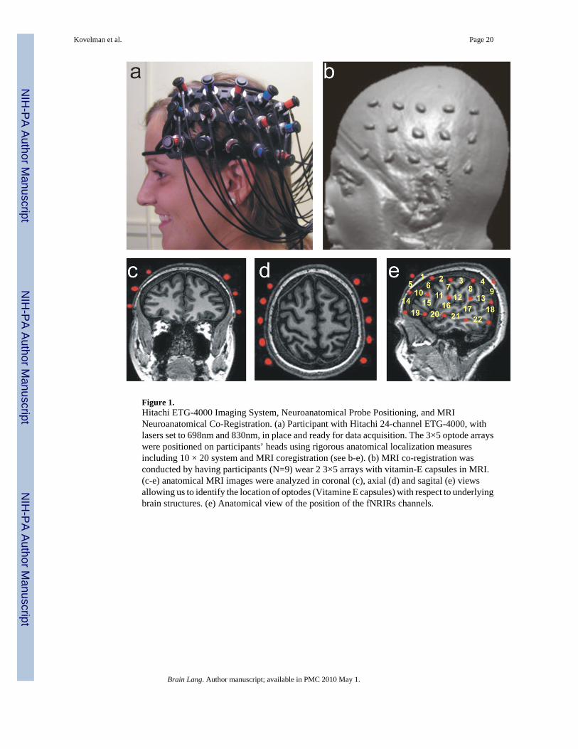
Figure 1.Hitachi ETG-4000 Imaging System, Neuroanatomical Probe Positioning, and MRINeuroanatomical Co-Registration. (a) Participant with Hitachi 24-channel ETG-4000, withlasers set to 698nm and 830nm, in place and ready for data acquisition. The 3×5 optode arrayswere positioned on participants’ heads using rigorous anatomical localization measuresincluding 10 × 20 system and MRI coregistration (see b-e). (b) MRI co-registration wasconducted by having participants (N=9) wear 2 3×5 arrays with vitamin-E capsules in MRI.(c-e) anatomical MRI images were analyzed in coronal (c), axial (d) and sagital (e) viewsallowing us to identify the location of optodes (Vitamine E capsules) with respect to underlyingbrain structures. (e) Anatomical view of the position of the fNRIRs channels.
Kovelman et al. Page 20
Brain Lang. Author manuscript; available in PMC 2010 May 1.
NIH
-PA Author Manuscript
NIH
-PA Author Manuscript
NIH
-PA Author Manuscript
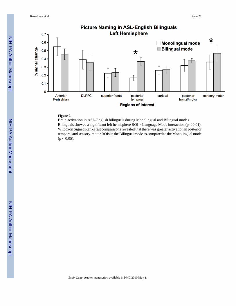
Figure 2.Brain activation in ASL-English bilinguals during Monolingual and Bilingual modes.Bilinguals showed a significant left hemisphere ROI × Language Mode interaction (p < 0.01).Wilcoxon Signed Ranks test comparisons revealed that there was greater activation in posteriortemporal and sensory-motor ROIs in the Bilingual mode as compared to the Monolingual mode(p < 0.05).
Kovelman et al. Page 21
Brain Lang. Author manuscript; available in PMC 2010 May 1.
NIH
-PA Author Manuscript
NIH
-PA Author Manuscript
NIH
-PA Author Manuscript
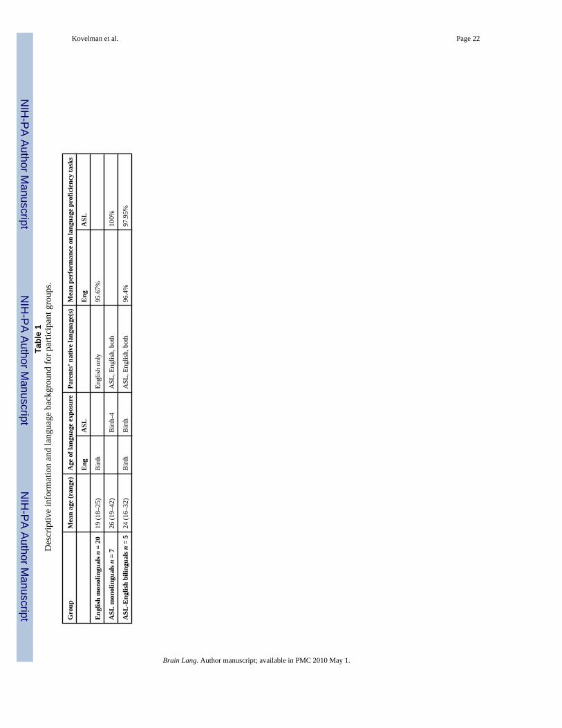
NIH
-PA Author Manuscript
NIH
-PA Author Manuscript
NIH
-PA Author Manuscript
Kovelman et al. Page 22Ta
ble
1D
escr
iptiv
e in
form
atio
n an
d la
ngua
ge b
ackg
roun
d fo
r par
ticip
ant g
roup
s.
Gro
upM
ean
age
(ran
ge)
Age
of l
angu
age
expo
sure
Pare
nts’
nat
ive
lang
uage
(s)
Mea
n pe
rfor
man
ce o
n la
ngua
ge p
rofic
ienc
y ta
sks
Eng
ASL
Eng
ASL
Eng
lish
mon
olin
gual
s n =
20
19 (1
8–25
)B
irth
Engl
ish
only
95.6
7%
ASL
mon
olin
gual
s n =
726
(19–
42)
Birt
h-4
ASL
, Eng
lish,
bot
h10
0%
ASL
-Eng
lish
bilin
gual
s n =
524
(16–
32)
Birt
hB
irth
ASL
, Eng
lish,
bot
h96
.4%
97.9
5%
Brain Lang. Author manuscript; available in PMC 2010 May 1.
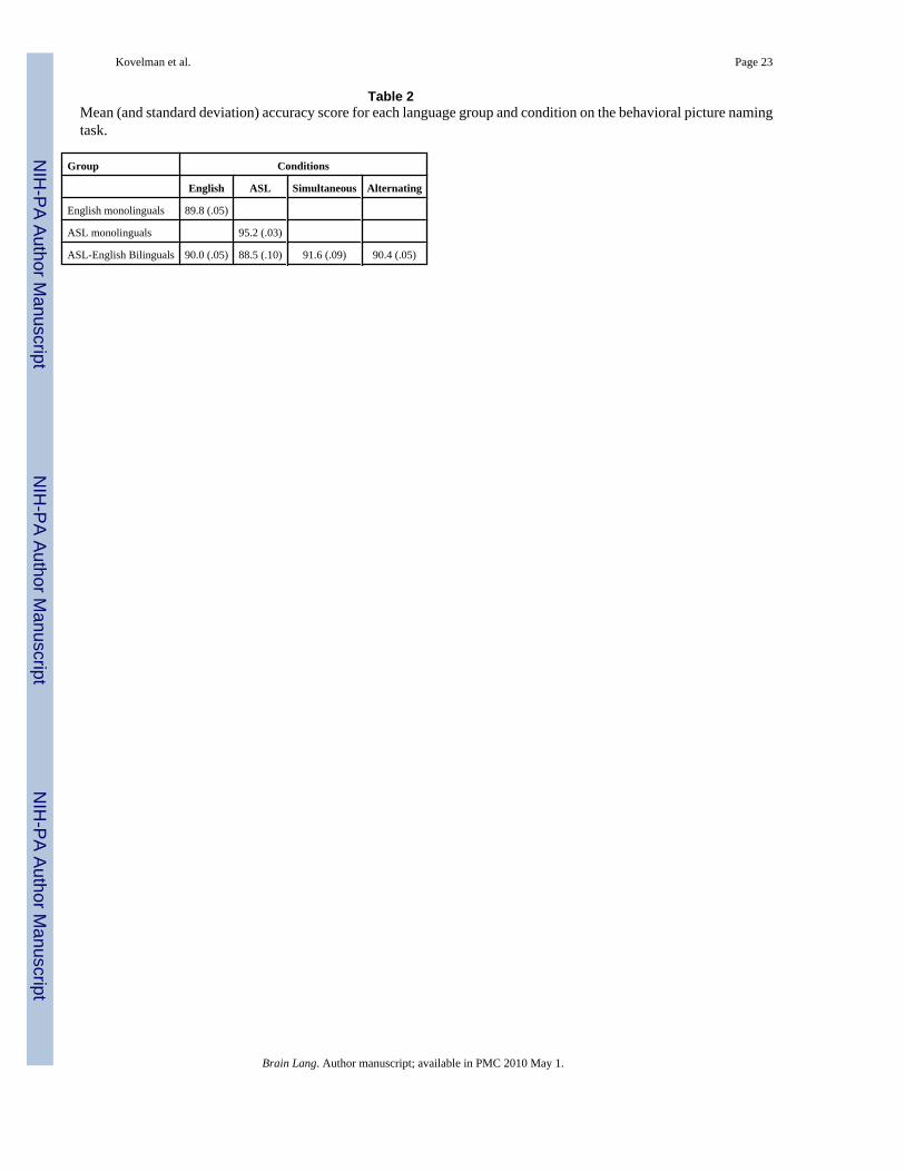
NIH
-PA Author Manuscript
NIH
-PA Author Manuscript
NIH
-PA Author Manuscript
Kovelman et al. Page 23
Table 2Mean (and standard deviation) accuracy score for each language group and condition on the behavioral picture namingtask.
Group Conditions
English ASL Simultaneous Alternating
English monolinguals 89.8 (.05)
ASL monolinguals 95.2 (.03)
ASL-English Bilinguals 90.0 (.05) 88.5 (.10) 91.6 (.09) 90.4 (.05)
Brain Lang. Author manuscript; available in PMC 2010 May 1.
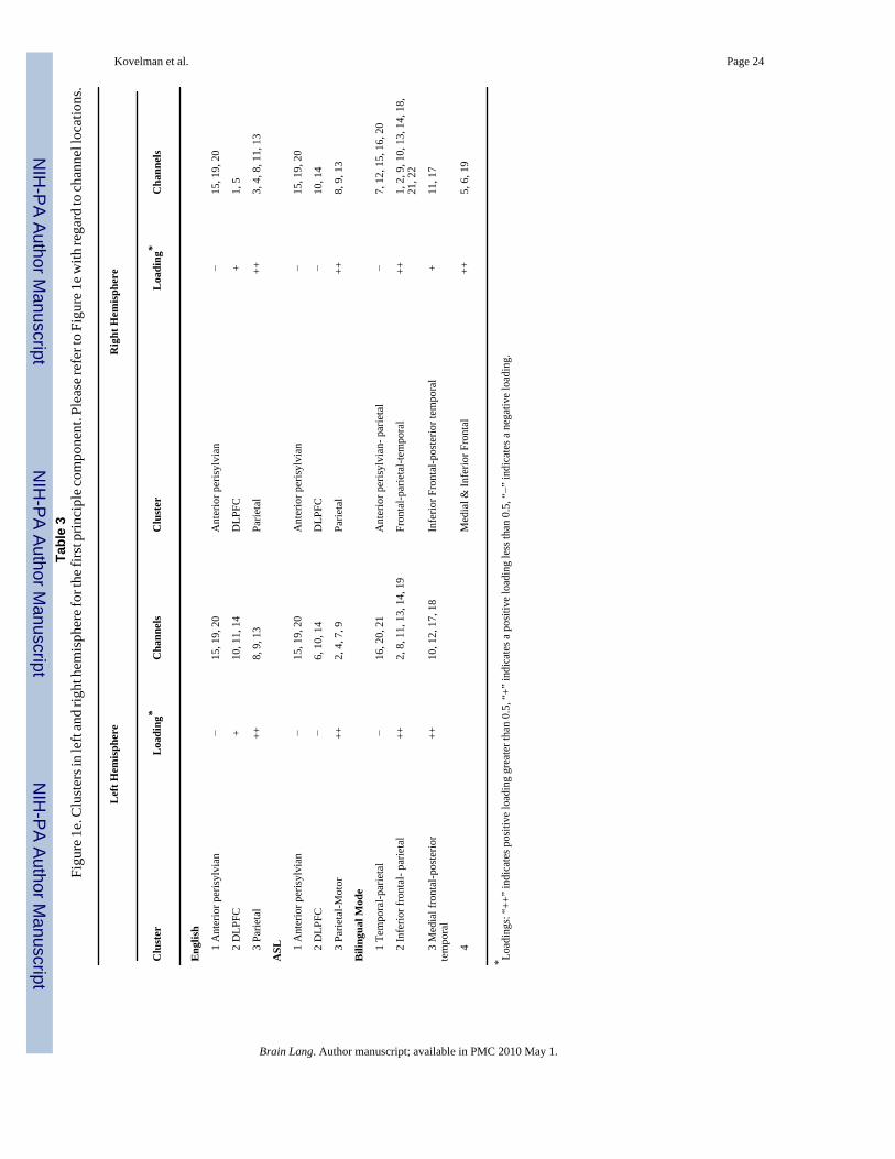
NIH
-PA Author Manuscript
NIH
-PA Author Manuscript
NIH
-PA Author Manuscript
Kovelman et al. Page 24Ta
ble
3Fi
gure
1e.
Clu
ster
s in
left
and
right
hem
isph
ere f
or th
e firs
t prin
cipl
e com
pone
nt. P
leas
e ref
er to
Fig
ure 1
e with
rega
rd to
chan
nel l
ocat
ions
.
Lef
t Hem
isph
ere
Rig
ht H
emis
pher
e
Clu
ster
Loa
ding
*C
hann
els
Clu
ster
Loa
ding
*C
hann
els
Eng
lish
1
Ant
erio
r per
isyl
vian
−15
, 19,
20
Ant
erio
r per
isyl
vian
−15
, 19,
20
2
DLP
FC+
10, 1
1, 1
4D
LPFC
+1,
5
3
Parie
tal
++8,
9, 1
3Pa
rieta
l++
3, 4
, 8, 1
1, 1
3
ASL
1
Ant
erio
r per
isyl
vian
−15
, 19,
20
Ant
erio
r per
isyl
vian
−15
, 19,
20
2
DLP
FC−
6, 1
0, 1
4D
LPFC
−10
, 14
3
Parie
tal-M
otor
++2,
4, 7
, 9Pa
rieta
l++
8, 9
, 13
Bili
ngua
l Mod
e
1
Tem
pora
l-par
ieta
l−
16, 2
0, 2
1A
nter
ior p
eris
ylvi
an- p
arie
tal
−7,
12,
15,
16,
20
2
Infe
rior f
ront
al- p
arie
tal
++2,
8, 1
1, 1
3, 1
4, 1
9Fr
onta
l-par
ieta
l-tem
pora
l++
1, 2
, 9, 1
0, 1
3, 1
4, 1
8,21
, 22
3
Med
ial f
ront
al-p
oste
rior
tem
pora
l++
10, 1
2, 1
7, 1
8In
ferio
r Fro
ntal
-pos
terio
r tem
pora
l+
11, 1
7
4
Med
ial &
Infe
rior F
ront
al++
5, 6
, 19
* Load
ings
: “++
” in
dica
tes p
ositi
ve lo
adin
g gr
eate
r tha
n 0.
5, “
+” in
dica
tes a
pos
itive
load
ing
less
than
0.5
, “−”
indi
cate
s a n
egat
ive
load
ing.
Brain Lang. Author manuscript; available in PMC 2010 May 1.
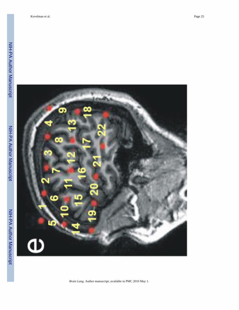
NIH
-PA Author Manuscript
NIH
-PA Author Manuscript
NIH
-PA Author Manuscript
Kovelman et al. Page 25
Brain Lang. Author manuscript; available in PMC 2010 May 1.
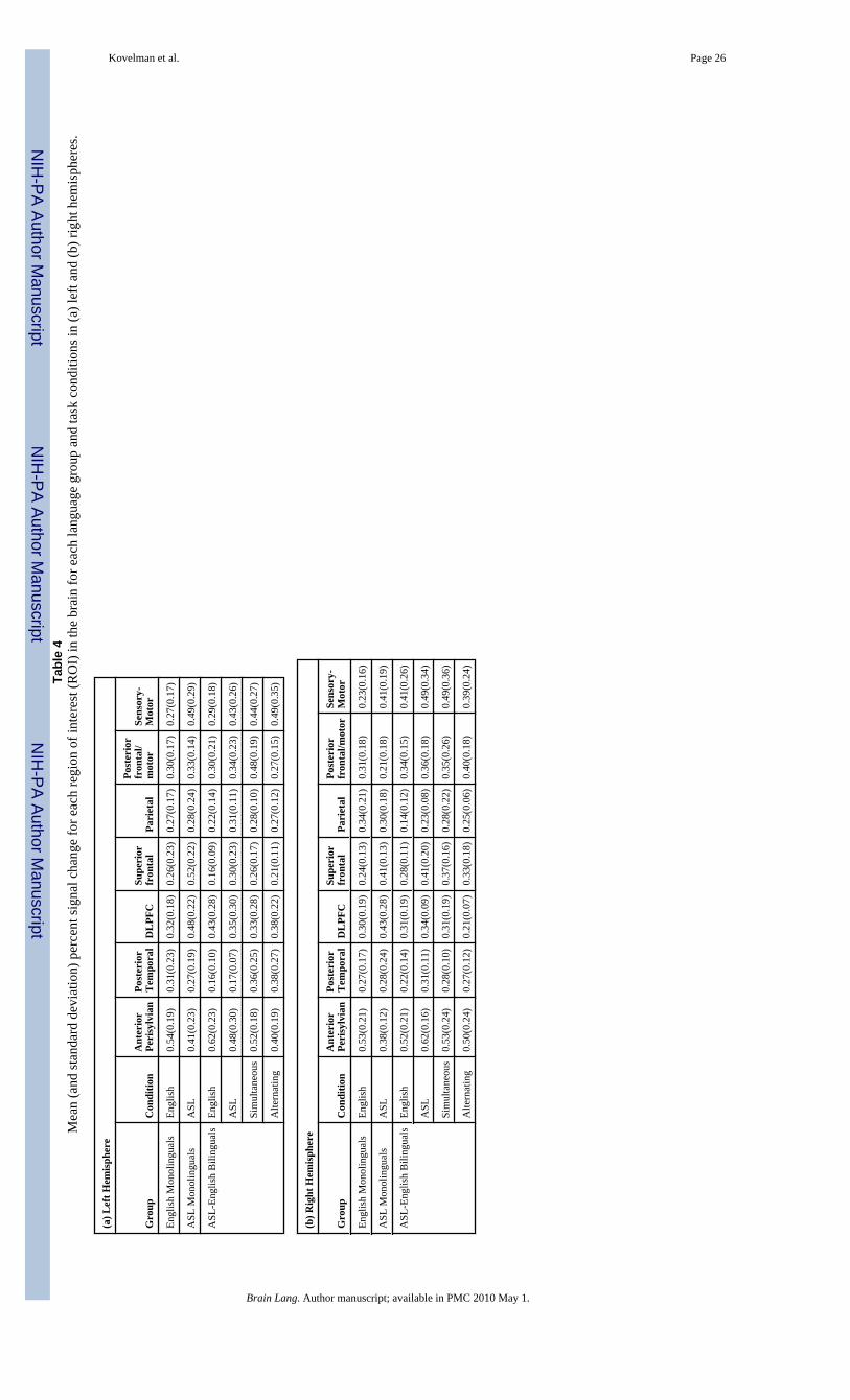
NIH
-PA Author Manuscript
NIH
-PA Author Manuscript
NIH
-PA Author Manuscript
Kovelman et al. Page 26Ta
ble
4M
ean
(and
stan
dard
dev
iatio
n) p
erce
nt si
gnal
cha
nge
for e
ach
regi
on o
f int
eres
t (R
OI)
in th
e br
ain
for e
ach
lang
uage
gro
up a
nd ta
sk c
ondi
tions
in (a
) lef
t and
(b) r
ight
hem
isph
eres
.
(a) L
eft H
emis
pher
e
Gro
upC
ondi
tion
Ant
erio
rPe
risy
lvia
nPo
ster
ior
Tem
pora
lD
LPF
CSu
peri
orfr
onta
lPa
riet
al
Post
erio
rfr
onta
l/m
otor
Sens
ory-
Mot
or
Engl
ish
Mon
olin
gual
sEn
glis
h0.
54(0
.19)
0.31
(0.2
3)0.
32(0
.18)
0.26
(0.2
3)0.
27(0
.17)
0.30
(0.1
7)0.
27(0
.17)
ASL
Mon
olin
gual
sA
SL0.
41(0
.23)
0.27
(0.1
9)0.
48(0
.22)
0.52
(0.2
2)0.
28(0
.24)
0.33
(0.1
4)0.
49(0
.29)
ASL
-Eng
lish
Bili
ngua
lsEn
glis
h0.
62(0
.23)
0.16
(0.1
0)0.
43(0
.28)
0.16
(0.0
9)0.
22(0
.14)
0.30
(0.2
1)0.
29(0
.18)
ASL
0.48
(0.3
0)0.
17(0
.07)
0.35
(0.3
0)0.
30(0
.23)
0.31
(0.1
1)0.
34(0
.23)
0.43
(0.2
6)
Sim
ulta
neou
s0.
52(0
.18)
0.36
(0.2
5)0.
33(0
.28)
0.26
(0.1
7)0.
28(0
.10)
0.48
(0.1
9)0.
44(0
.27)
Alte
rnat
ing
0.40
(0.1
9)0.
38(0
.27)
0.38
(0.2
2)0.
21(0
.11)
0.27
(0.1
2)0.
27(0
.15)
0.49
(0.3
5)
(b) R
ight
Hem
isph
ere
Gro
upC
ondi
tion
Ant
erio
rPe
risy
lvia
nPo
ster
ior
Tem
pora
lD
LPF
CSu
peri
orfr
onta
lPa
riet
alPo
ster
ior
fron
tal/m
otor
Sens
ory-
Mot
or
Engl
ish
Mon
olin
gual
sEn
glis
h0.
53(0
.21)
0.27
(0.1
7)0.
30(0
.19)
0.24
(0.1
3)0.
34(0
.21)
0.31
(0.1
8)0.
23(0
.16)
ASL
Mon
olin
gual
sA
SL0.
38(0
.12)
0.28
(0.2
4)0.
43(0
.28)
0.41
(0.1
3)0.
30(0
.18)
0.21
(0.1
8)0.
41(0
.19)
ASL
-Eng
lish
Bili
ngua
lsEn
glis
h0.
52(0
.21)
0.22
(0.1
4)0.
31(0
.19)
0.28
(0.1
1)0.
14(0
.12)
0.34
(0.1
5)0.
41(0
.26)
ASL
0.62
(0.1
6)0.
31(0
.11)
0.34
(0.0
9)0.
41(0
.20)
0.23
(0.0
8)0.
36(0
.18)
0.49
(0.3
4)
Sim
ulta
neou
s0.
53(0
.24)
0.28
(0.1
0)0.
31(0
.19)
0.37
(0.1
6)0.
28(0
.22)
0.35
(0.2
6)0.
49(0
.36)
Alte
rnat
ing
0.50
(0.2
4)0.
27(0
.12)
0.21
(0.0
7)0.
33(0
.18)
0.25
(0.0
6)0.
40(0
.18)
0.39
(0.2
4)
Brain Lang. Author manuscript; available in PMC 2010 May 1.
Related Documents

![Functional Near-Infrared Spectroscopy (fNIRS) during Apnoeabiosignalsplux.com/downloads/docs/technical-notes/... · A functional near-infrared spectroscopy (fNIRS) sensor [7] uses](https://static.cupdf.com/doc/110x72/5fbd4343fe93b80102432136/functional-near-infrared-spectroscopy-fnirs-during-a-functional-near-infrared.jpg)


