Drug Targeting Organ-Specific Strategies Edited by Grietje Molema and Dirk K. F. Meijer Drug Targeting Organ-Specific Strategies. Edited by G. Molema, D. K. F. Meijer Copyright © 2001 Wiley-VCH Verlag GmbH ISBNs: 3-527-29989-0 (Hardcover); 3-527-60006-X (Electronic)

Drug Targeting Organ-specific Strategies
Nov 03, 2014
science
Welcome message from author
This document is posted to help you gain knowledge. Please leave a comment to let me know what you think about it! Share it to your friends and learn new things together.
Transcript

Drug TargetingOrgan-Specific StrategiesEdited by Grietje Molema and Dirk K. F. Meijer
Drug Targeting Organ-Specific Strategies. Edited by G. Molema, D. K. F. MeijerCopyright © 2001 Wiley-VCH Verlag GmbH
ISBNs: 3-527-29989-0 (Hardcover); 3-527-60006-X (Electronic)

Methods and Principlesin Medicinal Chemistry
Edited byR. MannholdH. KubinyiH. Timmerman
Editorial Board
G. Folkers, H.-D. Höltje, J. Vacca,H. van de Waterbeemd, T. Wieland
Drug Targeting Organ-Specific Strategies. Edited by G. Molema, D. K. F. MeijerCopyright © 2001 Wiley-VCH Verlag GmbH
ISBNs: 3-527-29989-0 (Hardcover); 3-527-60006-X (Electronic)

Weinheim · New York · Chichester · Brisbane · Singapore · Toronto
Drug TargetingOrgan-Specific Strategies
Edited by Grietje Molema and Dirk K. F. Meijer
Drug Targeting Organ-Specific Strategies. Edited by G. Molema, D. K. F. MeijerCopyright © 2001 Wiley-VCH Verlag GmbH
ISBNs: 3-527-29989-0 (Hardcover); 3-527-60006-X (Electronic)

Series Editors:Prof. Dr. Raimund Mannhold Prof. Dr. Hugo Kubinyi Prof. Dr. Hendrik TimmermanBiomedical Research Center BASF AG Ludwigshafen Faculty of ChemistryMolecular Drug Research Group c/o Donnersbergstrasse 9 Dept. of PharmacochemistryHeinrich-Heine-Universität D-67256 Weisenheim am Sand Free University of AmsterdamUniversitätsstraße 1 Germany De Boelelaan 1083D-40225 Düsseldorf NL-1081 HV AmsterdamGermany The Netherlands
Volume Editors:Dr. Grietje Molema Prof. Dr. Dirk K. F. MeijerUniversity Centre for Pharmacy University Centre for PharmacyDepartment of Pharmacokinetics Department of Pharmacokineticsand Drug Delivery and Drug DeliveryAntonius Deusinglaan 1 Antonius Deusinglaan 1NL-9713 AV Groningen NL-9713 AV GroningenThe Netherlands The Netherlands
This book was carefully produced. Nevertheless, authors, editors and publisher do not warrant theinformation contained therein to be free of errors. Readers are advised to keep in mind that statements,data, illustrations, procedural details or other items may inadvertently be inaccurate.
Library of Congress Card No. applied for.
British Library Cataloguing-in-Publication Data: A catalogue record for this book is available from the BritishLibrary.
Die Deutsche Bibliothek – CIP Cataloguing-in-Publication-DataA catalogue record for this publication is available from Die Deutsche Bibliothek
ISBN 3-527-29989-0
© WILEY-VCH Verlag GmbH, D-69469 Weinheim (Federal Republic of Germany), 2001
Printed on acid-free paper.
All rights reserved (including those of translation into other languages). No part of this book may be reproducedin any form – by photoprinting, microfilm, or any other means – nor transmitted or translated into a machinelanguage without written permission from the publishers. Registered names, trademarks, etc. used in this book,even when not specifically marked as such, are not to be considered unprotected by law.
Composition: Datascan GmbH, D-64295 DarmstadtPrinting: betz-druck GmbH, D-63291 DarmstadtBookbinding: Wilhelm Osswald & Co., D-67433 Neustadt (Weinstraße)
Printed in the Federal Republik of Germany.
Drug Targeting Organ-Specific Strategies. Edited by G. Molema, D. K. F. MeijerCopyright © 2001 Wiley-VCH Verlag GmbH
ISBNs: 3-527-29989-0 (Hardcover); 3-527-60006-X (Electronic)
Preface
It is our prime intention to cover the topics of this series as comprehensively as possible.Thus, we are very pleased to introduce this volume focussing on organ specific strategies ofdrug targeting.
About hundred years ago Paul Ehrlich put forward his theory of “the magic bullet” as anapproach to tame disease. Scientists have ever since worked on the principle of drug target-ing based on this idea of specifically delivering drugs to diseased cells. Especially nowadaysthat by high-throughput screening and molecular modelling techniques highly potent drugsare being developed that interfere with general (signal transduction) processes in cells in thebody, the need for their application by a drug targeting approach has almost become in-evitable.
Progress in the field of drug targeting has been slow till thirty years ago. With the adventof the monoclonal antibody technology in the mid seventies of the last century as well as thedevelopment of liposomal devices as carriers did the drug targeting field expand and did theclinical application become a feasible aim.
Monoclonal antibodies, liposomes, polymers, proteins, and many other entities have eversince seen the light as carrier molecules. And, as with most technological developments, theyhave all encountered a vast array of difficulties, ranging from problems in the synthesis of thecarriers and drug conjugates to unfavorable pharmacokinetics and toxicity. Furthermore,lack of knowledge on the anatomical and physiological barriers in the body hampered appli-cation. However, many problems have been solved, not in the least due to the advent of re-combinant DNA technology to construct better defined carriers that can be produced inlarge amounts, and advanced pharmaceutical formulation technology. Similarly, the rapid de-velopments in molecular biology, cell biology and immunology led to a better understandingof the processes taking place in vivo upon administration of carriers and conjugates. Impor-tant conclusion is that drug targeting has become a multidisciplinary research area.
What has been achieved until now? In the year 2001, several liposome and antibody basedstrategies have been or will soon be approved for clinical application, some for the treatmentof cancer, some for the treatment of bacterial infections, some for chronic inflammatory dis-eases. Furthermore many monoclonal antibodies without a drug or pharmacologically activemolecule attached are in the clinic. Their intrinsic targeting and effector function is obvious-ly sufficient for the pharmacological effect.
Only a few polymer or protein based drug targeting strategies have reached the clinic andan important question in the coming years will be whether these strategies eventually willreach it.All will depend on their effectiveness and improved toxicity profiles as compared tofree drug only and the ease of their production at large scale.
The present volume is in several respects unique. It provides a map of the body from theviewpoint of drug targeting/drug delivery. Potentials and limitations of targeting strategies
Drug Targeting Organ-Specific Strategies. Edited by G. Molema, D. K. F. MeijerCopyright © 2001 Wiley-VCH Verlag GmbH
ISBNs: 3-527-29989-0 (Hardcover); 3-527-60006-X (Electronic)

are discussed in the light of organ related diseases for each organ separately. Furthermore,novel technologies are described that may be useful in the future to allow an even betterproduct to be developed that can be clinically exploited at a more rapid pace.
The series editors are grateful to the contributors to this volume, in particular GrietjeMolema and Dirk K. F. Meijer, as well as Wiley-VCH publishers, for the fruitful collaborationand the straightforward realization of this project.
January 2001 Raimund Mannhold, DüsseldorfHugo Kubinyi, LudwigshafenHenk Timmerman, Amsterdam
VI Preface

Foreword
It was in the mid-1970s I think, just a few years after Brenda Ryman and I introduced lipo-somes as a drug delivery system, when a well meaning colleague af mine advised me not toput all my eggs in one basket.The eggs were liposomes and the basket my career.At the sametime there were all sorts of prophecies and rumours from a variety of quarters about liposo-mal stability problems, expense, toxicity, difficulties with large scale manufacture, etc. Somewent as far as to dismiss the system as a flash in the pan phenomenon. Indeed, the yellowbrick road to the magic bullet is littered with systems that once made the headlines and thenfell by the wayside. So, such comments on liposomes, and later on on antibodies, were not sur-prising. I believe that what made many of us persevere throughout the decades in developingdrug carrier systems such as liposomes, and associated technologies was the realization that,for the foreseeable future at least, molecular modelling is not the answer to drug selectivityfor most therapeutics. The vagaries of the biological milieu in vivo ensures that optimal drugaction (seen in the test tube) is compromised by such factors as opsonins and proteolytic en-zymes in the bloodstream, membrane barriers, loss through the kidneys, and premature in-terception of therapeutics by the reticuloendothelial system. In the case of liposomes, mono-clonal antibodies and some polymers, carrier development was greatly facilitated by theirstructural versatility which enabled the design of advanced versions of unique sophistication.
The first generation of liposome-based systems approved for clinical use are believed tofunction on the basis of their passive uptake by the target tissues (e.g. the AmBisome and thevirosome vaccine Hepaxal) or by avoiding certain tissues (e.g. heart, kidneys) that are proneto damage by the drug when given as such (e.g. Doxil, Daunoxome). The next challenge is tocreate or build on the systems that can be actively targeted to specific tissues or circulationcells for which systems such as liposomes have little or no affinity. They include a variety ofmolecules with genuine targeting properties, for instance (neo-) glycoproteins, monoclonalantibodies and fragments thereof, applied either as a means to deliver drugs attached to thesebiopolymers, or as homing devices when attached to the surface of other drug delivery sys-tems, for instance liposomes and other particle-type carriers. Success to that end will greatlyenlarge the spectrum of therapeutics that can be selectively delivered, and widen the rangeof applications.
In this respect, Grietje Molema, Dirk K. F. Meijer and a team of drug delivery experts havetaken an important step with the present book. Unlike previous volumes, this one is not de-voted exclusively to liposome or antibody technologies. Rather, the book deals with organ-specific drug targeting strategies developed for the treatment of a wide spectrum of diseasesand includes a collection of novel techniques applied to drug targeting research. Thus, thebook provides a blueprint for both the experienced and the semi-experienced reader inter-ested in drug targeting and related optimization strategies.
London, 2001 Gregory Gregoriadis
Drug Targeting Organ-Specific Strategies. Edited by G. Molema, D. K. F. MeijerCopyright © 2001 Wiley-VCH Verlag GmbH
ISBNs: 3-527-29989-0 (Hardcover); 3-527-60006-X (Electronic)
List of Contributors
Jan-Willem Arends
Maastricht UniversityDepartment of PathologyPO Box 6166200 MD Maastrichtthe Netherlands
Sigridur A. Ásgeirsdóttir
Groningen University Institute for DrugExploration (GUIDE)Department of Pharmacokinetics and DrugDeliveryAnt. Deusinglaan 19713 AV Groningenthe [email protected]
Leonie Beljaars
Groningen University Institute for DrugExploration (GUIDE)Department of Pharmacokinetics and DrugDeliveryAnt. Deusinglaan 19713 AV Groningenthe [email protected]
Ulrich Bickel
Texas Tech University HSCSchool of PharmacyPharmaceutical Sciences1300 S Coulter Amarillo, Texas [email protected]
Anne H. de Boer
Groningen University Institute for DrugExploration (GUIDE)Department of Pharmaceutical Technologyand BiopharmacyAnt. Deusinglaan 19713 AV Groningenthe [email protected]
Maaike Everts
Groningen University Institute for DrugExploration (GUIDE)Department of Pharmacokinetics and DrugDeliveryAnt. Deusinglaan 19713 AV Groningenthe [email protected]
Drug Targeting Organ-Specific Strategies. Edited by G. Molema, D. K. F. MeijerCopyright © 2001 Wiley-VCH Verlag GmbH
ISBNs: 3-527-29989-0 (Hardcover); 3-527-60006-X (Electronic)
R. Folgert G. Haverdings
Groningen University Institute for DrugExploration (GUIDE)Department of Pharmacokinetics and DrugDeliveryAnt. Deusinglaan 19713 AV Groningenthe [email protected]
Wijnand Helfrich
Groningen University Institute for DrugExploration (GUIDE)Department of Pathology and LaboratoryMedicineMedical Biology SectionTumor Immunology LaboratoryHanzeplein 19713 GZ Groningenthe [email protected]
Hennie R. Hoogenboom
Dyax bvPO Box 58006202 AZ Maastrichtthe [email protected]
Jörg Huwyler
F. Hoffmann-LaRoche Ltd.CNS ResearchPRBN, Bldg. 68/323aCH-4070 [email protected]
Henderik W. Frijlink
Groningen University Institute for DrugExploration (GUIDE)Department of Pharmaceutical Technologyand BiopharmacyAnt. Deusinglaan 19713 AV Groningenthe [email protected]
Arjan W. Griffioen
Maastricht University/University Hospital MaastrichtDepartment of Internal MedicineTumor Angiogenesis LaboratoryPeter Debyelaan 256202 AZ Maastrichtthe [email protected]
Geny M. M. Groothuis
Groningen University Institute for DrugExploration (GUIDE)Department of Pharmacokinetics and DrugDeliveryAnt. Deusinglaan 19713 AV Groningenthe [email protected]
Marijke Haas
Groningen University Institute for DrugExploration (GUIDE)Department of Clinical PharmacologyAnt. Deusinglaan 19713 AV Groningenthe [email protected]
X List of Contributors

List of Contributors XI
Young-Sook Kang
Physiology and Pathophysiology LaboratoryCollege of PharmacySookmyung Women’s UniversityChungpa-dong 2 ga 53-12Yongsan gu, [email protected]
Yukio Kato
University of TokyoGraduate School of Pharmaceutical Sciences7-3-1 HongoBunkyo-kuTokyo [email protected]
Robbert J. Kok
Groningen University Institute for DrugExploration (GUIDE)Department of Pharmacokinetics and DrugDeliveryAnt. Deusinglaan 19713 AV Groningenthe [email protected]
Jos G. W. Kosterink
University Hospital GroningenDepartment of Hospital and Clinical PharmacyHanzeplein 19713 GZ Groningenthe [email protected]
Lou F. M. H. de Leij
Groningen University Institute for DrugExploration (GUIDE)Department of Pathology and LaboratoryMedicineMedical Biology SectionTumor Immunology LaboratoryHanzeplein 19713 GZ Groningenthe [email protected]
Claudia S. Leopold
University of LeipzigDepartment of Pharmaceutical TechnologySchönauer Str. 16004207 [email protected]
Dirk K. F. Meijer
Groningen University Institute for DrugExploration (GUIDE)Department of Pharmacokinetics and DrugDeliveryAnt. Deusinglaan 19713 AV Groningenthe [email protected]
Barbro N. Melgert
Groningen University Institute for DrugExploration (GUIDE)Department of Pharmacokinetics and DrugDeliveryAnt. Deusinglaan 19713 AV Groningenthe [email protected]

Grietje Molema
Groningen University Institute for DrugExploration (GUIDE)Department of Pharmacokinetics and DrugDelivery and Department of Pathology and LaboratoryMedicineMedical Biology SectionTumor Immunology LaboratoryAnt. Deusinglaan 19713 AV Groningenthe [email protected]
Frits Moolenaar
Groningen University Institute for DrugExploration (GUIDE)Department of Pharmacokinetics and DrugDeliveryAnt. Deusinglaan 19713 AV Groningenthe [email protected]
Ricardo Mutuberria
Maastricht UniversityDepartment of PathologyPO Box 6166200 MD Maastrichtthe Netherlands
Peter Olinga
Groningen University Institute for DrugExploration (GUIDE)Department of Pharmacokinetics and DrugDeliveryAnt. Deusinglaan 19713 AV Groningenthe [email protected]
Klaas Poelstra
Groningen University Institute for DrugExploration (GUIDE)Department of Pharmacokinetics and DrugDeliveryAnt. Deusinglaan 19713 AV Groningenthe [email protected]
Johannes H. Proost
Groningen University Institute for DrugExploration (GUIDE)Department of Pharmacokinetics and DrugDeliveryAnt. Deusinglaan 19713 AV Groningenthe [email protected]
S. Ramakrishnan
University of MinnesotaHealth Science CenterDepartment of Pharmacology321 Church StreetMinneapolisMinnesota [email protected]
XII List of Contributors

Daisy W. J. van der Schaft
Maastricht University/University Hospital MaastrichtDepartment of Internal MedicineTumor Angiogenesis LaboratoryPeter Debyelaan 256202 AZ Maastrichtthe [email protected]
Astrid J. Schraa
Groningen University Institute for DrugExploration (GUIDE)Department of Pathology and LaboratoryMedicineMedical Biology SectionTumor Immunology LaboratoryHanzeplein 19713 GZ Groningenthe [email protected]
Yuichi Sugiyama
University of TokyoGraduate School of Pharmaceutical Sciences7-3-1 HongoBunkyo-kuTokyo [email protected]
Kokichi Suzuki
Meiji Seika Kaisha Ltd760 Moro-oka-choKohoku-kuYokohama City 222-0002Japan
Willem R. Verweij
Groningen University Institute for DrugExploration (GUIDE)Department of Pharmacokinetics and DrugDeliveryAnt. Deusinglaan 19713 AV Groningenthe [email protected]
Dick de Zeeuw
Groningen University Institute for DrugExploration (GUIDE)Department of Clinical PharmacologyAnt. Deusinglaan 19713 AV Groningenthe [email protected]
List of Contributors XIII

Preface . . . . . . . . . . . . . . . . . . . . . . . . . . . . . . . . . . . . . . . . . . . . V
Foreword . . . . . . . . . . . . . . . . . . . . . . . . . . . . . . . . . . . . . . . . . . . VII
List of Contributors . . . . . . . . . . . . . . . . . . . . . . . . . . . . . . . . . . . . . XXI
1 Drug Targeting: Basic Concepts and Novel AdvancesGrietje Molema . . . . . . . . . . . . . . . . . . . . . . . . . . . . . . . . . . 1
1.1 Introduction . . . . . . . . . . . . . . . . . . . . . . . . . . . . . . . . . . . . . 11.2 Carriers used in Drug Targeting . . . . . . . . . . . . . . . . . . . . . . . . . . 2
1.2.1 Liposomes . . . . . . . . . . . . . . . . . . . . . . . . . . . . . . . . . 21.2.2 Monoclonal Antibodies and Fragments . . . . . . . . . . . . . . . . . 31.2.3 Modified (Plasma) Proteins . . . . . . . . . . . . . . . . . . . . . . . 41.2.4 Soluble Polymers . . . . . . . . . . . . . . . . . . . . . . . . . . . . . 41.2.5 Lipoproteins . . . . . . . . . . . . . . . . . . . . . . . . . . . . . . . . 61.2.6 Microspheres and Nanoparticles . . . . . . . . . . . . . . . . . . . . . 61.2.7 Polymeric Micelles . . . . . . . . . . . . . . . . . . . . . . . . . . . . 71.2.8 Cellular Carriers . . . . . . . . . . . . . . . . . . . . . . . . . . . . . . 7
1.3 Intracellular Routing of Drug–Carrier Complex . . . . . . . . . . . . . . . . 81.3.1 Passive Versus Active Drug Targeting . . . . . . . . . . . . . . . . . . 81.3.2 Lysosomes as a Cellular Target Compartment . . . . . . . . . . . . . 81.3.3 Cytoplasmic Delivery . . . . . . . . . . . . . . . . . . . . . . . . . . . 91.3.4 Nuclear Targeting . . . . . . . . . . . . . . . . . . . . . . . . . . . . . 91.3.5 Mitochondrial Targeting . . . . . . . . . . . . . . . . . . . . . . . . . 10
1.4 Drug Targeting Strategies in the Clinic . . . . . . . . . . . . . . . . . . . . . . 101.4.1 Liposome Based Therapies in the Clinic . . . . . . . . . . . . . . . . 111.4.2 Monoclonal Antibody Therapies in the Clinic . . . . . . . . . . . . . 111.4.3 Monoclonal Antibody Based Targeting Strategies in the Clinic . . . . 131.4.4 Other Drug Targeting Strategies in the Clinic . . . . . . . . . . . . . 13
1.5 Vaccination Strategies for Enhanced Immunity . . . . . . . . . . . . . . . . . 151.6 Drug Targeting as a Research Tool to Study Disease . . . . . . . . . . . . . . 16
Contents
Drug Targeting Organ-Specific Strategies. Edited by G. Molema, D. K. F. MeijerCopyright © 2001 Wiley-VCH Verlag GmbH
ISBNs: 3-527-29989-0 (Hardcover); 3-527-60006-X (Electronic)
1.7 Challenges in Drug Targeting Research . . . . . . . . . . . . . . . . . . . . . 181.8 Conclusions . . . . . . . . . . . . . . . . . . . . . . . . . . . . . . . . . . . . . 19References . . . . . . . . . . . . . . . . . . . . . . . . . . . . . . . . . . . . . . . . . . 20
2 Brain-Specific Drug Targeting StrategiesUlrich Bickel, Young-Sook Kang, Jörg Huwyler . . . . . . . . . . . . . . . . . 23
2.1 Introduction . . . . . . . . . . . . . . . . . . . . . . . . . . . . . . . . . . . . . 232.2 Overview of Central Nervous System Diseases . . . . . . . . . . . . . . . . . 23
2.2.1 Neurodegenerative Diseases . . . . . . . . . . . . . . . . . . . . . . . 232.2.1.1 Alzheimer Disease (AD) . . . . . . . . . . . . . . . . . . . 232.2.1.2 Parkinson’s Disease . . . . . . . . . . . . . . . . . . . . . . 24
2.2.2 Cerebrovascular Disease . . . . . . . . . . . . . . . . . . . . . . . . . 252.2.3 Brain Tumours . . . . . . . . . . . . . . . . . . . . . . . . . . . . . . . 252.2.4 HIV Infection . . . . . . . . . . . . . . . . . . . . . . . . . . . . . . . 25
2.3 BBB Biology and Pharmacology . . . . . . . . . . . . . . . . . . . . . . . . . 262.3.1 Physiological Transport Systems . . . . . . . . . . . . . . . . . . . . . 28
2.3.1.1 Nutrient Carriers Versus Diffusion-mediated Uptake . . . 282.3.1.2 Efflux Systems . . . . . . . . . . . . . . . . . . . . . . . . . 292.3.1.3 Receptor- and Absorptive-mediated Uptake . . . . . . . . 29
2.3.2 Techniques for Measurement of Brain Uptake . . . . . . . . . . . . . 312.3.2.1 In Vivo Methods . . . . . . . . . . . . . . . . . . . . . . . . 312.3.2.2 In Vitro Models . . . . . . . . . . . . . . . . . . . . . . . . . 34
2.4 Drug Delivery Strategies . . . . . . . . . . . . . . . . . . . . . . . . . . . . . . 342.4.1 Small Molecule Drug Delivery . . . . . . . . . . . . . . . . . . . . . . 352.4.2 Macromolecular Drug Delivery . . . . . . . . . . . . . . . . . . . . . 36
2.4.2.1 Intraventricular Route . . . . . . . . . . . . . . . . . . . . . 362.4.2.2 Intraparenchymal Route . . . . . . . . . . . . . . . . . . . . 372.4.2.3 Convective Flow . . . . . . . . . . . . . . . . . . . . . . . . 392.4.2.4 Delivery by Barrier Disruption . . . . . . . . . . . . . . . . 392.4.2.5 Vector-mediated Delivery . . . . . . . . . . . . . . . . . . . 402.4.2.6 Pharmacological Effects of Chimeric Peptides . . . . . . . 432.4.2.7 Chimeric Peptide Radiopharmaceuticals . . . . . . . . . . . 46
2.4.3 Liposomes as Drug Carriers . . . . . . . . . . . . . . . . . . . . . . . 472.4.3.1 Conventional Liposomes and Small Molecules . . . . . . . 472.4.3.2 Brain Targeting Using Immunoliposomes . . . . . . . . . . 472.4.3.3 Drugs of Interest for Targeting to the Brain . . . . . . . . . 48
2.5 Conclusions . . . . . . . . . . . . . . . . . . . . . . . . . . . . . . . . . . . . . 49References . . . . . . . . . . . . . . . . . . . . . . . . . . . . . . . . . . . . . . . . . . 50
XVI Contents

3 Pulmonary Drug Delivery: Delivery to and Through the LungAnne H. de Boer, Grietje Molema, Henderik W. Frijlink . . . . . . . . . . . . . 53
3.1 Introduction . . . . . . . . . . . . . . . . . . . . . . . . . . . . . . . . . . . . . 533.2 The Respiratory Tract . . . . . . . . . . . . . . . . . . . . . . . . . . . . . . . 55
3.2.1 Lung Capacities and Pulmonary Ventilation . . . . . . . . . . . . . . 553.3 Lung Deposition and Particle Size . . . . . . . . . . . . . . . . . . . . . . . . 573.4 Drug Absorption via the Lung . . . . . . . . . . . . . . . . . . . . . . . . . . 58
3.4.1 Systemic Delivery of Peptides and Proteins . . . . . . . . . . . . . . 603.5 Devices for Therapeutic Aerosol Generation . . . . . . . . . . . . . . . . . . 63
3.5.1 Nebulizers . . . . . . . . . . . . . . . . . . . . . . . . . . . . . . . . . 643.5.2 Metered Dose Inhalers . . . . . . . . . . . . . . . . . . . . . . . . . . 653.5.3 Dry Powder Inhalers . . . . . . . . . . . . . . . . . . . . . . . . . . . 65
3.6 Formulations for Inhalation Products . . . . . . . . . . . . . . . . . . . . . . 673.6.1 Formulations for Nebulizers . . . . . . . . . . . . . . . . . . . . . . . 673.6.2 Formulations for Dry Powder Inhalers . . . . . . . . . . . . . . . . . 673.6.3 Formulations for Peptides and Proteins . . . . . . . . . . . . . . . . . 69
3.7 Variables and Interactions in Dry Powder Inhalation . . . . . . . . . . . . . . 733.8 Airflow Resistance . . . . . . . . . . . . . . . . . . . . . . . . . . . . . . . . . 743.9 Inspiratory Pressure and Relevant Flow Parameters . . . . . . . . . . . . . . 75
3.9.1 Measurement of the Inspiratory Flow Curve . . . . . . . . . . . . . . 773.10 In Vitro Particle Size Analysis and Deposition Measurements . . . . . . . . . 783.11 In Vitro and In Vivo Deposition Efficacy of Inhalation Systems . . . . . . . . 803.12 Targeting Drugs to the Lungs via the Bloodstream . . . . . . . . . . . . . . . 813.13 Final Conclusions and Perspectives . . . . . . . . . . . . . . . . . . . . . . . . 82References . . . . . . . . . . . . . . . . . . . . . . . . . . . . . . . . . . . . . . . . . . 83
4 Cell Specific Delivery of Anti-Inflammatory Drugs to Hepatic Endothelialand Kupffer Cells for the Treatment of Inflammatory Liver DiseasesBarbro N. Melgert, Leonie Beljaars, Dirk K. F. Meijer, Klaas Poelstra . . . . . 89
4.1 Introduction . . . . . . . . . . . . . . . . . . . . . . . . . . . . . . . . . . . . . 894.2 The Liver . . . . . . . . . . . . . . . . . . . . . . . . . . . . . . . . . . . . . . 89
4.2.1 The Parenchymal Cell (PC) . . . . . . . . . . . . . . . . . . . . . . . 914.2.2 The Sinusoidal Endothelial Cell (SEC) . . . . . . . . . . . . . . . . . 91
4.2.2.1 Receptor-mediated Endocytosis . . . . . . . . . . . . . . . 924.2.2.2 Phagocytosis and Transcytosis . . . . . . . . . . . . . . . . . 934.2.2.3 Regulation of the Inflammatory Process by SECs . . . . . 93
4.2.3 The Kupffer Cell (KC) . . . . . . . . . . . . . . . . . . . . . . . . . . 934.2.3.1 Receptor-mediated Endocytosis . . . . . . . . . . . . . . . 944.2.3.2 Phagocytosis . . . . . . . . . . . . . . . . . . . . . . . . . . 944.2.3.3 Regulation of the Inflammatory process by the KC . . . . . 94
4.2.4 The Hepatic Stellate Cell (HSC) . . . . . . . . . . . . . . . . . . . . . 95
Contents XVII

4.3 Hepatic Inflammation and Fibrosis . . . . . . . . . . . . . . . . . . . . . . . . 964.4 Liver Cirrhosis: Causes and Therapy . . . . . . . . . . . . . . . . . . . . . . . 984.5 Drug Targeting to the Liver . . . . . . . . . . . . . . . . . . . . . . . . . . . . 99
4.5.1 Carriers Directed at SECs and KCs . . . . . . . . . . . . . . . . . . . 1004.5.1.1 Albumins . . . . . . . . . . . . . . . . . . . . . . . . . . . . 1004.5.1.2 Liposomes . . . . . . . . . . . . . . . . . . . . . . . . . . . . 1014.5.1.3 Carriers with Intrinsic Anti-inflammatory Activity . . . . . 102
4.5.2 Targeting to other Hepatic Cells . . . . . . . . . . . . . . . . . . . . . 1034.6 Anti-inflammatory Drugs . . . . . . . . . . . . . . . . . . . . . . . . . . . . . 103
4.6.1 Nonsteroidal Anti-inflammatory Drugs (NSAIDs) . . . . . . . . . . 1034.6.2 Glucocorticosteroids . . . . . . . . . . . . . . . . . . . . . . . . . . . 1044.6.3 Other Anti-inflammatory Drugs . . . . . . . . . . . . . . . . . . . . . 105
4.7 Anti-fibrotic Drugs . . . . . . . . . . . . . . . . . . . . . . . . . . . . . . . . . 1054.8 Testing Liver Targeting Preparations . . . . . . . . . . . . . . . . . . . . . . . 106
4.8.1 Distribution . . . . . . . . . . . . . . . . . . . . . . . . . . . . . . . . 1064.8.2 Cellular Processing . . . . . . . . . . . . . . . . . . . . . . . . . . . . 1084.8.3 Efficacy and Toxicity . . . . . . . . . . . . . . . . . . . . . . . . . . . 108
4.9 Targeting of Anti-inflammatory Drugs for the Treatment of Liver Fibrosis . . 1104.9.1 Targeting of NSAIDs . . . . . . . . . . . . . . . . . . . . . . . . . . . 1114.9.2 Targeting of Glucocorticosteroids . . . . . . . . . . . . . . . . . . . . 112
4.10 Selective Drug Delivery for the Treatment of Other Hepatic Disorders . . . 1144.11 Conclusions . . . . . . . . . . . . . . . . . . . . . . . . . . . . . . . . . . . . . 115References . . . . . . . . . . . . . . . . . . . . . . . . . . . . . . . . . . . . . . . . . . 115
5 Delivery of Drugs and Antisense Oligonucleotides to the Proximal TubularCell of the Kidney Using Macromolecular and Pro-drug ApproachesMarijke Haas, Yukio Kato, R. Folgert G. Haverdings, Frits Moolenaar,Kokichi Suzuki, Dick de Zeeuw, Yuichi Sugiyama, Dirk K. F. Meijer . . . . . . 121
5.1 Introduction . . . . . . . . . . . . . . . . . . . . . . . . . . . . . . . . . . . . . 1215.1.1 Kidneys and their Functions . . . . . . . . . . . . . . . . . . . . . . . 1215.1.2 Proximal Tubular Cells and their Functions . . . . . . . . . . . . . . . 1235.1.3 Cellular Targets for Drug Delivery in the Kidney . . . . . . . . . . . 1245.1.4 Renal Pathology and the Proximal Tubular Cell for Therapeutic
Intervention . . . . . . . . . . . . . . . . . . . . . . . . . . . . . . . . 1245.1.5 Targeting to the Proximal Tubular Cell . . . . . . . . . . . . . . . . . 125
5.2 Renal Delivery Using Pro-Drugs . . . . . . . . . . . . . . . . . . . . . . . . . 1265.2.1 The Alkylglycoside Approach . . . . . . . . . . . . . . . . . . . . . . 126
5.2.1.1 Introduction . . . . . . . . . . . . . . . . . . . . . . . . . . . 1265.2.1.2 Concept of the Alkylglycoside Approach . . . . . . . . . . 1265.2.1.3 Distribution of Alkylglycoside-derivatized AVP In Vivo . . 1265.2.1.4 Specific Binding of Alkylglycoside-derivatized AVP
in Kidney Plasma Membranes . . . . . . . . . . . . . . . . . 1295.2.1.5 Structure–Kinetic Relationship Studies . . . . . . . . . . . 129
XVIII Contents

5.2.1.6 Identification of Target Molecules for Alkylglycosides . . . 1315.2.1.7 Perspectives of Renal Delivery with Alkylglycoside Vectors 131
5.2.2 The Amino Acid Pro-drug Approach . . . . . . . . . . . . . . . . . . 1325.2.2.1 Introduction . . . . . . . . . . . . . . . . . . . . . . . . . . . 1325.2.2.2 The Concept of the Amino Acid Pro-drug . . . . . . . . . . 1325.2.2.3 Renal Specificity of Amino Acid Pro-drug and their Effects 1335.2.2.4 Benefits and Limitations of the Amino Acid Pro-drug . . . 1335.2.2.5 The Soft Drug Concept . . . . . . . . . . . . . . . . . . . . 134
5.2.3 The Folate Pro-drug Approach . . . . . . . . . . . . . . . . . . . . . . 1345.2.3.1 Introduction . . . . . . . . . . . . . . . . . . . . . . . . . . . 1345.2.3.2 Potential Renal Selectivity of Folate Constructs . . . . . . . 1345.2.3.3 Benefits and Limitations of Folate . . . . . . . . . . . . . . 135
5.3 Renal Delivery Using Macromolecular Carriers: The Low-Molecular Weight Protein Approach . . . . . . . . . . . . . . . . . . . . . . . . . . . . . 1355.3.1 Introduction . . . . . . . . . . . . . . . . . . . . . . . . . . . . . . . . 1355.3.2 Renal uptake of LMWP Conjugates . . . . . . . . . . . . . . . . . . . 137
5.3.2.1 Renal Uptake of Native LMWPs . . . . . . . . . . . . . . . 1375.3.2.2 Renal Delivery of Naproxen–Lysozyme . . . . . . . . . . . 1375.3.2.3 Renal Delivery of Captopril–Lysozyme . . . . . . . . . . . 138
5.3.3 Renal Catabolism of LMWP-conjugates . . . . . . . . . . . . . . . . 1395.3.3.1 Renal Catabolism of Native LMWPs . . . . . . . . . . . . . 1395.3.3.2 Renal Catabolism of Naproxen–Lysozyme . . . . . . . . . 1415.3.3.3 Renal Catabolism of Captopril–Lysozyme . . . . . . . . . . 141
5.3.4 Effects of Targeted Drugs Using an LMWP as Carrier . . . . . . . . 1415.3.4.1 Renal Effects of Naproxen–Lysozyme . . . . . . . . . . . . 1415.3.4.2 Renal and Systemic Effects of Captopril–Lysozyme . . . . 142
5.3.5 Renal Disease and LMWP Processing . . . . . . . . . . . . . . . . . 1425.3.6 Renal Delivery of High Doses of LMWPs . . . . . . . . . . . . . . . 1435.3.7 Limitations of the LMWP Strategy of Drug Delivery to the Kidney . 144
5.4 Renal Delivery of Antisense Oligodeoxynucleotides . . . . . . . . . . . . . . 1445.4.1 Introduction . . . . . . . . . . . . . . . . . . . . . . . . . . . . . . . . 1445.4.2 Mechanism of Action of Antisense Oligodeoxynucleotides . . . . . . 1455.4.3 Stabilization of Antisense Oligodeoxynucleotides . . . . . . . . . . . 1455.4.4 Pharmacokinetic Aspects of Antisense Oligodeoxynucleotides and
Renal Distribution . . . . . . . . . . . . . . . . . . . . . . . . . . . . 1465.4.5 Cellular Uptake of Antisense Oligodeoxynucleotides . . . . . . . . . 1475.4.6 Metabolism and Elimination of Antisense Oligodeoxynucleotides . . 1475.4.7 Effects of Antisense Targeting to the Proximal Tubule . . . . . . . . 1485.4.8 Benefits and Limitations of Antisense Oligodeoxynucleotides . . . . 149
5.5 Drugs for Renal Targeting . . . . . . . . . . . . . . . . . . . . . . . . . . . . . 1495.6 In Vitro and In Vivo Models for Renal Targeting . . . . . . . . . . . . . . . . 149
5.6.1 In Vitro Models . . . . . . . . . . . . . . . . . . . . . . . . . . . . . . 1495.6.2 In Vivo Models . . . . . . . . . . . . . . . . . . . . . . . . . . . . . . . 150
5.7 Concluding Remarks . . . . . . . . . . . . . . . . . . . . . . . . . . . . . . . . 151References . . . . . . . . . . . . . . . . . . . . . . . . . . . . . . . . . . . . . . . . . . 151
Contents XIX

6 A Practical Approach in the Design of Colon-specific Drug Delivery SystemsClaudia S. Leopold . . . . . . . . . . . . . . . . . . . . . . . . . . . . . . . . . 157
6.1 Introduction . . . . . . . . . . . . . . . . . . . . . . . . . . . . . . . . . . . . . 1576.2 Physiological Characteristics of the Colon . . . . . . . . . . . . . . . . . . . . 1576.3 Pathological Processes in the Colon . . . . . . . . . . . . . . . . . . . . . . . 1596.4 Approaches to Colon-specific Drug Delivery . . . . . . . . . . . . . . . . . . 160
6.4.1 pH-Controlled Drug Release . . . . . . . . . . . . . . . . . . . . . . . 1616.4.2 Enzyme-controlled Drug Release . . . . . . . . . . . . . . . . . . . . 1636.4.3 Time-controlled Drug Release . . . . . . . . . . . . . . . . . . . . . . 1666.4.4 Pressure-controlled Drug Release . . . . . . . . . . . . . . . . . . . . 167
6.5 Concluding Remarks . . . . . . . . . . . . . . . . . . . . . . . . . . . . . . . . 168References . . . . . . . . . . . . . . . . . . . . . . . . . . . . . . . . . . . . . . . . . . 168
7 Vascular Endothelium in Inflamed Tissue as a Target for Site SelectiveDelivery of DrugsMaaike Everts, Astrid J. Schraa, Lou F. M. H. de Leij, Dirk K. F. Meijer,Grietje Molema . . . . . . . . . . . . . . . . . . . . . . . . . . . . . . . . . . 171
7.1 Introduction . . . . . . . . . . . . . . . . . . . . . . . . . . . . . . . . . . . . . 1717.2 Regulation of Immune Responses in (Chronic) Inflammation . . . . . . . . . 171
7.2.1 Induction of an Immune Response . . . . . . . . . . . . . . . . . . . 1717.2.2 The Resolution of Inflammation . . . . . . . . . . . . . . . . . . . . . 173
7.3 Chronic Inflammatory Disorders . . . . . . . . . . . . . . . . . . . . . . . . . 1737.3.1 Pathophysiology of Chronic Inflammatory Disorders . . . . . . . . . 173
7.3.1.1 Rheumatoid Arthritis . . . . . . . . . . . . . . . . . . . . . 1737.3.1.2 Atherosclerosis . . . . . . . . . . . . . . . . . . . . . . . . . 1747.3.1.3 Inflammatory Bowel Disease . . . . . . . . . . . . . . . . . 1747.3.1.4 Other Diseases . . . . . . . . . . . . . . . . . . . . . . . . . 175
7.3.2 Angiogenesis in Chronic Inflammation . . . . . . . . . . . . . . . . . 1757.3.3 Activation Pathways of Endothelial Cells in Chronic Inflammation . 177
7.4 Targeting Drugs to the Endothelial Cell . . . . . . . . . . . . . . . . . . . . . 1797.4.1 Target Epitopes on Inflammatory Endothelium . . . . . . . . . . . . 1807.4.2 Targeting Devices . . . . . . . . . . . . . . . . . . . . . . . . . . . . . 180
7.4.2.1 Monoclonal Antibodies . . . . . . . . . . . . . . . . . . . . 1807.4.2.2 Peptides . . . . . . . . . . . . . . . . . . . . . . . . . . . . . 1817.4.2.3 Oligosaccharides . . . . . . . . . . . . . . . . . . . . . . . . 182
7.4.3 Drugs Inhibiting Endothelial Activation . . . . . . . . . . . . . . . . 1827.4.3.1 Inhibitors of NFκB and Other Intracellular Signalling
Pathways . . . . . . . . . . . . . . . . . . . . . . . . . . . . 1827.4.3.2 Glucocorticoids, NSAIDs and Others . . . . . . . . . . . . 1837.4.3.3 Antisense Oligonucleotides . . . . . . . . . . . . . . . . . . 1857.4.3.4 Drugs that Inhibit Angiogenesis-associated Events . . . . . 186
XX Contents

7.5 In Vitro Techniques for Studying Endothelial Cell Activation . . . . . . . . . 1877.5.1 Cell Cultures . . . . . . . . . . . . . . . . . . . . . . . . . . . . . . . . 1877.5.2 Read-out Systems . . . . . . . . . . . . . . . . . . . . . . . . . . . . . 187
7.6 In Vivo Animal Models for Studying Inflammation . . . . . . . . . . . . . . . 1897.6.1 Rheumatoid Arthritis . . . . . . . . . . . . . . . . . . . . . . . . . . . 1897.6.2 Inflammatory Bowel Disease . . . . . . . . . . . . . . . . . . . . . . . 1897.6.3 Atherosclerosis . . . . . . . . . . . . . . . . . . . . . . . . . . . . . . 1907.6.4 Angiogenesis . . . . . . . . . . . . . . . . . . . . . . . . . . . . . . . . 1907.6.5 General Inflammation Models . . . . . . . . . . . . . . . . . . . . . . 190
7.7 General Considerations and Practical Directions for Endothelial CellTargeting Research . . . . . . . . . . . . . . . . . . . . . . . . . . . . . . . . . 1917.7.1 The Choice of a Target Epitope . . . . . . . . . . . . . . . . . . . . . 1917.7.2 Disease Stage . . . . . . . . . . . . . . . . . . . . . . . . . . . . . . . 1917.7.3 Drugs of Choice . . . . . . . . . . . . . . . . . . . . . . . . . . . . . . 191
7.8 Conclusions . . . . . . . . . . . . . . . . . . . . . . . . . . . . . . . . . . . . . 193References . . . . . . . . . . . . . . . . . . . . . . . . . . . . . . . . . . . . . . . . . . 193
8 Strategies for Specific Drug Targeting to Tumour CellsJos G. W. Kosterink, Wijnand Helfrich, Lou F. M. H. de Leij . . . . . . . . . . 199
8.1 Introduction . . . . . . . . . . . . . . . . . . . . . . . . . . . . . . . . . . . . . 1998.2 Cancer Pathology . . . . . . . . . . . . . . . . . . . . . . . . . . . . . . . . . . 199
8.2.1 Cell Biology of Cancer . . . . . . . . . . . . . . . . . . . . . . . . . . 1998.2.2 Histogenesis . . . . . . . . . . . . . . . . . . . . . . . . . . . . . . . . 201
8.3 Currently Available Therapeutics . . . . . . . . . . . . . . . . . . . . . . . . . 2018.4 Barriers in Tumour-directed Therapies/Strategies . . . . . . . . . . . . . . . . 202
8.4.1 Tumour Structure and Physiology . . . . . . . . . . . . . . . . . . . . 2028.4.2 Physiological Barriers . . . . . . . . . . . . . . . . . . . . . . . . . . . 2038.4.3 Cellular and Biochemical Barriers, Multi-drug Resistance . . . . . . 2038.4.4 Pharmacokinetic Barriers . . . . . . . . . . . . . . . . . . . . . . . . . 204
8.5 Strategies to Deliver Drugs to Targets within the Tumour (Cells) . . . . . . . 2058.5.1 Monoclonal Antibody-mediated Therapeutics . . . . . . . . . . . . . 206
8.5.1.1 Antigenic Targets . . . . . . . . . . . . . . . . . . . . . . . . 2068.5.1.2 Unconjugated Antibodies . . . . . . . . . . . . . . . . . . . 209
8.5.1.2.1 Potential Disadvantages and Limitations ofthe MAb Approach . . . . . . . . . . . . . . . . 210
8.5.1.3 Recombinant Antibodies . . . . . . . . . . . . . . . . . . . 2118.5.1.3.1 Recombinant DNA Technology . . . . . . . . . 2118.5.1.3.2 Single Chain Fv Antibody Fragments . . . . . . 2118.5.1.3.3 Phage Display Library . . . . . . . . . . . . . . . 2128.5.1.3.4 Transgenic ‘Human’ Animals . . . . . . . . . . . 2128.5.1.3.5 Considerations for Recombinant Antibody
Production . . . . . . . . . . . . . . . . . . . . . 2128.5.1.4 Immunotoxins (ITs) . . . . . . . . . . . . . . . . . . . . . . 213
Contents XXI
8.5.1.5 Monoclonal Antibody–Drug Conjugates . . . . . . . . . . . 2138.5.1.6 Radioimmunoconjugates . . . . . . . . . . . . . . . . . . . 215
8.5.2 Bispecific Monoclonal Antibodies . . . . . . . . . . . . . . . . . . . . 2158.5.3 Pro-drug Strategy . . . . . . . . . . . . . . . . . . . . . . . . . . . . . 217
8.5.3.1 Antibody-directed Enzyme Pro-drug Therapy (ADEPT) . 2178.5.3.2 Pro-drug Monotherapy . . . . . . . . . . . . . . . . . . . . 217
8.5.4 (Synthetic) (co)Polymers . . . . . . . . . . . . . . . . . . . . . . . . . 2188.5.5 Liposomes . . . . . . . . . . . . . . . . . . . . . . . . . . . . . . . . . 220
8.6 Clinical Studies . . . . . . . . . . . . . . . . . . . . . . . . . . . . . . . . . . . 2218.6.1 MAb and MAb-based Constructs . . . . . . . . . . . . . . . . . . . . 2218.6.2 Pro-drugs . . . . . . . . . . . . . . . . . . . . . . . . . . . . . . . . . . 2248.6.3 (Synthetic) (co)Polymers . . . . . . . . . . . . . . . . . . . . . . . . . 2258.6.4 Liposomes . . . . . . . . . . . . . . . . . . . . . . . . . . . . . . . . . 225
8.7 Animal Models: their Predictive Value . . . . . . . . . . . . . . . . . . . . . . 2268.8 Conclusions and Future Perspectives . . . . . . . . . . . . . . . . . . . . . . . 226References . . . . . . . . . . . . . . . . . . . . . . . . . . . . . . . . . . . . . . . . . . 228
9 Tumour Vasculature TargetingDaisy W. J. van der Schaft, S. Ramakrishnan, Grietje Molema, Arjan W. Griffioen 233
9.1 Introduction . . . . . . . . . . . . . . . . . . . . . . . . . . . . . . . . . . . . . 2339.1.1 Functions of Vascular Endothelial Cells in the Body . . . . . . . . . . 2349.1.2 Molecular Control of Tumour Growth-related Angiogenesis . . . . . 234
9.1.2.1 Role of Growth Factors VEGF and FGF-2 . . . . . . . . . 2359.1.2.2 Role of Integrins . . . . . . . . . . . . . . . . . . . . . . . . 2369.1.2.3 Role of the Extracellular Matrix . . . . . . . . . . . . . . . 2369.1.2.4 Role of Subendothelial Support Cells . . . . . . . . . . . . 236
9.2 Angiogenesis Assays and Models . . . . . . . . . . . . . . . . . . . . . . . . . 2379.2.1 Endothelial Cell Sources . . . . . . . . . . . . . . . . . . . . . . . . . 2379.2.2 Functional Assays with Endothelial Cells . . . . . . . . . . . . . . . . 238
9.2.2.1 Cell Growth Assays . . . . . . . . . . . . . . . . . . . . . . 2389.2.2.2 Adhesion and Migration Assays . . . . . . . . . . . . . . . 239
9.2.3 In Vitro Angiogenesis Assays . . . . . . . . . . . . . . . . . . . . . . . 2399.2.4 In Vivo Assays to Study Angiogenesis and Targeting of Angiogenic
Blood Vessels . . . . . . . . . . . . . . . . . . . . . . . . . . . . . . . 2409.3 Tumour Vasculature Targeting and Pre-clinical Experience . . . . . . . . . . 241
9.3.1 Growth Factor Receptor Targeting . . . . . . . . . . . . . . . . . . . 2439.3.1.1 VEGF Receptor Targeting . . . . . . . . . . . . . . . . . . . 2439.3.1.2 Other Growth Factor Receptors Used for Targeting Strategies . . . . . . . . . . . . . . . . . . . . . . . . . . . . . . . . . . 245
9.3.2 Endoglin Targeting . . . . . . . . . . . . . . . . . . . . . . . . . . . . 2459.3.3 Targeting Integrins to Tumour Vasculature . . . . . . . . . . . . . . . 2469.3.4 Tumour Vasculature-specific Blood Coagulation Induction . . . . . . 2479.3.5 Other Potential Targets . . . . . . . . . . . . . . . . . . . . . . . . . . 249
XXII Contents
9.4 Tumour Vasculature targeting Potentials: Extrapolation of Animal Studiesto the Human Situation . . . . . . . . . . . . . . . . . . . . . . . . . . . . . . 250
9.5 Summary and Future Perspectives . . . . . . . . . . . . . . . . . . . . . . . . 251References . . . . . . . . . . . . . . . . . . . . . . . . . . . . . . . . . . . . . . . . . . 251
10 Phage Display Technology for Target Discovery in Drug Delivery ResearchRicardo Mutuberria, Jan-Willem Arends, Arjan W. Griffioen,Hennie R. Hoogenboom . . . . . . . . . . . . . . . . . . . . . . . . . . . . . . 255
10.1 Introduction . . . . . . . . . . . . . . . . . . . . . . . . . . . . . . . . . . . . . 25510.2 Phage Display Technology . . . . . . . . . . . . . . . . . . . . . . . . . . . . . 255
10.2.1 Introduction to the Technology . . . . . . . . . . . . . . . . . . . . . 25510.2.2 Phage Display Libraries . . . . . . . . . . . . . . . . . . . . . . . . . 258
10.2.2.1 Introduction . . . . . . . . . . . . . . . . . . . . . . . . . . . 25810.2.2.2 Peptide Display . . . . . . . . . . . . . . . . . . . . . . . . . 25910.2.2.3 Antibody Display . . . . . . . . . . . . . . . . . . . . . . . . 26010.2.2.4 Protein Scaffolds . . . . . . . . . . . . . . . . . . . . . . . . 26110.2.2.5 Engineering Proteins with Phage Libraries . . . . . . . . . 26210.2.2.6 cDNA Expression Libraries . . . . . . . . . . . . . . . . . . 262
10.3 Generation of Ligands Amenable for Targeting . . . . . . . . . . . . . . . . . 26310.3.1 Selection of Ligands to Defined Targets . . . . . . . . . . . . . . . . . 26310.3.2 Phage Display for Target Identification . . . . . . . . . . . . . . . . . 264
10.3.2.1 In Vitro Selections on Complex Antigens . . . . . . . . . . 26410.3.2.2 In Vivo Selections and Selections for Functional Activity . 266
10.4 Engineering and Optimization for Targeting . . . . . . . . . . . . . . . . . . . 26610.5 Discovery of Novel Therapeutics Using Phage Display Technology . . . . . . 26810.6 Conclusions . . . . . . . . . . . . . . . . . . . . . . . . . . . . . . . . . . . . . 270References . . . . . . . . . . . . . . . . . . . . . . . . . . . . . . . . . . . . . . . . . . 270
11 Development of Proteinaceous Drug Targeting Constructs Using Chemicaland Recombinant DNA ApproachesRobbert J. Kok, Sigridur A. Ásgeirsdóttir, Willem R. Verweij . . . . . . . . . . 275
11.1 Introduction . . . . . . . . . . . . . . . . . . . . . . . . . . . . . . . . . . . . . 27511.2 The Carrier . . . . . . . . . . . . . . . . . . . . . . . . . . . . . . . . . . . . . 276
11.2.1 Albumin . . . . . . . . . . . . . . . . . . . . . . . . . . . . . . . . . . 27711.2.2 Low Molecular Weight Proteins . . . . . . . . . . . . . . . . . . . . . 27711.2.3 Monoclonal Antibodies . . . . . . . . . . . . . . . . . . . . . . . . . . 27811.2.4 Transferrin . . . . . . . . . . . . . . . . . . . . . . . . . . . . . . . . . 278
11.3 The Homing Device . . . . . . . . . . . . . . . . . . . . . . . . . . . . . . . . 27911.3.1 Carbohydrate Ligands . . . . . . . . . . . . . . . . . . . . . . . . . . 28011.3.2 Folate . . . . . . . . . . . . . . . . . . . . . . . . . . . . . . . . . . . . 28111.3.3 Peptide Ligands . . . . . . . . . . . . . . . . . . . . . . . . . . . . . . 281
Contents XXIII

11.3.4 Modifications of the Physicochemical Properties of the Protein . . . 28211.4 The Active Drug . . . . . . . . . . . . . . . . . . . . . . . . . . . . . . . . . . 28311.5 The Linkage Between Drug and Carrier . . . . . . . . . . . . . . . . . . . . . 285
11.5.1 Intracellular Degradation . . . . . . . . . . . . . . . . . . . . . . . . . 28711.5.2 Extracellular Degradation . . . . . . . . . . . . . . . . . . . . . . . . 291
11.6 Recombinant DNA Approaches . . . . . . . . . . . . . . . . . . . . . . . . . 29211.7 Recombinant DNA Expression Systems . . . . . . . . . . . . . . . . . . . . . 292
11.7.1 Heterologous Gene Expression in Escherichia coli . . . . . . . . . . 29211.7.2 Fungal Expression Systems . . . . . . . . . . . . . . . . . . . . . . . . 29311.7.3 Baculovirus Expression Systems . . . . . . . . . . . . . . . . . . . . . 29411.7.4 Stable Transformations of Insect Cells . . . . . . . . . . . . . . . . . 29511.7.5 Expression Using Mammalian Cells . . . . . . . . . . . . . . . . . . . 29511.7.6 Expression Systems: Concluding Remarks . . . . . . . . . . . . . . . 295
11.8 Recombinant DNA Constructs . . . . . . . . . . . . . . . . . . . . . . . . . . 29611.8.1 Antibody-based Constructs . . . . . . . . . . . . . . . . . . . . . . . 29611.8.2 Receptor-targeted Constructs . . . . . . . . . . . . . . . . . . . . . . 300
11.8.2.1 Cytotoxins . . . . . . . . . . . . . . . . . . . . . . . . . . . . 30011.8.2.2 Toxin-targeted Constructs . . . . . . . . . . . . . . . . . . . 30011.8.2.3 TfR-directed Constructs . . . . . . . . . . . . . . . . . . . . 301
11.9 Recombinant Domains as Building Blocks for Drug Targeting Constructs . . 30211.9.1 Targeting Domain . . . . . . . . . . . . . . . . . . . . . . . . . . . . . 30211.9.2 Membrane Translocation Domain . . . . . . . . . . . . . . . . . . . . 30311.9.3 Assembly Domain . . . . . . . . . . . . . . . . . . . . . . . . . . . . . 303
11.10 Concluding Remarks . . . . . . . . . . . . . . . . . . . . . . . . . . . . . . . . 304References . . . . . . . . . . . . . . . . . . . . . . . . . . . . . . . . . . . . . . . . . . 304
12 Use of Human Tissue Slices in Drug Targeting ResearchPeter Olinga, Geny M. M. Groothuis . . . . . . . . . . . . . . . . . . . . . . . 309
12.1 Introduction . . . . . . . . . . . . . . . . . . . . . . . . . . . . . . . . . . . . . 30912.2 Preparation of Liver Slices . . . . . . . . . . . . . . . . . . . . . . . . . . . . . 31112.3 Incubation and Culture of Liver Slices . . . . . . . . . . . . . . . . . . . . . . 312
12.3.1 Incubation Systems . . . . . . . . . . . . . . . . . . . . . . . . . . . . 31212.3.2 Evaluation of Incubation Systems . . . . . . . . . . . . . . . . . . . . 31312.3.3 Incubation Systems for Human Liver Slices . . . . . . . . . . . . . . 31612.3.4 Oxygenation and Culture Media for Liver Slice Incubation . . . . . 31612.3.5 Pre-incubation of Liver Slices . . . . . . . . . . . . . . . . . . . . . . 317
12.4 Viability and Functionality of Liver Slices . . . . . . . . . . . . . . . . . . . . 31712.5 In Vitro Transport Studies . . . . . . . . . . . . . . . . . . . . . . . . . . . . . 318
12.5.1 Transport in Hepatocytes . . . . . . . . . . . . . . . . . . . . . . . . . 31812.5.2 Transport in Liver Slices . . . . . . . . . . . . . . . . . . . . . . . . . 319
12.6 The Use of Liver Slices in Drug Targeting Research . . . . . . . . . . . . . . 32112.6.1 Distribution and Transport of Drug Targeting Devices . . . . . . . . 321
12.7 Efficacy Testing of the Drug Targeting Device in the Liver . . . . . . . . . . . 323
XXIV Contents

12.8. Tissue Slices from Other Organs . . . . . . . . . . . . . . . . . . . . . . . . . 32712.9 Summary and Future Possibilities . . . . . . . . . . . . . . . . . . . . . . . . . 327References . . . . . . . . . . . . . . . . . . . . . . . . . . . . . . . . . . . . . . . . . . 328
13 Pharmacokinetic/Pharmacodynamic Modelling in Drug TargetingJohannes H. Proost . . . . . . . . . . . . . . . . . . . . . . . . . . . . . . . . . 333
13.1 Introduction . . . . . . . . . . . . . . . . . . . . . . . . . . . . . . . . . . . . . 33313.1.1 Drug Targeting and Effectiveness: The Role of Pharmacokinetics . . 33313.1.2 Pro-drugs and Drug–Carrier Conjugates . . . . . . . . . . . . . . . . 33413.1.3 Scope of this Chapter . . . . . . . . . . . . . . . . . . . . . . . . . . . 335
13.2 Pharmacokinetics and Pharmacodynamics, Modelling, Simulation, andData Analysis . . . . . . . . . . . . . . . . . . . . . . . . . . . . . . . . . . . . 33513.2.1 Pharmacokinetics . . . . . . . . . . . . . . . . . . . . . . . . . . . . . 335
13.2.1.1 Pharmacokinetic Processes . . . . . . . . . . . . . . . . . . 33513.2.1.2 Transport Mechanisms . . . . . . . . . . . . . . . . . . . . . 33613.2.1.3 Perfusion and Permeability . . . . . . . . . . . . . . . . . . 33613.2.1.4 Plasma Protein Binding and Tissue Binding . . . . . . . . . 337
13.2.2 Pharmacodynamics . . . . . . . . . . . . . . . . . . . . . . . . . . . . 33713.2.3 Model and Modelling . . . . . . . . . . . . . . . . . . . . . . . . . . . 33713.2.4 Pharmacokinetic Models . . . . . . . . . . . . . . . . . . . . . . . . . 338
13.2.4.1 Compartmental Models . . . . . . . . . . . . . . . . . . . . 33813.2.4.2 Physiologically-based Pharmacokinetic (PB-PK) Models . 34013.2.4.3 Compartmental Models Versus Physiologically-based
Models . . . . . . . . . . . . . . . . . . . . . . . . . . . . . . 34313.2.4.4 Principles of Modelling . . . . . . . . . . . . . . . . . . . . 343
13.2.5 Pharmacodynamic Models . . . . . . . . . . . . . . . . . . . . . . . . 34413.2.5.1 Sigmoid Emax Model . . . . . . . . . . . . . . . . . . . . . . 34413.2.5.2 Growth/Kill Models . . . . . . . . . . . . . . . . . . . . . . 34413.2.5.3 Empirical PK/PD Relationships . . . . . . . . . . . . . . . 345
13.2.6 Pharmacokinetic/Pharmacodynamic (PK/PD) Models . . . . . . . . 34513.2.7 Simulations . . . . . . . . . . . . . . . . . . . . . . . . . . . . . . . . . 34613.2.8 Data Analysis by Modelling . . . . . . . . . . . . . . . . . . . . . . . 346
13.2.8.1 Model Building . . . . . . . . . . . . . . . . . . . . . . . . . 34613.2.8.2 Defining the Objective Function . . . . . . . . . . . . . . . 34713.2.8.3 Searching the Best-fitting Set of Parameters . . . . . . . . . 34813.2.8.4 Identification of Model Parameters . . . . . . . . . . . . . . 34813.2.8.5 Goodness-of-Fit . . . . . . . . . . . . . . . . . . . . . . . . . 34913.2.8.6 Model Selection . . . . . . . . . . . . . . . . . . . . . . . . 350
13.3 Pharmacokinetic Models for Drug Targeting . . . . . . . . . . . . . . . . . . 35113.3.1 Model of Stella and Himmelstein . . . . . . . . . . . . . . . . . . . . 351
13.3.1.1 Disposition of DC . . . . . . . . . . . . . . . . . . . . . . . 35213.3.1.2 Delivery of the DC to the Target Site . . . . . . . . . . . . . 353
Contents XXV

13.3.1.3 Release or Activation of D at the Target Site . . . . . . . . 35313.3.1.4 Removal of D from the Target Site . . . . . . . . . . . . . . 35413.3.1.5 Release of D at Non-target Sites . . . . . . . . . . . . . . . 35513.3.1.6 Disposition of D . . . . . . . . . . . . . . . . . . . . . . . . 355
13.3.2 Model of Hunt . . . . . . . . . . . . . . . . . . . . . . . . . . . . . . . 35513.3.3 Model of Boddy . . . . . . . . . . . . . . . . . . . . . . . . . . . . . . 35713.3.4 Model of Rowland and McLachlan . . . . . . . . . . . . . . . . . . . 357
13.4 Measures of Effectiveness of Drug Targeting . . . . . . . . . . . . . . . . . . 35713.4.1 Therapeutic Availability (TA) . . . . . . . . . . . . . . . . . . . . . . 35813.4.2 Drug Targeting Index (DTI) . . . . . . . . . . . . . . . . . . . . . . . 35813.4.3 Targeting Index (TI) . . . . . . . . . . . . . . . . . . . . . . . . . . . 359
13.5 Evaluation of Effectiveness of Drug Targeting Using PK and PK/PD Modelling . . . . . . . . . . . . . . . . . . . . . . . . . . . . . . . 35913.5.1 Effectiveness of an Ideal Carrier . . . . . . . . . . . . . . . . . . . . . 35913.5.2 Implications of the DTI Concept . . . . . . . . . . . . . . . . . . . . 36113.5.3 Drug Candidates for Effective Targeting . . . . . . . . . . . . . . . . 36313.5.4 Limitations of PK and PK/PD Modelling . . . . . . . . . . . . . . . . 363
13.6 Examples of PK Modelling in Drug Targeting . . . . . . . . . . . . . . . . . . 36413.6.1 In Vivo Studies . . . . . . . . . . . . . . . . . . . . . . . . . . . . . . . 36413.6.2 In Vitro Studies . . . . . . . . . . . . . . . . . . . . . . . . . . . . . . 36513.6.3 Regional Drug Administration . . . . . . . . . . . . . . . . . . . . . . 36513.6.4 Controlled Drug Delivery . . . . . . . . . . . . . . . . . . . . . . . . 36613.6.5 Pharmacokinetic Properties of Macromolecular Carrier Systems . . 366
13.7 Software for PK and PK/PD Modelling . . . . . . . . . . . . . . . . . . . . . 36613.8 Perspectives and Conclusions . . . . . . . . . . . . . . . . . . . . . . . . . . . 367References . . . . . . . . . . . . . . . . . . . . . . . . . . . . . . . . . . . . . . . . . . 368
14 Drug Targeting Strategy:Scrutinize the Concepts Before Screening the ConstructsDirk K. F. Meijer . . . . . . . . . . . . . . . . . . . . . . . . . . . . . . . . . . 371
14.1 Introduction . . . . . . . . . . . . . . . . . . . . . . . . . . . . . . . . . . . . . 37114.2 Receptor-based drug targeting . . . . . . . . . . . . . . . . . . . . . . . . . . 37214.3 Concluding remarks . . . . . . . . . . . . . . . . . . . . . . . . . . . . . . . . 374
Index . . . . . . . . . . . . . . . . . . . . . . . . . . . . . . . . . . . . . . . . . . . . . 377
XXVI Contents

Aco aconitylated (Chapter 4); aconitic acid (Chapter 11)cis-Aco cis-aconitic acidACE angiotensin-converting enzymeAD Alzheimer’s diseaseADCC antibody dependent cellular cytotoxicityADEPT antibody-directed enzyme pro-drug therapyADP adenosine diphosphateAEA polyvinylacetal diethylaminoacetateAIA antigen-induced arthritisALL acute lymphoblastic leukaemiaAML acute myeloid leukaemiaALT alanine transaminaseAMP adenosine monophosphateANP atrial natriuretic peptideAOX alcohol oxidase (promoter)AP alkaline phosphataseAP-1 activator protein-1APL acylated poly lysineAPP amyloid precursor proteinAPC antigen presenting cellAPS aerodynamic particle sizerAS-ODN antisense oligodeoxynucleotideAST aspartate transaminaseATP adenosine triphosphateAUC area under the (plasma concentration-time) curveAVP arginine vasopressinAZTMP azidothymidine-monophosphate
BBB blood-brain barrierB-CSF-B blood-cerebrospinal fluid barrierBDL bile duct ligationBDNF brain derived neurotrophic factorBDO bile duct occlusionBMEC bovine microvessel endothelial cellBSA bovine serum albumin
Abbreviations and Acronyms
Drug Targeting Organ-Specific Strategies. Edited by G. Molema, D. K. F. MeijerCopyright © 2001 Wiley-VCH Verlag GmbH
ISBNs: 3-527-29989-0 (Hardcover); 3-527-60006-X (Electronic)

Bs(M)Ab bispecific (monoclonal) antibodyBUI brain uptake index
C proportionality constant (Chapter 3); complement (Chapter 8);(drug) carrier, any part of a drug-carrier conjugate which is notthe pharmacologically active moiety (Chapter 13)
Cp plasma concentrationCss drug concentration at steady stateCT tissue concentrationCAM chick chorio-allantoic membrane assayCAT catalase; chloramphenicol acetyl transferase (Chapter 3)CBF cerebral blood flowCCA cell cycle arrestCD cluster of differentiationCDR complementary determining regionCEA carcinoembryonic antigenCFC chlorofluorocarboncfu colony-forming unitsCHO Chinese hamster ovary cellsCL clearanceCLuptake,app apparent clearance uptakeCLuptake clearance uptakeCLL chronic lymphoblastic leukaemiaCMV cytomegalovirusCNS central nervous systemCOER controlled onset extended releaseCOPD chronic obstructive pulmonary diseaseCOS African green monkey kidney cellsCOX cyclooxygenaseCPG2 pseudomonas carboxypeptidase-2CSF cerebrospinal fluidCTDC colon-targeted delivery capsuleCTL cytotoxic T lymphocyteCTLA-4 cytotoxic T lymphocyte associated protein-4CVO circumventricular organ
D (active, free) drug, active form of the drug, not bound to drugcarrier
DA aerodynamic particle diameterDE equivalent volume diameterDAB diphtheria toxin enzymatic A domain and binding B
domainDC dendritic cell (Chapter 1); drug-carrier conjugate, the conjugate
of a drug and a drug carrier (Chapter 13)DDI drug delivery index
XXVIII Abbreviations and Acronyms

Dexa dexamethasoneDIVEMA divinyl ether and maleic anhydride copolymerDOC system dynamic organ culture systemDPI dry powder inhalerDSS dextran sodium sulphateDT diphtheria toxinDTH delayed-type hypersensitivityDTI drug targeting indexDTPA diethylenetriaminepenta acid
EC energy chargeECM extracellular matrixEF edema factorEF-2 elongation factor-2EGF epidermal growth factor EGP-2 epithelial glycoprotein-2ELISA enzyme-linked immunosorbent assayEMSA electric mobility shift assayEPOR erythropoietin receptor
fp plasma unbound fractionFab’ antibody fragment with antigen binding capacityF(ab’)2 antibody fragment consisting of two Fab’FACS fluorescent activated cell sortingFBP folate-binding proteinFEV1 forced expiratory volume in 1 s(a/b)FGF (acidic/basic)fibroblast growth factor (is FGF-1/-2)FIR flow increase rateForm formaldehyde-treatedFPF fine particle fractionFu(A) function of the cross section of a flow constriction
Gal galactoseGDNF glial cell-line derived neurotrophic factorGFP green fluorescent proteinGFR glomerular filtration rateGGT γ-glutamyl transpeptidaseGI gastrointestinalGlc glucoseGludopa γ-glutamyl pro-drug of l-dopaGOX glucose oxidasegp glycoproteinGR glucocorticoid receptorGRE glucocorticoid responsive elementGRO growth related protein
Abbreviations and Acronyms XXIX

GSH glutathioneγ-GTP γ-glutamyl transpeptidase
HAMA human anti-mouse antibodyHDL high-density lipoproteinHDMEC human dermal microvascular endothelial cellHFA hydrofluoroalkaneHGF hepatocyte growth factorHIV human immunodeficiency virusHPMA N(-2-hydroxypropyl)methacrylamideHRP horseradish peroxidaseHSA human serum albuminHSC hepatic stellate cellHUVEC human umbilical vein endothelial cell
IBD inflammatory bowel diseaseICAM intercellular adhesion moleculei.c.v. intracerebroventricularIFN interferonIGFII/M6P insulin-like growth factor II/mannose-6-phosphate receptorIgG immunoglobulinIgSF immunoglobulin superfamilyIκB inhibitory factor κBIKK IκB-kinaseIL interleukinIP-10 interferon γ-inducible protein 10IPTG isopropyl-β-D-thiogalactopyranosideIT immunotoxin
JAB JAK binding proteinJAK janus kinase
KC Kupffer cellkm Michaelis-Menten constant of transport
LACHSA lactosylated HSALAK lymphokine activated killer cellsLAT large neutral amino acid transporterLDH lactate dehydrogenase(ox)LDL (oxidized) low-density lipoproteinLF lethal factorLH-RH luteinizing hormone releasing hormoneLMWP low molecular weight proteinLPS lipopolysaccharideLU lucigenin
XXX Abbreviations and Acronyms

LRP lung resistance related proteinLT leukotrieneLZM lysozyme
mAb/MAb monoclonal antibodyMACS magnetic activated cell sortingMal maleylated (Chapter 4); maleic acid (Chapter 11)Man mannosylated (Chapter 4); mannose (Chapter 5)MARCO macrophage receptor with collagenous structureMBP maltose binding proteinMCP(-1) monocyte chemotactic protein(-1)(p)MDI (pressurized) metered dose inhalerMDR multi-drug resistanceMHC major histocompatibility complexMIP maximal inspiratory pressure (Chapter 3); macrophage inflam-
matory protein (–1α/β)MLV-MTP-PE multilamellar vesicles-muramyl tripeptide-phosphatidyletha-
nolamineMMAD mass median aerodynamic diameterMMP matrix metalloproteinaseMPEG monomethoxypolyethyleneglycolMPTP 1-methyl-4-phenylpyridiniumMRP multi-drug resistance related proteinMSLI multi stage liquid impingerMTT 3[4,5-dimethyl-thiazole-2-yl]-2,5-diphenyltetrazolium bromideMUC-1 mucin 1
Nap naproxenNa/Pi-2 co-transporter sodium/phosphate co-transporterNBD 4-nitrobenz-2-oxa-1,3-diazoleNCE new chemical entityNCS neocarzinostatinNFκB nuclear factor κBNGF nerve growth factorNHL non-Hodgkin’s lymphomaNIK NFκB-inducible kinaseNK natural killer cellNLA neutral avidinNLS nuclear localization sequenceNO nitric oxideNOx nitrite and nitrateiNOS inducible NO synthaseNSAID non steroidal anti-inflammatory drug
ODN oligodeoxynucleotide
Abbreviations and Acronyms XXXI
OROS-CT oral osmotic system for colon targetingOX26-NLA/SA conjugate of anti-transferrin receptor antibody OX26 and
neutral avidin/streptavidin
PA protective antigenPAF platelet activating factorPB-PK physiologically-based pharmacokinetic (modelling/models)PBC primary biliary cirrhosisPBMC peripheral blood mononuclear cellPC parenchymal cell/hepatocytePCNA proliferating cell nuclear antigenPD Parkinson’s disease (Chapter 2); pharmacodynamics (Chapter
13)PDGF platelet-derived growth factorPDTC pyrrolidine dithiocarbamatePE(40) Pseudomonas exotoxin (amino acid 1–40)PECAM platelet endothelial cell adhesion moleculePEF peak expiratory flow ratePEG polyethylene glycolPET positron emission tomographyPG(E2) prostaglandin (E2)PGA poly-glutamic acidP-gp P-glycoproteinPIFR peak inspiratory flow ratePK pharmacokineticsPKC protein kinase CPK/PD pharmacokinetic/pharmacodynamicPMN polymorphonuclear cellpro-drug inactive form of the drug, which is converted within the body to
the active drugPS phosphatidylserinePS-product permeability surface area productPSC primary sclerosing cholangitis
Qr renal plasma flow rate
RE external resistance (to airflow)RI internal resistance (to airflow)RTOT total resistance (to airflow)RA rheumatoid arthritisRANTES regulated upon activation, normal T-cell expressed and secretedRB Rhodamine BRe Reynolds numberRES reticuloendothelial systemRGD Arg-Gly-Asp
XXXII Abbreviations and Acronyms
ROS reactive oxygen speciesRSV respiratory syncytial virusRV residual volume
SA streptavidinscFv single chain antibody variable fragmentSEC sinusoidal endothelial cellSELEX systemic evolution of ligands by exponential enrichmentSHR spontaneously hypertensive ratS(L)T Shiga(-like) toxinSMA styrene-co-maleic acid/anhydrideαSMA α-smooth muscle actinSMANCS styrene-co-maleic acid/anhydride–neocarzinostatin conjugateSOCS suppressors of cytokine signallingSOD superoxide dismutaseSPARC secreted protein acidic and rich in cysteineSPECT single photon emission computed tomographySSI STAT induced STAT inhibitorSTAT signal transduction and activator of transcriptionSuc succinylated (Chapter 4); succinic acid (Chapter 11)SV40 Simian virus 40
TA therapeutic availabilityTAA tumour associated antigenTEER transendothelial electrical resistanceTES time-controlled explosion systemTfR transferrin receptorTGF transforming growth factorTI targeting indexTie-2 angiopoietin receptortTF truncated tissue factorTIMP tissue inhibitor of metalloproteinasesTLC total lung capacityTNBS trinitrobenzene sulfonic acidTNFα tumour necrosis factor αTOF time of flightTSP thrombospondinTTC Tetanus toxin (C-fragment)TUNEL terminal transferase-mediated UTP nick-end labelling
unbound drug active form of the drug not bound to plasma proteins or othermacromolecular tissue compounds
V variable; volume of distributionVc vital capacity
Abbreviations and Acronyms XXXIII

Vmax maximum transport (between compartments)VT tidal volumeVCAM vascular cell adhesion moleculeVEGF(R) vascular endothelial growth factor (receptor)VH heavy chain variable domainVIP vasoactive intestinal polypeptideVL light chain variable domainVMAD volume median aerodynamic diameter
χ dynamic shape factorΦ flow rate (air)ΦV volumetric flow rate (of air)ρA density of airρP particle density∆P pressure difference
XXXIV Abbreviations and Acronyms

1 Drug Targeting: Basic Concepts and NovelAdvances
Grietje Molema
1.1 Introduction
Since the early 1960s, many scientists have dedicated their research to the development ofdrug targeting strategies for the treatment of disease. In general, the aim of targeted thera-pies is to increase the efficacy and reduce the toxicity of drugs. The behaviour of the carriermolecules largely determines the pharmacokinetics and cellular distribution of the drug. Fur-thermore, selective delivery into the target tissue may allow a higher drug concentration at orin the target cells or even in specific compartments of the target cells. As a result, drug effi-cacy can be enhanced.
Whereas the majority of strategies studied so far have incorporated cytotoxic drugs for thetreatment of cancer, it is believed that novel pharmacologically active substances will becomemore and more subject to study in the coming years. With the advent of molecular biologicaltechniques, molecular mechanisms of disease become unravelled at an almost uncontrollablepace. As a result, new chemical entities (NCE) are generated that in principle can exert po-tent effects on disease processes but have a deficient distribution to the areas of disease. Inaddition, they can be highly toxic upon gaining access to healthy tissue.The chemical charac-teristics of NCEs may be such, that access to the site of action, in particular intracellular tar-get enzyme systems, is minimal. By attaching an NCE to a carrier molecule, its whole bodyand cellular disposition can be considerably manipulated. Similarly, therapeutic macromole-cules, gene transcription/translation modulating agents such as antisense oligonucleotidesand genes themselves will progressively gain territory in the field of drug development. Forthese treatment modalities to become major successes, the delivery and/or targeting of thesecompounds will be an essential component [1].
The aim of this chapter is to introduce the basic principles of drug targeting as they haveevolved over previous decades. The most important chemical features and biological behav-ioural characteristics of the carrier molecules exploited for drug targeting purposes will beaddressed. Novel advances in the understanding of cellular routing of naturally-occurring en-tities such as viruses have in recent years been applied for cellular delivery purposes. Fur-thermore, a selection of drug targeting preparations that are either in the stage of clinicaltesting or have been approved for application in the clinic is discussed. As the basis of drugdevelopment lies in the understanding of the molecular basis of diseases, selective interfer-ence with regulatory processes in health and disease by drug targeting will become a power-ful technology. Drug targeting can, in this respect, serve both as a therapeutic approach andas a research tool in unravelling the functions of these processes in normal physiology andunder pathophysiological conditions.
Drug Targeting Organ-Specific Strategies. Edited by G. Molema, D. K. F. MeijerCopyright © 2001 Wiley-VCH Verlag GmbH
ISBNs: 3-527-29989-0 (Hardcover); 3-527-60006-X (Electronic)

1.2 Carriers used in Drug Targeting
Drug delivery and drug targeting research is blooming in a quantitative sense, as exemplifiedby the increase in research publications in international pharmaceutical and biomedical lit-erature [1]. In the last three decades many strategies to deliver drugs in a controlled fashionhave been developed. The aim of this section is to give a brief introduction on drug carriersemployed in targeting strategies. For in-depth reviews on these subjects, the reader is re-ferred to various (special) issues of journals such as Advanced Drug Delivery Reviews, theJournal of Controlled Release and the Journal of Drug Targeting.
The choice of carrier system to be used in drug targeting strategies depends on which tar-get cells should be reached and what drug needs to be delivered. Carriers can be divided intoparticle type, soluble and cellular carriers. Particle type carriers include liposomes, lipid par-ticles (low and high density lipoproteins, LDL and HDL respectively), microspheres andnanoparticles, and polymeric micelles. Soluble carriers consist of monoclonal antibodies andfragments thereof, modified plasma proteins, peptides, polysaccharides, and biodegradablecarriers consisting of polymers of various chemical composition. For the site selective ex-pression of genes, vectors such as liposomes, whole cells and viruses are widely exploitednowadays. With the advancement of chemical and recombinant DNA technology, combina-tions of strategies (e.g. antibody-targeted liposomes, or bispecific antibody-mediated crosslinking of viral vectors and target cells) are now also under investigation.
1.2.1 Liposomes
Liposomes are small vesicles composed of unilamellar or multilamellar phospholipid bilay-ers surrounding one or several aqueous compartments. Charge, lipid composition and size(ranging from 20 to 10 000 nm) of liposomes can be varied and these variations strongly af-fect their behaviour in vivo. Many liposome formulations are rapidly taken up bymacrophages.They are exploited either for macrophage-specific delivery of drugs or for pas-sive drug targeting, allowing slow release of the drug over time from these cells into the gen-eral circulation. Cationic liposomes and lipoplexes have been extensively investigated fortheir application in non-viral vector mediated gene therapy.
The use of molecules such as polyethylene glycol (PEG) to prevent liposome recognitionby phagocytic cells led to the development of so called ‘stealth’ liposomes with longer circu-lation times and increased distribution to peripheral tissues in the body [2]. Furthermore, atargeting device or homing ligand can be included at the external surface of the liposome inorder to obtain target cell specificity as shown schematically in Figure 1.1. Although lipo-somes do not easily extravasate from the systemic circulation into the tissues, enhanced vas-cular permeability during an inflammatory response or pro-angiogenic conditions in tumourscan favour local accumulation. Another approach is the design of target sensitive liposomesor fusogenic liposomes that become destabilized after binding and/or internalization to/intothe target cells [2,3]. After two decades of development, the in vivo and pharmaceutical be-haviour of liposomes is now better understood and forms the basis for further developmentof liposome-mediated drug targeting strategies for clinical application [4].
2 1 Drug Targeting: Basic Concepts and Novel Advances

1.2.2 Monoclonal Antibodies and Fragments
The development of monoclonal antibodies by Köhler and Milstein in 1975 paved the way toantibody therapy for disease [5]. In the last 25 years, the number of pre-clinical and clinicalstudies with monoclonal antibodies and derivatives thereof have greatly increased. The ma-jority of strategies based on antigen recognition by antibodies have been developed for can-cer therapy. These strategies are mostly aimed at tumour associated antigens being presenton normal cells but overexpressed by tumour cells [6]. More recently, antibodies against oth-er molecules have been developed for clinical application. Examples are anti-TNFα anti-bodies for treatment of chronic inflammatory diseases and anti-VEGF (vascular endothelialgrowth factor) antibodies which inhibit new blood vessel formation or angiogenesis.
The advent of recombinant DNA technology led to the development of antibodies andfragments that are tailored for optimal behaviour in vivo [7,8]. Humanized and chimeric an-tibodies can be constructed to circumvent the human anti-mouse antibody response elicitedby mouse antibody treatment of patients, which severely hampers the application of thesepowerful molecules. The treatment of rheumatoid arthritis patients with doses of as high as10 mg kg–1 cA2 chimeric antibody specific for TNFα [9], emphasizes that at present the pro-duction and purification methods for these proteins have been optimized to such extent thatclinical studies can be considerably intensified.
1.2 Carriers used in Drug Targeting 3
Figure 1.1. Schematic representation of four major liposome types. Conventional liposomes are eitherneutral or negatively charged. Stealth liposomes are sterically stabilized and carry a polymer coating toobtain a prolonged circulation time in the body. Immunoliposomes are antibody targeted liposomes andcan consist of either conventional or sterically stabilized liposomes. Positive charge on cationicliposomes can be created in various ways. Reproduced from reference [112] with permission.

1.2.3 Modified (Plasma) Proteins
Modified plasma proteins are attractive carriers for drug targeting as they are soluble mole-cules with a relatively small molecular weight. They can easily be modified by covalent at-tachment of peptides [10] (see Figure 1.2), sugars [11,12], and other ligands, as well as drugsof interest. Particularly in the case of liver cell targeting, quite extensive modifications of pro-tein backbones such as albumins have been carried out. The carriers and drug–carrier conju-gates rapidly distribute to either the hepatocytes and/or the non-parenchymal cells, depend-ing on the net protein charge and hydrophobicity. If the target cells are, however, for exam-ple tumour cells or vascular endothelial cells in tumours or inflammatory lesions, rapid dis-tribution to the liver is an undesirable characteristic. As a consequence, only minor modifi-cations are allowed in the protein backbone [13], which may pose a serious drawback in us-ing these proteins for non-hepatic drug targeting.
4 1 Drug Targeting: Basic Concepts and Novel Advances
Figure 1.2. A novel strategy in the development of cell-specific carriers consists of the identification ofa stretch of amino acids/peptides within a cytokine molecule that is specific for receptor binding. Thesepeptides can serve as homing ligands for a macromolecular protein by covalent attachment to theprotein backbone. The resulting carrier can subsequently be conjugated with drug molecules. Besidesdelivering the drug at or into the target cells, carrier or conjugate binding to the cytokine receptor maybe able to inhibit or induce activation of signal transduction pathways. Adapted from Beljaars L, thesisGroningen University (1999) and reference [10].
1.2.4 Soluble Polymers
Soluble synthetic polymers have been widely employed as versatile drug carrier systems.Polymer chemistry allows the development of tailor made conjugates in which target moi-

eties as well as drugs are introduced into the carrier molecule. In the case of enhanced per-meability retention in e.g. tumour vasculature [14], the introduction of drugs into the polymermay suffice.As non-specific adherence to cells is an undesirable property, excessive charge orhydrophobicity should be avoided in the design of polymeric carriers.
For cancer therapy, the well established N(-2-hydroxypropyl)methacrylamide (HMPA)polymers have been extensively studied. PK1, a 28-kDa HPMA copolymer containing dox-orubicin (Figure 1.3) is now in clinical testing [15]. Other drugs that have been incorporated
1.2 Carriers used in Drug Targeting 5
in these polymers are platinates and xanthine oxidase, respectively [16,17]. Furthermore, con-jugates (so called SMANCS) of the anticancer drug neocarzinostatin (NCS) and styrene-co-maleic acid/anhydride (SMA) have been developed for therapy of liver cancer (see Table1.3). New polymers developed in the last few years include the cationic low molecular masschitosan polymers for DNA delivery [18] and highly branched, low dispersity dendrimersconsisting of various chemical origins [19]. Kopecek and colleagues furthermore reported onthe application of Fab’ antibody fragments that can copolymerize with HPMA and drug-con-taining monomers to yield a targetable HPMA copolymer–Mce6 conjugate. In vitro studiesshowed that, as a result, the photosensitizer Mce6 was more efficiently internalized by OVCAR-3 carcinoma cells than the non-targeted copolymer and hence had greater cytotox-icity [20].
Figure 1.3. Structure of PK1 (HPMA copolymer doxorubicin), a 28-kDa polymeric carrier–drugconjugate investigated for its anti-tumour activity in a phase I clinical study. Adapted from reference[15].
NHCH3O
OH
CH3
y
CH3
x
O Gly
Phe
Leu
Gly
NH
O
O
O
OH
OH
O
OH
OH
CH3
OH
OCH3
HPMA copolymer
peptidic linker
doxorubicin

1.2.5 Lipoproteins
Endogenous lipid particles such as LDL and HDL containing a lipid and apoprotein moietycan be seen as ‘natural targeted liposomes’. The lipid core can be used to incorporatelipophilic drugs or lipophilic pro-drugs [21], covalent binding of the drug to the carrier is notnecessary here.The apolipoprotein moiety of these particles can be glycosylated or modifiedwith other (receptor) targeting ligands. Furthermore, modifications at the level of glycolipidincorporation can be employed to introduce targeting moieties. As with the liposomes, thesize and charge of the particles determine their behaviour in vivo. Large particles will noteasily pass the endothelial barrier of organs containing blood vessels with a continuous en-dothelial cell lining.
The majority of the research on the use of LDL and HDL particles has been devoted tothe targeting of drugs to the liver [22]. With respect to hepatocyte targeting, antiviral agentsand anti-malaria drugs are good candidates for being delivered to the site using lactosylatedlipoproteins [23]. Kupffer cells and sinusoidal endothelial cells in the liver can more specifi-cally be reached using oxidized and acetylated LDL. Uptake of both LDL derivatives takesplace via scavenger receptors [24,25]. To overcome the difficulties in isolation and handlingof the lipoproteins, various artificial supramolecular systems have been developed to mimicendogenous lipoproteins. Examples of these are lipoprotein-mimicking biovectorized sys-tems [26] and lipid variants of the nanoparticles described below [27].
1.2.6 Microspheres and Nanoparticles
Microspheres and nanoparticles often consist of biocompatible polymers and belong eitherto the soluble or the particle type carriers. Besides the aforementioned HPMA polymericbackbone, carriers have also been prepared using dextrans, ficoll, sepharose or poly-L-lysineas the main carrier body. More recently alginate nanoparticles have been described for thetargeting of antisense oligonucleotides [28]. As with other polymeric carrier systems, thebackbone can be modified with e.g. sugar molecules or antibody fragments to introduce cel-lular specificity.
Nanoparticles are smaller (0.2–0.5 µm) than microspheres (30–200 µm) and may have asmaller drug loading capacity than the soluble polymers. Formulation of drugs into thenanoparticles can occur at the surface of the particles and at the inner core, depending on thephysicochemical characteristics of the drug. The site of drug incorporation significantly af-fects its release rate from the particle [29]. After systemic administration they quickly dis-tribute to and subsequently become internalized by the cells of the phagocytic system. Evencoating of these carriers with PEG does not completely divert them from distribution to thephagocytes in liver and spleen. Consequently, intracellular infections in Kupffer cells andother macrophages are considered a useful target for these systems.
Besides parenteral application of microspheres and nanoparticles for cell selective deliv-ery of drugs, they have more recently been studied for their application in oral delivery ofpeptides and peptidomimetics [30]. Immunological tolerance induction against beta-lac-toglobulin could be achieved by application of this protein in a poly-lactic-glycolide micros-phere formulation [31].
6 1 Drug Targeting: Basic Concepts and Novel Advances

1.2.7 Polymeric Micelles
Polymeric micelles are characterized by a core-shell structure [32]. They have a di-blockstructure with a hydrophilic shell and a hydrophobic core. The hydrophobic core generallyconsists of a biodegradable polymer that serves as a reservoir for an insoluble drug. Non- orpoorly biodegradable polymers can be used, as long as they are not toxic to cells and can berenally secreted. If a water-soluble polymeric core is used, it is rendered hydrophobic bychemical conjugation with a hydrophobic drug. The viscosity of the micellar core may influ-ence the physical stability of the micelles as well as drug release. The biodistribution of themicelle is mainly dictated by the nature of the shell which is also responsible for micelle sta-bilization and interactions with plasma proteins and cell membranes. The micelles can con-tain functional groups at their surface for conjugation with a targeting moiety [32].
Polymeric micelles are mostly small (10–100 nm) in size and drugs can be incorporated bychemical conjugation or physical entrapment. For efficient delivery activity, they shouldmaintain their integrity for a sufficient amount of time after injection into the body. Most ofthe experience with polymeric micelles has been obtained in the field of passive targeting ofanticancer drugs to tumours [33]. Attachment of antibodies or sugars, or introduction of apolymer sensitive to variation in temperature or pH has also been studied [32].
1.2.8 Cellular Carriers
Cellular carriers may have the advantage of their natural biocompatibility. However, theywill encounter endothelial barriers and can rather easily invoke an immunological response.Most of the approaches on cellular carriers have been applied to the field of cancer therapy.Antigen specific cytotoxic T lymphocytes have been studied as vehicles to deliver immuno-toxins to cancer cells in vivo. To achieve this, a CD8 positive T-cell line that specifically rec-ognized a murine leukaemia cell line was transfected with a retroviral vector encoding atruncated diphtheria-toxin molecule/IL-4 fusion protein. Intravenous injection of thesetransfected cells led to significant tumour growth inhibition without concomitant renal andhepatic toxicity common to this class of immunotoxins [34]. Whereas in this particular studythe intrinsic capacity of T cells to home to tumour tissue was exploited,Wiedle et al. attacheda homing device to a lymphoid cell line to improve homing ability [35]. By transfection of thelymphoid cells with a chimeric adhesion molecule consisting of the CD31 transmembranedomain and the disintegrin kristin, the lymphocytes specifically homed to αvβ3 expressingpro-angiogenic tumour endothelium.
The identification of endothelial progenitor cells in peripheral blood [36], has led to thehypothesis that these cells may in the future be exploited as drug carriers. It has been shownthat in diseases in which angiogenesis and/or vasculogenesis plays an important role, theseprogenitors represent a pool of cells that seed at the site of neovascularization [37]. In theo-ry, one can isolate the progenitors from the peripheral blood and transfect them ex vivo withgenes encoding e.g. anti-angiogenic proteins. Subsequent intravenous or local re-injection ofthe cells into the patient may lead to seeding of the transfected cells at the diseased site andhence, local delivery of the therapeutic protein of interest [38].
1.2 Carriers used in Drug Targeting 7
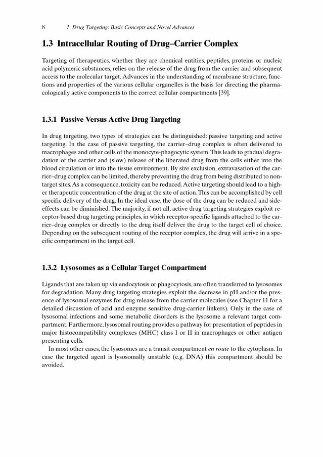
1.3 Intracellular Routing of Drug–Carrier Complex
Targeting of therapeutics, whether they are chemical entities, peptides, proteins or nucleicacid polymeric substances, relies on the release of the drug from the carrier and subsequentaccess to the molecular target. Advances in the understanding of membrane structure, func-tions and properties of the various cellular organelles is the basis for directing the pharma-cologically active components to the correct cellular compartments [39].
1.3.1 Passive Versus Active Drug Targeting
In drug targeting, two types of strategies can be distinguished: passive targeting and activetargeting. In the case of passive targeting, the carrier–drug complex is often delivered tomacrophages and other cells of the monocyte-phagocytic system.This leads to gradual degra-dation of the carrier and (slow) release of the liberated drug from the cells either into theblood circulation or into the tissue environment. By size exclusion, extravasation of the car-rier–drug complex can be limited, thereby preventing the drug from being distributed to non-target sites.As a consequence, toxicity can be reduced.Active targeting should lead to a high-er therapeutic concentration of the drug at the site of action.This can be accomplished by cellspecific delivery of the drug. In the ideal case, the dose of the drug can be reduced and side-effects can be diminished. The majority, if not all, active drug targeting strategies exploit re-ceptor-based drug targeting principles, in which receptor-specific ligands attached to the car-rier–drug complex or directly to the drug itself deliver the drug to the target cell of choice.Depending on the subsequent routing of the receptor complex, the drug will arrive in a spe-cific compartment in the target cell.
1.3.2 Lysosomes as a Cellular Target Compartment
Ligands that are taken up via endocytosis or phagocytosis, are often transferred to lysosomesfor degradation. Many drug targeting strategies exploit the decrease in pH and/or the pres-ence of lysosomal enzymes for drug release from the carrier molecules (see Chapter 11 for adetailed discussion of acid and enzyme sensitive drug-carrier linkers). Only in the case oflysosomal infections and some metabolic disorders is the lysosome a relevant target com-partment. Furthermore, lysosomal routing provides a pathway for presentation of peptides inmajor histocompatibility complexes (MHC) class I or II in macrophages or other antigenpresenting cells.
In most other cases, the lysosomes are a transit compartment en route to the cytoplasm. Incase the targeted agent is lysosomally unstable (e.g. DNA) this compartment should beavoided.
8 1 Drug Targeting: Basic Concepts and Novel Advances

1.3.3 Cytoplasmic Delivery
The majority of drugs exert their action in the cytoplasm of the cell where their target en-zymes are located. Consequently, they need to pass from one of the compartments of the en-docytotic pathway into the cytoplasm. The endosomal and lysosomal membranes can bedestabilized using fusogenic peptides derived from viruses, cyclodextrins and polyethyl-eneimine [39]. pH-sensitive liposomes or polymers become fusion competent at the acidicpH of the endosomes and subsequently release their contents into the cytoplasm. Particular-ly for the delivery of DNA into cells, this approach seems appropriate and quite successful[40]. Bacterial components such as listeriolysin O and alpha-haemolysin can form pores inphagosomal or plasma membranes [39]. It remains, however, to be established whether thesecomponents can be exploited for directing drug-targeting preparations in vivo to specific cel-lular compartments.
Schwarze et al. reported on the development of a recombinant fusion protein consisting ofthe protein transduction domain of HIV-derived TAT and the 120-kDa β-galactosidase. TheTAT protein was able to deliver the large molecular weight protein to the interior of the cellsin vitro. Interestingly, the enzymatic activity of intracellularly delivered β-galactosidasepeaked about 2 h later than did the intracellular concentration. This likely reflects a slowposttransduction refolding of the protein by intracellular chaperones. Intraperitoneal injec-tion of the fusion protein in mice resulted in delivery of the biologically active fusion proteinto all tissues, including the brain [41]. Similarly, the Herpes simplex virus tegument proteinVP22 is able to deliver proteins into the cytoplasm of cells [42]. Both approaches may proveuseful to enhance the delivery of e.g. enzymes for pro-drug protocols.
1.3.4 Nuclear Targeting
The three major obstacles to DNA accessibility in the nucleus of the target cells are low up-take across the plasma membrane, inadequate release of DNA with limited stability, and lackof nuclear targeting. Delivery systems of the future need to fully accommodate all steps in theinternalization and targeting routing in order to effectively guide the DNA into the nucleus.Due to space limitations, a complete overview of recent advances in this field will not be pro-vided. For more in-depth reading, the reader is referred to a recent concise review by Luoand Saltzman on novel strategies to accomplish optimal gene delivery [43].
In short, increased targeting and uptake of DNA by the cell using better delivery systemsis the basis for overcoming the plasma membrane hurdle. Increased stability of the DNAonce inside the cell can be achieved by chloroquine or branched cationic polymers which fa-cilitate the early release of the DNA from the endosomal pathway. Furthermore, bacterialsubunits and adenoviral capsids are capable of bypassing the endosomes, although it shouldbe realized that these approaches may be significantly hampered by inherent toxicity and/orimmunogenicity. Stabilization of the DNA in the ‘hostile’ environment of the cytosol can beachieved using PEG, PEG-poly-l-lysine block copolymers and others [43]. Intermediate sta-bility of DNA/delivery system interactions is most likely the prerequisite to achieve optimalliberation of DNA molecules once they have become available within the cytoplasm. To
1.3 Intracellular Routing of Drug-Carrier Complex 9

overcome the final obstacle, finding the nucleus, application of knowledge of viral infectionprocesses have led to the application of viral nuclear localization signals. Furthermore, theaforementioned viral tegument protein VP22 also localizes in the nuclear compartment. Inthe early stages of cell mitosis, VP22 translocates into the nucleus, binds to the condensingchromatin and remains bound [44]. It will be interesting to see whether these principles ofnuclear targeting can be exploited for the targeted delivery of DNA to the cell type of choice.
By assembling polypeptides that can code for all the necessary cellular transport tasks ona scaffold, Sheldon and colleagues developed so-called ‘loligomers’, branched squid-like pep-tides that can self-localize in the cytoplasm or nucleus [45]. In vitro application revealed goodtransfection properties of one of the nuclear localizing loligomers [46]. Its potential for ap-plication in drug targeting, i.e. the ability to combine cell specificity of DNA delivery withloligomer-orchestrated intracellular routing capacity, needs to be established.
1.3.5 Mitochondrial Targeting
Mitochondria are the ATP suppliers of the cells and have an important role in modulating in-tracellular calcium levels and cellular apoptosis. The mitochondrial respiratory chain is fur-thermore an important supplier of damaging free radicals. Evidence increases that mito-chondria are heavily involved in numerous diseases and therefore they may become impor-tant targets for the development of new drugs and therapies [47].
Both the large membrane potential across the inner membrane and the protein importmachinery of the mitochondria may be exploited for selectively delivering drugs to this cel-lular organelle. Lipophilic cations in particular, have been studied for mitochondrial target-ing purposes based on their mitochondrial accumulation potential. Using triphenylphospho-nium as a carrier, Smith et al. were able to selectively deliver antioxidant activity into the mi-tochondrial compartment of cells [48]. Similar to proteins that require nuclear localizationsequences for homing to this compartment, cellular proteins that need to be targeted to mi-tochondria require mitochondrial localization sequences [49]. Fusion of these signal se-quences with (model) proteins of interest redirects the proteins into the mitochondrial com-partments. Whether these intracellular targeting entities can be combined with other target-ing entities that specifically direct them into the desired cell type in the body, is howeverquestionable. One option may be to package the mitochondrial targeting system in immuno-liposomes that provide the cell specificity [47], but further research is awaited to provide in-sight into the potential and limitations of this approach.
1.4 Drug Targeting Strategies in the Clinic
Most of the drug delivery systems that have been studied for clinical application are capableof rate- and/or time-controlled drug release.The therapeutic advantages in these approacheslie in the in vivo predictability of release rate, minimized peak plasma levels, predictable andextended duration of action and reduced inconvenience of frequent re-dosing and hence, im-proved patient compliance [1].
10 1 Drug Targeting: Basic Concepts and Novel Advances

With the development of more advanced drugs such as therapeutic proteins, antisensemolecules and genes, not only controllable release, but also controllable delivery in the tar-get cells becomes desirable. As a consequence, targeting modalities need to be incorporatedinto the vehicles. Although drugs and drug formulation based on proteins were first consid-ered unfeasible, nowadays practice has proven this not to be the case. Since 1994, on averagemore than seven FDA approvals per year have been issued for protein-based therapies. It isfurthermore believed that the increasing yield of protein molecules produced by recombi-nant techniques will boost application of protein drugs in the near future [50].
In recent years, various reviews have been published in which the current status of lipo-some [2] and antibody [6] based drug targeting strategies have been summarized. Similarly,the current status of gene therapy in both pre-clinical and clinical settings have been recent-ly reviewed [51,52].Without trying to be complete, the next sections will give an overview onrecently published studies with drug targeting formulations in a clinical setting.
1.4.1 Liposome Based Therapies in the Clinic
Several liposomal formulations are either under investigation in phase I/II/III clinical trialsor have been approved by the US Food and Drug Administration or cleared for empiric ther-apy (see also Chapter 8 for FDA approved formulations in cancer therapy). They are in usefor the treatment of cancer or fungal infections and, consisting of non-targeted liposomes,aim at slow release of the encapsulated drugs over a prolonged period of time to circumventdose-limiting (cardiac) toxicity. Recently reported clinical studies with liposome formula-tions are summarized in Table 1.1. The reader is also referred to references [4] and [53] for amore detailed description of these liposome-based therapeutic strategies.
The general outcome of these studies was that the liposomal drugs were well tolerated andexerted a clinical effect, although larger studies need to be carried out to demonstrate the ex-tent of the effects. Furthermore, in the various studies of liposome-incorporated drugs, it wasobserved that cardiotoxicity was either absent or only limited, in contrast to studies with non-liposomally formulated drugs.
1.4.2 Monoclonal Antibody Therapies in the Clinic
Clinical studies on cancer therapy with antibodies have been elegantly summarized by Farahet al. [6] and more recently by Glennie and Johnson [54]. The majority of antibody-basedtherapies in the clinic have exploited the activity of the antibody per se. In the therapy of can-cer, the highest response rates have been observed in patients with haematological malig-nancies that are easily accessible to antibodies. In recent years, numerous clinical studies withantibodies directed against the B cell non-Hodgkin lymphoma-associated epitope CD20(Rituximab, Rituxan) and the breast carcinoma antigen Her-2 (also known as neu or erbB2)have been carried out. The mechanisms of action of these antibodies are believed to consistof complement-mediated lysis, antibody-dependent cellular cytotoxicity by macrophages andnatural killer cells, and signal transduction leading to apoptosis or growth arrest [55]. Novel
1.4 Drug Targeting Strategies in the Clinic 11

12 1 Drug Targeting: Basic Concepts and Novel Advances
Table 1.1. Overview of some recently reported clinical studies on the application of liposome-based drug formulations.
Liposome Drug Disease Clinical Remarks Referenceformulation incorporated responsea
DaunoXomeb Daunorubicin Acute leukaemia 2/23 CR [59]Heptocellular 2/12 SD [60]carcinomaRecurrent progressive 2/14 CR [61]brain tumours in 3/14 PRchildren 2/14 SD
Caelyxc Doxorubicin Hormone refractory 3/15 OR Severe [62]prostate cancer 2/15 SD mucocutaneous
toxicities Advanced/metastatic 3/25 PR Highest dose to [63]soft tissue sarcoma 2/25 MR treat not yet
17/25 SD reached Non-small-cell lung 3/15 CR Combination [64]cancer with fractionated Squamous cell head 11/15 CR radio therapy and neck cancer Tumour
microvessel density correlated with degree of Caelyx accumulation
TLC D-99d Doxorubicin Kaposi’s sarcoma 6/40 PR Dose dependent [65]26/40 SD response and
toxicityMetastatic breast 10/41 OR Combination with [66]cancer cyclophosphamide
and fluorouracil
AmBisome Amphotericin B Renal Tx recipients 4/4 treated [67]with viscer. Leishm.e successfullyHeart surgery with C. 1/1 treated [68]tropicalis successfullyendocardititisBone marrow Tx or Polyprophy- [69]chemotherapy lactic treatmentpatients with fungal did not reduce infections fungal infections
or requirementfor systemictherapy
TLC C-35 Prostaglandin E1 Acute respiratory 348 patients [70]distress syndrome evaluated: no
difference in number of daysto discontinuation of ventilation to placebo group; mortality not diffe-rent to placebo
a Number of patients responding/total number of evaluable patients. CR, complete remission/response; PR, partialresponse; MR, minor response; OR, objective response; SD, stable disease.
b Daunorubicin citrate liposome injection formula.c Also known as Doxil; PEG-coated liposome formulation with doxorubicin.d Phosphatidylcholine/cholesterol liposomal formulation with doxorubicin.e Tx, transplantation; visc. Leishm., visceral Leishmaniasis

advances in this field are the development of antibodies against angiogenesis-associated fac-tors and receptors such as VEGF and integrin αvβ3 [56,57]. Furthermore, antibodies direct-ed against the CD3 molecule on T lymphocytes, a treatment developed for prevention of or-gan rejection, are now under investigation for their immunomodulatory effects in cancer pa-tients [58].
Whereas initially the focus on antibody-based therapies was on cancer, anti-TNFα anti-bodies in particular have recently proven powerful in the therapy of chronic inflammatorydiseases such as inflammatory bowel disease and rheumatoid arthritis [71]. These antibodiescomplex serum TNFα, the clinical benefit to RA patients most likely being the reduction ofpro-inflammatory IL-6 and acute phase protein levels [9].Although they are directed againstsoluble proteins and as such will not serve as a drug carrier, they do show that targeted, i.e.selective, interference with a specific molecule or process can have a powerful effect withoutsignificant concomitant toxicity.
Although in the early 1990s several antibodies were developed that inhibited leuko-cyte–endothelial cell interaction to prevent e.g. allograft rejection or inflammatory processes[72], more effort is nowadays put into the development of small molecule antagonists and an-tisense oligonucleotides for this purpose [73,74]. A selection of more recently reported clini-cal studies with antibodies is summarized in Table 1.2.
1.4.3 Monoclonal Antibody Based Targeting Strategies in the Clinic
Not only can antibodies by themselves function as targeted effector molecules, they can alsobe used as carriers for the selective delivery of drugs, toxins, enzymes, radioisotopes, and ade-noviral vectors. Most of the strategies have been applied in humans [6,75–77]. Besides theseapproaches, cellular cytotoxicity can be redirected towards the tumour cells using bispecificantibodies that consist of a recognition site for specific cells of the immune system and tu-mour-associated antigens [78,79]. This strategy circumvents the MHC restriction of antigenrecognition and cellular cytotoxicity. It may therefore be exploited for the therapy of tu-mours which downregulate the expression of their MHC molecules and thereby avoid thenormal immune response.
In the development of these antibody-based targeting strategies, modifications that havebeen applied to improve the efficacy of the therapy include the use of chimerized or human-ized antibodies, more potent drugs and better linkages between drugs and carrier molecules.Furthermore, liposome encapsulation of drugs enabled a larger quantity of the drug to be de-livered per antibody molecule [75]. The development of transgenic mice capable of makingfully human antibodies now offers new opportunities for generating antibodies of therapeu-tic quality, and as a result has led to the revival of interest in antibody-based therapies [80].
1.4.4 Other Drug Targeting Strategies in the Clinic
Of the non-antibody, non-liposome based drug targeting strategies, most of the (limited) clin-ical experience has been obtained with polymer-based conjugates of anticancer drugs. Themost widely employed drugs for this application are cytotoxic agents such as doxorubicin and
1.4 Drug Targeting Strategies in the Clinic 13

14 1 Drug Targeting: Basic Concepts and Novel Advances
Table 1.2. Overview of recently reported clinical studies exploiting antibodies.
Antibody Antigen Disease treateda Clinical responseb Remarks Reference
Rituximab CD20 NHL 21/39 OR Similar results [81](rituxan) 14/39 SD or MR in follicular
and small lymphocytic lymphoma
PT-LPD after liver 2/2 CR: poly- [82]Tx morphic PT-LPD;
1/1 NR: large cell NHLPT-LPD after solid 15/26 CR [83]organ Tx 2/26 PRPT-LPD after BMT 5/6 CR [83]Mantle cell lym- 36/120 OR (varied Limited [84]phoma (MCL), im- with tumour type). activitymunocytoma, small CR only seen in SLLB cell lymphocytic with MCL lymphoma (SLL)
Herceptin; Her2/neu Metastatic breast 1/43 CR Responses [85]trastuzumab carcinoma 4/43 PR seen in
2/43 MR mediastinum,14/43 SD lymph nodes,
liver, chest wall lesions
MKC-454 Her2/neu Metastatic breast 2/18 OR [86]carcinoma
rhuMAb HER2 Her2/neu Advanced breast 9/37 PR Combination [87]carcinoma 9/37 MR – SD therapy with
19/37 DP cisplatinrhuMab VEGF VEGF Metastatic renal cell Phase III study in (clinical-
carcinoma progress trials.gov website)
hOKT3γ4 CD3 Various malignancies 3/24 responses [58]Infliximab TNFα Rheumatoid arthritis 428 patients: 20–50% Concomitant [71]
improvement in ~80% methotrexate of patients therapy
Psoriasis lesions Improved appearance [88]of a patient 2 weeksafter treatment
Synagis; RSV F - RSV infection 35 children < 2 years [89]palivizumab glyco- in infants of age: significant
protein reduction in tracheal RSV concentration, but not in nasal aspirate. No differencein disease severity compared to placebo
Abciximab Platelet gp Ischaemic complica- Platelet aggregation All patients [90]IIb/IIIa tions during balloon inhibition with received aspi-receptor angioplasty or abciximab provided rin co-medi-
atherectomy long-term clinical cation; pa-benefits after coronary tients with intervention com- stent implanta-pared to placebo tion received
ticlopidine co-medication
a BMT, bone marrow transplantation; NHL, non-Hodgkin lymphoma; PT-LPD, post-transplantation lymphoprolife-rative disorders; RSV, respiratory syncytial virus; Tx, transplantation.
b Number of patients responding/total number of evaluable patients. CR, complete remission/response; PR, partialresponse; MR, minor response; NR, no response OR, objective response; SD, stable disease; DP, disease progres-sion.

methotrexate, and the general aim of these conjugates is to lower the (dose-limiting cardio-)toxicity of these drugs. Enhanced permeability retention of the conjugates at the site of thetumour is most likely the basis of local accumulation. In the studies reported, some objectiveclinical responses were seen without concomitant toxicity of the drugs (Table 1.3). Althoughthe clinical responses until now seem rather disappointing, one should realize that the pa-tients who undergo clinical testing with these conjugates often carry large tumour burdenswhich have been non-responsive towards other therapies available.
1.5 Vaccination Strategies for Enhanced Immunity 15
1.5 Vaccination Strategies for Enhanced Immunity
Vaccination can be considered as a therapeutic modality that actively engages the immunesystem of the patient. It encompasses numerous principles derived from drug targeting re-search. Modification of the responses of the immune system may be an effective approach toimprove the disease status of patients with a variety of diseases. Either prevention of au-
Table 1.3. Overview of recently reported clinical studies with miscellaneous drug targeting strategies.
Carrier Drug Disease Clinical Remarksc Referenceincorporated responsea,b
Antibody 90Y Metastatic 2/25 OR Pre-targeted strategy: [91]colon cancer 2/25 PR mAb NR-LU-10 –
4/25 SD streptavidin, followedby biotin-gal-HSA clearing and biotin-DOTA-90Y
Non-Hodgkin 3/7 CR Pre-targeted strategy: [92]lymphoma 1/7 PR mAb Rituximab –
2/7 OR streptavidin, followedby biotin-N-acetyl-galactosamine clearingand biotin-DOTA90Y
HPMA Doxorubicin Various 2/36 PR Clinical proof of copolymer malignancies 2/36 MR principle of decrease [15](PK1) of doxorubicin toxicity
when polymer bound
Poly(styrene- Neocar- Renal cell Improved survival, SMANCS dissolved in [93]co-maleic acid) zinostatin carcinoma depending on lipiodol contrast polymers patients tumour size medium, administered (SMANCS) via the renal artery
Human serum Methotrexate Cancer patients 17 patients: 1 PR [94]albumin and 1 MR in RCC
patients; 1 MR inpleural mesothe-lioma patient
a Number of patients responding/total number of evaluable patients. CR, complete remission/respon-se; PR, partial response; MR, minor response; OR, objective response; SD, stable disease.
b RCC, renal cell carcinoma.c mAb, monoclonal antibody.

toimmune diseases or allergic responses, or enhancement of immune responses against in-fectious agents and tumour growth can be induced by these strategies [95,96]. Vaccinationstrategies can be divided into gene-, peptide-, protein- and cell-based strategies.
Antigen presenting cells (APC), particularly dendritic cells (DC), play a central role in theinduction of the desired immune response [97]. For successful (antitumour) vaccination ther-apy, either an in vitro or an in vivo approach can be followed. In the in vitro approach, DCsfrom an animal or a patient are isolated and manipulated by transfection with DNA or RNAencoding (tumour) antigens or pro-inflammatory factors, or by loading the cells with proteinsor peptides. After transfer back into the animal or patient, the cells can evoke antigen-spe-cific immune responses [98]. Similarly, in vitro transfected tumour cells encoding a combina-tion of genes involved in regulation of immune responses (e.g. MHC class II, a co-stimulato-ry molecule and a superantigen [96]) may serve as a vaccine.
The major drawback in the use of isolated DCs is the time-consuming isolation and culturemethods required to obtain the cell type of interest. Therefore, in vivo vaccination strategiesemploying DCs have been developed. For this purpose, fusion proteins of e.g. tumour anti-gens and molecules that are specifically recognized by DCs are under investigation. The DCtargeting molecules consist of e.g. DC-specific chemokines, mannose, the Fc moiety of im-munoglobulin, cytotoxic T lymphocyte-associated antigen (CTLA-4) molecule [99–103].They enable efficient uptake of the fusion protein by the DCs, induction of DC migratory ca-pacity into the lympoid organs and maturation into a dedicated APC. As an example, studiesby Biragyn et al. showed that active targeting of a tumour antigen into a receptor-mediateduptake route in APCs by the fusion of the antigen to APC-specific chemokines, elicited su-perior protection against a large tumour challenge in mice [99].
For a more detailed discussion of vaccines and vaccination strategies, the reader is referredto Volume 170 of Immunological Reviews (1999), in which various aspects of this area of re-search are discussed in greater detail.
1.6 Drug Targeting as a Research Tool to Study Disease
From an historical point of view, ‘magic bullets’ were initially proposed as novel therapeutictools for treatment of disease.Another, similarly attractive application of drug targeting is itsuse as a tool to study e.g. mechanisms of disease pathology. By selectively manipulating onespecific cell type or one specific protein, the role of this cell type or protein in disease pro-gression can be determined. The advent of knock-out and transgenic animal models has pro-vided the researcher in the laboratory with a powerful tool to investigate the relative contri-bution of gene products of interest. Yet, counter-regulatory processes that compensate forthe loss of a certain gene product may significantly hamper correct interpretation of the re-sults obtained in these models. Manipulating a gene in an appropriately matured animal us-ing drug targeting strategies can therefore be a valuable tool.
It is not always appreciated that the application of in vivo gene therapy actually representsa drug delivery protocol. By the incorporation of plasmids into e.g. adenoviral or liposomalcoats they are protected from degradation. Subsequent interaction with the target cells leadsto intracellular delivery and then to nuclear localization of the plasmid. Although transfec-
16 1 Drug Targeting: Basic Concepts and Novel Advances

tion efficiencies in in vivo gene therapy protocols are still not without problems, the tran-scription of genes of interest by transduced liver cells and muscle cells are quite satisfactorywhen using a strong promoter. Liver cells can be targeted by i.v. injection of the encapsulat-ed plasmids, whereas skeletal muscle cells can be locally transfected by intramuscular injec-tions. As an example, i.v. administration of recombinant adenovirus AdCMV-PDX-1 intomice resulted in specific transfection of liver cells. Expression of the PDX-1 gene, encoding aprotein that regulates insulin gene expression, led to increased plasma levels of insulin andameliorated hyperglycaemia in diabetic mice treated with streptozotocin [104].Transductionof skeletal muscle with liposome formulated human hepatocyte growth factor (HGF) geneinduced high plasma levels of protein, phosphorylation of the HGF target receptor, subse-quent suppression of transforming growth factor β production and improved survival rates ofanimals suffering from severe liver cirrhosis [105].
Proteins have also been selectively targeted to study a disease process. Selective inductionof oxidative vascular injury in the lungs was accomplished by targeting glucose oxidase(GOX) to the vasculature using anti-PECAM-1 (anti-CD31)-antibody–GOX conjugates. Asthe pulmonary vasculature represents approximately 30% of endothelial cells in the bodyand receives all the cardiac output, the majority of anti-PECAM-GOX conjugate accumulat-ed in the endothelium in the lung. By generating H2O2 locally from glucose, severe endothe-lial cell damage was induced. The injury was associated with the production of the oxidativemarker iPF2a-III isoprostane.This model of targeted induction of oxidative stress can now beapplied to study the effects of pharmacological agents [106].
Inhibition of tumour blood flow or the outgrowth of solid tumour vasculature by angio-genesis inhibition has been proposed as a powerful strategy to eradicate cancer ever since therecognition of the importance of blood supply for tumour growth. Subsequent developmentof compounds which inhibited the formation of new tumour blood vessels, demonstrated thedependence of solid tumour growth on blood vessel formation. It was however the selectiveinduction of blood coagulation in the vasculature of a well-developed tumour by a drug tar-geting strategy that really demonstrated the potency of instantaneous inhibition of bloodflow as a therapy for solid tumours [107]. Based on these and other ‘proof of principle’ stud-ies, enormous research effort is now put into the identification of target epitopes on humantumour vasculature to further develop this strategy for clinical application (see Chapter 9and reference [57] for a more detailed discussion on tumour vasculature targeting).
The inhibition of TNFα by neutralizing antibodies in rheumatoid arthritis patients is oneof the few examples of targeted interference with disease activity in humans that can provideus with new insights into the pathophysiology of the disease [108]. The concomitant reduc-tion in systemic levels of acute phase proteins, endothelial cell adhesion molecule expressionin synovial biopsies and inflamed joint-associated blood vessel density all suggest a centralrole for TNFα as a driving force in RA. Furthermore, it provides evidence for the existenceof a relationship between pro-inflammatory activity and the occurrence of angiogenesis, al-though it is at present unfeasible to conclude on cause and effect relations due to limitedknowledge on this subject.
In summary it can be said that besides being important in the development of therapeuticstrategies to combat disease with minimal toxicity and maximal effects, drug targeting may beof interest for more basic studies on the mechanistic background of diseases and the identi-fication of new molecules as targets for therapeutic intervention.
1.6 Drug Targeting as a Research Tool to Study Disease 17

1.7 Challenges in Drug Targeting Research
In 1984, Poznansky and Juliano published a critical review on the biological approaches tothe controlled delivery of drugs [109]. Already at that time, they had envisioned the use ofmolecular biology and molecular immunology in the delineation of structure and function ofimportant proteins and nucleic acids as being the true frontier in the drug delivery field. Theelucidation of signal transduction routes in cells responding to a variety of environmentalfactors of disease has indeed led to the development of numerous NCEs to be used in futuretherapies. Furthermore, the understanding of protein folding and minimal amino acid se-quences required for receptor recognition provided significant added value in the develop-ment of better carriers and ligands for drug targeting.
Numerous problems in the construction of clinically applicable drug targeting moietiesstill need to be solved. Of these issues, immunogenicity after repeated administration, coun-terproductive liver clearance, and production yields are the most important. Although theproblem of immunogenicity is believed to have been solved for monoclonal antibody thera-py by the development of humanized and fully human antibodies [110], for other carrier sys-tems such as modified plasma proteins and peptide modified polymers, this remains an im-portant issue.
For many drug targeting approaches, extensive uptake by the liver is undesirable when thetarget cells are of non-hepatic origin. In this respect, defining the optimal physicochemicalcharacteristics that would circumvent extensive hepatic clearance of the drug targetingpreparation would be of great value. This may, however, be wishful thinking more than a re-alistic aim, as hepatic elimination of (slightly) modified proteins is one method of protectingthe body from non-functional or aged proteins. Furthermore, drug targeting preparationshave to be eliminated from the body, as do regular drugs, so that their pharmacological ac-tivity can be controlled.
In general, validation of the targeting concept (therapeutic effectiveness, lack of toxicityeven after prolonged periods of time) for each drug targeting conjugate that is under devel-opment should be performed in vivo in animal models of disease. Not only is this the only un-ambiguous approach for showing proof of the targeting principle, but it also takes into ac-count the pharmacokinetic characteristics of the conjugate which determine the added valueof the targeting strategy compared to non-targeted therapy (see Chapter 13 for pharmacoki-netic considerations in drug targeting).
Most of the drug targeting strategies explored so far have been aimed at tumour cell killingor targeting of drugs and genes to the liver (i.e. Kupffer cell or hepatocyte). More recently thepotential of targeting other cells of interest has been put forward, e.g. endothelial cells liningtumour blood vessels (Chapter 9) or the vasculature in chronic inflamed tissue (Chapter 7),and hepatic stellate cells in the fibrotic liver (Chapter 4) [10,57].As the number of target cellsin these cases is often relatively low and at present little knowledge exists regarding basic cellregulatory processes such as responses to drugs or endocytotic activity, all aspects of drug tar-geting (pharmacological activity of the drug in the target cell, cellular handling and kineticsof release of the drug) need to be addressed in full.
One of the most frequently heard criticisms is that drug targeting preparations cannot beadministered orally and are therefore either of secondary or no interest for development intotherapeutics.Although significant research effort has been put into the development of oral-
18 1 Drug Targeting: Basic Concepts and Novel Advances

ly available protein therapeutics [111], there is a good chance that these preparations will notbe feasible because of the inherent limitations in manipulating the physiological barriers inhumans. Possibly, the understanding of the structural requirements for drug targeting prepa-rations may lead to the development of minimized proteins that may be orally administeredor become systemically available via e.g. pulmonary delivery (see Chapter 3). Furthermore,major efforts are being invested in the development of transdermal administration and pro-grammable infusion systems. These systems may lower the barriers for long-term parenteraladministration of drugs and may become common practice in pharmacotherapy. It is note-worthy that if a single dose of a targeted protein is potent enough to silence a disease for aprolonged period of time, the requirement of parenteral administration will not hamper itsdevelopment. This is exemplified by the development of the TNFα neutralizing antibodiesand receptors in the therapy of chronic inflammatory diseases such as rheumatoid arthritisand inflammatory bowel disease.
Finally, drug targeting technology should be integrated in the earliest phases of drug de-velopment. Many drugs that are pharmacologically active at low concentrations are with-drawn from the R & D pipeline due to toxicity in non-target cells. Would it be possible dur-ing an early phase in the development of a drug to construct cell-specific targeting conjugatesa large number of drugs could still be candidates for development into clinical therapeuticstrategies.
1.8 Conclusions
At present, many of the problems encountered during the development of drug targetingstrategies for clinical application, especially for cancer therapy, have been identified,analysed and solved. Several drug targeting preparations have entered the phases of clinicaltesting and/or have now been marketed. However, these strategies should be subjected tocontinuous evaluation in the light of advances in the understanding of the numerous process-es occuring in response to administration of the carriers and/or the drugs. New strategies un-der investigation will need to be optimized and extensively evaluated, taking advantage ofthe ‘bench to bed-side’ experience available today. Furthermore, in the coming years, com-bining expertise in the drug targeting field with the technological developments in molecularbiology and molecular medicine will facilitate the elucidation of the cellular and molecularprocesses underlying disease.
Acknowledgements
Drs E. S. Vitetta, D. K. F. Meijer and M. Everts are acknowledged for critically reading themanuscript. Drs G. Storm, R. J. Kok and L. Beljaars are thanked for their help with the prepa-ration of the figures.
1.8 Conclusions 19

References
[1] Breimer DD, Adv. Drug Deliv. Rev. 1998, 33, 265–268.[2] Mastrobattista E, Koning GA, Storm G, Adv. Drug Deliv. Rev. 1999, 40, 103–127.[3] Duzgunes N, Nir S, Adv. Drug Deliv. Rev. 1999, 40, 3–18.[4] Storm G, Crommelin DJA, PSTT 1998, 1, 19–31.[5] Köhler G, Milstein C, Nature 1975, 256, 495–497.[6] Farah RA, Clinchy B, Herrera L, Vitetta ES, Crit. Rev. Eukaryot. Gene Expr. 1998, 8, 321–356.[7] Willuda J, Honegger A, Waibel R, Schubiger PA, Stahel R, Zangemeister-Wittke U, Pluckthun
A, Cancer Res. 1999, 59, 5758–5767.[8] Worn A, Pluckthun A, Biochemistry 1999, 38, 8739–8750.[9] Charles P, Elliott MJ, Davis D, Potter A, Kalden JR, Antoni C, Breedveld FC, Smolen JS, Eberl
G, deWoody K, Feldmann M, Maini RN, J. Immunol. 1999, 163, 1521–1528.[10] Beljaars L, Molema G, Schuppan D, Geerts A, de Bleser PJ, Weert B, Meijer DKF, Poelstra K, J.
Biol. Chem. 2000, 275, 12743–12751.[11] Molema G, Meijer DKF, Adv. Drug Deliv. Rev. 1994, 14, 25–50.[12] Ogawara K, Hasegawa S, Nishikawa M, Takakura Y, Hashida M, J. Drug Target. 1999, 6, 349–360.[13] Stehle G, Sinn H, Wunder A, Schrenk HH, Schutt S, Maier Borst W, Heene DL, Anticancer Drugs
1997, 8, 677–685.[14] Maeda H, Wu J, Sawa T, Matsumura Y, Hori K, J. Control. Rel. 2000, 65, 271–284.[15] Vasey PA, Kaye SB, Morrison R, Twelves C, Wilson P, Duncan R, Thomson AH, Murray
LS, Hilditch TE, Murray T, Burtles S, Fraier D, Frigerio E, Cassidy J, Clin. Cancer Res. 1999, 5,83–94.
[16] Gianasi E, Wasil M, Evagorou EG, Keddle A, Wilson G, Duncan R, Eur. J. Cancer 1999, 35,994–1002.
[17] Sawa T, Wu J, Akaike T, Maeda H, Cancer Res. 2000, 60, 666–671.[18] Richardson SC, Kolbe HV, Duncan R, Int. J. Pharm. 1999, 178, 231–243.[19] Malik N, Wiwattanapatapee R, Klopsch R, Lorenz K, Frey H, Weener JW, Meijer EW, Paulus W,
Duncan R, J. Control. Rel. 2000, 65, 133–148.[20] Lu ZR, Kopeckova P, Kopecek J, Nature Biotechnol. 1999, 1, 1101–1104.[21] Kader A, Davis PJ, Kara M, Liu H, J. Control. Rel. 1998, 55, 231–243.[22] Meijer DKF, Molema G, Sem. Liver Dis. 1995, 15, 202–256.[23] de Vrueh RL, Rump ET, van De BE, van Veghel R, Balzarini J, Biessen EA, van Berkel TJ,
Bijsterbosch MK, Antimicrob. Agents Chemother. 2000, 44, 477–483.[24] Van Velzen AG, Da Silva RP, Gordon S, van Berkel TJ, Biochem. J. 1997, 322 (Pt 2), 411–415.[25] Ling W, Lougheed M, Suzuki H, Buchan A, Kodama T, Steinbrecher UP, J. Clin. Invest. 1997, 100,
244–252.[26] Utreja S, Khopade AJ, Jain NK, Pharm. Acta Helv. 1999, 73, 275–279.[27] Muller RH, Mader K, Gohla S, Eur. J. Pharm. Biopharm. 2000, 50, 161–177.[28] Aynie I, Vauthier C, Chacun H, Fattal E, Couvreur P, Antisense Nucl. Acid Drug Dev. 1999, 9,
301–312.[29] Soma CE, Dubernet C, Bentolila D, Benita S, Couvreur P, Biomaterials 2000, 21, 1–7.[30] Allemann E, Leroux J, Gurny R, Adv. Drug Deliv. Rev. 1998, 34, 171–189.[31] Pecquet S, Leo E, Fritsche R, Pfeifer A, Couvreur P, Fattal E, Vaccine 2000, 18, 1196–1202.[32] Jones M, Leroux J, Eur. J. Pharm. Biopharm. 1999, 48, 101–111.[33] Yokoyama M, Okano T, Sakurai Y, Fukushima S, Okamoto K, Kataoka K, J. Drug Target. 1999,
7, 171–186.[34] Vallera DA, Jin N, Baldrica JM, Panoskaltsis-Mortari A, Chen SY, Blazar BR, Cancer Res. 2000,
60, 976–984.[35] Wiedle G, Johnson-Leger C, Imhof BA, Cancer Res. 1999, 59, 5255–5263.[36] Asahara T, Murohara T, Sullivan A, Silver M, van der Zee R, Li T, Witzenbichler B, Schatteman
G, Isner JM, Science 1997, 275, 964–967.[37] Asahara T, Takahashi T, Masuda H, Kalka C, Chen D, Iwaguro H, Inai Y, Silver M, Isner JM,
EMBO J. 1999, 18, 3964–3972.[38] Isner JM, Asahara T, J. Clin. Invest 1999, 103, 1231–1236.[39] Moghimi SM, Rajabi-Siahboomi AR, Adv. Drug Deliv. Rev. 2000, 41, 129–133.[40] Stayton PS, Hoffman AS, Murthy N, Lackey C, Cheung C, Tan P, Klumb LA, Chilkoti A, Wilbur
FS, Press OW, J. Control. Rel. 2000, 65, 203–220.[41] Schwarze SR, Ho A, Vocero-Akbani A, Dowdy SF, Science 1999, 285, 1569–1572.
20 1 Drug Targeting: Basic Concepts and Novel Advances

[42] Dilber MS, Phelan A, Aints A, Mohamed AJ, Elliott G, Smith CI, O’Hare P, Gene Ther. 1999, 6,12–21.
[43] Luo D, Saltzman WM, Nature Biotechnol. 2000, 18, 33–37.[44] Elliott G, O’Hare P, J. Virol. 2000, 74, 2131–2141.[45] Sheldon K, Liu D, Ferguson J, Gariepy J, Proc. Natl. Acad. Sci. USA 1995, 92, 2056–2060.[46] Singh D, Bisland SK, Kawamura K, Gariepy J, Bioconjug. Chem. 1999, 10, 745–754.[47] Murphy MP, Smith RAJ, Adv. Drug Deliv. Rev. 2000, 41, 235–250.[48] Smith RA, Porteous CM, Coulter CV, Murphy MP, Eur. J Biochem. 1999, 263, 709–716.[49] Renold A, Koehler CM, Murphy MP, FEBS Lett. 2000, 465, 135–140.[50] Grindley J, Ogden J, 1999, SCRIP 59–646.[51] Vile RG, Russell SJ, Lemoine NR, Gene Ther. 2000, 7, 2–8.[52] Li S, Huang L, Gene Ther. 2000, 7, 31–34.[53] Lasic DD, Trends Biotechnol. 1998, 16, 307–321.[54] Glennie MJ, Johnson PW, Immunol. Today 2000, 21, 403–410.[55] Cragg MS, French RR, Glennie MJ, Curr. Opin. Immunol. 1999, 11, 541–547.[56] Schlaeppi JM, Wood JM, Cancer Metast. Rev 1999, 18, 473–481.[57] Griffioen AW, Molema G, Pharmacol. Rev 2000, 52, 237–268.[58] Richards J, Auger J, Peace D, Gale D, Michel J, Koons A, Haverty T, Zivin R, Jolliffe L, Blue-
stone JA, Cancer Res. 1999, 59, 2096–2101.[59] Cortes J, O’Brien S, Estey E, Giles F, Keating M, Kantarjian H, Invest. New Drugs 1999, 17, 81–87.[60] Yeo W, Chan KK, Mukwaya G, Ross M, Leung WT, Ho S, Chan AT, Johnson PJ, Cancer
Chemother. Pharmacol. 1999, 44, 124–130.[61] Lippens RJ, Pediatr. Hematol. Oncol. 1999, 16, 131–139.[62] Hubert A, Lyass O, Pode D, Gabizon A, Anticancer Drugs 2000, 11, 123–127.[63] Toma S, Tucci A, Villani G, Carteni G, Spadini N, Palumbo R, Anticancer Res. 2000, 20, 485–491.[64] Koukourakis MI, Koukouraki S, Giatromanolaki A, Archimandritis SC, Skarlatos J, Beroukas K,
Bizakis JG, Retalis G, Karkavitsas N, Helidonis ES, J. Clin. Oncol. 1999, 17, 3512–3521.[65] Cheung TW, Remick SC, Azarnia N, Proper JA, Barrueco JR, Dezube BJ, Clin. Cancer Res. 1999,
5, 3432–3437.[66] Valero V, Buzdar AU, Theriault RL, Azarnia N, Fonseca GA, Willey J, Ewer M, Walters RS,
Mackay B, Podoloff D, Booser D, Lee LW, and Hortobagyi GN, J. Clin. Oncol. 1999, 17,1425–1434.
[67] Boletis JN, Pefanis A, Stathakis C, Helioti H, Kostakis A, Giamarellou H, Clin. Infect. Dis. 1999,28, 1308–1309.
[68] Melamed R, Leibovitz E, Abramson O, Levitas A, Zucker N, Gorodisher R, Scand. J. Infect. Dis.2000, 32, 86–89.
[69] Kelsey SM, Goldman JM, McCann S, Newland AC, Scarffe JH, Oppenheim BA, Mufti GJ, BoneMarrow Transplant. 1999, 23, 163–168.
[70] Abraham E, Baughman R, Fletcher E, Heard S, Lamberti J, Levy H, Nelson L, Rumbak M, Ste-ingrub J, Taylor J, Park YC, Hynds JM, Freitag J, Crit. Care Med. 1999, 27, 1478–1485.
[71] Maini R, St Clair EW, Breedveld F, Furst D, Kalden J, Weisman M, Smolen J, Emery P, HarrimanG, Feldmann M, Lipsky P, Lancet 1999, 354, 1932–1939.
[72] Haug CE, Colvin RB, Delmonico FL, Auchincloss Jr H, Tolkoff-Rubin N, Preffer FI, Rothlein R,Norris S, Scharschmidt L, Cosimi AB, Transplantation 1993, 55, 766–772.
[73] Kelly TA, Jeanfavre DD, McNeil DW, Woska Jr JR, Reilly PL, Mainolfi EA, Kishimoto KM,Nabozny GH, Zinter R, Bormann BJ, Rothlein R, J. Immunol. 1999, 163, 5173–5177.
[74] Glover JM, Leeds JM, Mant TG, Amin D, Kisner DL, Zuckerman JE, Geary RS, Levin AA,Shanahan Jr WR, J. Pharmacol. Exp. Ther. 1997, 282, 1173–1180.
[75] Trail PA, Bianchi AB, Curr. Opin. Immunol. 1999, 11, 584–588.[76] Frankel AE, Kreitman RJ, Sausville EA, Clin. Cancer Res. 2000, 6, 326–334.[77] DeNardo SJ, Kroger LA, DeNardo GL, Curr. Opin. Immunol. 1999, 11, 563–569.[78] Molema G, Kroesen BJ, Helfrich W, Meijer DKF, de Leij LFMH, J. Control. Rel. 2000, 64,
229–239.[79] van Spriel AB, van Ojik HH, van de Winkel JG, Immunol. Today 2000, 21, 391–397.[80] Davis CG, Gallo ML, Corvalan JR, Cancer Metast. Rev 1999, 18, 421–425.[81] Hainsworth JD, Burris HA, Morrissey LH, Litchy S, Scullin DC, Bearden JD, Richards P, Greco
FA, Blood 2000, 95, 3052–3056.[82] Zompi S, Tulliez M, Conti F, Leblond V, Gaulard P, Blanche P, Durand F, Ghandi D, Dreyfus F,
Louvel A, Calmus Y, Bouscary D, J. Hepatol. 2000, 32, 521–527.
References 21

[83] Milpied N, Vasseur B, Parquet N, Garnier JL, Antoine C, Quartier P, Carret AS, Bouscary D,Faye A, Bourbigot B, Reguerre Y, Stoppa AM, Bourquard P, Hurault dL, Dubief F, Mathieu-Boue A, Leblond V, Ann. Oncol. 2000, 11 (Suppl. 1), 113–116.
[84] Foran JM, Rohatiner AZ, Cunningham D, Popescu RA, Solal-Celigny P, Ghielmini M, Coiffier B,Johnson PW, Gisselbrecht C, Reyes F, Radford JA, Bessell EM, Souleau B, Benzohra A, ListerTA, J. Clin. Oncol. 2000, 18, 317–324.
[85] Baselga J, Tripathy D, Mendelsohn J, Baughman S, Benz CC, Dantis L, Sklarin NT, Seidman AD,Hudis CA, Moore J, Rosen PP, Twaddell T, Henderson IC, Norton L, Sem. Oncol. 1999, 26,78–83.
[86] Tokuda Y, Watanabe T, Omuro Y, Ando M, Katsumata N, Okumura A, Ohta M, Fujii H, SasakiY, Niwa T, Tajima T, Br. J. Cancer 1999, 81, 1419–1425.
[87] Pegram MD, Lipton A, Hayes DF, Weber BL, Baselga JM, Tripathy D, Baly D, Baughman SA,Twaddell T, Glaspy JA, Slamon DJ, J. Clin. Oncol. 1998, 16, 2659–2671.
[88] Oh CJ, Das KM, Gottlieb AB, J. Am. Acad. Dermatol. 2000, 42, 829–830.[89] Malley R, DeVincenzo J, Ramilo O, Dennehy PH, Meissner HC, Gruber WC, Sanchez PJ, Jafri
H, Balsley J, Carlin D, Buckingham S, Vernacchio L, Ambrosino DM, J. Infect. Dis. 1998, 178,1555–1561.
[90] Lincoff AM, Califf RM, Moliterno DJ, Ellis SG, Ducas J, Kramer JH, Kleiman NS, Cohen EA,Booth JE, Sapp SK, Cabot CF, Topol EJ, N. Engl. J. Med. 1999, 341, 319–327.
[91] Knox SJ, Goris ML, Tempero M, Weiden PL, Gentner L, Breitz H, Adams GP, Axworthy D,Gaffigan S, Bryan K, Fisher DR, Colcher D, Horak ID, Weiner LM, Clin. Cancer Res. 2000, 6,406–414.
[92] Weiden PL, Breitz HB, Press O, Appelbaum JW, Bryan JK, Gaffigan S, Stone D, Axworthy D,Fisher D, Reno J, Cancer Biother. Radiopharm. 2000, 15, 15–29.
[93] Tsuchiya K, Uchida T, Kobayashi M, Maeda H, Konno T, Yamanaka H, Urology 1999, 55,495–500.
[94] Hartung G, Stehle G, Sinn H, Wunder A, Schrenk HH, Heeger S, Kranzle M, Edler L, Frei E,Fiebig HH, Heene DL, Maier-Borst W, Queisser W, Clin. Cancer Res. 1999, 5, 753–759.
[95] Ramsay AJ, Kent SJ, Strugnell RA, Suhrbier A, Thomson SA, Ramshaw IA, Immunol. Rev 1999,171, 27–44.
[96] Ostrand-Rosenberg S, Pulaski BA, Clements VK, Qi L, Pipeling MR, Hanyok LA, Immunol. Rev1999, 170, 101–114.
[97] Banchereau J, Briere F, Caux C, Davoust J, Lebecque S, Liu YJ, Pulendran B, Palucka K, Annu.Rev. Immunol. 2000, 18, 767–811.
[98] Nair SK, Heiser A, Boczkowski D, Majumdar A, Naoe M, Lebkowski JS, Vieweg J, Gilboa E, Na-ture Med. 2000, 6, 1011–1017.
[99] Biragyn A, Tani K, Grimm MC, Weeks S, Kwak LW, Nature Biotechnol. 1999, 17, 253–258.[100] Diebold SS, Kursa M, Wagner E, Cotten M, Zenke M, J. Biol. Chem. 1999, 274, 19087–19094.[101] Machy P, Serre K, Leserman L, Eur. J. Immunol. 2000, 30, 848–857.[102] van Bergen J, Ossendorp F, Jordens R, Mommaas AM, Drijfhout JW, Koning F, Immunol. Rev.
1999, 172, 87–96.[103] Biragyn A, Kwak LW, Nature Med. 2000, 6, 966–968.[104] Ferber S, Halkin A, Cohen H, Ber I, Einav Y, Goldberg I, Barshack I, Seijffers R, Kopolovic J,
Kaiser N, Karasik A, Nature Med. 2000, 6, 568–572.[105] Ueki T, Kaneda Y, Tsutsui H, Nakanishi K, Sawa Y, Morishita R, Matsumoto K, Nakamura T,
Takahashi H, Okamoto E, Fujimoto J, Nature Med. 1999, 5, 226–230.[106] Christofidou-Solomidou M, Pietra GG, Solomides CC, Arguiris E, Harshaw D, Fitzgerald GA,
Albelda SM, Muzykantov VR, Am. J. Physiol. Lung Cell. Mol. Physiol. 2000, 278, L794–L805.[107] Huang X, Molema G, King S, Watkins L, Edgington TS, Thorpe PE, Science 1997, 275, 547–550.[108] Maini RN, Taylor PC, Paleolog E, Charles P, Ballara S, Brennan FM, Feldmann M, Ann. Rheum.
Dis. 1999, 58 (Suppl. 1), I56–I60.[109] Poznansky MJ, Juliano RL, Pharmacol. Rev. 1984, 36, 277–336.[110] Clark M, Immunol. Today 2000, 21, 397–402.[111] Stoll BR, Leipold HR, Milstein S, Edwards DA, J. Control. Rel. 2000, 64, 217–228.[112] Lasic DD, Sci. Med. 1996, 3, 34–43.
22 1 Drug Targeting: Basic Concepts and Novel Advances

2 Brain-Specific Drug Targeting StrategiesUlrich Bickel, Young-Sook Kang, Jörg Huwyler
2.1 Introduction
The brain is unique as a target organ for drug delivery: while it ranks amongst organs with thegreatest blood supply and receives about 20% of the cardiac output in humans, access to thetissue is highly restricted by a tight vascular barrier, the blood–brain barrier (BBB). Due tothe existence of the BBB, the transport of potentially neuroactive drugs from blood intobrain is rarely blood-flow limited (for example for highly diffusible drugs like diazepam), butis in many cases extraction-limited. Therefore, drug delivery/targeting to the brain is primar-ily a permeability problem.
The major neurological diseases affecting the brain may be categorized as neurodegener-ative, cerebrovascular, inflammatory (infectious or autoimmune) and brain tumours.Progress is being made for most of these disorders in terms of identification of epidemiolog-ical risk factors and of the aetiology and pathophysiological mechanisms, as will be brieflydiscussed. This information will facilitate the development of more efficient pharmacothera-py [1]. Solving the BBB delivery problem must be an integral part of these efforts.
The present chapter discusses approaches to brain drug delivery for small molecularweight drugs and the macromolecular drugs which become available as a result of advancesin molecular biology and biotechnology, including peptides, monoclonal antibodies (mAb),and DNA- or RNA-based therapeutics (oligonucleotides, genes).
2.2 Overview of Central Nervous System Diseases
2.2.1 Neurodegenerative Diseases
2.2.1.1 Alzheimer Disease (AD)
AD affects an increasing number of elderly people. Its prevalence is currently estimated atabout 4 million in the USA alone [2]. The lack of an early diagnosis is by itself impedingprogress in the therapy of the disease. Although the aetiology is still not fully elucidated,overwhelming evidence has accumulated for the crucial involvement of the Aβ-peptide [3] inthe pathophysiological process. Aβ deposits occur as cerebrovascular amyloid or asparenchymal plaques (senile plaques). The 39–43-amino acid peptide is derived from theamyloid precursor protein (APP) [4], an ubiquitous cellular transmembrane protein of un-known function which is present in several splice variants. APP mutations have been identi-fied as rare causes of familial AD, but mutations in other proteins, the presenilins PS-1 and
Drug Targeting Organ-Specific Strategies. Edited by G. Molema, D. K. F. MeijerCopyright © 2001 Wiley-VCH Verlag GmbH
ISBNs: 3-527-29989-0 (Hardcover); 3-527-60006-X (Electronic)

PS-2, are found as the most frequent causes of early onset familial AD cases [5]. The prese-nilins are involved in APP processing by enzymes called secretases [6].
The massive amyloid deposition in the form of parenchymal plaques and/or in cere-brovascular amyloid (cerebral amyloid angiopathy) is associated with neuronal loss and dys-function. In particular the cholinergic neurons of the basal forebrain, which are involved inthe memory processes, are affected and neuron loss in these nuclei accounts for some of theAD symptoms.
According to clinico-pathological studies, the diagnosis of probable AD can be made withan accuracy of about 85% while the diagnosis of definite AD still relies on post mortem neu-ropathological proof, apart from the rare case where a brain biopsy can be performed. Amore sensitive and accurate diagnostic method, which should also be non-invasive, would behighly desirable [2].
Currently the only specific pharmacological therapeutic option available for AD patientsis treatment with cholinesterase inhibitors, which provide moderate benefits in a subset ofpatients for a limited period [7]. More efficient future therapeutic strategies may be directedat the metabolic events resulting in Aβ accumulation, for example by inhibition of β- or γ-sec-retase [7], or at the prevention of neuronal loss by neurotrophin therapy [6]. The availabilityof transgenic mouse models of the disease, such as mice overexpressing APP mutants [8], andthe utilization of primate models of cerebral amyloid [9] permits preclinical testing of noveldiagnostic and therapeutic approaches.
2.2.1.2 Parkinson’s Disease
The etiology of the clinical syndrome is heterogeneous and ranges from physical insults(head trauma) and specific chemical toxicity (1-methyl-4-phenylpyridinium, MPTP) to as yetunknown causes in the majority of patients (idiopathic Parkinson’s disease, PD). At the cel-lular level, PD manifests in a progressive loss of midbrain dopaminergic neurons of the sub-stantia nigra over several years and a concomitant development of a dopaminergic deficit inthe projection area, the striatum. Among the factors suspected of contributing to the prefer-ential vulnerability of dopaminergic neurons, is the oxidative stress that is associated withdopamine metabolism. Several mutations in a single gene located on chromosome 4 wereidentified in rare cases of familial PD. The gene codes for a protein of unknown function,α-synuclein, which is deposited in neurons of brains afflicted with PD as the main constituentof intracellular deposits known as Lewy bodies [10].
Although current drug therapy of PD is more successful compared to AD, it does not stopthe degenerative process, and pharmacotherapy unfortunately loses effectiveness with pro-gression of the disease. Drugs aimed at the reduction of the dopaminergic deficit (L-DOPA,monoamine oxidase B inhibitors, dopamine agonists) remain the mainstays of symptomaticdrug treatment. In addition to a loss of effect over time, the therapy is also accompanied byan increase in frequency and severity of side-effects [11]. Besides neurosurgical and electro-physiological based approaches, treatment options for the future include neuroprotectivetherapies. Analogous to efforts to save the cholinergic system affected in AD, neurotrophicfactors with the ability to promote the survival of dopaminergic central nervous system(CNS) neurons have been identified in vitro and in animal models. Here again, the drug de-
24 2 Brain-Specific Drug Targeting Strategies
livery dilemma of macromolecular drugs is an obstacle to the progression from experimentaltreatment to clinical testing.
Suitable animal models of the disease are available in both rodents and primates. A pro-gressive dopaminergic degeneration of substantia nigra neurons is the result of intrastriatal6-OH-dopamine injection in rats, and MPTP causes a Parkinson-like syndrome in monkeys[12].The availability of transgenic mice [13] based on α-synuclein further expands the exper-imental options.
2.2.2 Cerebrovascular Disease
Ischaemic stroke is the third leading cause of death in industrialized countries. The debilitat-ing or lethal consequences of transient or temporary reductions in cerebral blood flow arenot only caused by necrosis in the infarct zone itself, but also by pathophysiological events inthe peri-infarct zone [14]. Apparently, the release of inflammatory mediators such as cy-tokines and NO contributes to tissue inflammatory injury. There is also evidence for apopto-sis in the peri-infarct zone. These processes offer novel targets for therapeutic strategies. Inthis respect, the potential of neurotrophic factor treatment is described in Section 2.4.2.6.
2.2.3 Brain Tumours
Gliomas are the most frequent primary malignant brain tumours and range among the sec-ond to fourth most frequent causes of cancer deaths in younger age groups of men andwomen (from ages under 15 to 54 years, [15]). They present a particular diagnostic and ther-apeutic problem due the existence of a tight vascular barrier and the poor response tochemotherapy. Although the blood–tumour barrier is compromised compared to the normalBBB, it is nevertheless tighter than vascular barriers in peripheral organs. Methods of open-ing the blood–tumour barrier are therefore required [16].This point will be addressed in Sec-tion 2.4.2.4. Strategies for targeting to tumour cells and targeting tumour vasculature are dis-cussed in detail in Chapters 8 and 9. A specific opportunity for targeting glioma cells is theirtendency to overexpress functional epidermal growth factor (EGF) receptors, as described inSection 2.4.2.7.
2.2.4 HIV Infection
Although progress in antiretroviral therapy has reduced viral load in patients and also thefrequency of HIV-1-related disease of the CNS (AIDS–dementia complex) [17] a principleproblem remains. Once the virus has entered the brain via infected macrophages or as a freevirus particle, it can infect microglial cells and astrocytes [18]. Current therapeutics penetratethe BBB poorly, and the brain is therefore a sanctuary from where endogenous reinfectionmay occur, even if systemic therapy was able to eliminate the virus in the periphery. Hence,if eventual virus eradication is a therapeutic goal in HIV-infected patients, the developmentof brain drug delivery strategies is crucial.
2.2 Overview of Central Nervous System Diseases 25

2.3 BBB Biology and Pharmacology
Two major barrier systems separate the central nervous system from the circulation: the BBBand the blood–cerebrospinal fluid barrier (B-CSF-B). These barriers have distinct morpho-logical and physiological characteristics, according to their different tasks. Figure 2.1 high-lights the salient features of both barrier systems.
26 2 Brain-Specific Drug Targeting Strategies
(a)
(b)
(c)
(d)
(e)
(f)
Figure 2.1. The two main barrier systems in the mammalian brain are the blood–brain barrier (BBB)(a–c) and the blood–cerebrospinal fluid barrier (d–f). (a) Autoradiograph of a sagittal section through arat, which received an intravenous injection of [14C]-histamine 15 min earlier. The tracer distributes toall organs except for the central nervous system, where passage is prevented by the tight BBB. (b) Thesite of the permeability barrier at the level of the tight junctions between microvascular endothelial cells(arrow), shown schematically. (c) Silver enhanced immunogold staining of the dense brain capillary tree.Endothelial cells were labelled by perfusion in vivo of a rat brain with a gold-conjugated antibody to thetransferrin receptor followed by perfusion fixation. Scale bar = 10 µm. (d) Localization of thecircumventricular organs on a schematic median sagittal section of the human brain. (e) Schematicdiagram showing the difference between the barrier site and the BBB. The choroid plexus has leakyendothelium in its capillaries, yet a tight epithelial layer (arrow). (f) Scanning electron micrographshowing the epithelial cells of the ventricular surface of the choroid plexus. Reproduced with permissionfrom references [37,118-120].
The dual purpose of the BBB is to ensure a constant internal milieu within the CNS and toprovide the essential nutrient supply.The anatomical site of the BBB is the endothelial liningof the brain microvasculature [19]. Their endothelial cells are connected by complex strandsof tight junctions and form an epithelial-like, high resistance barrier. In vivo estimates of thetransendothelial resistance range up to values of 8000 ohm cm–2 [20]. Luminal and abluminalplasma membranes of the endothelial cells therefore represent the diffusion barrier forsolutes between the circulation and brain interstitial fluid. The endothelium rests on a base-ment membrane of approximately 20 nm in thickness, and that same basement membraneencloses another cell type, the pericyte, whose numbers are about 1/3 of that of endothelialcells. The abluminal surface of the basement membrane of intraparenchymal microvessels ismore than 99% invested by astrocyte foot processes. Glial sheathing and pericytes are essen-tial for the induction and integrity of the BBB, although they do not directly control perme-ability [21,22].
The BBB may be imagined as a very thin membranous structure covering a large surfacearea that amounts, in the adult human brain with a weight of about 1200 g, to an area of theBBB of about 12 m2 (approximately 100 cm2 g–1 tissue on average). At the same time, thecapillary volume and the endothelial cell volume itself constitute only approximately 1% and0.1% of the tissue volume, respectively [23].
In contrast to other organs, the endothelial cells of the brain microvessels have no fenes-trations or pores, and there is only very little pinocytosis [19]. An important angio-architec-tural feature is the mean intercapillary distance, which is in the order of only 40 µm in humanbrain [24], as is illustrated in Figure 2.1c. This allows for almost instantaneous solute equili-bration throughout the brain interstitial space for small molecules such as nutrients (glucose,oxygen). Once the endothelial barrier has been passed, a diffusion distance in that range is noobstacle for macromolecules either.
The other barrier, the B-CSF-B, is found within the circumventricular organs (CVO),shown in Figure 2.1d–f.As the name implies, these are specialized parts of tissue in or aroundthe brain ventricles. The largest CVO is the choroid plexus, which is found in the lateral ven-tricles, on the roof of the third ventricle, and in the fourth ventricle. The choroid plexus hasthe properties of a secreting epithelium, which actively forms the cerebrospinal fluid (CSF).Its leaky capillaries lack tight endothelial junctions. While the capillaries are similar to high-ly porous capillaries perfusing peripheral organs, the diffusion barrier is found at the level ofthe plexus epithelial cells which are connected by tight junctions. Though the composition ofthe CSF resembles a plasma ultrafiltrate, not all concentrations of electrolytes, nutrients, andproteins, show a simple linear relation to plasma concentrations [25]. The surface area of theB-CSF-B including the choroid plexus is estimated at 0.021 m2 [26]. Other CVOs include theneurohypophysis, the median eminence, the organum vasculosum laminae terminalis, thesubfornical organ, the subcommissural organ, the area postrema, and the pineal gland. Thecapillaries in these tiny areas are leaky and the diffusion barrier with tight intercellular junc-tions is found at the level of the covering ependymal cells, which locally seal the underlyingtissue from the ventricular space. The ependyma in the rest of the ventricular surface is notconnected by tight junctions.
2.3 BBB Biology and Pharmacology 27
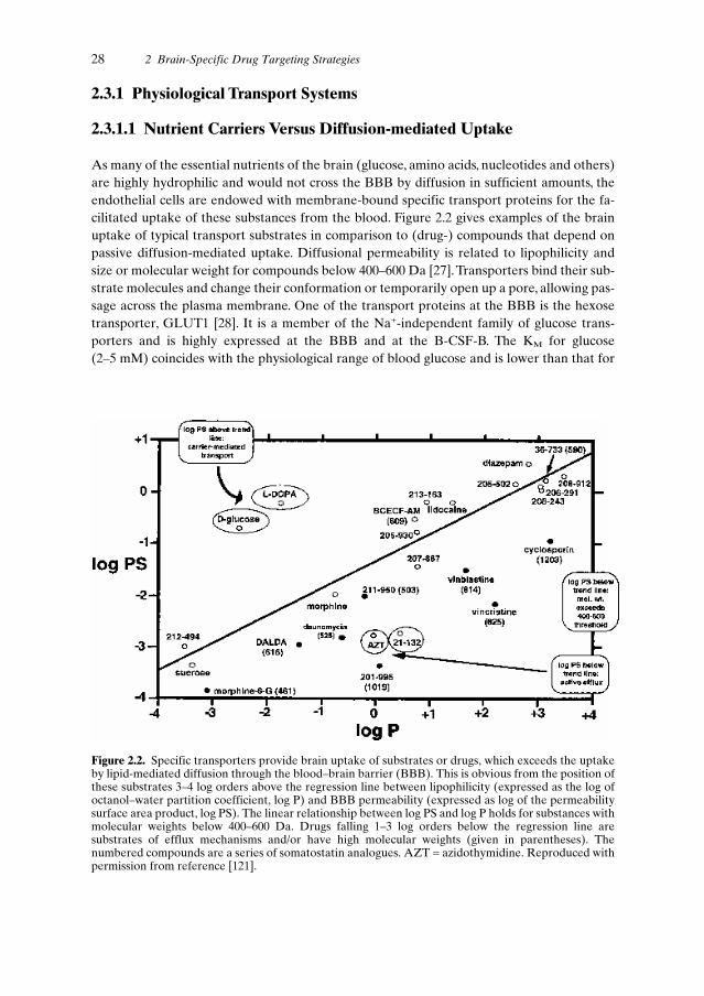
2.3.1 Physiological Transport Systems
2.3.1.1 Nutrient Carriers Versus Diffusion-mediated Uptake
As many of the essential nutrients of the brain (glucose, amino acids, nucleotides and others)are highly hydrophilic and would not cross the BBB by diffusion in sufficient amounts, theendothelial cells are endowed with membrane-bound specific transport proteins for the fa-cilitated uptake of these substances from the blood. Figure 2.2 gives examples of the brainuptake of typical transport substrates in comparison to (drug-) compounds that depend onpassive diffusion-mediated uptake. Diffusional permeability is related to lipophilicity andsize or molecular weight for compounds below 400–600 Da [27].Transporters bind their sub-strate molecules and change their conformation or temporarily open up a pore, allowing pas-sage across the plasma membrane. One of the transport proteins at the BBB is the hexosetransporter, GLUT1 [28]. It is a member of the Na+-independent family of glucose trans-porters and is highly expressed at the BBB and at the B-CSF-B. The KM for glucose(2–5 mM) coincides with the physiological range of blood glucose and is lower than that for
28 2 Brain-Specific Drug Targeting Strategies
Figure 2.2. Specific transporters provide brain uptake of substrates or drugs, which exceeds the uptakeby lipid-mediated diffusion through the blood–brain barrier (BBB). This is obvious from the position ofthese substrates 3–4 log orders above the regression line between lipophilicity (expressed as the log ofoctanol–water partition coefficient, log P) and BBB permeability (expressed as log of the permeabilitysurface area product, log PS). The linear relationship between log PS and log P holds for substances withmolecular weights below 400–600 Da. Drugs falling 1–3 log orders below the regression line aresubstrates of efflux mechanisms and/or have high molecular weights (given in parentheses). Thenumbered compounds are a series of somatostatin analogues. AZT = azidothymidine. Reproduced withpermission from reference [121].

other monosaccharides. Accordingly, GLUT1 mediates facilitated glucose uptake, glucosebeing the main energy source for the brain.
The transport of amino acids at the BBB differs depending on their chemical class and thedual function of some amino acids as nutrients and neurotransmitters. Essential large neutralamino acids are shuttled into the brain by facilitated transport via the large neutral aminoacid transporter (LAT) system [29] and display rapid equilibration between plasma andbrain concentrations on a minute time scale.The LAT-system at the BBB shows a much low-er KM for its substrates compared to the analogous L-system of peripheral tissues and itsmRNA is highly expressed in brain endothelial cells (100-fold abundance compared to othertissues). Cationic amino acids are taken up into the brain by a different facilitative trans-porter, designated as the y+ system, which is present on the luminal and abluminal endothe-lial membrane. In contrast, active Na+-dependent transporters for small neutral amino acids(A-system;ASC-system) and cationic amino acids (B0+ system), appear to be confined to theabluminal surface and may be involved in removal of amino acids from brain extracellularfluid [30]. Carrier-mediated BBB transport includes monocarboxylic acids (pyruvate),amines (choline), nucleosides (adenosine), purine bases (adenine), panthotenate, thiamine,and thyroid hormones (T3), with a representative substrate given in parentheses [31].
2.3.1.2 Efflux Systems
The sodium-dependent X- system is localized to the abluminal membrane and apparentlyfunctions as an efflux mechanism for the excitatory amino acids aspartate and glutamatefrom brain interstitial fluid. In accordance with such a direction of transport, acidic aminoacids do not undergo rapid uptake into the brain under physiological conditions. Other effluxpumps have recently been shown to exist at the BBB for substances belonging to diversechemical classes. The most studied example to date is the multi-drug resistance protein(MDR), also known as P-glycoprotein or P-gp, with its broad substrate specificity [32]. P-gpand structurally related transporters are members of the large ATP-binding cassette familyof membrane pumps. Several organic anion transport polypeptides have recently beencloned from animal and human tissue. The substrate specificity and direction of transport ofthese systems in vivo, which are expressed in liver, kidney, intestines as well as in the brain atthe BBB and B-CSF-B [33], has not been fully elucidated. In any case, the importance of ac-tive transport systems at the BBB is rapidly emerging. Evidence supports the view that manymore small molecular weight substances (including drugs) than previously thought, appar-ently do not cross the BBB by simple diffusion only, but are subject to active transport. Theinhibition of BBB efflux systems as a strategy for brain drug delivery has therefore been pro-posed [34].
2.3.1.3 Receptor- and Absorptive-mediated Uptake
Macromolecules such as polypeptides and proteins are excluded from uptake by diffusion orthrough pores at the BBB. However, the presence of receptor-mediated uptake and transportinto the brain has been found for a number of substances. Specific receptors for insulin and
2.3 BBB Biology and Pharmacology 29

transferrin were initially described. These receptors have been identified at the molecularlevel as being identical to their counterparts in peripheral tissues [35,36]. However, at theBBB the receptors mediate transcytosis of their respective ligands through the endothelialcell as opposed to endocytosis in other cells.
The process of transcytosis is illustrated in Figure 2.3 for the transferrin receptor (TfR)[37]. The receptor is heavily expressed at the BBB compared to other vascular beds [38].Transferrin or a monoclonal antibody to the extracellular domain of the receptor protein willbind from the luminal side of the BBB. This triggers cellular uptake by the mechanism of re-ceptor-mediated endocytosis, i.e. the invagination and budding off of parts of the cell mem-brane as a result of the formation of small vesicles (endosomes).The transcellular passage ofligand (transcytosis) is completed by exocytosis at the abluminal membrane, and the wholeprocess is completed within minutes in vivo.
Receptor-mediated uptake mechanisms have also been shown for insulin, insulin-likegrowth factors, and leptin. The fact that macromolecular complexes as large as LDL can un-
30 2 Brain-Specific Drug Targeting Strategies
vl
bm
(a)
(b)
(c)
Figure 2.3. An example of receptor-mediated transcytosis through the blood–brain barrier. OX26, amonoclonal antibody to the rat transferrin receptor, was conjugated with 5-nm colloidal gold andperfused through the internal carotid artery in rats in vivo. The brain was then perfusion-fixed. (a)Arrows indicate binding to the luminal plasma membrane of capillary endothelial cells and internalizedantibody in an endosome. (b) The endocytosed antibodies appear in multivesicular bodies (arrows) andare seen undergoing exocytosis at the abluminal cell membrane (arrowhead). vl, vascular lumen; bm,basement membrane. Scale bar = 100 nm. (c) A strategy for brain delivery can be based on receptor-mediated transcytosis. The non-permeable drug moiety ‘B’ is coupled to A by a linker, L. ‘A’ is a ligandor an anti-receptor antibody (e.g. OX26) which binds to its receptor on the luminal side and mediatesendocytosis. The chimeric peptide enters brain interstitial space by exocytosis from the endothelial celland is cleaved by local enzymes to release the drug. Reproduced with permission from references [37](a,b) and [81] (c).

dergo transcytosis [39] underlines the difference between vesicular transport and carrier-me-diated uptake.
Absorptive-mediated endocytosis and transcytosis of macromolecules through the BBB isrelated to receptor-mediated uptake, although not as specific. The process applies to certainlectins, for example wheat germ agglutinin [40], and to cationic proteins. Transport is trig-gered by glycoprotein binding (lectins) or by ionic interactions between negative charges onthe endothelial plasma membrane and positive surface charges on the proteins. Both nativeproteins (histones) and chemically modified proteins (cationized albumin and IgG) can un-dergo absorptive-mediated transcytosis. While the physiological function of that process atthe BBB remains to be identified, it offers a potential strategy for drug targeting/delivery[41].
2.3.2 Techniques for Measurement of Brain Uptake
2.3.2.1 In vivo Methods
For the correct interpretation of brain uptake studies in general and for the pharmacokinet-ic validation of a given delivery strategy, it is necessary to be familiar with characteristics andlimitations of the applied technique [41]. In vivo methods remain the gold standard, as thereare still no cell culture models available that fully represent the barrier characteristics.
Quantitative measurement of diffusional uptake and carrier-mediated transport of nutri-ents and drugs in experimental animals was greatly facilitated with the introduction of Old-endorf’s brain uptake index (BUI) [42]. Test and reference tracers are injected as an intraar-terial bolus into the carotid artery of the anaesthetized animal. After 5 s the animal is killedand the brain is removed for radioactivity counting. This method measures the ratio of theunidirectional brain extraction, E, of the test substance and of the reference ([3H]-water,[14C]-butanol), which are labelled with different isotopes, during a single passage through thebrain capillary bed:
BUI = Etest/Ereference (2.1)
Advantages of the method include technical simplicity, and control over the compositionof the injection fluid, making the technique suitable for competition and saturation experi-ments. Provided that the absolute value of Ereference is known, the absolute Etest may be cal-culated and, with the independently determined value of cerebral blood flow, F, Etest may beconverted into a permeability surface area (PS) product. The latter conversion follows fromthe application of the Kety–Renkin–Crone equation of capillary physiology:
E = 1 – ePS/F (2.2)
The major drawback of the BUI is its limited sensitivity for measurement of compoundswith low extraction.
Sensitivity was improved by at least two orders of magnitude with the internal carotidartery perfusion technique [43].An outline of the method is given in Figure 2.4. Here, the ex-traction can be measured over a time frame of 15 s to 10 min or more, while maintaining the
2.3 BBB Biology and Pharmacology 31

advantage of the experimental control of tracer concentrations and composition of the perfusion fluid. Gentle homogenization of brain tissue followed by density centrifugation,resulting in the separation into a vascular pellet and a ‘postvascular’ supernatant, differenti-ate the fractions of the test substance which have merely associated with the vasculaturefrom those which have fully penetrated the BBB [44]. This ‘capillary depletion’ is applicableto substances, which bind to capillaries with high affinity and/or which are internalized by specific mechanisms such as receptor-mediated endocytosis. It could be shown that for example the acetylated form of LDL is only endocytosed and then sequestered by brain capillary endothelium. Erroneous data may be observed for substances without specific uptake but low affinity non-specific adsorption to the endothelium because
32 2 Brain-Specific Drug Targeting Strategies
VD =Cbrain
Cperfusate
Internal carotid artery perfusion
VD
time
Supernatant
Pellet
Brain Homogenate
IV bolus kinetic
Cplasma
time
PS =AUCplasma(0-T)
Cbrain(T)
T
iep
o
c
(a) (b)
Figure 2.4. In vivo measurement of blood–brain barrier (BBB) permeability. (a) Internal carotid arteryperfusion technique (i) in the rat. Other branches of the carotid artery are ligated or electricallycoagulated (o, occipital artery; p, pterygopalatine artery). The external carotid artery (e) is cannulatedand the common carotid artery (c) ligated. Perfusion time may range from 15 s to 10 min, depending onthe test substance. It is necessary to subtract the intravascular volume, V0, from VD (apparent volume ofdistribution), to obtain true uptake values and this may be achieved by inclusion of a vascular marker inthe perfusate, for example labelled albumin. Time-dependent analysis of VD results in estimates of theunidirectional brain influx constant Kin (µl min–1 g–1) which is equivalent within certain constraints to thePS product. BBB permeability surface area product PS can be calculated from the increase in theapparent volume of distribution VD over time. Capillary depletion, i.e. separation of the vascularelements from the homogenate by density centrifugation, can discriminate capillary uptake fromtranscytosis. (b) i.v. bolus kinetics. The PS product is calculated from the brain concentration at thesampling time, T, and the area under the plasma concentration–time curve, AUC.

of redistribution into the postvascular supernatant during processing and centrifugation [45].
The most sensitive technique for measuring brain uptake is the intravenous bolus admin-istration or infusion and subsequent measurement of brain concentrations (Figure 2.4). De-pending on the pharmacokinetics of the test compound in plasma, brain sampling may beperformed after suitable circulation times ranging from minutes to hours or days.
The pharmacokinetic calculation of the unidirectional brain uptake rate, Kin, after intra-venous injection, uses the relation of the brain concentration and the area under the curve ofplasma concentration, AUC:
Kin = Cbrain/AUC (2.3)
Here Cbrain is the brain concentration after correction for intravascular content, and AUCis determined between time 0 and the final sampling time. Two assumptions must hold wheninterpreting the evaluation in the simple form described above: (1) the brain uptake of thecompound is linear, meaning Kin is dose independent, and (2) the analysis is performed with-in a time-frame where the efflux from tissue is negligible (tissue concentrations are suffi-ciently low compared to plasma concentrations).Violation of these assumptions requires ad-justments in experimental design and evaluation. For example, nonlinear kinetics may be ac-counted for by incorporation of a Michaelis–Menten term, while efflux can be treated bycompartmental analysis [46].
The i.v. approach has the distinct advantage of measurements being carried out under themost physiological conditions. On the other hand, the caveats include confounding effects ofperipheral metabolism, which may give rise to artifactual brain uptake of degradation prod-ucts. To exclude such a possibility the application of suitable analytical techniques to tissueand plasma samples is required.
Unless a test compound is available in tracer form suitable for noninvasive quantificationsuch as positron emission tomography (PET) or single photon emission computed tomogra-phy (SPECT), cerebrospinal fluid sampling is often used in human studies. It needs to be re-membered that such measurements pertain to permeability at the B-CSF-B, not at the BBB,otherwise erroneous conclusions may be derived, in particular when specific transportprocesses are involved. For example the rapid penetration of the anti-HIV drug azidothymi-dine into CSF [47] is due to a carrier for pyrimidine nucleotides in the choroid plexus. In con-trast, azidothymidine is subject to efflux at the BBB, which prevents it from reaching signifi-cant concentrations in the brain [48].
A caveat should be mentioned here concerning data relating to drug transport which hasbeen obtained using intracerebral microdialysis. While the method is appealing for reasonssuch as its ability to investigate extracellular fluid concentration–time courses and concen-tration-dependent uptake rates in each single animal [49], it has limitations, in particular inthe measurement of substances with low permeability. This is a consequence of the invasivenature of the placement of the microdialysis probe, which inevitably causes tissue damage, aninflammatory response and gliosis surrounding the dialysis probe. Therefore, the BBB maybe locally compromised, obviously jeopardizing the valid interpretation of experimental data[50].
2.3 BBB Biology and Pharmacology 33

2.3.2.2 In Vitro Models
Freshly isolated capillaries from different species are a valuable model system of the BBB[51], because they retain the native repertoire of receptors and enzymes. In particular, recep-tor binding and carrier-mediated uptake or receptor-mediated endocytosis can be conve-niently studied. On which side binding or uptake takes place cannot be easily determined, asboth the luminal and abluminal side are exposed in such a preparation.
Numerous modifications of in vitro culture systems have been developed for the estima-tion of BBB transfer [52]. Culture systems in use are either primary cultures of brain mi-crovessel endothelial cells (BMEC) or immortalized endothelial cell lines. BMEC may begrown in co-culture with astrocytes or in astrocyte-conditioned medium. Astrocyte-derivedfactors increase the tightness of the barrier as measured by transendothelial electrical resis-tance (TEER) and by the permeability of hydrophilic markers such as sucrose.They also up-regulate the expression of BBB-enriched enzymes such as γ-glutamyl transpeptidase (γ-GTP) and alkaline phosphatase.A setup of the in vitro technique in a transwell system fortransport studies is depicted in Figure 2.5.
34 2 Brain-Specific Drug Targeting Strategies
Microporous membrane(0.4 µM pore size)
6 well cluster plate
C6 Astroglioma
BBMECs
Abluminallower chamber
Luminal upper chamber
Millicell®�ERSTEER(ohms•�cm2)
C6 Astroglioma
Figure 2.5. Setup for in vitro measurement of blood–brain barrier permeability with a co-culture ofbovine brain microvascular endothelial cells (BBMEC) and an astroglioma cell line, C6. The BBMECare grown on top of a filter insert. The C6 cells are either grown on the opposite side of the filter or onthe bottom of the wells. Transport across the BBMEC monolayer is measured by adding the testsubstance to the upper chamber and sampling from the lower chamber. The tightness of the monolayeris also characterized by the transendothelial electrical resistance (TEER). Courtesy of T. Abbruscato.
2.4 Drug Delivery Strategies
The approaches for developing strategies for the delivery of drugs to the brain are shown ingroups in the scheme in Figure 2.6. Pharmacological-based and physiological-based methods
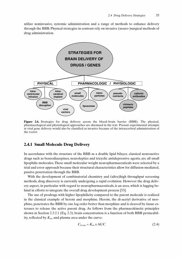
utilize noninvasive, systemic administration and a range of methods to enhance deliverythrough the BBB. Physical strategies in contrast rely on invasive (neuro-)surgical methods ofdrug administration.
2.4 Drug Delivery Strategies 35
STRATEGIES FOR
BRAIN DELIVERY OF
DRUGS / GENES
pseudo-nutrients
chimericpeptides
cationicantibodies
PHARMACOLOGIC / PHYSIOLOGICPHYSICAL
intra-ventricular
infusion
BBBdisruption
intra-cerebralimplant
smallmolecules
liposomes
nano-particles
viralvectors
Figure 2.6. Strategies for drug delivery across the blood–brain barrier (BBB). The physical,pharmacological and physiological approaches are discussed in the text. Present experimental attemptsat viral gene delivery would also be classified as invasive because of the intracerebral administration ofthe vector.
2.4.1 Small Molecule Drug Delivery
In accordance with the structure of the BBB as a double lipid bilayer, classical neuroactivedrugs such as benzodiazepines, neuroleptics and tricyclic antidepressive agents, are all smalllipophilic molecules. These small molecular weight neuropharmaceuticals were selected by atrial and error approach because their structural characteristics allow for diffusion-mediated,passive penetration through the BBB.
With the development of combinatorial chemistry and (ultra)high throughput screeningmethods, drug discovery is currently undergoing a rapid evolution. However the drug deliv-ery aspect, in particular with regard to neuropharmaceuticals, is an area, which is lagging be-hind in efforts to integrate the overall drug development process [53].
The use of prodrugs with higher lipophilicity compared to the parent molecule is realizedin the classical example of heroin and morphine. Heroin, the di-acetyl derivative of mor-phine, penetrates the BBB by one log order better than morphine and is cleaved by tissue es-terases to release the active parent drug. As follows from the pharmacokinetic principlesshown in Section 2.3.2.1 (Eq. 2.3), brain concentration is a function of both BBB permeabil-ity, reflected by Kin, and plasma area under the curve:
Cbrain = Kin x AUC (2.4)

While Kin increases with lipophilicity, AUC decreases due to higher uptake across all cellmembranes including those of peripheral tissues.Therefore limits are imposed on the gain inbrain delivery by the ‘lipidization’ strategy. In fact, for azidothymidine lipidization with thelipophilic adamantane moiety, CSF concentrations decreased by a factor of 10 as a result ofa decrease in AUC [54].
Lipidization is also an integral part of the chemical delivery approach that is based on theconcept of ‘retro-metabolism’ [55]. The underlying principle is the simultaneous derivatiza-tion of a drug with a ‘redox targeter’, e.g. dihydrotrigonellinate, and with a lipophilic moiety,e.g. cholesterol. This strategy has been applied to deliver a small enkephalin analogue to thebrain [55]. Once inside the target tissue, the redox targeter will be enzymatically oxidized toa positively charged derivative and serve to lock-in the prodrug. Further enzymatic steps re-lease the active drug by cleavage of the lipid modifier and the targeter. This strategy offersthe potential of tissue-selective delivery, yet it requires simultaneous optimization of a multi-tude of rates, including the influx and efflux of the prodrug, biotransformation of the targeterand eventual release of the active drug.
An alternative delivery strategy for small molecules is based on the presence of the nutri-ent transporters. Drugs that are structurally similar to substrates of a carrier system can un-dergo facilitated brain uptake as pseudoneutrients. The best example of this is the therapeu-tic use of L-DOPA in Parkinson’s disease. Unlike the neurotransmitter dopamine itself,which cannot cross the BBB in significant amounts, its precursor L-DOPA is a substrate forLAT, the transporter of large neutral amino acids [56]. Its uptake by the brain is saturable,and subject to competition by the other substrates of the carrier present in plasma.
L-DOPA therapy is an example of rational drug design based on knowledge of BBBtransport biology. A number of other small molecular weight drugs are known to undergocarrier-mediated transport at the BBB.These include the substrates of LAT, such as the anti-cancer drugs melphalan and acivicin, or the GABAB-agonist baclofen, and beta lactam an-tibiotics (e.g. benzylpenicilline, ceftriaxone, cefodizime) which are substrates for the organicacid carrier [31].
2.4.2 Macromolecular Drug Delivery
2.4.2.1 Intraventricular Route
At first glance, intracerebroventricular (i.c.v.) or intrathecal administration via catheters, typ-ically connected to a subcutaneous drug reservoir, appears as a logical mode of drug delivery,because the percentage of the dose reaching the target organ would be 100%.The i.c.v. routeis successfully used in disorders where pathogenetic events take place close to the brain sur-face. Clinical examples of intrathecal small drug delivery are the administration of glycopep-tide and aminoglycoside antibiotics in meningitis [57], the i.c.v. treatment of meningealmetastasis [58], intrathecal baclofen injection for treatment of spasticity [59] and the infusionof opioids for severe chronic pain [60]. However, in addition to the invasive character of theprocedure, it has to be taken into account that drug distribution into brain tissue is severelydiffusion-limited and that the continuous production and reabsorption of the CSF results inrapid clearance from that compartment.
36 2 Brain-Specific Drug Targeting Strategies

Although there is no cellular barrier preventing diffusion from the ventricular surface intobrain tissue (there are no tight junctions between the ependymal cells lining the ventricularsurface), the low speed of diffusion severely restricts tissue uptake of even small molecularweight drugs and practically prevents the penetration of large molecules such as peptides andproteins into deep tissue layers. Possible enzymatic inactivation and binding or sequestrationby brain cells along the diffusion path may even lower the actual drug concentrations in braininterstitial spaces to levels lower than predicted from the molecular size and diffusion coeffi-cients [61]. Figure 2.7a shows an example of a large molecule, the 26-kDa nerve growth fac-tor (NGF), that could not penetrate into rat brain deeper than 1–2 mm from the infused ven-tricle. This might be expected, as even small drugs show very steep concentration gradientsover a distance of only 2–3 mm from the ventricular surface (Figure 2.7b). Exceptions arefound in areas to which retrograde transport occurs, e.g. into the neurons of the basal cholin-ergic nuclei in the case of NGF (Figure 2.7a).
After i.c.v. injection, the rate of elimination from the CNS compartment is dominated bycerebrospinal fluid dynamics. The CSF, which is secreted by the choroid plexus epitheliumacross the apical membrane, circulates along the surface and convexities of the brain in a ros-tral to caudal direction. It is reabsorbed by bulk flow into the peripheral bloodstream at thearachnoid villi within both cranial and spinal arachnoid spaces [62]. Of note is that theturnover rate of total CSF volume is species dependent and varies between approximately1 h in rats and 5 h in humans. In adult human brain, the total CSF volume amounts to100–140 ml and the production rate is 21 ml h–1 [63]. Accordingly, the entire cerebrospinalvolume is exchanged regularly 4–5 times per day. This rapid drainage of CSF into peripheralblood leads to relatively high drug concentrations in the peripheral circulation. For instance,the concentration of methotrexate in peripheral blood reaches 1% of the ventricular CSFconcentration following intrathecal administration of the drug [64]. In Rhesus monkeys,parenchymal concentrations of methotrexate of 1% of the intraventricular concentrationhave been measured at a distance of 2 mm from the ependymal surface [65]. Therefore, theconcentration of methotrexate in the blood is actually higher than at tissue regions beyond2 mm from the ependymal surface following intrathecal application.
2.4.2.2 Intraparenchymal Route
Restricted diffusion also limits tissue distribution after intraparenchymal drug administra-tion, as shown in Figure 2.7c and d. Distribution has been measured in the rat brain after im-plantation of polymer discs containing NGF [66]. Drug concentrations decreased to less than10% of the values measured on the disc surface within a distance of 2–3 mm, even after pro-longed periods of several days. Therefore, applying this approach in the larger human brainwould require the stereotaxic placement of multiple intraparenchymal depots, as has beenevaluated in rat brain [67], on a repetitive schedule.
The same pharmacokinetic limitation is true in principle for the implantation of encapsu-lated genetically engineered cells, which synthesize and release neurotrophic factors [68].
2.4 Drug Delivery Strategies 37

Cti
ssu
e
CC
SF
cmfr
om
ven
tr.s
urf
ace
Dis
tan
cefr
om
po
lym
er,m
m
NGFconcentration,ng/ml
AB
2h
r6 h
r
10,1
2h
r
01.
01.
52.
00.
5
RelativeConcentration 0
1.0
2d
ay
Dis
tan
ce,c
m
(a)
(b)
(c)
(d)
(e)
(f)
38 2 Brain-Specific Drug Targeting Strategies

2.4.2.3 Convective Flow
Intraparenchymal high flow microinfusions with flow rates up to 4 µl min–1 result in almosthomogenous tissue concentrations of macromolecules (transferrin, mw 80 kDa) over a largevolume and over a distance of > 10 mm from the catheter tip within an infusion period of 2 h[69]. Figure 2.7 demonstrates the principle in an experimental study (Figure 2.7e) and the ex-pected tissue concentration profiles (Figure 2.7f). The plateau-like concentration profile atthe front of the convective flow is in contrast to the concentration gradients associated withdiffusion-mediated distribution (Figure 2.7b, d). The method has been applied in clinical tri-als for treatment of gliomas and metastatic brain tumours with a transferrin-targeted mutantdiphtheria toxin,Tf-CRM107 [70]. Current efforts are directed at overcoming the dose-limit-ing toxicities found in these studies in the CNS such as petechial haemorrhage and small ves-sel thrombosis [71].
2.4.2.4 Delivery by Barrier Disruption
The temporary physico-chemical disruption of brain endothelial barrier integrity is also con-sidered an invasive strategy for drug delivery, because it typically involves the intracarotid in-fusion of a barrier-opening agent. Barrier opening for low molecular weight tracers andmacromolecules (e.g. Evans blue-albumin) was experimentally demonstrated with intrac-arotid infusions of membrane active agents such as bile salts, oleic acid, cytostatic drugsetoposide and melphalan, and cytochalasin B. Intracarotid infusion of a low pH buffer alsoopens the BBB.
Most studies were performed with hyperosmolar solutions. Hypertonic disruption is underclinical evaluation for enhanced delivery of small molecular weight cytostatic agents to braintumours. Technically, the procedure is performed as a high-flow short-term infusion of 25%mannitol or arabinose under general anaesthesia.The underlying mechanism is a sequelae ofendothelial cell shrinkage, disruption of tight junctions and vasodilatation by osmotic shift [72].
2.4 Drug Delivery Strategies 39
Figure 2.7. (a) Autoradiograph of a brain section showing the limited distribution of [125I]-NGF (nervegrowth factor) in rat brain 18 h after injection into the lateral ventricle. Exception: cholinergic neuronswith retrograde transport to the cell body (arrow). (b) Brain–cerebrospinal fluid (CSF) concentrationgradient of four drugs relative to the distance from the ependymal surface. Curves from left to right:BCNU, thiotepa, cytosine arabinoside, hydroxyurea. Drugs were infused by the intracerebroventricularroute in a Rhesus monkey for a 1-h period. The gradients are steepest for highly diffusible substances(thiotepa, BCNU). (c) Autoradiograph (top) and unstained photograph (bottom) of coronal sections ofrat brains following implantation of [125I]-NGF-loaded polymers. Sections were obtained 2 days post-implanatation. Bar = 2.5 mm. (d) Concentration profile in the vicinity of the polymer implant in (c). (e)The convective-enhanced method allows homogenous delivery to precisely defined volumes, asdemonstrated here in a coronal section of monkey brain stained for biotin (black region). The animalreceived an infusion of biotinylated albumin into the globus pallidus internus (Gpi). Gpe, globus pallidusexternus; IC internal capsule; OT, optical tract; Put, Putamen. (f) Calculated concentration profiles formacromolecules in brain that can be achieved with high flow microinfusion. Dashed lines: steady-stateprofiles of molecules undergoing rapid metabolism with half-lives of 0.167 (A) or 1 h (B). Solid lines:profiles after 2, 6, 10 and 12 h for a molecule with long a half-life (33.5 h). The latter profiles showplateau-like tissue concentrations that extend with duration of infusion up to 1.5 cm from the tip of theinfusion catheter. Reproduced with permission from references [61] (a), [65] (b), [66](c,d); [122] (e) and[123] (f).
�

Morphological studies in rats, where the induction of neuropathological changes by os-motic opening was examined, provided evidence of uptake of macromolecules by the brain.The extravasation of plasma proteins such as fibrinogen and albumin was shown immuno-histochemically at the light microscopic level. Electron microscopy revealed ultrastructuralchanges such as swelling of astrocytic processes and severe mitochondrial damage in neurons[73]. There was also evidence of prolonged (24 h) cellular stress or injury in neurons and gliaas expressed by the induction of heat shock protein (HSP-70).While the nonspecific openingof the BBB to plasma proteins harbours a risk of eliciting neuropathological changes, osmot-ic disruption has been tested for its potential as a delivery method for macromolecular drugssuch as monoclonal antibodies against various tumour antigens or their Fab fragments. Inother studies, uptake after intracarotid administration of nanoparticles (20-nm iron oxideparticles) by normal brain, and uptake of recombinant adenovirus or herpes virus by normalbrain tissue and brain tumour xenografts in nude rats was postulated [74,75].
Compared to small molecules, barrier opening for high molecular weight compounds is ofshorter duration [72]. Furthermore, a characteristic difference exists in the degree of barrieropening in tumour versus normal brain tissue. Barrier disruption was consistently found tobe more pronounced for the normal BBB, which may limit the clinical applicability of hyper-osmolar barrier opening, at least for cytotoxic drugs [76].
BBB opening may also be achieved by receptor-mediated mechanisms. The vasoactivecompounds prostaglandins, histamine, serotonin, leukotriene C4 (LTC4), and bradykininhave all been shown to increase BBB permeability [16]. The effects of LTC4 and bradykininare more pronounced on the blood–tumour barrier than on the normal BBB. In the case ofLTC4 this effect is ascribed to the presence of an enzymatic barrier in normal brain tissue dueto the endothelial expression of γ-GTP. The enzyme metabolizes and inactivates LTC4 toLTD4. In contrast, tumour vessels are unable to express equivalent activities of γ-GTP, a factthat may be exploited for selective opening of the tumour barrier by intracarotid adminis-tration of LTC4. However, the effect is restricted to small molecules, as there was no increasein the tumour accumulation of a dextran tracer of molecular weight 70 kDa. On the otherhand, bradykinin also opens the barrier for the high molecular weight range. It acts on en-dothelial cells through B2-receptors located on the abluminal side. Normal brain tissue is pro-tected from barrier opening by bradykinin in the vascular lumen because the peptide cannotaccess these receptors. In tumour vessels the barrier integrity is sufficiently compromised toallow for additional bradykinin-mediated opening at low peptide concentrations. Whilebradykinin itself requires intracarotid administration, an analogue with prolonged half-life(RMP-7) is effective after intracarotid or intravenous application. A 4–5-fold increase in thedelivery of the cytokines interferon-γ , tumour necrosis factor α and interleukin-2 to experi-mental RG2 glioma in rats was demonstrated after intracarotid infusion of RMP-7 [77]. Thedrug is currently being evaluated in the therapy of human malignant gliomas to enhance de-livery of carboplatin to the tumour [78].
2.4.2.5 Vector-mediated Delivery
In this approach, ‘chimeric peptides’ [79] are generated as transportable drug derivatives tar-geting the receptor-mediated mechanism. Chimeric peptides are formed by linking a drug
40 2 Brain-Specific Drug Targeting Strategies

that is unable to cross the BBB to a vector (see Figure 2.8). Binding of the vector at the lu-minal membrane of brain capillary endothelial cells initiates receptor-mediated or adsorp-tive-mediated transcytosis (Figure 2.3c). Size and structure of the cargo may vary as long asbinding and cellular uptake of the vector is not inhibited by the drug moiety, and may only belimited by the size of the endocytotic vesicles.
Initial studies of brain delivery based on the chimeric peptide strategy used the absorptive-mediated uptake of cationized albumin which was chemically coupled to the opioid peptideβ-endorphin [80] or its metabolically stabilized analogue [D-Ala2]β-endorphin. Tracer ex-periments in which the chimeric peptide was labelled in the endorphin moiety provided evi-dence of internalization by isolated brain capillaries and transport into brain tissue in vivo[81].
Endogenous ligands for receptor-mediated systems may be unsuitable as vectors due tocompetition for transport or undesirable pharmacological effects. For example, plasma con-centrations of transferrin are in the range of 25 µM. Insulin as a vector would cause hypogly-caemia. A logical alternative as vectors are monoclonal antibodies specific to the extracellu-lar domain of a peptide or protein receptor at the BBB. These antibodies can be designed asnon-competitive, i.e. they bind to the receptors at a site distinct from the ligand binding siteand do not interfere with the endocytosis process.
Brain uptake data for some vectors are compared in Table 2.1. Quantitative comparisonswithin the same species are possible for the rat with vectors derived from the anti-TfR mon-oclonal antibody OX26 and from cationized human serum albumin. To put the efficiency ofbrain delivery into perspective, the comparison to a classical neuroactive drug may be infor-mative. In the rat, brain concentrations of morphine following systemic administration neverexceed 0.08% of injected dose per gram [%ID g–1] [82]. In contrast, OX26 easily reaches con-centrations in rat brain that are three to four times higher. Vectors based on cationized hu-
2.4 Drug Delivery Strategies 41
Vector Linker Drug
OX2684-15cHSA
(Strept)avidin -Biotin
PeptidesVIPDALDA
ProteinsNGFBDNF
OligonucleotidesPNA
Genesluciferase-Gal
Liposomeencapsulated drugs
Figure 2.8. Scheme of a chimeric peptide with examples for each of the distinct domains. OX26, anti-rattransferrin receptor monoclonal antibody (mAb); 84-15, anti-human insulin receptor mAb; cHSA,cationized human serum albumin; VIP, vasoactive intestinal polypeptide; DALDA, dermorphinanalogue; NGF, nerve growth factor; BDNF, brain-derived neurotrophic factor; PNA, peptide nucleicacid; β-gal, β-galactosidase.

man albumin reach lower brain concentrations compared to OX26 vectors. The difference ismainly caused by a corresponding difference in the PS products, i.e. the rate of uptake by ab-sorptive-mediated transcytosis of cationized albumin at the BBB is lower than the rate of re-ceptor-mediated uptake of OX26 (Table 2.1).
With regard to transport capacity, the introduction of the anti-human insulin receptor an-tibody (HIR MAb) 83-14 as a vector indicates the potential for future improvements inbrain-specific delivery vectors. Compared to anti-TfR monoclonal antibodies, the brain de-livery in primates is over 7-fold higher due to the high PS product of the HIR MAb.
A thorough characterization of vectors for drug delivery needs to take into account thesaturable character of receptor-mediated processes and therefore requires investigation ofdose dependence of uptake. While non-competing antibodies avoid the problem of competi-tion by endogenous ligands, the saturability of the antibody binding site remains. The valuesin Table 2.1 were obtained in uptake experiments with doses of vectors in the low µg kg–1
range, corresponding to plasma concentrations in the low nM range. Linear pharmacokinet-ics cannot be expected at higher doses and hence changes in both plasma AUC and apparentPS product are likely to occur at higher doses. Experimental evidence of saturability in vivowas seen for the OX26 antibody in rats and the 8D3 antibody in mice. In the case of 8D3, forexample, the brain uptake declined to virtually 0% ID g–1 brain at a dose of 4 mg kg–1 i.v. [83].Proof of saturability in vivo is direct evidence for a specific uptake mechanism.
The coupling step between vector and drug moiety may be performed by either chemicalor molecular biological approaches (see Chapter 11 for a more detailed discussion on conju-gation strategies in drug targeting research). While the options offered by chemical methods
42 2 Brain-Specific Drug Targeting Strategies
Table 2.1. Brain concentration, blood–brain barrier PS product, and plasma AUC (0–60 min) of braindelivery vectors after i.v. bolus injection.
Vector (species) %ID g-1a PS (µl min-1 g-1) AUC 0–60 (%ID min ml-1)
3H-OX26 (rat) 0.27 ± 0.04 1.92 ± 0.06 132 ± 19
OX26-Avb (rat) 0.041 ± 0.004 0.85 ± 0.02 49 ± 4
Anti-TfR IgG3-CH3-Avb,c 0.25 ± 0.09 2.25 ± 0.65 134 ± 29(rat, recombinant)
OX26-NLAb (rat) 0.17 ± 0.04 0.70 ± 0.10 232 ± 25
OX26-SAb (rat) 0.20 ± 0.03 0.92 ± 0.10 216 ± 28
cHSA-Avb (rat) 0.015 ± 0.006 0.26 ± 0.13 64 ± 7
cHSA-NLAb (rat) 0.061 ± 0.012 0.20 ± 0.04 300 ± 14125I-8D3 (mouse) 3.1 ± 0.2 3.3 ± 0.1 932 ± 57125I-HIR MAb (rhesus monkey) 3.8 ± 0.4 (100 g brain)d 5.4 ± 0.6 5.9 ± 1.2 (0–180 min)
a Percentage injected dose per g brain 60 min after i.v. bolus injection.b These proteins were labeled at the avidin moiety with [3H]biotin.c IgG3-CH3 fusion protein with variable region of OX26 and avidin.d %ID in total brain tissue after 3 h. AUC, area under curve of plasma concentration; Av, avidin; NLA, neutral avidin; SA, streptavidin;cHSA, cationized human serum albumin; HIR MAb, human insulin receptor mAb; PS product,permeability surface area product.
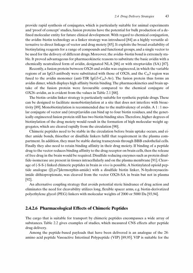
provide rapid synthesis of conjugates, which is particularly suitable for animal experimentsand ‘proof of concept’ studies, fusion proteins have the potential for bulk production of a de-fined molecular entity for future clinical development. With regard to chemical conjugation,the avidin–biotin technology as a linker strategy was introduced [84] as a highly versatile al-ternative to direct linkage of vector and drug moiety [85]. It exploits the broad availability ofbiotinylating reagents for a range of compounds and functional groups, and a single vector tobe used for the delivery of different drugs. Moreover, the avidin–biotin bond is extremely sta-ble. It proved advantageous for pharmacokinetic reasons to substitute the basic avidin with achemically neutralized form of avidin, designated NLA [86] or with streptavidin (SA) [87].
Recently, a fusion protein between OX26 and avidin was engineered, in which the variableregions of an IgG3-antibody were substituted with those of OX26, and the CH3 region wasfused to the avidin monomer (anti-TfR IgG3-CH3-Av). The fusion protein thus forms anavidin dimer, which displays high affinity biotin binding.The pharmacokinetics and brain up-take of the fusion protein were favourable compared to the chemical conjugate ofOX26–avidin, as is evident from the values in Table 2.1 [88].
The biotin–avidin linker strategy is particularly suitable for synthetic peptide drugs. Thesecan be designed to facilitate monobiotinylation at a site that does not interfere with bioac-tivity [89]. Monobiotinylation is recommended due to the multivalency of avidin. A 1 : 1 mo-lar conjugate of vector and (strept)avidin can bind up to four biotin residues, and the genet-ically engineered fusion protein still has two biotin binding sites.Therefore, higher degrees ofbiotinylation of the drug moiety would result in the formation of high molecular weight ag-gregates, which are cleared rapidly from the circulation [90].
Chimeric peptides need to be stable in the circulation before brain uptake occurs, and ei-ther amide bonds, thioether or disulfide linkers fulfil that requirement in the plasma com-partment. In addition, they must be stable during transcytosis through BBB endothelial cells.Finally they also need to retain binding affinity in their drug moiety. If binding of a peptidedrug to the vector reduces binding affinity to the drug receptor on brain cells, then the releaseof free drug in the brain would be required. Disulfide reducing enzymes such as protein disul-fide-isomerase are present in tissues intracellularly and on the plasma membrane [91]. Cleav-age of (-S-S-) linked chimeric peptides in brain in vivo is possible.A biotinylated opioid pep-tide analogue ([Lys7]dermorphin-amide) with a disulfide biotin linker, N-hydroxysuccin-imide dithiopropionate, was cleaved from the vector OX26-SA in brain but not in plasma[92].
An alternative coupling strategy that avoids potential steric hindrance of drug action andeliminates the need for cleavability utilizes long, flexible spacer arms, e.g. biotin-derivatizedpolyethylene glycol (PEG) linkers with molecular weights of 2000 or 5000 Da [93,94].
2.4.2.6 Pharmacological Effects of Chimeric Peptides
The cargo that is suitable for transport by chimeric peptides encompasses a wide array ofsubstances. Table 2.2 gives examples of studies, which measured CNS effects after peptidedrug delivery.
Among the peptide-based payloads that have been delivered is an analogue of the 28-amino acid peptide Vasoactive Intestinal Polypeptide (VIP) [89,95]. VIP is suitable for the
2.4 Drug Delivery Strategies 43
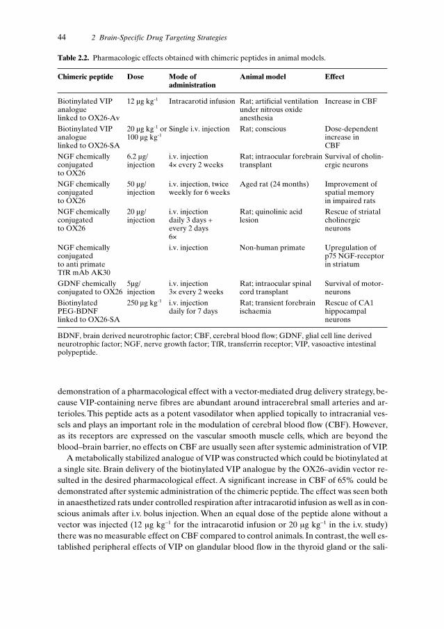
44 2 Brain-Specific Drug Targeting Strategies
Table 2.2. Pharmacologic effects obtained with chimeric peptides in animal models.
Chimeric peptide Dose Mode of Animal model Effectadministration
Biotinylated VIP 12 µg kg-1 Intracarotid infusion Rat; artificial ventilation Increase in CBFanalogue under nitrous oxide linked to OX26-Av anesthesia
Biotinylated VIP 20 µg kg-1 or Single i.v. injection Rat; conscious Dose-dependent analogue 100 µg kg-1 increase inlinked to OX26-SA CBF
NGF chemically 6.2 µg/ i.v. injection Rat; intraocular forebrain Survival of cholin-conjugated injection 4× every 2 weeks transplant ergic neuronsto OX26
NGF chemically 50 µg/ i.v. injection, twice Aged rat (24 months) Improvement of conjugated injection weekly for 6 weeks spatial memory to OX26 in impaired rats
NGF chemically 20 µg/ i.v. injection Rat; quinolinic acid Rescue of striatalconjugated injection daily 3 days + lesion cholinergic to OX26 every 2 days neurons
6×
NGF chemically i.v. injection Non-human primate Upregulation of conjugated p75 NGF-receptor to anti primate in striatumTfR mAb AK30
GDNF chemically 5µg/ i.v. injection Rat; intraocular spinal Survival of motor-conjugated to OX26 injection 3× every 2 weeks cord transplant neurons
Biotinylated 250 µg kg-1 i.v. injection Rat; transient forebrain Rescue of CA1 PEG-BDNF daily for 7 days ischaemia hippocampal linked to OX26-SA neurons
BDNF, brain derived neurotrophic factor; CBF, cerebral blood flow; GDNF, glial cell line derivedneurotrophic factor; NGF, nerve growth factor; TfR, transferrin receptor; VIP, vasoactive intestinalpolypeptide.
demonstration of a pharmacological effect with a vector-mediated drug delivery strategy, be-cause VIP-containing nerve fibres are abundant around intracerebral small arteries and ar-terioles. This peptide acts as a potent vasodilator when applied topically to intracranial ves-sels and plays an important role in the modulation of cerebral blood flow (CBF). However,as its receptors are expressed on the vascular smooth muscle cells, which are beyond theblood–brain barrier, no effects on CBF are usually seen after systemic administration of VIP.
A metabolically stabilized analogue of VIP was constructed which could be biotinylated ata single site. Brain delivery of the biotinylated VIP analogue by the OX26–avidin vector re-sulted in the desired pharmacological effect. A significant increase in CBF of 65% could bedemonstrated after systemic administration of the chimeric peptide.The effect was seen bothin anaesthetized rats under controlled respiration after intracarotid infusion as well as in con-scious animals after i.v. bolus injection. When an equal dose of the peptide alone without avector was injected (12 µg kg–1 for the intracarotid infusion or 20 µg kg–1 in the i.v. study)there was no measurable effect on CBF compared to control animals. In contrast, the well es-tablished peripheral effects of VIP on glandular blood flow in the thyroid gland or the sali-

2.4 Drug Delivery Strategies 45
Organ blood flow (µL / min /g)
SALINEn=8
VECTORn=5
VIPn=7
VIP/VECTORn=8
BRAIN BLOOD FLOWSALIVARY GLAND BLOOD FLOW
VECTOR = OX26 / SA without VIPaVIP = VIPa without VECTORVIP/VECTOR = BIO-xx-VIPa / OX26-SA
Figure 2.9. Differential pharmacological effect elicitedby vector-mediated delivery of a VIP analogue. Theorgan blood flow in brain and salivary gland wasmeasured in conscious rats after i.v. administration ofvehicle (saline), the brain delivery vector OX26-SA,the VIP peptide alone, or the chimeric peptide. Whilecerebral blood flow increased in the chimeric peptidegroup by 60% compared to the saline control, theincrease in salivary gland blood flow seen with thepeptide alone was abolished by coupling to the vector.The VIP analogue was biotinylated with a non-cleavable 14-atom spacer (biotin-XX) for coupling tothe vector. Data from reference [95].
vary gland were readily detectable [89,95], as shown in Figure 2.9. Notably, the effect on sali-vary gland blood flow was attenuated in animals treated with the chimeric peptide deliverysystem.Taking salivary gland blood flow in that respect as a potential adverse drug effect, thedelivery strategy of the VIP analogue to the brain not only resulted in the desired pharma-cological response at the target site, but it also diminished the effect at non-target sites andtherefore increased the therapeutic index [95].
Demonstrations of pharmacological effects of chimeric peptides have been achieved withdifferent neurotrophic factors in models of neurodegenerative diseases and ischaemia. Theinitial report by Friden et al. utilized an ocular graft model of fetal midbrain placed into theanterior eye chamber of adult rats [85].The vasculature of the grafted tissue retained its BBBproperties. Nerve growth factor was chemically coupled to the vector OX26 via a disulfidelinker. Repeated i.v. administration (four times bi-weekly) of the chimeric peptide promotedsurvival of the cholinergic neurons within the graft. Further proof of pharmacological effectof the same conjugate was obtained in aged rats with spatial learning deficits. They respond-ed to a 6-week treatment with twice weekly i.v. injections with improved performance in theso-called Morris water maze learning task and immunohistochemistry showed increased cellsize of cholinergic neurons in the medial septal area of these rats [96]. The NGF-OX26chimeric peptide was also effective in a quinolinic acid lesioning model of Huntington’s dis-ease [97].Treatment for 2 weeks significantly reduced the loss of intrastriatal cholinergic neu-rons induced by stereotaxic injection of quinolinic acid.
Animal models of Parkinson’s disease suggest that Glial Cell-Line Derived NeurotrophicFactor (GDNF) may be a suitable treatment modality for degenerative processes involvingdopaminergic midbrain neurons, and traumatic injury of spinal motor neurons.Therefore, theeffect of a GDNF-OX26 chimeric peptide was studied in another neural graft model [98].Thevector-mediated delivery of small i.v. doses equivalent to 5 µg of GDNF significantly pro-moted the survival of ocular implants of fetal spinal cord motor neurons in rats.
The potential therapeutic benefit of brain-derived neurotrophic factor BDNF for rescuingneurons after stroke was demonstrated in a forebrain ischaemia model [93]. In that study,

transient forebrain ischaemia was induced in rats by bilateral clamping of the carotid arter-ies. In order to achieve BDNF delivery with the chimeric peptide approach it was necessaryto modify the peptide by ‘pegylation’, i.e. the coupling of multiple PEG residues. NativeBDNF is a basic peptide with rapid clearance from plasma. The poor pharmacokinetic prop-erties persisted after coupling to OX26–streptavidin but could be overcome by pegylation.The PEG-BDNF could be delivered through the BBB by vector-mediated transport as effi-ciently as the OX26 antibody itself [90].Animals treated for 1 week after the ischaemic insultwith chimeric peptide (biotinylated PEG-BDNF coupled to OX26-SA) at a daily dose of250 µg kg–1 were fully protected from neuronal loss in the hippocampal CA1 region.
Oligodeoxynucleotides (ODN) represent another class of hydrophilic macromoleculardrug candidates, which require transcellular as well as intracellular delivery where brain celltargeting is concerned. Due to their highly charged, anionic character they also have the po-tential to impair the pharmacokinetics of the delivery system when used as the drug con-stituent of chimeric peptides. Coupling of a biotinylated phosphodiester ODN to OX26-NLA increases hepatic clearance of the complex and limits brain uptake by lowering theAUC [99]. On the other hand, phosphorothioate-modified ODNs show high plasma proteinbinding which may contribute to the low BBB transport measured for a PS-ODN/OX26-SAchimeric peptide. In contrast, the neutral peptide backbone of peptide nucleic acids makesthese compounds good drug candidates for chimeric peptides and allows for a substantialvector-mediated effect on brain targeting (28-fold increase, [100]). A potential therapeuticapplication of the ODN approach is the delivery of an antisense oligonucleotide to the revgene of HIV-1 through the BBB. The feasibility of such an approach was recently demon-strated using the OX26–avidin fusion protein [88].
2.4.2.7 Chimeric Peptide Radiopharmaceuticals
The potential of chimeric peptides for delivery of radiopharmaceuticals across the BBB, ei-ther for diagnostic or therapeutic purposes, has been explored in studies with radiolabelledsynthetic amyloid peptide and with EGF. Aß peptide in solution deposits specifically on pre-existing amyloid plaques and vascular amyloid.A pharmacokinetic study in Rhesus monkeyswith the insulin receptor antibody 83-14 as a vector showed brain accumulation of radiola-belled [125I]-Aβ only after vector-mediated delivery. The peptide alone was unable to crossthe BBB. In the monkeys, analysis of brain sections by phosphorimager quantitation of ra-dioactivity resulted in images comparable to scans obtained with the non-metabolized glu-cose analogue 2-deoxyglucose [101]. Labelling with a suitable radioisotope should enablequantitative detection by a neuroimaging method such as SPECT.
EGF receptors are abundantly expressed by gliomas and present a target both for diag-nostic imaging and radio-immunotherapy. A cerebral implant model in rats bearing humanU87 gliomas was utilized to test the brain delivery of [111In]-labelled EGF by vector mediat-ed transport with OX26 following i.v. injection. Brains were sampled after 2 h and cryosec-tioned for subsequent autoradiography. The tumours were clearly visualized on these au-toradiographs, but only when the labelled EGF was given as a chimeric peptide, not when in-jected without the vector [102].
46 2 Brain-Specific Drug Targeting Strategies

2.4.3 Liposomes as Drug Carriers
2.4.3.1 Conventional Liposomes and Small Molecules
Liposomes, in addition to oligonucleotides [104], are often used as carriers for low molecularweight drugs and peptides [103]. It has been demonstrated that encapsulation within lipo-somes can dramatically alter the fate of the encapsulated drug in vivo [105]. Liposomal for-mulations may protect against metabolic degradation and can influence plasma clearanceand tissue distribution of a variety of drugs. Loading efficiency, contents retention, plasmastability and pharmacokinetic properties can often be adjusted by appropriate formulationof conventional liposomal drug carriers [105,106]. However, conventional liposomes do notundergo significant blood–brain barrier transport [107]. This is also true for small unilamel-lar vesicles as demonstrated in a study where 60-nm liposomes radiollabeled with 111Indiumdid not penetrate the blood–brain barrier of a normal brain [108]. In this study brain pene-tration was only observed following non-specific pharmacological disruption of theblood–brain barrier by infusion of high doses (25 mg kg–1) of etoposide or at sites of brain tu-mours where the vasculature is porous.
2.4.3.2 Brain Targeting Using Immunoliposomes
Conventional liposomes are rapidly removed from the circulation by cells of the reticuloen-dothelial system [109].This rapid accumulation of conventional liposomes in the liver and thespleen and the resulting high plasma clearance can be slowed down by coating the liposomesurface with inert and hydrophilic polymers such as PEG [110]. The half-life of liposomescontaining PEG-derivatized lipids increases up to 100-fold [106]. Such liposomes are oftenreferred to as sterically-stabilized liposomes. The PEG polymers can also be used for cova-lent conjugation of an antibody or an antibody fragment to the liposome. In this case a chem-ically reactive linker lipid can be used (Figure 2.10) that consists of a bi-functional PEG mol-ecule covalently bound at one side to a phospholipid headgroup and at the other side to a thi-ol-reactive maleimide group. Thus modified antibodies bearing a thiol group can be coupledunder mild conditions to sterically-stabilized liposomes [111]. Such immunoliposomes retainboth their prolonged circulation properties and their target specificity in vivo. Similar resultscan be obtained using alternative coupling techniques such as biotin–avidin conjugation[112].
2.4 Drug Delivery Strategies 47
SH + N
O
n
O
(CH2)2O
NH
Phospholipid
O
SN
O
O
n(CH2)2O
NH
Phospholipid
O
Figure 2.10. Schematic diagram of coupling of athiolated antibody to a linker lipid (maleimide–PEG–phospholipid) which is part of a preformedliposome. The resulting thioether bond is meta-bolically stable. The strategy shown here was used tosynthesize OX26-immunoliposomes [111].

An antibody used for brain targeting of immunoliposomes has to meet several require-ments. First, the antibody should recognize a structure which is present exclusively at theblood–brain barrier. Second, the antibody should be able to cross the blood–brain barrier byan active transport mechanism such as receptor-mediated transcytosis. Third, the epitopeagainst which the antibody is targeted should preferably not be species specific. Fourth, highquantities of the antibody should be available. The OX26 mAb [38] meets several (but notall) of the above requirements. In vitro experiments have demonstrated that OX26-immuno-liposomes can be taken up specifically by living RG2 rat glioma cells overexpressing the rattransferrin receptor despite their particulate size of approximately 90 nm [113]. The fluores-cent-labelled OX26-immunoliposomes accumulated within an intracellular (endosomal)compartment [114]. Similar results were obtained by incubation of fluorescent OX26-im-munoliposomes with freshly isolated rat brain capillaries [115] which revealed binding to theluminal and basolateral membranes of the brain endothelium.
2.4.3.3 Drugs of Interest for Targeting to the Brain
Brain delivery of the anticancer drug daunomycin provides an example of the in vivo appli-cation of OX26-immunoliposomes [111]. Different formulations of [3H]-daunomycin werei.v. administered to rats either as the free drug or encapsulated in conventional liposomes,sterically-stabilized liposomes, or PEG-conjugated immunoliposomes (Table 2.3). Plasmasamples were taken at defined time points and after 1 h the animal was killed and drug con-centrations in brain tissue were determined.
Free daunomycin and not PEG-conjugated liposomes containing daunomycin, disappearrapidly from the circulation. Plasma clearance of the liposome was reduced 66-fold by PEG-conjugation. Coupling 29 OX26 monoclonal antibodies per PEG-liposome partially reversedthe effect of PEG-conjugation on plasma clearance.
Analysis of the blood–brain barrier permeability surface area (PS) product indicated thatdaunomycin, and to a lesser degree conventional liposomes, have the potential to penetratethe blood–brain barrier. However, brain tissue accumulation of free daunomycin or conven-tional liposomes was poor, being the result of their high systemic plasma clearance. The useof PEG-conjugated liposomes reduced the blood–brain barrier PS product value to zero. Nobrain uptake of the PEG-liposomes was observed, despite their marked increase in plasmacirculation time. Conversely, the use of PEG-conjugated OX26 immunoliposomes increasedthe blood–brain barrier PS product, relative to PEG-liposomes, resulting in increased brainuptake. Thus, optimal brain delivery of daunomycin was achieved using OX26 immunolipo-somes (see Table 2.3). Titration of the amount of OX26 conjugated per liposome (n between3 and 197) revealed an increase in plasma clearance and a decrease in the systemic volume ofdistribution of immunoliposomes at higher OX26 concentrations. Highest PS product valuesand brain tissue accumulation was observed for immunoliposomes with 29 OX26 mAb. Athigher OX26 densities on the liposome, a saturation effect was observed resulting in a re-duction in volume of distribution, PS product and brain tissue accumulation of OX26 im-munoliposomes.
Recently the OX26 immunoliposomes were used in a gene delivery approach to transportexpression vectors for luciferase or β-galactosidase through the BBB [116]. The plasmids
48 2 Brain-Specific Drug Targeting Strategies
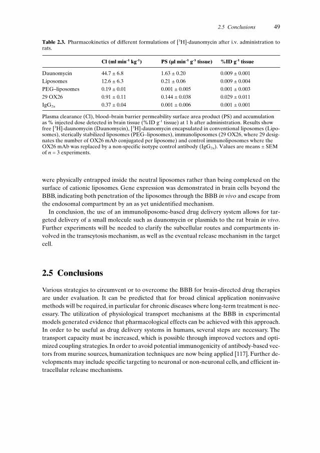
were physically entrapped inside the neutral liposomes rather than being complexed on thesurface of cationic liposomes. Gene expression was demonstrated in brain cells beyond theBBB, indicating both penetration of the liposomes through the BBB in vivo and escape fromthe endosomal compartment by an as yet unidentified mechanism.
In conclusion, the use of an immunoliposome-based drug delivery system allows for tar-geted delivery of a small molecule such as daunomycin or plasmids to the rat brain in vivo.Further experiments will be needed to clarify the subcellular routes and compartments in-volved in the transcytosis mechanism, as well as the eventual release mechanism in the targetcell.
2.5 Conclusions
Various strategies to circumvent or to overcome the BBB for brain-directed drug therapiesare under evaluation. It can be predicted that for broad clinical application noninvasivemethods will be required, in particular for chronic diseases where long-term treatment is nec-essary. The utilization of physiological transport mechanisms at the BBB in experimentalmodels generated evidence that pharmacological effects can be achieved with this approach.In order to be useful as drug delivery systems in humans, several steps are necessary. Thetransport capacity must be increased, which is possible through improved vectors and opti-mized coupling strategies. In order to avoid potential immunogenicity of antibody-based vec-tors from murine sources, humanization techniques are now being applied [117]. Further de-velopments may include specific targeting to neuronal or non-neuronal cells, and efficient in-tracellular release mechanisms.
2.5 Conclusions 49
Table 2.3. Pharmacokinetics of different formulations of [3H]-daunomycin after i.v. administration torats.
Cl (ml min-1 kg-1) PS (µl min-1 g-1 tissue) %ID g-1 tissue
Daunomycin 44.7 ± 6.8 1.63 ± 0.20 0.009 ± 0.001
Liposomes 12.6 ± 6.3 0.21 ± 0.06 0.009 ± 0.004
PEG–liposomes 0.19 ± 0.01 0.001 ± 0.005 0.001 ± 0.003
29 OX26 0.91 ± 0.11 0.144 ± 0.038 0.029 ± 0.011
IgG2a 0.37 ± 0.04 0.001 ± 0.006 0.001 ± 0.001
Plasma clearance (Cl), blood–brain barrier permeability surface area product (PS) and accumulationas % injected dose detected in brain tissue (%ID g-1 tissue) at 1 h after administration. Results showfree [3H]-daunomycin (Daunomycin), [3H]-daunomycin encapsulated in conventional liposomes (Lipo-somes), sterically stabilized liposomes (PEG–liposomes), immunoliposomes (29 OX26, where 29 desig-nates the number of OX26 mAb conjugated per liposome) and control immunoliposomes where theOX26 mAb was replaced by a non-specific isotype control antibody (IgG2a). Values are means ± SEMof n = 3 experiments.

References
[1] Shoulson I, Science 1998, 282, 1072–1074.[2] Small GW, Am. J. Geriatr. Psychiatry 1998, 6(2 Suppl. 1), S26–S33.[3] Glenner GG, Wong CW, Biochem. Biophys. Res. Commun. 1984, 120, 885–890.[4] Kang J, Lemaire HG, Unterbeck A, Salbaum JM, Masters CL, Grzeschik KH, Multhaup G,
Beyreuther K, Muller-Hill B, Nature 1987, 325, 733–736.[5] St George-Hyslop PH, Biol. Psychiatry 2000, 47, 183–199.[6] Selkoe DJ, Wolfe MS, Proc. Natl Acad. Sci. USA 2000, 97, 5690–5692.[7] Emilien G, Beyreuther K, Masters CL, Maloteaux JM, Arch. Neurol. 2000, 57, 454–459.[8] Chen KS, Masliah E, Grajeda H, Guido T, Huang J, Khan K, Motter R, Soriano F, Games D,
Prog. Brain Res. 1998, 117, 327–334.[9] Walker LC, Brain Res. Brain Res. Rev. 1997, 25, 70–84.
[10] Polymeropoulos MH, Lavedan C, Leroy E, Ide SE, Dehejia A, Dutra A, Pike B, Root H, Ruben-stein J, Boyer R, Stenroos ES, Chandrasekharappa S, Athanassiadou A, Papapetropoulos T,Johnson WG, Lazzarini AM, Duvoisin RC, Di Iorio G, Golbe LI, Nussbaum RL, Science 1997,276, 2045–2047.
[11] Dunnett SB, Bjorklund A, Nature 1999, 399 (Suppl.), A32–A39.[12] DeLong MR, Trends Neurosci. 1990, 13, 281–285.[13] Masliah E, Rockenstein E, Veinbergs I, Mallory M, Hashimoto M, Takeda A, Sagara Y, Sisk A,
Mucke L, Science 2000, 287, 1265–1269.[14] Dirnagl U, Iadecola C, Moskowitz MA, Trends Neurosci. 1999, 22, 391–397.[15] Mikkelsen T, Cancer Control 1998, 5, 150–162.[16] Black KL, Adv. Drug Delivery Rev. 1995, 15, 37–52.[17] Tardieu M, Curr. Opin. Neurol. 1999, 12, 377–381.[18] Nath A, Semin. Neurol. 1999, 19, 113–127.[19] Brightman MW, Exp. Eye Res. 1977, 25 (Suppl.), 1–25.[20] Smith QR, Rapoport SI, J. Neurochem. 1986, 46, 1732–1742.[21] Cancilla PA, Bready J, and Berliner J, In: The Blood–Brain Barrier – Cellular and Molecular Bi-
ology (Ed. Pardridge WM), pp. 29–46. Raven Press, New York, 1993.[22] Balabanov R, Dore-Duffy P, J. Neurosci. Res. 1998, 53, 637–644.[23] Pardridge WM, Triguero D, Yang J, Cancilla PA, J. Pharmacol. Exp. Ther. 1990, 253, 884–891.[24] Duvernoy H, Delon S, Vannson JL, Brain Res. Bull. 1983, 11, 419–480.[25] Segal M, In: Introduction to the Blood–Brain Barrier: Methodology, Biology and Pathology (Ed.
Pardridge WM), pp. 251–258. Cambridge University Press, Cambridge, 1998.[26] Dohrmann GJ, Brain Res. 1970, 18, 197–218.[27] Levin VA, J. Med. Chem. 1980, 23, 682–684.[28] Drewes LR, In: Introduction to the Blood–Brain Barrier: Methodology, Biology and Pathology
(Ed. Pardridge WM), pp. 165–174. Cambridge University Press, Cambridge, 1998.[29] Boado RJ, Li JY, Nagaya M, Zhang C, Pardridge WM, Proc. Natl Acad. Sci. USA 1999, 96,
12079–12084.[30] Smith QR, Stoll J, In: Introduction to the Blood–Brain Barrier: Methodology, Biology and Pathol-
ogy (Ed. Pardridge WM), pp. 188–197. Cambridge University Press, Cambridge, 1998.[31] Smith QR, In: Frontiers in Cerebral Vascular Biology: Transport and its Regulation (Eds Drewes
LR, Betz AL), pp. 83–93. Plenum Press, New York, 1993.[32] Borst P, Schinkel AH, In: Introduction to the Blood–Brain Barrier:Methodology, Biology and
Pathology (Ed. Pardridge WM), pp. 198–206. Cambridge University Press, Cambridge, 1998.[33] Gao B, Hagenbuch B, Kullak-Ublick GA, Benke D, Aguzzi A, Meier PJ, J. Pharmacol. Exp. Ther.
2000, 294, 73–79.[34] Sugiyama Y, Kusuhara H, Suzuki H, J. Control. Rel. 1999, 62, 179–186.[35] Pardridge WM, Eisenberg J, Yang J, J. Neurochem. 1985, 44, 1771–1778.[36] Pardridge WM, Eisenberg J, Yang J, Metabolism 1987, 36, 892–895.[37] Bickel U, Kang YS, Yoshikawa T, Pardridge WM, J. Histochem. Cytochem. 1994, 42, 1493–1497.[38] Jefferies WA, Brandon MR, Hunt SV, Williams AF, Gatter KC, Mason DY, Nature 1984, 312,
162–163.[39] Dehouck B, Fenart L, Dehouck MP, Pierce A, Torpier G, Cecchelli R, J. Cell Biol. 1997, 138,
877–889.[40] Broadwell RD, Balin BJ, Salcman M, Proc. Natl Acad. Sci. USA 1988, 85, 632–636.[41] Bickel U, Yoshikawa T,Pardridge WM, Adv. Drug Deliv. Rev. 1993, 10, 205–245.
50 2 Brain-Specific Drug Targeting Strategies

[42] Oldendorf WH, Brain Res 1970, 24, 372–376.[43] Takasato Y, Rapoport SI, Smith QR, Am. J. Physiol. 1984, 247(3 Pt 2), H484–H493.[44] Triguero D, Buciak J, Pardridge WM, J. Neurochem. 1990, 54, 1882–1888.[45] Samii A, Bickel U, Stroth U, Pardridge WM, Am. J. Physiol. 1994, 267(1 Pt 1), E124–E131.[46] Rapoport SI, Fitzhugh R, Pettigrew KD, Sundaram U, Ohno K, Am. J. Physiol. 1982, 242,
R339–R348.[47] Klecker Jr RW, Collins JM, Yarchoan R, Thomas R, Jenkins JF, Broder S, Myers CE, Clin. Phar-
macol. Ther. 1987, 41, 407–412.[48] Takasawa K, Suzuki H, Sugiyama Y, Biopharm. Drug Dispos. 1997, 18, 611–622.[49] de Lange EC, de Boer BA, Breimer DD, Adv. Drug Deliv. Rev. 1999, 36, 211–227.[50] Morgan ME, Singhal D, Anderson BD, J. Pharmacol. Exp. Ther. 1996, 277, 1167–1176.[51] Pardridge WM, In: Introduction to the Blood-Brain Barrier: Methodology, Biology and Pathology
(Ed. Pardridge WM), pp. 49–61. Cambridge University Press, Cambridge, 1998.[52] Audus KL, Rose JM, Wang W, Borchardt RT, In: Introduction To The Blood-Brain Barrier:
Methodology, Biology And Pathology (Ed. Pardridge WM), pp. 86–93. Cambridge UniversityPress, Cambridge, 1998.
[53] Pagliara A, Reist M, Geinoz S, Carrupt PA, Testa B, J. Pharm. Pharmacol. 1999, 51, 1339–1357.[54] Tsuzuki N, Hama T, Kawada M, Hasui A, Konishi R, Shiwa S, Ochi Y, Futaki S, Kitagawa K, J.
Pharm. Sci. 1994, 83, 481–484.[55] Bodor N, J. Control. Rel. 1999, 62, 209–222.[56] Wade LA, Katzman R, Am. J. Physiol. 1975, 228, 352–359.[57] Nau R, Sorgel F, Prange HW, Clin. Pharmacokinet. 1998, 35, 223–246.[58] DeAngelis LM, J. Neurooncol. 1998, 38, 245–252.[59] Penn RD, Savoy SM, Corcos D, Latash M, Gottlieb G, Parke B, Kroin JS, N. Engl. J. Med. 1989,
320, 1517–1521.[60] Lazorthes YR, Sallerin BA, Verdie JC, Neurosurgery 1995, 37, 422–428; discussion 428–429.[61] Ferguson IA, Schweitzer JB, Bartlett PF and Johnson Jr EM, J. Comp. Neurol. 1991, 313, 680–692.[62] Fishman RA, Cerebrospinal Fluid in Diseases of the Nervous System, W.B. Saunders Company,
Philadelphia, 1980.[63] Davson H, Segal MB, Physiology of the CSF and Blood–Brain Barriers, CRC Press, Boca Raton,
1996.[64] Shapiro WR, Young DF, Mehta BM, N. Engl. J. Med. 1975, 293, 161–166.[65] Blasberg RG, Patlak C, Fenstermacher JD, J. Pharmacol. Exp. Ther. 1975, 195, 73–83.[66] Krewson CE, Saltzman WM, Brain Res. 1996, 727, 169–181.[67] Mahoney MJ, Saltzman WM, Proc. Natl Acad. Sc.i USA 1999, 96, 4536–4539.[68] Kordower JH, Winn SR, Liu YT, Mufson EJ, Sladek Jr JR, Hammang JP, Baetge EE, Emerich
DF, Proc. Natl Acad. Sci. USA 1994, 91, 10898–10902.[69] Bobo RH, Laske DW, Akbasak A, Morrison PF, Dedrick RL, Oldfield EH, Proc. Natl Acad. Sci.
USA 1994, 91, 2076–2080.[70] Laske DW, Youle RJ, Oldfield EH, Nature Med. 1997, 3, 1362–1368.[71] Hagihara N, Walbridge S, Olson AW, Oldfield EH, Youle RJ, Cancer Res. 2000, 60, 230–234.[72] Robinson PJ, Rapoport SI, Am. J. Physiol. 1987, 253(3 Pt 2), R459–R466.[73] Salahuddin TS, Johansson BB, Kalimo H, Olsson Y, Acta Neuropathol. (Berl.) 1988, 77, 5–13.[74] Muldoon LL, Nilaver G, Kroll RA, Pagel MA, Breakefield XO, Chiocca EA, Davidson BL,
Weissleder R, Neuwelt EA, Am. J. Pathol. 1995, 147, 1840–1851.[75] Doran SE, Ren XD, Betz AL, Pagel MA, Neuwelt EA, Roessler BJ, Davidson BL, Neurosurgery
1995, 36, 965–970.[76] Zunkeler B, Carson RE, Olson J, Blasberg RG, DeVroom H, Lutz RJ, Saris SC, Wright DC,
Kammerer W, Patronas NJ, Dedrick RL, Herscovitch P, Oldfield EH, J. Neurosurg. 1996, 85,1056–1065.
[77] Nakano S, Matsukado K, Black KL, Neurol. Res. 1997, 19, 501–508.[78] Gregor A, Lind M, Newman H, Grant R, Hadley DM, Barton T, Osborn C, J. Neurooncol. 1999,
44, 137–145.[79] Pardridge WM, Endocrinol. Rev. 1986, 7, 314–330.[80] Kumagai AK, Eisenberg JB, Pardridge WM, J. Biol. Chem. 1987, 262, 15214–15219.[81] Pardridge WM, Triguero D, Buciak JL, Endocrinology 1990, 126, 977–984.[82] Wu D, Kang YS, Bickel U, Pardridge WM, Drug Metab. Dispos. 1997, 25, 768–771.[83] Lee HJ, Engelhardt B, Lesley J, Bickel U, Pardridge WM, J. Pharmacol. Exp. Ther. 2000, 292,
1048–1052.
References 51
[84] Yoshikawa T, Pardridge WM, J. Pharmacol. Exp. Ther. 1992, 263, 897–903.[85] Friden PM, Walus LR, Watson P, Doctrow SR, Kozarich JW, Backman C, Bergman H, Hoffer B,
Bloom F, Granholm AC, Science 1993, 259, 373–377.[86] Kang YS, Pardridge WM, J. Pharmacol. Exp. Ther. 1994, 269, 344–350.[87] Saito Y, Buciak J, Yang J, Pardridge WM, Proc. Natl Acad. Sci. USA 1995, 92, 10227–10231.[88] Penichet ML, Kang YS, Pardridge WM, Morrison SL, Shin SU, J. Immunol. 1999, 163,
4421–4426.[89] Bickel U, Yoshikawa T, Landaw EM, Faull KF, Pardridge WM, Proc. Natl Acad. Sci. USA 1993,
90, 2618–2622.[90] Pardridge WM, Wu D, Sakane T, Pharm. Res. 1998, 15, 576–582.[91] Mandel R, Ryser HJ, Ghani F, Wu M, Peak D, Proc. Natl Acad. Sci. USA 1993, 90, 4112–4116.[92] Bickel U, Kang YS, Pardridge WM, Bioconjug. Chem. 1995, 6, 211–218.[93] Wu D, Pardridge WM, Proc. Natl Acad. Sci. USA 1999, 96, 254–259.[94] Deguchi Y, Kurihara A, Pardridge WM, Bioconjug. Chem. 1999, 10, 32–37.[95] Wu D, Pardridge WM, J. Pharmacol. Exp. Ther. 1996, 279, 77–83.[96] Backman C, Rose GM, Hoffer BJ, Henry MA, Bartus RT, Friden P, Granholm AC, J. Neurosci.
1996, 16, 5437–5442.[97] Kordower JH, Charles V, Bayer R, Bartus RT, Putney S, Walus LR, Friden PM, Proc. Natl Acad.
Sci. USA 1994, 91, 9077–9080.[98] Albeck DS, Hoffer BJ, Quissell D, Sanders LA, Zerbe G, Granholm AC, Neuroreport 1997, 8,
2293–2298.[99] Boado RJ, Tsukamoto H, Pardridge WM, J. Pharm. Sci. 1998, 87, 1308–1315.
[100] Pardridge WM, Boado RJ, Kang YS, Proc. Natl Acad. Sci. USA 1995, 92, 5592–5596.[101] Wu D, Yang J, Pardridge WM, J. Clin. Invest. 1997, 100, 1804–1812.[102] Kurihara A, Pardridge WM, Cancer Res. 1999, 59, 6159–6163.[103] Chonn A, Cullis PR, Curr. Opin. Biotechnol. 1995, 6, 698–708.[104] Zelphati O, Szoka Jr FC, Proc. Natl Acad. Sci. USA 1996, 93, 11493–11498.[105] Allen TM, Drugs 1998, 56, 747–756.[106] Lasic DD, Papahadjopoulos D, Science 1995, 267, 1275–1276.[107] Micklus MJ, Greig NH, Tung J, Rapoport SI, Biochim. Biophys. Acta 1992, 1124, 7–12.[108] Gennuso R, Spigelman MK, Chinol M, Zappulla RA, Nieves J, Vallabhajosula S, Alberto Paciuc-
ci P, Goldsmith SJ, Holland JF, Cancer Invest. 1993, 11, 118–128.[109] Derksen JT, Morselt HW, Scherphof GL, Biochim. Biophys. Acta 1988, 971, 127–136.[110] Papahadjopoulos D, Allen TM, Gabizon A, Mayhew E, Matthay K, Huang SK, Lee KD, Woodle
MC, Lasic DD, Redemann C, Martin FJ, Proc. Natl Acad. Sci. USA 1991, 88, 11460–11464.[111] Huwyler J, Wu D, Pardridge WM, Proc. Natl Acad. Sci. USA 1996, 93, 14164–14169.[112] Hansen CB, Kao GY, Moase EH, Zalipsky S, Allen TM, Biochim. Biophys. Acta 1995, 1239,
133–144.[113] Huwyler J, In: Brain Barrier Systems, Vol. 45 (Eds Paulson OB, Moos Knudsen G, Moos T),
Munksgaard, Copenhagen, 1999.[114] Huwyler J, Yang J, Pardridge WM, J. Pharmacol. Exp. Ther. 1997, 282, 1541–1546.[115] Huwyler J, Pardridge WM, J. Neurochem. 1998, 70, 883–886.[116] Shi N, Pardridge WM, Proc. Natl Acad. Sci. USA 2000, 97, 7567–7572.[117] Coloma MJ, Lee HJ, Kurihara A, Landaw EM, Boado RJ, Morrison SL, Pardridge WM, Pharm.
Res. 2000, 17, 266–274.[118] Pardridge WM, Oldendorf WH, Cancilla P, Frank HJ, Ann. Intern. Med. 1986, 105, 82–95.[119] Weindl A, In: Frontiers in Neuroendocrinology (Eds Ganong WF, Martini L), pp. 3–32. Oxford
University Press, New York, 1973.[120] Clementi F, Marini D, Z. Zellforsch. Mikroskop. Anat. 1972, 123, 82–95.[121] Pardridge WM, J. Neurochem. 1998, 70, 1781–1792.[122] Lonser RR, Corthesy ME, Morrison PF, Gogate N, Oldfield EH, J. Neurosurg. 1999, 91, 294–302.[123] Morrison PF, Laske DW, Bobo H, Oldfield EH, Dedrick RL, Am. J. Physiol. 1994, 266(1 Pt 2),
R292–R305.
52 2 Brain-Specific Drug Targeting Strategies

3 Pulmonary Drug Delivery: Delivery To andThrough the Lung
Anne H. de Boer, Grietje Molema, Henderik W. Frijlink
3.1 Introduction
The respiratory tract is one of the oldest routes used for the administration of drugs. Anaes-thetics, aerosolized drugs, smoke or steam have been inhaled for medical purposes for cen-turies. Over the past decades inhalation therapy has established itself as a valuable tool in thelocal therapy of pulmonary diseases such as asthma or COPD (Chronic Obstructive Pul-monary Disease) [1]. This type of drug application in the therapy of these diseases is a clearform of targeted drug delivery: the major advantages are a rapid onset of the therapeutic ef-fect, a lowering of the required dose (as compared to systemic administration) and a reduc-tion in unwanted side-effects (increased therapeutic index). Currently, over 25 drug sub-stances are marketed as inhalation aerosol products for local pulmonary effects and aboutthe same number of drugs are in different stages of clinical development. Furthermore, thereare some drugs that are not marketed as inhalation aerosols per se but are formulated as suchby pharmacists.
The majority of the marketed products are used for asthma and COPD.Typical agents thatare used for these indications are β2-agonists such as salbutamol (albuterol), Terbutalin orformoterol, corticosteroids such as budesonide, Flixotide or beclomethasone and mast-cellstabilizers such as sodium cromoglycate or nedocromil.
Patients suffering from cystic fibrosis often use various aerosolized drugs. To reduce theviscosity of the mucus in the airways, recombinant human deoxyribonuclease is used. This enzyme is the first recombinant protein that has been developed for specific delivery to the lungs via the airways. It has a local action on the mucus in the airways and its absorptionis minimal. Another drug that decreases the viscosity of the mucus is acetylcysteine.Aerosolized antibiotics are a further group of therapeutics that is widely used by cystic fibrosis patients. Solutions of antibiotics like tobramycin or colistin are used in nebulizers toprevent exacerbation of the disease. Pentamidine has been used for the prophylaxis of Pneu-mocystis pneumonia in patients infected with HIV virus, while chronic rejection of lungtransplants provided a reason to develop an aerosol formulation of cyclosporine A.
The latest and probably one of the most promising applications of pulmonary drug ad-ministration is its use to achieve systemic absorption of the administered drug substances.Particularly for those drug substances that exhibit a poor bioavailability when administeredby the oral route, as for example peptides or proteins, the respiratory tract might be a conve-nient port of entry [2]. For this application a more or less contradictory situation occurs: ‘de-livery (into the lung) is required to obtain systemic absorption followed by a non-targeted
Drug Targeting Organ-Specific Strategies. Edited by G. Molema, D. K. F. MeijerCopyright © 2001 Wiley-VCH Verlag GmbH
ISBNs: 3-527-29989-0 (Hardcover); 3-527-60006-X (Electronic)

distribution of the drug substance’. In this case, the biopharmaceutical objective of improvedbioavailability and not the improved therapeutic index for the drug is the rationale for organdelivery.
Systemic absorption of pulmonary-delivered peptides and proteins has been the objectiveof many investigations [2].The most successful work in this field is the development of insulinformulations for inhalation. These dosage forms might, in the near future, become a suitablealternative for the current subcutaneous injection of insulin that is used to obtain meal-timeglucose control [3]. In spite of the strict requirements regarding dose variability for insulin,the pulmonary products under development seem to be as safe as the subcutaneous injections.
Numerous other peptides and proteins have been, or are still in development as inhalationproducts with the objective of systemic absorption. Examples of these are: calcitonin, LH-RHantagonists, recombinant human granulocyte colony-stimulating factor and growth hormone.
Protein inhalation products that have been developed for local use are interferon, alpha-1-antitrypsin and secretory leukoprotease inhibitor. Other therapeutic products that havebeen investigated with regard to delivery to the lungs are genetic material (plasmid DNA)and vaccines. For example, the delivery of the gene encoding the cystic fibrosis transmem-brane conductance regulator was extensively investigated [4]. Delivery of genes requiresspecific vector systems which enable the cellular transfection of the gene. Vectors that havebeen investigated are retroviral and adenovirus-associated vectors, recombinant adenovirus-es, cationic liposomes and DNA–ligand complexes. However, none of these approaches wasfound to be successful in clinical studies up to now. Other diseases for which gene therapy viathe lung was investigated are lung carcinoma malignant mesothelioma and alpha1-anti-trypsin deficiency [5].
Pulmonary drug delivery for local or systemic therapy comprises several aspects. Formula-tion of the drug, the generation of the aerosol, the lung deposition of inhaled particles andthe passage of the drug substance over the epithelial membranes of the respiratory tract, allrepresent crucial aspects of pulmonary drug administration. During the last decade many ofthese aspects have been studied extensively [1,2,6–18]. This chapter summarizes the aspectsthat are of relevance in the development of new drug products for inhalation. Focus will beplaced on the physical characteristics of the inspiratory flow curve since this is the drivingforce that finally brings the particles into the lung. This process is also relevant to under-standing the generation of the aerosol cloud as for example in dry powder inhalers. Furtheraspects that will be discussed are the apparatus to generate the aerosol, lung depositionmechanisms, typical formulation types for the pulmonary route as well as the mechanism oftransport over the alveolar membrane.
Principles of fluid and particle dynamics in the respiratory tract (physical and anatomicalparameters) are also discussed, as they are the starting point for the development of drugproducts for inhalation. In fact, they set the conditions used for in vitro and in vivo testing ofinhalation systems and define the specifications for new inhalation systems.
Finally, it should be noted that apart from the use of the pulmonary route for protein ab-sorption (targeting through the lung), the lung can also be an object for organ selective de-livery (targeting to the lung). The latter can in theory be achieved by two entirely differentroutes: deposition via the airways from the luminal side and targeting via the blood circula-tion.Whereas the main part of this chapter is dedicated to the former approach, the latter ap-proach will also be briefly discussed (Section 3.12).
54 3 Pulmonary Drug Delivery: Delivery To and Through the Lung
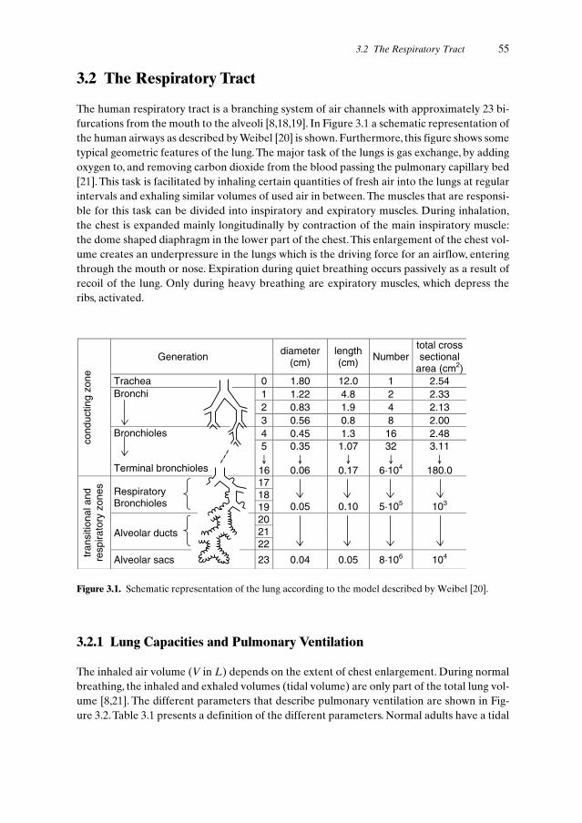
3.2 The Respiratory Tract
The human respiratory tract is a branching system of air channels with approximately 23 bi-furcations from the mouth to the alveoli [8,18,19]. In Figure 3.1 a schematic representation ofthe human airways as described by Weibel [20] is shown. Furthermore, this figure shows sometypical geometric features of the lung. The major task of the lungs is gas exchange, by addingoxygen to, and removing carbon dioxide from the blood passing the pulmonary capillary bed[21]. This task is facilitated by inhaling certain quantities of fresh air into the lungs at regularintervals and exhaling similar volumes of used air in between. The muscles that are responsi-ble for this task can be divided into inspiratory and expiratory muscles. During inhalation,the chest is expanded mainly longitudinally by contraction of the main inspiratory muscle:the dome shaped diaphragm in the lower part of the chest.This enlargement of the chest vol-ume creates an underpressure in the lungs which is the driving force for an airflow, enteringthrough the mouth or nose. Expiration during quiet breathing occurs passively as a result ofrecoil of the lung. Only during heavy breathing are expiratory muscles, which depress theribs, activated.
3.2 The Respiratory Tract 55
Generation diameter(cm)
length(cm) Number
total crosssectional
area (cm2)Trachea 0 1.80 12.0 1 2.54
1 1.22 4.8 2 2.332 0.83 1.9 4 2.13
Bronchi
3 0.56 0.8 8 2.004 0.45 1.3 16 2.48
cond
uctin
gzo
ne
Bronchioles
Terminal bronchioles
5↓�16
0.35↓�
0.06
1.07↓�
0.17
32↓�
6·104
3.11↓�
180.01718Respiratory
Bronchioles 19 0.05 0.10 5·105 103
2021Alveolar ducts22
tran
sitio
nala
ndre
spira
tory
zone
s
Alveolar sacs 23 0.04 0.05 8·106 104
Figure 3.1. Schematic representation of the lung according to the model described by Weibel [20].
3.2.1 Lung Capacities and Pulmonary Ventilation
The inhaled air volume (V in L) depends on the extent of chest enlargement. During normalbreathing, the inhaled and exhaled volumes (tidal volume) are only part of the total lung vol-ume [8,21]. The different parameters that describe pulmonary ventilation are shown in Fig-ure 3.2.Table 3.1 presents a definition of the different parameters. Normal adults have a tidal

volume (VT) of approximately 0.7 l, and inhale with a frequency of about 12 times a minuteat rest [8].The amount of air processed under these conditions is 12 m3 per day (with a rangeof 10–20 m3 per day). During heavy work, the tidal volume may be increased by a factor 3. Aresidual volume (RV) of 1 to 1.5 l is not exhaled during normal breathing: this volume is in-creased when a patient suffers from an obstruction e.g. in the case of asthma. Total lung ca-pacities (TLCs) of adults are estimated to be 5 to 7 l [1], maximal inspired volumes (vital ca-pacities: VCs) were found to be dependent on the external resistance and vary from less than
56 3 Pulmonary Drug Delivery: Delivery To and Through the Lung
7
0
volu
me
(l)
inspiratoryreservevolume
tidalvolume
expiratoryreservevolume
residualvolume
functionalresidualcapacity
vitalcapacity
totallung
capacity
inspiratorycapacity
Table 3.1. Definitions of the different parameters describing pulmonary ventilation.
Parameter Definition
Total lung capacity The volume of air in the lung after a maximal inspiratory effort
Inspiratory capacity The volume of air maximally inspired after a normal tidal expiration
Functional residual capacity The volume of air remaining in the lung at the end of normal tidal expira-tion
Vital capacity The maximum volume of air expired after a maximal forced inspiration
Inspiratory reserve volume The maximum volume of air inspired after a normal tidal inspiration
Tidal volume The volume of air entering or leaving the lung at each normal breath
Expiratory reserve volume The maximum volume of air expired after normal tidal expiration
Residual volume The volume of air left in the lung after a maximal forced expiratory effort
Figure 3.2. Schematic diagram of the different volumes describing pulmonary ventilation.

1 l (for high airflow resistances) to more than 2.5 l (for low resistances) for healthy adults[22].
Healthy subjects have hardly any alveolar dead space. However, disease may increase thedead space in the alveoli [15]. This might be of importance when alveolar deposition is de-sired, for example to obtain systemic absorption.
Attainable underpressures and inspiratory flow rates, which are especially relevant to theperformance of dry powder inhalers, are discussed more in detail in Section 3.9 as function ofthe patient’s effort, age and clinical condition and the (external) inhaler resistance. On thebasis of Weibel’s lung model (Figure 3.1), showing a strongly increasing total cross sectionalarea for airflow with increasing generation number, starting from the lobar bronchi, it can becalculated that the air velocity decreases with increasing penetration depth.At a common in-spiratory flow rate of 60 l min–1 through a dry powder inhaler, air velocity first increases fromapproximately 4 m s–1 in the trachea to a maximum of 4.6 m s–1 in the lobar bronchi. But start-ing at the segmental bronchi, a steep decrease in velocity occurs to 0.5 m s–1 in the terminalbronchi and not more than 0.05 m s–1 in the terminal bronchioles. So, in the periphery of thelungs, the air is practically still. A similar falling off can be calculated for the Reynolds num-ber, being 4800 in the trachea, 40 in the terminal bronchi and only 2 in the terminal bronchi-oles. This means that the flow is turbulent (at 60 l min–1) in the upper respiratory tract andlaminar in the central and deep lung. The decreasing air velocity is important for particle de-position in the lungs, as will be discussed in Section 3.3.
3.3 Lung Deposition and Particle Size
Airborne particles travelling through the respiratory tract are subjected to constantly chang-ing forces as a result of bends and the decreasing air velocity (Section 3.2.1). In the absenceof tribocharge, electrostatic forces play no role and particle behaviour is governed mainly byinertial forces, the drag force (Stokes’ law) and the force of gravity [8]. In the relatively wideupper airways, where the air velocity is highest (Section 3.2.1), inertial forces are dominant.Particles enter the airway system with (near) air velocity, unless they have been acceleratedto much higher speed, as by discharge from a metered dose aerosol [23].They will have to fol-low the streamlines of the air in bends and bifurcations in order to penetrate deeper, but areunable to do so when their inertia is too high (either from a high mass or a high velocity, orboth).Therefore, the largest particles are deposited by the mechanism of inertial impaction inthe throat and first bifurcations.As the remaining small particles move on to the central lung,the air velocity gradually decreases to much lower values and the force of gravity becomesimportant. Settling by sedimentation is the dominant deposition mechanism in this part of therespiratory tract. However, settling velocity is too low and residence time too short to removethe smallest particles in the aerosol cloud from the air by this mechanism. So, the finest frac-tions are able to enter the periphery of the lung where they can make contact with the wallsof the airways as the result of Brownian motion (particle diffusion). In rare cases, particle in-terception contributes to drug deposition, especially near obstructions in the smaller airways.
Particles entering the respiratory tract may not only vary in size and velocity, but also inshape and density, depending upon the type of drug and the inhalation system used for
3.3 Lung Deposition and Particle Size 57

aerosol generation (see Section 3.5). In order to be able to compare the behaviour of differ-ent types of aerosol particles with each other, the aerodynamic particle diameter (DA) hasbeen introduced. By definition, the aerodynamic diameter of a particle is the diameter of aunit density sphere (ρP = 1 g cm–3) having the same terminal settling velocity (in still air) asthe particle under consideration. Irregular particles can also be expressed in terms of equiv-alent volume diameter (DE) and dynamic shape factor (χ). The equivalent volume diameteris the diameter of a sphere having the same volume as the irregular particle, whereas the dy-namic shape factor is the ratio of the actual resistance force on a non-spherical particle to theresistance force on a sphere having the same volume and velocity. The aerodynamic diame-ter can be calculated from the equivalent volume diameter, which is an expression of the geo-metric particle size, when particle density and dynamic shape factor are known, using Eq. 3.1:
DA = DE.(ρP/χ)0.5 (3.1)
Deposition efficiencies for particles in the respiratory tract are generally presented as afunction of their aerodynamic diameter (e.g. [8,12]). Large particles (> 10 µm) are removedfrom the airstream with nearly 100% efficiency by inertial impaction, mainly in the orophar-ynx. But as sedimentation becomes more dominant, the deposition efficiency decreases to aminimum of approximately 20% for particles with an aerodynamic diameter of 0.5 µm.Whenparticles are smaller than 0.1 µm, the deposition efficiency increases again as a result of dif-fusional displacement. It is believed that 100% deposition due to Brownian motion might bepossible for particles in the nanometer range.
Both from deposition studies and force balances it can be derived that the optimum (aero-dynamic) particle size lies between 0.5 and 7.5 µm. Within this approximate range many dif-ferent subranges have been presented as most favourable, e.g. 0.1 to 5 µm [24], 0.5 to 8.0 µm[25], 2 to 7 µm [26] and 1–5 µm [27–29]. Particles of 7.5 µm and larger mainly deposit in theoropharynx [30] whereas most particles smaller than 0.5 µm are exhaled again [31]. All in-halation systems for drug delivery to the respiratory tract produce polydisperse aerosolswhich can be characterized by their mass median aerodynamic diameter (MMAD) and geo-metric standard deviation (σG). The MMAD is the particle diameter at 50% of the cumula-tive mass curve.
3.4 Drug Absorption via the Lung
During the past decade the lung has been (re)discovered as a suitable port of entry to the sys-temic circulation for various drugs. Among these drugs are many peptides and proteins [2],since the oral route cannot be used for these molecules.
In relation to systemic absorption of drugs, absorption in the lung can be described as thepassage of a series of barriers by the drug in order to enter the systemic circulation. It is im-portant to realize that physiological conditions in the lung differ widely from site to site.
Major physiological factors that affect pulmonary absorption are [10]:
• Mucociliary transport in the airways that constantly drains fluid and solid particles (bacte-ria) in a counter-current flow to the oral cavity. A drug that is deposited in the airways can
58 3 Pulmonary Drug Delivery: Delivery To and Through the Lung
be cleared by this mechanism within several hours. So, if systemic absorption over thebronchial membrane is required, this has to occur relatively fast.
• The epithelial cells in the alveoli are covered by a thin layer of so-called epithelial liningfluid. This fluid in turn is covered by a monolayer of lung surfactant, which also is presentin large amounts in the alveolar lining fluids. Moreover, the lining fluid often contain en-zymes that can metabolize drug substances.
• The epithelial cell layer forms the major barrier to absorption of drug molecules. In thelarge airways stratified epithelium occurs, whereas in the alveoli the epithelium is only onecell layer thick.
• After passing the alveolar epithelium, the molecule enters the interstitium being part ofthe extracellular space inside the tissue. In the alveoli this space is relatively small.
• Finally, for passage into the blood, the molecules have to pass the endothelial membraneof the capillaries, separating the interstitial space from the blood. The endothelial mem-brane is considered to be much ‘leakier’ than the epithelial membrane. Therefore it is notconsidered as a major barrier during drug absorption.
• Macrophages can also form a functional barrier for some particular drug substances dur-ing pulmonary absorption. Macrophages are able to ingest particulate material present inthe alveoli or airways. After phagocytosis the macrophages migrate either to the ciliatedbronchial airways or via the alveolar interstitial space to the lymphatic system. Infection orinflammation may increase their numbers significantly, and increased phagocytosis of par-ticles may occur. If protein drugs, deposited in the lung as particles, are internalized bymacrophages, the drug may be partly destroyed by the efficient proteolytic degradationmechanism of these cells.
For an efficient pulmonary absorption process, the alveolar membrane seems to be an op-timal absorption site for a number of reasons.
• In contrast to the airways, there is hardly any mucociliary clearance from the alveoli.• The alveolar membrane forms the largest surface area in the lung.• The area of the alveoli is 43 to 102 m2 which is large in comparison to the surface area of
the airways which have a cumulative area of about 2.5 m2 [10].• The alveolar epithelium is thinner and leakier than the bronchial epithelium.
If a drug is deposited in the alveoli, it will, in first instance, come into contact with the alve-olar lining fluids. Long chain phospholipids (often referred to as ‘lung surfactant’) are themajor constituent of this fluid. These amphiphilic insoluble molecules form a molecularmonolayer covering the epithelial surface fluids. Before any absorption of drug from thealveoli can occur, the drug will have to be dissolved. When dry powder inhalers are used dis-solution of the drug in the alveolar lining fluids might be significantly affected by the pres-ence of such phospholipids. The dissolution of lipophilic drug molecules in particular, is like-ly to be enhanced by their presence.
The fact that the volume of the epithelial lining fluids in the alveoli is small, implies that formany drugs this volume is insufficient to provide sink conditions for dissolution.The absorp-tion rate will therefore be highly dependent on the dissolution rate of the inhaled product.The micronization of drugs for inhalation is therefore not only a requirement for deep lungdeposition but is also useful for the rapid absorption of the drug through fast dissolution ofthe solid drug.
3.4 Drug Absorption via the Lung 59

The alveolar epithelium consists of so-called Type I and Type II cells. Type I cells coverover 90% of the alveolar surface, have a large surface, and are thin. Type II cells are larger innumbers but are small. Therefore, they cover only about 7% of the surface of the alveoli.Type II cells produce the phospholipids that make up the surfactant layer.
It should be noted that the permeability per surface unit of alveolar epithelium per se isnot particularly high. The significant absorption found for various substances after pul-monary administration is rather explained by a number of beneficial factors such as the largesurface area of the alveoli, the low volume of the epithelial lining fluid, the relatively thin dif-fusion layer, the absence of mucociliary clearance from the alveoli as well as the limited en-zymatic activity in the lining fluids.
Passage over the epithelial membrane from the apical to the basal site may occur via dif-ferent routes. The fast absorption found for molecules smaller than 40 kDa is generally ex-plained by paracellular transport through the tight junctions between the epithelial cells. Al-though rather incorrect from a physiological point of view, the estimation of ‘pore sizes’ ofalveolar epithelium on the basis of transport rates of solutes, may help to predict whether ornot a certain molecule can pass the alveolar epithelium. The value of 40 kDa is compatiblewith the presence of pore structures with a diameter of about 5 nm. The turnover of epithe-lial surface cells may be responsible for the transient existence of larger openings in the alve-olar epithelium. However, whether these pores have any significance to drug absorption isunknown.
The absorption of molecules that are larger than 40 kDa is generally slow and incomplete.These molecules probably cannot pass through the tight junctions of the epithelial mem-brane, but have to be transported by a transcytotic mechanism in order to be absorbed. Re-ceptor-mediated endocytosis is a crucial mechanism here. The subsequent transport throughthe epithelial cells may occur in coated and non-coated vesicles. The non-coated vesicles arecalled caveolae. Macromolecules (after receptor recognition) may be sequestered both incoated vesicles as well as in caveolae. In the alveolar Type I cells, large numbers of caveolaeare found (about 1.7 million per cell). The caveolae have internal diameters of 50 to 100 nmwhich is large enough to contain macromolecules with sizes over 400 kDa. However, in spiteof these well-defined physiological processes the evidence for massive transport of largermacromolecules via this pathway is scarce. In general it is doubtful whether transcytosis viacaveolae may significantly contribute to the absorption of macromolecules [10,32].
In relation to the above it is obvious that passage of the pulmonary epithelium may de-pend on characteristics of a drug molecule. Not only the size, but also its solubility, overallcharge, structural conformation and potential aggregation can have a significant effect on theabsorption rate and bioavailability of the drug after pulmonary deposition.
3.4.1 Systemic Delivery of Peptides and Proteins
Many studies have been carried out regarding the absorption of peptides and proteins afterpulmonary drug delivery. The perspectives of a non-parenteral route of administration forlarger proteins led to studies on the pulmonary absorption of proteins of different size. Todate, over 30 different proteins have been evaluated with regard to absorption rate and
60 3 Pulmonary Drug Delivery: Delivery To and Through the Lung

bioavailability [2,16]. In Table 3.2 data selected by Adjei [32] are shown, which illustrate thekinetic behaviour of proteins of different size after pulmonary administration.
Significant variations occur between results of studies using the same protein. Such varia-tions can be explained by differences in the experimental conditions used and the different
3.4 Drug Absorption via the Lung 61
Table 3.2. Absorption data after pulmonary administration of peptides and proteins. Data fromreference [32].
Substance Molecular Administration Formulation Absorption parametersa
(species) weight
DDAVP (rat) 1071 Da Aerosol Solution 84 % in adult rats; tmax 0.5–1 h;17–41 % in young rats
Hu leuprolide 1209 Da Aerosol (MDI) Solution/ 4–18 % independent of formula-acetate (human) suspension tion
Dog leuprolide 1209 Da IT instillation Solution 4.6–95% increasing with deposi-acetate (dog) tion distance from epiglottis
Insulin (human) 5786 Da Aerosol Solution pH 7 20–25 % vs. s.c. injection(nebulized)
Insulin (human) 5786 Da Aerosol Solution 75 % for smokers; 25 % in non-(nebulized) smokers; tmax 15–20 min
PTH–84 (rat) 9418 Da IT instillation Solution pH 5 > 20 %; tmax 15–90 min
G–CSF (hamster) 18.6 kDa IT instillation Solution pH 4 45 %; tmax 1–2 h
Interferon–α 19 kDa Aerosol Solution tmax 2–16 h; absorption < 5 %(human) (nebulized)
Interferon a 19 kDa IT instillation Solution > 56 %; tmax 3– 9 h(rat)
Growth hormone 22 kDa IT instillation Solution 36 %(rat)
Growth hormone 22 kDa Aerosol Solution 9–10 %; tmax 1–4 h(rat) (nebulized)
DNase I 32 kDa Aerosol Solution 15 % absorption in 24 h(rat)
DNase I 32 kDa Aerosol Solution < 2 %(monkey)
Peroxidase 40 kDa IT instillation Solution 0.1–4 % h-1 absorption rate(guinea pig)
Antitrypsin 52 kDa Aerosol Solution 50 % in 50 h lost from peripheral (sheep) (nebulized) lung; bioavailability. 16 % via
lymphatics
Albumin 68 kDa Aerosol Solution 50 % in 20 h lost from lung(human) (nebulized)
Albumin 68 kDa Bronchial Solution 1 % h-1 lost from lung(sheep) catheter
Albumin 68 kDa IT instillation Solution 4–5 % in 96 h; tmax 16–24 h.(rat)
IgG 150 kDa IT instillation Solution 1.5–1.8 % in 192 h, tmax 16 h(rat)a the relative bioavailability is given as the percentage of the administered dose. It should be mentio-
ned that some bioavailabilities are unrealistically high. For correct interpretation of data, it may beadvised to calculate the bioavailability from the deposited fraction of the dose.IT, intratracheal

ways in which, for example, the bioavailability can be expressed. Assuming that the alveoliare the major site of absorption for all proteins administered, it is obvious that the fraction ofthe protein actually reaching the alveoli will largely determine the amount of protein thatcan finally be absorbed. Unfortunately, the reported fraction of proteins actually reachingthe alveoli after pulmonary administration varies significantly between the different studies.Therefore, conclusions cannot be made with regard to the relative bioavailability after pul-monary administration. Moreover, it is not possible to estimate the fraction that passes theepithelium in relation to the amount of protein that has entered the alveoli.
When nebulizers are used to generate an aerosol, the fraction of the drug reaching thealveoli will be low and variable (see Section 3.5.1). Particularly for low molecular weight pro-teins (< 20 kDa), the fraction deposited in the alveoli might be the limiting factor for drug ab-sorption. For example, the low absorption found for aerosolized Leuprolide acetate in hu-mans is more likely to be explained by the low and variable portion of drug being depositedin the alveoli, than by the limited passage of the Leuprolide over the epithelial membrane. Inanimal studies the determination of the fraction absorbed is further complicated by the factthat it is often not feasible to establish the amount of drug that is actually inhaled. To over-come this problem instillation is often used as a method of administration. However, usingthis method of administration there remains uncertainty regarding the fraction of the ad-ministered protein that reaches the alveoli, it is likely that a significant part of the fluid onlyreaches the bronchi or the bronchioles.
In a study performed in rabbits, rhG-CSF in a powder formulation (aerodynamic diameter< 4 µm) was insufflated via an intratracheal tube and compared to intratracheal instillationof a solution of the drug. In this study it was shown that a direct relation exists between theamount of protein that was deposited deep into the lung and the relative bioavailability [33].
An alternative method which could be used to establish the fraction of protein that actu-ally reaches the alveoli is the so-called co-aerosolization. If a protein is aerosolized from a so-lution that also contains another low molecular weight substance (deposition marker), it canbe assumed that the fractions of protein and deposition marker reaching the alveoli will bethe same. The deposition marker should be a substance with a known alveolar epithelialmembrane passage (e.g. tobramycin or a decapeptide) which does not undergo absorption af-ter oral administration. The fraction of the deposition marker that is deposited in the alveolican be established from plasma (and urine) measurements of the deposition marker. Themaximum fraction of protein that can pass the alveolar membrane will then be known. Theratio between the deposited fraction and the fraction that has been absorbed into the sys-temic circulation (as can be established form plasma or urine analysis) will provide an esti-mation of the protein passage across the alveolar membrane.
Alternatively the membrane passage of human airway epithelial cell lines can be studiedin vitro. A number of bronchial epithelial cell lines is available, such as the 16HBE14o- andCalu–3 cell lines. These cell lines can be installed in diffusion chambers to measure transportrates [34]. A major disadvantage of the currently used cell lines is that they provide informa-tion about bronchial epithelial transport only. Since bronchial epithelium is very differentfrom alveolar epithelium, the information from these in vitro studies is of limited value forthe prediction of the bioavailability of pulmonary administered proteins.
The isolation and characterization of alveolar Type II cells which transform into alveolarType I cells has been described, as well as a monolayer culture of alveolar Type I cells [35,36].
62 3 Pulmonary Drug Delivery: Delivery To and Through the Lung

Recently, a monolayer of human alveolar epithelial cells was used to study the bioadhesiveproperties of lectins [37]. Lectins are sugar-recognizing adhesive molecules (they bind to ep-ithelial cells) and are thought to increase the bioavailability of larger molecules by triggeringvesicular transport processes.
Proteases occurring in the epithelial lining fluids are another source of variability in pro-tein absorption from the lung. In the epithelial lining fluids proteases and peptidases do oc-cur. Inhibition of proteases and peptidases with substances such as bacitracin or aprotininmight improve the bioavailability of proteins. For example bacitracin was shown to increaseinsulin bioavailability by a factor of 6.8 [3].
The enzymatic degradation of insulin was also shown to occur in the cytosol of alveolarcells, the pH optimum of the proteases being 7.4 [38]. To what extent intracellular proteasesplay a significant role in limiting the absorption of insulin is not clear, since the size of insulinlikely allows paracellular transport over the alveolar epithelium. However, for proteins ofhigher molecular weight, that require transcellular transport, these proteases might certainlylimit bioavailability.
For proteins with higher molecular weights the extent and rate of absorption tend to de-crease and variability in absorption increases even more (see Table 3.2). For these proteinsmolecular conformation, charge and self-aggregation can largely determine passage throughthe epithelial membrane. It is crucial that these molecules are presented in an optimal way tothe absorbing membrane. Consequently, formulation as discussed in Section 3.6.3 is criticalfor these proteins.
The addition of absorption enhancers, like bile salts (glycocholate), fatty acids (linoleicacid), surfactants (lecithins, polyoxyethylene-9-lauryl ether or N-lauryl-β-D-maltopyra-noside) and chelators (EDTA) can significantly increase the absorption of various proteins.However, the application of enhancers is limited by their toxicity. For example polyoxyethyl-ene-9-lauryl ether and sodium glycocholate caused serious oedema, haemorrhage and in-flammation of the lung after intratracheal instillation [39].
Since larger proteins are transported by the transcellular route it is important to investi-gate potential enzymatic degradation both in the coated and non-coated vesicles as well as inlysosomes. The alveolar endothelial cells were shown to contain various proteases [35,38],which (depending on the cellular routing of a particular protein) can influence its bioavail-ability. More basic knowledge concerning receptor-mediated, endocytotic and transcytoticprocesses should be acquired in order to utilize physiological transport systems for pul-monary absorption of macromolecules. In addition, it is necessary to study the influence ofvarious diseases on this route of administration.
3.5 Devices for Therapeutic Aerosol Generation
As described in Section 3.3 in more detail, particles in the aerosol cloud should preferablyhave an aerodynamic diameter between 0.5 and 7.5 µm. Currently, three different types ofdevices are used to generate aerosol clouds for inhalation: nebulizers (jet or ultrasonic),(pressurized) metered dose inhalers (pMDIs) and dry powder inhalers (DPIs). The basicfunction of these three completely different devices is to generate a drug-containing aerosolcloud that contains the highest possible fraction of particles in the desired size range.
3.5 Devices for Therapeutic Aerosol Generation 63

Pressurized metered dose inhalers are still the most frequently used systems and they haveproven their value in therapy. However, their application in early phases of biopharmaceuti-cal research and further development of dosage forms seems less convenient, since they re-quire special components including propellants, special containers, metering valves, and con-trolled filling conditions (pressure-filling or cold-filling).
Nebulizers and dry powder inhalers seem more appropriate systems to be used in the ear-ly stages of development of drug products for pulmonary drug delivery. However, it shouldnot be concluded from this that the development of formulations for nebulizers or DPIs iseasier and exhibits fewer theoretical and practical problems.
Which system is the most suitable for a particular drug or therapy is determined by boththe physicochemical properties of the drug as well as by patient condition in relation to thechosen therapy. Asthma and COPD treatment using drugs such as β2-agonists or corticos-teroids is carried out with MDIs and DPIs. For children, nebulizers seem to be preferred, butMDIs with spacers can also be used. For antibiotic (e.g. tobramycin or colistin) therapy in cys-tic fibrosis patients nebulizers still seem the device of choice. Probably the patient populationin this case is too small to make the development of DPIs or MDIs containing antibioticdrugs economically feasible.When peptide or protein delivery is considered, newer and moreadvanced systems such as the ‘AERx™ system’ or dry powder generators such as the ‘InhaleTherapeutic System (Innova™)’ have been developed [40,41].
3.5.1 Nebulizers
Nebulizers are applied to aerosolize drug solutions or suspensions.There are two basic types:the air jet and ultrasonic nebulizer [42]. Jet nebulizers have a two-fluid nozzle for atomizingthe drug solution. Compressed air passes through a narrow hole and entrains the drug solu-tion from one or more capillaries mainly by momentum transfer.The liquid break-up processdepends on the design of the nozzle, the air pressure and the physical properties of the drugsolution. Droplets in the required size range are entrained by the airflow from the nozzle.Larger droplets impact on a baffle and are returned to the reservoir. Auxiliary airflows, gen-erated by the patient, may pass through special vents to the nebulization cup in order to im-prove droplet entrainment from the nozzle area. In an ultrasonic nebulizer, droplets are pro-duced by a piezoelectric crystal vibrating at a high frequency. The frequency and again theproperties of the drug solution determine the droplet size distribution of the mist.
Many reviews on the relevant technical aspects for drug nebulization are available (e.g.[43–45]. The greatest disadvantages of nebulizers are their poor deposition efficiency (seeSection 3.11) and low output rate (e.g. [46]). Several developments have been reported to im-prove their efficacy, like the use of open vents or breath-assisted open vents [47] and adapt-ed aerosol delivery [48]. A renewed interest in nebulizer therapy may also come from thegeneration of special aerosols, such as liposomes [49].
The AERx™ pulmonary delivery system [40,41] can be regarded as a combination of aMDI and a nebulizer. This system forms an aerosol by extrusion of an aqueous drug-con-taining solution through a disposable nozzle containing an array of precisely micromachinedholes.The droplets are entrained by the airflow passing over the blister. Control over the sizedistribution of the holes enables the formation of droplets having a narrow size distribution.
64 3 Pulmonary Drug Delivery: Delivery To and Through the Lung

Moreover, the system will release the aerosol cloud only when the pre-programmed optimalinhalation flow is generated by the patient. These features enable a controlled and targeteddelivery to the lung.
3.5.2 Metered Dose Inhalers
The metered dose inhalers consist of four basic functional elements, container, meteringvalve, actuator and mouthpiece.
The drug is dissolved or suspended in the liquefied propellant which might contain otherexcipients. The energy for atomization of the drug suspension (or solution) from a metereddose inhaler is supplied by a liquefied propellant.When after actuation a small amount of thesuspension or solution is released from the metering valve connected to the pressurized con-tainer, the propellant starts evaporating rapidly, thereby disrupting the liquid into smalldroplets. Initial droplet size and droplet speed are too high for effective deposition in thelower respiratory tract (the target area), however. Evaporation and deceleration in the upperrespiratory tract (mouth and throat) is essential. Consequently, the inhalation manoeuvre isextremely relevant for deposition efficacy (particularly the co-ordination between firing andinhalation of a dose), in spite of the fact that no energy from the inspiratory air (except heatfor evaporation of the propellant) is required for fine droplet generation. If spacers are used,the inhalation manoeuvre becomes less critical. For the 3M Autohaler, no firing of a dose isnecessary, because dose release is breath triggered.
With respect to the formulations used in MDIs, the development over recent years has fo-cused on the replacement of chlorofluorocarbon (CFC) propellants by hydrofluoroalkanepropellants. Recently, new developments have been reviewed in a number of papers [50–52].
3.5.3 Dry Powder Inhalers
Dry powder inhalers have initially found their application in inhalation therapy as a CFC-free alternative for the older MDIs. However, nowadays they seem to have a much larger po-tential [14,53], because of the high lung deposition that can be attained and also because theyare suitable for the pulmonary delivery of therapeutic peptides and proteins [2,10,16].
Dry powder inhalers are generally described as ‘breath actuated’ devices, because the in-spiratory airstream releases the dose from the dose system and supplies the energy for thegeneration of fine drug particles from the powder formulation. Because the efficiency of doserelease and powder disintegration increases with increasing inspiratory flow rate for mostDPIs, these devices would be better described as ‘breath controlled’ devices. In Section 3.9,the effect of resistance and clinical conditions on the flow curve and relevant flow parame-ters for DPIs are discussed.
Basically, devices used as dry powder inhalers contain four basic functional elements, i.e.
• Powder container. Dry powder inhalers may contain the dry powder formulation in manydifferent forms. The first DPI, the Spinhaler™ contained single doses in capsules. Othersystems, like the Diskus™ or Diskhaler™ may contain the metered dose in blisters, where-as systems like the Turbohaler™, or Novolizer™, have multi-dose containers.
3.5 Devices for Therapeutic Aerosol Generation 65

• Dosing system.• Disintegration principle. In general, the powders in the inhaler are not formulated as sin-
gle particles, but as adhesive mixtures or spherical pellets (Figure 3.3). These mixtures orpellets are suitable for processing and metering. However, the particle size of these mix-tures or pellets is far too large for lung deposition. Therefore, the pellet or mixture has tobe disintegrated to make an aerosol cloud with the desired particle size (< 5 µm). Manydifferent disintegration principles exist. They may vary from a simple screen (Rota-haler™) to twisted powder channels (Turbuhaler™) or a cyclone chamber as used in deNovolizer™ [54].
• A mouthpiece. The mouthpiece may be used to control the direction of the aerosol cloudin the mouth and throat, in order to reduce drug deposition in the oropharyngeal cavities.De Boer et al. [55] use a so-called sheath flow to reduce mouth deposition.
66 3 Pulmonary Drug Delivery: Delivery To and Through the Lung
Figure 3.3. Scanning electron microscopy images of spherical pellets of budesonide (upper photograph)and of an adhesive mixture of lactose and micronized salbutamol (lower photograph).
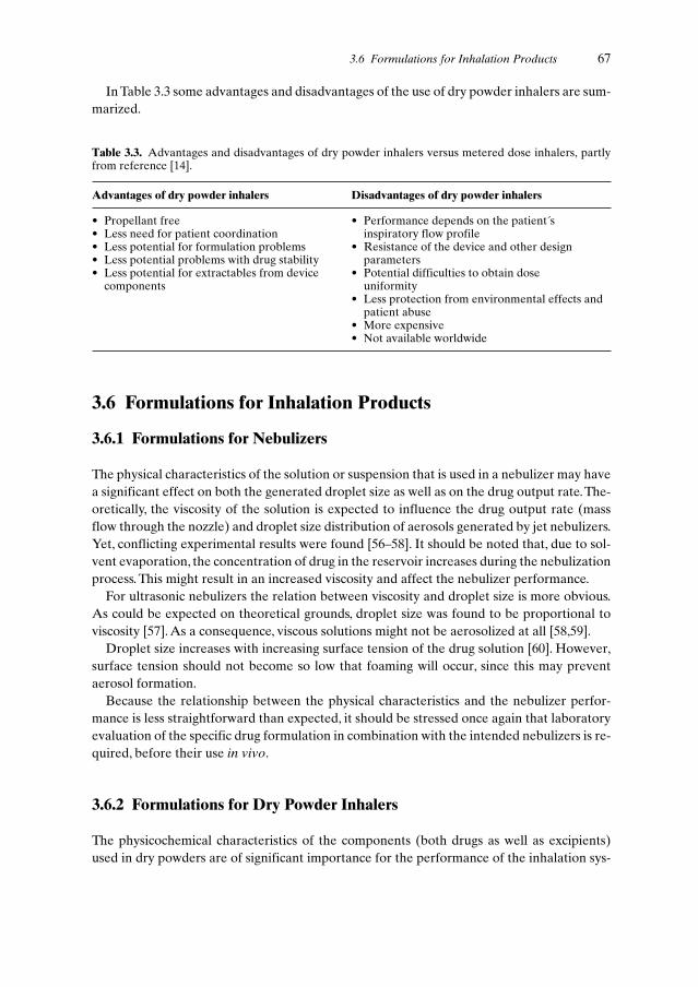
In Table 3.3 some advantages and disadvantages of the use of dry powder inhalers are sum-marized.
3.6 Formulations for Inhalation Products 67
Table 3.3. Advantages and disadvantages of dry powder inhalers versus metered dose inhalers, partlyfrom reference [14].
Advantages of dry powder inhalers Disadvantages of dry powder inhalers
• Propellant free • Performance depends on the patient´s• Less need for patient coordination inspiratory flow profile• Less potential for formulation problems • Resistance of the device and other design• Less potential problems with drug stability parameters• Less potential for extractables from device • Potential difficulties to obtain dose
components uniformity• Less protection from environmental effects and
patient abuse• More expensive• Not available worldwide
3.6 Formulations for Inhalation Products
3.6.1 Formulations for Nebulizers
The physical characteristics of the solution or suspension that is used in a nebulizer may havea significant effect on both the generated droplet size as well as on the drug output rate.The-oretically, the viscosity of the solution is expected to influence the drug output rate (massflow through the nozzle) and droplet size distribution of aerosols generated by jet nebulizers.Yet, conflicting experimental results were found [56–58]. It should be noted that, due to sol-vent evaporation, the concentration of drug in the reservoir increases during the nebulizationprocess. This might result in an increased viscosity and affect the nebulizer performance.
For ultrasonic nebulizers the relation between viscosity and droplet size is more obvious.As could be expected on theoretical grounds, droplet size was found to be proportional toviscosity [57]. As a consequence, viscous solutions might not be aerosolized at all [58,59].
Droplet size increases with increasing surface tension of the drug solution [60]. However,surface tension should not become so low that foaming will occur, since this may preventaerosol formation.
Because the relationship between the physical characteristics and the nebulizer perfor-mance is less straightforward than expected, it should be stressed once again that laboratoryevaluation of the specific drug formulation in combination with the intended nebulizers is re-quired, before their use in vivo.
3.6.2 Formulations for Dry Powder Inhalers
The physicochemical characteristics of the components (both drugs as well as excipients)used in dry powders are of significant importance for the performance of the inhalation sys-

tem. Staniforth [61] gave an overview of the required pre-formulation tests for dry powderformulation development.The nature of the surface (e.g. surface morphology, crystallinity orsurface energy) of the particles is of utmost importance and should be studied in detail. Tworeviews [62,63] described a number of methods for characterizing particle morphology.
Table 3.4 summarizes the major properties of powders which need to be characterized inpre-formulation. The effect of micronization (or other high energy processes), which is oftenapplied to the powder (surface), should also be investigated as this may alter the propertiesof the powder during processing [64].This may also alter the performance of the formulation.
Different dry powder formulations for inhalation were recently reviewed [53,65]. Spheri-cal pellets or adhesive mixtures are the most used formulation principles in dry powder in-haler systems (see Figure 3.3). Spherical pellets consist of the pure micronized drug or themicronized drug combined with a micronized excipient such as lactose or glucose. The smalldrug particles are formulated into the large spherical pellets to improve processing proper-ties such as flowability and precision of metering. The pellets should be strong enough towithstand the filling process as well as normal handling and shock as may occur during use bythe patient. On the other hand, it should be taken into account that optimal lung deposition
68 3 Pulmonary Drug Delivery: Delivery To and Through the Lung
Table 3.4. Major properties of powders to be determined during pre–formulation and some of themethods to be used.
Powder property Method
• Particle size and size distribution • Cascade impactor analysisWet or dry laser diffraction analysisMicroscopyCoulter counter analysisSieve analysisSedimentation analysisTime of flight measurements(Scanning electron) microscopy
• Particle surface area, shape and • Scanning electron microscopytexture (morphology) Atomic force microscopy
Particle flow (e.g. angle of repose)BET measurements
• Moisture sorption and desorption • Dynamic gravimetric sorption (DVS)
• Surface energy • Contact angle measurementsIsothermal microcalorimetryGravimetric sorptionInverse gas chromatography
• Crystallinity and crystal form • Differential scanning calorimetryThermogravimetric analysisIsothermal microcalorimetryInfra red analysisX–ray diffractionSorption and desorption measurements
(DVS)
• Solubility and dissolution rate
• Partition coefficient
• Stability in dry state and in solution
• Impurities

is achieved through full disintegration of the spheres into the primary particle size. This con-tradiction makes optimum formulation of spherical pellets difficult. Spherical pellets are pro-duced by low shear mixing procedures (e.g. tumbling or planetary mixers) with or withoutmixing aids such as small stainless steel balls, often followed by sieving procedures. Boere-fijn et al. [66] investigated the effects of agglomerate size and humidity on breakage of lactoseagglomerates. They showed that the extent of breakage was larger for the smaller particles,whereas humidity was found to decrease the propensity for breakage.
Adhesive mixtures require large carrier crystals to improve the handling properties of thepowders. Dispersion of the small drug particles over the larger carrier material should assuredose uniformity. However, the small drug particles should be removed from the carrier ma-terial during inhalation, to render an aerosol cloud of respirable particles. If the particles re-main on the carrier, mouth or throat deposition of the drug will occur, which might decreasetherapeutic efficacy or cause serious side-effects.
The adhesion of the drug particles to the carrier is largely dependent on the surface prop-erties of the carrier and drug as mentioned in Table 3.4. Variation in these properties can af-fect the dispersion of the drug over the carrier as well as the binding between drug and car-rier. Both may have a significant influence on the drug delivery performance of the formula-tion. Concessio et al. [67] correlated powder flow and particle detachment from solid surfacesto in vitro disintegration efficacy (deposition) as well as to in vivo efficacy (bronchodilation)in guinea pigs. A direct correlation between powder flow on the one hand and ease of parti-cle separation and aerosol dispersion on the other hand was observed. Furthermore it wasfound that formulations with a higher in vitro deposition had an increased in vivo efficacy.Lactose is the only carrier used in adhesive mixtures as yet; the carrier is added to the for-mulation to improve the processing and metering of the micronized drug. When increasingamounts of fine particles were used on the lactose, the drug delivery was found to increase[68]. Furthermore smoothing of the lactose surface increased the release of salbutamol sul-phate from the surface [69]. On the other hand, increased surface roughness and enlargingthe surface area were desirable to improve the release of pranlukast hydrate [70]. Obviously,detailed information concerning parameters determining the interaction between drug andcarrier lactose is still lacking.
Staniforth and co-workers managed to reduce the effect of the lactose surface by co-pro-cessing the carrier (e.g. co-milling, mixing or surface modification) with up to 2% L-leucine.This process is called corrasion. This approach significantly increased the release of be-clomethasone diproprionate from the carrier [14,53,61]. Another approach to modifying thesurface properties of carrier and drug could be the use of super critical fluid crystallization.This technique gives precise control over the particle size, shape and crystallinity of the par-ticles produced [71,72]. Lactose, trehalose and mannitol were also found to be suitable asdrug carriers for the pulmonary delivery of proteins. Sucrose was less suitable due to its hy-groscopicity [73].
3.6.3 Formulations for Peptides and Proteins
The formulation of small organic molecules in most cases uses established processes and onlya limited number of excipients (mainly lactose or a small number of propellants). In contrast,
3.6 Formulations for Inhalation Products 69

the formulation of peptide and protein powders for inhalation requires more advanced tech-niques and a wide variety of excipients and production processes [16,65,74]. The reason forthis difference is found in the more complex nature of the problems and requirements relat-ed to peptide and protein formulations.Table 3.5 summarizes a number of issues that need tobe considered when peptide or protein formulations for inhalation therapy are developed.Many of the characteristics mentioned in Table 3.5 can be affected by the processes used toprepare the protein or by the composition of the formulation used. Major formulation prob-lems connected to peptides and proteins are their low stability, hygroscopic nature, and ten-dency to form aggregates, which are too large to cross the alveolar membrane.
If possible, adhesive mixtures or spherical pellets, prepared using simple excipients such assugars are also preferred for protein formulations. For the preparation of dry peptide-con-taining formulations the most important techniques are lyophilization, spray freeze-drying,spray-drying, co-precipitation and super critical fluid extraction. When lyophilization is usedas the drying method, milling to obtain the desired particle size can be used. For spray-dryingor supercritical fluid extraction the desired particle size can be obtained immediately fromthe drying process. Lucas et al. [75] investigated different micronized bovine serum albu-min–maltodextrin (50 : 50) mixtures. Improved aerosolization behaviour was found for ad-hesive mixtures based on carrier lactoses with surfaces that were modified by micronized lac-tose or micronized polyethylene glycol 6000. Maa et al. [76] compared particles prepared byspray freeze-drying with particles prepared by spray-drying. The particles contained recom-binant human deoxyribonuclease-1, or anti-IgE monoclonal antibody and different sugars asexcipient. The large size of the spray freeze-dried particles (about 8–10 µm) in combinationwith their high porosity, turned out to result in improved aerosol performance compared tothe denser and smaller spray-dried particles. The lyophilization of proteins was recently re-viewed by Wang [77].
Protein instability can either be of a physical or chemical nature. The major mechanismsunderlying the degradation of proteins were recently extensively reviewed [78] Unfolding ofthe protein is the main cause of physical instability and may lead to denaturation, aggrega-tion or surface adsorption. Excipients that preserve the protein in its preferred state of hy-dration may be used to stabilize the protein. Several studies described the role of differentexcipients (often in combination with production processes) in the stabilization of proteins[65,79–82]. The major excipients used for stabilization of proteins are classified in Table 3.6.The incorporation of the proteins in amorphous solid matrices of sugar (often referred to assugar glasses), seems an effective method to stabilize the solid protein [83–85]. The stabiliza-
70 3 Pulmonary Drug Delivery: Delivery To and Through the Lung
Table 3.5. Different issues to consider for peptide or protein inhalation formulations.
• Particle size morphology and surface characteristics
• Moisture sorption behaviour
• Stability in dry state and dissolved
• Tendency to form aggregates
• Charge of the molecule, isoelectric point
• Solubility and dissolution behaviour
• Crystallinity and crystal form

tion is explained by the fact that in these amorphous sugar matrices hydrogen bonds betweenwater and the protein in an aqueous environment are replaced by hydrogen bonds betweenthe sugar and protein. This allows the protein to maintain its conformation and provides me-chanical protection. Furthermore, inclusion of the protein in the matrix protects it from theenvironment thereby preventing degradation processes such as hydrolysis or oxidation. It isessential that the sugar in these systems remains amorphous and has a glass transition tem-perature above storage temperature. In the rubbery state, the glasses are not stable; crystal-lization may occur and the protection from environmental influences disappears. The glasstransition temperatures of many sugars is above 50°C when the sugars are pure and com-pletely free of water. However, both moisture and the included protein may reduce the glasstransition temperature, which makes many sugars unsuitable for the formation of sugar glass-es. The moisture content of the products is not only important because of the plasticizing ef-fect, but also for their aerosol performance, since a high moisture content may increase pow-der cohesiveness. Compatibility of the sugar with the protein is necessary to obtain stable for-mulations. In this respect the use of reducing sugars such as sucrose or glucose is less satis-factory.
3.6 Formulations for Inhalation Products 71
Table 3.6. Excipients used for protein formulations for inhalation.
Class Excipient
• Carbohydrates SucroseLactoseTrehaloseInulinDextratesDextranCyclodextrins
• Polyols SorbitolMannitol
• Buffers Sodium citrateCitric acidSodium phosphateSodium biphosphateAmino acids
• Surfactants Polysorbate 80Tween 20Poloxamer 188Dipalmitoyl phosphatidylcholineAlkylbenzene sulfonate
• Polymers PEGPVP
• Amino acids GlycineLysine
• Proteins Albumin
• Salts Sodium chlorideCalcium chlorideSodium sulfate
• Chelators Disodium EDTA
Trehalose is often referred to as the sugar of choice for preparing sugar glasses. It is a non-reducing disaccharide with a glass transition temperature of about 120°C in the anhydrousstate. However, its glass transition temperature is rapidly decreased when the moisture con-tent in the sugar increases. Considering the hygroscopic nature of trehalose this is a potentialhazard and adequate moisture protection is essential. Furthermore, crystallization to the tre-halose dihydrate occurs easily at a relative humidity above 60%. From this perspective, theuse of a sugar polymer such as inulin (which is a fructose polymer terminating with a glucoseunit) seems much more suitable. Inulin is also a non-reducing sugar. By changing the chainlength (number of fructose units) of the molecule, physical characteristics like the glass tran-sition temperature can be changed. Moreover, due to the polymeric character of inulin, crys-tallization is less likely to occur.
Sugar glasses are prepared by spray-drying, freeze-drying or vacuum-drying. Freeze-dry-ing produces the lowest change in the sugar glass of degradation, whereas spray-drying mayresult in altering a large proportion of the particles to the preferred size range of 2 to 5 µm.In contrast to most other sugars or polyols that yield amorphous materials on spray-drying,mannitol was found to crystallize during spray-drying [73].
A final advantage of the use of sugar glasses is the fact that they include the proteins in amono-molecular form. In the glassy state, mobility in the systems is insufficient to allow ag-gregation of the proteins. Upon dissolution of the sugar matrix the protein is released in itsmono-molecular form which might enhance its passage through absorptive membranes.
Small amounts of surfactants may be used to prevent aggregation of proteins and may en-hance the refolding process when the dried protein dissolves. Buffers may also help to pre-vent aggregation of the dissolved drug. Similarly, polymers may be used as aggregation in-hibitors or to form matrices. Chan et al. [86] prepared crystalline powders of recombinant hu-man deoxyribonuclease with high fractions of sodium chloride. These powders were formu-lated as adhesive mixtures on lactose and mannitol and showed improved aerosolization be-haviour compared to the pure protein.
Preparation of high porosity particles may require special excipients, such as dipalmitoylphosphatidylcholine or special drying techniques such as spray freeze-drying [76,87,88].These large porous particles may combine the advantages that larger particle sizes contributeto the properties of powders with an improved aerosol performance. Furthermore, theselarge porous particles may be used to obtain sustained release of the incorporated drug [89,90].
Other techniques that have been used to obtain sustained release inhalation products are:the coating of the aerosol particles with paraffin wax or encapsulation or incorporation inbiodegradable polymers such as poly(L-lactic acid) or poly(DL-lactide-co-glycolide) [91,92].Talton et al. [93] described a new spray coating technique for applying ultra-thin coating lay-ers on particles. Finally, some authors describe the use of liposomes or other phospholipid-containing systems to prolong drug release or lung retention [94]. Liposome vaccine formu-lations have also been used for immunization via the pulmonary route. These developmentswill not be discussed in detail here.
One of the major questions in relation to absorption enhancers such as surfactants or sus-tained release products is their safety. Whether damage to lung tissue is caused by the differ-ent excipients is not yet clear. The results obtained so far are not very promising for sub-stances like surfactants [39]. What the effects of repetitive administration of insoluble orslowly (bio)degrading particles might be, remains to be established.
72 3 Pulmonary Drug Delivery: Delivery To and Through the Lung

When nebulizers are used to produce the aerosol cloud, the proteins should be dissolvedin aqueous solutions. The poor stability of many proteins in solution will make dissolution ofthe (freeze) dried protein just before nebulization necessary. The solvent may again containstabilizers such as buffers or salts. During jet and ultrasonic nebulization, high shear forcesmight be exerted on the solutions. Both jet and ultrasonic nebulization may cause aggrega-tion of the proteins in solution. Therefore, the resistance of the protein solution to nebuliza-tion should be investigated in vitro before use in vivo. Aggregation can be decreased by theaddition of Tween 20,Tween 80 or polyethylene glycol 8000 or by cooling the solution (for ul-trasonic nebulizers) [95,96].
A number of reports have described excipients and formulations for proteins used forMDIs [16,97,98].
3.7 Variables and Interactions in Dry Powder Inhalation
In Section 3.5.3, dry powder inhalers have been referred to as breath-controlled devices. Theefficacy of dry powder inhalation is a function of many factors, influencing the delivered doseof fine particles and the deposition of these particles in the respiratory tract. Figure 3.4 showsthat DPI performance is influenced both directly and indirectly by the design of the inhala-tion system. The powder formulation, the dose (measuring) system and the powder disinte-gration principle have to be designed correctly for release of sufficient fine drug particles in
3.7 Variables and Interactions in Dry Powder Inhalation 73
Figure 3.4. Relevant variables and interactions in dry powder inhalation therapy.
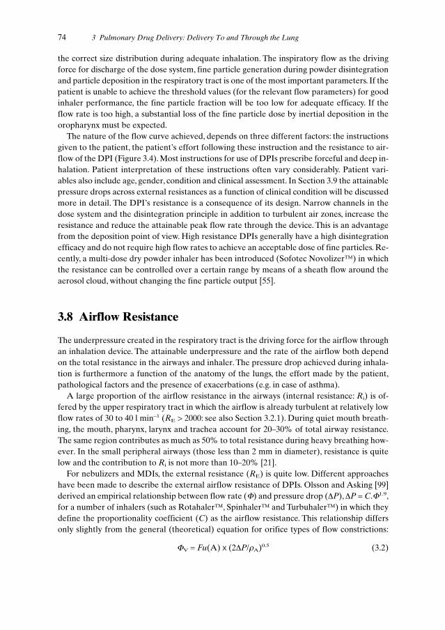
the correct size distribution during adequate inhalation. The inspiratory flow as the drivingforce for discharge of the dose system, fine particle generation during powder disintegrationand particle deposition in the respiratory tract is one of the most important parameters. If thepatient is unable to achieve the threshold values (for the relevant flow parameters) for goodinhaler performance, the fine particle fraction will be too low for adequate efficacy. If theflow rate is too high, a substantial loss of the fine particle dose by inertial deposition in theoropharynx must be expected.
The nature of the flow curve achieved, depends on three different factors: the instructionsgiven to the patient, the patient’s effort following these instruction and the resistance to air-flow of the DPI (Figure 3.4). Most instructions for use of DPIs prescribe forceful and deep in-halation. Patient interpretation of these instructions often vary considerably. Patient vari-ables also include age, gender, condition and clinical assessment. In Section 3.9 the attainablepressure drops across external resistances as a function of clinical condition will be discussedmore in detail. The DPI’s resistance is a consequence of its design. Narrow channels in thedose system and the disintegration principle in addition to turbulent air zones, increase theresistance and reduce the attainable peak flow rate through the device. This is an advantagefrom the deposition point of view. High resistance DPIs generally have a high disintegrationefficacy and do not require high flow rates to achieve an acceptable dose of fine particles. Re-cently, a multi-dose dry powder inhaler has been introduced (Sofotec Novolizer™) in whichthe resistance can be controlled over a certain range by means of a sheath flow around theaerosol cloud, without changing the fine particle output [55].
3.8 Airflow Resistance
The underpressure created in the respiratory tract is the driving force for the airflow throughan inhalation device. The attainable underpressure and the rate of the airflow both dependon the total resistance in the airways and inhaler. The pressure drop achieved during inhala-tion is furthermore a function of the anatomy of the lungs, the effort made by the patient,pathological factors and the presence of exacerbations (e.g. in case of asthma).
A large proportion of the airflow resistance in the airways (internal resistance: Ri) is of-fered by the upper respiratory tract in which the airflow is already turbulent at relatively lowflow rates of 30 to 40 l min–1 (RE > 2000: see also Section 3.2.1). During quiet mouth breath-ing, the mouth, pharynx, larynx and trachea account for 20–30% of total airway resistance.The same region contributes as much as 50% to total resistance during heavy breathing how-ever. In the small peripheral airways (those less than 2 mm in diameter), resistance is quitelow and the contribution to Ri is not more than 10–20% [21].
For nebulizers and MDIs, the external resistance (RE) is quite low. Different approacheshave been made to describe the external airflow resistance of DPIs. Olsson and Asking [99]derived an empirical relationship between flow rate (Φ) and pressure drop (∆P), ∆P = C.Φ1.9,for a number of inhalers (such as Rotahaler™, Spinhaler™ and Turbuhaler™) in which theydefine the proportionality coefficient (C) as the airflow resistance. This relationship differsonly slightly from the general (theoretical) equation for orifice types of flow constrictions:
ΦV = Fu(A) x (2∆P/ρA)0.5 (3.2)
74 3 Pulmonary Drug Delivery: Delivery To and Through the Lung

where ΦV is the volumetric flow rate, ρA is the density of the air (upstream of the flow con-striction) and Fu(A) is a function of the cross-section of the flow constriction. Several au-thors use a simplification of this formula, written in terms of driving force (√∆P), airflow re-sistance (RE) and volumetric airflow (ΦV) (e.g. references [22,100]).
√∆P = RE x ΦV (3.2a)
In this equation, RE is the reciprocal of Fu(A) x √(2/ρA). If there is no interaction betweenthe flow through the inhaler and the flow in the respiratory tract, total resistance (RTOT) maybe written as RTOT
2 = RI2 + RE
2. It has been reported that a high external resistance influ-ences the shape and width of the human pharynx and larynx in a manner favourable for deeplung deposition [101].
The resistance of dry powder inhalers can be calculated by measuring the volumetric flowrate and the pressure drop across the device simultaneously and applying Eq. 3.2a for the cal-culation. Values for some marketed DPIs are given in Table 3.7, showing that there are re-markable differences between devices.
3.9 Inspiratory Pressure and Relevant Flow Parameters 75
Table 3.7. Airflow resistances of some marketed dry powder inhalers in kPa0.5 min l–1.
Reference [100] [144] [145] Mean
Glaxo Rotahaler 0.013 0.013 0.014 0.0133
Fisons Spinhaler 0.016 0.015 0.015 0.0153
Pharbita Cyclohalera 0.017 0.018 0.018 0.0177
Sofotec Novolizer – – 0.026 0.0260
Glaxo Diskhalerc 0.021 0.018 0.030 0.0230
Glaxo Diskus – – 0.032 0.0320
Astra Turbuhaler 0.031 0.039 0.040 0.0367
Inhalator Ingelheimb 0.056 0.053 0.048–0.058 0.0540
Chiesi Inhaler – 0.093 – 0.0930
a Also known as ISF inhaler and Novartis (Foradil) Inhaler.b The range in reference [145] is a consequence of poor reproducibility in capsule piercing.c The spread for the Glaxo Diskhaler may come from the use of two different devices: four-dose [145]
and eight-dose [100, 144].1 technical bar equals 1000 mbar = 105 Pa (N m-2 or kg s-2 m-1) = 102 kPa Resistances have been (re-)calculated for standard liters per minute (at 20 °C: LS min-1 = 1,0733*LN min–1).
3.9 Inspiratory Pressure and Relevant Flow Parameters
Several authors measured attainable pressure drops as a function of the external resistancefor different groups of volunteers. Healthy male subjects (during maximal inspiration) areable to create a pressure drop (on average) of 6.7 kPa through an airflow resistance of0.038 kPa0.5 min l–1, (which is in the range of that of marketed DPIs, see Table 3.7), whereasfemales are able to create a pressure drop of 3.8 kPa under the same resistance [22]. Differ-ences between different groups of patients depend on the degree to which pulmonary func-

76 3 Pulmonary Drug Delivery: Delivery To and Through the Lung
Figure 3.5. The effect of external resistance and clinical conditions on the pressure drop generated atmaximum inhalation effort. The area between the dashed lines represents the 95% confidence interval.
tion has been deteriorated. However, pulmonary obstructions usually restrict the expiratoryperformance rather than the inhalation manoeuvre (e.g. [102,103]). Therefore, maximal in-spiratory pressure (MIP) values of asthmatic and COPD patients can be of the same order ofmagnitude as that of healthy subjects [104]. Only severe COPD patients may not be able togenerate MIPs higher than 1.5–2.0 kPa.The effect of airflow resistance and clinical conditionon the generated pressure drop across an external airflow resistance is summarized in Fig-ure 3.5 (data derived from reference [104]).The data show that in vitro testing of dry powderinhalers at only 4 kPa, as prescribed by various guidelines, is inadequate for the prediction oftheir performance in practice.
Attained flow parameters during inhalation can either be calculated (Eq. 3.1) from record-ed pressure drop curves or measured directly, using the equipment described in Section 3.9.1.Various studies report peak inspiratory flow rate (PIFR) values for healthy and diseasedadult subjects calculated from data on inhalation without external resistance (so-called ‘con-trol values’)(e.g. [22,100,105,106]). As for the attainable pressure drop, asthma, COPD andcystic fibrosis may decrease PIFR, depending on the severity of the disease. The addition ofan external airflow resistance, such as a DPI strongly affects the PIFR.Within the range of re-sistances for marketed DPIs, the average PIFR at maximal inhalation by healthy adults maydecrease from 159 l min–1 (for R = 0.015 kPa0.5 min l–1, equals the Rhone Poulenc Spin-haler™) to only 62 l min–1 (for R = 0.040 kPa0.5 min l–1, equals the Astra Turbuhaler™) [22].A maximal average flow rate of approximately 60 l min–1 through the Turbuhaler has alsobeen reported for asthmatics (e.g. references [103,107,108]).
Various flow parameters may be relevant to good DPI performance.The effect of PIFR onthe in vitro dose of fine particles from DPIs has been the subject of many studies (e.g.[109–112]. The actual dose of the fine particle fraction produced varies with the ranges offlow rates applied. The flow rate being the measuring principle used for the study and defin-ition of the fine fraction. Most studies show a strong increase in fine particle output with in-creasing PIFR for the ASTRA Turbuhaler™ [110–112]. For the Glaxo Diskus™ andDiskhaler™, the effect of PIFR is less extreme: in some cases an more or less constant, butalso a much lower output of approximately 15 to 25% of the label claim, has been obtained[109,110]. Some recent studies refer to the fact that the Pulmicort Turbuhaler™ also has aconstant fine particle yield if the acceleration towards peak flow (flow increase rate: FIR) ishigh enough [113,114]. For a FIR > 8 l s–2, the fine particle output is already maximal at flowrates above 40 l min–1. For capsule inhalers, the inhalation time has to be long enough for allthe particles to pass rapidly through the narrow holes pierced in the capsule ends before in-halation stops.
3.9.1 Measurement of the Inspiratory Flow Curve
Many different techniques are available for flow measurement and for recording of respira-tory functions or flow parameters in particular (e.g. [115,116]). However, not all methods areappropriate for measurement of inhalation flows, either because they have low frequency re-sponses or they influence the shape of the inspiratory flow curve by a large volume or by theinertia of the measuring instrument (e.g. rotameters). They may also interfere with theaerosol cloud from the inhalation device during drug deposition studies.
3.9 Inspiratory Pressure and Relevant Flow Parameters 77

Electronic equivalents of traditional spirometers (pneumotachographs, pneumotachome-ters or anemometers) are often used in clinical practice. They integrate expiratory flow ratesin order to compute flow–volume curves, from which expiratory parameters such as PEF(peak expiratory flow rate) and FEV1 (forced expiratory volume in 1 s) are derived (e.g.[22,117]). They are often laminar flow meters (e.g. Vitalograph 2100 Spirometer, Jaeger Mas-terscreen IOS), measuring the volumetric flow rate (Φv) at the upstream pressure of the flowhead.They can also be used for measuring the inspiratory flow curve, but the flow rate has tobe corrected for pressure and air density when they are used with an add-on resistance (e.g.during simulation of inhalation through dry powder inhalers) [118]. Similar corrections, in-cluding discharge and correction coefficients, are necessary for head meters (e.g. venturi ororifice meters) [119]. Thermal mass flow meters, also referred to as ‘hot-wire pneumota-chographs’ do not require these corrections, but they have a high internal resistance, whichmakes them inappropriate for patient characterization studies. Another great disadvantageof the techniques mentioned so far is that they are in-line components of the total flowscheme, which makes them difficult to use for in vitro drug deposition studies. For this rea-son, on-line pressure drop measurement across a flow constriction in the flow set-up is oftenrecommended. The flow constriction can be the add-on resistance (during patient character-ization) or the inhalation device (during drug deposition measurement). Once the airflow re-sistance of the flow constriction is known from previous calibration (as in Table 3.7 for DPIs),the flow curve can be monitored (or adjusted) on the differential pressure signal without in-terfering with the aerosol cloud or adding additional resistance to the flow set-up usingEq. 3.2a for the calculations.
3.10 In Vitro Particle Size Analysis and DepositionMeasurements
Methods for analysis of the particle size distribution in the aerosol cloud include techniquessuch as time of flight measurement (TOF), inertial impaction and laser diffraction. Dynamiclight scattering (photon correlation spectroscopy) is confined to particles (in suspension) inthe submicron range. In addition to the size distribution, the particle velocity distribution canbe measured with the Phase Doppler technique.
Inertial impaction is most widely applied for the characterization of inhalation systems.The principles of particle separation on the basis of inertial and drag forces have been welldescribed for many different applications. Theoretical cut-off diameters (for particles with50% collection efficiency) of impactors can be calculated on the basis of Stokes numbers fornozzles of a particular design [8,120]. Many different designs are available, but only a few aredescribed in the United States and European Pharmacopoeia [121,122].
Inertial impaction has many inaccuracies and limitations and there are also some relevantdifferences between deposition in vitro (impactor) and in vivo (respiratory tract) which causepoor correlation between impactor data and lung deposition data. The most important dif-ference is that deposition in vitro is by inertial impaction only, whereas deposition in vivo isby sedimentation and diffusional deposition as well. Except for the possible passage of the fi-nal stage (by the finest particles), particle collection in vitro is almost 100% efficient. In con-
78 3 Pulmonary Drug Delivery: Delivery To and Through the Lung

trast, collection efficiency in vivo decreases with decreasing particle size (deposition mecha-nism) to a minimum of 20% for particles with an aerodynamic diameter of 0.5 µm (e.g. [8]).The classification by impactors into a small number of classes for the relevant particle sizes(one to seven) may provide insufficient discrimination. For example, the theoretical cut-off(with 50% collection efficiency) for the third stage of the multistage liquid impinger (MSLI)is 3.1 µm at 60 l min–1 for particles with a true density of 1.5 g cm–3. So, the fraction retainedfrom the fourth impactor stage plus filter is smaller than 3.1 µm. If the MMAD of this frac-tion is 2 µm, deposition efficiency in the respiratory tract can be nearly 40%. If however, theMMAD of this fraction is only 1 µm, the deposition efficiency is only about 20%, which istwice as low. This difference cannot be judged from the cascade impactor result.
Usually, the inspiratory flow through impactors is an on–off function: a (solenoid) valve isopened to allow suction at a pre-set flow rate through the inhalation device over a certain pe-riod. Some recent guidelines prescribe a flow rate corresponding with 4 kPa pressure dropfor DPIs. Asking and Olsson derived a useful relationship for the effective cut-off diametersfor the different stages of the MSLI as function of the flow rate for the range between 30 and100 l min–1 [123]. Impactors have relatively large volumes and high airflow resistances whichconfine the range of adjustable PIFR and FIR. Probably the major drawback of cascade im-pactor analysis is that the technique is extremely laborious and time consuming.
Laser diffraction is a fast alternative for analysis of the size distribution of particles in anaerosol cloud.The theory of laser diffraction is well understood [124,125]) but this techniquerequires special measures to test inhalation devices and to interpret the results correctly. Oneof the major problems is that flow adjustment through the inhaler is not possible. Further-more, the presence of carrier particles from adhesive mixtures may disturb the measurementof the fine drug particles and the size distribution obtained is of an unknown delivered massfraction of the dose.These practical problems and limitations have been solved by the designof a new modular inhaler adapter for the Sympatec™ laser diffraction apparatus (Figure 3.6).
3.10 In Vitro Particle Size Analysis and Deposition Measurements 79
Preseparator
Fine particle collector
Figure 3.6. Schematic representation of a new inhaler adapter for laser diffraction characterization ofthe aerosol cloud.

The adapter for DPI testing consists of a closed central housing with a pre-separator for large(carrier) particles and a fine particle collector for analysis of the fine particle mass fraction[126].The adapter for nebulizer testing can be tilted in order to fit the angled mouthpieces ofthis type of inhalation device. The closed system allows accurate control of the inspiratoryflow rate through the inhaler without limitations regarding PIFR and FIR.
The application of the laser diffraction technique is sometimes questioned because it mea-sures geometric instead of aerodynamic particle diameters. However, the aerodynamic di-ameter can be calculated when the dynamic shape factor and density are known. Moreover,the dynamic shape factor (χ) of micronized particles will often be only slightly higher than 1and so is the true particle density (1.0 < ρP < 1.4 g cm–3). As a consequence, the aerodynamicdiameter differs only slightly from the equivalent volume diameter (see Eq. 3.1).
3.11 In Vitro and In Vivo Deposition Efficacy of InhalationSystems
Many studies present and discuss in vitro and in vivo drug deposition results obtained withinhalation systems. It is often difficult to compare in vitro results from different studies, be-cause different testing equipment and different definitions for the fine particle dose mayhave been used.
The aerosol clouds from nebulizers have been investigated both with the laser diffractiontechnique (e.g.[48,60]) and with cascade impactors [45,49]. Droplet size distributions gener-ated by 14 different devices (including eight jet and six ultrasonic) from laser diffractionanalysis were presented by Le Brun et al. for an aqueous 10% tobramycin solution [127].They found volume median diameters ranging from 1.3 to 3.3 µm at an inspiratory flow rateof 40 l min–1 (X90-values ranging from 2.3 to 7.9 µm). Other factors that may influence thedroplet size (distribution) are surface tension and viscosity of the drug solution, nozzle pres-sure and inspiratory flow rate (e.g. [45,57,60]). The in vivo deposition from standard nebuliz-ers is furthermore influenced by the patient’s breathing pattern (waste of aerosol during ex-halation) and the output (rate) of the device (e.g. [47,48]). As a result of all these critical fac-tors, lung deposition from nebulizers is generally low, between 2 and 12% of the dose for jetnebulizers [47] and between 1 and 32% for ultrasonic devices [23].
Many studies refer to the particle size (distribution) of droplets in the aerosol from MDIs(e.g. [51,128]). Droplet sizes from conventional CFC-MDIs are often not optimal for deeplung deposition: the MMADs range from approximately 2.7 to 4.8 µm [51,52,128,129].That iswhy in vitro fine particle (< 5 µm) fractions may be as low as 3 to 11% of the delivered dosefor MDIs with the drug in solution (e.g. reference [130]) versus 20 to 35% for suspensionMDIs [130,131]. Because initial particle velocities are high (exceeding 30 m s–1 at the actua-tor orifice), considerable losses occur in the oropharynx [23]. Under optimal conditions, in-cluding the inhalation technique, no more than 15 to 20% of the dose is deposited in the lungs(e.g. reference [29,131,132]). Newly formulated HFA(hydrofluoroalhane)-MDIs may be bet-ter, with MMADs between 1 and 2 µm [51,128,129], although it has been shown that themean particle diameter depends on the type of HFA and increases with increasing diameterof the actuator orifice as well as increasing the concentration of non-volatile components in
80 3 Pulmonary Drug Delivery: Delivery To and Through the Lung

the drug formulation [51]. In vitro fine particle fractions may be increased to 20–40% of themetered dose. Recently, an even higher in vitro FPF of 60% has been reported for the 3MQVAR™ Beclomethasone-HFA MDI [133], corresponding to a lung deposition of 54.1% [134].
It has been shown that the maximal in vitro FPF of most marketed devices is between ap-proximately 20 and 50% of the nominal dose, but the maxima are achieved at different flowrates [109,110]. Lung depositions from a great number of DPI studies, summarized in two re-view articles [112,135], show that most systems have the same efficacy as MDIs (between 5and 20% of the metered dose) with a few exceptions, such as the Easyhaler™, Turbuhaler™and Novolizer™, which yield lung depositions between 20 and 30% of the dose, or even high-er [135–137].
3.12 Targeting Drugs to the Lungs via the Bloodstream
This chapter mainly deals with theoretical backgrounds and strategies for the pulmonary de-livery of drugs for the treatment of lung diseases, or for the administration of systemically-re-quired drugs. For the treatment of lung diseases, one may also apply drug targeting constructsvia the bloodstream.As already mentioned, this is not the focus of this chapter.Yet, a few ex-amples will be highlighted here to give a brief view on the approaches that are under inves-tigation in this respect.
Various studies deal with gene targeting, e.g. as a treatment modality for cystic fibrosis orto induce mucosal immune responses. To determine delivery and expression efficiency, plas-mids encoding reporter genes such as chloramphenicol acetyltransferase (CAT), luciferaseor alkaline phosphatase are used. DODAC : DOPE (dioleoyldimethylammonium-Cl : di-oleoylphosphatidyl-ethanolamine) liposomes complexed with reporter plasmid DNA, de-posited DNA in the alveolar region in the lung after i.v. administration. In comparison, intra-tracheal administration of the same formulation predominantly led to the deposition ofDNA in epithelial cells lining the bronchioles [138]. A similar result was obtained with a for-mulation that consisted of DNA encoding CAT complexed with a ninth generation polyami-doamine (PAMAM) dendrimer.This polymer is a 467-kDa spherical molecule with a diame-ter of ~114 Å. Repeated i.v. administration allowed prolonged transgene expression [139].Macroaggregated albumin can also be used to target plasmid DNA to the lung after i.v. in-jection, particles being mostly distributed into the alveolar interstitium. Using this approach,human growth hormone as an antigen-encoding plasmid elicited both mucosal and systemicimmune responses in mice [140]. An important observation was the fact that inflammatoryconditions significantly altered the expression of functional proteins that were systemicallydelivered as polycationic liposome-formulated plasmids. Although plasmid delivery per sewas not affected by the disease, gene expression by the microvascular endothelium was al-tered to some extent [141].
Whereas all of the above-mentioned approaches are based on passive retardation of theparticles in the lungs, several active targeting strategies aimed at the endothelial cells liningthe pulmonary blood vessels have also been explored. Efficiency of targeting genes and theenzyme glucose oxidase, e.g. by anti-PECAM-1 antibody carriers, is based on the fact that thepulmonary vasculature contains roughly one-third of the endothelial cells in the body. Upon
3.12 Targeting Drugs to the Lungs via the Bloodstream 81
i.v. injection, all drug conjugates will encounter the endothelial lining of the lungs. Using acarrier consisting of a cationic polymer polyethylenimine and anti-PECAM-1 antibody, Liand colleagues were able to selectively deliver model plasmid DNA into pulmonary en-dothelium.This was associated with a decrease in circulating TNFα levels as compared to thelevels seen with the injection of polyethylenimine/plasmid, indicating less toxic side-effects ofthe targeted strategy [142]. Using a similar approach with anti-PECAM-1 antibody, the en-zyme glucose oxidase was selectively delivered to the pulmonary endothelium serving as amodel for oxidative pulmonary vascular injury [143].
In general, when the cells of the endothelium in the lungs are the target cells of interest(see Chapters 7 and 9 on aspects of targeting drugs to endothelium in inflammatory diseasesand cancer, respectively), systemic administration seems the route of choice. Bronchial ep-ithelium on the other hand can more easily be reached via the pulmonary route. The accessi-bility of other cells in the lungs is most likely governed by disease conditions, factors that canaffect epithelial permeability and vascular permeability, and others as described earlier.
3.13 Final Conclusions and Perspectives
Pulmonary drug administration is likely to become a rapidly growing field in drug deliveryover the next two decades. Its potential to serve as a port of entry for the systemic adminis-tration of peptides and proteins makes this route an attractive choice for many of the com-pounds evolving from the rapidly growing field of biotechnology. However, many of the ex-citing possibilities that have been described over past years, lack sufficient substantiation atthis time. Much experimental work is still required for many products in development beforethey can be introduced as a pulmonary dosage form that guarantees reproducible deliverythrough the lung.
Too often results are compromised by a poor experimental set-up of the studies and non-transparent data. Even essential information such as the relevant physicochemical charac-teristics of the drug in relation to the chosen aerosol system or the fraction that is depositedin the alveoli is often not provided. This makes it impossible to evaluate the impact of suchstudies. As a result, it is unclear until now to what extent and at what rate macromoleculardrugs (> 20 kDa) can be absorbed by the lung. Moreover, the routes by which macromole-cules pass through the different pulmonary membranes, especially the alveolar membrane,are unknown. Appropriate experiments and models that provide adequate answers to thesequestions are required in the coming years.
With regard to the systemic administration of smaller proteins (<20 kDa), the develop-ment of insulin for inhalation has shown that the pulmonary route is a feasible route of ad-ministration. However, advanced inhalation devices and formulations were required to ob-tain a reproducible lung deposition. It will be especially necessary to deal with the problemsthat occur when drugs with a small therapeutic window are administered. To enable wide-spread use of the lung as port of entry for these small proteins, future developments shouldbe directed towards more simple inhalation devices which still give a high and reproduciblelung deposition. The formulations that will be required for these proteins are likely to bemuch more complex and advanced than those that are currently used. Examples are formu-
82 3 Pulmonary Drug Delivery: Delivery To and Through the Lung

lations that not only stabilize the protein, but also deliver it in the adequate physicochemicalstate to the absorbing membrane. In addition, it will be necessary to monitor closely the dis-solution rate of drug in order to control the rate of absorption. Only if these requirements aremet, will it be possible for a significant number of potential therapeutic proteins to be ad-ministered by the pulmonary route.
With regard to delivery to the lung, local therapies for asthma or COPD have an estab-lished position. New developments will focus on inhalation devices and formulations that al-low a more reproducible and easy generation of the aerosol cloud and a less critical inhala-tion procedure. Generally, improved deposition of the drug in the airways is desirable to im-prove dosing accuracy and decrease side-effects. Together with the introduction of new drugsubstances technical improvements can still significantly improve the therapy of pulmonarydiseases. For example, a major improvement in the treatment of cystic fibrosis can beachieved in the coming 5 years due to the development of new inhalation therapies for an-tibiotic drugs. Currently, the pulmonary use of about 10 antibiotic drugs has been reportedbut only two (tobramycin and colistin) have found a place in regular prophylactic use andtherapy. Moreover, these drugs are still administered by quite inefficient nebulizers that pro-vide only a deep lung deposition of less than 10% of the administered dose. Both the devel-opment of effective antibiotics against microorganisms such as Pseudomonas aeruginosa andBurkholderia cepacia as well as the development of innovative formulations and devices toimprove lung deposition can largely optimize antibiotic therapy. Among others, improve-ments in this field can lead to better treatment of cystic fibrosis and increase life expectations.On the longer term, pulmonary administered gene therapy might even further improve cys-tic fibrosis therapy.Yet, significant technical hurdles have to be overcome before widespreaduse in therapy can be achieved.
In conclusion, it can be stated that the pulmonary administration of drugs is likely to ex-pand rapidly in the coming years. Yet many questions still exist and extensive basic researchis required before its therapeutic potential can be fully exploited in daily therapeutic prac-tice.
Acknowledgements
Dirk K. F. Meijer is acknowledged for critically reading the manuscript. Bert Stok is thankedfor his help in preparing the reference list.
References
[1] Pharmaceutical Inhalation Aerosol Technology (Ed. Hickey AJ), Marcel Dekker Inc., New York,1992.
[2] Inhalation Delivery of Therapeutic Peptides and Proteins (Eds Adjei AL, Gupta PK), MarcelDekker Inc., New York, 1997.
[3] Patton JS, Bukar J, Nagarajan S, Adv. Drug Deliv. Rev. 1999, 35, 235–247.[4] Crook K, Porteous DJ, In: Inhalation Delivery of Therapeutic Peptides and Proteins (Eds Adjei
AL, Gupta PK), pp. 555–585. Marcel Dekker Inc., New York, 1997.[5] Johnson LG, Boucher RC, In: Inhalation Delivery of Therapeutic Peptides and Proteins (Eds Ad-
jei AL, Gupta PK), pp. 515–553. Marcel Dekker Inc., New York, 1997.
References 83

[6] Inhalation Aerosols: Physical and Biological Basis for Therapy (Ed. Hickey AJ). Marcel DekkerInc., New York, 1996.
[7] In: Proceedings of the International Workshop on Aerosol Inhalation, Lung Transport, Depositionand the Relation to the Environment: Recent Research Frontiers, Warsaw, Poland, 14–16 September1995 (Eds Marijnissen JCM, Grado’n L). Kluwer Academic Publishers, Dordrecht, 1996.
[8] Aerosol Technology: Properties, Behaviour, and Measurement of Airborne Particles (Ed. HindsWC) John Wiley & Sons, New York, 1982.
[9] Ganderton D, Jones T, Drug Delivery to the Respiratory Tract. Ellis Horwood Ltd., Chichester,1987.
[10] Patton JS, Adv. Drug Deliv. Rev. 1996, 19, 3–36.[11] Wu-Pong S, Byron PR, Adv. Drug Deliv. Rev. 1996, 19, 47–71.[12] Schulz H, Pharm. Sci. Technol. Today 1998, 1, 336–344.[13] Li W-I, Perzl M, Ferron GA, Batycky R, Heyder J, Edwards DA, J. Aerosol Sci. 1998, 29,
995–1010.[14] Ashurst I, Malton A, Prime D, Sumby B, Pharm. Sci. Technol. Today 2000, 3, 246–256.[15] Yu J, Chien Y, Crit. Rev. Ther. Drug Carrier Syst. 1997, 14, 395–453.[16] Niven RW, Crit. Rev. Ther. Drug Carrier Syst. 1995, 12, 151–231.[17] Li W-I, Edwards DA, Adv. Drug Deliv. Rev. 1997, 26, 41–49.[18] Morrow PE, Yu CP, In: Aerosols in Medicine: Principles, Diagnosis and Therapy (Eds Morén F,
Newhouse M, Dolovich M), pp. 149–191. Elsevier Scientific Publishers, Biomedical Division, Lon-don, 1985.
[19] Martonen TB, Yang Y, Hwang D, Fleming JS, Comput. Biol. Med. 1995, 25, 431–446.[20] Weibel ER, Morphometry of the Human Lung. Springer Verlag, Berlin, 1963.[21] Netter FH, In: Respiratory System: A Compilation of Paintings Depicting Anatomy and Embryol-
ogy, Physiology, Pathology, Pathophysiology, and Clinical Features and Treatment of Diseases(Eds Divertie MB, Brass A), pp. 46–59. CIBA Pharmaceutical Company, Summit, NJ, 1980.
[22] de Boer AH, Winter HMI, Lerk CF, Int. J. Pharm. 1996, 130, 231–244.[23] Newman SP, Deposition and Effects of Inhalation Aerosols, ISBN: 91-86058-03-07, AB DRACO,
Lund, 1983.[24] Staniforth JN, Int. Patent Publication: WO97/03649, 1997.[25] Timsina MP, Martin GP, Marriott C, Ganderton D, Yianneskis M, Int. J. Pharm. 1994, 101, 1–13.[26] Kirk WF, Pharm. Int. 1986, 7, 150–154.[27] Byron PR, Drug Develop. Ind. Pharm. 1986, 12, 993–1015.[28] Moren F, Drug Develop. Ind. Pharm. 1987, 13, 695–728.[29] Jackson WF, Inhalers in Asthma: The New Perspective, ISBN: 1-899520-02-3, Astra Draco AB,
Lund, 1995.[30] Newman SP, Pavia D, In: Aerosols in Medicine: Principles, Diagnosis and Therapy (Eds Morén F,
Newhouse MT, Dolovich MB), pp. 193–218. Elsevier Scientific Publishers, Biomedical Division,London, 1985.
[31] Morén F, Andersson J, Int. J. Pharm. 1980, 6, 295–300.[32] Adjei AL, In: Inhalation Delivery of Therapeutic Peptides and Proteins (Eds Adjei AL, Gupta
PK), pp. 771–814. Marcel Dekker Inc., New York, 1997.[33] Niven RW, Lott FD, Yp AY, Cribbs JM, Pharm. Res. 1994, 11, 1101–1109.[34] Forbes B, Pharm. Sci. Technol. Today 2000, 3, 18–27.[35] Forbes B, Wilson CG, Gumbleton M, Int. J. Pharm. 1999, 180, 225–234.[36] Elbert KJ, Schäfer UF, Schäfers H-J, Kim K-J, Lee VHL, Lehr C-M, Pharm. Res. 1999, 16,
601–608.[37] Lehr C-M, J. Control. Rel. 2000, 65, 19–29.[38] Shen Z, Zhang Q, Wei S, Nagai T, Int. J. Pharm. 1999, 192, 115–121.[39] Suzuki M, Machida M, Adachi K, Otabe K, Sugimoto T, Hayashi M, Awazu S, J. Toxicol. Sci.
2000, 25, 49–55.[40] Gonda I, Schuster JA, Rubsamen RM, Lloyd P, Cipolla D, Farr SJ, J. Control. Rel. 1998, 53,
269–274.[41] Rubsamen R, In: Inhalation Delivery of Therapeutic Peptides and Proteins (Eds Adjei AL, Gupta
PK), pp. 703–731. Marcel Dekker Inc., New York, 1997.[42] Newman SP, Nebulizer Therapy: Scientific and Technical Aspects. AB DRACO, Lund, 1989.[43] Le Brun PPH, de Boer AH, Heijerman HGM, Frijlink HW, Pharm. World Sci. 2000, 22, 75–81.[44] McCallion ONM, Taylor KMG, Bridges PA, Thomas M, Taylor AJ, Int. J. Pharm. 1996, 130,
1–11.
84 3 Pulmonary Drug Delivery: Delivery To and Through the Lung

[45] Niven RW, Brain JD, Int. J. Pharm. 1994, 104, 73–85.[46] Le Brun PPH, de Boer AH, Gjaltema D, Hagedoorn P, Heijerman HGM, Frijlink HW, Int. J.
Pharm. 1999, 189, 205–214.[47] O’Callaghan C, Barry PW, Thorax 1997, 52, S31–S44.[48] Denyer J, Nikander K, In: Proceedings of Respiratory Drug Delivery VI (Hilton Head, SC), pp.
311–314. Interpharm Press, Buffalo Grove, IL, 1998.[49] Bridges PA, Taylor KMG, Int. J. Pharm. 1998, 173, 117–125.[50] Vervaet C, Byron PR, Int. J. Pharm. 1999, 186, 13–30.[51] Brambilla G, Ganderton D, Garzia R, Lewis D, Meakin B, Ventura P, Int. J. Pharm. 1999, 186,
53–61.[52] Keller M, Int. J. Pharm. 1999, 186, 81–90.[53] Malcolmson RJ, Embleton JK, Pharm. Sci. Technol. Today 1998, 1, 394–398.[54] Reiners M, Stang N, Wolf-Heuss E, In: Respiratory Drug Delivery VII (Eds Dalby RN, Byron PR,
Farr SJ, Peart J), pp. 333–336. Serentec Press, Raleigh, 2000.[55] de Boer AH, Hagedoorn P, Gjaltema D, Eur. Respir. J. 1997, 10, 236S–236S.[56] Newman SP, Pellow PGD, Clarke SW, Atomiz. Spray Technol. 1987, 3, 1–11.[57] McCallion ONM, Taylor KMG, Thomas M, Taylor AJ, Pharm. Res. 1995, 12, 1682–1688.[58] Le Brun PPH, de Boer AH, Gjaltema D, Hagedoorn P, Heijerman HGM, Frijlink HW, Int. J.
Pharm. 1999, 189, 215–225.[59] Boucher RMG, Kreuter J, Ann. Allergy 1968, 26, 591–600.[60] McCallion ONM, Taylor KMG, Thomas M, Taylor AJ, Int. J. Pharm. 1996, 129, 123–136.[61] Staniforth JN, In: Respiratory Drug Delivery V: Program and Proceedings. Proceedings of the
Symposium on Respiratory Drug Delivery, Phoenix, Arizona, 28 April–2 May 1996 (Eds DalbyRN, Byron PR, Farr SJ), pp. 65–73. Interpharm Press, Buffalo Grove, 1996.
[62] Hickey AJ, Concessio NM, Adv. Drug Deliv. Rev. 1997, 26, 29–40.[63] Buckton G, Adv. Drug Deliv. Rev. 1997, 26, 17–27.[64] Buckton G, Choularton A, Breezer AE, Chatham SM, Int. J. Pharm. 1988, 47, 121–128.[65] Johnson KA, Adv. Drug Deliv. Rev. 1997, 26, 3–15.[66] Boerefijn R, Ning Z, Ghadiri M, Int. J. Pharm. 1998, 172, 199–209.[67] Concessio NM, VanOort MM, Knowles MR, Hickey AJ, Pharm. Res. 1999, 16, 828–834.[68] Larhrib H, Zeng XM, Martin GP, Marriott C, Pritchard J, Int. J. Pharm. 1999, 191, 1–14.[69] Zeng XM, Martin GP, Marriott C, Pritchard J, Int. J. Pharm. 2000, 200, 93–106.[70] Kawashima Y, Serigano T, Hino T, Yamamoto H, Takeuchi H, Int. J. Pharm. 1998, 172, 179–188.[71] Shekunov BY, York P, J. Crystal Growth 2000, 211, 122–136.[72] York P, Hanna M, In: Respiratory Drug Delivery V: Program and Proceedings. Proceedings of the
Symposium on Respiratory Drug Delivery, Phoenix, Arizona, 28 April–2 May 1996 (Eds DalbyRN, Byron PR, Farr SJ), pp. 231–239. Interpharm Press, Buffallo Grove, 1996.
[73] Byron PR, Naini V, Philips EM, In: Respiratory Drug Delivery V: Program and Proceedings. Pro-ceedings of the Symposium on Respiratory Drug Delivery, Phoenix, Arizona, 28 April–2 May 1996(Eds Dalby RN, Byron PR, Farr SJ), pp. 103–111. Interpharm Press, Buffalo Grove, 1996.
[74] Adjei AL and Gupta PK, In: Inhalation Delivery of Therapeutic Peptides and Proteins (Eds AdjeiAL, Gupta PK), pp. 625–665. Marcel Dekker Inc., New York, 1997.
[75] Lucas P, Anderson K, Staniforth JN, Pharm. Res. 1998, 15, 562–569.[76] Maa Y-F, Nguyen P-A, Sweeney T, Shire SJ, Hsu CC, Pharm. Res. 1999, 16, 249–254.[77] Wang W, Int. J. Pharm. 2000, 203, 1–60.[78] Wang W, Int. J. Pharm. 1999, 185, 129–188.[79] Wong D, Parasrampuria J, Pharm. Technol. 1997, 21, 34–50.[80] Andya JD, Maa Y-F, Costantino HR, Nguyen P-A, Dasovich N, Sweeney TD, Hsu CC, Shire SJ,
Pharm. Res. 1999, 16, 350–358.[81] Costantino HR, Carrasquillo KG, Cordero RA, Mumenthaler M, Hsu CC, Griebenow K, J.
Pharm. Sci. 1998, 87, 1412–1420.[82]Won CM, Molnar TE, McKean RE, Spenlehauer GA, Int. J. Pharm. 1998, 167, 25–36.[83] Fox KC, Science 1995, 267, 1922–1923.[84] Levine H, Slade L, Cryo-letters 1988, 9, 21–63.[85] Angell CA, Science 1995, 267, 1924–1935.[86] Chan H-K, Clark A, Gonda I, Mumenthaler M, Hsu C, Pharm. Res. 1997, 14, 431–437.[87] Vanbever R, Mintzes JD, Wang J, Nice J, Chen D, Batycky R, Langer R, Edwards DA, Pharm.
Res. 1999, 16, 1735–1742.[88] Edwards DA, Ben-Jebria A, Langer R, J. Appl. Physiol. 1998, 85, 379–385.
References 85

[89] Vanbever R, Ben-Jebria A, Mintzes JD, Langer R, Edwards DA, Drug Develop. Res. 1999, 48,178–185.
[90] Ben-Jebria A, Chen D, Eskew ML, Vanbever R, Langer R, Edwards DA, Pharm. Res. 1999, 16,555–561.
[91] Pillai RS, Yeates DB, Miller IF, Hickey AJ, J. Appl. Physiol. 1998, 84, 717–725.[92] El-Baseir MM, Kellaway IW, Int. J. Pharm. 1998, 175, 135–145.[93] Talton J, Fitz-Gerald J, Rajiv Singh, Hochhaus G, In: Respiratory Drug Delivery VII (Eds Dalby
RN, Byron PR, Farr SJ, Peart J), pp. 67–81. Serentec Press, Raleigh, 2000.[94] McAllister SM, Alpar HO, Teitelbaum Z, Bennet DB, Adv. Drug Deliv. Rev. 1996, 19, 89–110.[95] Niven RW, Prestrelski SJ, Treuheit MJ, Ip AY, Arakawa T, Int. J. Pharm. 1996, 127, 191–201.[96] Ip AY, Arakawa T, Silvers H, Ransone CM, Niven RW, J. Pharm. Sci. 1995, 84, 1210–1214.[97] Williams RO, III, Liu J, Pharm. Res. 1997, 14, S634.[98] Quinn EA, Forbes RT, Williams AC, Oliver MJ, McKenzie L, Purewal TS, Int. J. Pharm. 1999,
186, 31–41.[99] Olsson B, Asking L, J. Aerosol Med. Deposition Clear. Eff. Lung 1994, 7, 201–204.
[100] Clark AR, Hollingworth AM, J. Aerosol Med. 1993, 6, 99–110.[101] Svartengren K, Lindestad P, Svartengren M, Philipson K, Bylin G, Camner P, Am. J. Respir. Crit.
Care Med. 1995, 152, 32–37.[102] Brown PH, Ning ACWS, Greening AP, McLean A, Crompton GK, Eur. Respir. J. 1995, 8,
1940–1941.[103] Meijer RJ, van der Mark ThW, Aalders BJ, Postma DS, Koëter GH, Thorax 1996, 51, 433–434.[104] de Koning JP, van der Mark ThW, Rutgers SR, Coenegracht PMJ, Lerk CF, Am. J. Respir. Crit.
Care Med. 1998, 157, A635.[105] Timsina M, Martin GP, Marriott C, Ganderton D, Lee KC, Suen KO, Yianneskis M, J. Aerosol
Med. 1993, 6, 41.[106] Sarinas PSA, Robinson TE, Clark AR, Canfield J, Chitkara RK, Fick Jr RB, Chest 1998, 114,
988–992.[107] Engel T, Heinig JH, Madsen F, Nikander K, Eur. Respir. J. 1990, 3, 1037–1041.[108] Newman SP, Morén F, Trofast E, Talaee N, Clarke SW, Eur. Respir. J. 1989, 2, 247–252.[109] de Boer AH, Hagedoorn P, Gjaltema D, Eur. Respir. J. 1996, 23 (Suppl.), 206S.[110] Olsson B, J. Aerosol Med. Deposition Clear. Eff. Lung 1995, 8, S13–S19.[111] Ross DL, Schultz RK, J. Aerosol Med. Deposition Clear. Eff. Lung 1996, 9, 215–226.[112] Prime D, Grant AC, Slater AL, Woodhouse RN, J. Aerosol Med. Deposition Clear. Eff. Lung
1999, 12, 75–84.[113] de Boer AH, Bolhuis GK, Gjaltema D, Hagedoorn P, Int. J. Pharm. 1997, 153, 67–77.[114] Everard ML, Devadason SG, Le Souëf PN, Respir. Med. 1997, 91, 624–628.[115] Bonfig KW, Technische Durchflussmessung: mit Besonderer Berücksichtigung Neuartiger Durch-
flussmessverfahren. Vulkan-Verlag, Essen, 1977.[116] Sullivan WJ, Peters GM, Enright PL, Respir. Care 1984, 29, 736–749.[117] Holmann JCh, Schibler A, Kraemer R, Schweiz. Med. Wochenschr. 1989, 119, 1713–1718.[118] Kreit JW, Sciurba FC, Am. J. Respir. Crit. Care Med. 1996, 154, 913–917.[119] Perry RH, Chilton CH, Chemical Engineers’ Handbook. McGraw-Hill, New York, 1973.[120] John W, J. Aerosol Sci. 1999, 30, 1317–1320.[121] Physical tests and determinations on aerosols. In: United States Pharmacopoeia XXII, NF XVII,
Ninth Supplement, pp. 3584–3591, 1993.[122] Preparations for inhalation: aerodynamic assessment of fine particles. In: European Pharma-
copoeia, pp. 143–152. European Pharmacopoeia, Strasbourg, 1997.[123] Asking L, Olsson B, Aerosol Sci. Technol. 1997, 27, 39–49.[124] Müller RH, Schumann R, Teilchengrössenmessung in der Labor Praxis (Paperback APV). Wis-
senschaftliche Verlagsgesellschaft GmbH, Stuttgart, 1996.[125] Heuer M, Leschonski K, Part. Charact. 1985, 2, 7–13.[126] de Boer AH, Gjaltema D, Witt W, Frijlink HW, In: Respiratory Drug Delivery VII (Eds Dalby
RN, Byron PR, Farr SJ, Peart J), pp. 585–587. Serentec Press, Raleigh, 2000.[127] Le Brun PPH, de Boer AH, Gjaltema D, Hagedoorn P, Heijerman HGM, Frijlink HW, Int. J.
Pharm. 1999, 189, 205–214.[128]Tzou T-Z, Int. J. Pharm. 1999, 186, 71–79.[129] Stein SW, Int. J. Pharm. 1999, 186, 43–52.[130] Bell JH, Brown K, Glasby J, J. Pharm. Pharmacol. 1973, 25, 32P–36P.[131] Newman SP, Clarke SW, Chest 1993, 103, 1442–1446.
86 3 Pulmonary Drug Delivery: Delivery To and Through the Lung

[132] Newman SP, Weisz AWB, Talaee N, Clarke SW, Thorax 1991, 46, 712–716.[133] Leach CL, Respir. Med. 1998, 92, 3–8.[134] Conway JH, Walker P, Eur. Respir. J. 1999, 14, 196s.[135] Pauwels R, Newman S, Borgström L, Eur. Respir. J. 1997, 10, 2127–2138.[136] Selroos O, Pietinalho A, Riska H, Clin. Immunother. 1996, 6, 273–299.[137] Newman SP, Pitcairn GR, Hirst PH, Bacon RE, O’Keefe E, Reiners M, Hermann R, Eur. Respir.
J. 2000, 16, 178–183.[138] Griesenbach U, Chonn A, Cassady R, Hannam V, Ackerley C, Post M, Tanswell AK, Olek K,
O’Brodovich H, Tsui L-C, Gene Ther. 1998, 5, 181–188.[139] Kukowska–Latallo JF, Raczka E, Quintana A, Chen C, Rymaszewski M, Baker Jr JR, Hum Gene
Ther. 2000, 11, 1385–1395.[140] Orson FM, Kinsey BM, Hua PJ, Bhogal BS, Densmore CL, Barry MA, J. Immunol. 2000, 164,
6313–6321.[141] Tyler RC, Fagan KA, Unfer RC, Gorman C, McClarrion M, Bullock C, Rodman DM, Am. J.
Physiol. 1999, 277, L1199–L1204.[142] Li S, Tan Y, Viroonchatapan E, Pitt BR, Huang L, Am. J. Physiol. Lung Cell. Mol. Physiol. 2000,
278, L504–L511.[143] Christofidou-Solomidou M, Pietra GG, Solomides CC, Arguiris E, Harshaw D, Fitzgerald GA,
Albelda SM, Muzykantov VR, Am. J. Physiol. Lung Cell. Mol. Physiol. 2000, 278, L794–L805.[144] Olsson B, Asking L, J. Aerosol Med. 1994, 7, S43–S47.[145] de Boer AH, In: Proceedings of The Second Annual Henry Stewart Update on Understanding
Equivalence Testing for Inhaled Drugs, London, 22–23 June 1998, Russell House Publ., London,1998.
References 87

4 Cell Specific Delivery of Anti-InflammatoryDrugs to Hepatic Endothelial and KupfferCells for the Treatment of InflammatoryLiver Diseases
Barbro N. Melgert, Leonie Beljaars, Dirk K. F. Meijer, Klaas Poelstra
4.1 Introduction
Fibrosis or scarring of the liver occurs after damage to liver tissue. Most chronic liver diseaseseventually result in excess scarring leading to liver cirrhosis. This fatal disease, to date, canonly be effectively treated with a liver transplantation. Since this is a costly procedure, ham-pered by the lack of donor organs among other technical factors, much effort has been putinto developing new drugs.The drugs available are not sufficiently effective and/or cause toomany adverse side-effects.Therefore drug targeting is an option in trying to maximize effica-cy and minimize adverse drug reactions.
Chronic liver diseases are characterized by an inflammatory and a fibrotic component,both of which can be targets for pharmacological intervention. This chapter focuses on thetreatment of liver fibrosis through the targeting of anti-inflammatory drugs. The target cellswithin the liver for anti-inflammatory treatment and possible entry mechanisms in these tar-get cells will be identified. In addition, the different drug carriers and drug targeting prepa-rations will be reviewed.
Since drug targeting implies the manipulation of drug distribution in the whole body, em-phasis should be put on in vivo studies. In contrast to in vitro studies, studies in the intact or-ganism will provide more definite insight into the cell specificity of carrier systems, the po-tential toxicity, immunogenicity, and the ability of the carrier system to pass anatomical bar-riers en route to the target cells. Moreover, it is of the utmost importance that these parame-ters are also studied in the diseased state, since the targeting potential of carriers can changedramatically under pathological conditions. In vitro studies with various liver preparationscan be used to study endocytosis, carrier degradation and intracellular release of the target-ed drug in more detail. In addition, the concept of drug targeting should also be tested in hu-man tissue. Possibilities to include early (kinetic) screening in human tissue will also be dis-cussed in this chapter.
4.2 The Liver
At the crossroads between the digestive tract and the rest of the body resides the largest sol-id organ of the body: the liver. Because of its interposition, the liver has a dual blood supply.
Drug Targeting Organ-Specific Strategies. Edited by G. Molema, D. K. F. MeijerCopyright © 2001 Wiley-VCH Verlag GmbH
ISBNs: 3-527-29989-0 (Hardcover); 3-527-60006-X (Electronic)

Nutrient-rich blood arrives through the portal vein and oxygen-rich blood through the he-patic artery. Together these channels import a large variety of endobiotics and xenobiotics,ranging from nutrients to toxic substances derived from the digestive system.The main func-tion of the liver, therefore, is to maintain the body’s metabolic homeostasis. This includes theefficient uptake of amino acids, carbohydrates, lipids and vitamins and their subsequent stor-age, metabolic conversion, and release into blood and bile; synthesis of serum proteins; he-patic biotransformation of circulating compounds, a process which converts hydrophobicsubstances into water-soluble derivatives that can be secreted into bile or urine, as well asphagocytosis of foreign macromolecules and particles such as bacteria.
Classically the liver has been divided into hexagonal lobules centred around the terminalhepatic venules. Blood enters the liver through the portal tracts that are situated at the cor-ners of the hexagon. The portal tracts are triads of a portal vein, an hepatic artery, and acommon hepatic bile duct. The vast expanse of hepatic tissue, mostly consisting of parenchy-mal cells (PC) or hepatocytes, is serviced via terminal branches of the portal vein and hepat-ic artery, which enters the tissue at intervals.The hepatocytes are organized into cords of cellsradially disposed about the central hepatic venule. Between these cords are vascular sinu-soids that transport the blood to the central hepatic venules. The blood is collected throughthe hepatic venules into the hepatic vein which exits the liver into the inferior vena cava (Fig-ure 4.1).
90 4 Cell Specific Delivery of Anti-Inflammatory Drugs to Hepatic Cells
Figure 4.1. Schematic representation of the architecture of the liver. Blood enters the liver through theportal vein (PV) and hepatic arteries (HA), flows through the sinusoids, and leaves the liver again viathe central vein (CV). KC, Kupffer cells; SEC, sinusoidal endothelial cells; HSC, hepatic stellate cells;BD, bile duct. Modified from reference 98.
The sinusoids are lined by the discontinuous and fenestrated sinusoidal endothelial cells(SEC) that demarcate the extrasinusoidal space of Disse. The abundant microvilli of the he-patocytes protrude into this space, which also contains the fat-containing lipocyte or hepaticstellate cell (HSC). At a strategic position along the luminal side of the endothelial cells arethe resident tissue macrophages, the Kupffer cells (KC). Also located on the endothelial lin-ing are the Pit cells, that correspond to large granular lymphocytes with natural killer activi-ty. Between the abutting hepatocytes are bile canaliculi: channels in between the plasmamembranes of facing hepatocytes, that are delineated from the vascular space by tight junc-tions. These intercellular spaces constitute the outermost reaches of the biliary tree. Thecanaliculi emanate from the centrilobular regions, progressively drain into the canals of Her-ing at the fringes of the portal tracts, and biliary fluid finally collects in the interlobular bileducts.
4.2.1 The Parenchymal Cell (PC)
The liver consists mainly of parenchymal cells, or hepatocytes. Most drug-targeting prepara-tions designed for liver targeting of therapeutic compounds are directed towards this celltype, generally aiming at the asialoglycoprotein receptor using galactose residues coupled toa core molecule for binding. This chapter, however, will not discuss this type of targeting, butfurther information can be found in several reviews [1–3].
Hepatocytes make up 60–70% of the total number of liver cells. They have a well-orga-nized intracellular structure with huge numbers of cell organelles to maintain the high meta-bolic profile. At the apical side or canalicular membrane the cell is specialized for the secre-tion of bile components. There are several ATP-dependent transport carriers located on thisside of the membrane, which transport bile salts, lipids and xenobiotics into the canaliculus.On the sinusoidal side, the cells specialize in uptake and secretion of a wide variety of com-ponents.To increase the surface of the membrane for this exchange with the bloodstream, thesinusoidal domain of the membrane is equipped with irregular microvilli. The microvilli areembedded into the fluid and matrix components of the space of Disse and are in close con-tact with the sinusoidal blood because of the discontinuous and fenestrated SECs. To facili-tate its metabolic functions numerous membrane transport mechanisms and receptors aresituated in the membrane.
4.2.2 The Sinusoidal Endothelial Cell (SEC)
The endothelial lining of the sinusoids in the liver differs from the other capillaries in thebody and is adapted to form a selective barrier between blood and hepatocytes. The base-ment membrane is composed of non-fibril-forming collagens including types IV,VI and XIV,glycoproteins and proteoglycans. The lining is discontinuous and the SECs are perforated bynumerous fenestrae that lack diaphragms. This allows direct contact of the hepatocytes withmost plasma proteins in the space of Disse, but prevents direct contact with blood cells, largechylomicrons, bacteria and viruses. SECs play an important role in the pathogenesis of sev-
4.2 The Liver 91

eral acute and chronic inflammatory liver diseases. Consequently they are attractive targetcells for anti-inflammatory therapies.
The SECs account for 20% of all liver cells and are the first cells, together with the KCs, toencounter potentially harmful materials present in the portal blood. They are thereforeequipped with scavenger capabilities and certain defence mechanisms to prevent damage toother cell types.The SECs have an active scavenging system for the majority of physiologicaland foreign soluble (waste) macromolecules [4,5]. Clearance mechanisms include receptor-mediated endocytosis, transcytosis, and phagocytosis.To regain local homeostasis after inges-tion of injurious substances and after other detrimental events, the SECs can also produce cy-tokines, eicosanoids, and adhesion molecules for the mobilization of other hepatic cell typesand cells of the immune system.
4.2.2.1 Receptor-mediated Endocytosis
Targeting to SECs should be directed at specific receptors present on this cell type. A widerange of proteins and other molecules can be taken up by SECs through receptor-mediatedendocytosis. For example, SECs play an important role in the uptake of degradation productsof the extracellular matrix. For this purpose they have hyaluronan [6], (pro)collagen, and fi-bronectin receptors [7]. The first two receptors are uniquely located on SECs. Elevated lev-els of serum hyaluronan and fibronectin, that are often found in liver disease [8], are usuallythe result of dysfunction of the clearance capacity of SECs combined with an increased pro-duction by HSCs [9].
Scavenger receptors on the SECs are instrumental in another important endocytic mech-anism.They recognize and endocytose modified proteins that have a high net negative charge[9]. SECs predominantly express two types of scavenger receptors: the class AI and the classAII scavenger receptor [10]. Physiological substrates for these receptors were found to bethe N-terminal propeptides of types I and III procollagen [11] and the lipid A moiety of en-dotoxin [12]. Most studies, however, have used non-physiological substrates such as nega-tively-charged albumins [13] and acetylated low-density lipoproteins (LDL) [14] to charac-terize these receptors.Yet, the binding of both physiological and non-physiological substratesis Ca2+-independent and is followed by rapid endocytosis and degradation in lysosomes.The SECs are further equipped with a receptor that specifically interacts with mannose- andN-acetylglucosamine-terminated glycoproteins. Unlike the scavenger receptor, binding ofligands to this so-called mannose receptor is Ca2+-dependent, but is also followed by rapidendocytosis and degradation in lysosomes [15]. The receptor is thought to be involved in theuptake of micoorganisms like yeasts, bacteria, and parasites [16], but has also been shown tobe involved in the uptake of tissue-type plasminogen activator [17]. In addition, the receptoris involved in antigen uptake for subsequent antigen presentation [18]. This indicates thatSECs may also be involved in cell-mediated immune responses in the liver.
Other uptake-linked receptors found on the SECs are the Fc receptor for the uptake ofimmunoglobulins [19], the CD14 receptor for the binding of lipopolysaccharide (LPS) boundto LPS binding protein [20], the platelet derived growth factor AA receptor [21] and theglucagon receptor [22].
92 4 Cell Specific Delivery of Anti-Inflammatory Drugs to Hepatic Cells

4.2.2.2 Phagocytosis and Transcytosis
SECs are normally able to internalize only small particles (up to 0.23 µm). In conditions ofimpaired KC function, however, they have also been found to phagocytose larger particles[23]. They are also responsible for the receptor-mediated transcytosis of several compounds,such as insulin [24] and transferrin [25].
4.2.2.3 Regulation of the Inflammatory Process by SECs
Exposure of the SECs to pathogens or cytokines produced by other cells during stress in-duces activation of the SECs and subsequent production of cytokines, eicosanoids, and/oradhesion molecules. For instance, after activation with LPS, a main component of the walls of gram-negative bacteria and a major inducer of inflammation and non-specific immune functions[20], SECs produce a number of pro- and anti-inflammatory cytokines. Pro-inflammatory cy-tokines shown to be produced were: tumour necrosis factor alpha (TNFα) [26]; interleukin-1alpha/beta(IL-1α/β) [27]; the major inducer of acute phase proteins interleukin-6 (IL-6) [28];and the neutrophil chemo-attractant interleukin-8 (IL-8) [29]. Anti-inflammatory cytokinesshown to be produced were: interleukin-10 (IL-10) [27] and hepatocyte growth factor (HGF) [30].
Eicosanoids are the oxidative metabolites derived from the cell membrane componentarachidonic acid. Arachidonic acid is released from the cell membrane by phospholipase A2
and enzymatically converted to either prostaglandins (PGs) by cyclo-oxygenase orleukotrienes (LTs) by lipoxygenase. Eicosanoids is the collective name of prostaglandins andleukotrienes. SECs and KCs are the major sources of eicosanoids, whereas the PCs are con-sidered to be the most important target cells for them. The main eicosanoid produced bySECs was found to be PGE2 [31], although PGD2 has also been reported to be a major prod-uct [32]. The type of PG released may be a result of the difference in the induction stimulusused. Eicosanoid production is induced by many circulating substances; LPS, interferon gam-ma (IFNγ), TNFα, and platelet activating factor (PAF). PGE2 is postulated to be involved inliver regeneration [33] and inhibition of hormone-stimulated glycogenolysis [31], PGD2 wasfound to induce glycogenolysis [34].
SECs, like the vascular endothelium, play an active part in the control of leucocyte re-cruitment in cases of acute and chronic inflammatory conditions. Leucocyte recruitmentfrom the blood compartment is a crucial determinant for the induction of immunity and in-flammation. SECs control this process by producing cytokines that activate leucocytes and byexpressing adhesion molecules. Under inflammatory conditions upregulation of intracellularadhesion molecule 1 (ICAM-1) and vascular cell adhesion molecule 1 (VCAM-1) was found[35;36], as well as expression of E-selectin and P-selectin [37]. Together with the expressionof CD4 on SECs it has been postulated that these adhesion molecules might also be involvedin the adhesion of KC cells to the sinusoidal wall [20].
4.2.3 The Kupffer Cell (KC)
Kupffer cells are the largest reservoir of fixed-tissue macrophages and are quantitatively themost important cell type for the removal of circulating microorganisms, LPS, tumour cells,
4.2 The Liver 93

immune complexes, and other circulating tissue and microbial debris [38]. They account forabout 15% of the liver cell population in number and they are preferentially located in theperiportal areas [39].
4.2.3.1 Receptor-mediated Endocytosis
Similar to the targeting of compounds to SECs, drug targeting preparations designed to mod-ify KC functions have to be directed at KC-specific receptors. KCs are able to remove nu-merous soluble and particulate substances from the circulation and they possess many re-ceptor systems that mediate this clearance, some of which have also been described for SECs.Like SECs, they possess fibronectin receptors, mannose receptors, Fc receptors, CD14 recep-tors, and the scavenger receptors class AI and AII [40]. In addition to these receptors, KCsalso possess the novel member of the class A scavenger receptor family, the macrophage re-ceptor with collagenous structure (MARCO) [41]. Besides these types of scavenger recep-tors, they also have macrosialin scavenger receptors for the uptake of oxidized LDL [10] andscavenger receptors class BI for the removal of high-density lipoproteins (HDL) [42]. For theuptake of unmodified LDL, KCs also have special LDL receptors [43].
Mannose receptors on KCs essentially recognize the same molecules as the mannose re-ceptors present on SECs, but they exhibit different kinetics [44]. Besides the mannose recep-tors, KCs have two other carbohydrate-specific receptors. One is the galactose particle re-ceptor, recognizing galactose-terminated oligosaccharides on particles and mediating endo-cytosis of desialylated erythrocytes [45]. The other is the fucose receptor which interacts notonly with fucose-terminated glycoproteins, but also with galactose-exposing neoglycopro-teins [46].
KCs also possess receptors for the complement components C1q and C3b [47;48]. Thecomplement system is one of the main defence mechanisms of the body against invadingpathogens. It is composed of a group of serum proteins that are part of a multienzymatic cas-cade. Activation of complement generates membranolytic components and protein frag-ments that enhance phagocytosis and mediate immune responses. KCs have the optimal ca-pacity to remove complexes coated with complement from the circulation.
4.2.3.2 Phagocytosis
Not all KCs are phagocytic to the same extent; periportal KCs generally have a higher levelof phagocytic activity than those in other regions of the liver [49]. Prior to phagocytosis, par-ticulate material like viruses, bacteria and erythrocytes may be opsonized and bound by spe-cific receptors, but this is not essential for phagocytosis [50].
4.2.3.3 Regulation of the Inflammatory Process by the KC
As is the case for SECs, endocytosis of substances represents more than just circulatory clear-ance mechanisms.The uptake of potentially toxic material can activate KCs to function as ei-
94 4 Cell Specific Delivery of Anti-Inflammatory Drugs to Hepatic Cells

ther inflammatory cells or accessory cells. As accessory cells they express major histocom-patibility complex (MHC) class II molecules on their surface , synthesize IL-1β and presentantigens to T cells [51].As inflammatory cells, KCs enhance chemotaxis, phagocytosis, and ox-idative metabolism of inflammatory cells [52] by producing cytokines, eicosanoids and reac-tive oxygen species (ROS). After LPS stimulation, KCs produce chemokines such as mono-cyte chemotactic protein (MCP-1), macrophage inflammatory protein-1α/β (MIP-1α/β),RANTES (Regulated upon Activation, Normal T-cell Expressed and Secreted) and IL-8[53,54], in addition to TNFα, IL-1α/β, interferon alpha/beta (IFNα/β), and IL-6 [55]. Releaseof these mediators will lead to activation and local infiltration of inflammatory cells and acti-vation of other resident hepatic cells. KCs also produce transforming growth factor beta (TGFβ),which stimulates collagen synthesis by HSCs, while inhibiting proliferation of these cells [56].
Besides LPS, other particulate and soluble agents are known to stimulate the formation ofeicosanoids, e.g. PGE2, PGD2, and thromboxane [57].These agents also elicit nitric oxide andsuperoxide anion formation, which may help to destroy phagocytosed microorganisms orparticles [58].
4.2.4 The Hepatic Stellate Cell (HSC)
Another resident hepatic cell that is important in the pathogenesis of chronic liver diseasesis the hepatic stellate cell (also known as fat-storing cell, Ito cell, lipocyte, perisinusoidal cell).They are located in the space of Disse and represent 5–8% of all liver cells. With cytoplas-matic extensions encircling the sinusoid they regulate blood flow through the sinusoidal lu-men, in response to endothelin-1, nitric oxide, angiotensin-II, thromboxane A2, and theprostaglandins F2α, I2, and E2 [59]. They also contain many vitamin A-rich lipid dropletswhich account for 75% of the total amount of retinoids stored in the body. As well as con-trolling the uptake, storage, and release of retinoids, HSCs are the major regulators of the ex-tracellular matrix composition after activation. They produce and secrete matrix proteinssuch as collagens I, III, IV, V and VI, fibronectin, laminin, tenascin, undulin, hyaluronic acidand proteoglycans [60], as well as extracellular matrix degrading metalloproteinases andtheir inhibitors, the tissue inhibitors of metalloproteinases (TIMPs) [61].
HSCs have a dual phenotype. In healthy livers they have the quiescent phenotype, regu-lating retinoid storage and blood flow. In response to liver injury, however, they acquire anactivated myofibroblast-like phenotype. During the transition to the activated phenotypethere is a gradual loss of lipid droplets and an increased expression of α-smooth muscle actin(αSMA). In rat livers this is also accompanied by a loss of desmin expression [62]. A conse-quence of HSC activation is the change in synthetic activity towards production of excess col-lagen I and III molecules and other matrix molecules.These matrix proteins are deposited inthe space of Disse obstructing efficient exchange of proteins and reducing the diameter ofthe sinusoids, thereby impeding blood flow. This process is called capillarization. It is also ac-companied by a loss of fenestration of the sinusoidal endothelial lining, which further ham-pers the diffusion of proteins between plasma and hepatic cells. The alterations in the ap-pearance of the sinusoid are the hallmark of fibrosis.
The transdifferentiation of HSCs to myofibroblasts, producing extracellular matrix con-stituents is characterized by an increased expression of several receptors, including the
4.2 The Liver 95

platelet derived growth factor (PDGF) receptor, the collagen type VI receptor, and the in-sulin-like growth factor II/mannose-6-phophate receptor (IGFII/M6P). For reviews on thissubject see Beljaars et al. [63], Li et al. [64], and Bissell [65].
4.3 Hepatic Inflammation and Fibrosis
Virtually any insult to the liver can cause hepatocyte destruction and parenchymal inflam-mation. If the insult is minor and occurs only once, local restoration mechanisms will sufficeto repair the damage. If, however, the insult is major or persistent, an inflammatory responsewill be generated. This inflammation is the result of cytokine-mediated activation of sinu-soidal cells, their subsequent release of pro-inflammatory cytokines and their expression ofadhesion molecules for the recruitment of circulating leucocytes. Once the damage is undercontrol and the inciting insult has been eliminated, the inflammatory process will end and lo-cal mechanisms will proceed until the damage is repaired. Usually little scar tissue will be de-tectable, because of extracellular matrix remodelling. During conditions of chronic liver in-jury, however, the repair process does lead to scar tissue formation, which is deposited with-in the liver until impairment of liver function occurs. This process is called liver fibrogenesisand the end stage, or irreversible stage, is referred to as liver cirrhosis (Figure 4.2).
96 4 Cell Specific Delivery of Anti-Inflammatory Drugs to Hepatic Cells
Figure 4.2. Diagram outlining the pathogenesis of liver fibrosis. Injury to parenchymal cells (PC) resultsin the activation of Kupffer cells (KC) and sinusoidal endothelial cells (SEC) and the recruitment ofinflammatory cells (IC). These cells release cytokines, growth factors and reactive oxygen species thatinduce activation and proliferation of hepatic stellate cells (HSC). HSCs gradually transform intomyofibroblasts (MF), the major producers of extracellular matrix (ECM) proteins.

After damage or infection, monocytes and KCs in the area detect the damaged cells or in-fectious agent and respond with release of primary mediators such as TNFα, IL-1 and someIL-6.These cytokines activate the surrounding cells, that respond with a secondary, amplifiedrelease of cytokines. This second wave includes large amounts of IL-6, which induce the syn-thesis of acute phase proteins in hepatocytes and chemoattractants such as IL-8 and MCP-1.These events will then lead to the typical inflammatory reactions. Both IL-1 and TNFα acti-vate the central regulatory protein of many reactions involved in immunity and inflamma-tion, nuclear factor kappa B (NFκB).These cytokines cause dissociation of NFκB from its in-hibitor IκB, which makes translocation of NFκB to the nucleus possible. In the nucleus activeNFκB induces the transcription of the ‘second wave’ cytokines (see also Chapter 7 for themolecular mechanisms of cytokine-mediated cell activation).
The release of TNFα and IL-1 also upregulates adhesion molecules like ICAM-1 andVCAM-1 on SECs, that are subsequently responsible for the adhesion and recruitment of cir-culating neutrophils. KCs and PCs release IL-8, which is a potent neutrophil chemoattrac-tant. The attracted neutrophils and KCs are stimulated to release large amounts of reactiveoxygen species (ROS: hydrogen peroxide, superoxide anion and nitric oxide (NO) radicals).The production of NO is also mediated through the NFκB pathway. The enzyme responsiblefor the increased synthesis of NO, inducible NO synthetase (i-NOS), is increasingly ex-pressed through NFκB-mediated stimulation of the i-NOS promotor region.
TGFβ and TNFα produced by KCs and PDGF produced by SECs subsequently play animportant role in the activation of HSCs.TGFβ appears to be the most important cytokine instimulating the production of scar tissue components like collagens by HSCs.The mechanismof activation is probably via the IGF-II/M6P receptor, which is also increasingly expressed onactivated HSCs. As yet unknown factors produced by KCs [66] stimulate expression ofPDGF receptors on the surface of HSCs. In the presence of PDGF the HSC will now prolif-erate as well. On chronic stimulation, HSC stimulation and proliferation will result in pro-duction of excess extracellular matrix and the onset of fibrosis. KC-produced mediators ap-pear to be important for HSC stimulation, but substances directly released by PCs are alsofound to be mitogenic [67].
Since not every insult necessarily results in liver fibrosis, counter-regulatory mechanismsmust also exist. During inflammation, elimination of ROS by SECs and KCs is enhanced viaincreased expression of radical scavengers like superoxide dismutases and glutathione per-oxidase. The radical nitric oxide itself also has an anti-inflammatory role. It has been de-scribed to prevent leucocyte adhesion to the endothelium [68] and to block an activationpathway of thrombocytes by stimulating guanylyl cyclase [69]. Furthermore, both PGE2 andIL-10 can downregulate cytokine production by macrophages [70,71] and can also inhibit theantigen-presenting properties of SECs and KCs [18,72]. HGF produced by KCs, SECs, andquiescent HSCs is a potent mitogen for PCs and stimulates liver regeneration. It is probablyaided by PGE2 which also stimulates DNA synthesis in PCs [73]. Finally, scar tissue forma-tion is not only regulated by production of extracellular matrix components, but also by thedegradation of matrix components. Activated and quiescent HSCs, KCs, and SECs producematrix metalloproteinases that are responsible for matrix degradation [74].
Whether liver regeneration will dominate over scar tissue formation depends on many fac-tors, including the nature and the duration of the injury and the genetic background of the in-dividual. It is still unclear at which point liver regeneration is no longer possible and fibroge-
4.3 Hepatic Inflammation and Fibrosis 97

nesis will progress to cirrhosis. When fibrogenesis takes over, however, collagens type I andIII which are normally concentrated in the portal tracts and around central veins, are de-posited throughout the liver. Collagen IV and VI and other components of the extracellularmatrix are also increasingly expressed. The normal liver only contains 1–2% connective tis-sue, but in patients with cirrhosis this can increase up to a maximum of 50% [75]. The in-creased amount of extracellular matrix results in severe disruption of blood flow and im-paired diffusion of solutes between PCs and plasma, which may have implications for drugtargeting preparations to hepatic cells. Deposition of collagens in the space of Disse is alsoaccompanied by the loss of fenestrations in SECs, which further impairs the movement ofproteins between PCs and plasma. The subsequent resistance to portal flow induces portalhypertension and together with the reduced metabolic capacity of the liver this leads to fourmajor clinical consequences: development of ascites, the formation of portosystemic venousshunts leading to dangerous esophagogastric varices, congestive splenomegaly causinghaematologic abnormalities, and hepatic encephalopathy because of the exposure of thebrain to an altered metabolic milieu. Other complications arising from the progressing fibro-sis of the liver are the appearance of renal failure (hepatorenal syndrome), endotoxemia andhepatic failure. When loss of the hepatic functional capacity exceeds 80–90%, liver trans-plantation is usually the only option for survival. Many new pharmacological approaches tothe therapy of fibrosis are being explored, but lack of effectiveness or a small therapeuticwindow remain major obstacles. These approaches may therefore benefit from drug target-ing strategies [3].
4.4 Liver Cirrhosis: Causes and Therapy
Cirrhosis is among the top 10 causes of death in the Western World. This is largely the resultof alcohol abuse, viral hepatitis and biliary diseases [75]. The causes for cirrhosis can beroughly divided into six categories:
1. Chronic exposure to toxins such as alcohol, drugs or chemicals,2. Viral hepatitis resulting from infection with the hepatitis B, C or D viruses,3. Metabolic disorders such as Wilson’s disease (copper storage disease) and haemochro-
matosis (iron overload disease),4. Autoimmune diseases such as primary biliary cirrhosis (PBC), primary sclerosing cholan-
gitis (PSC) and autoimmune hepatitis,5. Venous outflow obstruction,6. Cirrhosis of unknown causes.
Obviously the best treatment for cirrhosis is removal of the injurious event. In the case ofviral hepatitis, viral load can at least be temporarily reduced with anti-viral agents such aslamivudine, ribavirin and/or IFNα [76]. Unfortunately, complete removal of the injuriousevent is frequently not possible. Moreover, by the time cirrhosis is diagnosed the fibroticprocess has usually progressed beyond ‘the point of no return’ and removal of the injuriousevent will have little effect. Successful pharmacological treatment to reverse the fibrotic
98 4 Cell Specific Delivery of Anti-Inflammatory Drugs to Hepatic Cells

process is not yet available. Several drugs have been tested in clinical trials, but the most ef-fective treatment remains a liver transplantation.
The bile acid ursodeoxycholic acid has shown some promise in slowing down the fibroticprocess in cholestatic patients, especially those suffering from PBC and PSC [77,78]. Itsmechanism of action, however, is still a matter of debate.
Penicillamine, an inhibitor of collagen crosslinking, was evaluated in PBC, but failed todemonstrate any efficacy [79]. More promising results were found for colchicine, which in-hibits collagen synthesis and secretion and enhances collagenase activity. Long-term use ofcolchicine prolonged survival in patients with mild to moderate cirrhosis, regardless of thecause [77,80]. Other types of collagen synthesis inhibitors, like the prolyl hydroxylase in-hibitors, have been studied in experimental animal models [81], but have not yet found theirway into the clinic.
Several types of immunosuppression have also been tried. Azathioprine alone was foundto have no effect on PBC [82], but additional benificial effects were found in combinationwith ursodeoxycholic acid and corticosteroids [78]. Cyclosporin showed some success, espe-cially in corticosteroid-resistant autoimmune hepatitis [83], but its use is generally consider-ably limited by severe side-effects. Corticosteroids were effective in the management of sev-eral types of autoimmune chronic active hepatitis [84,85] and in the management of acute al-coholic hepatitis [86]. Their use, however, has to be brief in order to minimize side-effects. Inthe treatment of PBC, corticosteroids alone were found to be toxic and had only limited effi-cacy [77].
A promising new development in drug therapy is the endothelin-antagonists [87,88].Though not yet clinically tested, these compounds show potential in the management of por-tal hypertension, a hallmark of cirrhosis. Again, uptake of these antagonists by other parts ofthe body hampers their applicability [89], which might be circumvented by drug targeting.
4.5 Drug Targeting to the Liver
With no effective drugs available and the unacceptable side-effect profile of those drugswhich have been studied so far, liver cirrhosis might benefit from the targeting of drugs tocells within the liver. There are several ways to intervene in the fibrotic process. One way isthe targeting of drugs to SECs and KCs to modulate their release of pro-inflammatory me-diators.This may arrest the inflammatory process leading to cirrhosis.Another way is the de-livery of drugs to HSCs to inhibit collagen production or to enhance their extracellular ma-trix degrading capabilities.This chapter will focus on targeting to KCs and SECs to influencethe inflammatory process that is the basis of most forms of liver cirrhosis. As mentioned be-fore these cells have a number of specific entry mechanisms that could be used for cell-spe-cific delivery of drugs. By either enclosing drugs in particles or by coupling drugs to macro-molecular carriers with high affinity for certain uptake mechanisms, drugs can be concen-trated in the target cells without causing side-effects elsewhere in the body. The choice for atype of carrier is determined by a number of considerations, depending on the specificity ofthe carrier, the potency of the drug and the entry mechanism during pathological conditions.The possible carriers directed to KCs and SECs show a considerable overlap, because these
4.5 Drug Targeting to the Liver 99

cells share many receptor-mediated endocytotic uptake mechanisms, such as uptake mediat-ed by scavenger receptors or mannose receptors. Most of the carriers directed to SECs andKCs are designed for these receptors and are reviewed below.
4.5.1 Carriers Directed at SECs and KCs
4.5.1.1 Albumins
Albumin is one of the soluble macromolecular carriers available for drug targeting purposes.With a molecular weight of approximately 67 kDa, it is small in size as compared to other po-tential carriers. It can be derivatized with molecules that will determine its cell specificity, andwith drug molecules. A maximum of about 60 molecules can be coupled to albumin throughthe ε-NH2 of the lysine residues. Table 4.1 shows the modified albumins that have been usedfor targeting to SECs and KCs.
Albumin modified with negatively charged groups like succinic acid (Suc-HSA) andaconitic acid (Aco-HSA) are avidly taken up by SECs via the scavenger receptors type A.These receptors are also present on KCs, but have a slightly different substrate specificity.This was elegantly shown with formaldehyde-treated HSA (Form-HSA). The scavenger re-ceptors on SECs take up the monomeric negatively-charged Form-HSA, whereas these re-ceptors on KCs take up the polymeric Form-HSA [90]. Scavenging receptors are also in-volved in the uptake of maleylated albumin (Mal-BSA), which was designed for the target-ing of chemotherapeutics to macrophages. It was found to be taken up by the non-parenchy-mal cells of the liver and by peritoneal macrophages, uptake by SECs was not determined[91].
The subtle differences in substrate specificity were found for the mannose receptor as well.Both SECs and KCs have mannose receptors, but mannosylated albumin (Man10-HSA) is al-most exclusively taken up by KCs. The relatively low mannose substitution (10 molecules ofmannose per HSA molecule) combined with the extra negative charge added to the albuminmolecule by the coupling of the mannose molecules to the lysine-residues of HSA, directs
100 4 Cell Specific Delivery of Anti-Inflammatory Drugs to Hepatic Cells
Table 4.1. Albumin carriers designed for targeting to SEC and KC.
Carrier SEC KC
Suc-HSA +++ –
Aco-HSA +++ –
Form-HSA ++ ++
Man10-HSA + +++
Mal-BSA ND +++
Nap20-HSA +++ +
Dexa10-HSA +++ +
HAS, human serum albumin; BSA, bovine serum albumin; Suc, succinic acid; Aco, aconitic acid; Form,formaldehyde; Man, mannose; Mal, maltose; Nap, naproxen; Dexa, dexamethasone. ND, not done; –,no uptake; +, small uptake; ++, moderate uptake; +++, abundant uptake.

this carrier to the KCs.When the mannose substitution is increased, the uptake by SECs alsoincreases [92]. For a long time this carrier has been assumed to be inert. However, recentstudies from our laboratory indicate that the carrier Man10-HSA may activate KCs and in-duce an immunological response [93].Whether this limits the use of this carrier remains to beestablished. The subsequent coupling of dexamethasone to Man10-HSA attenuated this im-munological response [93].
Direct modification of albumin with drugs like naproxen (Nap20-HSA) and dexametha-sone (Dexa10-HSA) changes the protein into a substrate for the scavenging receptors type A.These drugs are coupled to the free ε-NH2-groups of the lysine residues in albumin. Normal-ly these NH2-groups are positively charged through protonation. Coupling of a drug mole-cule inhibits this protonation. The albumin molecule is left with a relative negative chargeand becomes a substrate for the scavenger receptors. Apart from the net negative charge, ithas been postulated that the added hydrophobicity of these drug molecules is an importantfeature in determining their affinity for the scavenger receptors [94].
After interaction of the aforementioned carriers with specific receptors, the carrier is thentaken up by endocytosis and transported intracellularly to acidified endosomes and lyso-somes.The carrier is proteolytically degraded in the lysosomes and if a drug is coupled to thecarrier, it is then released to diffuse into the cytoplasmic compartment.
4.5.1.2 Liposomes
Liposomes are small vesicles composed of unilamellar or multilamellar phospholipid bilay-ers enclosing an aqueous space. Soluble drugs can readily be incorporated into this aqueousspace and lipophilic drugs can be incorporated into the lipid bilayer.The loading capacity fordrugs is therefore much greater than that of the modified albumins. Elimination from the cir-culation is dependent on the lipid composition, charge, and size of the liposomes. Common li-posomes such as neutral and negatively-charged liposomes, are however, primarily clearedby the phagocytotic processes of the cells of the reticuloendothelial system (RES), the KCshaving the greatest responsibility for this process.This feature of liposomes can seriously lim-it the use of liposomes in targeting other sites in the body [95]. It has been shown for instancethat the targeting of cytostatic agents such as adriamycine to tumours is associated with lossof KC function [96], thereby contributing to the immuno-suppressed status of patients. Thehigh KC uptake has been suprisingly under-exploited in drug targeting approaches to treatliver diseases. Liposomes have been used for the targeting of anti-Leishmania drugs [97,98]and immunomodulators [99] and have greatly increased the efficacy of these drugs in Leish-mania infections and metastatic tumour growth, respectively. However, intervening in the fi-brotic process by modulating KC or SEC functions with liposome-encapsuled drugs has notyet been attempted.
The exact mechanism responsible for the uptake of liposomes by KCs and SECs is notclear. Most studies confirm internalization of whole liposomes in an energy-dependentphagocytic process in which the liposomes are delivered to the lysosomes. The liposomallipids are completely degraded and the encapsulated solutes released. Neutral liposomesconsisting of lipids such as cholesterol and phosphatidylcholine are probably cleared by re-ceptor-mediated mechanisms, due to the adsorption of opsonizing proteins onto the lipid bi-
4.5 Drug Targeting to the Liver 101

layer. Some of the opsonizing proteins that have been found to play a role are complementfactors [100] and fibronectin [101].The opsonization of liposomes by plasma proteins, in par-ticular complement factors C3bi and C5a, affects the cell selectivity of this carrier, causinguptake by the RES and by neutrophils [102]. The uptake of liposomes containing the nega-tively-charged phospholipid phophatidylserine (PS) is still a matter of debate. These lipo-somes may be taken up by the RES via specific PS receptors or via scavenger receptors, buthere too uptake appears to be mediated predominantly by plasma proteins that bind in a PS-specific manner to liposomes [103]. The influence of plasma proteins on the uptake route ofPS-containing liposomes was shown by Kamps et al. In vitro studies with liposomes contain-ing 30% PS showed scavenger receptor-mediated uptake in KCs and SECs, but subsequentin vivo studies did not reveal a significant contribution of scavenger receptors to the KC up-take of these liposomes [104]. Specific scavenger-mediated uptake of liposomes by SECs wasachieved by coating liposomes with negatively-charged albumins [105].
Lipoproteins are endogenous carriers for the transport of cholesterol and other lipids inthe blood circulation and can be regarded as ‘natural liposomes’. Because they are endoge-nous, they are not immunogenic and escape recognition by the RES. They are cleared fromthe circulation by specific lipoprotein receptors that recognize the apolipoproteins [106].They can be directed to non-lipoprotein receptors as well, by chemical modification of theapolipoprotein moiety. Specific scavenger receptor-mediated uptake by SECs was achievedby the acetylation of LDL, whereby oxidized LDL was specifically taken up by KCs via thescavenger receptors and lactosylated LDL via the galactose-particle receptors [106–108].
The lipid core can be used to incorporate lipophilic drugs, whereas more hydrophilic drugshave to be provided with a lipophilic anchor to enable incorporation. Oleyl, retinyl and cho-lesteryl residues have been used for this purpose [109]. Chemical derivatization will howev-er, alter the pharmacological activity of the parent drug in most cases. The anchors shouldtherefore be easily removable inside the cell, yielding the original drugs. These liposomeshave not as yet been used much to target drugs to KCs and SECs. Just one study describedthe enhancement of the tumouricidal activity of KCs with the immunomodulator mu-ramyldipeptide incorporated in lactosylated LDL [110].
4.5.1.3 Carriers with Intrinsic Anti-inflammatory Activity
Another approach to drug targeting is the use of carriers with an intrinsic pharmacologicalactivity. In this ‘dual targeting’ strategy a beneficial effect is achieved both from the carrier it-self and the drug it carries. The negatively-charged HSA carriers, for instance, developed forthe targeting of drugs to HIV-infected cells, exert strong antiviral activity themselves [111].Possible carriers with intrinsic anti-inflammatory activity are superoxide dismutase (SOD)and alkaline phosphatase (AP).
SOD is a major oxygen free radical scavenging enzyme, which may therefore have benefi-cial effects in liver fibrosis. Through mannosylation or coupling to the polyanion DIVEMA,SOD was made more liver specific. Both conjugates showed superior inhibition of intrahep-atic ROS production in fibrotic rats as compared to unmodified SOD. DIVEMA-SOD, how-ever, exhibited the most potent inhibitory effects [112].Although their mode of action is mostprobably extracellular free radical scavenging, Man-SOD and DIVEMA-SOD are likely to
102 4 Cell Specific Delivery of Anti-Inflammatory Drugs to Hepatic Cells

be taken up rapidly by mannose receptors and scavenger receptors, respectively. However,depending on the dose, a considerable fraction may be present on the cell surface, eitherbound to the receptor or through re-exposure via retroendocytosis after prior internalization[113]. Therefore, sufficient enzymatic activity might still be obtained in the extracellularspace.
AP is a membrane-anchored protein, that can be shed into the general circulation, whichwas shown to be able to detoxify LPS in vivo through dephosphorylation [114]. This dephos-phorylating activity could be enhanced by increasing the negative charge of the enzymethrough succinylation [115]. Using AP as a carrier for anti-inflammatory drugs to KCs, themain site of LPS uptake, it could intrinsically contribute to therapeutic efficacy in cirrhosisthrough detoxicification of LPS. The LPS-detoxifying activity of KCs in cirrhotic livers is im-paired and consequently LPS may promote the fibrotic process [116].
4.5.2 Targeting to other Hepatic Cells
Selective delivery of anti-fibrotic drugs to HSCs would be an elegant option in the design ofeffective anti-fibrotic therapy. Only recently the first carriers targeted to HSCs were devel-oped: albumin modified with mannose 6-phosphate groups for uptake via mannose 6-phos-phate/insulin-like growth factor II receptor and albumin derivatized with cyclic peptides con-taining amino acid sequences that mimic the binding site of either collagen type VI or PDGFto their receptors [117,118]. In addition to being used as drug carriers, these carriers couldalso be intrinsically active.The mannose 6-phosphate/insulin-like growth factor II receptor isinvolved in the activation of the fibrogenic mediator TGFβ [119,120], which, in theory, couldbe competively inhibited by the mannose 6-phosphate-modified albumin. The same compe-tition between carrier and endogenous ligands can be anticipated for collagen type VI andPDGF receptors. The approach of using cyclic peptides with the receptor-recognizing do-mains of various cytokines or growth factors, that will mediate binding to their respective re-ceptors, can also be exploited for the design of other dual active carriers to cell types such asSEC and KC.
4.6 Anti-inflammatory Drugs
Several classes of drugs can be potentially used to reduce the release of pro-inflammatorymediators by SECs and KCs in the fibrotic process.
4.6.1 Nonsteroidal Anti-inflammatory Drugs (NSAIDs)
NSAIDs are drugs related to acetylsalicylic acid which inhibit cyclooxygenase (COX), theenzyme in the synthesis of PGs and thromboxanes from arachidonic acid. There are two iso-forms of cyclooxygenase, COX-1 and COX-2 [121]. The former is constitutively expressed inblood vessels, stomach and kidney, while COX-2 is normally not present at these sites. It can,
4.6 Anti-inflammatory Drugs 103

however, be induced under inflammatory conditions by certain serum factors, cytokines, andgrowth factors [122]. Most of the currently used NSAIDs non-selectively inhibit both COX-1 and COX-2. They are widely used in inflammatory disorders of the joints such as arthritis,of the tendons and of the bursae.The side-effects are to some extent the result of the non-se-lective inhibition of the constitutive COX-1 production by PGs in other tissues. In the kid-neys this may lead to renal insufficiency and in the gastrointestinal tract to the formation ofulcers and bleeding [123,124].
Acetylsalicylic acid was shown to prevent cirrhosis under certain experimental conditions[125]. Naproxen and indomethacin partially protected against LPS and D-galactosamine-in-duced hepatotoxicity [126] Acetylsalicylic acid and ibuprofen were also protective in endo-toxic shock [127]. Endotoxaemia is one of the complications in cirrhotic patients [128] and isprobably caused by an impaired ability of the liver to take up and detoxify gut-derived LPS[116]. The presence of portosystemic shunts in cirrhotic patients may also contribute to thisspill-over of LPS into the systemic circulation [129]. NSAIDs, however, are also reported toprovoke deleterious effects on renal function in cirrhosis [130], and can therefore not be usedin cirrhotic patients. Cell-specific delivery of NSAIDs to SECs and/or KCs may make appli-cation of these drugs in cirrhosis feasible by circumventing the renal side-effects.
4.6.2 Glucocorticosteroids
Glucocorticosteroids are the synthetic derivatives of the adrenal gland hormone cortisol. Atpharmacological doses they prevent or suppress inflammation and other immunologicallymediated processes. These drugs are therefore used for a variety of inflammatory diseasessuch as allergic diseases, rheumatic disorders, renal diseases, bronchial asthma, skin and gas-trointestinal diseases [122]. The anti-inflammatory and immunosuppressive activities of glu-cocorticosteroids are most likely due to the inhibition of the production of a wide range ofcytokines, chemokines, eicosanoids, and metalloproteinases in many cell types. Inmacrophages they block the release of numerous cytokines (IL-1, IL-6, TNFα), inhibit theexpression of the MHC class II antigens, depress production and release of pro-inflammato-ry PGs and LTs, and depress tumouricidal and microbicidal activities of activatedmacrophages [131]. In the case of neutrophils they inhibit neutrophil adhesion to endothelialcells, thereby reducing the infiltration of neutrophils at inflamed sites. At pharmacologicaldoses they only modestly block neutrophil functions such as lysosomal enzyme release andrespiratory burst [132]. Glucocorticosteroids also have profound effects on the activation andsubsequent function of endothelial cells. Besides inhibiting cytokine and eicosanoid release,they depress vascular permeability, LPS-induced upregulation of adhesion molecules ICAM-1 and endothelial leucocyte adhesion molecule 1 or E-selectine, and expression of MHC classII antigens [133,134]. Moreover, they inhibit the secretion of the complement pathway pro-teins C3 and factor B [135].
The molecular mechanisms underlying glucocorticosteroid inhibition of inflammatory re-sponses are slowly being unravelled (see also Chapter 7). After entering the cell, glucocorti-costeroids bind to the glucocorticoid receptor (GR) present in the cytoplasm. Following lig-and binding, the GR is redirected to the nucleus where it can interact with specific DNA se-quences. The expressions of many proteins involved in inflammatory reactions are regulated
104 4 Cell Specific Delivery of Anti-Inflammatory Drugs to Hepatic Cells
by the transcriptional regulatory proteins Activator Protein-1 (AP-1) and NFκB. The lig-and–GR complex decreases the AP-1-dependent activation of some pro-inflammatory genesby interacting directly with AP-1, thereby sequestering AP-1 away from its binding site [136].The NFκB-dependent activation of pro-inflammatory genes is inhibited in a different way.Glucocorticosteroids stimulate the synthesis of IκBα inhibitory protein, which traps the acti-vated NFκB in inactive cytoplasmic complexes [137]. Recently it has been shown that morecomplex mechanisms like nuclear competition for limiting amounts of coactivators betweenthe GR and the p65 component of NFκB, also contribute to the effect of glucocorticosteroids[138].
The continued use of glucocorticosteroids at supraphysiological doses will lead to severalside-effects, some of them potentially life-threatening. These include increased susceptibilityto infections, osteoporosis, hyperglycaemia, myopathy, behavioural disturbances and hyper-tension [122]. The severity of these side-effects limits the use of glucocorticosteroids, butwould also justify a drug targeting approach. Beneficial effects of targeted glucocorticos-teroids that inhibit many of the harmful mediators of the fibrotic process are therefore an-ticipated.
4.6.3 Other Anti-inflammatory Drugs
Most NSAIDs decrease COX activity without decreasing the generation of lipoxygenase-produced LTs. These substances also contribute to the inflammatory response through a va-riety of effects, such as that on smooth muscle contractility (LTC4, LTD4, LTE4); neutrophilaggregation, degranulation and chemotaxis (LTB4); vascular permeability (LTC4, LTD4,LTE4); and on lymphocytes (LTB4). In recent years, a large number of drugs have been de-veloped that act either as lipoxygenase inhibitors or as LT receptor antagonists. Studies so farhave shown only limited toxicity of these drugs [139].
Several other agents are under study that are designed to produce combined blockade ofCOX and lipoxygenase. One such example is tenidap sodium, a novel antiarthritic agent,which also appears to block IL-1 formation and action [140].
Another drug that has been found to have anticytokine activity is pentoxifylline. It was ini-tially characterized as a haemorheologic agent for the treatment of peripheral vascular dis-eases [141]. In addition, it was also found to be capable of inhibiting the pro-inflammatory ac-tions of IL-1 and TNFα on neutrophil function and cytokine production by monocytic cells[142]. Its mechanism of action is the inhibition of phosphodiesterases, leading to increased in-tracellular levels of cyclic adenosine monophosphate [143]. Besides its effects on the cytokinenetwork, pentoxifylline also exerted an anti-fibrogenic action in cultures of fibroblasts and inanimal models of fibrosis [144] and could therefore be an attractive candidate for targetinghepatic inflammation.
4.7 Anti-fibrotic Drugs
HSCs are the major contributors to the deposition of extracellular matrix in fibrotic liversand should therefore be the target for anti-fibrotic therapy. With the recent development of
4.7 Anti-fibrotic Drugs 105

carriers for this cell type [117,118] targeting of anti-fibrotic drugs has become a realistic op-tion. Using the carriers that are internalized by activated HSCs, potential anti-fibrotic drugsinclude collagen synthesis inhibitors, e.g. the prolyl hydroxylase inhibitors [81,145], inhibitorsof HSC activation, e.g. NFκB inhibitors or histone deacetylase inhibitors (trichostatin A)[146], and inhibitors of portal hypertension, the endothelin antagonists [87,88]. With the car-riers that stay at the outer surface of activated HSCs it is possible to deliver anti-fibroticdrugs to the extracellular microenvironment of HSCs. Interesting candidates would be met-alloproteinase activators [145], TGFβ-neutralizing compounds, and PDGF-binding mole-cules.
4.8 Testing Liver Targeting Preparations
We have developed albumin-conjugates of dexamethasone and naproxen for targeting toSECs and KCs.These conjugates have been extensively tested in vitro and in vivo in rats withbile duct ligation-induced liver fibrosis [93,147–150].
4.8.1 Distribution
Biodistribution is one of the first things that should be tested after construction of a drug tar-geting preparation for the liver. Is the drug delivered to the intended site within the liver andis the drug released from the carrier? In our opinion only in vivo studies can give a correctanswer to the first question. Once the success of targeting is established, in vitro studies canbe used to provide insights into the cellular handling of the preparations.
There are several ways to investigate the biodistribution of drug targeting preparations.Uptake in the target organ as compared to other organs in the body can be studied by radio-labelling the drug targeting preparation and measuring radioactivity in the organs either byorgan harvesting or with positron emission tomography or gamma camera studies. Ideally,the labelling should not influence the distribution of the carrier, but this cannot be excludedin all cases [151]. Alternatively, a drug that is easily detectable can be measured in ho-mogenates of the organs without prior radiolabelling. In our laboratory, we developed anti-bodies against dexamethasone coupled to an albumin carrier allowing the detection of unla-belled dexamethasone-containing drug targeting preparations in human and rat tissue [152].The initial distribution of the compound can be assessed at 10 min after injection, whereas atlater time points degradation, metabolism and redistribution can be studied. The pharmaco-kinetic behaviour of the drug targeting preparations can be investigated using ELISA meth-ods.
Since the structure of the liver is completely changed during fibrosis, distribution of a liv-er targeting compound has to be tested in the pathological state as well. Parts of the liver maybecome inaccessible by portosystemic shunts and individual cells may be hampered in theuptake of compounds by the excess extracellular matrix deposited around them. Uptakeprocesses themselves may also be impaired. The phagocytic activity of KCs has been report-ed to be depressed in some forms of fibrosis [153,154] and some receptors, such as the
106 4 Cell Specific Delivery of Anti-Inflammatory Drugs to Hepatic Cells

hyaluronic acid receptor, are found to be downregulated [155], whereas others can be upreg-ulated [156,157]. There are several animal models for liver fibrosis available. Those that aremost frequently used are fibrosis induced by bile duct ligation or occlusion (BDL or BDO)and fibrosis induced by carbon tetrachloride [158–160].
Once specific liver uptake is established, the intrahepatic distribution needs to be ad-dressed. The importance of this issue is exemplified by the apparently high uptake of untar-geted Dexa by the liver. Dexa itself was taken up exclusively by the hepatocytes, whereas tar-geted Dexa was taken up by SECs and KCs [152], the target cells for anti-inflammatory ther-apy.
Qualitative analysis of intrahepatic distribution is possible with immunohistochemistry.With antibodies against the carrier or the carrier-bound drug this compound can be localizedin liver sections [152].To identify the cell type(s) involved in the uptake, the sections can sub-sequently be double stained with markers for the different cell types. In the rat liver, the mon-oclonal antibodies HIS52 (anti-rat endothelial cell antigen-1 or anti-RECA-1), ED2, thecombination of anti-desmin and anti-glial fibrillary acidic protein, and anti-αSMA are gen-erally used to identify SECs, KCs, quiescent HSCs, and activated HSCs, respectively.
To determine the uptake by the different cell types quantitatively, the cells can be isolatedfrom the liver after injection of the drug targeting preparation. The amount of targeting con-jugate can be detected in the subsequent cell fractions by determining the amount of drugpresent or counting the amount of radioactivity when the compound is labelled. Rat and hu-man liver cells can be isolated after perfusion of the liver with collagenase and/or pronase[161,162]. Separation of the different cell types is performed by centrifugal elutriation, bydensity gradients (using Percoll, Nycodenz, stractan, or sucrose), or by magnetic retention ofthe cells with specific antibodies attached to insoluble magnetic beads [163].There are sever-al drawbacks to using these methods: the cell fractions obtained are usually not 100% pure,which makes interpretation of the results problematic [162]; selection of normal or diseasedcells from fibrotic livers cannot be excluded; and isolation of cells from fibrotic livers is verydifficult with respect to viability and purity of the fractions; pronase, which is needed for theisolation of HSCs, affects the viability of hepatocytes [164]. Isolation of all cell types from oneliver is therefore impossible.
For obvious reasons it is impossible to determine the biodistribution of liver targetingpreparations in humans. It is, however, possible to determine the intrahepatic distribution inhuman liver tissue using two in vitro methods. The first makes use of pieces of human liver,both non-diseased and cirrhotic, in a perfusion set-up, the so-called human liver lobe perfu-sion [165].These pieces of liver can be perfused with a liver targeting preparation for at least90 min, after which time the intrahepatic distribution can be determined by subsequent cellisolation or immunohistochemical analysis. The liver targeting compound and the cell typesinvolved are again identified with specific antibodies. Human SECs, KCs and HSCs can bedetected with anti-GP96, anti-CD68 and anti-α-SMA, respectively. The second method usesslices of human liver tissue, as described in Chapter 12. After incubation of precision-cut liv-er slices with a drug targeting compound, the latter can be localized by immunohistochemi-cal analysis of cryostat-cut sections.
4.8 Testing Liver Targeting Preparations 107

4.8.2 Cellular Processing
To study the uptake mechanism of drug targeting preparations and the release of a drug fromthe carrier several in vitro techniques are available in addition to in vivo studies. Besides liv-er slices and isolated perfused liver tissue of both rat and human origin [166], cultured cellsderived from cell lines or from cell isolations of rat or human liver tissue may be used. Theprimary cultures generally reflect the in vivo situation better than the immortalized cells of acell line culture. However, one should bear in mind that during the isolation procedure targetreceptors may be destroyed or damaged by the enzymes collagenase and/or pronase. Liverslices and perfused liver tissue, therefore, seem to be the most attractive alternatives for invitro studies. Both methods use liver tissue which is unaffected by isolation and culture pro-cedures in which the hepatic cells still have their normal cell–cell contacts. This allows thestudy of the cellular processing of liver targeting preparations within the organ, in the pres-ence of all other resident cell types and their secreted mediators.
The receptors involved in the uptake and the route of internalization can be assessed us-ing specific ligands and inhibitors. The binding of a targeting compound to a certain receptoris generally studied at 4ºC when internalization is low or absent.The preparation under studyis incubated with increasing amounts of specific ligand for a potential receptor. Inhibition ofthe binding of the preparation indicates that the two compounds compete for the same re-ceptor. Ligands used for the scavenger receptor are Form-HSA, Suc-HSA, polyinosinic acid,and acetylated LDL. For the mannose receptor mannan is mostly used.
With inhibitors affecting different levels of the endocytotic pathway, the route of internal-ization can be clarified. The vesicles formed after internalization of receptor-bound liver tar-geting preparation are endosomes that are subsequently acidified through an ATP-depen-dent proton pump [167]. This acidification is necessary to dissociate the receptor and carrier.Vesicles containing receptor molecules often recycled to the membrane, while vesicles containing targeting molecules are trafficked to the lysosomes [168]. This acidic organellecontains a variety of aggressive enzymes to degrade proteins, lipids and oligosaccharides[169]. Chloroquine and ammonium chloride neutralize the acidic pH of the endosomes andlysomes [170]. The ligand can no longer dissociate from the receptor and a used receptor is recycled to the cell membrane and this subsequently blocks the uptake of new ligands.Colchicine is a microtubule depolymerizing drug. Microtubules are important for both theintracellular organization of vesicles and their routing between compartments. Consequent-ly, colchicine inhibits endocytosis of the liver targeting preparation, the movement of endocytotic vesicles, and receptor recycling [171]. Monensin also prevents acidification of en-dosomes and lysosomes, but additionally it inhibits the release of the receptor–ligand-con-taining vesicles from the microtubules [172].Although all of these compounds block endocy-tosis, none of them inhibit the initial binding of the targeting preparation to its specific receptor.
4.8.3 Efficacy and Toxicity
Initially, testing whether drug targeting conjugates have pharmacological or toxic effects canbest be done in vitro. A large variety of drug targeting preparations can be tested under sev-
108 4 Cell Specific Delivery of Anti-Inflammatory Drugs to Hepatic Cells

eral experimental conditions, using only few experimental animals.The most promising com-pounds can then be studied in vivo. In addition to using individual cell cultures, the methodof precision-cut liver slices seems to be an excellent procedure for the rapid testing of livertargeting preparations.The effect of anti-inflammatory targeting preparations can be studiedin liver slices by the inhibition of mediator release after activation of resident cells with LPS.Dexa10-HSA, for instance, was shown to inhibit LPS-induced TNFα release more effectivelythan an equimolar quantity of free dexamethasone [152]. Another method is to study the ef-fects on LPS-induced cell activation in a liver perfusion set-up system. Isolated perfused liv-ers of rats pretreated with Corynebacterium parvum and subsequently challenged with LPSare a well-established model for hepatic inflammation. LPS-induced cholestasis and hepaticdamage in this model were successfully prevented by the administration of Nap23-HSA,whereas an equimolar dose of uncoupled naproxen did not have any effect on these parame-ters [173]. Currently, we are also investigating whether LPS-induced cellular activation in thehuman liver lobe perfusion set-up can be exploited for efficacy studies in human tissue [165].With the information gathered from experiments concerning uptake mechanisms, cellularprocessing and in vitro efficacy, combined with in vivo pharmacokinetic data of the targetingconjugate and the free drug, it may be possible to make estimations about treatment effica-cy. The liver targeting compounds can then be studied in models of acute hepatic inflamma-tion and chronic hepatic inflammation or fibrosis. Acute inflammation is usually induced byi.v. LPS injection, after which plasma levels of relevant cytokines such as TNFα and IL-1β canbe measured. This acute hepatic inflammatory response can be induced in either normal orfibrotic rats [147,152]. Livers of these rats may be analysed (immuno)histochemically for KCactivation, infiltration of inflammatory cells and other signs of inflammation. This model ofacute inflammation was used to study the effects of Dexa10-HSA or Nap23-HSA. Fibrotic ratsreceiving LPS after Dexa10-HSA or Nap23-HSA administration showed less signs of toxicityand a prolonged survival as compared to vehicle-treated controls [147,152].
Chronic inflammatory processes like fibrosis can be induced physically or chemically. Asstated before, bile duct ligation (BDL)/bile duct occlusion (BDO) and tetrachloride-inducedmodels of fibrosis are the most frequently used models. BDL/BDO-induced fibrosis is char-acterized by portal proliferation of bile ducts with collagen deposition and neutrophil infil-tration, and periportal hepatocellular necrosis [158,174,175]. The combination of increasingintrabiliary pressure, anoxia, release of inflammatory mediators, and the cytotoxic effect ofbile acids induce inflammatory processes leading to HSC proliferation and transformation tomyofibroblasts. These cells produce large quantities of extracellular matrix molecules whichare characteristic of hepatic fibrosis. In the experimental model of carbon tetrachloride-in-duced fibrosis on the other hand, hepatotoxicity is mediated by free radicals.The ensuing ox-idative stress leads to lipid peroxidation and mitochondrial dysfunction which perpetuatecell damage leading to micronodular fibrosis [176].
Unfortunately, there are few serum parameters available to determine the degree of fi-brosis [177]. Those that best show the correlation with the degree of fibrosis are serumlaminin, hyaluronic acid, and procollagen type III peptide levels. However, these parametersare not sensitive enough to detect small differences in the fibrotic process. Therefore, the ef-fect of targeted anti-inflammatory treatment has to be examined invasively, either by takinga liver biopsy or examining the whole liver. Again, (immuno)histochemical methods can beused to examine collagen deposition, KC/SEC activation, HSC proliferation and transforma-
4.8 Testing Liver Targeting Preparations 109

tion, and infiltration of inflammatory cells. In addition to these methods, deposition of colla-gen and other matrix molecules can be measured quantitatively after careful tissue extrac-tion [178,179].
4.9 Targeting of Anti-inflammatory Drugs for the Treatment ofLiver Fibrosis
As described above there are several carriers available for targeting the key cells in the he-patic inflammatory process. The method of loading a carrier with anti-inflammatory drugslargely depends on the proposed entry mechanism of the carrier into the cell. Most drugs arenot active when coupled to albumin or incorporated in liposomes and have to be releasedfrom the carrier first. Ideally, the carrier should be stable enough in the bloodstream for thedrug to be released only within the target cell. In the case of drug-filled liposomes and drugmolecules covalently linked to albumin this means the carrier must be degraded in the targetcell for the drug to be released. Most of the receptors described above which are responsiblefor the uptake of carriers are linked to a lysosomal degradation route. After receptor-medi-ated uptake most of the carrier is thus lysosomally degraded and a pharmacologically activedrug can be released. For covalently attached drugs this means enzymatic (lysosomal hydro-lases or reductases) or hydrolytic (acid environment) degradation of the chemical bond be-tween the drug and the carrier.
The advantage of using liposomal carriers is that drugs can be easily incorporated by dis-solving them in the aqueous phase of the liposome core or in the lipid phase. The drawbackof chronic treatment with a particle-based carrier system for KC or SEC targeting, however,is the possible blockade of cell functions if the carrier is slowly degraded and accumulates inthe cells [180,181]. Moreover, because of their size liposomes might be impeded in reachingtheir target cells during fibrosis when extensive ECM deposition is present in the liver. Incontrast, the soluble macromolecular carriers are readily able to penetrate fibrotic tissue andreach their target cells without hinderance [152,174]. Other complications with the use of li-posomes are related to the adsorption of plasma proteins to the surface of the liposomes.Thismay lead to the loss of cell-selectivity due to opsonization by the RES and the activation ofthe complement system (see Section 4.5.1.2).
In the case of soluble macromolecular carriers, like the albumins, drugs have to be cova-lently attached to the carrier protein. Drugs can be coupled directly to the carrier or via anacid-labile or enzymatically-sensitive spacer (see also Chapter 11) [182]. The free aminogroups of the lysine residues in the protein are attractive candidates for coupling drugs andhoming devices. In HSA up to 60 lysine molecules can be derivatized. The carrier Man-HSAhas fewer lysine residues available, because some of the lysine amino groups have been usedto attach the mannose molecules [183]. However, the higher the loading the more deviationsfrom the normal charge distribution of the protein will occur. This may lead to immuno-genicity and loss of cell specificity. Therefore, the number of drugs and homing devices cou-pled to the carrier protein should not exceed a threshold beyond which the surface chargeand tertiary structure of the protein are likely to be significantly affected.
110 4 Cell Specific Delivery of Anti-Inflammatory Drugs to Hepatic Cells

4.9.1 Targeting of NSAIDs
For targeting with a soluble macromolecular carrier, the NSAID naproxen (Nap) was cou-pled via its carboxyl groups to the free amino groups of the lysine residues in the (Man-)HSAmolecule, resulting in a direct amide linkage (Figure 4.3). This type of bond is not very sensi-tive to proteolytic degradation and incubation with lysosomal lysates showed release of a ly-sine conjugate of Nap [184]. This Nap-lysine, however, was equipotent to Nap itself with re-spect to inhibition of PGE2 synthesis in sensitized guinea-pig trachea challenged with antigen[185].
Distribution, pharmacokinetics and efficay of a conjugate consisting of approximately 20molecules of Nap coupled to one molecule of HSA were studied in detail. As compared tofree Nap, Nap coupled to HSA was preferentially taken up by the liver, mainly by SECs, butto a lesser extent also by KCs. Scavenger receptors were responsible for this uptake[148,184]. Liver fibrosis induced significant alterations in the pharmacokinetic behaviour ofNap20-HSA. The initial plasma concentration of Nap20-HSA was markedly lower in fibroticrats and was accompanied by an increase in the volume of distribution during the terminalelimination phase [149]. LPS-induced acute inflammation did not significantly change thepharmacokinetics of Nap20-HSA. Positive effects of treatment with Nap20-HSA were ob-served in two separate models of hepatic inflammation. In isolated perfused livers of rats pre-treated with Corynebacterium parvum and subsequently challenged with LPS, Nap20-HSAtreatment was protective in a dose-dependent manner at concentrations 30 times lower thanconventional doses [173].As well as inhibiting alanine aminotransferase release, Nap20-HSAalso prevented cholestasis and increased vascular resistance in this experimental set-up.Treatment of endotoxaemic fibrotic rats with Nap20-HSA was found to significantly increasesurvival and markedly reduce toxic effects to the kidneys [147].
4.9 Targeting of Anti-inflammatory Drugs for the Treatment of Liver Fibrosis 111
O
OHCH3
CH3O
Naproxen
DCC
NHSN
O
OCH3
CH3O
O
O
Naproxen-active ester
pH 8.5NH2
NH2
NH2
NH2Human serum albumin
NH2
NH2
NH2
O
CH3
CH3O
NH
Naproxen-HSA
Figure 4.3. The chemical synthesis of naproxen-HSA. Naproxen is first converted to an ester and is thencoupled to the free ε-NH2 of the lysine residues in human serum albumin (HSA). NHS: N-hydroxysuccinimide, DCC: dicyclohexylcarbodiimide.

A pronounced alteration in the intrahepatic distribution of Nap was observed when Napwas coupled to mannosylated HSA as compared to Nap coupled to HSA. Coupling to Man10-HSA resulted in a major shift in intrahepatic distribution from mainly SECs to mainly KCs[184].
One study has described the use of NSAID-loaded liposomes for the targeting of inflam-matory lesion sites for the treatment of postoperative pain and pain related to various typesof cancer [186]. In this study they showed strong and immediate analgesic effects in relievingpain with NSAID-loaded liposomes, but did not compare this with the analgesic effects offree NSAID.
4.9.2 Targeting of Glucocorticosteroids
The glucocorticosteroid dexamethasone (Dexa) was coupled to HSA and Man10-HSA fortargeting to SECs and KCs. Dexa itself could not be coupled directly to the protein, and
112 4 Cell Specific Delivery of Anti-Inflammatory Drugs to Hepatic Cells
NH2
NH2
NH 2
Reactive intermediate
NH2
NH2
NH2
NH2Human serum albumin
Dexamethasone-HSA
Dexamethasone hemisuccinate Isobutylchlorocarbonate
HCl
Tri-n-butylamine
Cl-C-O-C-CH3
CH3
CH3
O
O
O
O
CH3
HCH3
OH
O
F H
OHCH3
O OC(CH3)3
O O
O
O
COOH
O
CH3
HCH3
OH
O
F H
OHCH3
HCl
Tri-n-butylamine
O
O
O
CH3
HCH3
OH
O
F H
OHCH3
NH
O
Figure 4.4. The chemicalsynthesis of dexamethaso-ne-HSA. Dexamethasonehemisuccinate is first con-verted to a reactive inter-mediate with isobutylchlo-rocarbonate and is thencoupled to the free ε-NH2
of the lysine residues inhuman serum albumin(HSA).

therefore had to be derivatized to create a reactive compound. As described by Fiume et al.[187] succinic acid was coupled to the alcohol group on C21 yielding Dexa hemisuccinate.The introduced carboxyl group could then easily be coupled to the free amino groups of thelysine residues in the HSA molecule yielding Dexa10-HSA and Dexa5-Man10-HSA (Fig-ure 4.4) [152]. The ester bond between native Dexa and the succinate spacer proved to bemore sensitive to proteolytic enzymes than the amide bond between the succinate spacer andthe protein. Lysosomal degradation of the Dexa-HSA conjugate, therefore, yielded the na-tive Dexa.
4.9 Targeting of Anti-inflammatory Drugs for the Treatment of Liver Fibrosis 113
Figure 4.5. Tissue and intrahepatic distribution of 125I-Dexa5-Man10-HSA 10 min after injection innormal (n = 4) and cirrhotic rats (n = 5). NPC, non-parenchymal cells; PC, parenchymal cells.
In rats, both Dexa10-HSA and Dexa5-Man10-HSA were mostly taken up by the liver, inhealthy as well as fibrotic livers (Figure 4.5). Intrahepatic distribution studies showed thatDexa10-HSA was taken up by SECs and KCs, whereas Dexa5-Man10-HSA was taken up byKCs [93,152]. Interestingly, in human livers Dexa10-HSA was found to be taken up by SECsand KCs of healthy livers, but in cirrhotic livers only by KCs [165]. In liver slices, these twoconjugates showed superior inhibition of LPS-induced release of TNFα as compared to un-targeted Dexa, indicating specific inhibition of KC and SEC function.
The efficacy of Dexa10-HSA in vivo was shown in a model of acute inflammation. Fibroticrats pretreated with conjugated and non-conjugated Dexa showed increased survival afterLPS injection. Although the anti-inflammatory effects of Dexa10-HSA could be demonstrat-ed, superiority of conjugated Dexa as compared to free Dexa could not be established in thismodel.
When Dexa5-Man10-HSA was administered chronically to rats with BDL, reduced infil-tration of ROS-producing cells and an increased glycogen content in hepatocytes was found,suggesting more efficient liver function, this was not the case for non-conjugated Dexa [93].The specific inhibition of KC function in this model, however, also accelerated the develop-ment of fibrosis. The mechanism causing this acceleration is currently under investigation,
but is not related to the immunogenicity of the conjugate [93]. These studies may indicatethat in addition to the pro-fibrotic actions of KCs, these cells are also endowed with anti-fi-brotic capacities that are downregulated by dexamethasone.
Dexa has also been incorporated into several particle-type carriers, although most of themwere not designed for the specific targeting of KCs or SECs. Yokoyama and Watanabe in-corporated Dexa-21-palmitate into lipid microspheres for targeting inflammatory cells andmacrophages in the treatment of rheumatoid arthritis [188]. They demonstrated high uptakeof lipid-incorporated Dexa by macrophages, up to five times higher anti-inflammatory activ-ity of lipid-incorporated Dexa as opposed to free Dexa and a significantly higher rate of im-provement in patients with rheumatoid arthritis treated with encapsulated Dexa. Magnaniet al. used Dexa-21-phosphate encapsulated in human erythrocytes for this same purpose[189]. They also showed high concentrations of corticosteroids in macrophages and effectiveinhibition of the respiratory burst of stimulated macrophages by encapsulated Dexa , whichwas not found for free Dexa. Recently, we incorporated Dexa disodium phosphate into lipo-somes especially designed for KC targeting [190]. These liposomes are currently used tostudy their effects on the development of fibrosis in rats subjected to bile duct ligation.
4.10 Selective Drug Delivery for the Treatment of OtherHepatic Disorders
Drug targeting preparations have been given to patients with various infectious diseases. Forthe treatment of Leishmaniasis, liposomes as well as mannosylated HSA have been used todeliver antiparasitic drugs, such as methotrexate, amphotericin B, doxorubicin and muramyldipeptide to KCs [97,98,191–193]. These conjugates all inhibited the growth of Leishmaniaparasites in Kupffer cells as well as in splenic macrophages in mice infected with this parasite.Liposomal amphotericin B was also tested in immunocompetent patients with visceral Leish-maniasis and was proven to be an effective treatment. Side-effects typical for amphotericin B(hypokalaemia, nephrotoxicity) occurred significantly less frequently after treatment withthe liposomal formulation as compared to the convential formulation of the drug [98].
Drug targeting preparations based on lactosylated HSA have been used for the treatmentof chronic viral hepatitis, because these viruses reside in hepatocytes. Fiume et al. coupledseveral anti-viral nucleoside analogues to this carrier for the treatment of hepatitis [194,195].The conjugate of adenine arabinoside monophosphate and lactosylated HSA (araAMP-lacHSA) has been studied in animals as well as in humans [1,196]. From the clinical trials itwas concluded that use of this conjugate allowed more prolonged treatment of chronic he-patitis B than free araAMP, because of the lack of side-effects after chronic application of theconjugate, which enhanced its chemotherapeutical index. To date, however, no follow-up hasbeen published.
The treatment of tumours in the liver with drug targeting preparations is hampered by thelack of tumour specifity of most preparations. Liposomes incorporating the immunomodula-tor muramyl tripeptide phosphatidylethanolamine have been used as an ‘aspecific’ approachto increasing the number of tumouricidal macrophages in the liver in order to prevent the de-velopment of metastases [99]. To date, the greatest tumour cell specificity has been obtained
114 4 Cell Specific Delivery of Anti-Inflammatory Drugs to Hepatic Cells
with monoclonal and bispecific antibody carriers [197]. In addition to these tumour-directedstrategies, targeting is also aimed at the tumour vasculature. This is extensively reviewed inChapter 9.
4.11 Conclusions
Hepatic inflammation and fibrosis of the liver are multifactorial processes that cannot betreated successfully with drugs currently on the market. These drugs either lack suitable effi-cacy or cause too many side-effects. New directions for therapy include the targeting of anti-inflammatory drugs to the key players in the chronic inflammatory process: the Kupffer cellsand liver endothelial cells. Several studies have shown that targeting of, for instance, the anti-inflammatory drugs dexamethasone and naproxen to these cell types is feasible, but few invivo studies have been conducted to investigate the effects of targeted therapeutic interven-tion. Initial studies in our laboratory with dexamethasone targeted to KCs revealed thatthese cells may have important anti-fibrotic abilities and that KC-delivered dexamethasoneyields not only anti-inflammatory, but also pro-fibrotic effects. These results show that indrug targeting research more emphasis should be placed on studying therapeutic interven-tions in pathological models. However, it should be noted that therapeutic interventionaimed at one cell type may not be sufficient to treat the disease, since all hepatic cell typescontribute to the development of liver fibrosis. It may therefore be necessary to target sever-al drugs to different cell types simultaneously.
It should also be emphasized that results obtained from studies with experimental animalscan not be directly translated to the human situation. We have shown that the characteristicintercellular distribution of the drug targeting preparation Dexa10-HSA is different in hu-man and rat cirrhotic liver tissue [165]. Careful evaluation of animal experimental datashould therefore be combined with studies in human test models. Cell culture systems are avaluable technique, but for drug targeting purposes a more integrated system is preferred. Inthe case of the liver these could include precision-cut liver slices or a liver lobe perfusion ofhuman liver tissue. In contrast to liver cell cultures, liver slices and liver lobes contain all thedifferent liver cell types, and the complex cell–cell contacts and interactions that exist in vivoare maintained. The major challenge in the near future will be to establish the relevance ofthe concept of drug targeting in experimental models of disease, in human tissue in vitro, andfinally in patients with liver diseases.
References
[1] Fiume L, Di Stefano G, Busi C, Mattioli A, Bonino F, Torrani-Cerenzia M, Verme G, RapicettaM, Bertini M, Gervasi GB, J. Viral. Hepat. 1997, 4, 363–370.
[2] Smith RM, Wu GY, Semin. Liver. Dis. 1999, 19, 83–92.[3] Meijer DKF, Molema G, Semin. Liver Dis. 1995, 15, 202–256.[4] Martinez I, Sveinbjornsson B, Vidal Vanaclocha F, Asumendi A, Smedsrod B, Porcheron J, Marg-
onari J, Gorry F, Boillot O, Biochem. Biophys. Res. Commun. 1995, 27, 1658–1641.[5] Melkko J, Hellevik T, Risteli L, Risteli J, Smedsrod B, J. Exp. Med. 1994, 179, 405–412.[6] Mcgary CT, Yannariello-Brown J, Kim DW, Stinson TC, Weigel PH, Hepatology 1993, 18,
1465–1476.
References 115

[7] Smedsrod B, De Bleser PJ, Braet F, Lovisetti P, Vanderkerken K, Wisse E, Geerts A, Gut 1994,35, 1509–1516.
[8] Ichida T, Sugitani S, Satoh T, Matsuda Y, Sugiyama M, Yonekura K, Ishikawa T, Asakura H, Liver 1996, 16, 365–371.
[9] Smedsrod B, Pertoft H, Gustafson S, Laurent TC, Biochem. J. 1990, 266, 313–327.[10] van Oosten M, van de Bilt E, Van Berkel TJC, Kuiper J, Infect. Immun. 1998, 66, 5107–5112.[11] Smedsrod B, Melkko J, Araki N, Sano H, Horiuchi S, Biochem. J. 1997, 322, 567–573.[12] Hampton RY, Golenbock DT, Penman M, Krieger M, Raetz CR, Nature 1991, 352, 342–344.[13] Yoshioka T, Yamamoto K, Kobashi H, Tomita M, Tsuji T, Liver 1994, 14, 129–137.[14] Van Berkel TJC, Van Velzen A, Kruijt JK, Suzuki H, Kodama T, Biochem. J. 1998, 331, 29–35.[15] Magnusson S, Berg T, Biochem. J. 1989, 257, 651–656.[16] Ezekowitz RAB, Williams DJ, Koziel H, Armstrong MYK, Warner A, Richards FF, Rose RM,
Nature 1991, 351, 155–158.[17] Noorman F, Rijken DC, Fibrin. Proteol. 1997, 11, 173-186.[18] Knolle PA, Uhrig A, Hegenbarth S, Loser E, Schmitt E, Gerken G, Lohse AW, Clin. Exp. Im-
munol. 1998, 114, 427–433.[19] Iwamura S, Enzan H, Saibara T, Onishi S, Yamamoto Y, Hepatology 1995, 22, 1456–1461.[20] Scoazes JY, Feldman G, Hepatology 1991, 14, 789–797.[21] Gressner AM ,Bachem MG, Digestion 1995, 56, 335–346.[22] Watanabe J, Kanai K, Kanamura S, J. Histochem. Cytochem. 1988, 36, 1081–1089.[23] Kanai M, Murata Y, Mabuchi Y, Kawahashi N, Tanaka M, Ogawa T, Doi M, Soji T, Herbert DC,
Anat. Rec. 1996, 244, 175–181.[24] Bergman RN, Recent. Prog. Horm. Res. 1997, 52, 359–385.[25] Omoto E, Minguell JJ, Tavassoli M, Pathobiology 1992, 60, 284–288.[26] Nagano T, Kita T, Tanaka N, Int. J. Exp. Pathol. 1992, 73, 675–683.[27] Rieder H, Meyer zum Büschenfelde K-H, Ramadori G, J. Hepatol. 1992, 15, 237–250.[28] Knolle PA, Loser E, Protzer U, Duchmann R, Schmitt E, Zumbuschenfelde KHM, Rosejohn S,
Gerken G, Clin. Exp. Immunol. 1997, 107, 555–561.[29] Ohkubo K, Masumoto T, Horiike N, Onji M, J. Gastroenterol. Hepatol. 1998, 13, 696–702.[30] Noji S, Tashiro K, Koyama E, Nohno T, Ohyama K, Taniguchi S, Nakamura T, Biochem. Biophys.
Res. Commun. 1990, 173, 42–47.[31] Hashimoto N, Watanabe T, Shiratori Y, Ikeda Y, Kato H, Han K, Yamada H, Toda G, Kurokawa
K, Hepatology 1995, 21, 1713–1718.[32] Kuiper J, Zijlstra FJ, Kamps JA, Van Berkel TJ, Biochim. Biophys. Acta 1988, 959, 143–152.[33] Tsujii H, Okamoto Y, Kikuchi E, Matsumoto M, Nakano H, Gastroenterology 1993, 105, 495–499.[34] Kuiper J, De Rijke YB, Zijlstra FJ, Van Waas MP, Van Berkel TJ, Biochem. Biophys. Res. Com-
mun. 1988, 157, 1288–1295.[35] van Oosten M, van de Bilt E, de Vries HE, Van Berkel TJC, Kuiper J, Hepatology 1995, 22,
1538–1546.[36] Steinhoff G, Behrend M, Schrader B, Duijvestijn AM, Wonigeit K, Am. J. Pathol. 1993, 142,
481–488.[37] Lopez S, Prats N, Marco AJ, Am. J. Pathol. 1999, 155, 1391–1397.[38] West MA, Heaney ML, In: Hepatocyte and Kupffer Cell Interactions (Eds Billiar TR, Curran RD),
pp. 209–241. CRC Press, London, Tokyo, 1992.[39] Bioulac Sage P, Kuiper J, Van Berkel TJ, Balabaud C, Hepatogastroenterology 1996, 43, 4–14.[40] Toth CA, Thomas P, Hepatology 1992, 16, 255–266.[41] van der Laan LJ, Dopp EA, Haworth R, Pikkarainen T, Kangas M, Elomaa O, Dijkstra CD, Gor-
don S, Tryggvason K, Kraal G, J. Immunol. 1999, 162, 939–947.[42] Fluiter K, vanderWesthuijzen DR, Vanberkel TJC, J. Biol. Chem. 1998, 273, 8434–8438.[43] Kamps JA, Kruijt JK, Kuiper J, Van Berkel TJ, Biochem. J. 1991, 276 (Pt 1), 135–140.[44] Sano A, Taylor ME, Leaning MS, Summerfield JA, J. Hepatol. 1990, 10, 211–216.[45] Shimada K, Kamps JAAM, Regts J, Ikeda K, Shiozawa T, Hirota S, Scherphof GL, BBA–Bio-
membranes 1997, 1326, 329–341.[46] Biessen EA, Bakkeren HF, Beuting DM, Kuiper J, Van Berkel TJ, Biochem. J. 1994, 299 (Pt 1),
291–296.[47] Armbrust T, Nordmann B, Kreissig M, Ramadori G, Hepatology 1997, 26, 98–106.[48] Maruiwa M, Mizoguchi A, Russell GJ, Narula N, Stronska M, Mizoguchi E, Rabb H, Arnaout
MA, Bhan AK, J. Immunol. 1993, 150, 4019–4030.[49] Bouwens L, Baekeland M, De Zanger RB, Wisse E, Hepatology 1986, 6, 718–722.
116 4 Cell Specific Delivery of Anti-Inflammatory Drugs to Hepatic Cells

[50] Coleman DL, Eur. J. Clin. Microbiol. 1986, 5, 1–5.[51] Knolle P, Lohr H, Treichel U, Dienes HP, Lohse A, Schlaack J, Gerken G, Z. Gastroenterology
1995, 33, 613–620.[52] Crawford JM, Curr. Opin. Gastroenterol. 1997, 13, 175–185.[53] Kopydlowski KM, Salkowski CA, Cody MJ, Van Rooijen N, Major J, Hamilton TA, Vogel SN, J.
Immunol. 1999, 163, 1537–1544.[54] Yamaguchi Y, Matsumura F, Takeya M, Ichiguchi O, Kuratsu J-I, Horiuchi T, Akizuki E, Matsu-
da T, Okabe K, Ohshiro H, Liang J, Mori K, Yamada S, Takahashi K, Ogawa M, Hepatology 1998,27, 727–734.
[55] Luster MI, Germolec DR, Yoshida T, Kayama F, Thompson M, Hepatology 1994, 19, 480–488.[56] Roth S, Gong W, Gressner AM, J. Hepatol. 1998, 29, 915–922.[57] Zhang F, Warskulat U, Wettstein M, Schreiber R, Henninger HP, Decker K, Haussinger D,
Biochem. J. 1995, 312 (Pt 1), 135–143.[58] Vogl S, Petermann H, Dargel R, Liver 1996, 16, 313–320.[59] Rockey DC, Weisiger RA, Hepatology 1996, 24, 233–240.[60] Ramadori G, Knittel T, Saile B, Digestion 1998, 59, 372–375.[61] Arthur MJP, Digestion 1998, 59, 376–380.[62] Kawada N, Histol. Histopathol. 1997, 12, 1069–1080.[63] Beljaars E, Melgert BN, Meijer DKF, Molema G, Poelstra K, In: Drug Targeting Technology: A
Critical Analysis of Physical, Chemical and Biological Methods (Ed. Schreier H). Marcel DekkerInc., New York, 2001.
[64] Li D, Friedman SL, J. Gastroenterol. Hepatol. 1999, 14, 618–633.[65] Bissell DM, J. Gastroenterol. 1998, 33, 295–302.[66] Friedman SL, Arthur MJ, J. Clin. Invest. 1989, 84, 1780–1785.[67] Gressner AM, J. Hepatol. 1995, 22, 28–36.[68] Kanwar S, Kubes P, New. Horiz. 1995, 3, 93–104.[69] Radziszewski W, Chopra M, Zembowicz A, Gryglewski R, Ignarro LJ, Chaudhuri G, Int. J. Car-
diol. 1995, 51, 211–220.[70] Goss JA, Mangino MJ, Callery MP, Flye MW, Am. J. Physiol. 1993, 264, G601–G608.[71] Goss JA, Mangino MJ, Flye MW, J. Surg. Res. 1992, 52, 422–428.[72] Fennekohl A, Schieferdecker HL, Jungermann K, Puschel GP, J. Hepatol. 1999, 30, 38–47.[73] Callery MP, Mangino MJ, Flye MW, Hepatology 1991, 14, 368–372.[74] Alcolado R, Arthur MJP, Iredale JP, Clin. Sci. 1997, 92, 103–112s.[75] Scholmerich J, Holstege A, Drugs. 1990, 40 (Suppl. 3), 3–22.[76] Hoofnagle JH, Digestion 1998, 59, 563–578.[77] Kaplan MM, Semin. Liver. Dis. 1997, 17, 129–136.[78] Wolfhagen FH, van Hoogstraten HJ, van Buuren HR, Berge-Henegouwen GP, Ten Kate FJ, Hop
WC, van der Hoek EW, Kerbert MJ, van Lijf HH, den Ouden JW, Smit AM, de Vries RA, vanZanten RA, Schalm SW, J. Hepatol. 1998, 29, 736–742.
[79] Messner M, Brissot P, Drugs 1990, 40 (Suppl. 3), 45–57.[80] Kershenobich D, Vargas F, Garcia-Tsao G, Perez TR, Gent M, Rojkind M, N. Engl. J. Med. 1988,
318, 1709–1713.[81] Bickel M, Baringhaus K-H, Gerl M, Günzler V, Kanta J, Schmidts L, Stapf M, Tschank G, Weid-
mann K, Werner U, Baringhaus KH, Gunzler V, Hepatology 1998, 28, 404-411.[82] Crowe J, Christensen E, Smith M, Cochrane M, Ranek L, Watkinson G, Doniach D, Popper H,
Tygstrup N, Williams R, Gastroenterology 1980, 78, 1005–1010.[83] Fernandes NF, Redeker AG, Vierling JM, Villamil FG, Fong TL, Am. J. Gastroenterol. 1999, 94,
241–248.[84] Chazouilleres O, Wendum D, Serfaty L, Montembault S, Rosmorduc O, Poupon R, Hepatology
1998, 28, 296–301.[85] Czaja AJ, Ann. Intern. Med. 1996, 125, 588–598.[86] Batey RG, Alcohol Suppl. 1994, 2, 327–333.[87] Kawada N, Seki S, Kuroki T, Kaneda K, Biochem. Biophys. Res. Commun. 1999, 266, 296–300.[88] Baveja R, Kresge N, Bian X, Yokoyama Y, Sonin NV, Clemens MG, Zhang JX, Hepatology 1999,
30, 232A.[89] Poo JL, Jimenez W, Maria MR, Bosch-Marce M, Bordas N, Morales-Ruiz M, Perez M, Deulofeu
R, Sole M, Arroyo V, Rodes J, Gastroenterology 1999, 116, 161–167.[90] Jansen RW, Molema G, Harms G, Kruijt JK, Van Berkel TJC, Hardonk MJ, Meijer DKF,
Biochem. Biophys. Res. Commun. 1991, 180, 23–32.
References 117

[91] Chaudhuri G, Mukhopadhyay A, Basu SK, Biochem. Pharmacol. 1989, 38, 2995–3002.[92] Ogawara K, Hasegawa S, Nishikawa M, Takakura Y, Hashida M, J. Drug. Target. 1999, 6,
349–360.[93] Melgert BN, Weert B, Molema G, Meijer DKF, Poelstra K, Hepatology 1999, 30, 329A.[94] Skogh T, Stendahl O, Sundqvist T, Edebo L, Int. Arch. Allergy Appl. Immunol. 1983, 70, 238–244.[95] Rooijen Nv, Sanders A, Int. J. Pharm. 1998, 162, 45–50.[96] Daemen T, Hofstede G, Ten Kate MT, Bakker-Woudenberg IAJM, Scherphof GL, Int. J. Cancer
1995, 61, 716–721.[97] Kole L, Das L, J. Infect. Dis. 1999, 180, 811–820.[98] Coukell AJ, Brogden RN, Drugs 1998, 55, 585–612.[99] Thomas K, Nijenhuis AM, Dontje BH, Daemen T, Scherphof GL, Clin. Exp. Metast. 1995, 13,
328–336.[100] Harashima H, Sakata K, Funato K, Kiwada H, Pharm. Res. 1994, 11, 402–406.[101] Rossi JD, Wallace BA, J. Biol. Chem. 1983, 258, 3327–3331.[102] Liu DX, Adv. Drug. Deliv. Rev. 1997, 24, 201–213.[103] Scherphof GL, Kamps JAAM, Adv. Drug. Deliv. Rev. 1998, 32, 81–97.[104] Kamps JA, Morselt HW, Scherphof GL, Biochem. Biophys. Res. Commun. 1999, 256, 57–62.[105] Kamps JAAM, Morselt HWM, Swart PJ, Meijer DKF, Scherphof GL, Proc. Natl Acad. Sci. USA
1997, 94, 11681–11685.[106] Bijsterbosch MK, Van Berkel TJC, Adv. Drug. Deliv. Rev. 1990, 5, 231–251.[107] De Rijke YB, Biessen EA, Vogelezang CJ, Van Berkel TJ, Biochem. J. 1994, 304, 69–73.[108] Kuiper J, Bakkeren HF, Biessen EAL, Van Berkel TJC, Biochem. J. 1994, 299, 285–290.[109] Firestone RA, Pisano JM, Falck JR, McPhaul MM, Krieger M, J. Med. Chem. 1984, 27, 1037–1043.[110] van de Water B, Van Berkel TJC, Kuiper J, Mol. Pharmacol. 1994, 45, 971–977.[111] Kuipers ME, Van den Berg M, Swart PJ, Laman JD, Meijer DKF, Koppelman MHGM, Huisman
H, Biochem. Pharmacol. 1999, 57, 889–898.[112] Swart PJ, Hirano T, Kuipers ME, Ito Y, Smit C, Hashida M, Nishikawa M, Beljaars L, Meijer
DKF, Poelstra K, J. Hepatol. 1999, 30, 1034–1043.[113] Takakura Y, Masuda S, Tokuda H, Nishikawa M, Hashida M, Biochem. Pharmacol. 1994, 47, 853-
858.[114] Poelstra K, Bakker WW, Klok PA, Kamps JA, Hardonk MJ, Meijer DKF, Am. J. Pathol. 1997,
151, 1163–1169.[115] Poelstra K, Grasmeijer R, Meijer DKF, Hepatology 1996, 24, 327A.[116] Nolan JP, Hepatology 1989, 10, 887–891.[117] Beljaars L, Molema G, Schuppan D, Geerts A, De Bleser PJ, Weert B, Meijer DKF, Poelstra K,
J. Biol. Chem. 2000, 275, 12743–12751.[118] Beljaars L, Molema G, Weert B, Bonnema H, Olinga P, Groothuis GMM, Meijer DKF, Poelstra
K, Hepatology 1999, 29, 1486–1493.[119] De Bleser PJ, Jannes P, Van Buul-Offers SC, Hoogerbrugge CM, Van Schravendijk CFH, Niki T,
Rogiers V, Van den Brande JL, Wisse E, Geerts A, Hepatology 1995, 21, 1429–1437.[120] Weiner JK, Chen AP, Davis BH, J. Biol. Chem. 1998, 273, 15913–15919.[121] Smith WL, Am. J. Physiol. 1992, 263, F181–F191.[122] Goodman A, Gilman AZ, In: Goodman and Gilman’s the pharmacological basis of therapeutics.
McGraw-Hill, Inc., New York, 1993.[123] Langman MJ, Weil J, Wainwright P, Lawson DH, Rawlins MD, Logan RF, Murphy M, Vessey
MP, Colin-Jones DG, Lancet 1994, 343, 1075–1078.[124] Gentilini P, Cirrhosis, J. Hepatol. 1993, 19, 200–203.[125] Endoh T, Tang Q, Denda A, Noguchi O, Kobayashi E, Tamura K, Horiguchi K, Ogasawara H,
Tsujiuchi T, Nakae D, Sugimura M, Konishi Y, Carcinogenesis 1996, 17, 467–475.[126] Badger AM, Olivera D, Talmadge JE, Hanna N, Circ. Shock 1989, 27, 51–61.[127] Wise WC, Cook JA, Eller T, Halushka PV, J. Pharmacol. Exp. Ther. 1980, 215, 160–164.[128] Lin RS, Lee FY, Lee SD, Tsai YT, Lin HC, Lu RH, Hsu WC, Huang CC, Wang SS, Lo KJ, J. He-
patol. 1995, 22, 165–172.[129] Nolan JP, Hepatology 1981, 1, 458–465.[130] Boyer TD, Zia P, Reynolds TB, Gastroenterolgy 1979, 77, 215–222.[131] Boumpas DT, Chrousos GP, Wilder RL, Cupps TR, Balow JE, Ann. Intern. Med. 1993, 119,
1198–1208.[132] Sternberg EM, Wilder RL, In: Arthritis and Allied Conditions (Eds McCarthy DJ, Koopman WJ),
pp. 665–682. Lea & Febiger, Philadelphia, 1993.
118 4 Cell Specific Delivery of Anti-Inflammatory Drugs to Hepatic Cells

[133] Cronstein BN, Kimmel SC, Levin RI, Martiniuk F, Weissmann G, Proc. Natl Acad. Sci. USA 1992,89, 9991–9995.
[134] Zimmerman GA, Prescott SM, McIntyre TM, Immunol. Today 1992, 13, 93–100.[135] Coulpier M, Andreev S, Lemercier C, Dauchel H, Lees O, Fontaine M, Ripoche J, Clin. Exp. Im-
munol. 1995, 101, 142–149.[136] Didonato JA, Saatcioglu F, Karin M, Am. J. Respir. Crit. Care Med. 1996, 154, S11–5.[137] Auphan N, Didonato JA, Rosette C, Helmberg A, Karin M, Science 1995, 270, 286–290.[138] Sheppard KA, Phelps KM, Williams AJ, Thanos D, Glass CK, Rosenfeld MG, Gerritsen ME,
Collins T, J. Biol. Chem. 1998, 273, 29291–29294.[139] Chanarin N, Johnston SL, Drugs 1994, 47, 12–24.[140] Brooks PM, Agents Actions (Suppl.), 1993, 44, 161–163.[141] Muller R, J. Med. 1981, 12, 209–235.[142] Sullivan GW, Carper HT, Novick Jr WJ, Mandell GL, Infect. Immun. 1988, 56, 1722–1729.[143] Ward A, Clissold SP, Drugs 1987, 34, 50–97.[144] Windmeier C, Gressner AM, Gen. Pharmacol. 1997, 29, 181–196.[145] Schuppan D, Strobel D, Hahn EG, Digestion 1998, 59, 385–39[146] Niki T, Rombouts K, De Bleser PJ, de Smet K, Rogiers V, Schuppan D, Yoshida M, Gabbiani G,
Geerts A, Hepatology 1999, 29, 858–867.[147] Albrecht C, Meijer DKF, Lebbe C, Sägesser H, Melgert BN, Poelstra K, Reichen J, Hepatology.
1997, 26, 1553–1559.[148] Melgert BN, Wartna E, Lebbe C, Albrecht C, Molema G, Poelstra K, Reichen J, Meijer DKF, J.
Drug Target. 1998, 5, 329–342.[149] Albrecht C, Melgert BN, Reichen J, Poelstra K, Meijer DKF, J. Drug Target. 1998, 6, 105–117.[150] Melgert BN, Poelstra K, Jack VK, Molema G, Meijer DKF, Hepatology 1998, 28, 440A.[151] Rosenmund A, Kuyas C, Heberli A, Biochem. J. 1986, 240, 239–245.[152] Melgert BN, Olinga P, Jack VK, Molema G, Meijer DKF, Poelstra K, J. Hepatol. 2000, 32,
603–611.[153] Noda T, Mimura H, Orita K, Hepatogastroenterology 1990, 37, 319–323.[154] Petermann H, Heymann S, Vogl S, Dargel R, J. Hepatol. 1996, 24, 468–477.[155] Laurent TC, Laurent UB, Fraser JR, Ann. Med. 1996, 28, 241–253.[156] Weiner JA, Chen A, Davis BH, J. Biol. Chem. 1998, 273, 15913–15919.[157] Wong L, Yamasaki G, Johnson RJ, Friedman SL, J. Clin. Invest. 1994, 94, 1563–1569.[158] Kountouras J, Billing BH, Scheuer PJ, Br. J. Exp. Pathol. 1984, 65, 305–311.[159] Fischer-Nielsen A, Poulsen HE, Hansen BA, Hage E, Keiding S, J. Hepatol. 1991, 12, 110–117.[160] Boigk G, Stroedter L, Herbst H, Waldschmidt J, Riecken EO, Schuppan D, Hepatology 1997, 26,
643–649.[161] Braet F, De Zanger R, Sasaoki T, Baekeland M, Janssens P, Smedsrod B, Wisse E, Lab. Invest.
1994, 70, 944–952.[162] Geerts A, Niki T, Hellemans K, De Craemer D, Van den Berg K, Lazou J-M, Stange G, Van de
Winkel M, De Bleser PJ, Hepatology 1998, 27, 590–598.[163] Dong QG, Bernasconi S, Lostaglio S, De Calmanovici RW, Martin Padura I, Breviario F, Gar-
landa C, Ramponi S, Mantovani A, and Vecchi A, Arterioscler. Thromb. Vasc. Biol. 1997, 17,1599–1604.
[164] Kuiper J, Brouwer A, Knook DL, Van Berkel TJC, In: The Liver: Biology and Pathobiology (EdsArias IM, Boyer JL, Fausto N, Jakoby WB, Schachter DA, Shafritz DA), pp. 791–818. RavenPress Ltd., New York, 1994.
[165] Melgert BN, Olinga P, Weert B, Slooff MJH, Meijer DKF, Poelstra K, Groothuis GMM, DrugMetab. Dispos. 2001, 29, 361–367.
[166] Olinga P, Meijer DKF, Slooff MJH, Groothuis GMM, Toxicol. In Vitro 1997, 12, 77–100.[167] Yamashiro DJ, Maxfield FR, J. Cell Biochem. 1984, 26, 231–246.[168] Geuze HJ, Slot JW, Strous GJAM, Lodish HF, Schwartz AL, Cell 1983, 32, 1277–1287.[169] De Duve C, Wattiaux R, Annu. Rev. Physiol. 1966, 28, 435–492.[170] Thorens B, Vassalli P, Nature 1986, 321, 618–620.[171] Clarke BL, Weigel PH, Biochem. J. 1989, 262, 277–284.[172] Goltz JS, Wolkoff AW, Novikoff PM, Stockert RJ, Satir P, Proc. Natl Acad. Sci. USA 1992, 89,
7026–7030.[173] Lebbe C, Reichen J, Wartna E, Sägesser H, Poelstra K, and Meijer DKF, J. Drug Target. 1997, 4,
303–310.[174] Beljaars L, Poelstra K, Molema G, Meijer DKF, J. Hepatol. 1998, 29, 579–588.
References 119

[175] Hinz S, Franke H, Machnik G, Muller A, Dargel R, Exp. Toxicol. Pathol. 1997, 49, 281–288.[176] Castilla-Cortazar I, Garcia M, Muguerza B, Quiroga J, Perez R, Santidrian S, Prieto J, Gastroen-
terology 1997, 113, 1682–1691.[177] Schuppan D, Stolzel U, Oesterling C, Somasundaram R, J. Hepatol. 1995, 22, 82–88.[178] Gerling B, Becker M, Waldschmidt J, Rehmann M, Schuppan D, J. Hepatol. 1996, 25, 79–84.[179] Creemers LB, Jansen DC, Veen-Reurings A, van den BT, Everts V, Biotechniques 1997, 22,
656–658.[180] Allen TM, Murray L, MacKeigan S, Shah M, J. Pharmacol. Exp. Ther. 1984, 229, 267–275.[181] Storm G, Oussoren C, Peeters PAM, Barenholz Y, In: Liposome Technology, (Ed. Gregoriadis
G), pp. 345–383. CRC Press, Boca Raton, 1993.[182] Soyez H, Schacht E, Van Der Kerken S, Adv. Drug Deliv. Rev. 1996, 21, 81–106.[183] Kataoka H, Tavassoli M, Blood 1985, 65, 1165–1171.[184] Franssen EJF, Jansen RW, Vaalburg M, Meijer DKF, Biochem. Pharmacol. 1993, 45, 1215–1226.[185] Franssen EJF, Van Amsterdam RGM, Visser J, Moolenaar F, De Zeeuw D, Meijer DKF, Pharm.
Res. 1991, 8, 1223–1230.[186] Ohmukai O, Adv. Drug Deliv. Rev. 1996, 20, 203–207.[187] Fiume L, Chiricolo M., Busi C., Mattioli A., Spinelli C., Spinosa G., Bartolini G., Minghetti L., Or-
landi M., Tomasi V. European Community Patent no.: 91117891.1 (EP0482540A1), 1992.[188] Yokoyama K, Watanabe M, Adv. Drug Deliv. Rev. 1996, 20, 195–201.[189] Magnani M, D’Ascenzo M, Chiarantini L, Antonelli A, Naftalin R, Drug Deliv. 1995, 2, 151–155.[190] Kamps JAAM, Morselt HWM, Scherphof GL, Prog. Drug Deliv. Syst. 1996, 5, 89–92.[191] Sett R, Sarkar HS, Das PK, J. Antimicrob. Chemother. 1993, 31, 151–159.[192] Sett R, Sarkar K, Das PK, J. Infect. Dis. 1993, 168, 994–999.[193] Sarkar K, Das PK, J. Immunol. 1997, 158, 5357–5365.[194] Ponzetto A, Fiume L, Forzani B, Song SY, Busi C, Mattioli A, Spinelli C, Marinelli M, Smedile A,
Chiaberge E, Bonino F, Gervasi GB, Rapicetta M, Verme G, Hepatology 1991, 14, 16–24.[195] Zahm FE, d’Urso N, Bonino F, Ponzetto A, Liver 1996, 16, 88–93.[196] Cerenzia MT, Fiume L, Venon WD, Lavezzo B, Brunetto MR, Ponzetto A, Di Stefano G, Busi C,
Mattioli A, Gervasi GB, Bonino F, Verme G, Hepatology 1996, 23, 657–661.[197] Fanger MW, Morganelli PM, Guyre PM, Crit. Rev. Immunol. 1992, 12, 101–124.[198] Muto M, Arch. Histol. Jpn. 1975, 37, 369–386.
120 4 Cell Specific Delivery of Anti-Inflammatory Drugs to Hepatic Cells

5 Delivery of Drugs and AntisenseOligonucleotides to the Proximal TubularCell of the Kidney Using Macromolecularand Pro-drug Approaches
Marijke Haas, Yukio Kato, R. Folgert G. Haverdings, Frits Moolenaar, Kokichi Suzuki,Dick de Zeeuw, Yuichi Sugiyama, Dirk K.F. Meijer
5.1 Introduction
The need for specific delivery of drugs to their site(s) of action is evident in the case of ex-tremely toxic agents that have to be administered in high doses such as anti-tumour drugs.But what would be the rational for specific drug targeting to the kidney? Clearly, the kidneyis one of the organs with the highest exposure to drugs circulating in the body. Around 25%of the cardiac output flows through the two kidneys. In addition, many compounds are con-centrated in the proximal tubule by active transport processes while often high luminal con-centrations of drugs are reached due to water reabsorption. So, at first glance, one may arguethat little extra would be gained by renal selective targeting. However, we believe that therecan be several good reasons for renal-specific drug delivery. First, although many drugs usedin the treatment of renal diseases do reach the kidney in sufficient quantities, they may causeundesirable extra-renal effects. Second, the intra-renal transport of a drug may not be opti-mal in relation to the target cell within the organ. Third, some drugs are largely inactivatedbefore they reach the site of action in the kidneys. Finally, pathological conditions such as ab-normalities in glomerular filtration, tubular secretion, or the occurrence of proteinuria canaffect the normal renal distribution of a drug. Renal-specific drug targeting therefore can bean attractive option to overcome such problems and to improve the therapeutic index of adrug. Furthermore, cell-specific drug targeting within the kidney may provide an interestingtool in understanding the mechanisms of drug action and to manipulate renal physiology.
5.1.1 Kidneys and their Functions
The major function of kidneys is to filter the redundant nutrients and metabolites out of theblood, including those that come from the natural breakdown of tissues as well as those thatwe ingest with food intake. In this way, the kidneys maintain the homeostatic balance with re-spect to water and electrolytes as well as nutrients and metabolites. In addition, the actions ofthe kidneys also regulate blood pressure and erythropoiesis.
To do this, the kidney is equipped with nephrons, the basic units of the kidney.The nephronconsists of a glomerulus and a tubule (Figure 5.1).The tubule is subdivided into the proximal
Drug Targeting Organ-Specific Strategies. Edited by G. Molema, D. K. F. MeijerCopyright © 2001 Wiley-VCH Verlag GmbH
ISBNs: 3-527-29989-0 (Hardcover); 3-527-60006-X (Electronic)

122 5 Delivery of Drugs and Antisense Oligonunucleotides to the Proximal Tubular Cell
Figure 5.1. The functional nephron with representative blood supply. Reprinted with permission fromreference [154].
5.1 Introduction 123
convoluted tubule, proximal straight tubule, Henle’s loop, the distal tubule and the collectingduct. In the tubule, a monolayer of epithelial cells separates the tubular lumen from theblood. A close network of arterial and venous capillaries provides close contact between theblood circulation and the tubular cells.
The blood first reaches the glomerulus, the filter unit of the nephron. The glomerular fil-trate, i.e. blood deprived of macromolecules and blood cells, passes through the tubular lu-men. The blood which is not filtered, flows through the efferent arteriole into the network ofcapillaries around the tubules supplying the proximal and distal tubules with blood.
5.1.2 Proximal Tubular Cells and their Functions
The proximal tubular cell plays a major role in the elimination of both inorganic and organ-ic substrates. The cells have two distinct membrane domains. The basolateral membrane is incontact with the blood, and the apical brush-border membrane lines the tubular lumen.
Methods of traversing the basolateral membrane include uptake systems for organiccations and anions via facilitated diffusion and/or active transport [1]. Organic anions andcations cross the basolateral membrane via ATP-driven or secondary active processes (H+-antiport) [2]. Basolateral uptake processes include the gamma-glutamyl transport system [3]and those for glycoproteins [4]. Certain proteins (insulin, epidermal growth factor (EGF))are transcytosed across the tubular cells from the blood to the tubular lumen via receptor-mediated uptake [5].
In healthy individuals, useful endogenous compounds that are freely filtered by theglomerulus, only appear in the urine in small quantities. These compounds are ‘rescued’ bytubular reabsorption. These ‘rescue mechanisms’ consist of a variety of, mostly, carrier-medi-ated processes at the luminal site of the tubular cell. Substances transported by reabsorptivesystems include sugars [6], amino acids [7], dipeptides [8], urate [9], folate [10], nucleosides[11] and proteins [12].
Apart from the elimination function, the kidney disposes of many endogenous and exoge-nous substances through metabolic conversion. Many compounds are highly concentrated inthe proximal tubular lumen before being eliminated in the urine [13]. Therefore the drivingforce for metabolic conversion can be high. For instance metabolic clearance of in-domethacin occurs predominantly by renal glucuronidation due to efficient enterohepaticrecycling/deconjugation processes followed by carrier-mediated accumulation in the tubularcells [14].
For exogenous compounds such as drugs, various enzymes involved in both phase I andphase II metabolic routes are present, e.g. various isoforms of cytochrome p450, cytochromeb5, glucuronyl transferase and sulfotransferase [15].
In addition, renal tubular cells contain various proteases for the degradation of proteinsand oligopeptides. These enzymes are located predominantly in the lysosomes and micro-somes of these cells, but some have been reported on the brush-border membranes [16].Degradative enzymes include various endopeptidases, exopeptidases and esterases [17].
In principle, the above-mentioned transport and metabolic functions of the tubule can beused for renal delivery and (re-)activation of (pro-)drugs and macromolecular drug targetingpreparations.

5.1.3 Cellular Targets for Drug Delivery in the Kidney
The renal glomerulus consists of endothelial cells, glomerular epithelial cells and mesangialcells.
The mesangial cells of the glomerulus and the proximal tubular cells are the first choicetargets for renal drug delivery. Both cell types play a central role in many disease processesin the kidney.
The mesangium is a specialized pericapillary tissue. It contains predominantly mesangialcells constituting contractile endocytic capillary pericytes embedded in the extracellular ma-trix. There is a continuous flow of blood plasma into the mesangium through mesangial fen-estrations including the sieving of even relatively large particles. The mesangial cells are par-ticularly highly reactive to foreign substances and pathogenic agents. As a consequence ofsuch noxious triggers, mesangial cells respond with the synthesis of a host of inflammatoryfactors [18]. Consequently this cell type is an interesting target for renal drug delivery in thecase of acute and chronic inflammatory conditions.
Several factors have been identified that trigger activity of the proximal tubular cell.Glomerular and systemically-derived cytokines and growth factors reach the tubular cells byfiltration, peritubular secretion or diffusion through the interstitium [19]. Hypoxia, is-chaemia, nephron loss and luminal obstruction cause tubular cell activation in an adaptiveresponse to compensate for loss of function. Furthermore, tubular protein overload as a re-sult of glomerular proteinuria and high tubular delivery of glucose in the diabetic state areconsidered to be important factors causing tubular activation (Figure 5.2).
As a consequence of such noxious triggers, proximal tubular cells respond with the syn-thesis of a host of inflammatory mediators [20]. Because of this, the proximal tubular cell is acentral target for drug delivery.
To date, only a limited number of studies have focused on drug delivery to the mesangiumcell and only a modest degree of selectivity has been obtained in this respect [21,22]. Moreextensive studies have been performed on targeting drugs to the proximal tubular cell.Therefore, in this chapter, only targeting to the proximal tubular cell will be addressed.
5.1.4 Renal Pathology and the Proximal Tubular Cell for TherapeuticIntervention
Targeting of anti-inflammatory and anti-fibrotic drugs to the proximal tubular cell may pre-vent tubulointerstitial inflammation and scarring secondary to systemic and glomerular in-fection and proteinuria. Furthermore, tubular drug delivery may be beneficial during shock,renal transplantation, ureter obstruction, diabetes, proteinuria, renal carcinoma and sometubular defect diseases such as Fanconi and Bartter’s syndrome.
An argument against the concept of cell-specific drug delivery to the kidney is that, in mostcases, the aforementioned diseases are not associated with only one cell-type in the kidney.However, after being released into the proximal tubular cell, the targeted drug may be redis-tributed locally through diffusion out of the cell, after which it becomes active in interstitiumand downstream cells. Furthermore, cell-specific drug delivery will allow more aggressive
124 5 Delivery of Drugs and Antisense Oligonunucleotides to the Proximal Tubular Cell

treatment of the targeted cell and because of that, may improve the therapy when given incombination with the conventional treatment.
5.1.5 Targeting to the Proximal Tubular Cell
In this chapter, three aspects of drug targeting to the proximal tubular cell will be discussedin the light of recent advances in this field. First, various pro-drug concepts designed for se-lective renal delivery with emphasis on the use of alkylglycoside vectors, will be described.Subsequently, the use of low-molecular weight proteins as potential drug carriers will be dis-
5.1 Introduction 125
Figure 5.2. Pathogenic mechanisms that are potentially involved in tubulointerstitial fibrogenesis inglomerular kidney disorders. Reprinted with permission from reference [19].

cussed and finally, the delivery of antisense oligonucleotides to the proximal tubules is re-viewed.
5.2 Renal Delivery Using Pro-Drugs
5.2.1 The Alkylglycoside Approach
5.2.1.1 Introduction
The alkylglycoside vector is a kidney-specific delivery system that has recently been estab-lished [23–25]. This vector is efficiently taken up from the basal side of the renal epitheliumin a blood flow-limited manner and it can be used with several types of therapeutic mole-cules. The following sections summarize and discuss, first, how the novel kidney-specificalkylglycoside vector was identified, second, its structural and size requirements and third,the potential limitations of delivery to the kidney and the characterization of its binding siteson kidney cell membranes.
5.2.1.2 Concept of the Alkylglycoside Approach
To identify a novel target for tissue-specific drug delivery, Suzuki et al. [23–25] focused onsugars as probes. The reason for this was that sugar-recognition has been reported to play akey role in cell–cell, cell–matrix, and cell–molecule interactions including receptor-mediatedendocytosis [26]. For instance, it has been established that the galactose moiety in carbohy-drate chains determines the systemic clearance of glycoproteins [27]. Most of the carbohy-drate receptors, such as the galactose- and mannose-specific receptors, are located in specif-ic cell types in the liver (see Chapter 4 for more details on this subject). Some studies havedescribed sugar moieties as sites for drug delivery in organs other than the liver [28]. In thetubular cells of the kidney various carrier-mediated processes for basolateral and apicalmembrane transport have been described, including sugar transport. Until recently, mem-brane transporters for sugars and glycoprotein receptors have not been studied as targets fordrug delivery to the kidney.Therefore, studies were undertaken in which several types of sug-ars were introduced into a model peptide drug (arginine vasopressin, AVP) via differentlengths of alkyl-chain spacer (Figure 5.3a). Subsequently, their tissue distribution character-istics were examined [23,24].
5.2.1.3 Distribution of Alkylglycoside-derivatized AVP In Vivo
Because tissue-specific vectors are aimed at increasing the influx of a drug into the target, as-sessment of unidirectional transport from the circulating plasma into the target organ is es-sential. In this context, integration plot analysis is a convenient in vivo method in which atracer amount of vector is injected intravenously and the plasma (Cp) and tissue (CT) con-
126 5 Delivery of Drugs and Antisense Oligonunucleotides to the Proximal Tubular Cell

centration profiles are monitored (Figure 5.4a) [29]. Figure 5.3b shows the CLuptake for sev-eral sugar-modified AVPs. It shows that the tissue-distribution profile largely depends on thesugar moiety, and that glucose- (Glc), mannose- (Man) and 2dGlc-O-C8-AVP exhibit kid-ney-specific distribution [23].
In the kidney, the CLuptake includes glomerular filtration as well as renal binding andor uptake from the basal site. Hence, such CLuptake should give the apparent value (CLuptake,app):
CLuptake,app = fpGFR + CLuptake,kidney (5.1)
where fp, GFR, and CLuptake,kidney represent respectively, the unbound plasma fraction, theglomerular filtration rate, and the CLuptake value representing the unidirectional drug associ-
5.2 Renal Delivary Using Pro-Drugs 127
Figure 5.3. (a) Structure and (b) tissue uptake clearance of alkylglycoside-derivatized AVP. *Positionof 3H-label. Adapted from reference [23].
CLuptake (ml/min/g)

ation from the basal site of the kidney (Figure 5.4b). Based on this equation, the targeting ef-ficiency from the basal site can be estimated as CLuptake,kidney.A similar analysis for p-amino-hippurate and inulin enabled us to estimate the renal plasma flow rate (Qr) and GFR, re-spectively. The CLuptake,app for Glc-O-C8-AVP was close to Qr (~2.4 ml min–1 g–1) and muchhigher than fpGFR (~0.13 ml min–1 g–1), suggesting that renal accumulation of Glc-O-C8-AVP occurs mainly from the blood (fpGFR << CLuptake,kidney) [23]. Semimicro- and microau-toradiography revealed that this renal uptake occurs in the cortex, and Glc-O-C8-AVP is dis-tributed in the proximal convoluted tubules rather than in the glomeruli [23].
128 5 Delivery of Drugs and Antisense Oligonunucleotides to the Proximal Tubular Cell
Figure 5.4. Schematic diagram for (a) the integration plot analysis and (b) renal processing ofalkylglycoside.
(a)
(b)

5.2.1.4 Specific Binding of Alkylglycoside-derivatized AVP in KidneyPlasma Membranes
Possible explanations for a blood flow-limited uptake in kidney include the existence of spe-cific uptake mechanisms, such as receptor-mediated endocytosis and carrier-mediated trans-port. Since the former mechanism is initiated by binding of the ligand to the cell-surface re-ceptor, the specific binding of alkylglycoside compounds to isolated tubular plasma mem-branes was examined [23,24].
Scatchard analysis revealed specific binding of Glc-, Man- and 2dGlc-O-C8-AVP exhibit-ing kidney-specific distribution in vivo (Figure 5.3b), with a dissociation constant (Kd) of10–60 nM.This did not occur with Gal- and Man(α)-O-C8-AVP. Saturation of the CLuptake ofGlc-O-C8-AVP in the kidney in vivo occurred at a similar concentration (40–80 nM) of un-bound ligand in the renal capillary space [23]. These results suggest that specific bindingsite(s) are involved in the renal distribution of alkylglycoside conjugates of AVP.
5.2.1.5 Structure–Kinetic Relationship Studies
To develop alkylglycoside moieties as drug delivery vectors, a systematic analysis was per-formed to identify the structural requirements for both vectors and drugs. This allowed us tounderstand the spectrum and limitations of compounds that can be delivered by this system.A binding study using isolated tubular membranes enabled the investigation of such struc-
5.2 Renal Delivery Using Pro-Drugs 129
Figure 5.5. Inhibition of specific binding of Glc-O-C8-AVP to rat kidney membrane fraction. Adaptedfrom reference [24].
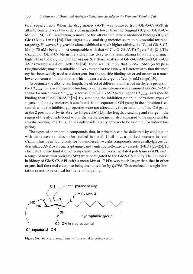
tural requirements. When the drug moiety (AVP) was removed from Glc-O-C8-AVP, itsaffinity constant was two orders of magnitude lower than the original (IC50 of Glc-O-C7-Me ~ 3 µM) [24]. In addition, removal of the alkyl-chain almost abolished binding (IC50 ofGlc-O-Me > 1 mM) [24]. Thus, sugar, alkyl, and drug moieties seem to be essential for renaltargeting. However, S-glycoside alone exhibited a much higher affinity, the IC50 of Glc-S-C7-Me (~ 70 nM) being almost comparable with that of Glc-O-C8-AVP (Figure 5.5) [24]. TheCLuptake of Glc-S-C7-Me in the kidney was close to the renal plasma flow rate and muchhigher than the CLuptake in other organs. Scatchard analysis of Glc-S-C7-Me and Glc-S-C8-AVP revealed a Kd of 10–20 nM [24]. These results imply that Glc-S-C7-Me (octyl β-D-thioglucoside) may be a suitable delivery vector for the kidney. It is noteworthy that this moi-ety has been widely used as a detergent, but the specific binding observed occurs at a muchlower concentration than that at which it exerts a detergent effect (~ mM range) [30].
To optimize the alkyl-chain length, the effect of different numbers of methylene groups onthe CLuptake in vivo and specific binding to kidney membranes was examined. Glc-S-C5-AVPshowed a much lower CLuptake whereas Glc-S-C11-AVP had a higher CLuptake and specificbinding than Glc-S-C8-AVP [24]. By screening the inhibition potential of various types ofsugars and/or alkyl moieties, it was found that an equatorial OH group in the 4 position is es-sential, while the inhibitory properties were not affected by the orientation of the OH groupat the 2 position or by its absence (Figure 5.6) [25]. The length, branching and charge in theregion of the glycoside bond within the methylene group also appeared to be important forspecific binding [25]. Thus, the alkylglycoside moiety appears to be essential for kidney tar-geting.
The types of therapeutic compounds that, in principle, can be delivered by conjugationwith this vector remains to be studied in detail. Until now, a marked increase in renal CLuptake has been found only for low-molecular-weight compounds such as alkylglycoside-derivatized AVP, oxytocin, tryptamine, and 4-nitrobenz-2-oxa-1,3- diazole (NBD) [23–25].Toelucidate the size limitation of compounds to be delivered, acylated polylysines (APL) witha range of molecular weights (Mw) were conjugated to the Glc-S-C8 moiety. The CLuptakein kidney of Glc-S-C8-APL with a mean Mw of 17 kDa was much larger than that in otherorgans, half the renal clearance being accounted for by fpGFR. Thus, molecular weight limi-tation seems to be critical for the renal targeting.
130 5 Delivery of Drugs and Antisense Oligonunucleotides to the Proximal Tubular Cell
Figure 5.6. Structural requirements for a renal targeting vector.

5.2.1.6 Identification of Target Molecules for Alkylglycosides
So far, the target molecule that binds alkylglycosides in the kidney has not been conclusivelyidentified. It is known that several types of transporters are localized on the proximaltubules. These include organic anion transporters, organic cation transporters, and oligopep-tide transporters [31,32]. However, since the renal distribution of the alkylglycoside vector ishighly dependent on the structure of the sugar moieties, and both basic and neutral peptides(AVP and oxytocin, respectively) are recognized, the involvement of these transportersseems to be minor. Sugar transporters are classified as facilitated sugar transporters andNa+/glucose co-transporters [33,34]. The facilitated transporters do not concentrate the lig-and inside the cells whereas the secondary active Na+/glucose co-transporter that is locatedon the brush-border membrane can transport molecules through the existing Na+-gradient[34]. Since the degree of inhibition of alkylglycoside binding to the kidney membrane by glu-cose (10–100 mM) is minor [25], involvement of this transporter is also unlikely. In addition,kidney lectin [35] which recognizes acidic sugars, does not seem to be a potential transporter.Further studies are therefore needed to clarify the major transport system.
Downregulation of CLuptake is one of the methods used to discriminate between carrier-mediated transport and receptor-mediated endocytosis [29,36]. This concept is based on thegeneral finding that an excess concentration of a ligand can induce internalization and sub-sequent degradation of cell-surface receptors, resulting in a reduction of the cell-surface re-ceptor density. After intravenous administration of excess unlabelled Glc-O-C8-AVP, theCLuptake of tracer Glc-O-C8-AVP initially declined followed by a gradual recovery, suggest-ing that Glc-O-C8-AVP is taken up predominantly via receptor-mediated endocytosis [23].However, it is possible that the temporary reduction of CLuptake can also be explained bycompetitive inhibition by the unlabelled ligand instead of a downregulation of the receptor[23]. Recently, cross-linking 125I-labelled alkylglycoside to the renal plasma membrane re-vealed the presence of a binding protein with a Mw of 62 kDa. This band disappeared in thepresence of excess unlabelled ligand and was clearly located at the basolateral membranes,and not in the brush-border membranes [37].
5.2.1.7 Perspectives of Renal Delivery with Alkylglycoside Vectors
The most notable feature of the alkylglycoside vector system is the highly efficient uptake viathe basal site of the renal plasma membranes. This may provide a higher rate of target deliv-ery compared to the targeting methods that exploit glomerular filtration with subsequent re-absorption from the apical position, such as the low-molecular weight protein method de-scribed below in this chapter [38,39].The CLuptake in the latter case cannot exceed the fpGFRbased on Eq. 5.1 (Figure 5.4b). Furthermore, delivery via the basal site will not be negativelyaffected by proteinuria.
Concentration as a result of uptake and subsequent retention of the ligand in the proximaltubules may be suitable for certain types of therapeutic agents. The plasma concentration ofGlc-S-C8-NBD fell rapidly while the kidney concentration of the intact ligand remained al-most constant, the kidney-to-plasma concentration ratio being ~ 200 at 30 min after injection
5.2 Renal Delivery Using Pro-Drugs 131

[24]. Uptake which has the effect of concentrating the therapeutic agent is one of the advan-tages of using receptor- and/or transporter-mediated drug delivery.
The critical factors that need to be addressed include the limited range of drugs that can bedelivered by this system. The present findings suggest that the system cannot be applied to macro-molecules such as genes and proteins. For application to low-molecular-weight compounds,the therapeutic activity of the drug needs to be regained after release from its vector in thekidney. The pharmacological activity of AVP was affected by derivatization [40,41] and ourrecent findings suggest that the kidney-targeting potential is low for certain types of anionic drugs.
It should be noted also that distribution may occur to organs other than the kidney. For ex-ample, oxytocin derivatives also exhibited CLuptake in the small intestine. A similar phenom-enon was observed for Glc-S-C8-tryptamine where the CLuptake in small intestine and liveralso increased by derivatization [24]. These findings cannot be explained simply by the exis-tence of a single binding site for alkylglycoside vectors in the different organs. The presenceof multiple binding sites is supported by the finding that inhibition of the specific binding ofGlc-S-C8-tyrosine by Glc-S-C8-AVP cannot be fitted to a single site kinetic model [37]. Toclarify the renal and extra-renal transport mechanisms, kinetic analysis performed by chang-ing the structure of the ligand may not be sufficient and molecular biological analysis may behelpful, for example by characterizing the target binding protein. This should reveal thescope and limitations of this alkylglycoside strategy in clinical and pathological situations.
5.2.2 The Amino Acid Pro-drug Approach
5.2.2.1 Introduction
Most research on tubular cell-specific drug delivery has been focused on the development ofpro-drugs that should be activated by more or less kidney-selective enzymes.The relevant lit-erature will be reviewed briefly and discussed with regard to the benefits and limitationscompared to the alkylglycoside and macromolecular approaches of renal drug targeting.The‘soft drug’ concept will be discussed as a potential method by which targeted drugs are inac-tivated efficiently after reentering the circulation.
5.2.2.2 The Concept of the Amino Acid Pro-drug
In the design of drugs, the usefulness of renal-specific enzymes which enable the site-specif-ic release of the active drug, should be taken into account.The design of kidney-selective pro-drugs is based upon the relatively higher amounts of certain enzymes in the proximal tubu-lar cells than elsewhere in the body.
These strategies are aimed at either cytosolic enzymes, such as L-amino acid decarboxy-lase, β-lyase and N-acetyl transferase, or enzymes that are expressed at the brush border ofthe proximal tubule and to a lesser extent on the basolateral membrane, such as γ-glutamyltranspeptidase (GGT).
The technology involves one or more chemical modifications of the parent compound us-ing chemical moieties that, with regard to size, are comparable to or even smaller than the
132 5 Delivery of Drugs and Antisense Oligonunucleotides to the Proximal Tubular Cell

parent drug. This type of pro-drug may be degraded intracellularly into the active drug, re-sulting in its release and subsequent secretion into the tubular lumen or via the interstitiumback into the circulation.
Alternatively, the pro-drug may be a substrate for brush-border enzymes of the proximaltubular cell, resulting in release of the active drug in the tubular lumen and subsequent reab-sorption at distal sites or elimination in the urine.
5.2.2.3 Renal Specificity of Amino Acid Pro-drugs and their Effects
Several drugs have been coupled to gamma-glutamyl transferase, γ-glutamyl. The γ-glutamylpro-drug of l-dopa (gludopa) showed a higher renal specificity [42–44] than the pro-drug ofdopamine [45]. Gludopa induced renal-specific effects such as increases in renal blood flowand salt excretion while systemic blood pressure remained unaffected [42,43].
Another example is the pro-drug γ-glutamyl-sulphamethoxazole. This pro-drug did notshow renal selectivity, either because of its rapid removal from the kidney, or due to cleavagein non-target tissues containing a low concentration of the enzyme. On the other hand the N-acetyl-γ-glutamyl derivative showed pronounced renal specificity [46,47]. In this respect, pro-drug accumulation in the kidney was due to carrier-mediated transport at the basolateralmembrane site, which is sensitive to buthionine sulfoximine and probenecid. Other N-acetyl-γ-glutamyl pro-drugs have been developed and tested on the basis of the same principle. Ofthe derivatives tested, N-chloroacetyl-γ-glutamyl-sulfamethoxazole appeared to have thehighest renal selectivity [46]. N-acetyl-γ-glutamyl-aminowarfarin was not a successful pro-drug since it was not rapidly secreted via the tubule and therefore did not reach the enzymesite [48]. In fact, this pro-drug was selectively excreted in the bile.
The N-acetyl-γ-glutamyl pro-drug of the hydralazine-like vasodilator CGP 18137 showeda higher renal-selective activity than the parent compound, CGP 18137. In contrast to theparent drug, the pro-drug caused a decrease in renal resistance without any effect on bloodpressure [49].
Because of the high phosphatase activity in the kidney, dopamine-phosphate ester pro-drugs have been synthesized, e.g. SIM 2055 (N-methyl-dopamine-4-O-phosphate) [50]. Al-though the mechanism of renal selectivity of this compound is not yet understood in detail, itis thought to be due to the high renal blood flow and the high renal phosphatase activity to-gether with the high affinity of the kidney for the released drug.
Cysteine-S-conjugates have also been proposed as kidney-selective pro-drugs. Renal me-tabolism of S-6-(purinyl)-L-cysteine resulted in the formation of 6-mercaptopurine by the ac-tion of β-lyase [51]. However, besides formation of the intended parent compound, other S-conjugates may be formed by various radical reactions, which may induce renal toxicity.
5.2.2.4 Benefits and Limitations of the Amino Acid Pro-drug
A potential benefit of the pro-drug approach is that the compounds can, in principle, be de-signed for oral administration. Furthermore, immunogenicity which results from using pro-tein conjugates as drug carriers, will not be a problem. However, in contrast to the LMWP ap-
5.2 Renal Delivery Using Pro-Drugs 133

proach, in which the kinetics of the protein carrier overrule the intrinsic kinetics of the drugto be targeted by accumulation after the absorptive process, conjugation with amino acids orsmall peptides does not necessarily lead to higher specificity for renal uptake. Therefore, foreach drug, different derivatives should be synthesized and tested for the desired kinetic pro-file.
5.2.2.5 The Soft Drug Concept
When drugs that are activated in the kidney are transported back into the circulation theymay be deposited elsewhere in the body. This undesirable consequence may be overcome by using the so-called soft drug approach [52]. After administration of N-acetyl-γ-glutamyl-CGP 18137, active CGP 18137 is specifically generated within the kidney but partly diffusesback into the circulation. However, in the circulation, this vasodilating agent is rapidly inac-tivated by a chemical reaction [49]. For reasons unknown to us, this innovative product wasnot developed further. However, this example illustrates the potential of soft drugs in thefield of drug targeting. Irrespective of the fact that the drug is generated from a pro-drug orreleased from a carrier, inactivation of the active compound after being released from thekidney into the blood circulation, could evidently add to the renal selectivity and therapeuticsafety.
5.2.3 The Folate Pro-drug Approach
5.2.3.1 Introduction
The kidney has an important role in conserving folate to counteract a potential deficiency ofthis essential vitamin. Circulating folate, in the form of 5-methyltetrahydrofolate, is filteredthrough the glomeruli and extensively reabsorbed within the nephron into the renal vascularcirculation. The kidney contains a high affinity folate-binding protein (FBP) that is concen-trated in the proximal tubule cells [53,54]. Immunocytochemical studies have located FBP tothe brush-border membrane, endocytic vacuoles and dense apical tubules, indicating a reab-sorption of folate through endocytosis of the FBP–folate complex followed by dissociationand recycling of FBP [55]. In this study no significant labelling was found in lysosomes at anytime, implying that there is no transport of FBP to lysosomes for degradation.
Recently it was shown that folate transport from the basolateral site occurs as readily asthat from the luminal site, indicating that changes in secretion can mediate excess urinary fo-late excretion [56].
5.2.3.2 Potential Renal Selectivity of Folate Constructs
It has been hypothesized that folate receptor-mediated endocytosis can be exploited for theselective delivery of drugs by covalent attachment to folate via its γ-carboxyl group.This con-cept was primarily designed for the targeting of various biomolecules to solid tumours. For a
134 5 Delivery of Drugs and Antisense Oligonunucleotides to the Proximal Tubular Cell

number of human tumours, having a high over-expression of a membrane-associated folatereceptor, in-vitro studies have shown that folic acid derivatization allowed selective deliveryto cancer cells in the presence of normal cells.Thus high tumour selectivity was achieved withfolate-targeted imaging agents [57], antineoplastic drugs [58,59], protein toxins [60], lipo-somes [61] and antisense oligonucleotides [62].
Interestingly, after intravenous administration of a radiolabelled folate conjugate (111-In-dium-diethylenetriaminepenta acid (DTPA)-folate) in the rat, the conjugate was rapidly ex-creted in the urine. Moreover, after intravenous administration to athymic mice with a hu-man tumour cell implant, the radiotracer was not only taken up by the subcutaneous tumourbut was also taken up by the kidneys in significant quantities [63], indicating substantial re-nal selectivity of the folate conjugate. In addition to the kidney, the liver also has a high con-centration of the folate-receptor [64].
5.2.3.3 Benefits and Limitations of Folate
To date, the possibility of using folate binding for the purpose of renal drug targeting has notbeen studied. Since the kidney is not the only organ containing folate-receptors, the physico-chemical properties of the conjugate may be important determinants of the success of tar-geting.
5.3 Renal Delivery Using Macromolecular Carriers:The Low Molecular Weight Protein Approach
5.3.1 Introduction
Low molecular weight proteins (LMWP) are freely filtered proteins with a molecular weightof less than 30 000 Dalton and are considered to be suitable as renal-specific drug carriers.The concept is based on four principles:
• The carrier has functional groups allowing drug attachment.• The LMWP accumulates specifically in the kidney, in particular in the tubular cells
through a reabsorption mechanism.• The physicochemical properties of the LMWP overrule those of the linked drug.• The drug–LMWP conjugate is stable in the circulation but after arrival in the kidney, the
active drug is released in the catabolically-active lysosomes of the proximal tubular cells(Figure 5.7).
As reviewed by Franssen et al. [65], drugs can be directly coupled to LMWPs via the lysineamino group of the protein to form an amide bond.Alternatively, the drug can be coupled tothe protein via different spacers such oligopeptides (amide bond), (poly)-alpha-hydroxyacids (ester bond), pH-sensitive cis-aconityl spacers (acid-sensitive amide bond) and SPDPspacers (disulfide bond) (see Chapter 11).The ability of the kidney to release the parent drugfrom such drug-spacer derivatives and drug–LMWP conjugates by enzymatic or chemical hy-
5.3 Renal Delivery Using Macromolecular Carriers: The Low Molecular Weight Protein Approach 135

drolysis of the bond, have been tested in renal cortex homogenates and lysosomal lysates aswell as in in vivo studies. It was found that lysosmal proteases can cleave the peptide bond be-tween the carboxylic acid group of a drug and an α-amino group of an amino acid. However,the bond between the carboxylic acid group of the drug and the ε-amino group of lysinecould not be cleaved. Since the conjugation of drugs to amino groups of a protein will pre-dominantly occur at the ε-lysine residues and only to a small extent at the N-terminal α-amino group, direct conjugation of a drug via its carboxylic acid group will not result in thequantitative regeneration of the parent compound [66]. Drugs with a terminal carboxylgroup, such as naproxen [67], can be released as the parent drug from LMWP conjugates us-ing ester spacers such as L-lactic acid. Increasing spacer length by intercalating a tetra (L-lac-tic acid) moiety between the drug and the protein further increases the rate of drug release,indicating increased accessibility of the bond to the enzymes.
Drugs that have primary amino groups available for conjugation, for instance dopamineand doxorubicin, can in principle be coupled to LMWPs via oligopeptides. In contrast to thecarboxypeptidases, the aminopeptidases appear to possess a broader specificity. To allow therelease of terminal amino group-containing drugs in the acid environment of the lysosomeswithout the requirement of enzymes, an acid-sensitive spacer can be used.
Drugs coupled via a disulfide bond like, captopril, are rapidly released from the protein-spacer moiety of the conjugate, enzymatically by β-lyase and/or non-enzymatically by thiol-disulfide exchange with endogenous thiols [68].
The different aspects of drug targeting using LMWPs that have been studied to date arediscussed below. As an example, we use the data of two conjugates, naproxen–lysozyme andcaptopril–lysozyme.
136 5 Delivery of Drugs and Antisense Oligonunucleotides to the Proximal Tubular Cell
Figure 5.7. Schematic representation of the mechanism by which drug targeting to the proximal tubularcell of the kidney might be achieved using a low molecular weight protein (LMWP) as a carrier.

5.3.2 Renal uptake of LMWP Conjugates
5.3.2.1 Renal Uptake of Native LMWPs
Comparison of the kinetic features of different LMWPs revealed that all LMWPs tested sofar (such as lysozyme, cytochrome-c and aprotinin) are quickly cleared from the circulationand accumulate rapidly in the kidney [38]. The fractions of the injected LMWP that are re-ported to be taken up by the kidney vary between 40–80 % of the injected dose. In our stud-ies, using external counting of radioactivity, at least 80 % of the intravenously injected LMW-Ps was finally taken up by the kidneys, which is in agreement with renal extraction studies[69,70]. However, studies in which the actual amount of LMWP in the kidney was measureddirectly in the tissue, indicated a lower, but still substantial accumulation of 40% of the in-jected dose [71,72]. Apart from the kidney, LMWPs do not seem to accumulate elsewhere inthe body (Figure 5.8).
From this we concluded that LMWPs are potentially suitable to serve as renal-specificdrug carriers: a drug–LMWP conjugate will be rapidly removed from the circulation and thedrug can be intra-renally released. Consequently, major distribution to extra-renal tissue andrelated unwanted effects elsewhere in the body can, in principle, be avoided. It is assumedthat secondary redistribution of the generated drug from the kidney is relatively slow so thatsystemic concentrations remain below the therapeutic window for extra-renal effects.
5.3 Renal Delivery Using Macromolecular Carriers: The Low Molecular Weight Protein Approach 137
5.3.2.2 Renal Delivery of Naproxen–Lysozyme
Targeting of nonsteroidal anti-inflammatory drugs (NSAIDs) such as naproxen could be ofinterest for the treatment of proteinuria and tubular defects such as Fanconi syndrome andBartter’s syndrome [73,74]. Although a conjugate with an ester spacer is preferred to a con-jugate with a direct peptide linkage [66,67], we continued our research using naproxen di-
Figure 5.8. Renal specificity of a radiolabelled LMWP. Gamma-camera imaging after an intravenousinjection of a radiolabelled low molecular weight protein (LMWP) in the rat, showing the predominantuptake of the LMWP by the kidneys.

rectly conjugated to lysozyme. The synthesis of the conjugate with an ester spacer (naprox-en–L-lactic acid–lysozyme) is cumbersome, but fortunately the catabolite of the conjugatewith the direct peptide linkage (naproxen–lysine) appeared to have an inhibitory effect onprostaglandin synthesis in vitro which was equivalent to that of the parent drug [66].
The coupling of 2 moles of naproxen to 1 mole of lysozyme did not affect the renal uptakeof lysozyme in the rat: like native lysozyme, the conjugate rapidly accumulated in the kidney[75]. Focusing on the drug moiety of the conjugate, it was shown that conjugation of naprox-en to lysozyme distinctly altered the kinetics of the drug. Conjugation to lysozyme resulted ina 70-fold increase in naproxen concentrations in the kidney (Figure 5.9a) [76].
138 5 Delivery of Drugs and Antisense Oligonunucleotides to the Proximal Tubular Cell
Figure 5.9. The concentration–time course of (a) naproxen and (b) captopril in the kidney afterintravenous injection of the parent drug or the drug–lysozyme (LZM) conjugate. Values are given asmeans + SEM.
5.3.2.3 Renal Delivery of Captopril–Lysozyme
Angiotensin-converting enzyme (ACE) inhibitors such as captopril exert a long-term reno-protective effect. Among other effects, they lower systemic blood pressure and renal plasmaflow and effectively reduce urinary protein excretion. Renal delivery of ACE-inhibitors mayincrease this efficacy and reduce extra-renal side-effects. Renal targeting of an ACE-in-hibitor can also be useful in clarifying the contribution of local ACE inhibition to these reno-protective effects.
A spacer was used to link captopril via a disulfide bond to the LMWP lysozyme. Conjuga-tion of captopril to lysozyme resulted in a 6-fold increase in captopril accumulation in the ratkidney (Figure 5.9b) [77]. This modest enrichment, as compared to that achieved withnaproxen–lysozyme, was due to fact that, in contrast to naproxen, free captopril is clearedvery efficiently by the kidney itself. Thus, delivery via lysozyme reabsorption only leads to alimited improvement of renal accumulation of captopril.
a) b)

5.3 Renal Delivery Using Macromolecular Carriers: The Low Molecular Weight Protein Approach 139
Figure 5.10. Accumulation of a radiolabelled LMWP in the lysosomes of the proximal tubular cell.Electron microscope autoradiography of renal proximal tubular cells from a rat injected i.v. with [125I]-tyramine-cellobiose-labelled cytochrome-c, 4 h prior to fixation through the abdominal aorta. Anintense lysosomal accumulation of the protein is observed in three dark electron-dense lysosomes . Afew grains are seen over the apical endocytic apparatus. Part of the luminal brush border is found in theupper right hand corner. Magnification, x 25 000. Unpublished data from E. I. Christensen, Arhus,Denmark, and M. Haas, Groningen, Netherlands.
5.3.3 Renal Catabolism of LMWP-conjugates
5.3.3.1 Renal Catabolism of Native LMWPs
Morphological (Figure 5.10) and biochemical studies have established that after endocytosisby the proximal tubular cell, LMWPs migrate via endosomes to the proteolytically activelysosomes [78,79]. Within the lysosomes the LMWPs are degraded into small peptides andsingle amino acids.Whereas the renal uptake rate of various LMWPs appeared to be similar,LMWPs are catabolized with distinct individual differences in their catabolic rate as indicat-ed from the difference in the rate of decline of radioactivity in the kidney (Figure 5.11). Therate of catabolism seemed unrelated to the size or charge of the protein alone [80,81]. Prob-ably multiple structural factors play a role in this process. A crucial factor may be the differ-ent endosomal migration times of LMWPs from the tubular lumen to the lysosomes. Where-as cytochrome-c accumulated in the lysosomes within 3 min, lysozyme seemed to migrate for20 min before the commencement of degradation [72, 82].Also the intrinsic activity of the re-absorbed protein may play a role. For instance, the long renal half-life of aprotinin, an in-hibitor of proteolytic enzymes, may be explained by an inhibition of its own degradation, assuggested by Bianchi [71]. These studies suggest that the LMWP method of renal drug tar-geting results in cell-selective delivery followed by controlled drug release which can be ma-nipulated at various stages of the renal deposition process. The lysosomes are stacked with avariety of proteolytic enzymes in an acidic environment. Programmed drug release from adrug–carrier conjugate may therefore be achieved using peptide, ester or acid-labile bonds

between the drug and protein carrier. Consequently both the differences in rate of catabo-lism between LMWPs as well as the rate of hydrolysis of the bond between the drug and car-rier may be used to manipulate the rate of drug release in the kidney. The variable migrationtimes of different LMWPs and their conjugates after endocytosis may have consequences forthe intracellular concentration profiles. For instance, in order to achieve relatively constantcellular levels of the drug, an LMWP which is only slowly degraded might be preferred as adrug carrier. In contrast, if short-term peak levels of the drug are preferred, treatment with arapidly processed protein (with a short migration time) may be a more appropriate choice.Certain drugs (e.g. peptides and nucleotides) should be released before entering lysosomesto prevent inactivation by degradative enzymes. For such drugs, a prolonged endosomal mi-gration time combined with simple hydrolysis of the drug–protein linkage in the acidic envi-ronment of the endosomes, will be preferred to achieve adequate drug release and preventan abortive route to the lysosomes.
5.3.3.2 Renal Catabolism of Naproxen–Lysozyme
The coupling of 2 moles of naproxen to 1 mole of lysozyme did not affect the catabolism oflysozyme in rat kidney [66,75]. After delivery to the kidney, naproxen in the form of naprox-
140 5 Delivery of Drugs and Antisense Oligonunucleotides to the Proximal Tubular Cell
Figure 5.11. Time course of clearance from the kidney of radiolabelled LMWPs after intravenousinjection. After renal uptake, the radiolabelled protein is gradually catabolized and the radioactivebreakdown products released from the kidney, as shown by the decline of renal radioactivity over time.
en–lysine was gradually released from the conjugate. This catabolite was subsequently elimi-nated from the kidney and after a single injection, drug levels in the renal tissue gradually de-creased with a t1/2 of 160 min (Figure 5.9a).
No detectable amounts of naproxen or its lysine conjugates were found in the plasma af-ter administration of the conjugate and it can be inferred that excretion into the urine is thecrucial process which determines the elimination rate t1/2. The lack of diffusion into thebloodstream is a favourable property in relation to unwanted extra-renal effects.
5.3.3.3 Renal Catabolism of Captopril–Lysozyme
After renal uptake, captopril was rapidly released from the conjugate as indicated by therapid decrease in renal captopril levels with time (Figure 5.9b). The difference in renal t1/2 ofnaproxen and captopril after delivery with lysozyme is likely to be due to an unequal rate ofrelease from the lysozyme conjugates. Whereas naproxen–lysozyme requires a peptidase forcleavage, captopril is released from the conjugate enzymatically by β-lyase and/or non-enzy-maticaly by thiol-disulfide exchange with endogenous thiols. To reduce the rate of capto-pril–lysozyme breakdown, two different cross-linking reagents, SPDP and SMPT, were test-ed. Although an SMPT link between two proteins is in principle less susceptible to disulfidereduction [83], no difference in degradation rate was found between the SPDP and theSMPT captopril–lysozyme conjugates (Kok et al., unpublished data).
5.3.4 Effects of Targeted Drugs Using an LMWP as Carrier
5.3.4.1 Renal Effects of Naproxen–Lysozyme
Having obtained promising kinetic profiles, the potential renal effects of naproxen–lysozymein the rat were investigated [84]. Naproxen, as an inhibitor of cyclooxygenase, blocksprostaglandin synthesis. Among other effects, naproxen reduced furosemide-stimulated uri-nary excretion of prostaglandin E2 (PGE2) as well as the natriuretic and diuretic effects offurosemide. Studies with the conjugate showed that naproxen–lysozyme treatment clearlyprevents furosemide-induced excretion of PGE2. This occurred with a dose of naproxen thatwas not effective in the unconjugated form. Surprisingly, this effect occurred in the absenceof a change in natriuretic and diuretic response to furosemide. In this respect the pharmaco-logical effect differed from treatment with a high dose of free naproxen. An explanation forthese differences remains to be found. One possibility is that there is a difference in the in-tra-renal kinetics of the NSAID compared with the parent drug. Free naproxen is extensive-ly reabsorbed in the distal tubule of the kidney via which route it may effectively inhibitprostaglandin synthesis in the medullary interstitial cells. On the other hand, naproxen–ly-sine is more hydrophilic and may be unable to reach the sites of prostaglandin synthesis in-volved in the furosemide-induced excretion of sodium and water. These data shows that re-nal drug targeting preparations can also be used as a tool to unravel the mechanisms of renaltherapeutic effects.
5.3 Renal Delivery Using Macromolecular Carriers: The Low Molecular Weight Protein Approach 141

5.3.4.2 Renal and Systemic Effects of Captopril–Lysozyme
With regard to the pharmacological effects of the captopril–lysozyme conjugate, the follow-ing observations were made (Kok et al., unpublished data). The extent of ACE-inhibition inthe plasma and kidney tissue was measured after i.v. administration of captopril–lysozymeand an equimolar dose of free captopril. It was shown that conjugation to lysozyme caused asimilar though more sustained inhibition of renal ACE-activity by captopril.The inhibition ofplasma ACE-activity was clearly reduced but not entirely prevented by conjugation of cap-topril to lysozyme. Possibly, the S-S linked drug conjugate is partly degraded in the circula-tion. It is also possible that after degradation of the conjugate in the kidney, captopril wastransported back into the bloodstream. The rapid intracellular release may provide a suffi-cient driving force for transport across the basolateral membranes.
Captopril–lysozyme did not significantly affect systemic blood pressure whereas anequimolar dose of captopril alone decreased blood pressure significantly. Whereas free cap-topril (5 mg kg–1) completely prevented an angiotensin-I-induced blood pressure increase, anequimolar amount of captopril–lysozyme did not. However, in line with the direct ACE ac-tivity measurements in renal tissue and plasma, in captopril–lysozyme-treated rats the an-giotensin-I-induced blood pressure increase was lower than in untreated rats, suggesting thatsystemic activity was not fully prevented.
Neither free nor conjugated captopril affected glomerular filtration. Renal plasma flow in-creased to the same degree after treatment with free or conjugated captopril (1 mg kg–1).Al-though the complete dose–effect relationship was not studied, we can conclude that conju-gation of captopril to lysozyme did not prevent the drug from acting on the renal plasmaflow. Whether this effect is determined by intra-renal or systemic ACE-inhibition remains tobe investigated.
At present, the synthesis of lysozyme conjugates with ACE-inhibitors other than captoprilis under investigation. Some of these ACE-inhibitors may be advantageous for renal delivery.The amount of conjugate required for therapy can be reduced when using an ACE-inhibitorwith a higher affinity for ACE (e.g. lisinopril). Furthermore, the stability of the conjugate inplasma may be increased by using an ACE-inhibitor which is conjugated to lysozyme via alinkage that is highly stable in plasma (e.g. lisinopril can in principle be coupled via an acid-sensitive spacer).
5.3.5 Renal Disease and LMWP Processing
Proteinuria is one of the most prominent abnormalities found in renal disease and is one ofthe factors held responsible for the progressive loss of renal function. As a consequence ofthe glomerular leakage of proteins, the proximal tubular cells are exposed to increasingamounts of protein. This pathological condition can be anticipated to influence the deposi-tion and metabolism of protein-linked drugs. It is likely, in such a situation, that drug–LMWPconjugates will have to compete with the overload of protein for tubular uptake as well as forcatabolism. The effect of proteinuria on the renal processing of LMWPs has been examinedin a number of studies [85–92]. Collectively, these studies clearly indicate that the effect ofproteinuria on renal uptake and degradation of LMWPs depends on the severity and dura-
142 5 Delivery of Drugs and Antisense Oligonunucleotides to the Proximal Tubular Cell
tion of the protein leakage. However, it should be noted that tubular reabsorption of LMW-Ps is only slightly reduced during adriamycin-induced chronic proteinuria [92]. With respectto LMWP catabolism, the data suggest that protein overload will lead to reduced proteolyt-ic degradation. In that case, an acid labile spacer or a disulfide bond should be chosen toguarantee an adequate rate of drug release.
We found a difference in susceptibility to proteinuria between cationic LMWP cy-tochrome-c and neutral LMWP myoglobulin with respect to their catabolism. This may indi-cate that the effect of proteinuria on LMWP catabolism is determined by the proximal tubu-lar segment in which the LMWPs and the protein overload are processed [88,89,93,94]. Wespeculate that, through coupling to a specific LMWP, drugs can be delivered specifically tothose proximal tubular cells that are predominantly affected by proteinuria.This might be es-sential for drugs chosen to protect the tubular cell from further damage by proteinuria. In ad-dition, it may be possible to use certain LMWPs as drug carriers to circumvent the protein-uria-affected cells. In that case, treatment of diseases unrelated to proteinuria will not be hin-dered by the severity of proteinuria.
5.3.6 Renal Delivery of High Doses of LMWPs
The renal cell responsible for the uptake of LMWPs is the proximal tubular cell. LMWPs arerelatively freely filtered by the glomerulus and subsequently reabsorbed by the proximaltubular cell by megalin/gp330 receptor-mediated endocytosis [95]. In healthy individuals, therelatively moderate amounts of endogenous LMWPs are completely reabsorbed by theproximal tubular cells. However, for drug targeting purposes, larger doses of LMWP may berequired. We compared the urinary loss of intact LMWP after intravenous administration ofdifferent doses of LMWP by either single dose injections or by continuous infusions inhealthy rats. From these studies, we concluded that after a continuous low-dose infusion thenon-reabsorbed fraction is considerably less than that after single high-dose injections. How-ever, infusion could not entirely prevent the loss of intact LMWP into the urine (the loss was8% of the dose after 100 mg lysozyme kg–1 over 6 h and rose to 33% following1000 mg lysozyme kg–1 over 6 h).
Cojocel et al. demonstrated clear adverse effects after relatively high doses of lysozyme[96]. We studied these aspects in more detail and concluded that lysozyme should be given in a dose of less than 100 mg–1 kg–1 over 6 h to minimize the negative effects on systemicblood pressure, glomerular filtration and renal blood flow. From these data, it emerged thatLMWPs are suitable to serve as drug carriers to the proximal tubular cell of the kidney. How-ever, the conjugate should preferably be administered in low-dose by constant infusion tolimit the systemic and renal toxicity and to reduce the urinary loss of the intact conjugate(unpublished data).
5.3.7 Limitations of the LMWP Strategy of Drug Delivery to the Kidney
Among the disadvantages of the LMWP strategy for the treatment of chronic renal diseaseare the requirement for parenteral administration and the possible immunogenicity of the
5.3 Renal Delivery Using Macromolecular Carriers: The Low Molecular Weight Protein Approach 143

drug conjugate. With respect to the administration route, the conjugate could possibly be ad-ministered subcutaneously or intramuscularly. This is a common administration route forpolypeptide drugs such as insulin. If immunogenicity appears to be a serious limitation forchronic treatment, a synthetic polymer may be used as the ‘reabsorptive’ carrier instead[97,98].
For short-term clinical interventions with the aim of protecting the kidney during acutereperfusion or preventing allograft rejection after transplantation, the prerequisite of par-enteral administration does not constitute a serious limitation.
5.4 Renal Delivery of Antisense Oligodeoxynucleotides
5.4.1 Introduction
Various macromolecular and pro-drug technologies designed to achieve selective renal drugaccumulation and action have been discussed in the previous sections of this chapter. In theseapproaches, traditional drugs have been modified through coupling to carrier molecules. It isgenerally accepted that, at least in theory, antisense oligodeoxynucleotides (AS-ODN) offera new approach for selective treatment [99,100].
In view of the preferential distribution of some AS-ODNs to the kidney the oligonu-cleotide backbone could even be employed for renal-specific drug delivery because of boththeir intrinsic activity and the potential of coupling of other agents to them.
Antisense refers to the use of single-stranded synthetic oligonucleotides to inhibit gene ex-pression [99,100].The striking advantage of the antisense approach in comparison to tradi-tional drugs is its potential for specificity. The binding affinity between the oligonucleotideand its target receptor is many orders of magnitude higher compared to that at other bindingsites, as a result of the multiple interaction sites that exist on the target receptors [101]. Sinceaffinity is proportional to the number of interactions between a drug and its receptor, thespecificity of an AS-ODN depends on its length. The base pairing specificity of an AS-ODNof about 15-17 nucleotides in length appeared to be sufficient to inhibit only one target genewithin the entire human genome [99]. For successful inhibition in vivo, the plasma and intra-cellular stability and the pharmacokinetic profile of the antisense molecule along with theturnover time of the inhibited gene are important determinants.
First, we will briefly review the different aspects that are of importance in the use of anti-sense for in vivo therapy. Second, we will describe the effects of antisense targeting to theproximal tubule of the kidney that have been obtained so far.
5.4.2 Mechanism of Action of Antisense Oligodeoxynucleotides
AS-ODN are designed to be complementary to the coding (sense) sequence of the mRNA inthe cell. After hybridization to target sequences, translational arrest occurs via one of sever-al putative mechanisms. The first mechanism is inhibition of transcription. Secondly, AS-ODN can prevent the synthesis of fully mature mRNA in the cytosol at the level of splicing,
144 5 Delivery of Drugs and Antisense Oligonunucleotides to the Proximal Tubular Cell

processing and transport across the nuclear membrane. The third mechanism is inhibition oftranslation by hybridization of the AS-ODN to the sense sequence and thereby preventingthe ribosome from reading the mRNA code.Translation can be inhibited by AS-ODN whichbind to important sites for translation such as translation initiation sites, poly(A) signals, andprotein-binding regulatory sites. Finally, AS-ODN hybridization to the mRNA initiates spe-cific cleavage of the RNA strand by activated RNase H and this cleavage results in destruc-tion of the coding sequence and inhibition of mRNA translation [101].
5.4.3 Stabilization of Antisense Oligodeoxynucleotides
Phosphodiester AS-ODN are poor candidates for use as therapeutic agents in vivo due totheir sensitivity to 3′- and 5′- exo/endonucleases. Because of this, various chemical modifica-tions to the oligonucleotide backbone have been introduced to improve enzymatic stabilitywhile preserving their ability to hybridize cognate targets. Most common examples includethe phosphorothioated and methylphosphonated analogues which have a sulfur atom and amethyl group, respectively, substituted for a non-bridging oxygen atom (Figure 5.12).
Phosphorothioated AS-ODN retain their negatively charged groups in the phosphodiesterbackbone and have the ability to induce mRNA degradation via RNase H. However, thesecompounds have a somewhat lower binding affinity to the target sequence. Moreover, non-sequence-specific activity has been reported for phosphorothioated AS-ODN, probably dueto their stronger protein binding capacity [102,103].
Unlike phosphorothioates, methylphosphonated AS-ODN are uncharged compoundswith a higher cellular uptake than unmodified AS-ODN. Unfortunately, these compoundsappeared to be ineffective in some cell lines. This might be explained by the formation of
5.4 Renal Delivary of Antisense Oligodeoxynucleotides 145
Figure 5.12. Chemical structure of antisense oligodeoxynucleotides (AS-ODN). Phosphorothioate andmethylphosphonate AS-ODN have a sulfur atom and a methyl group respectively, substituted for a non-bridging oxygen atom to increase stability to nucleases.

diastereomers or the inability of methylphosphonates to induce mRNA degradation viaRNase H [104].
To avoid the problem of chirality and to improve the potency and limit the non-specific ac-tions of AS-ODN, new compounds are required. Synthesis of new AS-ODNs has further im-proved their nuclease stability, enhanced of cellular uptake and affinity through modificationof the base, sugar and phosphate moieties of the oligonucleotides [105–108].
5.4.4 Pharmacokinetic Aspects of Antisense Oligodeoxynucleotides andRenal Distribution
The tissue distribution of AS-ODN after a single intravenous injection has been studied ex-tensively in many species including mouse [109], rat [110], monkey [111] and man [112]. Themajority of pharmacokinetic studies have been performed using phosphorothioated AS-ODNs. In general, the pharmacokinetic profiles of AS-ODNs of varying lengths (up to20mer) and base compositions are remarkably similar in all species.
In plasma, most of the phosphorothioated AS-ODNs are protein bound [113,114]. Cossumand co-workers revealed that albumin and α2-macroglobulin are responsible for this binding[114]. The protein binding capacity in rat was elevated after administration of doses higherthan 15–20 mg kg–1 resulting in a dose-dependent increase in distribution volume and an in-crease in plasma clearance [115–117].
The rapid elimination from plasma following intravenous administration of phosphoro-thioated AS-ODN can be explained by a two compartment model in all species, i.e. an initialplasma half-life of less than 1 h [111,113,114] and a slower elimination half-life ranging be-tween 20 and 50 h [111,113].
The kidneys and the liver primarily take up phosphorothioated AS-ODN after parenteraladministration, accumulating more than 10% each, while the rest of the organs all accumu-late less than 1% of the injected dose [110,111,114,116]. It is noteworthy that renal AS-ODNtissue levels exceed that of any other organ [110,113,114], as confirmed by the tissue to plas-ma ratios of approximately 85 and 20 for kidney and liver, respectively [113,118].
Autoradiographic studies of the kidney have shown the accumulation of AS-ODN to oc-cur almost exclusively in the proximal tubular cells [110,119]. Oberbauer et al. reported thatintravenously injected AS-ODN accumulated in proximal tubular cells, and electron mi-croscopy revealed that AS-ODN did accumulate only in the brush border or lysosomal com-partment. This implies that the AS-ODNs were not completely degraded after being takenup by the proximal tubule [110].
In the last 2 years, several AS-ODNs with modified backbone structures and sugar moi-eties have been developed and these are characterized by a significanty increased stability inplasma [107,108]. Chimeric AS-ODNs, consisting of a mixture of phosphorothioate andmethylphosphonate nucleotides, also exhibited increased stability in plasma [106]. It is worthnoting that these AS-ODNs also appeared to be more stable in various tissues including thekidney [106,107]. Agrawal et al. [105] and Crooke et al. [108] have shown that changes in thesugar moieties can further improve the tissue distribution of AS-ODNs in favour of the kid-ney.
146 5 Delivery of Drugs and Antisense Oligonunucleotides to the Proximal Tubular Cell

5.4.5 Cellular Uptake of Antisense Oligonucleotides
Cellular uptake of AS-ODNs is restricted because of their large molecular mass as well astheir polyanionic character. When added directly to cells in culture, only 1–2% of the AS-ODNs will be cell-associated. Therefore, enhanced AS-ODN uptake is a critical considera-tion in developing these agents for therapeutic applications.
The cellular uptake of AS-ODN is an energy-dependent process and takes place in a sa-turable and sequence-independent manner [120,121]. The exact mechanism of uptake re-mains controversial. From in vitro experiments, some authors have proposed that the uptakeis endocytic and mediated by membrane receptor proteins. The receptor responsible for thecellular uptake of AS-ODNs was reported to consist of both a 30-kDa protein [122] and an80-kDa membrane protein [121]. However, other workers have argued that AS-ODN bind-ing to membrane proteins is relatively non-specific and is mostly charge associated, consis-tent with adsorptive endocytosis or fluid-phase pinocytosis [101]. As a result of these con-flicting reports, it is unlikely that in vitro data can be safely extrapolated to what occurs in theintact organism.
In the kidney, AS-ODNs are filtered and subsequently reabsorbed by the proximal tubu-lar cells. The AS-ODNs most likely accumulate in the proximal tubular cells via a receptor-dependent mechanism [110,123]. This hypothesis supports the apparent saturation of AS-ODN uptake in the kidney as reflected by a reduction of degree of renal uptake with in-creasing AS-ODN dose [110,116,117]. Moreover, Rappaport et al. described the existence of40 and 97-kDa binding proteins for 18mer phosphorothioates in the renal brush bordermembrane [123]. In another study, a protein with a molecular weight of approximately50 kDa which may serve as a transmembrane channel transporting AS-ODN into the tubu-lar cell was described [124]. These channels have previously been reported for the uptake ofproteins and phage DNA. The presence of such channels might explain why uptake in theproximal tubular cells is dependent on the nucleotide length as was demonstrated by Lokeand co-workers [121]. It is noteworthy that scavenger receptors located at the basolateral sitemay also be responsible for additional tubular accumulation of AS-ODN [125].
5.4.6 Metabolism and Elimination of Antisense Oligodeoxynucleotides
A prerequisite to acquire an antisense effect is the maintenance of AS-ODN within the tar-get cells. Several studies have reported that the majority of phosphorothioated AS-ODNstaken up by the kidney remains intact for several hours [110,113]. In fact, 4 days after ad-ministration, 3% of the infused dose was still present in the kidney intactly [110]. Althoughseveral studies have confirmed the presence of intact AS-ODN in the kidney, concomitantmetabolism in the kidney of 20% after 6 h [113], 50% after 48 h [118,126] and 50% after4 days [114] has also been reported.
In spite of the improved stability to nucleases, achieved through chemical modification,AS-ODN degradation in plasma still occurs, predominantly from the 3′-terminus. In the li-ver and kidney, the major sites of metabolism, AS-ODNs are degraded from the 5′-terminusas well [127,128].
5.4 Renal Delivery of Antisense Oligodeoxynucleotides 147

Elimination of phosphorothioated AS-ODN takes place primarily via the urine. Approxi-mately 30% of the injected dose is found in the urine within 24 h [110,113].Althought in mostcases only metabolites of AS-ODN could be demonstrated in the urine [110,118], Agrawaland co-workers described the excretion of intact AS-ODN in the urine after a dose of30 mg kg–1 [126]. The saturation of plasma protein binding and proximal tubular uptakecould explain this observation [114,116].
Excretion via faeces is a minor route of elimination, accounting for less than 10% of theadministered dose [113, 126].
5.4.7 Effects of Antisense Targeting to the Proximal Tubule
Noiri et al. used AS-ODN to inhibit production of inducible nitric oxide synthase (iNOS) inan attempt to prevent NO production in an ischaemic kidney. A single intravenous injectionof iNOS AS-ODN attenuated acute renal failure and reduced the morphological abnormali-ties [129].
Oberbauer et al. reported inhibition of a sodium/phosphate (Na/Pi-2) co-transporter byphosphorothioated AS-ODN. A single intravenous injection of the AS-ODN inhibited boththe mRNA and the protein for the Na/Pi-2 co-transporter, and consequently suppressed lu-minal uptake of phosphate by the proximal tubules [130].
Wang et al. injected a Texas-red-labelled phosphorothioated AS-ODN into the dopamine1A receptor in the rat renal interstitium. Fluorescence was detected after 24 h in both tubu-lar epithelium and intra-renal vasculature. Treatment resulted in a 35% decrease in thedopamine 1A receptor protein, causing a reduction in urinary sodium excretion and urineoutput [131].
Rat kidneys were perfused ex situ with phosphorothioate intercellular adhesion molecule(ICAM)-1 AS-ODN and exposed to 30 min cold or warm ischaemia. After this time the kid-neys were transplanted to syngeneic nephrectomized rats.Treatment with 10 mg antisense re-duced the harmful effect of transplantation on renal function [132]
Cheng et al. showed that intravenous infusion of intracellular adhesion molecule (ICAM)AS-ODN markedly reduced ICAM-1 expression, alleviated infiltration of inflammatory cellsand accumulation of extracellular matrix in the obstructed kidney of mice with unilateral ob-struction of the ureter [133].
Repeated intravenous injection of osteopontin AS-ODN to Goodpasture syndrome ratsblocked tubular osteopontin expression, attenuated monocyte infiltration and preserved re-nal plasma flow. No changes were found in osteopontin mRNA level, glomerular histology orproteinuria. The data suggest that interstitial inflammation as a consequence of glomerulardisease can be prevented through a selective inhibition of tubular osteopontin expression u-sing osteopontin AS-ODN [134].
Continuous infusion of transforming growth factor-β (TGF-β) AS-ODN in diabetic mice,decreased kidney TGF-β levels and attenuated the increase in kidney weight, and decreasedlevels of α1(IV)collagen and fibronectin mRNAs.
The above described studies show that the renal proximal tubular cell is a good target forantisense therapy [135].
148 5 Delivery of Drugs and Antisense Oligonunucleotides to the Proximal Tubular Cell

5.4.8 Benefits and Limitations of Antisense Oligodeoxynucleotides
The introduction of therapy through the delivery of antisense oligodeoxynucleotides holdspromise for the treatment of several diseases. It is more specific than conventional drugswhile, in contrast with gene targeting, the effect is temporary so that therapy can be termi-nated when desired. Pharmacokinetic studies have revealed that the proximal tubular cell ofthe kidney is a suitable target for antisense therapy. Although recent studies have shown an-tisense oligodeoxynucleotides to be effective in the treatment of renal diseases, antisense tar-geting is however, a new approach to therapy and all the risks associated with it are not yetknown.
5.5 Drugs for Renal Targeting
For the treatment of kidney diseases, several kinds of drugs are currently used. At present,angiotensin-converting enzyme inhibitors are the first choice drugs for the treatment ofchronic kidney diseases that are characterized by loss of renal function and proteinuria [136].These drugs exhibit only moderate side-effects. However, renal targeting of an ACE-in-hibitor may improve the therapy in certain cases. For example, when proteinuria is accompa-nied by normal blood pressure, hypotension due to ACE-inhibition limits the amount of drugthat can be given. Renal inflammation such as glomerulonephritis and tubulointerstitial in-flammation are treated with corticosteroids [137]. These drugs have serious side-effects. Re-nal targeting of these drugs may allow a more aggressive treatment of the inflammation.Also, local suppression of the immune system may be useful to prevent transplant rejection.However, it is as yet unknown whether suppression of the local immune system is sufficientor whether the systemic system should also be suppressed to prevent rejection [138]. Renaltumours are characterized by insensitivity to the common anti-tumour drugs [139]. This isprobably due to the unfavourable kinetic profile of these drugs. By renal targeting, an anti-tumour drug may reach the renal tumour while the extra-renal side-effects will be reduced.
5.6 In-Vitro and In-Vivo Models for Renal Targeting
5.6.1 In-vitro Models
In the isolated perfused kidney model, the artery of the kidney is perfused and urinary sam-ples as well as venous blood samples can be collected to determine the drug concentration.Aserious drawback of the model is that isolation and artificial perfusion greatly affect the func-tion of the organ as shown by a dramatic drop in the glomerular filtration rate. Another in-vitro model is the isolated tubule in which samples can be taken from both the luminal andbasolateral sites of the tubule [140,141]. The disadvantage of this technique as well as of theisolated kidney model, is that they require specific equipment and expertise and thereforecan only be performed in rather specialized laboratories. Experiments using freshly isolatedor cultured cells are more simple to carry out [142,143]. Tubular cells can be grown in a po-
5.6 In-Vitro and In-Vino Models for Renal Targeting 149

larized fashion enabling the addition of drugs and removal of samples from both the luminaland basolateral sites of the cell.
5.6.2 In vivo Models
Obviously, none of the existing animal models of renal diseases are perfect reflections of thehuman situation. The natural model of progressive loss of renal function is the 5/6 nephrec-tomy. Drawbacks of this model are the large wound in the remaining kidney and the limitedamount of tissue available for analysis. The two-kidney, one-clip Goldblatt model is a goodmodel of renal vascular hypertension. Progressive loss of renal function due to essential hy-pertension can be studied using the spontaneously hypertensive rat (SHR). Several animalmodels for diabetic nephropathy exist [144]. Streptozotocin induces diabetes, resulting in amild proteinuria and tubular dysfunction during the progression of the disease [145]. Also,animal models of spontaneous diabetes have been described [146].The diabetic nephropathythat develops in these models is likely to be a good reflection of the human situation since itis a consequence of the same initial disease.
Several models of toxic nephritis have been developed. After an intravenous injection ofadriamycin or puromycin, a chronic nephropathy develops which is characterized by severeproteinuria and glomerular sclerosis [147,148]. The severity of the proteinuria is much high-er compared to human proteinuria, while the reduction in the glomerular filtration rate islimited. The progression of proteinuria after puromycin injection occurs in two phases, whileadriamycin causes a gradual continuous increase in proteinuria. Overload proteinuria is amodel in which bovine albumin is repeatedly injected into rats in large quantities [149]. Theproteinuria is less aggressive than in the adriamycin and puromycin models and the modelseems to be a better reflection of the human situation. However, this model is more difficultto set up. Several toxic agents, like cyclosporin and cadmium, accumulate in the proximaltubular cell, causing severe tubular damage. These models are a good reflection of tubulardamage by toxic agents in humans.
For glomerulonephritis, several immunological models are available [150]. For example,injection of an antibody against thymocytes (anti-Thy 1.1 nephritis) causes a rapid mesangi-olysis followed by proliferation [151,152].
In addition to other models [153], tubulointerstitial inflammation and fibrosis can be ob-tained by ureter obstruction. The inflammation develops very rapidly and is severe. The model is a good reflection of ureter obstruction in humans. However, a serious drawback in using this model for tubular drug delivery studies is the fact that glomerular filtration is absent.
5.7 Concluding Remarks
In this chapter, macromolecular and pro-drug approaches for cell-selective therapeutic in-tervention in the proximal tubular cell have been described. Using a low-molecular weightprotein as a drug carrier, the drug is delivered to the lysosomes of the proximal tubular cell
150 5 Delivery of Drugs and Antisense Oligonunucleotides to the Proximal Tubular Cell

via reabsorption from the luminal site. Lysosomal delivery allows drug attachment via anacid-sensitive spacer or via biodegradable peptide or ester moieties. Using the alkylglycosidevector as a drug carrier, the drug is taken up via the basolateral site into the proximal tubu-lar cell. It is as yet unknown to which compartment of the proximal tubular cell the drug isdelivered using this carrier, and the subsequent stages such as drug release as well as the ki-netics and dynamics during renal diseases remain to be studied. Yet, a basolateral deliverymay be advantageous during severe reduction of glomerular filtration and presence of pro-teinuria. On the other hand, with low-molecular weight proteins a broader range of drugs(with respect to their physicochemical properties) can be delivered to the proximal tubularcells.
Oligonucleotide targeting to the kidney is more feasible than to many other tissues as a re-sult of the glomerular filtration and tubular reabsorption of these poly-anionic agents.The ef-fect is temporary allowing the therapy to be terminated when desired. Up until now, data hasonly been available on the kinetics and some renal and extra-renal effects of oligonucleotidesin healthy animals.
The most relevant studies examining the effects of drug targeting in experimental disease,are yet to come.These studies may provide clues to the role of the proximal tubular cell in thevarious renal diseases and may determine whether treatment of renal disease can be accom-plished by drug targeting to the proximal tubular cell. A further goal of renal targeting is thespecific delivery of drugs to the filtration unit of the kidney, the glomerulus, which is also be-lieved to play an important role in the progression of renal disease. Until recently only limit-ed research has been focused on this target in the kidney [76].
References
[1] Ito S, Pediatr. Nephrol. 1999, 13, 980–988.[2] Pritchard JB, Miller DS, Am. J. Physiol. 1991, 261, R1329–R1340.[3] Welbourne TC, Matthews JC, Am. J. Physiol. 1999, 277, F501–F505.[4] Suzuki K, Susaki H, Okuno S, Yamada H, Watanabe HK, Sugiyama Y, J. Pharmacol. Exp. Ther.
1999, 288, 888–897.[5] Orlando RA, Rader K, Authier F, Yamazaki H, Posner BI, Bergeron JJ, Farquhar MG, J. Am.
Soc. Nephrol. 1998, 9, 1759–1766.[6] Hoffman BB, Ziyadeh FN, Curr. Opin. Nephrol. Hypertens. 1995, 4, 406–411.[7] Murer H, Contrib. Nephrol. 1982, 33, 14–28.[8] Adibi SA, Am. J. Physiol. 1997, 272, E723–E736.[9] Maesaka JK, Fishbane S, Am. J. Kidney Dis. 1998, 32, 917–933.
[10] Christensen EI, Birn H, Verroust P, Moestrup SK, Int. Rev. Cytol. 1998, 180, 237–284.[11] Leung S, Bendayan R, Can. J. Physiol. Pharmacol. 1999, 77, 625–630[12] Birn H, Vorum H, Verroust PJ, Moestrup SK, Christensen EI, J. Am. Soc. Nephrol. 2000, 11,
191–202.[13] van Ginneken CA, Russel FG, Clin. Pharmacokin. 1989, 16, 38–54.[14] Moolenaar F, Cancrinius S, Visser J, de Zeeuw D, Meijer DKF, Pharm. Weekbl. (Sci). 1992, 14,
191–195.[15] Pacifici GM, Viani A, Franchi M, Gervasi PG, Longo V, Di Simplicio P, Temellini A, Romiti P,
Santerini S, Vanucci L, Mosca F, Pharmacology 1989, 39, 299–308.[16] Minard FN, Grant DS, Cain JC, Jones PH, Kyncl J, Biochem. Pharmacol. 1980, 29, 69–75.[17] Haga HJ, Int. J. Biochem. 1989, 21, 343–345.[18] Kitamura M, Fine LG, Kidney Int. 1999, 55, 1639–1671.[19] Vleming LJ, Bruijn JA, van Es LA, Neth. J. Med. 1999, 54, 114–128.[20] Johnson RJ, Kidney Int. 1997, 63 (Suppl.), S2–S6.
References 151

[21] Manil L, Davin JC, Duchenne C, Kubiak C, Foidart J, Couvreur P, Mahieu P, Pharm. Res. 1994,11, 1160–1165.
[22] Kitamura M, Taylor S, Unwin R, Burton S, Shimizu F, Fine LG, J. Clin. Invest. 1994, 94, 497–505.[23] Suzuki K, Susaki H, Okuno S, Yamada H, Watanabe HK, Sugiyama Y, J. Pharmacol. Exp. Ther.
1999, 288, 888–897.[24] Suzuki K, Susaki H, Okuno S, Sugiyama Y, J. Pharmacol. Exp. Ther. 1999, 288, 57–64.[25] Suzuki K, Ando T, Susaki H, Mimori K, Nakabayashi S, Sugiyama Y, Pharm. Res. 1999, 16,
1026–1034.[26] Varki A, Glycobiology. 1993, 3, 97–130.[27] Ashwell G, Harford J, Annu. Rev. Biochem. 1982, 51, 531–554.[28] Takakura Y, Hashida M, Pharm. Res. 1996, 13, 820–831.[29] Kato Y, Sugiyama Y, Crit. Rev. Ther. Drug Carrier Syst. 1997, 14, 287–331.[30] Tsuchiya T, Saito S, J. Biochem (Tokyo) 1984, 96, 1593–1597.[31] Sweet DH, Pritchard JB, Cell. Biochem. Biophys. 1999, 31, 89–118.[32] Endou H, Toxicol. Lett. 1998, 102–103, 29–33.[33] Hediger MA, Rhoads DB, Physiol. Rev. 1994, 74, 993–1026.[34] Thorens B, Am. J. Physiol. 1996, 270, G541–G553.[35] Matsumoto I, Kitagaki H, Iida N, Saito Y, Seno N, Carbohydr. Res. 1986, 151, 261–270.[36] Liu KX, Kato Y, Kino I, Nakamura T, Sugiyama Y, Am. J. Physiol. 1998, 275, E835–E842.[37] Watanabe Y, Suzuki H, Suzuki K, Ando T, Nakabayashi S, Sugiyama Y, Pharm. Res. 2000, 17,
49–54.[38] Haas M, De Zeeuw D, van Zanten A, Meijer DKF, Kidney Int. 1993, 43, 949–954.[39] Franssen EJF, Moolenaar F, De Zeeuw D, Meijer DKF, Adv. Drug Deliv. Rev. 1994, 14, 67–88.[40] Susaki H, Suzuki K, Ikeda M, Yamada H, Watanabe HK, Chem. Pharm. Bull. (Tokyo) 1994, 42,
2090–2096.[41] Susaki H, Suzuki K, Ikeda M, Yamada H, Watanabe HK, Chem. Pharm. Bull. (Tokyo) 1998, 46,
1530–1537.[42] Lee MR, J. Auton. Pharmacol. 1990, 10, s103–s108.[43] Barthelmebs M, Caillette A, Ehrhardt JD, Velly J, Imbs JL, Kidney Int. 1990, 37, 1414–1422.[44] Drieman JC, van Kan FJ, Thijssen HH, van Essen H, Smits JF, Struyker-Boudier HA, Br. J. Phar-
macol. 1994, 111, 1117–1122.[45] Minard FN, Grant DS, Cain JC, Jones PH, Kyncl J, Biochem. Pharmacol. 1980, 29, 69–75.[46] Orlowski M, Mizoguchi H, Wilk S, J. Pharmacol. Exp. Ther. 1980, 212, 167–172.[47] Drieman JC, Thijssen HH, Struyker-Boudier HA, J. Pharmacol. Exp. Ther. 1990, 252, 1255–1260.[48] Drieman JC, Thijssen HH, J. Pharmacol. Exp. Ther. 1991, 259, 766–771.[49] Drieman JC, Thijssen HH, Zeegers HH, Smits JF, Struyker-Boudier HA, Br. J. Pharmacol. 1990,
99, 15–20.[50] Elfarra AA, Hwang IY, Drug Metab. Dispos. 1993, 21, 841–845.[51] Casagrande C, Merlo L, Ferrini R, Miragoli G, Semeraro C, J. Cardiovasc. Pharmacol. 1989, 14,
S40–S59.[52] Hofbauer KG, Sonnenburg C, Stalder R, Criscione L, Kraetz J, Fuhrer W, Habicht E, J. Pharma-
col. Exp. Ther. 1985, 232, 838–844.[53] Selhub J, Nakamura S, Carone FA, Am. J. Physiol. 1987, 252, F757–F760.[54] Christensen EI, Birn H, Verroust P, Moestrup SK, Int. Rev. Cytol. 1998, 180, 237–284.[55] Birn H, Selhub J, Christensen EI, Am. J. Physiol. 1993, 264, C302–C310.[56] Morshed KM, Ross DM, McMartin KE, J. Nutr. 1997, 127, 1137–1147.[57] Mathias CJ, Wang S, Waters DJ, Low PS, Green MA, J. Nucl. Med. 1996, 37, 347P–348P.[58] Turner FB, Andreassi JL, Ferguson J, Titus S, Tse A, Taylor SM, Moran RG, Cancer Res. 1999,
59, 6074–6079.[59] Wang S, Low PS, J. Control. Release 1998, 53, 39–48.[60] Leamon CP Low PS, J. Drug Target. 1994, 2, 101–112.[61] Lee RJ, Low PS, J. Biol. Chem. 1994, 269, 3198–3204.[62] Wang S, Lee RJ, Cauchon G, Gorenstein DG, Low PS, Proc. Natl Acad. Sci. USA. 1995, 92,
3318–3322.[63] Wang S, Luo J, Lantrip DA, Waters DJ, Mathias CJ, Green MA, Fuchs PL, Low PS, Bioconjug.
Chem. 1997, 8, 673–679.[64] Clifford AJ, Heid MK, Muller HG, Bills ND, J. Nutr. 1990, 120, 1633–1639.[65] Franssen EJF, Koiter J, Kuipers CAM, Bruins AP, Moolenaar F, De Zeeuw D, Kruizinga WH,
Kellogg RM, Meijer DKF, J. Med. Chem. 1992, 35, 1246–1259.
152 5 Delivery of Drugs and Antisense Oligonunucleotides to the Proximal Tubular Cell

[66] Franssen EJF, Moolenaar F, De Zeeuw D, Meijer DKF, Pharm. Res. 1993, 10, 963–969.[67] Franssen EJF, van Amsterdam RGM, Visser J, Moolenaar F, De Zeeuw D, Meijer DKF, Pharm.
Res. 1991, 8, 1223–1230.[68] Bertani T, Poggi A, Pozzoni R, Delaini F, Sacchi G, Thoua Y, Mecca G, Remuzzi G, Donati MB,
Lab. Invest 1982, 46, 16–23.[69] Kau ST, Maack T, Am. J. Physiol. 1977, 233, F445–F454.[70] Wallace AL, Stacy BD, Thorburn GD, Pflugers Arch. 1972, 331, 25–37.[71] Bianchi C, Donadio C, Tramonti G, Auner I, Lorusso P, Deleide G, Lunghi F, Salvadori P, Con-
trib. Nephrol. 1988, 68, 37–44.[72] Hysing J, Tolleshaug H, Curthoys NP, Acta Physiol. Scand. 1990, 140, 419–427.[73] Vriesendorp R, Donker AJ, De Zeeuw D, De Jong PE, van der Hem GK, Brentjens JR, Am. J.
Med. 1986, 81, 84–94.[74] Usberti M, Pecoraro C, Federico S, Cianciaruso B, Guida B, Romano A, Grumetto L, Carbonaro
L, Pediatrics 1985, 75, 501–507.[75] Haas M, Kluppel ACA, Wartna ES, Moolenaar F, Meijer DKF, De Jong PE, De Zeeuw D, Kid-
ney Int. 1997, 52, 1693–1699.[76] Haas M, Meijer DKF, Moolenaar F, De Jong PE, De Zeeuw D, In: International Yearbook
of Nephrology 1996 (Eds Andreucci VE, Fine LG), pp. 3–11. Oxford University Press, Oxford,1996.
[77] Kok RJ, Grijpstra F, Walthuis RB, Moolenaar F, De Zeeuw D, Meijer DKF, J. Pharmacol. Exp.Ther. 1999, 288, 281–285.
[78] Maack T, Park CH, Camargo MJF, In: The Kidney, Physiology and Pathophysiology (Eds SeldinDW, Giebisch G), pp. 1773–1803. Raven Press, New York, 1985.
[79] Christensen EI, Nielsen S, Semin. Nephrol. 1991, 11, 414–439.[80] Haas M, De Zeeuw D, Meijer DKF, Contrib. Nephrol. 1993, 101, 78–84.[81] Bianchi C, Donadio C, Tramonti G, Auner I, Lorusso P, Deleide G, Lunghi F, Vannucci C, Vitali
S, Ricchiuti V, Contrib. Nephrol. 1990, 83, 39–46.[82] Christensen EI, Kidney Int. 1976, 10, 301–310.[83] Thorpe PE, Wallace PM, Knowles PP, Relf MG, Brown AN, Watson GJ, Knyba RE,
Wawrzynczak EJ, Blakey DC, Cancer Res. 1987, 47, 5924–5931.[84] Haas M, Moolenaar F, Meijer DKF, De Jong PE, De Zeeuw D, Clin. Sci. 1998, 95, 603–609.[85] Thielemans N, Lauwerys R, Bernard A, Nephron 1994, 66, 453–458.[86] Rustom R, Grime JS, Maltby P, Stockdale HR, Jackson MJ, Critchley M, Bone JM, Clin. Sci. 1993,
85, 733–736.[87] Rustom R, Jackson MJ, Critchley M, Bone JM, Miner. Electrolyte. Metab 1992, 18, 108–112.[88] Olbricht CJ, Cannon JK, Tisher CC, Kidney Int. 1987, 32, 354–361.[89] Eisenberger U, Fels LM, Olbricht CJ, Stolte H, Renal Physiol. Biochem. 1995, 18, 89–96.[90] Olbricht CJ, Cannon JK, Garg LC, Tisher CC, Am. J. Physiol. 1986, 250, F1055–F1062.[91] Schaefer L, Gilge U, Heidland A, Schaefer RM, Kidney Int. 1994, 47 (Suppl.), S64–S67.[92] Haas M, De Boer E., Navis G., Meijer D. K. F., De Jong P. E., De Zeeuw D. J. Am. Soc. Nephrol.
1997, 8, A2876.[93] Olbricht CJ, Gutjahr E, Cannon JK, Irmler H, Tisher CC, Kidney Int. 1990, 37, 918–926.[94] Tojo A, Endou H, Am. J. Physiol. 1992, 263, F601–F606.[95] Cui S, Verroust PJ, Moestrup SK, Christensen EI, Am. J. Physiol. 1996, 271, F900–F907.[96] Cojocel C, Franzen-Sieveking M, Berndt W, Baumann K, Pflugers. Arch. 1984, 402, 34–38.[97] Haas M, De Boer E, Moolenaar F, Navis G, Meijer DKF, De Jong PE, De Zeeuw D, J. Am. Soc.
Nephrol. 1997, 8, A2877.[98] van Kooten C, Langers AM, Bruijn JA, Daha MR, Kidney Blood Press. Res. 1999, 22, 53–61.[99] Stein CA, Cheng YC, Science 1993, 261, 1004–1012.
[100] Wagner RW, Nature 1994, 372, 333–335.[101] Rojanasakul Y, Adv. Drug. Deliv. Rev. 1996, 18, 115–131.[102] Agrawal S, Zhao Q, Curr. Opin. Chem. Biol. 1998, 2, 519–528.[103] Lipkowitz MS, Klotman ME, Bruggeman LA, Nicklin P, Hanss B, Rappaport J, Klotman PE, Am.
J. Kidney Dis. 1996, 28, 475–492.[104] Hélène C, Toulmé J-J, Biochim. Biophys. Acta 1990, 1049, 99–125.[105] Agrawal S, Jiang Z, Zhao Q, Shaw D, Cai Q, Roskey A, Channavajjala L, Saxinger C, Zhang R,
Proc. Natl Acad. Sci. USA. 1997, 94, 2620–2625.[106] Zhang R, Iyer RP, Yu D, Tan W, Zhang X, Lu Z, Zhao H, Agrawal S, J. Pharmacol. Exp. Ther.
1996, 278, 971–979.
References 153
[107] Zhang R, Lu Z, Zhao H, Zhang X, Diasio RB, Habus I, Jiang Z, Iyer RP, Yu D, Agrawal S,Biochem. Pharmacol. 1995, 50, 545–556.
[108] Crooke ST, Graham MJ, Zuckerman JE, Brooks D, Conklin BS, Cummins LL, Greig MJ, Guinos-so CJ, Kornbrust D, Manoharan M, Sasmor HM, Schleich T, Tivel KL, Griffey RH, J. Pharmacol.Exp. Ther. 1996, 277, 923–937.
[109] Agrawal S, Temsamani J, Galbraith W, Tang J, Clin. Pharmacokinet. 1995, 28, 7–16.[110] Oberbauer R, Schreiner GF, Meyer TW, Kidney Int. 1995, 48, 1226–1232.[111] Grindel JM, Musick TJ, Jiang Z, Roskey A, Agrawal S, Antisense Nucleic Acid Drug Dev. 1998, 8,
43–52.[112] Maeshima Y, Kashihara N, Sugiyama H, Sekikawa T, Okamoto K, Kanao K, Morita Y, Yamasa-
ki Y, Makino H, Ota Z, J. Am. Soc. Nephrol. 1994, 5, 835–835.[113] Raynaud FI, Orr RM, Goddard PM, Lacey HA, Lancashire H, Judson IR, Beck T, Bryan B, Cot-
ter FE, J. Pharmacol. Exp. Ther. 1997, 281, 420–427.[114] Cossum PA, Truong L, Owens SR, Markham PM, Shea JP, Crooke ST, J. Pharmacol. Exp. Ther.
1993, 267, 1181–1190.[115] Nicklin PL, Craig SJ, Phillips JA, In: Antisense Research and Application (Ed. Crooke ST), pp.
141–168. Springer Verlag, Berlin, 1998.[116] Geary RS, Leeds JM, Henry SP, Monteith DK, Levin AA, Anticancer Drug Des. 1997, 12,
383–393.[117] Rifai A, Brysch W, Fadden K, Clark J, Schlingensiepen KH, Am. J. Pathol. 1996, 149, 717–725.[118] Sands H, Gorey Feret LJ, Cocuzza AJ, Hobbs FW, Chidester D, Trainor GL, Mol. Pharmacol.
1994, 45, 932–943.[119] Carome MA, Kang YH, Bohen EM, Nicholson DE, Carr FE, Kiandoli LC, Brummel SE, Yuan
CM, Nephron 1997, 75, 82–87.[120] Stein CA, Mori K, Loke SL, Subasinghe C, Cohen JS, Neckers LM, Gene 1988, 72, 333–341.[121] Loke SL, Stein CA, Zhang XH, Mori K, Nakanishi M, Subasinghe C, Cohen JS, Neckers LM,
Proc. Natl Acad. Sci. USA. 1989, 86, 3474–3484.[122] Bennett RM, Gabor GT, Morritt MJ, J. Clin. Invest. 1985, 76, 2182–2190.[123] Rappaport J, Hanss B, Kopp JB, Copeland TD, Bruggeman LA, Coffman TM, Klotman PE, Kid-
ney Int. 1995, 47, 1462–1469.[124] Hanss B, Leal-Pinto E, and Klotman PE, J. Am. Soc. Nephrol. 1994, 6, 362–362.[125] Sawai K, Mahato RI, Oka Y, Takakura Y, Hashida M, J. Pharmacol. Exp. Ther. 1996, 279,
284–290.[126] Agrawal S, Temsamani J, Tang JY, Proc. Natl Acad. Sci. USA 1991, 88, 7595–7599.[127] Temsamani J, Tang JY, Padmapriya A, Kubert M, Agrawal S, Antisense Res. Dev. 1993, 3,
277–284.[128] Temsamani J, Roskey A, Chaix C, Agrawal S, Antisense Nucleic Acid Drug Dev. 1997, 7,
159–165.[129] Noiri E, Peresleni T, Miller F, Goligorsky MS, J. Clin. Invest. 1996, 97, 2377–2383.[130] Oberbauer R, Schreiner GF, Biber J, Murer H, Meyer TW, Proc. Natl Acad. Sci. USA 1996, 93,
4903–4906.[131] Wang ZQ, Felder RA, Carey RM, Hypertension 1999, 33, 504–510.[132] Chen W, Bennett CF, Wang ME, Dragun D, Tian L, Stecker K, Clark JH, Kahan BD, Stepkows-
ki SM, Transplantation 1999, 68, 880–887.[133] Cheng QL, Chen XM, Li F, Lin HL, Ye YZ, Fu B, Kidney Int. 2000, 57, 183–190.[134] Okada H, Moriwaki K, Kalluri R, Takenaka T, Imai H, Ban S, Takahama M, Suzuki H, Am. J.
Physiol. 2000, 278, F110–F121.[135] Han DC, Hoffman BB, Hong SW, Guo J, Ziyadeh FN, Am. J. Physiol. 2000, 278, F628–F634.[136] Weir MR, Am. J. Hypertens. 1999, 12, 195S–203S.[137] Briggs WA, Gimenez LF, Samaniego-Picota M, Choi MJ, Nadasdy T, Eustace J, Scheel PJ, Jr,
J. Clin. Pharmacol. 2000, 40, 115–123.[138] Gruber SA, Immunol. Rev. 1992, 129, 5–30.[139] Motzer RJ, Russo P, J. Urol. 2000, 163, 408–417.[140] Edwards RM, Stack EJ, Trizna W, J. Pharmacol. Exp. Ther. 1999, 290, 38–42.[141] Muller-Berger S, Nesterov VV, Fromter E, Pflugers Arch. 1997, 434, 383–391.[142] Takakura Y, Morita T, Fujikawa M, Hayashi M, Sezaki H, Hashida M, Borchardt RT, Pharm. Res.
1995, 12, 1968–1972.[143] Baer PC, Tunn UW, Nunez G, Scherberich JE, Geiger H, Exp. Nephrol. 1999, 7, 306–313.[144] Janssen U, Phillips AO, Floege J, J. Nephrol. 1999, 12, 159–172.
154 5 Delivery of Drugs and Antisense Oligonunucleotides to the Proximal Tubular Cell

[145] Van Zwieten PA, Kam KL, Pijl AJ, Hendriks MG, Beenen OH, Pfaffendorf M, Pharmacol. Res.1996, 33, 95–105.
[146] Velasquez MT, Kimmel PL, Michaelis OE, FASEB J. 1990, 4, 2850–2859.[147] Wapstra FH, Van Goor H, Navis G, De Jong PE, de Zeeuw D, Clin. Sci. 1996, 90, 393–401.[148] Marinides GN, Groggel GC, Cohen AH, Border WA, Kidney Int. 1990, 37, 749–757.[149] Rollason TP, Brewer DB, J. Pathol. 1981, 134, 39–56.[150] Kluth DC, Rees AJ, J. Nephrol. 1999, 12, 66–75.[151] Bagchus WM, Hoedemaeker PJ, Rozing J, Bakker WW, Lab. Invest. 1986, 55, 680–687.[152] Jefferson JA, Johnson RJ, J. Nephrol. 1999, 12, 297–307.[153] Wilson CB, Kidney Int. 1989, 35, 938–953.[154] Jungquiera LC, Carneiro J, Coutopoulos AN, In: Basic Histology, pp. 354–371. Lange Medical
Publications, Los Altos, 1975.
References 155

6 A Practical Approach in the Design ofColon-specific Drug Delivery Systems
Claudia S. Leopold
6.1 Introduction
Drug delivery to the large intestine has become attractive to researchers whose main inter-est is the treatment of colonic disorders and the delivery of peptide drugs to the colon. Incontrast to colon-specific drug delivery, drug delivery to the small intestine can be easilyachieved by using enteric coating polymers that are soluble in the neutral environment of thesmall intestine. The development and the design of colon-specific drug formulations repre-sents a technological challenge as these dosage forms must pass through the upper gastroin-testinal (GI) tract in intact form before delivering the drug to the colon.
Colon-specific drug delivery does not appear to make much sense at first because of thesmall area of absorption and the strong barrier properties of the colonic epithelium. Howev-er, the colon has some unique features, which make this organ attractive for site-specific drugdelivery. On the one hand, the peptidase activity in the large intestine is significantly lowerthan that in the stomach and the small intestine and the colonic transit time is much longerthan that of the upper GI tract. This allows the delivery of unstable peptide drugs and drugswith a low permeability to this lower intestinal region. On the other hand, the topical treat-ment of colonic disorders may lead to the reduction of both drug dose and side effects.
There are currently four strategies that are pursued to achieve colon specificity: first, by re-lying on the pH difference between the small and the large intestine (pH-controlled drug re-lease); second, by exploiting the enzymatic activity of the colonic microflora (enzyme-con-trolled drug release); third, by relying on the relatively constant small intestinal transit time(time-controlled drug release) and fourth, by taking advantage of the increase in the luminalpressure in the colon due to strong peristaltic waves (pressure-controlled drug release). Thischapter gives an overview of the delivery concepts in colon-specific drug delivery which arecurrently employed (see also Table 6.1).
6.2 Physiological Characteristics of the Colon
Drug delivery to the colon has become attractive to researchers interested in the delivery ofpeptide drugs to the large intestine and the topical treatment of colonic disorders. Because ofthe unique physiological characteristics of the large intestine, drug delivery to the colon canbe achieved in different ways. One such feature is the colonic microflora (bacterial count:1011–1012 cfu ml–1), which consist mainly of anaerobic or facultative anaerobic microorgan-isms [1] (Figure 6.1) that produce a variety of enzymes [2].The ability of the colon to support
Drug Targeting Organ-Specific Strategies. Edited by G. Molema, D. K. F. MeijerCopyright © 2001 Wiley-VCH Verlag GmbH
ISBNs: 3-527-29989-0 (Hardcover); 3-527-60006-X (Electronic)

anaerobic bacterial flora is shown by its redox potential in the range of – 250 and – 480 mV[3,4]. Further characteristics are the slightly acidic environment in the proximal colon (pH6.0–6.4), which results from the degradation of poly- and oligosaccharides to short-chain fat-ty acids, and a neutral or slightly alkaline environment in the distal colon (pH 7.0–7.4) [5](Figure 6.2). Moreover, as a result of the strong and prolonged propulsive motility in the dis-tal colon that occurs once or twice a day, the luminal pressure and thus the potentially de-structive forces increase temporarily in this lower part of the large intestine, where solid fae-ces are formed [6]. Drug absorption from the colon is affected by the small effective surfacearea available for absorption and the tightly packed colon epithelium; however, colonic tran-
158 6 A Practical Approach in the Design of Colon-specific Drug Delivery Systems
Table 6.1. Overview of potential possibilities for achieving drug delivery to the colon.
Delivery method Principle Achieved with Examples
pH-controlled Difference in pH between pH-dependent dissolution Enteric coatings, basic drug release the small and large of polymeric coatings polymers
intestine
pH-dependent Acrylic polymerspolymer swellingof hydrogels
pH-dependent drug Insulin + gelatin Brelease from drug/ion Olsalazine + anionexchange resin complexes exchange resin
Enzyme-controlled Degradation of dosage Degradable pro-drugs Mono-, oligo- or polymers drug release form components by the with degradable drug–
enzymes of the colonic carrier bondsmicroflora
Coating materials with de- Azo polymers, polymers gradable bonds including with glycosidic bondscapsule shells
Hydrogels and matrices Cross-linked guar, pectin, consisting of cross-linked, dextran, inulin, azo degradable polymers polymers
Sustained release coating Ethylcellulose or Eudra-materials with degradable git® RS with galactoman-domains (pore formers) nans, β-cyclodextrin,
glassy amylose, inulin
Time-controlled Relatively constant transit Time-dependent Cellulose ethers, drug release time in the small intestine swellable polymers Eudragit® sustained-
of about 3 h release coatings
Slow build-up of an COER-24TM
osmotic pressure in thedosage form
Polymer layers with time- Cellulose ethers; dependent erosion or Eudragit® E, chitosan (in dissolution combination with an acid
in the dosage form)
Pressure-controlled Disintegration of the Thick coating consisting Hard gelatin capsule with drug release dosage form in the colon of water-insoluble, non- inner ethylcellulose
by intra-luminal pressure swellable polymers coatingresulting from strong peristaltic waves

sit time can last for up to 78 h, thereby allowing the absorption of drugs of even low perme-ability such as peptides.
6.3 Pathological Processes in the Colon
Ulcerative or inflammatory lesions may affect the physiology of the small and large intestine.Ulcer formation entails a circumscribed loss of tissue from the surface of an organ, which re-sults from necrosis following cell destruction by chemicals and the like, or by restriction ofthe blood supply. Ulcers are among the most common and important lesions. Those that donot penetrate the muscularis mucosa are called erosions. Ulcerative conditions in humansmust be differentiated from malignant ulcers, which are associated with neoplasia. Among
6.3 Pathological Processes in the Colon 159
ora
l cav
ity
sto
mac
h
du
od
enu
m
jeju
nu
m
ileu
m
cecu
m
rect
um
2
3
4
5
6
7
8
9
10
11lo
g v
iab
le c
ou
nt
/ g c
on
ten
tBacteroides
Eubacteria
Anaerobic streptococci
Bifidobacteria
Enterococci
Coliform bacteria
Lactobacilli
Veillonellae
Clostridium perfringens
Figure 6.1. Bacterial flora of the human GI tract. Modified from reference [4].
Figure 6.2. pH profile in the GI tract of a healthy subject, measured with a radiotelemetry capsule.Modified from reference [5].
0
1
2
3
4
5
6
7
8
9
10
0 2 4 6 8 10 12 14 16 18 20
pH
time after ingestion of a radiotelemetry capsule [h]
small intestine large intestine
rightcolon
midcolon
leftcolon
prox
imal
sm
all i
ntes
tine
mid
sm
all i
ntes
tine
dist
al s
mal
l int
estin
e
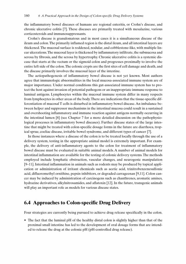
the inflammatory bowel diseases of humans are regional enteritis, or Crohn’s disease, andchronic ulcerative colitis [7]. These diseases are primarily treated with mesalazine, variouscorticosteroids and immunosuppressants.
Crohn’s disease is granulomatous and in most cases it is a simultaneous disease of theileum and colon.The primarily inflamed region is the distal ileum, and all intestinal layers arethickened.The mucosal surface is reddened, nodular, and cobblestone-like, with multiple lin-ear ulcerations.The mucosal layer is thickened by inflammatory infiltrate, the submucosa andserosa by fibrosis, and the serosa by hypertrophy. Chronic ulcerative colitis is a systemic dis-ease that starts at the rectum or the sigmoid colon and progresses proximally to involve theentire left side of the colon.The colonic crypts are the first sites of cell damage and death, andthe disease primarily involves the mucosal layer of the intestine.
The aetiopathogenesis of inflammatory bowel disease is not yet known. Most authorsagree that immunologic abnormalities in the local mucosa-associated immune system are ofmajor importance. Under normal conditions this gut-associated immune system has to pro-tect the host against invasion of potential pathogens or an inappropriate immune response toluminal antigens. Lymphocytes within the mucosal immune system differ in many respectsfrom lymphocytes in other areas of the body.There are indications that the tissue-specific dif-ferentiation of mucosal T cells is disturbed in inflammatory bowel disease. An imbalance be-tween helper and suppressor mechanisms in the intestinal mucosa could result in a sustainedand overshooting inflammatory and immune reaction against antigens normally occurring inthe intestinal lumen [8] (see Chapter 7 for a more detailed discussion on the pathophysio-logical processes in inflammatory bowel diseases). Further disease states of the large intes-tine that might be treated with colon-specific dosage forms in the future are diarrhoea, trop-ical sprue, coeliac disease, irritable bowel syndrome, and different types of cancer [7].
In those instances where a disease of the colon is to be treated locally through the use of adelivery system, testing in the appropriate animal model is extremely important. For exam-ple, the delivery of anti-inflammatory agents to the colon for treatment of inflammatorybowel disease must be evaluated in suitable animal models. A number of animal models forintestinal inflammation are available for the testing of colonic delivery systems.The methodsemployed include lymphatic obstruction, vascular changes, and neurogenic manipulation[9–11]. Intestinal inflammation in animals such as rodents may be produced by topical appli-cation or administration of irritant chemicals such as acetic acid, trinitrobenzenesulfonicacid, difluoromethyl ornithine, pepsin inhibitors, or degraded carrageenan [9,11]. Colon can-cer may be induced by administration of carcinogens such as chanthrenes, aromatic amines,hydrazine derivatives, alkylnitrosamides, and aflatoxin [12]. In the future, transgenic animalswill play an important role as models for various disease states.
6.4 Approaches to Colon-specific Drug Delivery
Four strategies are currently being pursued to achieve drug release specifically in the colon.
• The fact that the luminal pH of the healthy distal colon is slightly higher than that of theproximal small intestine has led to the development of oral dosage forms that are intend-ed to release the drug at the colonic pH (pH-controlled drug release).
160 6 A Practical Approach in the Design of Colon-specific Drug Delivery Systems

• Colonic microflora produce a variety of enzymes that are not present in the stomach or thesmall intestine and can therefore be used to deliver drugs to the colon after enzymaticcleavage of degradable formulation components or drug carrier bonds (enzyme-con-trolled drug release). It should be taken into consideration that because of the negative re-dox potential in the colon, enzymatic or chemical reduction reactions are favoured.
• The relatively constant transit time in the small intestine of approximately 3–4 h is anoth-er physiological characteristic which can be exploited to achieve colon specificity (time-controlled drug release). After gastric emptying, a time-controlled drug delivery system isintended to release the drug after a predetermined lag phase.
• Another strategy relies on the strong peristaltic waves in the colon that lead to a tem-porarily increased luminal pressure (pressure-controlled drug release). Pressure-sensitivedrug formulations release the drug as soon as a certain pressure limit is attained, i.e. de-struction force is exceeded.
Using mostly anti-inflammatory model drugs or drugs that are absorbable in the colon,many colon-specific dosage forms have been developed in the past, including pro-drugs,cross-linked hydrogels, matrices and coated dosage forms. However, whereas the synthesis ofpro-drugs is possible only if the drug has suitable functional groups that can be bound to acarrier molecule, biodegradable hydrogels and matrices are problematic insofar as polymerdegradation rates and thus drug release are often too slow. Most colon-specific drug deliverysystems belong to the group of coated dosage forms because of the flexibility in the design ofthe latter and the improved coating procedures that have been developed in the past.With regard to peptide and protein absorption poor membrane permeability, enzymatic in-stability, and large molecular size are three factors that have remained major hurdles for pep-tide formulations. Absorption-enhancing agents that have been effective, at least in researchenvironments with smaller drug candidates, have also shown some limited efficacy in smallanimal models with certain peptides. In most cases, however, effective formulations haveonly achieved fairly low peptide absorption (< 10%) and have also resulted in significant al-terations in the normal cellular morphology of the gastrointestinal tract, at least on a tran-sient basis [13]. Current data suggest that the successful development of oral peptide formu-lations remains a significant challenge.
6.4.1 pH-Controlled Drug Release
Many of the marketed dosage forms developed for colon-specific drug delivery, such as theenteric coated formulations Asacolitin®, Azulfidine®, Claversal®, Salofalk®, Colo-Pleon®,Entocort® and Budenofalk® rely on the physiological pH difference between the small andthe distal large intestine. In healthy subjects this pH difference amounts to about 0.5 pH units[4,5] (Figure 6.2). However, it has been shown that this difference in pH between the smalland the large intestine is too small to guarantee reliable drug release in the colonic region[14–16]. Moreover, in patients with inflammatory bowel disease the luminal colonic pH dropsto values between 2.5 and 4.7 [17–19], a fact that has been attributed to a failure of bicarbon-ate secretion rather than excessive bacterial fermentation [18].
Enteric coating materials not only protect a dosage form from the acidic environment inthe stomach and allow drug delivery to the small intestine, they may also pass through the
6.4 Approaches to Colon-specific Drug Delivery 161
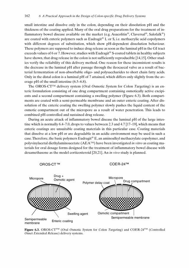
small intestine and dissolve only in the colon, depending on their dissolution pH and thethickness of the coating applied. Many of the oral drug preparations for the treatment of in-flammatory bowel disease available on the market (e.g. Asacolitin®, Claversal®, Salofalk®)are coated with enteric polymers such as Eudragit® L or S, i.e. methacrylic acid copolymerswith different degrees of substitution, which show pH-dependent dissolution behaviour.These polymers are supposed to induce drug release as soon as the luminal pH in the GI tractexceeds values of 6 or 7. However, studies with Eudragit® S-coated tablets in healthy subjectshave shown, that drug release in the colon is not sufficiently reproducible [14,15]. Other stud-ies verify the reliability of this delivery method. One reason for these inconsistent results isthe decrease in the luminal pH after passage through the ileocaecal valve as a result of bac-terial fermentation of non-absorbable oligo- and polysaccharides to short chain fatty acids.Only in the distal colon is a luminal pH of 7 attained, which differs only slightly from the av-erage pH of the small intestine (6.5–6.8).
The OROS-CT™ delivery system (Oral Osmotic System for Colon Targeting) is an en-teric formulation consisting of one drug compartment containing osmotically active excipi-ents and a second compartment containing a swelling polymer (Figure 6.3). Both compart-ments are coated with a semi-permeable membrane and an outer enteric coating. After dis-solution of the enteric coating the swelling polymer slowly pushes the liquid content of theosmotic compartment out of the micropore as a result of water penetration. This leads tocombined pH-controlled and sustained drug release.
During an acute attack of inflammatory bowel disease the luminal pH of the large intes-tine which is normally 6.4–7.0, drops to values between 2.3 and 4.7 [17–19], which means thatenteric coatings are unsuitable coating materials in this particular case. Coating materialsthat dissolve at a low pH or are degradable in an acidic environment may be used in such acase.Therefore, the basic polymers Eudragit® E, an aminoalkyl methacrylate copolymer, andpolyvinylacetal diethylaminoacetate (AEA™) have been investigated in vitro as coating ma-terials for oral dosage forms designed for the treatment of inflammatory bowel disease withdexamethasone as the model corticosteroid [20,21]. An in vivo study is planned.
162 6 A Practical Approach in the Design of Colon-specific Drug Delivery Systems
Micropore
Polymer delay coat
Semipermeable membrane
Drug compartment
Osmotic compartment
Drug +Osmotic agent
Enteric coating
OROS-CT ™ COER-24™
Semipermeablemembrane
Micropore
Swelling agent
Figure 6.3. OROS-CTTM (Oral Osmotic System for Colon Targeting) and COER-24TM (ControlledOnset Extended Release) delivery systems.

pH-sensitive ion exchange systems represent another approach to how to achieve pH-con-trolled drug release in the colon. Drug ions, bound to an ion exchange resin, may be releasedpH-dependently into the large intestine, as in the case of insulin [22] or mesalazine [23]. Inthe latter case the drug is used in the form of its ionized pro-drug (olsalazine) and drug re-lease occurs after microbial cleavage of the drug–carrier bonds. The ion exchange resinserves as polymer to prevent premature absorption of the pro-drug in the small intestine.
6.4.2 Enzyme-controlled Drug Release
Enzyme-controlled drug release takes advantage of the existence of enzyme-producing mi-croorganisms in the colon (Figure 6.1). The colonic microflora produce a variety of enzymes,the activity of which may be exploited for colon-specific drug delivery.Among these enzymesare the azoreductase, various glycosidases, esterases and peptidases. Because of this physio-logical characteristic of the colon, biodegradable pro-drugs, coating materials, hydrogels andmatrices have been developed (see below).
Pro-drugs are conjugates of drugs with carrier molecules mostly of inert nature. The mi-crobial enzymes in the colon are responsible for the cleavage of the drug–carrier bond.A va-riety of pro-drugs have been synthesized, mainly azo compounds, glycosides, esters andamides [24].
Pro-drugs must not be cleaved by digestive enzymes of the upper GI tract and should notbe susceptible to chemical hydrolysis. Moreover, pro-drug absorption in the small intestineshould be negligible. Because of these requirements, the hydrophilicity, the molecular weightand the charge of the carrier molecules have to be regarded as critical parameters.
The azo pro-drug sulfasalazine (Azulfidine®, Colo-Pleon®), consisting of the drugmesalazine and the carrier molecule sulfapyridine, was the first pro-drug available for thetreatment of inflammatory bowel disease. Because of side-effects caused by the pharmaco-logically active sulfapyridine carrier, other carrier molecules such as sulfanilic acid, p-aminobenzoic acid and its amino acid derivatives (benzalazine; ipsalazide; balsalazide, Co-lazide®) and mesalazine itself (olsalazine, Dipentum®) have been used in its place. In the caseof glycoside pro-drugs, which were developed primarily for use with corticosteroids, mono-saccharides have been intensively investigated as inert carrier molecules [25,26]. In addition,a variety of inert hydrophilic carriers of oligomeric as well as polymeric nature, some of thembeing enzymatically degradable themselves, have been used to prevent premature pro-drugabsorption in the small intestine. Examples are β-cyclodextrin [27,28], dextran [29–34] andthe polyanionic poly(L-aspartic acid) [35,36].
The Drug Delivery Index (DDI) allows a quantification of the reduction in the drug doseand the systemic exposure observed after drug release specifically to the colon [37]. It may becalculated using AUC (Area Under the plasma drug concentration–time Curve) data or drugconcentrations in blood and colonic tissues under steady-state conditions:
AUC Tissue (Pro-drug)
AUC Tissue (Drug)
AUC Blood (Pro-drug)
AUC Blood (Drug)
6.4 Approaches to Colon-specific Drug Delivery 163
DDI (Pro-drug vs. Drug) = (6.1a)

Css Tissue (Pro-drug)
Css Tissue (Drug)
Css Blood (Pro-drug)
Css Blood (Drug)
where Css is the steady-state drug concentration
The numerator of Eq. 6.1a describes to what extent drug concentrations are increased inthe target tissue (colonic mucosa or muscle tissue) after pro-drug administration as opposedto drug administration. It may be regarded as the factor by which the pro-drug dose could bereduced in comparison to the drug dose. The denominator of Eq. 6.1a, which corresponds tothe relative bioavailability of the drug released from the pro-drug, provides a measure of thereduction in the systemic exposure and thus the decrease of the systemic toxicity. In Eq. 6.1bthe DDI is defined as the quotient of the tissue to blood concentration ratios after pro-drugand drug administration. With glucoside, glucuronide and dextran pro-drugs DDI values upto 9.7 have been reported in rats [25,38,39].
A disadvantage of the use of pro-drugs is the need for suitable functional groups such asamino-, hydroxy- or carboxy groups in both drug and carrier molecules (see Chapter 11 formore details on chemical synthesis routes applied in drug targeting/delivery strategies).Sometimes spacer molecules are necessary to link the drug to the carrier molecule, which of-ten leads to complicated drug release kinetics. Another disadvantage of the pro-drug ap-proach is the fact that for the approval of any newly synthesized pro-drug a toxicity study isrequired by regulatory agencies.
The group of enzymatically degradable coating materials comprises film-forming azo poly-mers and polymers with glycosidic bonds as well as conventional sustained release coatingmaterials based on acrylates or ethylcellulose with biodegradable pore formers. The filmsmust not be soluble or degradable in the upper GI tract. They should only be degradable inthe colon and their degradation products need to be toxicologically harmless.
Cross-linked azo polymers were the first coating materials that were investigated with re-gard to their enzymatic degradability in the colon using insulin as the model drug [40,41]. Aproblem observed with these polymers was the rather slow degradation rate, probably a re-sult of the lipophilicity of these compounds. Sufficiently hydrophilic linear azo polymers andpH-sensitive terpolymers based on acrylates were found to be more suitable coating materi-als [42–45].
In general, several problems have to be considered if azo polymers are used as an enzymatically degradable component of a colon-specific dosage form [46]. As a result of the enzymatic reduction to primary aromatic amines in the colon, the toxicity of these polymers is a critical issue. Moreover, the rate of microbial reduction of the polymeric azocompounds is often too slow. The reduction reaction may stop at the level of the hydrazocompounds instead of leading to the amines, which may influence the drug release mechanism and rate. In addition, the reduction reaction does not necessarily depend on thepresence of the azoreductase, but may be induced by the negative redox potential in thecolon. The latter also applies to the degradation of pro-drugs or polymers with disulfidebonds, which is the result of a chemical reduction step with no enzymatic reaction involved[47].
164 6 A Practical Approach in the Design of Colon-specific Drug Delivery Systems
DDI (Pro-drug vs. Drug) = (6.1b)

Biodegradable polymer coatings with glycosidic bonds, that have been developed in thepast, are mainly based on galactomannans [48–50], chitosan [51] or the high molecular weightdextran fatty acid ester lauroyl dextran [52]. A special enzyme-controlled delivery systemwith pH-induced drug release has been developed by Watanabe et al. [53]. Drug release fromthis enteric tablet formulation begins after microbial degradation of an outer disaccharidelayer (lactulose) to short-chain fatty acids in the colon and subsequent dissolution of the un-derlying basic Eudragit® E film.The colon-specific dosage form is available on the market asCodes™ and can incorporate any drug suitable for colonic drug delivery.
Degradable matrix films, consisting of a sustained release coating material and a poorlywater-soluble but degradable pore former, are used if the pore former itself does not form anhomogeneous coating film. These pore formers guarantee drug release in the colon after mi-crobial degradation and the formation of pores in the film. As pore formers several oligo-and polysaccharides were investigated such as β-cyclodextrin [54], galactomannans [55],glassy amylose [56,57], pectin [58–60] and inulin [61].A coated dosage form for colon-specif-ic drug delivery with a matrix film consisting of ethylcellulose and the pore former glassyamylose is available on the market as Colal™.
If a biodegradable polymer does not exhibit satisfactory film-forming properties, it mayalso be used as a compression coat requiring a compaction process onto a drug-containingcore [62–64].
Further examples of enzymatically degradable ‘drug formulation wrappings’ are capsuleshells made of the polysaccharides chitosan [65,66] or cross-linked dextran [67].
Biodegradable polymers that cannot be used as coating materials for colon-specific drugdelivery because they are readily water soluble and/or do not form films, may be used in theform of hydrogels and matrices. Hydrogels, consisting of cross-linked polymers, have been de-veloped based on acrylates, polyether-esters and polysaccharides. In the case of acrylate- andpolyether-ester-copolymers, azo-aromatic compounds have been used for cross-linking pur-poses and to guarantee degradability in the colon [68–70]. The higher the cross-linking den-sity of these polymers, the lower their tendency to swell and the slower the degradation rateby azoreductase, ultimately resulting in slower drug release. Long chain azo-aromatic cross-linking compounds lead to faster polymer degradation and thus higher drug release rates. Ingeneral, the azo-aromatic cross-linking compounds that are used should have a rather smallnegative redox potential in order to ensure rapid degradation [71].
In the design of coatings, hydrogels and matrices based on azo polymers, the number of azobonds in the polymers should not be too high, as this could lead to enzyme-saturated condi-tions slowing down the degradation process and thus drug release [72].
Polymer cross-linking leads to a decrease in the water solubility of many readily solublepolysaccharides, low water solubility being a requirement for colon-specific drug delivery.Dextrans [73,74], the mucopolysaccharide chondroitin sulfate [75,76], guar gum [77,78],pectin [79–82] and inulin [83–85] have all been investigated in cross-linked forms.Again, witha higher degree of cross-linking, the swelling properties of these polymers tend to be lowerand this leads to a slower degradation rate and thus slower release of the drug. Poorly water-soluble drugs are usually released by an erosion-type mechanism.
6.4 Approaches to Colon-specific Drug Delivery 165

6.4.3 Time-controlled Drug Release
Time-controlled drug release mechanisms rely on the fairly constant small intestinal transittime during which no measurable drug release occurs. Only after arrival of the dosage formin the colon may drug delivery begin. Such a delayed release mechanism can be achieved us-ing compounds with swelling properties or osmotically active excipients, which leads to an in-crease in volume by water uptake resulting in a build-up of pressure inside the dosage form.After a predetermined lag phase the drug is released in a more or less pulsatile fashion, oftenaccompanied by rupture of the outer coating layer. Such a lag phase may also be achievedwith slowly eroding or dissolving coating layers [21]. In general, drug release from time-con-trolled delivery systems may be pH-induced, induced by swelling, pressure-induced or ero-sion-induced.
Time-controlled drug release with pH-induced drug delivery is a delivery method that doesnot depend on changes in the luminal pH of the GI tract but on a pH change within thedosage form itself.
Colon-specific drug formulations relying on the time-dependent dissolution of basic poly-mer layers such as Eudragit® E and chitosan under acidic conditions have been developed byIshibashi et al. (CTDC: Colon-Targeted Delivery Capsule) [86–88] and Yamada et al. [89]. Asolid organic acid incorporated in the dosage forms which dissolves as soon as it comes intocontact with the penetrating intestinal fluid generates an acidic environment and induces thedissolution of the basic polymer layers and thus drug release. The onset of drug release de-pends on the thickness of the basic polymer layer or shell.
The organic acid-induced sigmoidal release system developed by Narisawa et al. consists of a drug core containing a solid organic acid which is coated with an ammonio metha-crylate copolymer sustained-release coating (Eudragit® RS) [90,91]. During a lag phase,the drug permeability of the Eudragit® RS film increases drastically as a result of diffusion ofthe organic acid into the film thereby facilitating polymer hydration and inducing drug release.
Drug delivery induced by swelling may be achieved with swellable polymer layers based oncellulose ethers or acrylates, where with the latter pH-dependent swelling behaviour is feasi-ble [92]. Examples are the oral Chronotopic® delivery system [93], the Time-controlled Ex-plosion System (TES) [94,95] and the TIME-CLOCK™-System [96–98] (Figure 6.4).
Swelling polymers may also be used as plugs, i.e. stoppers, to seal water-insoluble capsulebodies, as in the case of Pulsincap® [99,100]. The delayed drug release observed after plugejection at the end of the lag phase, depends on the swelling properties of the polymer plugas well as on the geometry and chemical structure of the plug.
Swelling sustained-release coating polymers such as Eudragit® NE 30 D, i.e. poly(ethyl-acrylate-methylmethacrylate) [101,102] or Eudragit® RS [90], lead to a delay in drug releasewhich is dependent on the thickness of the coating since these films have slow rates ofswelling.
One dosage form available on the market which relies on pressure-induced delivery is theCOER-24TM delivery system (Controlled Onset Extended Release).This formulation, devel-oped for drugs that can be absorbed in the colon, is similar to the OROS-CTTM system (Fig-ure 6.3). Here, a polymeric delay coat is responsible for the delayed drug release. During thelag phase the osmotic compartment swells resulting in pressure being applied to the drug
166 6 A Practical Approach in the Design of Colon-specific Drug Delivery Systems

compartment. Micropores in the outer semi-permeable film allow a sustained drug releaseafter dissolution and extrusion of the underlying delay coat.
For an erosion-induced drug delivery system compactable cellulose ethers are suitablepolymers [103]. Drug release, which is controlled by the erosion/dissolution of these poly-meric layers, may be pH-dependent if an acid or basic polymer is used.
In summary, the lag time of drug release may be controlled by the rate at which water pen-etrates through the coating or shell, the rate of fluid absorption of the polymer layer, the os-motic activity of salts and osmopolymers, the erosion and dissolution rate of the polymer lay-ers and the thickness of the layers or coatings.
6.4.4 Pressure-controlled Drug Release
Pressure-controlled delivery systems take advantage of the temporary increase in intra-lu-minal pressure in the colon in the event of a peristaltic wave.A drug formulation relying on theresulting destructive force has been developed and consists of a conventional hard gelatincapsule with a thick inner layer of ethylcellulose [104,105]. The rupture of the ethylcelluloseshell with subsequent drug release is induced by an increase in the intra-luminal pressure andsubsequent destructive force. Drug absorption from this formulation has been investigated invivo in dogs and humans with mesalazine, carbamazepine [105,106] and caffeine [104] as themodel drugs. The capsule contains the drug in solution because otherwise there may be in-sufficient fluid available in the distal portion of the colon for the drug to dissolve in.
6.4 Approaches to Colon-specific Drug Delivery 167
Swelling agent layer(low substituted Hydoxypropylcellulose)
Drug layer
Sucrose bead
Water insoluble polymermembrane (Ethylcellulose)
Hydrophobic surfactant layer(wax, Polysorbate 80, Hydroxypropylmethylcellulose)
Tablet coreEnteric coating
Drug core
Eudragit ® S(optional) Eudragit ® L
Hydroxypropylmethylcellulose(high viscosity)
™
™
Figure 6.4. Swelling-induced time-controlled drug delivery systems.

6.5 Concluding Remarks
All the methods for colon-specific drug delivery presented in this overview are more or lesssusceptible to changes in the physiological environment (diet, disease state, medication, etc.).This may affect reliability and reproducibility of the delivery systems. However, most of thesystems described have already been investigated in vivo with regard to colon specificity.Methods such as planar γ-scintigraphy [86,97,107] and three-dimensional magnetic markermonitoring [108,109] appear to be the methods of choice for dynamic visualization of thedosage form in the GI tract and the determination of the location and time of onset of drugrelease. In animal models, the measurement of blood and intestinal tissue concentrations ofthe perorally administered drug in order to quantify the reduction in the systemic exposure,is an additional approach [110]. However, most formulations are tested in healthy humans oranimals in spite of the fact, that pathological conditions such as inflammatory bowel diseasemay have a significant influence on gut physiology.As yet, little is known regarding the effectof disease states on the intestinal transit time, GI tract motility, luminal pH, intra-luminalcolonic pressure and composition of colonic microflora. It appears that time-controlled drugrelease systems, which rely on a constant transit time through the small intestine, are the mostpromising colon-specific delivery systems, as this transit time is only marginally influenced bypathological events in the GI tract. pH-controlled systems are reliable only if there is a sig-nificant difference in the luminal pH between the small and the large intestine. Under phys-iological conditions this pH difference is often too small. In certain pathological conditionssuch as inflammatory bowel disease however, a significant decrease in pH in the affected GItract regions may be observed, and this has led to the development of novel pH-controlleddelivery systems. Enzyme-controlled delivery systems are dependent on the activity of themicrobial enzymes in the large intestine, which are susceptible to many environmental fac-tors, particularly diet and medication. The reliability of pressure-controlled delivery systemsis expected to be highly dependent on variations in intestinal peristalsis despite promising invivo data.
References
[1] Simon GL, Gorbach SL, Gastroenterology 1984, 86, 174–193.[2] Drasar BS, Hill MJ, In: Human Intestinal Flora, pp. 103-123. Academic Press, London, 1974.[3] Bragger JL, Yazaki E, Evans DF, Pharm. Res. 1997, 14 (11 Suppl.), S656–S657.[4] Mitsuoka T, Intestinal Bacteria and Health. Harcourt Brace Jovanovich, Tokyo, 1978.[5] Evans DF, Pye G, Bramley R, Clark AG, Dyson TJ, Hardcastle JD, Gut 1988, 29, 1035–1041.[6] Shameem M, Katori N, Aoyagi N, Kojima S, Pharm. Res. 1995, 12, 1049–1054.[7] Haeberlin B, Friend DR, In: Oral Colon-Specific Drug Delivery (Ed. Friend DR), pp. 1–43. CRC
Press, Boca Raton, 1992.[8] Zeitz M, Digestion 1997, 58 (Suppl. 1), 59–61.[9] Bhatti MA, Hodgson HJF, Int. J. Exp. Pathol. 1995, 76, 309–315.
[10] Morteau O, Bueno L, Gastroenterol. Clin. Biol. 1995, 19, 204–214.[11] Warren BF, Watkins PE, J. Pathol. 1994, 172, 313–316.[12] Pories SE, Ramchurren N, Summerhayes I, Steele G, Arch. Surg. 1993, 128, 647–653.[13] Fix JA, J. Pharm. Sci. 1996, 85, 1282–1285.[14] Ashford M, Fell JT, Attwood D, Woodhead PJ, Int. J. Pharm. 1993, 91, 241–145.[15] Ashford M, Fell JT, Attwood D, Sharma H, Woodhead PJ, Int. J. Pharm. 1993, 95, 193–199.[16] Dew MJ, Ryder REJ, Evans N, Evans BK, Rhodes J, Br. J. Clin. Pharmacol. 1983, 16, 185–187.
168 6 A Practical Approach in the Design of Colon-specific Drug Delivery Systems

[17] Fallingborg J, Christensen LA, Jacobsen BA, Rasmussen SN, Dig. Dis. Sci. 1993, 38, 1989–1993.[18] Roediger W, Lawson MJ, Kwok V, Grant AK, Pannall PR, J. Clin. Pathol. 1984, 37, 704–707.[19] Sasaki Y, Hada R, Nakajima H, Fukuda S, Munakata A, Am. J. Gastroenterol. 1997, 92, 114–118.[20] Leopold CS, Eikeler D, J. Drug Target. 1998, 6, 85–94.[21] Leopold CS, Eikeler D, Drug Dev. Ind. Pharm. 2000, 26, 1239–1246.[22] Wunderlich J-C, Schick U, Werry J, Freidenreich J, International Patent WO 93/10767, 1993.[23] Pitzele BS, Jones PH, US Patent 4,374,932, 1983.[24] Leopold CS, Pharmazie in unserer Zeit 1997, 26, 289–298.[25] Friend DR, Tozer TN, J. Control. Rel. 1992, 19, 109–120.[26] Friend DR, STP Pharma Sci. 1995, 5, 70–76.[27] Hirayama F, Minami K, Uekama K, J. Pharm. Pharmacol. 1996, 48, 27–31.[28] Hirayama F, Minami K, Uekama K, Proc. 2nd World Meeting APGI/APV 1998, 2, 935–936.[29] Harboe E, Larsen C, Johansen M, Olesen HP, Pharm. Res. 1989, 6, 919–923.[30] Larsen C, Harboe E, Johansen M, Olesen HP, Pharm. Res. 1989, 6, 995–999.[31] Larsen C, Jensen BH, Olesen HP, Acta Pharm. Nord. 1991, 3, 71–76.[32] McLeod AD, In: Oral Colon-specific Drug Delivery (Ed. Friend DR), pp. 213–231. CRC Press,
Boca Raton, 1992.[33] McLeod AD, Tolentino L, Tozer TN, Biopharm. Drug Dispos. 1994, 15, 151–161.[34] McLeod AD, Fedorak RN, Friend DR, Tozer TN, Cui NR, Gastroenterology 1994, 106, 405–413.[35] Leopold CS, Friend DR, Int. J. Pharm. 1995, 126, 139–145.[36] Leopold CS, Friend DR, J. Pharmacokinet. Biopharm. 1995, 23, 397–406.[37] McLeod AD, Tozer TN, In: Oral Colon-specific Drug Delivery (Ed. Friend DR), pp. 85–114. CRC
Press, Boca Raton, 1992.[38] Fedorak RN, Haeberlin B, Empey LR, Cui N, Nolen H, III, Jewell LD, Friend DR, Gastroen-
terology 1995, 108, 1688–1699.[39] Tozer TN, Rigod J, McLeod AD, Gungon R, Hoag MK, Friend DR, Pharm. Res. 1991, 8, 445–454.[40] Saffran M, Kumar GS, Savariar C, Burnham JC, Williams F, Neckers DC, Science 1986, 233,
1081–1084.[41] Saffran M, Kumar GS, Neckers DC, Peña J, Jones RH, Field JB, Biochem. Soc. Trans. 1990, 18,
752–754.[42] Van den Mooter G, Samyn C, Kinget R, Int. J. Pharm. 1992, 87, 37–46.[43] Van den Mooter G, Samyn C, Kinget R, Pharm. Res. 1994, 11, 1737–1741.[44] Van den Mooter G, Samyn C, Kinget R, Pharm. Res. 1995, 12, 244–247.[45] Van den Mooter G, Maris B, Samyn C, Augustijns P, Kinget R, J. Pharm. Sci. 1997, 86, 1321–1327.[46] Lloyd AW, Martin GP, Soozandehfar SH, Int. J. Pharm. 1994, 106, 255–260.[47] Schacht E, Wilding I, International Patent WO 91/11175, 1991.[48] Bauer KH, Deutsche Patentschrift 42 09 160, 1993.[49] Betzing J, Bauer KH, Pharm. Ztg. Wiss. 1992, 137, 131–134.[50] Sarlikiotis AW, Bauer KH, Pharm. Ind. 1992, 54, 873–880.[51] Sekigawa F, Onda Y, US Patent 5,217,720, 1993.[52] Kesselhut JF, Bauer KH, Pharmazie 1995, 50, 263–269.[53] Watanabe S, Kawai H, Katsuma M, Fukui M, International Patent WO 95/28963, 1995.[54] Siefke V, Weckenmann H, Bauer K, Proc. Int. Symp. Control. Rel. Bioact. Mater. 1993, 20,
182–183.[55] Lehmann K, Dreher KD, Proc. Int. Symp. Control. Rel. Bioact. Mater. 1991, 18, 331–332.[56] Milojevic S, Newton JM, Cummings JH, Gibson GR, Bothman RL, Ring SG, Allwood MC, Stock-
ham M, STP Pharma Sci. 1995, 5, 47–53.[57] Siew LF, Newton JM, Pharm. Res. 1997, 14 (11 Suppl.), S-658.[58] Macleod GS, Fell JT, Collett JH, Int. J. Pharm. 1997, 157, 53–60.[59] Wakerly Z, Fell JT, Attwood D, Parkins D, Pharm. Res. 1996, 13, 1210–1212.[60] Wakerly Z, Fell JT, Attwood D, Parkins D, Int. J. Pharm. 1997, 153, 219–224.[61] Vervoort L, Kinget R, Int. J. Pharm. 1996, 129, 185–190.[62] Krishnaiah YSR, Satyanarayana S, Prasad YVR, Rao SN, Int. J. Pharm. 1998, 171, 137–146.[63] Krishnaiah YSR, Satyanarayana S, Prasad YVR, Drug Dev. Industr. Pharm. 1999, 25, 651–657.[64] Rubinstein A, Radai R, Eur. J. Pharm. Biopharm. 1995, 41, 291–295.[65] Chen RH, Tsaih ML, Lin WC, Carbohydr. Polymers 1996, 31, 141–148.[66] Tozaki H, Komoike J, Tada C, Maruyama T, Terabe A, Suzuki T, Yamamoto A, Muranishi S, J.
Pharm. Sci. 1997, 86, 1016–1021.[67] Brøndsted H, Andersen C, Hovgaard L, J. Control. Rel. 1998, 53, 7–13.
References 169
[68] Brøndsted H, Kopecek J, Pharm. Res. 1992, 9, 1540–1545.[69] Kakoulides EP, Smart JD, Tsibouklis J, J. Control. Rel. 1998, 54, 95–109.[70] Samyn C, Kalala W, Van den Mooter G, Kinget R, Int. J. Pharm. 1995, 121, 211–216.[71] Bragger JL, Lloyd AW, Soozandehfar SH, Bloomfield SC, Marriott C, Martin GP, Int. J. Pharm.
1997, 157, 61–71.[72] Van den Mooter G, Offringa M, Kalala W, Samyn C, Kinget R, STP Pharma Sci. 1995, 5, 36–40.[73] Chiu HC, Hsiue GH, Lee YP, Huang LW, J. Biomater. Sci. 1999, 10, 591–608.[74] Hovgaard L, Brøndsted H, J. Control. Rel. 1995, 36, 159–166.[75] Rubinstein A, Nakar D, Sintov A, Int. J. Pharm. 1992, 84, 141–150.[76] Rubinstein A, Nakar D, Sintov A, Pharm. Res. 1992, 9, 276–278.[77] Rubinstein A, Gliko-Kabir I, STP Pharma Sci. 1995, 5, 41–46.[78] Rubinstein A, Gliko-Kabir I, Penhasi A, Yagen B, Proc. Int. Symp. Control. Rel. Bioact. Mater.
1997, 24, 839–840.[79] Ashford M, Fell J, Attwood D, Sharma H, Woodhead P, J. Control. Rel. 1993, 26, 213–220.[80] Ashford M, Fell J, Attwood D, Sharma H, Woodhead P, J. Control. Rel. 1994, 30, 225–232.[81] Rubinstein A, Radai R, Ezra M, Pathak S, Rokem JS, Pharm. Res. 1993, 10, 258–263.[82] Wakerly Z, Fell J, Attwood D, Parkins D, J. Pharm. Pharmacol. 1997, 49, 622–625.[83] Vervoort L, Van den Mooter G, Augustijns P, Bousson R, Toppet S, Kinget R, Pharm. Res. 1997,
14, 1730–1737.[84] Vervoort L, Van den Mooter G, Augustijns P, Kinget R, Int. J. Pharm. 1998, 172, 127–135.[85] Vervoort L, Rombaut P, Van den Mooter G, Augustijns P, Kinget R, Int. J. Pharm. 1998, 172,
137–145.[86] Ishibashi T, Pitcairn GR, Yoshino H, Mizobe M, Wilding IR, J. Pharm. Sci. 1998, 87, 531–535.[87] Ishibashi T, Hatano H, Kobayashi M, Mizobe M, Yoshino H, Int. J. Pharm. 1998, 168, 31–40.[88] Ishibashi T, Hatano H, Kobayashi M, Mizobe M, Yoshino H, J. Control. Rel. 1999, 57, 45–53.[89] Yamada A, Wato T, Uchida N, Fujisawa M, Takama S, Inamoto Y, US Patent 5,395,628, 1995.[90] Narisawa S, Nagata M, Danyoshi C, Yoshino H, Murata K, Hirakawa Y, Noda K, Pharm. Res.
1994, 11, 111–116.[91] Narisawa S, Nagata M, Hirakawa Y, Kobayashi M, Yoshino H, J. Pharm. Sci. 1996, 85, 184–188.[92] Abramowitz R, Ranadive S, Jain NB, Varia SA, Pharm. Res. 1997, 14 (11 Suppl.), S656.[93] Gazzaniga A, Sangalli ME, Giordano F, Eur. J. Pharm. Biopharm. 1994, 40, 246–250.[94] Ueda S, Ibuki R, Kawamura A, Murata S, Takahashi T, Kimura S, Hata T, J. Drug Target. 1994,
2, 133–140.[95] Ueda S, Hata T, Asakura S, Yamaguchi H, Kotani M, Ueda Y, J. Drug Target. 1994, 2, 35–44.[96] Pozzi F, Furlani P, Gazzaniga A, Davis SS, Wilding IR, J. Control. Rel. 1994, 31, 99–108.[97] Steed KP, Hooper G, Monti N, Strolin Benedetti M, Fornasini G, Wilding IR, J. Control. Rel.
1997, 49, 115–122.[98] Wilding IR, Davis SS, Pozzi F, Furlani P, Gazzaniga A, Int. J. Pharm. 1994, 111, 99–102.[99] Binns J, Stevens HNE, McEwen J, Pritchard G, Brewer FM, Clarke A, Johnson ES, McMillan I,
J. Control. Rel. 1996, 38, 151–158.[100] Hegarty M, Atkins G, International Patent WO 95/10263, 1995.[101] Kraeling MEK, Ritschel WA, Methods Find. Exp. Clin. Pharmacol. 1992, 14, 199–209.[102] Rao SS, Ritschel WA, Int. J. Pharm. 1992, 86, 35–41.[103] Shah NH, Phuapradit W, Railkar A, US Patent 5,482,718, 1996.[104] Muraoka M, Hu ZP, Shimokawa T, Sekino S, Kurogoshi R, Kuboi Y, Yoshikawa Y, Takada K, J.
Control. Rel. 1998, 52, 119–129.[105] Takaya T, Sawada K, Suzuki H, Funaoka A, Matsuda KI, Takada K, J. Drug Target. 1997, 4,
271–276.[106] Takaya T, Niwa K, Muraoka M, Ogita I, Nagai N, Yano R, Kimura G, Yoshikawa Y, Yoshikawa
H, Takada K, J. Contr. Rel. 1998, 50, 111–122.[107] Adkin DA, Kenyon CJ, Lerner EI, Landau I, Strauss E, Caron D, Penhasi A, Rubinstein A, Wild-
ing IR, Pharm. Res. 1997, 14, 103–107.[108] Weitschies W, Kotitz R, Cordini D, Trahms L, J. Pharm. Sci. 1997, 86, 1218–1222.[109] Weitschies W, Cardini D, Karaus M, Trahms L, Semmler W, Pharmazie 1999, 54, 426–430.[110] Tozer TN, Friend DR, McLeod AD, STP Pharma Sci. 1995, 5, 5–12.
170 6 A Practical Approach in the Design of Colon-specific Drug Delivery Systems

7 Vascular Endothelium in Inflamed Tissueas a Target for Site Selective Delivery of Drugs
Maaike Everts, Astrid J. Schraa, Lou F. M. H. de Leij, Dirk K. F. Meijer, Grietje Molema
7.1 Introduction
Chronic inflammatory disorders are characterized by an abundant leucocyte infiltration inthe affected tissue due to the continuous recruitment of these inflammatory cells. Althoughthese events are part of the body’s defence mechanisms, excessive responses occur in the vi-cious circle of cell activation and cell destruction, leading to acute and chronic inflammatorydisorders. Many current therapeutic approaches are based on attempts to control leucocyteactivation. Recently there has been a growing interest in the role of the endothelial cells inleucocyte recruitment and the possibility of manipulating endothelial cell activation as atherapeutic approach [1,2]. The endothelium plays an important role in the inflammatorycascade, and has the advantage of being easily accessible for drug targeting preparations dueto direct contact with the blood as well as the presence of endocytotic processes [3]. There-fore endothelial cells are attractive targets for cell-selective pharmacological interventionemploying drug targeting strategies.
The aim of this chapter is to describe potential target epitopes on endothelial cells inchronic inflammation, the design of targeting devices on the basis of this knowledge and theuse of these targeting devices in the intervention of endothelial cell activation. Several in vitro and in vivo test models for studying endothelial cell responses in inflammation are cur-rently available and will be briefly described. Since drug targeting to the inflamed endotheli-um is a relatively new approach, the number of research reports dedicated to this topic is, atpresent, relatively small. Therefore, this chapter will include some general considerations re-garding drug targeting to the activated endothelial cell. Adequate cell-selective delivery ofpotent anti-inflammatory agents may provide an important tool for increasing the efficacyand reducing the side-effects of such agents in the treatment of chronic inflammatory diseases.
7.2 Regulation of Immune Responses in (Chronic)Inflammation
7.2.1 Induction of an Immune Response
After an antigen (a ‘non-self’ molecular entity) has entered the body, it is recognized by thecells of the immune system.An important step in eliciting an efficient immune response to an
Drug Targeting Organ-Specific Strategies. Edited by G. Molema, D. K. F. MeijerCopyright © 2001 Wiley-VCH Verlag GmbH
ISBNs: 3-527-29989-0 (Hardcover); 3-527-60006-X (Electronic)
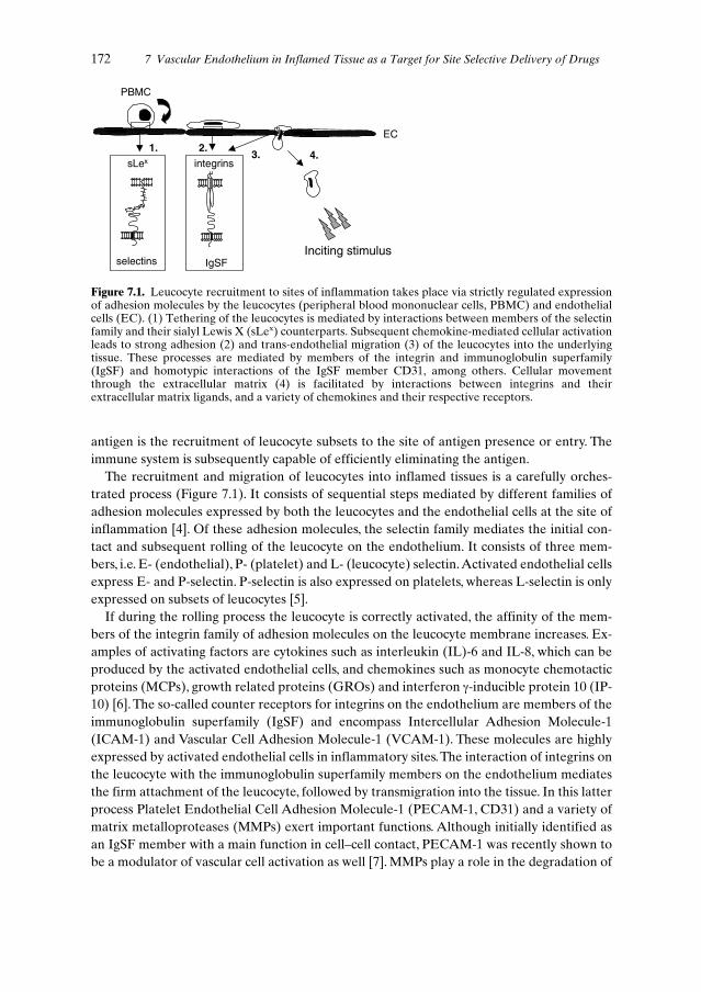
antigen is the recruitment of leucocyte subsets to the site of antigen presence or entry. Theimmune system is subsequently capable of efficiently eliminating the antigen.
The recruitment and migration of leucocytes into inflamed tissues is a carefully orches-trated process (Figure 7.1). It consists of sequential steps mediated by different families ofadhesion molecules expressed by both the leucocytes and the endothelial cells at the site ofinflammation [4]. Of these adhesion molecules, the selectin family mediates the initial con-tact and subsequent rolling of the leucocyte on the endothelium. It consists of three mem-bers, i.e. E- (endothelial), P- (platelet) and L- (leucocyte) selectin.Activated endothelial cellsexpress E- and P-selectin. P-selectin is also expressed on platelets, whereas L-selectin is onlyexpressed on subsets of leucocytes [5].
If during the rolling process the leucocyte is correctly activated, the affinity of the mem-bers of the integrin family of adhesion molecules on the leucocyte membrane increases. Ex-amples of activating factors are cytokines such as interleukin (IL)-6 and IL-8, which can beproduced by the activated endothelial cells, and chemokines such as monocyte chemotacticproteins (MCPs), growth related proteins (GROs) and interferon γ-inducible protein 10 (IP-10) [6]. The so-called counter receptors for integrins on the endothelium are members of theimmunoglobulin superfamily (IgSF) and encompass Intercellular Adhesion Molecule-1(ICAM-1) and Vascular Cell Adhesion Molecule-1 (VCAM-1). These molecules are highlyexpressed by activated endothelial cells in inflammatory sites.The interaction of integrins onthe leucocyte with the immunoglobulin superfamily members on the endothelium mediatesthe firm attachment of the leucocyte, followed by transmigration into the tissue. In this latterprocess Platelet Endothelial Cell Adhesion Molecule-1 (PECAM-1, CD31) and a variety ofmatrix metalloproteases (MMPs) exert important functions. Although initially identified asan IgSF member with a main function in cell–cell contact, PECAM-1 was recently shown tobe a modulator of vascular cell activation as well [7]. MMPs play a role in the degradation of
172 7 Vascular Endothelium in Inflamed Tissue as a Target for Site Selective Delivery of Drugs
1. 2.3.
sLex
selectins
integrins
IgSF
EC
PBMC
4.
Inciting stimulus
Figure 7.1. Leucocyte recruitment to sites of inflammation takes place via strictly regulated expressionof adhesion molecules by the leucocytes (peripheral blood mononuclear cells, PBMC) and endothelialcells (EC). (1) Tethering of the leucocytes is mediated by interactions between members of the selectinfamily and their sialyl Lewis X (sLex) counterparts. Subsequent chemokine-mediated cellular activationleads to strong adhesion (2) and trans-endothelial migration (3) of the leucocytes into the underlyingtissue. These processes are mediated by members of the integrin and immunoglobulin superfamily(IgSF) and homotypic interactions of the IgSF member CD31, among others. Cellular movementthrough the extracellular matrix (4) is facilitated by interactions between integrins and theirextracellular matrix ligands, and a variety of chemokines and their respective receptors.
the basal membrane and the migration of the leucocyte through the tissue in the direction ofthe antigen [8–11].
7.2.2 The Resolution of Inflammation
In normal, non-pathological inflammation, the resolution of an inflammatory reaction isstrictly controlled. Elimination of the antigen by the immune system leads to the shutdownof the inflammatory reaction. Without the inciting stimulus being present, infiltrated leuco-cytes die by apoptosis as was demonstrated in a delayed-type hypersensitivity (DTH) re-sponse in healthy volunteers.This increase in apoptotic T cells which preceded the resolutionof the reaction was probably due to an increased CD95 ligand (FasL) expression on the cellsof the perivascular infiltrate and the deprivation of stimulatory cytokines such as IL-2 an IL-15.This process is furthermore associated with a decreased ratio of anti-apoptotic (e.g. Bcl-2)and pro-apoptotic (e.g. Bax) protein expression by the infiltrated leucocytes [12]. Collective-ly this indicates that the expression of pro- and anti-apoptotic proteins in leucocytes plays animportant role in controlling inflammation. Similarly, these processes exert central regulato-ry functions in endothelial cell activation (see Section 7.3.3). In contrast to the above-de-scribed role in the resolution of inflammation, programmed cell death can also initiate in-flammation. In an ischaemia–reperfusion model in mice, inhibition of apoptosis effectivelyprevented subsequent inflammation and tissue injury. Inhibition of early apoptosis of tissueparenchymal cells is most likely the mechanism behind this effect [13].The events described above demonstrate the complexity and fine balance between variousprocesses resulting in either resolution or enhancement of inflammation. Failure to controlone or more of these, and possibly other presently unknown, regulatory mechanisms maylead to chronic activation of the immune system resulting in chronic inflammatory diseases.
7.3 Chronic Inflammatory Disorders
7.3.1 Pathophysiology of Chronic Inflammatory Disorders
In chronic inflammatory disorders there is a general over-activation of the immune system,the cause of which is often unknown. In all of these diseases, an increased number of infil-trated immune cells can be found in the inflamed tissues. Furthermore, increased expressionof adhesion molecules on the endothelium can often be observed, although the adhesionmolecules are expressed differentially in the various diseases.
7.3.1.1 Rheumatoid Arthritis
Rheumatoid arthritis (RA) is an inflammatory disease of the synovium which results in ero-sion, deformity and finally the destruction of joints. Inflammation of the joints is associatedwith a villous hypertrophy of the synovial membrane, which on microscopy shows prolifera-tion of the lining layer with an inflammatory infiltrate.There is extensive expression of HLA-
7.3 Chronic Inflammatory Disorders 173

DR on T cells, B cells and synovial lining cells, indicating strong immunological activity. RAis thought to be an autoimmune reaction, caused by an interaction between constitutionaland environmental factors [14].
In human biopsies of patients with RA, an increased expression of the adhesion moleculesE-selectin, VCAM-1, ICAM-1 and the CD11c integrin has been demonstrated [15,16]. Also,soluble mediators produced by perivascular cells (e.g. the chemokines Macrophage Inflam-matory Protein (MIP)-1α and MIP-1β) or by endothelial cells themselves (e.g. IL-8 and IL-15) are important in the regulation of leucocyte infiltration in RA [17].Tumour Necrosis Fac-tor α (TNFα) is thought to be a particularly important inflammatory mediator contributingto the pathology of arthritis, as demonstrated by the beneficial effects of the TNFα-neutral-izing therapies that are currently being explored [18]. However, present therapies for RAmainly aim at the inhibition of cyclooxygenase enzymes which are responsible for the over-production of inflammatory mediators like prostaglandin E2 in arthritis-affected joints [19].
7.3.1.2 Atherosclerosis
Atherosclerosis is a generalized degenerative disease that affects large and medium-sized ar-teries. The atherosclerotic plaque contains increased numbers of smooth muscle cells (whichare morphologically abnormal), increased connective tissue and lipid, mostly cholesterol.Monocyte-derived macrophages and lymphocytes are also found in the plaques. Endothelialdamage is believed to be the essential trigger for the development of atherosclerosis. Onceinjury of the endothelium has occurred, platelets and smooth muscle cells will adhere and ag-gregate. Subsequently smooth muscle cells will proliferate, collagen and elastin productionwill increase, and lipid is allowed to accumulate in the vessel wall through enhanced perme-ability at the site of injury [14].
Two important molecular participants in the atherosclerotic process are the transcriptionfactor nuclear factor κB (NFκB) and the adhesion molecule CD40. Using immunohisto-chemical techniques the activated form of NFκB has been shown to be present in human ath-erosclerotic lesions in smooth muscle cells, macrophages and endothelial cells. In contrast, invessels lacking atherosclerotic processes little or no activated NFκB was present [20]. Re-cently, there has been an increasing interest in the role of CD40 and CD40L (CD154), mem-bers of the TNF receptor and TNF family respectively, in chronic inflammation. Ligation ofCD40 on vascular wall cells promotes upregulation of endothelial adhesion molecules suchas E-selectin, ICAM-1 and VCAM-1 [21]. Furthermore, it stimulates mononuclear cell re-cruitment, participates in the weakening of the plaque and sets the stage for thrombosis byinducing tissue factor expression [22]. Additionally, CD40 ligation signals pro-angiogenicprocesses which are also prominent in atherosclerosis [23].
7.3.1.3 Inflammatory Bowel Disease
Inflammatory Bowel Disease (IBD) comprises several diseases, including ulcerative colitisand Crohn’s disease. Ulcerative colitis is a disease of the colon, originating in the rectum andextending proximally to a variable extent. It frequently affects the entire colon but never
174 7 Vascular Endothelium in Inflamed Tissue as a Target for Site Selective Delivery of Drugs

causes significant involvement of the small intestine. Crohn’s disease may affect any part ofthe gastrointestinal tract, although an ileocolitis is the most common localization. The aetiol-ogy of both diseases is unknown. Many humoral and immune phenomena are reputed to beinvolved, and it seems likely that immunological effector mechanisms are mainly responsiblefor causing chronic disease [14,24]. For instance, abnormal T cell responses to components ofnormal gut flora have been described [25], and it has been suggested that deficiencies in sup-pressive regulatory T cells are involved in the pathogenesis of IBD [26].
An important mechanism for the observed tissue accumulation of polymorphonuclearcells (PMNs) in IBD might be the high production of granulocyte colony-stimulating factor,resulting in a delay of neutrophil apoptosis [27]. In addition, PMNs of IBD patients have anincreased capacity to produce cytokines TNFα and IL-1β, which is also reflected by the pres-ence of TNFα in the affected tissues and in stool of IBD patients [28–30]. Furthermore, an in-creased expression of the chemokines IP-10, IL-8, MCP-1 and MCP-3 in infiltrated cells inthe lamina propria in colitis patients was detected [31]. All these inflammatory mediatorscontribute to activation of endothelial cells, as shown by an increased expression of E- and P-selectin and ICAM-1 in biopsies of IBD patients, particularly at the base of ulcers in activelyinflamed tissue [32]. The high expression of these adhesion molecules mediates the ongoingrecruitment of inflammatory cells, resulting in a vicious circle of leucocyte infiltration and tis-sue damage (Figure 7.2).
7.3.1.4 Other Diseases
In a variety of other diseases inflammation is a pathological hallmark.These diseases includeasthma, psoriasis and organ transplant rejection episodes [13]. Even Alzheimer’s disease ex-hibits an inflammatory component [33]. Although the aetiology of these diseases differs to alarge extent, they are all characterized by ongoing leucocyte recruitment and cytokine pro-duction. Therefore, they represent potential target diseases for treatment with endothelium-directed drug targeting constructs. Due to space limitations, these diseases will not howeverbe discussed further.
7.3.2 Angiogenesis in Chronic Inflammation
A process that was more recently identified as being important in chronic inflammatory dis-eases is angiogenesis, the formation of new blood vessels from pre-existing capillaries orpost-capillary venules. Angiogenesis is important in normal physiological conditions such asembryogenesis, wound healing and in the female reproductive cycle. It also plays a role inpathological conditions such as cancer, diabetic retinopathy and chronic inflammatory dis-eases [34].
The switch to the angiogenic phenotype involves a change in the local equilibrium be-tween pro- and anti-angiogenic factors. The most extensively studied pro-angiogenic factorsare vascular endothelial growth factor (VEGF) and basic fibroblast growth factor (bFGF).Following a pro-angiogenic stimulus, endothelial cells become activated and start to expressproteolytic enzymes to break down the basement membrane and extracellular matrix. Sub-
7.3 Chronic Inflammatory Disorders 175

176 7 Vascular Endothelium in Inflamed Tissue as a Target for Site Selective Delivery of Drugs
(d)
(b)
(a)
(c)
Figure 7.2. (a) Schematic representation of the different layers of the gut wall. From reference [153](b–c) Haematoxylin/eosin staining of biopsies of a Crohn’s disease lesion in the human colon. Biopsyorientation: intestinal lumen at the upper side of the photographs; (b) excessive leucocyte infiltrationthrough the disintegrated muscle layer (muscularis mucosae; arrowhead); (c) active Crohn’s disease isaccompanied by blood coagulation within the blood vessels (asterisk) throughout the lesion; (d) theintegrity of the epithelial layer of the intestinal tissue is lost (arrowhead) and excessiveneovascularization can be observed (asterisks). Figure 7.2 (b to d) by courtesy of Dr G. Dijkstra,Department of Gastroenterology, University Hospital Groningen, The Netherlands.

sequently, they proliferate and migrate towards the tumour or inflammatory site. Finally theyform a capillary lumen, after which vessel maturation takes place (for a more detailed de-scription of the molecular regulation of angiogenesis, see Chapter 9) [34].
The putative role of angiogenesis in chronic inflammatory diseases is the maintenance ofthe inflammatory state by allowing ongoing recruitment of inflammatory cells and by sup-plying nutrients and oxygen to proliferating inflamed tissue. The increased endothelial sur-face creates an enormous capacity for the production of cytokines, adhesion molecules, andother inflammatory stimuli [35].
In many chronic inflammatory diseases, angiogenesis can be identified in the inflamed le-sions. For example, in rheumatoid arthritis extensive neovascularization is present in the in-flamed synovium where it is one of the earliest histopathological findings [36]. Since in RAsynoviocytes exhibit characteristics of tumour cells, including somatic mutations in key regu-latory genes such as H-ras and the p53 tumour suppressor, RA can be viewed as a multicen-tric tumour-like mass that invades and destroys its local environment [37]. Concurrent in-creased endothelial cell turnover may contribute to microvascular dysfunction and therebyfacilitate persistent synovitis.
Although the identity of the factors that promote angiogenesis in RA specifically still re-main unclear, both synovial tissue and fluid are enriched in angiogenesis-promoting factors.These include cytokines such as bFGF,VEGF, and IL-8, and soluble VCAM-1 and E-selectin[38].
In other chronic inflammatory diseases elevated levels of angiogenic factors were alsofound. In patients with psoriasis, skin lesions over-expressed IL-8 while the expression of oneof the inhibitors of angiogenesis, thrombospondin-1, was downregulated [39].
Neovascularization in artherosclerotic lesions may be regulated by VEGF, as this factor isover-expressed by different cells in the plaque tissue [40–42]. The increased serum levels ofVEGF that correlate with disease activity in patients with Crohn’s disease and ulcerative col-itis, indicate a role for this cytokine in promoting inflammation. Most likely, increased vascu-lar permeability and/or wound healing via its pro-angiogenic activity are the basis for this ef-fect [43].
There is at present no consensus on whether inflammation and angiogenesis can exist in-dependently from each other. This is due to the lack of suitable marker-epitopes that coverall stages of angiogenesis, resulting in an underestimation or misinterpretation of the occur-rence of angiogenesis.As cells of the immune system contribute significantly to the local pro-duction of pro-inflammatory and pro-angiogenic factors, it is likely though, that inflamma-tion and angiogenesis affect each other to a considerable extent.
7.3.3 Activation Pathways of Endothelial Cells in Chronic Inflammation
Activation of endothelial cells leads to changes in endothelial cell properties such as loss ofvascular integrity, expression of adhesion molecules, antithrombotic to prothrombotic phe-notype changes, cytokine production and the upregulation of HLA molecules. All these di-verse effects can be attributed to the activation of transcription factors [44]. Of the presentlyknown transcription factors, NFκB is believed to be one of the most important in the regula-tion of endothelial cell activation. After a stimulus at the cell surface which is caused by e.g.
7.3 Chronic Inflammatory Disorders 177

interaction between a cytokine (IL-1, TNFα) and its receptor, UV radiation, lipopolysaccha-ride (LPS) or oxidized low density lipoproteins (oxLDL), NFκB-inducible kinase (NIK) isactivated, which in turn phosphorylates IκB-kinase-1 (IKK-1), and perhaps IKK-2. The IKKmolecules phosphorylate the inhibitor of κB (IκB) at serine-residues, resulting in ubiquitina-tion and degradation of IκB by the proteasome machinery in the cell cytoplasm. The nuclearlocalization sequence (NLS) of the NFκB dimer then becomes exposed, after which the tran-scription factor travels to the nucleus and induces transcription of many pro-inflammatorygenes (Figure 7.3) [45]. Since NFκB is a key component in the inflammation process, this im-portant transcription factor is controlled by several autoregulatory loops. The expression ofthe inhibitory protein IκBα to which NFκB is bound in the cytoplasm, becomes upregulatedwhen NFκB is activated, thereby repressing the transcription of VCAM-1 and E-selectin for
178 7 Vascular Endothelium in Inflamed Tissue as a Target for Site Selective Delivery of Drugs
nucleus
IκBα
p50 p65
NFκB
cell membrane
cytoplasm
receptor
Cytokine
IκB kinases
inflammatory gene
PUb
Inflammatoryproteins
protein
mRNAIκBdegradation
PUb
PP
P
P PJAK JAK
PP
PP
Cytokine
STAT
inflammatory gene
receptor
Figure 7.3. Schematic representation of some of the intracellular signal transduction pathways ofcytokine receptor signalling. Interleukin (IL)-1 and TNFα, among others, induce cell activation via theNFκB pathway, although their respective signal transduction routes upstream of the NFκB system differsignificantly. On interaction with their receptors, NFκB-inducible kinase (NIK) is activated resulting inIκB-kinase (IKK) phosphorylation. As a result, IκBα is phosphorylated, thereby becoming a substratefor ubiquitination and proteasome-mediated degradation. The released p50/p65 NFκB complextranslocates to the nucleus, which results in the expression of inflammatory genes. The JAK/STAT routecan be activated by cytokine (e.g. IL-6, IL-10 or IL-15) binding to their receptors. JAK phosphorylationis followed by phosphorylation of receptor subunits which function as a docking site for STATmolecules. Phosphorylated STAT molecules subsequently dimerize, translocate to the nucleus andmodulate gene transcription.
instance [46]. Large quantities of NO produced by the inducible NO-synthase enzyme(iNOS) prevent further NFκB activation either by S-nitrosylation of cysteine 62 of the p50subunit of NFκB or by stabilization of the inhibitory IκB-α protein [47,48]. As describedabove, the anti-apoptotic proteins Bcl-2,A20 and Bcl-xL play an important role in controllinginflammation, as well as in endothelial cell activation. This is partly due to their ability to in-hibit NFκB activation in endothelial cells. In vitro, these proteins block the induction of pro-inflammatory genes such as cytokines, pro-coagulant and adhesion molecules, and henceserve as a regulatory mechanism to restrain activation and injury [49,50].
Class I and II cytokine receptors that do not have direct tyrosine kinase activity mediatesignal transduction in cells via JAK (janus kinase) and STAT (signal transduction and acti-vator of transcription) molecules. Among these are receptors for IL-15 and GM-CSF, cy-tokines involved in T cell recruitment to rheumatoid arthritis lesions and pro-angiogenic re-sponses of endothelium, respectively [17]. Each cytokine activates a pre-defined set of JAKsthrough interaction between one of the receptor subunits and a JAK molecule. The JAKsthen become activated by reciprocal transphosphorylation and in turn phosphorylate sub-strates such as receptor subunits (Figure 7.3). This creates a docking site for signalling mole-cules leading to subsequent signal transduction cascades. Downstream of JAK activation liesa variety of targets, including the low molecular weight G protein Ras and its targets PI3-ki-nase and the serine/threonine kinase Akt or protein kinase B. In parallel, members of a fam-ily of STAT transcription factors sharing a central DNA-binding domain, can become phos-phorylated. Their subsequent dimerization leads to nuclear localization and DNA-binding,although this is not sufficient for their transactivation. Through physical and functional in-teractions between STATs and other transcription factors gene transcription can take placeto modulate cellular functions [51]. It has been reported that the c-Jun N-terminal kinasegroup of MAP kinases plays a role in endothelial cell signal transduction and activation uponexposure to inflammatory cytokines, in a manner similar to that described above. Due tospace limitations, this will not be discussed further: a detailed description of the role of thisregulatory pathway in various inflammatory conditions can be found in reference [52].
7.4 Targeting Drugs to the Endothelial Cell
In most, if not all, chronic inflammatory diseases endothelial cells are prominently involvedin the disease process. This is demonstrated by an increased expression of adhesion mole-cules and production of cytokines, and their pro-angiogenic behaviour.This leads to continu-ous recruitment of leucocytes into the inflamed area, without (detectable) antigen present inthe affected tissue, resulting in a vicious circle of tissue damage and leucocyte recruitment.Targeting inhibitory agents (in)to the endothelial cell may interrupt in this process by con-trolling the activation status of this cell type.
The advantage of endothelial cell targeting is the localization of the endothelial cells. Sincethey are in direct contact with the blood, the ‘homing’ devices do not have to cross the en-dothelial barrier to find their targets [53].The expression of adhesion molecules and epitopesinvolved in angiogenesis are, at least in theory, suitable target epitopes for drug targeting pur-poses. In this respect it is important to distinguish between potential carriers that only bind
7.4 Targeting Drugs to the Endothelial Cell 179

to the target cells at the external surface, and carrier molecules that after binding are inter-nalized and intracellularly processed. This will be discussed further in Section 7.7.
7.4.1 Target Epitopes on Inflammatory Endothelium
A rational approach in the development of drug targeting carriers for endothelial cells in in-flamed tissue is to identify disease-induced target epitopes in these cells [54,55].As discussedin Sections 7.2.1 and 7.3, in this respect E- and P-selectin, VCAM-1 and ICAM-1 are consid-ered candidate target epitopes.
Over the past decade, many target epitopes have been described to be upregulated on an-giogenenic blood vessels. Recently, several targets that are under investigation for the clini-cal development of anti-tumour agents, have also been studied under conditions of chronicinflammation. One of them is the αvβ3 integrin receptor, which is upregulated on synovialblood vessels in antigen-induced arthritis (AIA) in rabbits and in human RA [36]. Further-more, the VEGF-receptors VEGF-R1 (Flt-1/flt-1) and VEGF-R2 (KDR/flk-1) are stronglyexpressed by microvascular endothelial cells in RA, psoriatic epidermis and atheroscleroticlesions [42,56,57].
In addition to the above-mentioned target epitopes, other endothelial molecules can beconsidered as targets. For instance, chemokine receptors have been identified to be presenton activated endothelial cells [58]. A chemokine that may be useful in endothelial cell tar-geting is Fractalkine, a chemokine with a CX3C motif on a membrane-bound mucin-likestalk.This molecule has chemoattractant activity and promotes strong adhesion of T cells andmonocytes through the Fractalkine receptor CX3CR1. It can, however, also exist in a solubleform and thereby provide an undesirable sink for Fractalkine-targeted compounds. Further-more, it is also expressed to various extents by monocytes and microglial cells [59–61]. Be-sides the specificity of cellular expression of the target epitopes, other important considera-tions should be taken into account when selecting a target epitope. These considerations willbe discussed in more detail in Section 7.7.
At present, many questions regarding the use of the target epitopes for endothelium-di-rected drug targeting strategies still remain to be answered. The efforts that are being putinto the development of drug targeting strategies directed at these epitopes should, in thenear future, eventually lead to a better understanding of the potential of this area of drug de-velopment.
7.4.2 Targeting Devices
7.4.2.1 Monoclonal Antibodies
Various antibody-based targeting moieties have been described, either as directly actingcompounds or as targeting devices. Antibodies against adhesion molecules have been widelyused as blocking agents, presumably because of their high specificity and their relative easeof production. For instance, an anti-α4 integrin monoclonal antibody significantly attenuatedcolitis in the cotton-top tamarin by intervention in leucocyte adhesion and possibly in other
180 7 Vascular Endothelium in Inflamed Tissue as a Target for Site Selective Delivery of Drugs

integrin-mediated events such as T cell aggregation, T cell-stromal interactions and lympho-cyte homing [62]. Furthermore, Jamar et al. reported the use of an anti-E-selectin antibodyfor imaging rheumatoid arthritis in humans. This antibody was 111In-labelled and its localiza-tion was compared with a non-specific 99Tcm-labelled immunoglobulin. The anti-E-selectinantibody was found to have a more specific distribution and higher sensitivity of detectionthan the control antibody. As the 99Tcm-label is preferred for its physical properties, furtherinvestigations now comprise the production of a 99Tcm-labelled F(ab′)2-fragment of the anti-E-selectin antibody [63].
In studies of tumour-induced angiogenesis, the monoclonal antibody LM609 against theαvβ3 integrin receptor was used for imaging purposes [64]. Its humanized counterpart Vitax-in, which has now entered clinical anti-cancer trials, could in theory also be used for targetingto endothelium in chronically inflamed sites where αvβ3 is upregulated.
There are a few examples of the use of antibodies as targeting devices to vascular en-dothelium. Kiely et al. used the monoclonal antibody H18/7 to target hirudin, an anti-throm-bin agent, to the surface of activated human vascular endothelial cells in vitro [65]. The sameantibody was also incorporated in liposomes to target doxorubucin to activated endothelium[66]. A bispecific antibody recognizing both E-selectin and an adenovirus (AdZ.FLAG) wasused to specifically transduce activated endothelium with a gene encoding β-galactosidase,resulting in the production of this protein [67]. However, all of these studies were carried outin vitro only and at present, no definite conclusions on their pharmacological potential invivo can be drawn.
In another approach, antibodies directed against ICAM-1, angiotensin converting enzyme(ACE) or CD31 conjugated to catalase were successfully used to protect perfused rat lungsagainst oxidative stress [68,69].The lungs receive the entire cardiac blood output and contain30% of the endothelial cell population in the body. In addition, ICAM-1 and ACE are con-stitutively expressed on the pulmonary vasculature. Therefore, this approach may result insignificant accumulation in the lungs of ICAM-1- or ACE-directed conjugates, even when in-flammatory processes in other organs are the desired target. Consequently, these conjugatesare likely to be useful only for the treatment of lung disorders.
7.4.2.2 Peptides
Peptides, like antibodies, have until now mostly been exploited as direct-acting moieties inimaging or inhibition studies. An E-selectin binding peptide has been used for imaging inarthritis models.This peptide has the advantage of recognizing and binding to murine, rat andhuman E-selectin, in contrast to antibodies which lack cross reactivity with E-selectins fromother species [70,71]. Peptides recognizing and blocking the function of other selectins,ICAM-1, VCAM-1 and chemokines have also been documented [1,72].
Peptides containing an RGD motif can be used to target the αvβ3 integrin receptor on an-giogenic blood vessels. Specifically the dicyclic peptide RGD-4C, an 11mer with two disulfidebridges, exhibits a high affinity for the αvβ3 integrin receptor due to its constrained confor-mation. Arap et al. showed that this peptide can selectively deliver chemotherapeutics at theαvβ3 integrin receptor on angiogenic blood vessels in solid tumours [73]. Furthermore, anapoptosis-inducing peptide was specifically delivered to angiogenic endothelium by a chem-
7.4 Targeting Drugs to the Endothelial Cell 181
ically derived peptide dimer [74]. Recently, Storgard and colleagues demonstrated thatmonocyclic RGDfV peptides target the synovial blood vessels in antigen-induced arthritis inrabbits [36]. A general consideration concerning peptides is the relatively short half-life ofthese molecules compared to larger proteins [75]. Whether this is an advantage or disadvan-tage depends on the application. For nuclear imaging purposes a short half-life is favourable,whereas a longer circulation time would be useful for therapeutic purposes.
Peptides as well as oligosaccharides (discussed below) that specifically bind to moleculeson the endothelium may be used as homing ligands in larger constructs. Covalent attachmentto e.g. a protein backbone or liposomes may lead to carriers with multivalent binding sites.Furthermore, drug loading of the peptide/oligosaccharide-modified carrier can be increased,in contrast to the 1 : 1 ratio in the case of direct coupling of the drug to the peptide oroligosaccharide.
7.4.2.3 Oligosaccharides
Selectins mediate contact by binding to carbohydrate-containing receptors on leucocytesthrough their N-terminal lectin domain. Sialyl-Lewis X (Neu5Acα2-3-Galβ1-4(Fucα1-3)Glc-NAc) and derivatives thereof were shown to bind to the selectins and subsequently inhibit is-chaemia-induced leucocyte infiltration in the liver [76–78]. A similar compound preventedantigen-induced late bronchial responses and airway hyper-responsiveness in allergic sheep[79].
The binding potency of the native sialyl-Lewis X can be increased with three orders ofmagnitudes by conjugating this saccharide to BSA. This is probably due to the clustering ofsaccharides that may favour binding to E-selectin [80].
It should be borne in mind, that targeting the selectins with Sialyl-Lewis X-derived hom-ing devices will not result in selective targeting to the endothelium. As well as the activatedendothelium, naïve T-lymphocytes bearing L-selectin will also be a target for such a prepara-tion.Whether this poses a problem by creating a site of non-target cell binding and hence lossof the drug targeting preparation, or whether it is beneficial from a therapeutic point of viewbecause it may inhibit T cell activation, remains to be investigated.
7.4.3 Drugs Inhibiting Endothelial Activation
7.4.3.1 Inhibitors of NFκB and Other Intracellular Signalling Pathways
Based on its central role in pro-inflammatory reactions, the transcription factor NFκB has at-tracted much of attention as a target for therapeutic intervention. Drugs like pyrrolidinedithiocarbamate (PDTC), N-acetylcysteine or α-tocopheryl succinate prevent IκB degrada-tion, thereby stabilizing the NFκB complex and inhibiting cytokine production, adhesionmolecule expression and preventing vascular injury both in vitro and in vivo [81–86]. How-ever, these drugs work in a rather non-specific fashion since they influence general cellularmechanisms such as the redox state and proteasome function.
182 7 Vascular Endothelium in Inflamed Tissue as a Target for Site Selective Delivery of Drugs

A more selective inhibition of NFκB can be achieved by transfecting cells with DNA cod-ing for the natural inhibitor IκBα or a mutant IκB protein that lacks 36 N-terminal aminoacids, and consequently becomes proteolysis resistant. In this way expression of adhesionmolecules and monocyte adhesion and transmigration can be inhibited [87,88]. The poten-tials and limitations of these latter types of therapy are however not fully understood as yet.Different transfection systems (adenoviral, retroviral, non-viral) are available for gene deliv-ery purposes, all with their own potentials and restrictions.
Inhibitors of IκBα phosphorylation have been described which irreversibly inhibit cy-tokine-induced phosphorylation without affecting constitutive phosphorylation. One suchcompound (Bay 11-7083 ((E)3-[4-t-butylphenyl)-sulfonyl]-2-propenenitrile)) was found tobe effective in two animal models of inflammation after intraperitoneal administration [89].In addition to the effect it has on the expression of adhesion molecules in pro-inflammatoryresponses, inhibition of the transcription factor NFκB will also have an effect on angiogene-sis. Endothelial cells can produce growth factors and cytokines which have pro-angiogenic ef-fects. Some of these factors, e.g. IL-8,TNFα and MCP-1 are known to be produced via NFκB-mediated endothelial cell activation [90,91].The importance of NFκB-mediated responses inpro-angiogenic endothelium was reflected in studies in which the NFκB inhibitor PDTC de-creased retinal neovascularization in the eye of mice [92].
Taking their central role in cytokine signalling into account, it seems evident that mole-cules that inhibit JAK/STAT signalling are also interesting candidates for targeting to en-dothelial cells in chronic inflammatory lesions. Endogenous factors such as suppressors of cy-tokine signalling (SOCS), IL-4, STAT-induced STAT-inhibitor-1 (SSI-1) and JAK bindingprotein (JAB), have been reported to intervene effectively at this level of cytokine signallingin a variety of cell types [93–95]. In addition, a number of new chemical entities are under de-velopment to fulfil this function [96–99]. Although at present most of these compounds havebeen tested in oncology research, it seems likely that they can also affect JAK/STAT sig-nalling in other cells such as pro-inflammatory endothelium.
It is anticipated that elucidation of the fine tuning of the regulatory processes in endothe-lial cells under pro-inflammatory conditions, will lead to the identification of additional nov-el pharmacological targets in the near future.
7.4.3.2 Glucocorticoids, NSAIDs and Others
Glucocorticoids are commonly used in inflammatory disorders owing to their broad anti-in-flammatory and immune suppressive effects in a wide variety of diseases. A major drawbackof these compounds is the serious side-effects associated with their use. They have at least apartial inhibitory effect on the expression of adhesion molecules by the endothelium and inhibit cytokine production [2]. For instance, the glucocorticoid dexamethasone impaired the increase of E-selectin and ICAM-1 expression on HUVEC, at least partially by an in-hibitory effect on NFκB (Figure 7.4) [100–102]. In IBD patients, prednisolone, another glu-cocorticoid, blocked NFκB activation thereby inducing healing of colonic inflammation[103]. Barnes and Karin also underlined the beneficial effect of glucocorticoids in chronic inflammatory diseases via NFκB-mediated mechanisms [90]. The anti-angiogenic effects of glucocorticoids were demonstrated by the ability of dexamethasone to inhibit capillary
7.4 Targeting Drugs to the Endothelial Cell 183

tube formation in vitro and to decrease the vascular density of brain tumours in vivo[104,105].
Non-steroidal anti-inflammatory drugs (NSAIDs) exert their effect by inhibiting the en-zymes cyclooxygenase-1 and -2 (COX-1 and -2), which are instrumental in the synthesis ofprostaglandins. COX-2 is thought to be responsible for the enhanced production ofprostaglandins in inflammation, whereas COX-1 is associated with the production of protec-tive prostaglandins in e.g. the gastrointestinal tract. The well-known gastrointestinal side-ef-fects of NSAIDs are likely to be mediated by the non-selective inhibition of both COX en-zymes, resulting in e.g. a decreased protection of the gut by COX-1-related prostaglandins.NSAIDs are thought to predominantly mediate downregulation of leucocyte adhesion mol-ecules such as L-selectin [106]. Furthermore, they inhibit expression of endothelial cell adhe-sion molecules. For example, diclofenac inhibited the adhesion of HL60 cells to HUVEC, invitro [107]. NSAIDs such as ibuprofen and acetylsalicylic acid did not exert such an effect in
184 7 Vascular Endothelium in Inflamed Tissue as a Target for Site Selective Delivery of Drugs
p50 p65
NFκBIκBα
mRNA
protein
Inflammatoryproteins
inflammatory gene
Glucocorticoid
mRNA
nucleus
Glucocorticoidreceptor
(1)
(2)
e.g. ΙκΒα geneGRE
(3)
cell membrane
cytoplasm
cytokine receptor
Cytokine
Figure 7.4. Glucocorticoids affect intracellular signal transduction pathways and gene transcription in(at least) three ways. Glucocorticoid–receptor complexes can prevent NFκB-mediated activation of pro-inflammatory genes by binding to the p65 subunit of NFκB (1). Glucocorticoid–receptor complexes canfurthermore bind as dimers to the glucocorticoid-responsive element (GRE) in the promoter region ofthe IκBα gene, leading to IκBα expression and subsequent complexation with, and hence inactivation ofNFκB (2). Lastly, the monomeric glucocorticoid–receptor complex can intervene in transcriptionalactivation of various genes which are activated when NFκB binds to κB sites in their promotor region(3). Adapted from reference [90].

this experimental set-up. In a study by Hofbauer et al., ibuprofen was able to inhibit leucocytemigration through endothelial monolayers [108]. Sodium salicylate decreased E-selectin,ICAM-1 and VCAM-1 expression, probably via inhibition of IκB-phosphorylation [109], andP-selectin expression in an NFκB-independent fashion [110]. COX enzymes are unlikely tobe involved in these effects, since indomethacin, a non-salicylate COX-inhibitor had no effecton surface expression of adhesion molecules. Recently, it was demonstrated that both COXselective and non-selective NSAIDs inhibited angiogenesis through a direct interaction withmolecular processes such as inhibition of MAP kinase (ERK2) activity and intervention inERK nuclear translocation in endothelial cells [111].
Finally there has been a growing interest in the development of selective leukotriene inhibitors. Bay Y 1015 (R-(-)-2-cycloheptyl-N-methylsulfonyl-(4-(2-quinolinyl-methoxy)phenyl)-acetamide) is a quinoline-type 5-lipoxygenase-activating protein inhibitor whichwas effective in inhibiting inflammation in a dextran sulfate model of mouse colitis [112].Whether these compounds can also exert their anti-inflammatory action through inhibitionof endothelial cell activation needs to be established.
7.4.3.3 Antisense Oligonucleotides
A selective method of preventing the expression of adhesion molecules or cytokines is theuse of antisense oligonucleotides.These oligonucleotides are short sequences of nucleic acidscomplementary to mRNA sequences of specific proteins of interest. If delivered to the cyto-plasmic compartment of cells these oligonucleotides are able to form a complex with theirtarget mRNA. In this way the translation of mRNA into protein by ribosomes is inhibited.The subsequent mRNA degradation by RNAse H results in reduced expression of the pro-tein (see also Chapter 5 for a description of antisense oligonucleotides as therapeutic modal-ities).
ICAM-1, VCAM-1 and E-selectin synthesis was successfully blocked in vitro using thesetypes of molecules [113]. In vivo, the systemic administration of ICAM-1 antisense oligonu-cleotide prevented and reversed murine colitis without serious side-effects [114]. In a place-bo-controlled trial of the human analogue, the antisense oligonucleotide was effective andwell tolerated [115].
Local and systemic administration of an NFκB p65 subunit antisense phophorothioateoligonucleotide effectively inhibited experimental colitis in mice [116].
It is important to realize however that these antisense molecules were not specifically tar-geted to the endothelium. Consequently, the contribution of the endothelial cells to the ef-fects observed is unknown. Furthermore, in these studies adequate control experiments withmismatched oligonucleotides are essential, since polyanionic agents such as antisenseoligonucleotides can exert a broad range of non-specific antisense effects due to non-specif-ic binding to proteins [117].
This non-antisense-based feature of oligonucleotides is used in Systematic Evolution ofLigands by Exponential Enrichment (SELEX) technology, a process based on oligonu-cleotide combinatorial chemistry. SELEX can lead to the development of high affinity bind-ing antagonists for various molecules, including adhesion molecules, growth factors, nucleicacid binding proteins, enzymes, and peptides. This is exemplified by the development of so-
7.4 Targeting Drugs to the Endothelial Cell 185

called aptamers that bind to P-selectin with subnanomolar affinities, and, in theory, may serveas candidates for homing devices in drug targeting preparations [118].
7.4.3.4 Drugs that Inhibit Angiogenesis-associated Events
Inhibition of angiogenesis is a relatively new approach in the treatment of chronic inflam-matory diseases. Angiogenesis is a complex, multi-step process that can be inhibited at manylevels. For example, antibodies or soluble receptors can block the action of angiogenic factorssuch as VEGF and bFGF. Matrix degradation can be inhibited by MMP inhibitors such asMarimastat or by plasminogen activator inhibitors, whereas endothelial cell proliferationand migration can be affected by the fumagillin analogue AGM-1470 or by an anti-αvβ3monoclonal antibody (see Chapter 9 for a more detailed description of the application ofthese anti-angiogenic drugs in cancer therapy).
That angiogenesis inhibition positively affects chronic inflammatory disorders was demon-strated in several animal studies. The fumagillin analogue AGM-1470 (also called TNP-470)prevented pannus formation in rat arthritis models [119,120] and also reduced artheroscle-rosis in aortas of apoE –/– mice [121]. Intra-articular administration of cyclic RGD peptidesin bFGF-augmented antigen-induced arthritis (AIA) in rabbits resulted in increased vascu-lar apoptosis leading to inhibition of synovial angiogenesis. This therapy also reduced thesymptoms of arthritis such as joint swelling, synovial infiltrate, and pannus formation [36].Thalidomide, a drug with many pharmacological activities, may also be a suitable drug for usein anti-angiogenic strategies in inflammatory disorders [122,123]. Besides having an im-munomodulatory effect, it is believed to inhibit the upregulation of endothelial integrin ex-pression [124].
It should be noted, however, that mechanisms of action of most anti-angiogenic com-pounds are not well understood at present. For example, trombospondin-1 (TSP-1) is able toinhibit tumour-associated angiogenesis, but when TSP-1 pellets were implanted into the an-kles of AIA rats, it enhanced joint swelling and body weight loss in a dose- and time-depen-dent manner. These, possibly indirect, effects may be due to the involvement of TSP-1 in celladhesion, as well as to its interactions with other adhesion molecules and inflammatory me-diators [125].
Although in animal studies little to no toxicity of the various angiogenesis inhibitors wasreported, the recent withdrawal of two MMP inhibitors from clinical trials indicates thatthese drugs may have serious, deleterious side-effects [126]. A novel approach that is there-fore worthwhile investigating, is the selective delivery of anti-angiogenic drugs to the pro-an-giogenic endothelium. It can be envisioned that increased availability of these agents at thesite of the angiogenic endothelium may improve therapeutic outcome and diminish toxicity.In general, many drugs with various mechanisms of action were shown to be potent inhibitorsof inflammatory responses in animals. Lack of effectiveness and/or severe toxicity in earlyclinical testing justifies studying the added value of selective targeting of these compounds tothe endothelial compartment at the diseased site.
186 7 Vascular Endothelium in Inflamed Tissue as a Target for Site Selective Delivery of Drugs

7.5 In Vitro Techniques for Studying Endothelial CellActivation
7.5.1 Cell Cultures
Endothelial cells from various origins in the body can be used in endothelial cell research(see also Chapter 9). The most widely used cells are human umbilical vein endothelial cells(HUVEC), primary cells that can be isolated from the umbilical cord. Because of limitedavailability of umbilical cords and the limitations associated with the number of cell passagesthat can be used, several immortalized endothelial cell lines have been developed [127,128].Although these cell lines are simple to use, most lines have lost particular characteristics. Forexample, the spontaneously transformed HUVEC cell line ECV304 and the cell lineEaHy926 do not express VCAM-1 and E-selectin in response to TNFα stimulation [129].Furthermore, chemokine receptor expression and responses to pharmacological stimuli aredifferentially regulated in HUVEC and ECV304 [58]. The endothelium differs to a large ex-tent between larger and smaller vessels with respect to responses to stimuli and their regula-tory processes, thereby e.g. influencing the relative expression of adhesion molecules [130].Therefore, the use of microvascular cells, for instance human dermal microvascular endothe-lial cells (HDMEC), may be an alternative option. In this cell type E-selectin was shown tobe degraded and internalized more slowly than in HUVEC [3]. This seems to be a generalcharacteristic of microvasculature endothelium: human intestinal microvascular endothelialcells also displayed prolonged expression of E-selectin after stimulation [131].
Murine endothelial cell lines can be used to more explicitly follow up on observations in invivo mouse models or for rapid screening purposes of murine-directed drug targeting prepa-rations. Examples of these cell lines are H5V, 1G11 and SIEC, which, to a variable extent, ex-press the adhesion molecules E-selectin, ICAM-1 and VCAM-1 on stimulation with e.g.TNFα [127]. The binding and processing of carrier molecules, in addition to the pharmaco-logical effects of intracellularly delivered drugs can in principle be studied in the cell typesmentioned.
7.5.2 Read-out Systems
As stated earlier, activation of endothelial cells by pro-inflammatory stimuli leads to the ex-pression of cell adhesion molecules and cytokines such as IL-6 and IL-8. The expression andhence modulation of surface expressed adhesion molecules by e.g. targeted delivery of in-hibitors of NFκB, can be measured using flow cytometric analysis or whole cell ELISA tech-niques. Cytokine production can be measured in the supernatant of cultured cells or in bio-logical fluids. Furthermore, competitive or quantitative RT-PCR analysis of mRNA levels ofcell adhesion molecules or cytokines, allows the transcriptional activity of the genes of inter-est to be estimated.
To investigate the effects of drugs on NFκB activation at the molecular level, the ElectricMobility Shift Assay (EMSA) is a useful read-out system.With this technique the nuclear lo-calization of this transcription factor following activation and subsequent translocation can
7.5 In Vitro Techniques for Studying Endothelial Cell Activation 187

be semi-quantitatively assessed. Drugs intervening in this process can thus be tested. Somedrugs, however, inhibit NFκB-mediated gene expression at levels downstream of NFκB bind-ing to its DNA consensus sequence. For these drugs the EMSA technique is not suitable. Fur-thermore, antibodies specifically recognizing the NFκB nuclear localization sequence can beapplied in immunohistochemical analysis to determine the activation and nuclear localiza-tion of this transcription factor [132].
EMSA assays can also be exploited to measure STAT nuclear localization, which is, simi-lar to NFκB localization, a measure of STAT activity. Determination of JAK phosphorylationis carried out by immunoprecipitation of the JAK proteins from cell lysates, followed bySDS-PAGE electrophoresis, immunoblotting with antiphosphotyrosine antibody and JAK-specific antibody re-probing [99].
Another molecular biological approach to the measurement of (inhibition of) NFκB ac-tivity exploits transfection of endothelial cells with artificial reporter genes.These genes con-tain a κB-promotor region preceding a gene encoding a reporter protein such as Green Flu-orescent Protein (GFP) or luciferase. If NFκB becomes activated it will activate the promo-tor of the gene construct, resulting in the production of the reporter protein. The expressionlevel of this protein can subsequently be determined by flow cytometry or enzymatically andis a direct measure of NFκB activity. The same principle can be applied using, for instance, areporter construct with a promotor containing a Glucocorticoid Responsive Element (GRE)to study the cellular delivery of targeted glucocorticoids such dexamethasone. These trans-fection systems can be useful tools in the investigation of the basic principles and character-istics of drug targeting to endothelial cells.
The general aim in inhibiting endothelial cell function by the targeted delivery of anti-in-flammatory drugs is to inhibit local leucocyte recruitment. In vitro, interactions between leu-cocytes and endothelium, and pharmacological intervention can be analysed using quantita-tive flow cytometry. In such an assay, endothelium is cultured on collagen gels. After incuba-tion of leucocytes with endothelium under different experimental conditions, adherent leu-cocytes are released by trypsin treatment. Migrated leucocytes are retrieved by digestion ofthe collagen using collagenase. For quantitation purposes, known amounts of fluorescentbeads are added to the cell samples prior to flow cytometry analysis. In combination with cellidentification based on forward scatter/side scatter characteristics, accurate determination ofthe numbers of leucocytes which adhered to or migrated through the endothelium can be ob-tained [133].
The various steps of angiogenesis can be investigated in vitro in assays studying endothe-lial cell migration, proliferation or tube formation. These types of assays will be described indetail in Chapter 9, as most of them have been applied in cancer research. Because angio-genesis in vivo does not occur with only one cell type present, cell co-culture models were de-veloped. In these models endothelial cells were grown in the presence of other cells, e.g. tu-mour cells, keratinocytes, astroglia cells or fibroblasts, which can induce angiogenesis by pro-ducing soluble factors or by inducing cell–cell contact. Villaschi and Nicosia have examinedthe communication between endothelial cells and fibroblasts in a rat aorta explant model. Inthese cultures, fibroblasts stabilized microvessel sprouts and hence allowed the study ofprocesses such as proliferation, migration, tube formation, and later events such as pericytemigration and extracellular matrix deposition [35,134].
188 7 Vascular Endothelium in Inflamed Tissue as a Target for Site Selective Delivery of Drugs
7.6 In Vivo Animal Models for Studying Inflammation
It is difficult to mimic in vitro the complex environment of the endothelium that prevails atsites of inflammation. The extracellular matrix (ECM) is subjected to a continuous remodel-ling during endothelial cell activation and leucocyte recruitment. Changes in blood flow ve-locities may alter the cellular behaviour of endothelial and non-endothelial support cells.Therefore, drug targeting strategies aimed at the endothelium need to be investigated in vivoto establish their effects on pathological processes.
Many animal models have been described in which inflammation is induced and activationof the endothelium occurs. Here, some disease-specific models, in addition to some generalinflammatory experimental models will be summarized.
7.6.1 Rheumatoid Arthritis
There are several spontaneous and induced animal models of arthritis available. In MRL/lprmice that have a defective Fas gene, arthritis develops spontaneously, but the immune mech-anisms underlying joint disease in these mice are not known [135]. T cell-mediated arthritiscan be induced in susceptible strains of mice and rats by immunization with type II collagen,the collagen type found in cartilage.This model has long been used to study mechanisms andeffects of anti-arthritic drugs, for instance liposome-encapsulated drugs [136]. However, inthe human situation there is no evidence for collagen-specific immunity.Arthritis can also beinduced using various bacterial antigens. However, in these models the resemblance to hu-man disease is, at best, questionable [137].
7.6.2 Inflammatory Bowel Disease
There is a wide variety of animal models that mimic aspects of inflammatory bowel disease.The choice of an appropriate model depends on the question being addressed in a particularstudy. There are multiple factors contributing to IBD, including for example, environmentalor genetic susceptibility. Common experimental models differ largely with regard to the fac-tors that contribute most prominently to the pathogenesis of IBD.Therefore not only shouldthe model be chosen carefully, but the intrinsic limitations in the interpretation of resultsshould also be recognized. For instance, to test new anti-inflammatory drugs a simple and re-producible model involving non-specific inflammation might be selected. Examples are dex-tran sodium sulphate (DSS)-induced colitis in which the cellular toxicity of DSS induces aninflammatory reaction, or trinitrobenzene sulfonic acid (TNBS)-induced colitis, which is adelayed-type hypersensitivity response to this contact allergen. In addition to these ‘non-spe-cific’ models, several murine knock-out models have become available for IBD research.These include IL-2 and IL-10 knock-out mice, as well as mice with disrupted T-cell receptorα-chain genes, which all develop severe colitis. The intestinal lesions in these animal modelsresemble at least to some extent those that can be found in the human situation, including forinstance, cellular infiltrates or ulcerations. In a detailed review Elson et al. discussed these dif-ferent models and their most useful applications [138].
7.6 In Vivo Animal Models for Studying Inflammation 189
7.6.3 Atherosclerosis
Various animal species have been exploited in experimental atherosclerosis research, butnowadays most of the research is performed in mice [139]. This species has the advantage ofdeveloping atherosclerotic lesions in a relatively short period of time. The emergence of abroad variety of knock-out and transgenic mouse strains has led to a huge increase in the ath-erosclerosis research performed in this species. Mice are resistant to atherosclerosis when feda normal low-fat chow diet, but they can develop atherosclerotic lesions after hypercholes-terolaemia has been induced. The three most widely used models in research on atheroscle-rosis are diet-induced, apoE deficiency-induced or LDL receptor deficiency-induced [139].
Rabbits are the second most used species in this type of research, in particular the LDL re-ceptor deficient Watanabe heritable hyperlipidaemic rabbits.These animals show expressionof VCAM-1 in atherosclerotic lesions [140], but resemblance to human lesions is generallylow [139,141].
7.6.4 Angiogenesis
One of the animal models of angiogenesis that is driven by inflammatory stimuli is ocularneovascularization. Ocular angiogenesis is induced by implantation of pellets containing pro-angiogenic factors in the avascular cornea in mice. Angiogenic effects were observed withpellets containing VEGF or bFGF, and also with IL-1β or TNFα. Implantation of inflamma-tory cells such as macrophages or fibroblasts also induces angiogenesis in this model [35]. Avast number of animal models used for angiogenesis research in general have been devel-oped over recent years. Although it is believed that in many conditions angiogenesis and in-flammation are directly related, the variations in inflammatory responses in the various mod-els warrants great care in extrapolating data from one model to another.
7.6.5 General Inflammation Models
In addition to disease-specific models, several models of general inflammation, involving leu-cocyte adherence and transmigration into inflamed sites have been developed. These can beused to study general kinetics, homing characteristics and effects of anti-inflammatory drugtargeting conjugates. An example of a rapid method of general induction of adhesion mole-cule expression in vivo, is the systemic administration of bacterial lipopolysaccharide (LPS).This leads to expression of E-selectin, P-selectin, ICAM-1 and VCAM-1 within different vas-cular beds [142]. In a similar manner, systemic IL-1 administration in pigs leads to general-ized E-selectin expression as determined by using a radiolabelled monoclonal antibody[143]. Intra-dermal administration of inflammatory mediators such as IL-1, LPS or TNFαresults in a local inflammatory reaction and adhesion molecule expression in the endotheli-um of the skin [143].
Several immune-regulated models have been described, of which the delayed-type hyper-sensitivity reaction in the skin is an example. In several species application of allergic or con-tact-sensitizing substances results in a local inflammatory reaction involving adhesion mole-
190 7 Vascular Endothelium in Inflamed Tissue as a Target for Site Selective Delivery of Drugs

cules and leucocyte recruitment [144,145]. These models of pathological mechanisms and ef-fects of anti-inflammatory drugs have been studied in mice in particular [146].
The models described herein are just a few examples of general experimental models usedto study inflammatory reactions. A wide variety of other models exist which can be used indrug targeting research. It is however beyond the scope of this chapter to list all the modelswhich have been used.
7.7 General Considerations and Practical Directions forEndothelial Cell Targeting Research
7.7.1 The Choice of a Target Epitope
Identifying an appropriate epitope for the desired drug targeting strategies is a complexprocess in which several considerations have to be taken into account. First, an importantfeature of a target epitope is its cellular processing, i.e. its internalization characteristics andits route into the cell. For instance, E-selectin is directed to the lysosomes and subsequentlydegraded, whereas P-selectin is re-routed through the Golgi-apparatus to the Weibel Paladebodies in which it is stored [147–150]. In contrast, antibodies directed against ICAM-1 orVCAM-1 are not internalized, but remain surface bound to the endothelial cell [147,148].Second, the tissue-specific distribution pattern of constitutively expressed target epitopesmay largely determine the selectivity of accumulation of the chosen targeting device in thediseased tissue. For instance, under normal physiological circumstances ICAM-1 andVCAM-1 are expressed on non-inflammatory endothelium, dendritic cells and leucocytes.Another example is the constitutive expression of P-selectin on platelets, in addition to beingexpressed on activated endothelial cells. When aiming at target epitopes that are not specifi-cally induced in diseased tissue, effective targeting to chronically activated endothelium doesnot seem to be feasible. Third, the level of expression of the target epitope has to be takeninto account. Hypothetically, differences in target epitope density may allow discriminationbetween diseased and non-diseased tissues, as has been reported for tumour-associated anti-gens. In this case the mechanism of action and the therapeutic window of the drug will de-termine whether delivery of small amounts to the non-diseased endothelium will lead to un-desirable toxicity. Fourth, cleavage of target molecules from the target cell membrane, re-sulting in soluble adhesion molecules, may frustrate the process of targeting to the endothe-lial cell as a result of undesirable systemic complex formation.
Bearing these considerations in mind, of the presently identified endothelial adhesionmolecules, E-selectin in particular seems to be a suitable epitope for targeting chronic in-flammatory endothelial cells.
7.7.2 Disease Stage
In tumour models different stages of angiogenesis are thought to prevail, leading to varyingdegrees of responsiveness to anti-angiogenic treatment.Therefore anti-angiogenic drugs mayprove most efficacious when targeted at distinct stages of the angiogenic process [151].
7.7 General Considerations and Practical Directions for Endothelial Cell Targeting Research 191

The same may hold true for the different stages in endothelial cell activation during flare-upsin chronic inflammatory disorders. Therefore, the use of a combination of different drug tar-geting preparations aimed at disease stage-specific epitopes is likely to be a prerequisite fortargeting endothelial cells at various stages of activation.
7.7.3 Drugs of Choice
Similar to drug targeting strategies aimed at multiple target epitopes, a drug that intervenesat various stages of cell activation may be exploited for effective blockade of endothelial cellinvolvement in chronic inflammation.
In angiogenic processes in cancer the induction of endothelial apoptosis is a promisingstrategy in the battle against tumour growth. Not only does this approach inhibit the forma-tion of new blood vessels, but the change in phenotype of apoptotic endothelial cells result-ing in their becoming pro-adhesive to non-activated platelets, contributes to the anti-tumoureffects, since this leads to prothrombotic effects and coagulation [152]. Unexpectedly, the in-duction of endothelial apoptosis by blocking the αvβ3 integrin receptor with either antibod-ies or peptides also resulted in an improved therapeutic outcome in an experimental arthri-tis model [36]. It needs to be established whether under inflammatory conditions the pro-ad-hesiveness of endothelial cells contributes to the effects observed. These studies exemplifythe complexity of the immune system in inflammatory and angiogenic processes, and stressthe need for further investigation in this regard. Finally, it should be noted that informationconcerning angiogenic processes in inflammatory disorders is still limited, and this is partic-ularly true with regard to attempts to inhibit angiogenesis in the treatment of chronic in-flammatory disorders. It is the remit of future research to further clarify the potentials andlimitations of these approaches.
Some of the new drugs described, for instance inhibitors of NFκB and JAK/STAT signaltransduction pathways, have been selected because of their potency in in vitro screening pro-cedures. In most cases no data are available on in vivo effectiveness and toxicity, either in an-imals or in human patients.Taking into account the high potency but lack of cell selectivity ofthese compounds, considerable toxicity can be expected. In these cases, drug targeting tech-nology should be considered as a valuable tool to improve the chances of these compoundsbecoming therapeutically useful in the future.
Apart from the primary delivery process, the rate of intracellular release of the drug fromthe carrier and the potential back-flux of the released agent from the target cell into the sys-tem have to be considered. In fact, the targeting efficiency (the cellular levels obtained witha targeted drug in comparison with the levels obtained with a non-targeted drug) will be de-termined by the half-life of elimination from the target cell after cell selective delivery andthe half-life following parent drug administration. A slow (rate-limiting) release from thecarrier will obviously be favourable since this will determine the length of time that the drugremains in the target cell (see also Chapter 13 on pharmacokinetic considerations in drug tar-geting).
A large amount of clinical data is available on some of the common anti-inflammatorydrugs such as glucocorticoids and NSAIDs. Coupling of these drugs to drug carriers mayfavourably affect kinetics and metabolism, thereby improving effectiveness and safety. So,
192 7 Vascular Endothelium in Inflamed Tissue as a Target for Site Selective Delivery of Drugs

despite the available knowledge concerning these classical drugs, new evaluation of their useas a part of drug targeting conjugates in a clinical setting seems obligatory.
7.8 Conclusions
The importance of the endothelium in the pathology of chronic inflammatory diseases isnowadays appreciated. Therefore, the endothelium is an attractive target for therapeutic in-tervention. Drug targeting strategies can be helpful in the treatment of these disorders. En-dothelial cells have the advantage of being easily accessible for drug targeting conjugates.Preliminary results from in vitro studies demonstrated the potential of endothelium-directedtargeting strategies. This has stimulated current efforts to determine the therapeutic poten-tial of delivery modalities in in vivo models of chronic inflammation. Future studies will elu-cidate whether intervention in endothelial activation is able to abrogate the vicious circle ofleucocyte recruitment and tissue damage, thereby improving the diseased state.
References
[1] Osborn L, In: Adhesion Molecules and Chemokines in Lymphocyte Trafficking (Ed. Hamann A),pp. 217–236. Harwood Academic Publ. GmbH, Chur, 1997.
[2] Bell CJ, Wallace JL, BioDrugs 1997, 7, 273–284.[3] Kluger MS, Johnson DR, Pober JS, J. Immunol. 1997, 158, 887–896.[4] Panes J, Perry M, Granger DN, Br. J. Pharmacol. 1999, 126, 537–550.[5] Malik AB, Lo SK, Pharmacol. Rev. 1996, 48, 213–229.[6] Wuyts A, Struyf S, Proost P, Van Damme J, In: The Cytokine Network and Immune Function (Ed.
Theze J), pp. 125–145. Oxford University Press, Oxford, 1999.[7] Newman PJ, J. Clin. Invest. 1999, 103, 5–9.[8] Mantovani A, Sozzani S, Vecchi A, Introna M, Allavena P, Thromb. Haemost. 1997, 78, 406–414.[9] Carlos TM, Harlan JM, Blood 1994, 84, 2068–2101.
[10] Bianchi E, Bender JR, Blasi F, Pardi R, Immunol. Today 1997, 18, 586–591.[11] Gonzalez-Amaro R, Diaz-Gonzalez F, Sanchez-Madrid F, Drugs 1998, 56, 977–988.[12] Orteu CH, Poulter LW, Rustin MH, Sabin CA, Salmon M, Akbar AN, J. Immunol. 1998, 161,
1619–1629.[13] Daemen MA, van’t Veer C, Denecker G, Heemskerk VH, Wolfs TG, Clauss M, Vandenabeele P,
Buurman WA, J. Clin. Invest. 1999, 104, 541–549.[14] Brostoff J, Scadding GK, Male D, Roitt IM, Clinical Immunology. Mosby, London, 1994.[15] Koch AE, Burrows JC, Haines GK, Carlos TM, Harlan JM, Leibovich SJ, Lab. Invest. 1991, 64,
313–320.[16] Kriegsmann J, Keyszer GM, Geiler T, Lagoo AS, Lagoo-Deenadayalan S, Gay RE, Gay S, Arthri-
tis Rheum. 1995, 38, 750–754.[17] Oppenheimer-Marks N, Brezinschek RI, Mohamadzadeh M, Vita R, Lipsky PE, J. Clin. Invest.
1998, 101, 1261–1272.[18] Fox DA, Arch. Intern. Med. 2000, 160, 437–444.[19] Amin AR, Attur M, Abramson SB, Curr. Opin. Rheumatol. 1999, 11, 202–209.[20] Brand K, Page S, Rogler G, Bartsch A, Brandl R, Knuechel R, Page M, Kaltschmidt C, Baeuerle
PA, Neumeier D, J. Clin. Invest. 1996, 97, 1715–1722.[21] Karmann K, Min W, Fanslow WC, Pober JS, J. Exp. Med. 1996, 184, 173–182.[22] Mach F, Schonbeck U, Libby P, Atherosclerosis 1998, 137 (Suppl.), S89–S95.[23] Mach F, Schonbeck U, Fabunmi RP, Murphy C, Atkinson E, Bonnefoy JY, Graber P, Libby P,
Am. J. Pathol. 1999, 154, 229–238.[24] Stallmach A, Strober W, Macdonald TT, Lochs H, Zeitz M, Immunol. Today 1998, 438–441.
References 193
[25] Powrie F, Mauze S, Coffman RL, Res. Immunol. 1997, 148, 576–581.[26] Groux H, Powrie F, Immunol. Today 1999, 20, 442–445.[27] Ina K, Kusugami K, Hosokawa T, Imada A, Shimizu T, Yamaguchi T, Ohsuga M, Kyokane K,
Sakai T, Nishio Y, Yokoyama Y, Ando T, J. Gastroenterol. Hepatol. 1999, 14, 46–53.[28] Nikolaus S, Bauditz J, Gionchetti P, Witt C, Lochs H, Schreiber S, Gut 1998, 42, 470–476.[29] Murch SH, Braegger CP, Walker Smith JA, Macdonald TT, Gut 1993, 34, 1705–1709.[30] Braegger CP, Nicholls S, Murch SH, Stephens S, Macdonald TT, Lancet 1992, 339, 89–91.[31] Uguccioni M, Gionchetti P, Robbiani DF, Rizzello F, Peruzzo S, Campieri M, Baggiolini M, Am.
J. Pathol. 1999, 155, 331–336.[32] Nakamura S, Ohtani H, Watanabe Y, Fukushima K, Matsumoto T, Kitano A, Kobayashi K,
Nagura H, Lab. Invest. 1993, 69, 77–85.[33] McGeer PL, McGeer EG, J. Leukoc. Biol. 1999, 65, 409–415.[34] Folkman J, Angiogenesis in cancer, vascular, rheumatoid and other disease. Nature Med. 1995, 1,
27–31.[35] Jackson JR, Seed MP, Kircher CH, Willoughby DA, Winkler JD, FASEB J. 1997, 11, 457–465.[36] Storgard CM, Stupack DG, Jonczyk A, Goodman SL, Fox RI, Cheresh DA, J Clin. Invest. 1999,
103, 47–54.[37] Firestein GS, J. Clin. Invest. 1999, 103, 3–4.[38] Koch AE, Arthritis Rheum. 1998, 41, 951–962.[39] Nickoloff BJ, Mitra RS, Varani J, Dixit VM, Polverini PJ, Am. J. Pathol. 1994, 144, 820–828.[40] Chen YX, Nakashima Y, Tanaka K, Shiraishi S, Nakagawa K, Sueishi K, Arterioscler. Thromb.
Vasc. Biol. 1999, 19, 131–139.[41] Couffinhal T, Kearney M, Witzenbichler B, Chen D, Murohara T, Losordo DW, Symes J, Isner
JM, Am. J. Pathol. 1997, 150, 1673–1685.[42] Inoue M, Itoh H, Ueda M, Naruko T, Kojima A, Komatsu R, Doi K, Ogawa Y, Tamura N, Takaya
K, Igaki T, Yamashita J, Chun TH, Masatsugu K, Becker AE, Nakao K, Circulation 1998, 98,2108–2116.
[43] Bousvaros A, Leichtner A, Zurakowski D, Kwon J, Law T, Keough K, Fishman S, Dig. Dis. Sci.1999, 44, 424–430.
[44] Hunt BJ, Jurd KM, Br. Med. J. 1998, 316, 1328–1329.[45] Verma IM, Stevenson J, Proc. Natl Acad. Sci. USA 1997, 94, 11758–11760.[46] Read MA, Neish AS, Gerritsen ME, Collins T, J. Immunol. 1996, 157, 3472–3479.[47] Barnes PJ, Int. J. Biochem. Cell. Biol. 1997, 29, 867–870.[48] Umansky V, Hehner SP, Dumont A, Hofmann TG, Schirrmacher V, Droge W, Schmitz ML, Eur.
J. Immunol. 1998, 28, 2276–2282.[49] Bach FH, Hancock WW, Ferran C, Immunol. Today 1997, 18, 483–486.[50] Bach FH, Ferran C, Hechenleitner P, Mark W, Koyamada N, Miyatake T, Winkler H, Badrichani
A, Candinas D, Hancock WW, Nature Med. 1997, 3, 196–204.[51] Itoh T, Arai KI, In: The Cytokine Network and Immune Function (Ed. Theze J), pp. 169–190. Ox-
ford University Press, Oxford, 1999.[52] Ip YT, Davis RJ, Curr. Opin. Cell Biol. 1998, 10, 205–219.[53] Jain RK, Annu. Rev. Biomed. Eng. 1999, 01, 241–263.[54] Jacobson BS, Stolz DB, Schnitzer JE, Nature Med. 1996, 2, 482–484.[55] Beljaars L, Molema G, Schuppan D, Geerts A, De Bleser PJ, Weert B, Meijer DKF, Poelstra K,
J. Biol. Chem. 2000, 275, 12743–12751.[56] Fava RA, Olsen NJ, Spencer-Green G, Yeo KT, Yeo TK, Berse B, Jackman RW, Senger DR,
Dvorak HF, Brown LF, J. Exp. Med. 1994, 180, 341–346.[57] Detmar M, Brown LF, Claffey KP, Yeo KT, Kocher O, Jackman RW, Berse B, Dvorak HF, J.
Exp. Med. 1994, 180, 1141–1146.[58] Murdoch C, Monk PN, Finn A, Cytokine 1999, 11, 704–712.[59] Bazan JF, Bacon KB, Hardiman G, Wang W, Soo K, Rossi D, Greaves DR, Zlotnik A, Schall TJ,
Nature 1997, 385, 640–644.[60] Imai T, Hieshima K, Haskell C, Baba M, Nagira M, Nishimura M, Kakizaki M, Takagi S, Nomiya-
ma H, Schall TJ, Yoshie O, Cell 1997, 91, 521–530.[61] Wang JM, Shen W, Su S, Trends Cardiovasc. Med. 1998, 8, 169–174.[62] Podolsky DK, Lobb R, King N, Benjamin CD, Pepinsky B, Sehgal P, deBeaumont M, J. Clin. In-
vest. 1993, 92, 372–380.[63] Jamar F, Chapman PT, Manicourt DH, Glass DM, Haskard DO, Peters AM, Br. J. Radiol. 1997,
70, 473–481.
194 7 Vascular Endothelium in Inflamed Tissue as a Target for Site Selective Delivery of Drugs

[64] Sipkins DA, Cheresh DA, Kazemi MR, Nevin LM, Bednarski MD, Li KCP, Nature Med. 1998, 4,623–626.
[65] Kiely JM, Cybulsky MI, Luscinskas FW, Gimbrone Jr MA, Arterioscler. Thromb. Vasc. Biol. 1995,15, 1211–1218.
[66] Spragg DD, Alford DR, Greferath R, Larsen CE, Lee KD, Gurtner GC, Cybulsky MI, Tosi PF,Nicolau C, Gimbrone MA, Proc. Natl Acad. Sci. USA 1997, 94, 8795–8800.
[67] Harari OA, Wickham TJ, Stocker CJ, Kovesdi I, Segal DM, Huehns TY, Sarraf C, Haskard DO,Gene Ther. 1999, 6, 801–807.
[68] Atochina EN, Balyasnikova IV, Danilov SM, Granger DN, Fisher AB, Muzykantov VR, Am. J.Physiol. 1998, 275, L806–L817.
[69] Muzykantov VR, Christofidou Solomidou M, Balyasnikova I, Harshaw DW, Schultz L, FisherAB, Albelda SM, Proc. Natl Acad. Sci. USA 1999, 96, 2379–2384.
[70] Martens CL, Cwirla SE, Lee RY, Whitehorn E, Chen EY, Bakker A, Martin EL, Wagstrom C,Gopalan P, Smith CW, Tate E, Koller KJ, Schatz PJ, Dower WJ, Barrett RW, J. Biol. Chem. 1995,270, 21129–21136.
[71] Zinn KR, Chaudhuri TR, Smyth CA, Wu Q, Liu HG, Fleck M, Mountz JD, Mountz JM, ArthritisRheum. 1999, 42, 641–649.
[72] Reckless J, Grainger DJ, Biochem. J. 1999, 340 ( Pt 3), 803–811.[73] Arap W, Pasqualini R, Ruoslahti E, Science 1998, 279, 377–380.[74] Ellerby HM, Arap W, Ellerby LM, Kain R, Andrusiak R, Rio GD, Krajewski S, Lombardo CR,
Rao R, Ruoslahti E, Bredesen DE, Pasqualini R, Nature Med.1999, 5, 1032–1038.[75] Haubner R, Wester HJ, Reuning U, Senekowitsch-Schmidtke R, Diefenbach B, Kessler H, Stock-
lin G, Schwaiger M, J. Nucl. Med. 1999, 40, 1061–1071.[76] Palma-Vargas JM, Toledo-Pereyra L, Dean RE, Harkema JM, Dixon RA, Kogan TP, J. Am. Coll.
Surg. 1997, 185, 365–372.[77] Kogan TP, Dupre B, Bui H, McAbee KL, Kassir JM, Scott IL, Hu X, Vanderslice P, Beck PJ,
Dixon RA, J. Med. Chem. 1998, 41, 1099–1111.[78] Simanek EE, McGarvey GJ, Jablonowski JA, Wong CH, Chem. Rev. 1998, 98, 833–862.[79] Abraham WM, Ahmed A, Sabater JR, Lauredo IT, Botvinnikova Y, Bjercke RJ, Hu X, Revelle
BM, Kogan TP, Scott IL, Dixon RA, Yeh ET, Beck PJ, Am. J. Respir. Crit. Care. Med. 1999, 159,1205–1214.
[80] Welply JK, Abbas SZ, Scudder P, Keene JL, Broschat K, Casnocha S, Gorka C, Steininger C,Howard SC, Schmuke JJ, Graneto M, Rotsaert JM, Manger ID, Jacob GS, Glycobiology 1994,4, 259–265.
[81] Liu SF, Ye X, Malik AB, Mol. Pharmacol. 1999, 55, 658–667.[82] Munoz C, Pascual Salcedo D, Castellanos MC, Alfranca A, Aragones J, Vara A, Redondo MJ, de
Landazuri MO, Blood 1996, 88, 3482–3490.[83] Ferran C, Millan MT, Csizmadia V, Cooper JT, Brostjan C, Bach FH, Winkler H, Biochem. Bio-
phys. Res. Commun. 1995, 214, 212–223.[84] Faruqi RM, Poptic EJ, Faruqi TR, De La Motte C, DiCorleto PE, Am. J. Physiol. 1997, 273,
H817–H826.[85] Erl W, Weber C, Wardemann C, Weber PC, Am. J. Physiol. 1997, 273, H634–H640.[86] Boyle Jr EM, Kovacich JC, Canty Jr TG, Morgan EN, Chi E, Verrier ED, Pohlman TH, Circula-
tion 1998, 98, II282–II288.[87] Lockyer JM, Colladay JS, AlperinLea WL, Hammond T, Buda AJ, Circ. Res. 1998, 82,
314–320.[88] Weber KS, Draude G, Erl W, de Martin R, Weber C, Blood 1999, 93, 3685–3693.[89] Pierce JW, Schoenleber R, Jesmok G, Best J, Moore SA, Collins T, Gerritsen ME, J. Biol. Chem.
1997, 272, 21096–21103.[90] Barnes PJ, Karin M, N. Engl. J. Med. 1997, 336, 1066–1071.[91] Marumo T, Schini-Kerth VB, Busse R, Diabetes 1999, 48, 1131–1137.[92] Yoshida A, Yoshida S, Ishibashi T, Kuwano M, Inomata H, Ophthalmol. Vis. Sci. 1999, 40,
1624–1629.[93] Castro A, Sengupta TK, Ruiz DC, Yang E, Ivashkiv LB, J. Immunol. 1999, 162, 1261–1269.[94] Naka T, Narazaki M, Hirata M, Matsumoto T, Minamoto S, Aono A, Nishimoto N, Kajita T, Taga
T, Yoshizaki K, Akira S, Kishimoto T, Nature 1997, 387, 924–929.[95] Sakamoto H, Yasukawa H, Masuhara M, Tanimura S, Sasaki A, Yuge K, Ohtsubo M, Ohtsuka A,
Fujita T, Ohta T, Furukawa Y, Iwase S, Yamada H, Yoshimura A, Blood 1998, 92, 1668–1676.[96] Levitzki A, Pharmacol. Ther. 1999, 82, 231–239.
References 195
[97] Sudbeck EA, Liu XP, Narla RK, Mahajan S, Ghosh S, Mao C, Uckun FM, Clin. Cancer Res. 1999,5, 1569–1582.
[98] Wasik MA, Nowak I, Zhang Q, Shaw LM, Leuk. Lymph. 1998, 28, 551–560.[99] Wang LH, Kirken RA, Erwin RA, Yu CR, J. Immunol. 1999, 162, 3897–3904.
[100] Cronstein BN, Kimmel SC, Levin RI, Martiniuk F, Weissmann G, Proc. Natl. Acad. Sci. USA1992, 89, 9991–9995.
[101] Didonato JA, Saatcioglu F, Karin M, Am. J. Respir. Crit. Care Med. 1996, 154, S11–S15.[102] Brostjan C, Anrather J, Csizmadia V, Natarajan G, Winkler H, J. Immunol. 1997, 158,
3836–3844.[103] Ardite E, Panes J, Miranda M, Salas A, Elizalde JI, Sans M, Arce Y, Bordas JM, Fernandezcheca
JC, Pique JM, Br. J. Pharmacol. 1998, 124, 431–433.[104] Lansink M, Koolwijk P, Van Hinsberg V, Kooistra T, Blood 1998, 92, 927–938.[105] Wolff JE, Molenkamp G, Hotfilder M, Laterra J, Klin. Padiatr. 1997, 209, 275–277.[106] Diaz Gonzalez F, Sanchez Madrid F, Immunol. Today 1998, 19, 169–172.[107] Sakai A, Life Sci. 1996, 58, 2377–2387.[108] Hofbauer R, Speiser W, Kapiotis S, Life Sci. 1998, 62, 1775–1781.[109] Pierce JW, Read MA, Ding H, Luscinskas FW, Collins T, J. Immunol. 1996, 156, 3961–3969.[110] Xia L, Pan J, Yao L, McEver RP, Blood 1998, 91, 1625–1632.[111] Jones MK, Wang H, Peskar BM, Levin E, Itani RM, Sarfeh IJ, Tarnawski AS, Nature Med. 1999,
5, 1418–1423.[112] Murthy S, Murthy NS, Coppola D, Wood DL, Inflamm. Res. 1997, 46, 224–233.[113] Bennett CF, Condon TP, Grimm S, Chan H, Chiang MY, J. Immunol. 1994, 152, 3530–3540.[114] Bennett CF, Kornbrust D, Henry S, Stecker K, Howard R, Cooper S, Dutson S, Hall W, Jacoby
HI, J. Pharmacol. Exp. Ther. 1997, 280, 988–1000.[115] Yacyshyn BR, BowenYacyshyn MB, Jewell L, Tami JA, Bennett CF, Kisner DL, Shanahan WR,
Gastroenterology 1998, 114, 1133–1142.[116] Neurath MF, Pettersson S, Meyer zum Büschenfelde K-H, Strober W, Nature Med. 1996, 2,
998–1004.[117] Akhtar S and Agrawal S, Trends Pharmacol. Sci. 1997, 18, 12–18.[118] Jenison RD, Jennings SD, Walker DW, Bargatze RF, Parma D, Antisense Nucleic Acid Drug Dev.
1998, 8, 265–279.[119] Peacock DJ, Banquerigo ML, Brahn E, J. Exp. Med. 1992, 175, 1135–1138.[120] Oliver SJ, Banquerigo ML, Brahn E, Cell Immunol. 1994, 157, 291–299.[121] Moulton KS, Heller E, Konerding MA, Flynn E, Palinski W, Folkman J, Circulation 1999, 99,
1726–1732.[122] D’Amato RJ, Loughnan MS, Flynn E, Folkman J, Proc. Natl Acad. Sci.USA 1994, 91, 4082–4085.[123] Moreira AL, Sampaio EP, Zmuidzinas A, Frindt P, Smith KA, Kaplan G, J. Exp. Med. 1993, 177,
1675–1680.[124] Marriott JB, Muller G, Dalgleish AG, Immunol. Today 1999, 20, 538–540.[125] Koch AE, Szekanecz Z, Friedman J, Haines GK, Langman CB, Bouck NP, Clin. Immunol. Im-
munopathol. 1998, 86, 199–208.[126] Anonymous. SCRIP 1999, October (2477), 20.[127] Dong QG, Bernasconi S, Lostaglio S, De Calmanovici RW, Martin Padura I, Breviario F, Gar-
landa C, Ramponi S, Mantovani A, Vecchi A, Arterioscler. Thromb. Vasc. Biol. 1997, 17,1599–1604.
[128] van Leeuwen EBM, Veenstra R, van Wijk R, Molema G, Hoekstra A, Ruiters MHJ, van der MeerJ, Blood Coagul. Fibrinol. 2000, 11, 15–25.
[129] Lidington EA, Moyes DL, McCormack AM, Rose ML, Transpl. Immunol. 1999, 7, 239–246.[130] Tamaru M, Tomura K, Sakamoto S, Tezuka K, Tamatani T, Narumi S, Arterioscler. Thromb.
Vasc. Biol. 1998, 18, 1292–1303.[131] Haraldsen G, Kvale D, Lien B, Farstad IN, Brandtzaeg P, J. Immunol. 1996, 156, 2558–2565.[132] Wilson SJ, Leone BA, Anderson D, Manning A, Holgate ST, J. Pathol. 1999, 189, 265–272.[133] Molema G, Mesander G, Kroesen BJ, Helfrich W, Meijer DK, de Leij LF, Cytometry 1998, 32,
37–43.[134] Villaschi S, Nicosia RF, Lab. Invest. 1994, 71, 291–299.[135] Horizoe T, Nagakura N, Chiba K, Shirota H, Shinoda M, Numata H, Kobayashi S, Abe C, In-
flamm. Res. 1999, 48, 432–436.[136] Konigsberg PJ, Debrick JE, Pawlowski TJ, Staerz UD, Biochim. Biophys. Acta 1999, 1421,
149–162.
196 7 Vascular Endothelium in Inflamed Tissue as a Target for Site Selective Delivery of Drugs
[137] Abbas AK, Lichtman AH, Pober JS, Cellular and Molecular Immunology, pp. 424–438. W. B.Saunders Company, Philadelphia, 1997.
[138] Elson CO, Sartor RB, Tennyson GS, Riddell RH, Gastroenterology 1995, 109, 1344–1367.[139] Smith JD, Breslow JL, J. Intern. Med. 1997, 242, 99–109.[140] Cybulsky MI, Gimbrone Jr MA, Science 1991, 251, 788–791.[141] Carew TE, Schwenke DC, Steinberg D, Proc. Natl Acad. Sci. USA 1987, 84, 7725–7729.[142] Mori N, Horie Y, Gerritsen ME, Anderson DC, Granger DN, Gut 1999, 44, 186–195.[143] Keelan ETM, Licence ST, Peters AM, Binns RM, Haskard DO, Am. J. Physiol. 1994, 266,
H278–H290.[144] Silber A, Newman W, Reimann KA, Hendricks E, Walsh D, Ringler DJ, Lab. Invest. 1994, 70,
163–175.[145] Grabbe S, Schwarz T, Immunol. Today 1998, 19, 37–44.[146] Austrup F, Vestweber D, Borges E, Lohning M, Brauer R, Herz U, Renz H, Hallmann R, Schef-
fold A, Radbruch A, Hamann A, Nature 1997, 385, 81–83.[147] von Asmuth EJU, Smeets EF, Ginsel LA, Onderwater JJM, Leeuwenberg JFM, Buurman WA,
Eur. J. Immunol. 1992, 22, 2519–2526.[148] Kuijpers TW, Raleigh M, Kavanagh T, Janssen H, Calafat J, Roos D, Harlan JM, J. Immunol.
1994, 152, 5060–5069.[149] Chuang PI, Young BA, Thiagarajan RR, Cornejo C, Winn RK, Harlan JM, J. Biol. Chem. 1997,
272, 24813–24818.[150] Yoshida M, Szente BE, Kiely JM, Rosenzweig A, Gimbrone Jr MA, J. Immunol. 1998, 161,
933–941.[151] Bergers G, Javaherian K, Lo KM, Folkman J, Hanahan D, Science 1999, 284, 808–812.[152] Bombeli T, Schwartz BR, Harlan JM, Blood 1999, 93, 3831–3838.[153] Gastroenterology, Electronic reference and review, Lippincott-Raven.
References 197

8 Strategies for Specific Drug Targeting to Tumour Cells
Jos G. W. Kosterink, Wijnand Helfrich, Lou F. M. H. de Leij
8.1 Introduction
Cancer is the second most common cause of death among adults in most Western countries.Great progress has been made in the treatment of selected malignancies and approximately50% of all malignancies can be cured by current treatment strategies. The majority of thesecures are achieved by surgery, that is if the disease has not spread throughout the whole body.Radiotherapy and chemotherapy used alone or in combination have greatly improved themanagement of patients with a variety of solid and haematologic malignancies. Chemother-apy has curative potentials in patients with various haematologic malignancies, testicularcancer and germ cell tumours. Despite improvements in the treatment of most metastatic sol-id tumours, these remain largely incurable. Reasons for this are insufficient tumour selectiv-ity of anti-cancer agents and poor penetration within the tumour mass [1,2]. Another prob-lem is that after surgical removal of the solid tumour, metastatic cells that are resistant toconventional chemotherapy often remain. The same holds for tumours with high metastaticcapacity and high proliferation rates, even though these might be sensitive initially to chemo-or radiotherapy. Relapse may occur with therapy-resistant recurrences. New therapeutic ap-proaches are under investigation to address these obstacles.
The purpose of this chapter is to describe briefly the pathology of cancer, the cell types in-volved in cancer disease, the currently available therapeutics and problems/hurdles to tu-mour-directed drug delivery/targeting.The following will be discussed in more detail: surfaceepitopes for targeting, molecular targets within the cells for therapeutic intervention, suitabletargeting devices, drugs of choice for targeting and in-vitro and in-vivo techniques which areavailable to study the various approaches. Finally the current clinical experience with target-ed anti-cancer drugs will be discussed.
8.2 Cancer Pathology
8.2.1 Cell Biology of Cancer
In the cell cycle, dividing cells undergo one mitosis (M) after another, passing through G1, S(DNA synthesis phase), and G2 phases. Some cells leave the cycle temporarily, entering a G0
state from which they can be rescued by appropriate mitogenic stimuli. Other cells leave thecycle permanently, entering terminal differentiation.
Drug Targeting Organ-Specific Strategies. Edited by G. Molema, D. K. F. MeijerCopyright © 2001 Wiley-VCH Verlag GmbH
ISBNs: 3-527-29989-0 (Hardcover); 3-527-60006-X (Electronic)

Any population of cells can grow in number by any one of three mechanisms: shorteningthe length of the cell cycle, decreasing the rate of cell death, and moving G0 cells into the cellcycle. All three mechanisms operate in normal and abnormal growth. In most tumours, allthree mechanisms are important in determining the growth of the tumour, which is bestcharacterized by its doubling time. Doubling time of tumours range from as little as 17 daysfor Ewing sarcoma to more than 600 days for certain adenocarcinomas of the colon and rec-tum. However, the fastest growing tumour is probably Burkitt’s lymphoma, with a mean dou-bling time of less than 3 days.
Cancer is a multi-step process in which multiple genetic alterations must occur, usuallyover a span of years, to have a cumulative effect on the control of cell differentiation, cell di-vision, and growth [3].
As in cancer predisposing syndromes, these genetic alterations are sometimes carried inthe germline.Among human tumours, heritable mutations are an exception. Most alterationsare acquired in somatic life in the form of chromosomal translocations, deletions, inversions,amplifications or point mutations. Certain oncogenic viruses play important roles in a few hu-man tumours. Examples are human papilloma-virus in cervical cancer and skin tumours, Ep-stein-Barr virus in nasopharyngeal carcinoma and Burkitt’s lymphoma, and human T-cellleukaemia viruses (e.g. HTLV-I, HTLV-II) in T-cell leukaemia.
In recent past decades there has been an extraordinary progress in the understanding ofthe mechanisms of oncogenesis. The application of molecular biological techniques in thefield of tumour virology, cytogenetics, and cell biology led to the discovery of the transform-ing genes of tumour viruses, the genes activated at the breakpoints of non-random chromo-somal translocations of lymphomas and leukaemias, the correlation between growth factorsor growth factor receptors and certain transforming genes, and the existence of transforminggenes that are activated in vivo and in vitro by direct-acting chemical carcinogens [4–6]. Thetransforming genes are collectively called oncogenes. Oncogene products are positive effec-tors of transformation.They impose their activity on the cell to elicit the transformed pheno-type and can be considered positive regulators of growth. To the transformed cell, they rep-resent a gain in function. Tumour suppressor gene products are negative growth regulatorsand their loss of function results in expression of the transformed phenotype.
The normally functioning cellular counterparts of the oncogenes, called protooncogenesare also important regulators of biological processes.They are localized in different cell com-partments, are expressed at different stages of the cell cycle, and appear to be involved in thecascade of events that maintain the ordered procession through the cell cycle.
The cell cycle is regulated by external mitogens (e.g. growth factors, peptide and steroidhormones, lymphokines), which activate a process called signal transduction by which specif-ic signals are transmitted within the cell to the nucleus. The process is also mediated by non-integral-membrane-associated proteins belonging to the tyrosine kinase, RAS gene families,and members of the MAPK family. Signals generated by mitogenic stimulation can lead tothe expression of specific genes coding for proteins localized in the nucleus. Certain membersof the nuclear oncogene protein family have been shown to be transactivators of specificRNA transcripts.
200 8 Strategies for Specific Drug Targeting to Tumour Cells

8.2.2 Histogenesis
The traditional principle of tumour histogenesis is that neoplasms characterized by a certainphenotype arise from normal cells of similar phenotype. Considerable evidence has accumu-lated in recent years which indicates that this histogenetic assumption is incorrect. Most, ifnot all, neoplasms arise from immature cells, which in the course of neoplastic transformationacquire phenotypic features equivalent to those of one or more normal cell types. Often thisdifferentiation develops along lines analogous to those expected under normal conditions forthat particular cell.
The characterization and genesis of neoplasms on the basis of morphological (shape-relat-ed) features is needed for the evaluation of the common traits and differences among themany types of neoplasms that can affect the human body. Such identification and classifica-tion confirms that many different cell types can be involved in cancer.
The traditional classification of neoplasms on the basis of their behaviour postulates be-nign (non-metastatic) and malignant (metastatic) types. These designations are determinedby the expected behaviour of the tumour rather than its microscopic appearance. This divi-sion of tumours into benign and malignant represents a gross oversimplification of the widebehavioural range exhibited by tumour cells in terms of local aggressiveness and metastaticpotential.
Determination of the microscopic type of a malignancy does not always provide all the in-formation needed to predict the clinical course of the disease or to choose the appropriatetherapy. Microscopic grading is an attempt to determine the degree of malignancy indepen-dently from cell type and is based on the evaluation of several parameters, which vary de-pending on the system being studied. They include cellularity, pleomorphism, mitotic activi-ty, type of margins, amount of matrix formation, and presence of haemorrhage, necrosis andinflammation.
The number of grades varies from system to system, but in general the three-grade system(well differentiated, moderately differentiated, and poorly differentiated, or undifferentiat-ed; or grades I, II and III, respectively) has proved to be the most reproducible and the bestsuited to prediction of survival.
For more detailed information about cancer pathology readers are referred to De Vitaet al. [7].
8.3 Currently Available Therapeutics
Non-surgical methods of cancer treatment, primarily radiation therapy and chemotherapy,rely almost exclusively on procedures that kill cells.The main problem with these treatmentsis that they do not provide specificity for cancer cells. In the case of radiation therapy, a de-gree of specificity is achieved by localizing the radiation to the tumour and its immediate sur-rounding normal tissue. For anti-cancer drugs, it is primarily the rapid proliferation of manyof the cancer cells that makes them more sensitive to cell killing than their normal counter-parts. However, both modalities are limited by their cytotoxic effects on normal cells. In thecase of radiotherapy, normal tissue surrounding the tumour limits the radiation dose, where-
8.3 Currently Available Therapeutics 201
as for anti-cancer drugs, it is usually the killing of rapidly dividing normal cells such as thosein the bone marrow, hair follicles, and epithelial cells lining the gastrointestinal tract, that lim-its the dose that can be given.
8.4 Barriers in Tumour-directed Therapies/Strategies
The success of treating tumours, especially solid tumours, by systemic therapy depends onvarious characteristics of the tumour. Besides the importance of intrinsic drug activity andthe potential targets within the tumour cells, drug pharmacokinetics and whole body distrib-ution, site of delivery and the ability of site-specific targeting (affinity) are important fea-tures.
In the following sections tumour cell-directed targeting and intracellular delivery of drugswill be discussed. This includes crucial factors such as tumour structure and physiology aswell as physiological, cellular, molecular, biochemical and pharmacokinetic barriers.
8.4.1 Tumour Structure and Physiology
At the simplest level, the successful delivery of cytotoxic agents, either as small molecules orassociated with polymers or liposomes, to a solid tumour depends on the relationship be-tween the tumour cells and the blood vessels supporting their growth. Therefore the first re-quirement for effective delivery is a fully functional vasculature with respect to perfusionfunction. For those strategies of treatment where blockade of the tumour blood flow or inhi-bition of tumour growth is associated with angiogenesis, other considerations need to be tak-en into account as will be discussed in Chapter 9.
In solid tumours the criterion of adequate perfusion is rarely met. Solid tumours compriseof sheets or nests of neoplastic cells interspersed within a supporting stroma. The stromalcomponent of the tumour is composed of fibroblasts, inflammatory cells, and blood vessels,and may represent as much as 90% of the mass of a tumour, depending on the tumour type[8]. The supporting stroma plays a critical role, in particular in the formation of new bloodvessels in the growth of solid tumours [9]. It is not possible for a tumour to grow in excess of1–2 mm in diameter without evoking a new blood supply. Neovascularization is necessary forgrowth of the tumour in order to maintain the supply of nutrients and to remove the resul-tant catabolites. This process of new vessel formation, or angiogenesis, is the result of a com-plex programme of proteolytic and migratory events involving the endothelial cell [10] (seealso Chapter 9). There is much evidence to support the observation that this process is medi-ated by growth factors produced by tumour cells or by immune competent effector cells in-filtrating the tumour parenchyma, or both [11,12]. As a result of the intense local angiogenicpressures, the vasculature of many tumours appears abnormal [13]. This abnormality occursat the level of the vessel wall itself which is often characterized by an interrupted endotheli-um and/or an incomplete basement membrane.
Abnormalities of vessel architecture on a macroscopic scale are also frequently observed.Pre-existing arterioles and venules inevitably incorporated into the growing tumour mass
202 8 Strategies for Specific Drug Targeting to Tumour Cells
may become obstructed and compressed, while other arterioles appear to be maximally di-lated, displaying loss of vasomotion. Similarly, the neovasculature arising from pre-existingvenules often displays a range of abnormalities, including increased blood vessel tortuosityand elongation, as well as abnormal and heterogeneous capillary density. The overall picturewill depend on the nature and developmental stage of the tumour.
8.4.2 Physiological Barriers
Overall, the pattern of perfusion in human tumours is non-uniform, and human tumours con-tain well-perfused, rapidly growing regions, as well as poorly-perfused, often necrotic, re-gions. So the first obstacle to effective systemic treatments is the heterogenicity of the distri-bution of areas of growth within the tumour.
The next barrier to appropriate delivery of cytotoxic agents is the transport of agentsacross the blood vessel wall into the interstitium. In normal tissues an intact endothelium actsas a selective barrier to all but the smallest molecules and ions. Larger molecules may pene-trate by para- or trans-cellular pathways and in some cases by active transport. Barrier func-tion in tumours is often inadequate due to compromised endothelial integrity. Because of thisreduced integrity, access for drugs and macromolecules such as antibodies and liposomes canbe increased. However, hydrodynamics and solute behaviour influence the movement ofsuch agents and the net effect of diffusive and convective forces may differ considerably fromthat predicted from observations on normal tissues [14].
Diffusion, particularly of macromolecules, plays a minor role in transport across this barri-er. Convection due to leaky blood vessels, on the other hand, should enhance delivery; yet themovement of drugs and macromolecules into the interstitium is often surprisingly limited.This is generally attributed to a diminished hydrostatic pressure gradient between the vascu-lar compartment and the interstitium, which is explained by decreased vascular pressure orincreased interstitial pressure, or both.
There are several consequences of these anomalies in pressure gradients for the deliveryand distribution of drugs and macromolecules within the tumour interstitium. First, high in-terstitial pressures mean that the central regions of the tumour, already poorly perfused,demonstrate low or non-existent convective flow into the interstitium. Furthermore, intersti-tial convective flow will tend to radiate outward from the centre, towards the periphery andregions of lower interstitial pressure. Therefore, only small amounts of drugs or macromole-cules will reach cells in the centre of the tumour. At the tumour periphery, where convectivetransfer across the blood vessel wall might take place, further movement towards the centreof the tumour will be impeded by bulk flow in the opposite direction.
In summary, in solid tumours the laws of hydrodynamics and transport of solutes mitigateagainst the successful delivery of drugs and macromolecules to tumour cells.
8.4.3 Cellular and Biochemical Barriers, Multi-drug Resistance
The first barrier at the level of the single cell is the cell membrane. Although the majority ofdrugs gains access into cells by passive diffusion, a number of anti-metabolites is actively
8.4 Barriers in Tumour-directed Therapies/Strategies 203

transported.Also, there are certain membrane proteins which act as energy-dependent effluxpumps for a number of commonly used chemotherapy drugs. Examples of these proteins areP-glycoprotein (P-gp), first described by Juliano and Ling [15], as well as the multi-drug re-sistance related protein (MRP), [16] and the lung resistance related protein (LRP) [17,18].These proteins are either alone or in concert operative in the phenomenon known as multi-drug resistance (MDR).
Frequently used cytostatic agents which are involved in MDR are the anthracyclines (dox-orubicin, daunorubicin), vinca-alkaloids (vincristine, vinblastine), epipodohyllotoxins(etoposide), and taxanes (paclitaxel). The most extensively studied mechanism is the over-expression of P-gp, which is a 170-kDa transmembrane drug efflux pump encoded by theMDR1 gene in humans. Another mechanism is the over-expression of MRP, a 190-kDa drugefflux pump, encoded by the MRP1 gene.A third mechanism which is involved in MDR is theheterotopic expression of LRP. This protein is extensively expressed in a variety of normaltissues, especially in the bronchus, renal proximal tubulus, canalicular domain of the hepato-cyte [19], macrophages and adrenal cortex. In vitro studies also suggest that LRP has a rolein the compartmentalization and transport of chemotherapeutic drugs out of the tumourcells.
Once a drug has entered the cell, detoxification mechanisms within the cytoplasm can po-tentially inactivate cytotoxic drugs. These include the activity of glutathione and the glu-tathione-S-transferase enzyme. At the nuclear level there is a wide variety of proteins avail-able to protect the cell against chemotherapy-induced damage. The topoisomerase enzymes[20] are common targets for cytotoxic drugs. Topoisomerases are nuclear enzymes, which areinvolved in DNA replication. Inhibitors of the topoisomerase-1 include agents based on thecamptothecin structure, topotecan and irinotecan. They stabilize the covalent complex be-tween DNA and topoisomerase-1 resulting in DNA breakdown and finally cell death. In-hibitors of topoisomerase-2 include etoposide, teniposide and doxorubicin.
The malignant cell, similar to the normal cell, has a complex array of enzymes involved inrecognizing and repairing DNA damage. Increased levels of DNA repair enzymes have beenidentified in models of resistance to cytotoxic drugs, in particular to methylating agents, withelevations in O-methyltransferase, and in resistance to platinum-based drugs. So, in additionto the tumour structure and physiological barriers, there is a variety of ways by which an in-dividual tumour cell can escape adequate targeting of drugs and/or their cytotoxic effects.
8.4.4 Pharmacokinetic Barriers
Before reaching the site of action (tumour cells), basic pharmacokinetic tolerance and wholebody distribution patterns of cytotoxic drugs play an important role in the final outcome ofdrug treatment. As a result of unfavourable pharmacokinetics, patients are often unable totolerate effective doses due to unacceptable toxicity. This holds true especially for the moreconventional cytotoxic drugs. There is also a variability between patients in pharmacokinetictolerance of cytotoxic drugs, e.g. in parameters such as oral bio-availability of drugs, differ-ences in excretion rate (partially P-gp mediated), and altered metabolism through variationsin cytochrome P-450 iso-enzyme activities, particularly in the elderly.The vast majority of cy-totoxic drugs are metabolized via cytochrome P-450-dependent mechanisms, and many of
204 8 Strategies for Specific Drug Targeting to Tumour Cells

these drugs are excreted through the kidneys and liver at least partially by the P-gp systems[19].
The use of so-called reversal agents to block P-gp in order to decrease multi-drug resis-tance, will therefore also affect the elimination rate of those anti-cancer agents that are sub-strates for this transport system [19].
The pharmacokinetic processing of macromolecules used as targeting devices or drug car-rier systems is different from that of conventional cytotoxic drugs and plays an importantrole in e.g. the targeting efficiency of these cytotoxic agents coupled to the macromolecules.
8.5 Strategies to Deliver Drugs to Targets within the Tumour(Cells)
As discussed above there are several hurdles to overcome in attempting to enhance the de-livery of the drug to the tumour cell. In addition to the use of high dose chemotherapy withconcomitant protection of normal tissues, a number of other approaches have been devel-oped. Local perfusion is used with significant benefit in some cancers. This technique is how-ever limited to cancers localized to a single site, e.g. to one of the extremities. This approachwill not be discussed here.
Other approaches have been exploited in attempting to increase the therapeutic index byimproving the specificity and efficacy of the drug and reducing the toxicity. One example ofthis is to target the cytotoxic agent to the tumour cells.To increase specificity and reduce tox-icity, trigger mechanisms have been designed to activate cytotoxic agents synthesized in theirpro-drug/inactive forms, in a site selective manner.Triggering signals can be either exogenousfactors such as light or chemicals or endogenous (cellular) factors such as enzymes. The in-herent features of cancer cells can also be used in the development of targeting agents for tu-mour cells. Cancer cells often over-express specific (tumour) antigens, carbohydrate struc-tures, or growth factor receptors on their cell surface. In addition to tumour cell membrane-specific antigens, some cells also express unique proteases. Based on the above concepts, var-ious strategies for targeting cytotoxic agents are under development and are currently beingtested in pre-clinical and/or clinical settings. These include:
(1) Monoclonal antibodies (MAb) against tumour-associated antigens or growth factors us-ing their intrinsic activity or used as carriers to target cytotoxic drugs, radionuclides andtoxins (Section 8.5.1).
(2) Bispecific monoclonal antibodies (BsMAb) which combine the specificity of two differ-ent antibodies within one molecule and cross-link an effector cell or a toxic molecule withthe target cell (Section 8.5.2).
(3) Pro-drugs in conjunction with enzymes or enzyme–MAb conjugates (Section 8.5.3).(4) Synthetic copolymers as drug carriers (Section 8.5.4);(5) Liposomes as carriers for drug delivery (Section 8.5.5).
The following sections will discuss these different approaches in more detail. Only those ap-proaches which are of interest for potential development into clinical strategies will be dis-cussed.
8.5 Strategies to Deliver Drugs to Targets within the Tumour (Cells) 205

8.5.1 Monoclonal Antibody-mediated Therapeutics
The ground-breaking development of monoclonal antibodies by Köhler and Milstein [21]initiated the development of antibody-mediated therapeutics for cancer. Because of theirunique specificity, MAb were predicted to become the magic bullets in the battle against can-cer. Over the last two and a half decades MAbs have moved from clone to clinic for the treat-ment of various malignancies. Several MAbs are currently entering clinical trials and shouldappear on the market in the next few years. The first MAb for cancer therapy was approvedin the US in 1997.
MAbs have been used in a natural, fragmented, chemically modified, or recombinant formin a variety of settings (Figure 8.1a–d). They have been coupled to drugs, toxins, enzymes, ra-dionuclides, cytokines, superantigens and drug-filled liposomes (Figure 8.1e–f).The develop-ment of each construct, their advantages and disadvantages will be discussed as well as theirapplications in animal models and patient populations. For a more detailed review readersare also referred to Farah et al. [22].
As the specificity and availability of the target epitope expressed by the tumour cells areimportant determinants for therapeutic outcome, the most interesting antigenic targets willbe discussed below.
8.5.1.1 Antigenic Targets
Many different tumour-associated antigens (TAAs) have been described for targeted im-munotherapy. General considerations that rationalize the choice of a target antigen are:
(1) The expression of the antigen on the tumour cells should be homogenous throughout thetumour and high enough to ensure the effective binding of the antibody of choice.
206 8 Strategies for Specific Drug Targeting to Tumour Cells
Figure 8.1. MAb Constructs. (a) A murine immunglobulin IgG molecule contains two heavy chains (H)and two light chains (L). The heavy and light chains are linked by an interchain disulfide bond indicatedby a horizontal line between the CH1 and CL domains. The heavy chains are also linked to each otherby one or more interchain disulfide bonds (indicated by horizontal lines in the CH2 domain). (b)Fragments of IgG generated by enzymatic digestion. (1) F(ab)′2 fragments are generated by pepsindigestion of IgG. (2) F(ab)′ fragments are generated by reduction of F(ab)′2 fragments. (3) Fabfragments are generated by papain digestion. (c) Recombinant MAbs. (1) Chimeric MAbs have mouse F(ab)′2 portions (white) and human Fc portions (black). (2) Humanized MAbs contain mousehypervariable regions (CDRs) (white) and human framework regions (black). (3) Recombinantfragments: (I) Fv fragments are heterodimers composed of noncovalenty associated variable domains ofthe heavy and light chains, (II) single chain Fvs (scFvs) are covalently linked through a polypeptidelinker that can be introduced to stabilize interchain association, (III) a diabody consists of two scFvmolecules, (IV) a triabody consists of three Fvs. These are a few of more common constructs that havebeen described to date. (d) Bispecific MAbs. (1) Tetravalent, chemically cross-linked molecules. TwoMAbs are held together covalently by a chemical cross-linker. This construct has four binding sites: twofor antigen A, and two for antigen B. (2) Divalent quadromas are obtained from the secretions of hybridhybridomas. The quadroma receives one set of heavy and light chains from each parent hybridoma,creating one binding site for antigen A and one for antigen B. (3) Divalent recombinant molecules aresingle-chain fusion proteins with one binding site for antigen A and for antigen B. (e) Immuno-conjugates are MAbs linked to toxic agents or effector molecules. The linkage can be chemical or theimmunconjugate can be generated by genetic engineering. (f) Immunoliposomes consists of a toxic agentencapsulated within a lipid vesicle with multiple MAbs attached to the vesicle as targeting moieties.
�

8.5 Strategies to Deliver Drugs to Targets within the Tumour (Cells) 207

(2) Expression of the antigen by normal tissues should be limited or, if the antigen is ex-pressed on normal tissue, it should be inaccessible to antibodies in these tissues.
(3) The antigen should be membrane bound and not shed from the cell surface. One of thepositive exceptions to this rule is carcinoembryonic antigen (CEA) which is also presentin the serum of patients in significant concentrations.
The heterogeneity of tumours as well as the fact that their antigenic make-up resemblesthat of the equivalent normal tissues, has made it difficult to identify suitable target mole-cules. In the following, a number of potential target antigens for such an approach are dis-cussed [23].The surface Ig idiotype sequences present in B-cell malignancies are close to ide-al with respect to specificity as they truly represent a tumour specific antigen. However, anti-idiotype targeting has several drawbacks that are difficult to overcome. First, the unique in-trinsic specificity of the surface Ig implies that new antibodies have to be generated for everydistinct B-cell clone. Second, soluble malignant B-cell-produced antibody present in theserum may act as a scavenger for the therapeutic anti-idiotypic antibodies thereby prevent-ing them from binding to their membrane bound target [24]. Other B-cell-specific target anti-gens include the normal B-cell markers such as CD19 or CD20, which are present on a widerange of B-cell-derived malignancies. Immunotherapy directed against normal B-cell-specif-ic markers holds the risk of compromising the natural immune response by eradication of thecomplete B-cell repertoire. However it may be anticipated that this immune ‘gap’ can be re-stored by new, bone-marrow-derived B-cells.
Carcinomas are frequently occurring solid tumours. Examples of carcinoma-associatedantigens that have been exploited in therapeutic protocols are c-erbB-1 or epidermal growthfactor (EGF) receptor, c-erbB-2 or HERs/neu antigen, the folate receptor or folate-bindingprotein (FBP) and the epithelial glycoprotein-2 (EGP-2) [25]. Over-expression of the c-erbB-1 proto-oncogene product was reported in squamous cell carcinomas of the lung[26,27], adenocarcinomas and large cell carcinomas [28]. The proto-oncogene product c-erbB-2 is amplified in a variety of adenocarcinomas and squamous cell carcinomas, includ-ing lung, breast, gastric and colon cancer [29,30].The antigen is also expressed in normal lungtissue [28].
A number of both pre-clinical and clinical studies have used the folate receptor or FBP asa target for immunotherapy of ovarian carcinoma [31,32]. Expression of this tumour-associ-ated antigen by normal tissues is restricted [33]. The carcinoma-associated antigen, EGP-2,also called EpCAM, is a 38-kDA transmembrane glycoprotein, present on the majority ofsimple, stratified and transitional epithelia [34].The biological function of EGP-2 has not yetbeen established.
Another approach in solid tumour therapy is to target antibodies to antigens expressed onthe tumour vasculature, rather than to tumour-associated antigens of solid tumours. This hasshown impressive activity in pre-clinical models [35,36]. Directing therapy to the accessiblevascular compartment reduces the impact of the physical barriers of solid tumours, such asheterogeneous blood flow and elevated interstitial pressure [14]. Identification of appropri-ate target antigens that are expressed on the tumour vasculature, but not on cells of normalvessels, is an area of ongoing interest (see also Chapter 9).
Monoclonal antibodies against tumour-associated antigens or growth factors have beenused to target the delivery of cytotoxic drugs, radionuclides and (bacterial) toxins [22]. Simi-
208 8 Strategies for Specific Drug Targeting to Tumour Cells

larly cytotoxic immune effector cells have been redirected to kill tumour cells using bispecif-ic antibodies [37]. These approaches will be discussed below.
8.5.1.2 Unconjugated Antibodies
Some unconjugated or ‘naked’ MAbs can induce anti-tumour effects by mechanisms that in-clude the activation of the effector cells of the immune system, or the fixation of complement(C) (Figure 8.2). The former, called antibody dependent cellular cytotoxicity (ADCC), de-pends on the ability of lymphocytes, macrophages, and granulocytes to recognize the Fc re-gion (see Figure 8.1a) of the tumour cell-bound antibody.The latter involves activation of theC cascade that eventually punches holes in the plasma membrane of the target cell. Unfortu-nately, one of the inherent weaknesses of using mouse MAbs to treat humans is their inabil-ity to effectively activate human ADCC or human C because of structural differences be-tween the Fc portions of mouse and human Igs [38]. Of the different subclasses of mouse
8.5 Strategies to Deliver Drugs to Targets within the Tumour (Cells) 209
Figure 8.2. Monoclonal antibodies can block tumor growth using many mechanisms. Top, monoclonalantibodies recognize antigens on the target cell, in this case a cancer cell. 1. Monoclonal antibody boundto antigen activates complement components (small ring between the two antibody molecules), leadingto opsonization of cancer cells by phagocytic cells expressing complement receptors (half-circles), directlysis of tumor cells and inflammation with recruitment of inflammatory cells. 2. Monoclonal antibodybinds to activating Fc receptors on the effector cells, leading to anti-body.dependent cellular cytotoxicity(ADCC) or release of cytokines. 3. Monoclonal antibody binds to inhibitory Fc receptors (or to bothactivation and inhibitory Fc receptors), inhibiting effector cell activation. 4. Monoclonal antibody bindsdirectly to growth factor receptors or other signaling molecules on the cancer cell, leading to cell deathreprinted with permission from [155].
IgGs, IgG2a is the one which is most efficient in mediating human ADCC, whereas IgG3 canmediate potent C-dependent cytolysis [39].
Some MAbs have the ability to signal target cells to undergo cell cycle arrest (CCA) orapoptosis.The prototypic example of such a MAb is anti-Fas that signals apoptosis in all Fas-positive cells [40,41]. However, because of the ubiquitous expression of Fas, administration ofanti-Fas is lethal. Other MAbs, particularly when used as homodimers [42] which hyper-crosslink their antigenic targets, can induce CCA or apoptosis. Both anti-CD19 and anti-CD22 induce CCA in several Burkitt’s lymphoma cell lines both in vitro and in micexenografted with human tumours [43]. Anti-Id MAbs are also thought to be of therapeuticvalue because of their ability to direct negative signals to tumour cells [44].
More recently, our knowledge of cellular signalling pathways has led to the developmentof MAbs which target molecules involved in the regulation of tumour cell growth. Cytostat-ic or cytotoxic effects can result from the binding of a MAb to growth factors or cellulargrowth factor receptors which are required for tumour cell survival [45]. For example, manyadult carcinomas depend, in part, on the autocrine or paracrine effects of epidermal growthfactor (EGF) or transforming growth factor-α (TGF-α).As a result, some anti-EGF receptorMAbs have anti-tumour activity in tumours of the breast, vulva, cervix, and in squamous cellcarcinomas [45]. Other MAbs targeting various cell surface growth factor receptors have alsoeffectively induced CCA or apoptosis in tumour cells [40,46,47].
To potentiate the cytotoxic effects of MAbs with low endogenous activity, cytokines andactivated effector cells have been co-administered [48]. Cytokines can increase extravasationof MAbs into the tumour and, by inducing local inflammatory responses, enhance the influxof effector cells. For example, the addition of interleukin-2 (IL-2) or the concomitant adop-tive transfer of lymphokine-activated killer cells (LAKs) can enhance the activity of MAbs.Other cytokines, such as interferon-gamma (IFNγ) and IFNα can augment the delivery ofMAbs to tumour targets by upregulating antigen expression [48,49]. The use of activated ef-fector cells (peripheral blood mononuclear cells or granulocytes) in combination with MAbshas also resulted in their increased cytotoxicity to various tumours [48].
8.5.1.2.1 Potential Disadvantages and Limitations of the MAb Approach
Unfortunately, the clinical efficacy of MAb-directed therapy is often limited. One importantfactor in this respect is that the target antigen is expressed on normal as well as malignantcells. With the exception of MAbs to idiotypic domains of B-lymphocytes, MAbs which areexclusively tumour-specific have not been identified. Rather, most currently used MAbs rec-ognize tumour-associated antigens expressed at higher density on malignant cells relative tonormal cells. Furthermore, MAbs are murine in origin, particularly those used in past re-search.As a consequence, human anti-mouse antibody (HAMA) responses developed in pa-tients treated with murine MAbs, led to accelerated clearance of the administered MAb andblocking of the therapeutic effect.
As mentioned in Section 8.4.2, elevated interstitial pressure, heterogeneous and reducedfunctional vasculature, and the relatively large distances that Mabs have to travel in the tu-mour interstitium, are hurdles which need to be overcome in the pursuit of efficient drug tar-geting. The relatively large molecular weight of Mabs (approximately 150 kDa) [2,14], also
210 8 Strategies for Specific Drug Targeting to Tumour Cells

contributes to limited tumour penetration and minimal efficacy especially if MAb-directedtherapies are used as single agents in patients with advanced disease.
Several modifications have been explored to improve efficacy. The problem of relativelylarge molecular weight can be partially resolved by using fragments of IgG generated by en-zymatic digestion (Figure 8.1b). With advances in protein engineering, efforts are beingaimed at reducing the size of the MAb, as well as reducing immunogenicity by using chimericor humanized MAbs [39] (see Section 8.5.1.3). Despite these concerns, adverse effects ofnaked MAb, even after repetitive administration, are uncommon, and when they occur theyare usually readily reversible.
8.5.1.3 Recombinant Antibodies
8.5.1.3.1 Recombinant DNA Technology
Recombinant DNA technology can be exploited to deal with the above-mentioned problemsand has been used not only to manipulate the size, but also the shape, affinity, and immuno-genicity of the MAb molecule. Chimeric versions of murine MAbs can be constructedthrough combination of variable chains of the original murine MAb with the constant do-mains of human Ig. This serves to enhance effector functions and reduce the chances of aHAMA response occurring (Figure 8.1c).Alternatively, the six hypervariable loops (comple-mentarity determining regions, CDRs) forming the antigen binding site of a murine antibodycan be transplanted into a human framework resulting in a CDR-grafted or humanized anti-body (Figure 8.1c).
8.5.1.3.2 Single Chain Fv Antibody Fragments
In addition to modified complete Ig molecules, recombinant DNA technology has been usedto construct small antibody-like molecules called single chain Fv fragments (scFv) [50,51](Figure 8.1c). Briefly scFvs are recombinant antibody fragments consisting of only the vari-able light chain (VL) and variable heavy chain (VH) domains covalently connected by a flex-ible polypeptide linker typically composed of 15 amino acid residues ((Glycine4Serine)3).Due to their relatively small size (approximately 26 kDa), scFvs are rapidly distributed and asignificant improvement in penetration into solid tumours has been shown in vivo. MurinescFv fragments can be produced by PCR-based gene assembly using mRNA templates iso-lated from the corresponding hybridoma cell line [52].
Functional expression (display) of scFv proteins on the surface of bacteriophage has beenwidely exploited in the selection of scFvs which have retained the binding properties charac-teristic of the MAb from which they were derived (see also Chapter 10 on the application ofphage display for target antigen-specific scFv identification). The inherent advantage ofphage display technology is its direct link between the DNA sequence and the protein func-tion [53]. Large numbers of clones can be rapidly screened for antigen binding, making it themethod of choice for hybridoma Ig cloning. It is clear that molecular cloning and sequencingof scFv forms the basis for further antibody engineering and modelling.
8.5 Strategies to Deliver Drugs to Targets within the Tumour (Cells) 211

8.5.1.3.3 Phage Display Library
As an alternative to immunization and hybridoma construction procedures, it is possible nowto construct large (synthetic) human antibody gene repertoires entirely in vitro (see alsoChapter 10). This procedure can generate a huge library of recombinant filamentous bacte-riophages that express hundreds of millions of different human scFvs on their tips fused tothe phage minor coat protein III [38,54]. The scFvs displayed by these phages can show anti-gen binding activity and phages with the desired binding characteristics and specificity can beselected by panning on the antigen.The selected phage (including the genetic information ofthe displayed scFv inside) can be rescued and grown after each round of panning after whichthe ‘enriched’ phage library is again subjected to selection so that even rare phages (< 1/108)can be isolated. Using this strategy human antibodies and/or their fragments have been iso-lated with specificities against foreign and self antigens [55].
8.5.1.3.4 Transgenic ‘Human’ Animals
A further advance in antibody technology is the development of transgenic mouse ‘human’strains. XenoMouse animals have been engineered in such a way that they now produce ex-clusively human antibodies rather than murine antibodies when immunized. The use ofXenoMouse animals to produce MAbs avoids the need for any engineering of the antibodygenes, since the products are already 100% human protein. XenoMouse animals are fullycompatible with standard hybridoma technology and can be readily adopted by laboratoriesexperienced in monoclonal antibody production [56].
8.5.1.3.5 Considerations for Recombinant Antibody Production
When the biodistribution of scFv (Figure 8.1c), Fab′, (Fab)2′ (Figure 8.1b), and IgG (Fig-ure 8.1a) were compared, most of the intact IgG delivered to tumours was concentrated inthe region immediately adjacent to the blood vessels.The Fab′ and F(ab)2′ fragments demon-strated intermediate degrees of tumour penetration, while the scFv was distributed moreevenly throughout the tumour [57].
In mice with human breast carcinoma xenografts, a humanized IgG anti-HER-2 MAberadicated well-established tumours [58]. In addition, a humanized version of an IgG anti-CD33 MAb (HuM 195) mediated ADCC in vitro [59] and had an 8.6-fold higher avidity thanthe parent murine Mab. Recombinant antibody fragments may have valuable properties asdiscussed above, but their biophysical behaviour, production yield and low thermostabilityleaves much to be desired and thereby limits their usefulness for in-vivo applications so far[60]. One possibility to improve these characteristics of scFv fragments with suboptimal sta-bility and/or folding yield, is the grafting of their CDRs onto the framework of a different,more stable scFv [61,62].
Another valuable tool for the development of scFv-based therapeutics consists of a versatileexpression vector for the rapid construction and evaluation of scFv-based fusion proteins andbispecific scFv [63]. The vector was used for grafting a number of biological effector princi-
212 8 Strategies for Specific Drug Targeting to Tumour Cells

ples onto anti-EGP-2 scFv. Biologically-active fusion proteins were produced by directing themthrough the endoplasmic reticulum-based protein folding machinery of eukaryotic cells.Thisprocedure may help to identify those fusion proteins that which desirable characteristics suchstability and biological activity in the presence of serum and at low protein concentrations.
Biophysical properties such as high thermal stability are thus of paramount importance inthe decision as to whether or not these molecules are useful in vivo.The above described ap-proaches may provide a strategy to meet these requirements and may eventually result in at-tractive modalities for the targeting of solid tumours in patients.
8.5.1.4 Immunotoxins (ITs)
The conjugates referred to as ITs are hybrid molecules consisting of MAbs linked to power-ful toxins (or toxin subunits) purified from plants, fungi, or bacteria [64] (Figure 8.1e andTable 8.1). These toxins inhibit protein synthesis after internalization, leading to death of thetargeted cell. Small quantities of ITs when compared with unconjugated MAbs, are neededfor effective target cell killing. In fact, a single toxin molecule in the cytosol can kill a targetcell, and, unlike chemotherapeutic agents, ITs will kill both resting and dividing cells.
Limitations to IT therapy include their immunogenicity and toxicity. Dose-limiting side-ef-fects of IT therapy include hepatotoxicity and vascular leak syndrome.
8.5 Strategies to Deliver Drugs to Targets within the Tumour (Cells) 213
Table 8.1. Toxins ued for the preparation of ITs.
Source Toxin Enzymatic activity
Plant Type I RIPs* (single chain) N-glycosidase for 28s rRNAPokeweed anti-viral protein (PAP)Saporin (SAP)GeloninMomordinTrichosanthinBarley toxin
Type II RIPs (two chains)AbrinRicinViscumin
Bacteria Diphtheria toxin (DT) ADP Ribosylation of EF2Pseudomonas exotoxin (PE)
Fungi α-Sarcin Ribonuclease for 28 S RNARestrictocin
* RIP: Ribosome-Inactivating Proteins
8.5.1.5 Monoclonal Antibody–Drug Conjugates
MAb–drug conjugates offer the advantages of improving the therapeutic index by increasingdrug uptake by tumour cells, reducing drug toxicity to normal cells, and prolonging bioavail-ability of the drug and thus more extensive exposure to tumour cells.

Conventional cytotoxic drugs such as doxorubicin, idarubicin, bleomycin, methotrexate,cytosine arabinoside, chlorambucil, cisplatin, vinca alkaloids, and mitomycin C have all beenconjugated to tumour-binding MAbs [65–69] (Figure 8.1e). Drug conjugates can be preparedby covalently coupling drugs directly to a MAb or indirectly through an intermediate spacermolecule such as dextran, human serum albumin, poly-glutamic acid, carboxymethyl dextran,or amino-dextran. An indirect linkage facilitates the attachment of more drug molecules toone MAb molecule, in theory resulting in an increased delivery of drug molecules to the tu-mour (see Chapter 11 on drug–carrier conjugate synthesis strategies).
The members of the enediyne family of antibiotics are highly potent drugs that are goodcandidates for attachment to MAbs [70]. Calicheamicin conjugates of e.g. the MAb CT-M-01,exerted strong cell specific activity against s.c. breast-tumour xenografts in athymic mice [71].Similar impressive anti-tumour activity was shown with a calicheamicin conjugate of anti-ganglioside-GD2 MAb used to treat experimental liver metastases in immunocompetentmice [72].
214 8 Strategies for Specific Drug Targeting to Tumour Cells
Table 8.2. MAb-drug conjugates that have been developed for cancer therapy.
Class of anti-neoplastic drug Drug Disease
Antimetabolites Methotrexate Lung cancer, Colon cancer, Teratocarcinoma, T cell lymphoma
5-Fluorouracil B leukaemia
Cytosine arabinoside B leukaemia
Aminopterin Murine thymoma
5-Fluoro-2’-deoxyuridine Colon carcinoma
Alkylating agents Chlorambucil Murine thymoma, Murine lymphoma, Melanoma
Melphalan Colon Cancer, Murine thymoma
Mitomycin C Lung cancer, Various dissemina-ted refractory malignancies, Gastric cancer
Cisplatinum Ovarian carcinoma
Trenimon Hepatoma
Anthracyclines Doxorubicin/adriamycin Melanoma, Ovarian carcinoma, T-cell lymphoma, Colon carcinoma, B-cell lymphoma, Various disseminated refractory malignancies, Breast cancer, Lung cancer, Pancreatic cancer, Liver cancer, Neuroblastoma
Daunomycin Soft-tissue sarcomas, Mammaliancarcinoma, Hepatoma
Antimitotic agents Vinca alkaloids Lung adenocarcinoma
Miscellaneous agents Bleomycin LeukaemiaIdarubicin Murine thymomaMaytansine Colon cancerCalicheamicins Human breast
carcinoma xenografts

In the case of drug–monoclonal antibody conjugates, the entire conjugate may be inter-nalized after which the drug can be released intracellularly.The drug may also be cleaved ex-tracellularly and subsequently taken up into tumour cells by diffusion or active transport.Success with drug–MAb conjugates has been limited thus far because of poor uptake of theMAb–conjugates especially in solid tumours. Drug delivery is also limited by the number ofdrug molecules that can be efficiently carried by each antibody molecule. Furthermore chem-ical conjugation is usually a complex procedure that can damage both the MAb and the drugand MAb–conjugates require intra- or extracellularly active biochemicals and/or enzymes tocleave the active drug from the antibody. Table 8.2 gives an overview of the various anti-body–drug conjugates that have been developed for cancer therapy to date.
8.5.1.6 Radioimmunoconjugates
Another way of using MAbs as therapeutic agents is to couple them to radionuclides (Fig-ure 8.1e and Table 8.3). Radioimmunoconjugates offer many advantages in the treatment ofcancer. Cell killing does not rely on the host’s immune system and occurs by the ionizing ef-fects of emitted radioactive particles [73]. These radioactive cytotoxic particles are effectiveover a distance of several cell diameters, allowing eradication of antigen-negative cells by theradioimmunoconjugate bound to the adjacent antigen-positive tumour cells. This is usefulconsidering the heterogeneity of antigen expression in some tumours. Finally, the amount ofradioactive MAb delivered to a tumour can be measured non-invasively by imaging [67].Themost important factors for therapeutic efficacy of radioimmunoconjugates are good pene-tration, favourable pharmacokinetics, and a prolonged time of retention in the tumour [74].
8.5 Strategies to Deliver Drugs to Targets within the Tumour (Cells) 215
Table 8.3. Isotopes used forradioimmunotherapy in cancer
Radioisotope
Beta-emitters lodine-131Yttrium-90Rhenium-188Rhenium-186Copper-67
Alpha-emitters Bismuth-212Astatine-211
Electron capture Iodine-125
8.5.2 Bispecific Monoclonal Antibodies
Another approach to selectively inducing tumour cell killing is by the use of bispecific mon-oclonal antibodies (BsMAb). They combine the specificity of two separate antibodies withinone molecule and cross-link an effector killer cell or a toxic molecule with the target cell tobe destroyed [75]. There are three major approaches for creating BsMAbs. They can be ob-tained by chemical cross-linking of two MAbs, by fusing two hybridomas [76], or by genetic

engineering [77]. Each method has its advantages and disadvantages. Chemical conjugateshave a well-defined linkage and can be produced in high yield. However, there is lot-to-lotvariability in purity and activity. Quadromas, produced by fusing two hybridomas, can alsoproduce large quantities of BsMAbs. However, in addition to the desired BsMAb, theparental MAb and every possible combination of heavy and light chain matches and mis-matches are also produced. Furthermore, quadromas are often genetically unstable and re-quire frequent subcloning.With recombinant fusion proteins, it is possible to make new com-binations of Fab or Fv segments or to combine human and mouse gene segments. Yields andcorrect folding of the purified fusion protein can present problems as discussed earlier.
Cytotoxic drugs including toxins such as saporin, ricin A chain, vinca alkaloids, and ra-dioisotopes have been delivered to tumour cells with BsMAbs that bind to the drug/toxinwith one arm and to a surface molecule on the targeted cell with the other arm.This approachhas proven successful in animals as e.g. shown by Schmidt et al. [78].
Cytotoxic effector cells have also been cross-linked to tumour cells via BsMAb (Fig-ure 8.3). The BsMAb activates the cytotoxic activity of the effector cell on bridging it to thetarget cell. Several effector cells, including phagocytic cells, natural killer (NK) cells and Tlymphocytes, can mediate cellular cytotoxity [37,75,79,80].Adequate pre-activation of the ef-fector cells is an important requirement in these methods of drug delivery. In the case of T
216 8 Strategies for Specific Drug Targeting to Tumour Cells
Figure 8.3. Schematic representation of bispecific antobody mediated tumor cell recognition by animmune effector cell. Summarised are effector cell types, trigger molecules and tumor associatedantigens used as a targed as reported in the literature. From reference [37].
cells, the presence of co-stimulatory molecules such as CD28 and cytokines is a prerequisiteto achieve this, whereas granulocytes and macrophages can be activated with granulocytemacrophage colony stimulating factor (GM-CSF).
Heteroconjugates made by cross-linking two different IgGs are twice as large as MAbsand thus are limited in their ability to penetrate tumours, although this problem can besolved by combining two different scFvs resulting in BsMAb formats with minimal molecu-lar mass.As is the case for all mouse Mab-based therapies, HAMA is generated, but with thehelp of humanizing and chimerizing technologies this should become less of a problem in thefuture.
8.5.3 Pro-drug Strategy
8.5.3.1 Antibody-directed Enzyme Pro-drug Therapy (ADEPT)
Pro-drugs in combination with enzyme–MAb conjugates can also be used to target tumourcells [81,82]. The so-called antibody-directed enzyme pro-drug therapy (ADEPT) approachinvolves the use of antibody–enzyme conjugates directed against tumour-associated antigensthat achieve in situ activation of subsequently administered pro-drugs. Pro-drugs are inactivedrug precursors that are not readily taken up by cells and hence are less toxic to healthy cells.The pro-drug can be converted locally in the tumour into the active drug by a specific enzymewhich is covalently linked to tumour-specific antigen-targeted MAbs. When the active formof the drug is released, it will then distribute to the nearby tumour cells, resulting in celldeath. A number of such pro-drug/MAb–enzyme conjugates have been developed and test-ed in vitro and in vivo [83,84]. One of the significant advantages of this approach is that thetargeted enzyme can be effective without being endocytosed. Another beneficial aspect isthat a large amount of the drug can be enzymatically generated at the tumour site. Table 8.4shows some ADEPT strategies developed in recent years. Limitations of ADEPT includesuboptimal tumour uptake due to heterogeneity in antigen expression, development of im-mune responses against the enzyme component, the risk of diffusion of the active drug awayfrom the tumour site and the complexity of dosing schedules.
8.5.3.2 Pro-drug Monotherapy
Another pro-drug strategy under development is the concept of ‘monotherapy’. An at-tractive feature of pro-drug monotherapy, unlike ADEPT, is that antibody–enzyme conju-gates are not required. In the case of pro-drug monotherapy, local production of elevated lev-els of enzymes by the tumour is exploited to release the active drug. Pro-drug monotherapyworks well with anthracycline pro-drugs that are activated by β-glucuronidase [85,86] whichcan be found in elevated concentrations in necrotic areas of tumour tissue [87]. De Grootet al. [88] also developed anthracycline pro-drugs that can be activated by the tumour-associ-ated protease plasmin. The plasmin system plays a key role in tumour invasion and metasta-sis by its matrix degrading activity and its involvement in tumour growth, most likely by itsparticipation in growth factor activation and angiogenesis.
8.5 Strategies to Deliver Drugs to Targets within the Tumour (Cells) 217

One of the limitations of pro-drug monotherapy may be the risk of diffusion of the activedrug away from the tumour site.
8.5.4 (Synthetic) (co)Polymers
Polymers or synthetic copolymers are believed to accumulate in solid tumours due to en-hanced vascular permeability of tumour blood vessels combined with a lack of lymphaticdrainage in the tumour tissue [89,90]. Polymer-based targeting strategies can be divided intotwo main categories, i.e. polymer–protein conjugates (so far the most widely studied) andpolymer–drug conjugates, particularly those containing conventional anti-tumour agents.Polymer–drug conjugation can be used to alter the biodistribution, elimination rate and rateof metabolism of covalently bound drugs. In the case of protein adducts, polymer conjugationcan prolong the protein plasma elimination half-life, reduce proteolytic degradation and mayhave the added benefit of reducing immunogenicity. Polyethylene glycol (PEG) is the mostwidely used polymer for protein conjugation (Figure 8.4).
218 8 Strategies for Specific Drug Targeting to Tumour Cells
Table 8.4. ADEPT strategies developed for cancer therapy.
Enzyme Pro-drug Active drug
Carboxypeptidase G2 Benzoic acid Benzoic acidMustard glutamates mustards
Carboxypeptidase A Methotrexate-alanine Methotrexate
Alkaline phosphatase Etoposide phosphate EtoposideMitomycin phosphate MitomycinDoxorubicin phosphate DoxorubicinPhenolmustard phosphate PhenolmustardMitomycin phosphate Mitomycin
Beta-glucuronidase Phenolmustard-glucuronide PhenolmustardEpirubicin-glucuronide EpirubicinDoxorubicin-glucuronide Doxorubicin
Penicillin amidase Palytoxin-4- PalytoxinhydroxyphenylacetamideDoxorubicin-phenoxyacetamide DoxorubicinMelphalan-phenoxyacetamide Melphalan
Beta-lactamase Cephalosporin vinca alkaloid Desacetylvinblastinehydrazide
Cephalosporin mustard Phenylenediaminemustard
Cephalosporin mitomycin C Mitomycin CCephalosporin doxorubicin Doxorubicin
Carboxylesterase Paclitaxel carbonate PaclitaxelCarbonyloxycamptothecin Camptothecin
Cytosine deaminase 5-Fluorocytosine Fluorouracil
Plasmin Doxorubicin tripartate DoxorubicinDaunorubicin tripartate Daunorubicin

Soluble polymer conjugates have also been proposed as macromolecular pro-drugs forcontrolled release and targeting of various low molecular weight, (non-protein) chemicals[91,92]. In this case, polymer conjugation not only serves to alter drug biodistribution by re-stricting cellular capture to the lysosomotropic route, but the polymer–drug linkage can alsobe designed to allow site-specific enzymatic or hydrolytic cleavage. Thus, both the rate andthe site of drug delivery can in principle be controlled. Enhanced permeability of the mi-crovasculature at certain sites, particularly within solid tumours, can be exploited to facilitatesite-specific accumulation of polymer–drug conjugates [93]. Other (co)polymers of interestbesides PEG are SMANCS (styrene-co-maleic anhydride neocarzinostatin; zinostatin sti-malar) and HPMA (N-(2-hydroxypropyl) methylacrylamide).
Targeting moieties such as sugars (galactose, mannose), proteins and antibodies have beenincorporated into the conjugates to promote receptor-mediated recognition.Thus, cell- or or-gan-specific localization of therapy may be achieved [98]. It should be noted however, thatvarious cell types in the liver and spleen are important target cells for sugar-derivatized pro-teins (see Chapter 4) and that hepatic clearance will always compete with extrahepatic dis-tribution.
Polymer conjugates are most useful in the context of immuno-conjugates. Other proteinconstructs such as fusion proteins can assist their future development. Soluble polymer con-
8.5 Strategies to Deliver Drugs to Targets within the Tumour (Cells) 219
Linker
PEG
Protein
NH-L-asparaginase
Monomethoxy-polyethylene glycol
succinic acid
CH3–(OCH2CH2)–O–C–CH2–CH2–C
OO
Figure 8.4. Schematic diagram showing the structure of a typical polyethylene glycol (PEG) conjugateand the chemical structure of PEG-asparaginase.

jugates have now also been introduced into clinical practice [90,94] as will be described inSection 8.6.3.
8.5.5 Liposomes
Selective targeting of drugs using liposomes is expected to optimize the pharmacologicaleffect and toxicities of encapsulated drugs with the advantage that liposomal components arenon-toxic, non-immunogenic and biodegradable [95,96].Through encapsulation of drugs in amacromolecular carrier such as a liposome, the volume of distribution is significantly re-duced and the concentration of drug in the tumour is increased. Under optimal conditions,the drug is carried through the body associated with the lipid and/or aqueous moiety of theliposome. On arrival at the tumour the system should leak at a sufficient rate for the encap-sulated drug to become bioavailable. The liposome protects the drug from metabolism andinactivation in the plasma. Due to size limitations in the transport of large molecules acrosshealthy endothelium, the drug will accumulate to a reduced extent in healthy tissues. Dis-continuities in the endothelium of the tumour vasculature, on the other hand, may result inan increased extravasation of large molecules and increased accumulation of liposomal drugin the tumour. However, this increased penetration phenomenon may be highly dependenton the type of tumour and the stage of tumour development.
Initially, liposomes seemed to be optimal drug carrier systems, but further research in gen-eral showed disappointing results [97]. The clinical utility of what are now called ‘conven-tional’ liposomes was limited by their rapid uptake by phagocytic cells of the immune system,predominantly in the liver and spleen, resulting in their largely uncontrollable propertiesupon administration in vivo.
Interest in liposomes as drug carriers was rejuvenated by the introduction of new ideasfrom membrane biophysics. Liposomes can now be designed as non-reactive (sterically sta-bilized) particles, as well as cationic or fusogenic liposomes. The non-reactive liposomes canalso be designed to induce tumour-specific targeting, while cationic or fusogenic liposomescan exhibit high specificity for nucleic acid and cell membrane interactions. Because of theirreduced recognition and uptake by the phagocytic system, these liposomes are referred to as‘stealth’ liposomes [98].These may prove to be useful in cancer therapy, although it should benoted that even if distribution to macrophages is slowed down, they will eventually find theirway into these cells.
In sterically-stabilized liposomes, the lipid bilayer contains hydrophilic polymers or hy-drophilic glycolipids, PEG and the ganglioside GM being the subjects of the most detailedstudies. These liposomes remain in the blood for up to 100 times longer than conventional li-posomes and thus can increase the pharmacological efficacy of encapsulated agents. Conse-quently, on chronic administration side-effects related to macrophage function are certainlynot excluded and can become dose limiting [99].
The choice of the drug for delivery via liposomes is essential. To be effective as a carrier, aliposome must be able to efficiently balance stability in the circulation with the ability tomake the drug bioavailable at the tumour. Furthermore the drug must have adequate activi-ty against the chosen tumour. A drug such as doxorubicin with a relatively broad activityagainst a variety of tumour types is an ideal choice in this regard [100]. The drug also has to
220 8 Strategies for Specific Drug Targeting to Tumour Cells
be efficiently loaded into the liposomal carrier [101,102]. Liposomes bearing attached anti-bodies or other ligands accumulate much more readily in target cells than conventional lipo-somes (Figure 8.1f). The encapsulation of drugs in MAb-targeted liposomes can be used toselectively increase the concentration of drug delivered to antigen-expressing cells[103–105]. Immunoliposomes utilizing internalizing MAbs, such as anti-HER-2 [106] or anti-CD19 [107], can be used to selectively deliver high concentrations of drug into the cytoplasmof antigen-expressing cells.
Encapsulation of immunomodulators, e.g. muramyl tripeptide analogues, into liposomeshas been designed to stimulate host immunity [108] and can be used in combination with oth-er treatment modalities.The systemic activation of macrophages provides an additional ther-apeutic modality for the eradication of cancer and cancer metastases.
Liposomes have also been tested as carriers in gene therapy. Cationic lipids in particular,can condense DNA and increase transfection yields in vitro by several orders of magnitude.Reports on transfections in vivo stimulated intense interest in the use of liposomes for genetherapy, but data so far have been quite disappointing [109]. With the generation of more so-phisticated multifunctional liposomal systems containing steric stabilization, homing devicesand/or fusogenic/controlled released properties, studies on liposomal gene delivery systemsare now focused on the development of small, circulation-stable lipid–DNA complexes.These complexes can be administered systemically and, once accumulated at the tumour site,be specifically taken up by tumour cells via endocytosis or direct fusion with the tumour plas-ma cell membrane [110,111].
Perhaps the most interesting and potentially most powerful therapeutic application of li-posome technology for cancer therapy, may be in combining therapeutics aimed at various(newly identified) molecular targets with conventional cytotoxic drugs. Furthermore, optimaltumour specificity and therapeutic activity may be achieved by synergistically combining theselectivity benefits of tumour cell molecular targets with pharmacokinetic targeting. In fact,both therapeutic modalities can be delivered in liposomal form [112].
For a review on optimizing liposomes for delivery of chemotherapeutic agents to solid tu-mours, readers are referred to Drummond et al. [113].
8.6 Clinical Studies
8.6.1 MAb and MAb-based Constructs
MAb-based constructs represent, as described, a heterogeneous class of anti-tumour agentswith remarkable efficacy in the treatment of experimental cancers in animals. Several MAband immunoconjugates have been evaluated further in cancer patients, and the results haveindicated that some have activity at safe doses.
Clinical trials with MAbs began in the late 1970s, primarily in patients with haematologi-cal malignancies. The first successful results were obtained using anti-Id MAb, where im-pressive, long-lasting responses were induced in patients with Non-Hodgkin’s lymphoma(NHL) [114]. Unfortunately, anti-Ids were expensive to generate and were useful in only alimited number of patients. Therefore, commercial development was considered risky. Other
8.6 Clinical Studies 221

successful results have been observed using anti-IL-2 receptor MAb in T-cell acute lym-phoblastic leukaemia (ALL) [115,116].An early trial using a MAb against CD10 showed dra-matic reductions in peripheral blood leukaemia cells in three patients [115]. A MAb againstthe Lewis Y antigen which is abundantly expressed on the surface of cells from several hu-man carcinomas, induced four minor responses in 12 patients with advanced breast cancer[58]. Finally, MAbs against the asialoganglioside antigens, GD2 and GD3, which are presenton tumours of neuroectodermal origin including melanoma and neuroblastoma, were suc-cessfully applied in a number of studies [117–121].
An important demonstration of the efficacy of a MAb in minimal residual disease was achieved using MAb 17-1A (directed against the EGP-2 or EpCAM antigen as de-scribed previously) in patients with stage III colorectal cancer. Following surgical resection,MAb therapy reduced the overall death rate by 32% and the rate of recurrence by 23%[122].
In general, experience with unlabelled MAbs has clearly demonstrated that therapy israrely associated with toxicity even when HAMA responses are evoked, although circulatingimmune complexes can lead to serum sickness and organ damage in rare instances. Clinicaltrials with murine and human MAbs have clearly led to an appreciation of the complexity oftreating patients with these reagents. Hence, issues such as (1) the pharmacokinetics of MAbvis-à-vis the presence of circulating target antigens; (2) the number of available target anti-gens and binding avidity of the MAb; and (3) the importance of dosing schedules vis-à-vis thegeneration of HAMA, have been addressed in numerous trials. In general, it has been con-cluded that MAb therapy will probably be most effective in the treatment of minimal resid-ual disease and/or of micrometastasis, following routine chemotherapy, radiotherapy, orsurgery.
Humanized and chimerized MAbs have been developed for the treatment of non-Hodgkin lymphoma, renal cell carcinoma, ovarian carcinoma, breast cancer, melanoma, andneuroblastoma [117,119,120,123,124]. Patients with relapsed or refractory myeloidleukaemias that have been treated with HuM95, did not develop significant HAMA re-sponses [59].
Multicentre studies have demonstrated the efficacy of rituximab, a chimeric IgG-1 direct-ed against CD20, in the treatment of relapsed low-grade and follicular non-Hodgkin lym-phoma. In 166 patients, receiving 375 mg • m–2 rituximab in four weekly doses, the overall re-sponse rate was 50% in 161 evaluable patients who had previously received chemotherapy[125]. In addition, combination therapy trials of rituximab with CHOP (cyclophosphamide,doxorubicin, vincristine, prednisone) in refractory and newly diagnosed patients suggeststhat rituximab may also have a role in the eradication of residual disease. This combinationappears to be a viable treatment option for relapsed low-grade non-Hodgkin lymphoma[126].
CD52 MAb (Campath-1H) has also been extensively evaluated for its capacity to lyse ma-lignant lymphopoietic cells [127]. CD52 MAb proved to be effective for chronic leukaemiasof T-cell or B-cell origin that may be resistant to conventional chemotherapy. Patients with T-cell polylymphocytic leukaemia, including those with a large tumour burden and high pe-ripheral-blood-cell counts, showed complete remission after using the Campath-1H MAb.Trastuzumab, the recombinant humanized anti-p185 HER2/neu monoclonal antibody, hasbeen investigated in the treatment of breast cancer. In a single-arm clinical study of 222 pa-
222 8 Strategies for Specific Drug Targeting to Tumour Cells
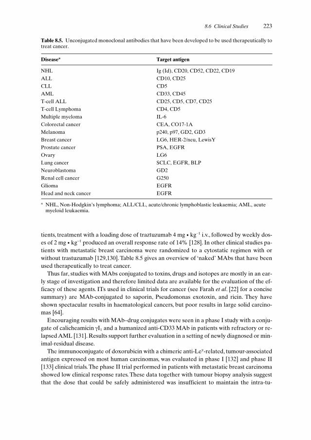
tients, treatment with a loading dose of traztuzumab 4 mg • kg–1 i.v., followed by weekly dos-es of 2 mg • kg–1 produced an overall response rate of 14% [128]. In other clinical studies pa-tients with metastatic breast carcinoma were randomized to a cytostatic regimen with orwithout trastuzumab [129,130]. Table 8.5 gives an overview of ‘naked’ MAbs that have beenused therapeutically to treat cancer.
Thus far, studies with MAbs conjugated to toxins, drugs and isotopes are mostly in an ear-ly stage of investigation and therefore limited data are available for the evaluation of the ef-ficacy of these agents. ITs used in clinical trials for cancer (see Farah et al. [22] for a concisesummary) are MAb-conjugated to saporin, Pseudomonas exotoxin, and ricin. They haveshown spectacular results in haematological cancers, but poor results in large solid carcino-mas [64].
Encouraging results with MAb–drug conjugates were seen in a phase I study with a conju-gate of calicheamicin γI1 and a humanized anti-CD33 MAb in patients with refractory or re-lapsed AML [131]. Results support further evaluation in a setting of newly diagnosed or min-imal-residual disease.
The immunoconjugate of doxorubicin with a chimeric anti-Ley-related, tumour-associatedantigen expressed on most human carcinomas, was evaluated in phase I [132] and phase II[133] clinical trials. The phase II trial performed in patients with metastatic breast carcinomashowed low clinical response rates. These data together with tumour biopsy analysis suggestthat the dose that could be safely administered was insufficient to maintain the intra-tu-
8.6 Clinical Studies 223
Table 8.5. Unconjugated monoclonal antibodies that have been developed to be used therapeutically totreat cancer.
Diseasea Target antigen
NHL Ig (Id), CD20, CD52, CD22, CD19
ALL CD10, CD25
CLL CD5
AML CD33, CD45
T-cell ALL CD25, CD5, CD7, CD25
T-cell Lymphoma CD4, CD5
Multiple myeloma IL-6
Colorectal cancer CEA, CO17-1A
Melanoma p240, p97, GD2, GD3
Breast cancer LG6, HER-2/neu, LewisY
Prostate cancer PSA, EGFR
Ovary LG6
Lung cancer SCLC, EGFR, BLP
Neuroblastoma GD2
Renal cell cancer G250
Glioma EGFR
Head and neck cancer EGFR
a NHL, Non-Hodgkin’s lymphoma; ALL/CLL, acute/chronic lymphoblastic leukaemia; AML, acutemyeloid leukaemia.

moural concentration of doxorubicin at a level required to achieve regression. It is likelytherefore that immunoconjugates of doxorubicin will be only effective in minimal-diseasesettings.
In general, trials with radioimmunoconjugates reported higher response rates in patientswith haematological malignancies when compared with patients with solid tumours. Themost impressive studies with non-myeloablative regimens reported objective responses in70–80% of patients with chemotherapy-refractory B-cell lymphomas, a median response du-ration of 12 months and minimal toxicity using 131-I-labelled anti-CD20 [134]. In trials withmyeloablative regimens, performed in conjunction with autologous haematopoeitic bonemarrow or stem cell transplantation, responses have been seen in 95% of patients, completeresponses in 85%, with a progression-free survival of 62% and overall survival of 93% with amedian follow-up of 2 years [135,136].The estimated 4-year overall and progression-free sur-vival rates were reported to be 68 and 42%, respectively [137].
Anti-CD45 antibody BC8 labelled with 131-I may reduce the rate of tumour relapse andhas acceptable toxicity in patients with AML,ALL or myelodysplastic syndrome who under-went stem-cell rescue [138]. Of 25 patients treated with advanced AML and myelodysplasticsyndrome, seven were disease-free at 15–89 months post-transplantation. Of nine patientswith advanced ALL, three were disease-free at 23, 58 and 70 months post-transplantation, re-spectively.
As with the monoclonal antibody therapies described above, B-cell malignancies would bethe most attractive targets for anti-CD3 bispecific monoclonal antibody-based immunother-apy [139]. Several phase I/II clinical trials have been described using BsMAb for non B-cellmalignancies. BIS-1, a BsMAb directed against the TAA EGP-2 and the CD3/T-cell receptorcomplex on T lymphocytes was studied in renal [140] and lung cancer [141]. In carcinoma pa-tients with EGP-2 positive malignant ascites or pleural exudates, local administration of ex-vivo IL-2-activated autologous lymphocytes and BIS-1, resulted in both anti-tumour effectsand a strong local inflammatory reaction [141]. In patients with advanced breast or ovariancancer administration of BsMAb directed against FcγRI or FcγRIII and HER-2/neu resultedin elevated plasma levels of cytokines [142,143].
The biological effects observed at tumour sites indicate that BsMAbs effectively penetratetissue. However, trials are limited by the toxicity caused by induction of a ‘cytokine storm’ orby the complex pharmacokinetics. Furthermore, T-cell directed BsMAb approaches are hin-dered by difficulties in mobilizing and activating T and NK effector cells. Recent attention,therefore, has focused on BsMAbs which target myeloid effectors [79].
8.6.2 Pro-drugs
ADEPT strategies have been described but only the carboxypeptidase G2 approach hasbeen tested in patients so far. In a phase I clinical trial, patients with non-resectable metasta-tic or locally recurrent colorectal carcinoma were treated with ADEPT. CarboxypeptidaseG2 activity was found in metastatic tumour biopsies.The pro-drug was converted into the ac-tive drug but leakage into the bloodstream also occurred [22,144].
Clinical trials with ‘monotherapy’ pro-drug strategies are in progress, but have not yetbeen evaluated.
224 8 Strategies for Specific Drug Targeting to Tumour Cells

8.6.3 (Synthetic) (co)Polymers
Soluble polymer conjugates have been introduced into clinical practice in the last decade[90,94]. Several PEG conjugates have been evaluated clinically [145] for cancer therapy in-cluding a PEG conjugate of asparaginase in the treatment of ALL in patients hypersensitiveto the native enzyme [146], and a PEG conjugate of IL-2 [147].
A phase I clinical and pharmacokinetic study of PK1 comprising doxorubicin covalentlybound to N-(2-hydroxypropyl)-methacrylamide copolymer by a peptidyl linker, was carriedout in 36 patients with refractory or resistant cancers [94]. PK1 demonstrated anti-tumour ac-tivity, and that polymer–drug conjugation decreased doxorubicin dose-limiting toxicities.Phase II studies are in progress.
8.6.4 Liposomes
There are three liposomal forms of doxorubicin or daunorubicin on the market (Table 8.6).Doxil® and DaunoXome® have been approved for the treatment of AIDS-related Kaposi’ssarcoma and are being evaluated in clinical trials for the treatment of a variety of cancers[148–151]. Evacet® (liposomal doxorubicin) has recently been tested in large phase II and IIIclinical trials for the treatment of metastatic breast cancer and is awaiting approval by theFDA [151]. Data obtained from trials thus far suggest that all three liposomal drugs offer sig-nificant therapeutic benefit compared with the free drug [113].
8.6 Clinical Studies 225
Table 8.6. Monoclonal antibody based products and liposome formulations registered for cancertherapy in the USA and/or Europe.
Generic name Trade name Company Indication
Rituximaba Rituxan Roche Relapsed or refractory low-(Mabthera)c grade/follicular non-
Hodgkin’s lymphoma
Trastuzumaba Herceptin Genentech-Roche Metastatic breast cancer
Edrecolomaba Panorex Glaxo-Wellcome Post-operative adjuvanttherapy Dukes C colorectal carcinoma
Doxorubicinb Doxil Alza-corporation AIDS-related Kaposi’s (Caelyx)d sarcoma
Daunorubicinb DaunoXome Nexstar AIDS-related Kaposi’s Pharmaceuticals sarcoma
Doxorubicinb Evacet The Liposome Metastatic breast cancer(Myocet)e Company, Inc.
a MAb-based.b Liposome formulation.c Rituxan is known as Mabthera in Europe.d Doxil is known as Caelyx in Europe.e Evacet is known as Myocet in Europe.

Liposomal preparations can be therapeutically beneficial based on their ability to de-crease non-specific toxicities associated with the drug, or by being more efficacious against aspecific type of cancer, increasing the response frequency, and/or the average time to relapseor response duration.
Liposome-encapsulated immunomodulators are currently under investigation in differentpatient groups although this development has certainly not advanced as far as that with theliposomal anthracyclines. MLV-MTP-PE (multilamellar vesicles-muramyl tripeptide-phos-phatidylethanolamine) was studied in several clinical trials in osteosarcoma patients who de-veloped pulmonary metastases during adjuvant chemotherapy [108]. The intravenous ad-ministration of MLV-MTP-PE induced tumouricidal properties in monocytes as well as in-crease in serum IL-1 shortly after intravenous infusion. Furthermore elevations in C-reactiveprotein, β2-microglobulin and ceruloplasmin were frequently observed. Even higher anti-tu-mour activity was observed in combination with ifosfamide. These preliminary results sug-gests that liposome-encapsulated immunomodulators in combination with chemotherapymay be an appropriate treatment for recurrent disease.
In Table 8.6 a summary is given of MAb and liposome-based formulations registered forcancer therapy.
8.7 Animal Models: their Predictive Value
Studies in pre-clinical models with human tumours are often carried out in (immuno)defi-cient mice. However, particularly in the case of monoclonal antibody-directed therapy, it isimportant to recognize that these models, while useful, frequently over-predict activity andunder-predict toxicity because the target antigen is tumour-specific in the animal but only tu-mour-associated in man.
Besides mouse models, rat models are used for experimental in vivo studies of MAbs forimmunotherapy of cancers. Kroesen et al. [80] described a rat model to investigate the po-tential of targeted anti-tumour treatment against EGP-2-positive, rat syngenic tumours.However, possible adverse side-effects on healthy tissues could not be studied in this modelas the target epitope was not expressed on tissues other than tumour cells. To overcome thisproblem, a transgenetic rat model expressing the EGP-2 protein in various organs was de-veloped for evaluation of anti-carcinoma-associated-antigen-EGP-2-directed immunothera-py strategies [152].
Other limitations of mouse/rat models are that tumour growth is different in the animalmodel compared to the situation in man.Tumour growth is more rapid in the rat/mouse mod-el which has an effect on vascularization and intra-tumoural pressure for example.These fac-tors can, as discussed in Section 8.4, have great impact on tumour penetration and uptake ofthe MAb-based drug-targeting constructs.
8.8 Conclusions and Future Perspectives
The studies presented above represent the most advanced pre-clinical and clinical therapyprogrammes for cancer management including drug targeting strategies. Patience is required
226 8 Strategies for Specific Drug Targeting to Tumour Cells

to evaluate the large variety of results, to understand the reasons for limited success, and thegreat number of concepts to improve the treatment strategies. It should be emphasized thatafter more than 15 years of experience with monoclonal antibody therapy of cancer, we haveno definite idea of essential mechanisms required for clinical activity. The findings of e.g.Clynes et al. [153,154] support the idea that the anti-tumour effects of MAbs depend on manymechanisms. For instance, myeloid cells, probably macrophages and monocytes that expressboth activation and inhibitory Fc receptors, are essential immune effector cells for MAbtherapy. Blocking inhibitory receptors or MAbs that selectively trigger activating Fc recep-tors without affecting inhibitory Fc receptors could increase potency. The underlying mecha-nisms of the therapeutic effects of MAbs may also vary from one MAb to another. Thesefindings should be considered in the improvement of drug-targeting strategies based onMAbs, including Mab derivatives such as those used in ADEPT. The same holds true for ap-proaches such as modulating growth factor receptor functions or inducing immune respons-es using BsMAbs or the combination of tumour cell-directed strategies combined with Bs-MAbs delivering agents which inhibit the supply of blood to the tumour.
Liposomal drugs have been suggested to be the ‘magic bullet’ of cancer therapy due totheir ability to accumulate selectively in the tumour. The problem remains that not all can-cers and patients respond in the same way.The drug actually delivered to the required site ofaction plays an important role in the response achieved, while the potential toxicity of thesurface modified liposomes in the macrophage system have to be taken into account onchronic administration.
Multi-drug resistance represents a significant obstacle in the use of standard chemothera-py regimens to cure cancer. It is unlikely that tumours resistant to free drug therapy will beeradicated by liposome-encapsulated or any other targeted form of drug. Additional studiesthat attempt to encapsulate drugs which exhibit non-overlapping modes of drug resistanceand significant activity against a particular form of cancer, or combine free drugs with non-overlapping modes of drug resistance with currently available liposomal drugs, will addressthe potential and limitations of these approaches. There is also a need to develop liposomalformulations of other drugs and therapeutic regimens designed to stimulate host immunityalone or in combination with other treatment modalities.
In conclusion, research needs to be aimed at improving the potency and reducing the im-munogenicity and toxicity of drug formulations in order to increase the therapeutic index.Methods to increase the overall therapeutic index of a drug, e.g. by using tumour-specific lig-ands, increasing extravasation of liposomes into tumours and increasing the bioavailability ofthe drug selectively at the site of the tumour, should be explored further.
In addition to this, research must focus on optimizing the biophysical properties of the car-riers and drug-conjugates, as these properties are of major importance to the decision ofwhether or not to use these molecules in vivo. A wealth of innovative strategies involving re-combinant molecules with novel effector functions are in the early stages of clinical evalua-tion and hold great promise for the future. These developments will initiate other studieswhich should focus on long-term safety and cost–benefit analyses. Only adequately designedcase–control studies supported by international collaboration can show the long-term safetyof these new therapies. The same holds true for studies evaluating the costs of these thera-peutic regimens and the resultant improvement in the quality of life, in relation to the cumu-lative social and health-care costs of current therapies. These multidisciplinary collaborative
8.8 Conclusions and Future Perspectives 227
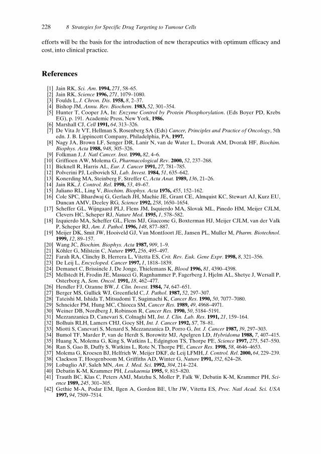
efforts will be the basis for the introduction of new therapeutics with optimum efficacy andcost, into clinical practice.
References
[1] Jain RK, Sci. Am. 1994, 271, 58–65.[2] Jain RK, Science 1996, 271, 1079–1080.[3] Foulds L, J. Chron. Dis. 1958, 8, 2–37.[4] Bishop JM, Annu. Rev. Biochem. 1983, 52, 301–354.[5] Hunter T, Cooper JA, In: Enzyme Control by Protein Phosphorylation. (Eds Boyer PD, Krebs
EG), p. 191. Academic Press, New York, 1986.[6] Marshall CJ, Cell 1991, 64, 313–326.[7] De Vita Jr VT, Hellman S, Rosenberg SA (Eds) Cancer, Principles and Practice of Oncology, 5th
edn. J. B. Lippincott Company, Philadelphia, PA, 1997.[8] Nagy JA, Brown LF, Senger DR, Lanir N, van de Water L, Dvorak AM, Dvorak HF, Biochim.
Biophys. Acta 1988, 948, 305–326.[9] Folkman J, J. Natl Cancer. Inst. 1990, 82, 4–6.
[10] Griffioen AW, Molema G, Pharmacological Rev. 2000, 52, 237–268.[11] Bicknell R, Harris AL, Eur. J. Cancer 1991, 27, 781–785.[12] Polverini PJ, Leibovich SJ, Lab. Invest. 1984, 51, 635–642.[13] Konerding MA, Steinberg F, Streffer C, Acta Anat. 1989, 136, 21–26.[14] Jain RK, J. Control. Rel. 1998, 53, 49–67.[15] Juliano RL, Ling V, Biochim. Biophys. Acta 1976, 455, 152–162.[16] Cole SPC, Bhardwaj G, Gerlach JH, Machie JE, Grant CE, Almquist KC, Stewart AJ, Kurz EU,
Duncan AMV, Deeley RG, Science 1992, 258, 1650–1654.[17] Scheffer GL, Wijngaard PLJ, Flens JM, Isquierdo MA, Slovak ML, Pinedo HM, Meijer CJLM,
Clevers HC, Scheper RJ, Nature Med. 1995, 1, 578–582.[18] Izquierdo MA, Scheffer GL, Flens MJ, Giaccone G, Boxterman HJ, Meijer CJLM, van der Valk
P, Scheper RJ, Am. J. Pathol. 1996, 148, 877–887.[19] Meijer DK, Smit JW, Hooiveld GJ, Van Montfoort JE, Jansen PL, Muller M, Pharm. Biotechnol.
1999, 12, 89–157.[20] Wang JC, Biochim. Biophys. Acta 1987, 909, 1–9.[21] Köhler G, Milstein C, Nature 1997, 256, 495–497.[22] Farah RA, Clinchy B, Herrera L, Vitetta ES, Crit. Rev. Euk. Gene Expr. 1998, 8, 321–356.[23] De Leij L, Encycloped. Cancer 1997, 1, 1818–1839.[24] Demanet C, Brissincle J, De Jonge, Thielemans K, Blood 1996, 81, 4390–4398.[25] Mellstedt H, Frodin JE, Masucci G, Ragnhammer P, Fagerberg J, Hjelm AL, Shetye J, Wersall P,
Osterborg A, Sem. Oncol. 1991, 18, 462–477.[26] Hendler FJ, Ozanne BW, J. Clin. Invest. 1984, 74, 647–651.[27] Berger MS, Gullick WJ, Greenfield C, J. Pathol. 1987, 52, 297–307.[28] Tateishi M, Ishida T, Mitsudomi T, Sugimachi K, Cancer Res. 1990, 50, 7077–7080.[29] Schneider PM, Hung MC, Chiocca SM, Cancer Res. 1989, 49, 4968–4971.[30] Weiner DB, Nordberg J, Robinson R, Cancer Res. 1990, 50, 5184–5191.[31] Mezzanzanica D, Canevari S, Colnaghi MI, Int. J. Clin. Lab. Res. 1991, 21, 159–164.[32] Bolhuis RLH, Lamers CHJ, Goey SH, Int. J. Cancer 1992, S7, 78–81.[33] Miotti S, Canevari S, Menard S, Mezzanzanica D, Porro G, Int. J. Cancer 1987, 39, 297–303.[34] Bumol TF, Marder P, van de Herdt S, Borowitz MJ, Apelgren LD, Hybridoma 1988, 7, 407–415.[35] Huang X, Molema G, King S, Watkins L, Edgington TS, Thorpe PE, Science 1997, 275, 547–550.[36] Ran S, Gao B, Duffy S, Watkins L, Rote N, Thorpe PE, Cancer Res. 1998, 58, 4646–4653.[37] Molema G, Kroesen BJ, Helfrich W, Meijer DKF, de Leij LFMH, J. Control. Rel. 2000, 64, 229–239.[38] Clackson T, Hoogenboom M, Griffiths AD, Winter G, Nature 1991, 352, 624–28.[39] Lobuglio AF, Saleh MN, Am. J. Med. Sci. 1992, 304, 214–224.[40] Debatin K-M, Krammer PH, Leukaemia 1995, 9, 815–820.[41] Trauth BC, Klas C, Peters AMJ, Matzhu S, Moller P, Falk W, Debatin K-M, Krammer PH, Sci-
ence 1989, 245, 301–305.[42] Gethie M-A, Podar EM, Ilgen A, Gordon BE, Uhr JW, Vitetta ES, Proc. Natl Acad. Sci. USA
1997, 94, 7509–7514.
228 8 Strategies for Specific Drug Targeting to Tumour Cells
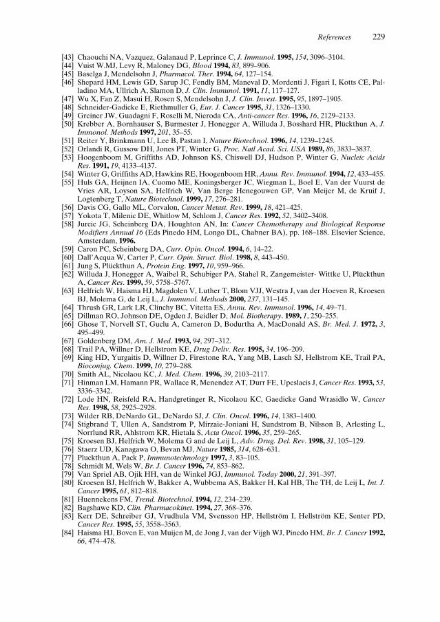
[43] Chaouchi NA, Vazquez, Galanaud P, Leprince C, J. Immunol. 1995, 154, 3096–3104.[44] Vuist W.MJ, Levy R, Maloney DG, Blood 1994, 83, 899–906.[45] Baselga J, Mendelsohn J, Pharmacol. Ther. 1994, 64, 127–154.[46] Shepard HM, Lewis GD, Sarup JC, Fendly BM, Maneval D, Mordenti J, Figari I, Kotts CE, Pal-
ladino MA, Ullrich A, Slamon D, J. Clin. Immunol. 1991, 11, 117–127.[47] Wu X, Fan Z, Masui H, Rosen S, Mendelsohn J, J. Clin. Invest. 1995, 95, 1897–1905.[48] Schneider-Gadicke E, Riethmuller G, Eur. J. Cancer 1995, 31, 1326–1330.[49] Greiner JW, Guadagni F, Roselli M, Nieroda CA, Anti-cancer Res. 1996, 16, 2129–2133.[50] Krebber A, Bornhauser S, Burmester J, Honegger A, Willuda J, Bosshard HR, Plückthun A, J.
Immonol. Methods 1997, 201, 35–55.[51] Reiter Y, Brinkmann U, Lee B, Pastan I, Nature Biotechnol. 1996, 14, 1239–1245.[52] Orlandi R, Gussow DH, Jones PT, Winter G, Proc. Natl Acad. Sci. USA 1989, 86, 3833–3837.[53] Hoogenboom M, Griffiths AD, Johnson KS, Chiswell DJ, Hudson P, Winter G, Nucleic Acids
Res. 1991, 19, 4133–4137.[54] Winter G, Griffiths AD, Hawkins RE, Hoogenboom HR, Annu. Rev. Immunol. 1994, 12, 433–455.[55] Huls GA, Heijnen IA, Cuomo ME, Koningsberger JC, Wiegman L, Boel E, Van der Vuurst de
Vries AR, Loyson SA, Helfrich W, Van Berge Henegouwen GP, Van Meijer M, de Kruif J,Logtenberg T, Nature Biotechnol. 1999, 17, 276–281.
[56] Davis CG, Gallo ML, Corvalon, Cancer Metast. Rev. 1999, 18, 421–425.[57] Yokota T, Milenic DE, Whitlow M, Schlom J, Cancer Res. 1992, 52, 3402–3408.[58] Jurcic JG, Scheinberg DA, Houghton AN, In: Cancer Chemotherapy and Biological Response
Modifiers Annual 16 (Eds Pinedo HM, Longo DL, Chabner BA), pp. 168–188. Elsevier Science,Amsterdam, 1996.
[59] Caron PC, Scheinberg DA, Curr. Opin. Oncol. 1994, 6, 14–22.[60] Dall’Acqua W, Carter P, Curr. Opin. Struct. Biol. 1998, 8, 443–450.[61] Jung S, Plückthun A, Protein Eng. 1997, 10, 959–966.[62] Willuda J, Honegger A, Waibel R, Schubiger PA, Stahel R, Zangemeister- Wittke U, Plückthun
A, Cancer Res. 1999, 59, 5758–5767.[63] Helfrich W, Haisma HJ, Magdolen V, Luther T, Blom VJJ, Westra J, van der Hoeven R, Kroesen
BJ, Molema G, de Leij L, J. Immunol. Methods 2000, 237, 131–145.[64] Thrush GR, Lark LR, Clinchy BC, Vitetta ES, Annu. Rev. Immunol. 1996, 14, 49–71.[65] Dillman RO, Johnson DE, Ogden J, Beidler D, Mol. Biotherapy. 1989, 1, 250–255.[66] Ghose T, Norvell ST, Guclu A, Cameron D, Bodurtha A, MacDonald AS, Br. Med. J. 1972, 3,
495–499.[67] Goldenberg DM, Am. J. Med. 1993, 94, 297–312.[68] Trail PA, Willner D, Hellstrom KE, Drug Deliv. Res. 1995, 34, 196–209.[69] King HD, Yurgaitis D, Willner D, Firestone RA, Yang MB, Lasch SJ, Hellstrom KE, Trail PA,
Bioconjug. Chem. 1999, 10, 279–288.[70] Smith AL, Nicolaou KC, J. Med. Chem. 1996, 39, 2103–2117.[71] Hinman LM, Hamann PR, Wallace R, Menendez AT, Durr FE, Upeslacis J, Cancer Res. 1993, 53,
3336–3342.[72] Lode HN, Reisfeld RA, Handgretinger R, Nicolaou KC, Gaedicke Gand Wrasidlo W, Cancer
Res. 1998, 58, 2925–2928.[73] Wilder RB, DeNardo GL, DeNardo SJ, J. Clin. Oncol. 1996, 14, 1383–1400.[74] Stigbrand T, Ullen A, Sandstrom P, Mirzaie-Joniani H, Sundstrom B, Nilsson B, Arlesting L,
Norrlund RR, Ahlstrom KR, Hietala S, Acta Oncol. 1996, 35, 259–265.[75] Kroesen BJ, Helfrich W, Molema G and de Leij L, Adv. Drug. Del. Rev. 1998, 31, 105–129.[76] Staerz UD, Kanagawa O, Bevan MJ, Nature 1985, 314, 628–631.[77] Pluckthun A, Pack P, Immunotechnology 1997, 3, 83–105.[78] Schmidt M, Wels W, Br. J. Cancer 1996, 74, 853–862.[79] Van Spriel AB, Ojik HH, van de Winkel JGJ, Immunol. Today 2000, 21, 391–397.[80] Kroesen BJ, Helfrich W, Bakker A, Wubbema AS, Bakker H, Kal HB, The TH, de Leij L, Int. J.
Cancer 1995, 61, 812–818.[81] Huennekens FM, Trend. Biotechnol. 1994, 12, 234–239.[82] Bagshawe KD, Clin. Pharmacokinet. 1994, 27, 368–376.[83] Kerr DE, Schreiber GJ, Vrudhula VM, Svensson HP, Hellström I, Hellström KE, Senter PD,
Cancer Res. 1995, 55, 3558–3563.[84] Haisma HJ, Boven E, van Muijen M, de Jong J, van der Vijgh WJ, Pinedo HM, Br. J. Cancer 1992,
66, 474–478.
References 229

[85] Leenders RGG, Damen EWP, Bijsterveld EJA, Scheeren HW, Houba PHJ, van der Meulen-Muileman IH, Boven E, Haisma HJ, Bioorg. Med. Chem. Lett. 1999, 718, 1597–1610.
[86] Bosslet K, Straub R, Blumrich M, Czech J, Gerken M, Sperker B, Kroemer HH, Gesson JP, KochM, Monneret C, Cancer Res. 1998, 58, 1195–1219.
[87] Bosslet K, Czech J, Hoffman D, Tumour Target. 1995, 1, 45–50.[88] de Groot FMH, de Bart ACW, Verheijen JH, Scharen HW, J. Med. Chem. 1999, 42, 5277–5283.[89] Takakura Y, Hashida M, Crit. Rev. Oncol. Hematol. 1995, 18, 207–231.[90] Duncan R, Spreafico F, Clin. Pharmacokinet. 1994, 27, 290–306,.[91] Duncan R, Anti-Cancer Drugs 1992, 3, 175–210.[92] Sezaki H, Takakura Y, Hashida M, Adv. Drug Delivery Rev. 1989, 3, 247–266.[93] Seymour LW, CRC Crit. Rev. Ther. Drug Carrier. Syst. 1992, 9, 135–187.[94] Vasey PA, Kaye SB, Morrison R, Twelves C, Wilson P, Duncan R, Thomson AH, Murray LS,
Hilditech TE, Murray T, Burtles S, Fraier D, Frigerio E, Cassidy J, Clin. Cancer Res. 1999, 5, 83–94.[95] Lasic DD, Papahadjopoulos D, Science 1995, 267, 1275–1276.[96] Gregoriadis G, Trends Biotechnol. 1995, 13, 527–537.[97] Lasic DD, From Physics to Applications. Elsevier, Amsterdam, 1993.[98] Lasic DD, Martin FJ, (Eds) Stealth Liposomes. CRC, Boca Raton, FL, 1995.[99] Daemen T, Regts J, Meesters M, Ten Kate MT, Bakker-Woudenberg IAJM, Scherphof GL, J.
Control. Rel. 1997, 44, 1–9.[100] Doroshaw JH, Cancer Chemotherapy and Biotherapy: Principles and Practice. (eds Chabner BA
and Longo DL) pp. 409–433. Lippincott-Raven, Philadelphia, PA. 1996.[101] Harrigan PR, Wong KF, Redelmeier TE, Wheeler JJ, Cullis PR, Biochim. Biophys. Acta 1993,
1149, 329–338.[102] Cullis PR, Hope MJ, Bally MB, Madden TD, Mayer LD, Fenske DB, Biochem. Biophys. Acta
1997, 1331, 187–211.[103] Park JW, Hong K, Kirpotin DB, Meyer O, Papahadjopoulos D, Benz CC, Cancer Lett. 1997, 118,
153–160.[104] Crosasso P, Brusa P, Dosio F, Arpicco S, Pacchioni D, Schuber F, Cattel L, J Pharm. Sci. 1997, 86,
832–839.[105] Tseng YL, Hong RL, Tao MH, Chang FH, Int. J. Cancer 1999, 80, 723–730.[106] Kirpotin D, Park JW, Hong K, Zalipsky S, Li WL, Carter P, Benz CC, Papahadjopoulos D, Bio-
chemistry 1997, 36, 66–75.[107] Lopes de Menezes DE, Pilarski LM, Allen TM, Cancer Res. 1998, 58, 3320–3330.[108] Killion JJ, Fidler IJ, Pharmacol. Ther. 1998, 78, 141–154.[109] Dass CR, Walker TL, DeCruz EE, Burton MA, Drug Deliv. 1997, 4, 151–165.[110] Li S, Huang L, Gene Therapy 1997, 4, 891–900.[111] Kaneda Y, Saeki Y, Morischita R, Mol. Med. Today 1999, 5, 298–303.[112] Mayer LD, Cancer Metast. Rev. 1998, 17, 211–218.[113] Drummond DC, Meijer O, Hong K, Kirpotin DB, Papahadjopoulos D, Pharmacol. Rev. 1999, 51,
691–743.[114] Brown SL, Miller RA, Levy R, Semin. Oncol. 1989, 16, 199–210.[115] Dillman RO, J. Clin. Oncol. 1994, 12, 1497–1515.[116] Renner C, Trümper L, Pfreundschuh M, Leukaemia 1997, 11, S55–S59.[117] Irie RF, Morton DL, Proc. Natl Acad. Sci. USA 1986, 83, 8694–8698.[118] Cheung N-KV, Lazarus H, Miraldi FD, Abramowsky CR, Kallick S, Saarinen UM, Spitzer T,
Strandjord SE, Coccia PF, Berger NA, J. Clin. Oncol. 1987, 51, 430–1440.[119] Vadhan-Raj S, Cordon-Cardo C, Carswell E, Mintzer D, Dantis L, Duteau C, Templeton MA,
Oettgen HF, Old LJ, Houghton AN, J. Clin. Oncol. 1988, 6, 1636–1648.[120] Handgretinger R, Baader P, Dopfer R, Klingebiel T, Reuland P, Treuner J, Reisfeld RA, Ni-
ethammer D, Cancer Immunol. Immunother. 1992, 35, 199–204.[121] Saleh MN, Khazaeli MB, Wheeler RH, Dropcho E, Liu T, Urist M, Miller DM, Lawson S, Dixon
P, Russell CH, LoBuglio A, Cancer Res. 1992, 52, 4342–4347.[122] Riethmuller G, Holz e, Schlimok G, Schmiegel W, Raab R, Hoffken K, Gruber R, Funke I,
Pichlmaier H, Hirche H, J. Clin. Oncol. 1998, 16, 1788–1794.[123] Oosterwijk E, Debruyne FMJ, Schalken JA, Semin. Oncol. 1995, 22, 34–41.[124] Weiner LM, Semin. Oncol. 1999, 26, 41–50.[125] McLaughlin P, Grillo-Lopez AJ, Link BK, Levy R, Czuczman MS, Williams ME, Heyman MR,
Bence-Bruchler I, White CA, Canabillas F, Jain V, Ho AD, Frister J, Wey K, Shen D, DallaizeBK, J. Clin. Oncol. 1998, 16, 2824–2833.
230 8 Strategies for Specific Drug Targeting to Tumour Cells

[126] Czuczman MS, Grillo-Lopez AJ, White CA, Saleh M, Gordon L, LoBuglio AF, Jonas C, Klip-penstein D, Dallaize B, Warms C, J. Clin. Oncol. 1999, 26, 268–276.
[127] Dyer MJS, Semin. Oncol. 1999, 26, 52–57.[128] Baselga J, Tripathy D, Mendelsohn J, Baughman S, Benz CC, Dantis L, Shlarin NT, Seidman AD,
Hudis CA, Moore J, Rosen PP, Twaddell T, Henderson IC, Norton L, J. Clin. Oncol. 1996, 14,737–744.
[129] Pegram MD, Lipton A, Hayes DF, Weber BL, Baselga MM, Tripathy D, Baly D, Baighman SA,Twaddell T, Glaspy JA, Slalom DJ, J. Clin. Oncol. 1998, 16, 2659–2663.
[130] Baselga J, Tripathy D, Mendelsohn J, et al., Semin. Oncol. 1999, 26, 78–83.[131] Sievers EL, Appelbaum FR, Spielberger RT, Forman SJ, Flowers D, Smith FO, Shannon-Dorcy
K, Berger MS, Berstein ID, Blood, 1999, 93, 3678–3684.[132] Saleh MN, LoBuglio AF, Trail PA, In: Monoclonal Antibody-Based Therapy of Cancer (Ed.
Grossband M), pp. 397–416. Marcel Dekker, New York, 1998.[133] Tolcher AW, Sugarman S, Gelmon KA, Cohen R, Saleh M, Isaacs C, Young L, Healey D, Onet-
to N Slichenmyer W, J. Clin. Oncol. 1999, 17, 478–484.[134] Kaminski MS, Zasadny KR, Francis IR, Fenner MC, Ross CW, Milik AW, Estes J, Tude M, Re-
gan D, Fisher S, Glenn SD, Wahl RL, J. Clin. Oncol. 1996, 14, 1974–1981.[135] Press OW, Eary JF, Appelbaum FR, Martin PJ, Badger CC, Nelp WB, Glenn S, Butchko G, Fish-
er D, Porter B, N. Engl. J. Med. 1993, 329, 1219–1224.[136] Press OW, Eary JF, Appelbaum FR, Martin PJ, Nelp WB, Glenn S, Fisher DR, Porter B,
Matthews DC, Gooley T and Bernstein ID, Lancet 1995, 346, 336–340.[137] Liu SY, Eary JF, Petersdorf SH, Martin PJ, Maloney DG, Appelbaum FR, Matthews DC, Bush
SA Duracke LD, J. Clin. Oncol. 1998, 16, 3270–3278.[138] Matthews DC, Appelbaum FR, Eary JF, Fischer DR, Durack LD, Hui TE, Martin PJ, Mitchell D,
Press OW, Storb R, Bernstein ID, Blood 1999, 94, 1237–1247.[139] De Gast GC, Haagen IA, van Houten AA, Klein SC, Duits AJ, de Weyer RA, Vroom TM, Clark
MR, Phillips J, Van Dijk AJ, Cancer Immunol. Immunother. 1995, 40, 390–396.[140] Janssen RAJ, Kroesen BJ, Buter J, Mesander G, Sleijfer DT, The TH, Mulder NH, de Leij L, Br.
J. Cancer 1995, 72, 795–799.[141] Kroesen BJ, Nieken J, Sleijfer DT, Molema G, de Vries EGE, Groen HJM, Helfrich W, The TH,
Mulder NH, de Leij L, Cancer Immunol. Immunother. 1997, 45, 203–206.[142] Valone FH, Kaufman PA, Guyre PM, Lewis LD, Memoli V, Deo Y, Graziano R, Fisher JL, Mey-
er L, Mrozek–Orlowski M, Wardwell K, Guyre V, Morley TL, Arvinu C, Fanger MW, J. Clin. On-col. 1995, 13, 2281–2292.
[143] Weiner LM, Clark JI, Davey M, Li WS, Garcia de Palazzo I, Ring DB, Alpaugh RK, Cancer Res.1995, 55, 4586–4593.
[144] Martin J, Stribbling SM, Poon GK, Begent RH, Napier M, Sharma SK, Springer CJ, CancerChemother. Pharmacol. 1997, 40, 189–201.
[145] Fuertges F, Abuchowski A, J. Control. Rel. 1990, 11, 139–148.[146] Ho DH, Brown NS, Yen A, Holmes R, Keating M, Abuchowski A, Newman RA, Kozahoff IH,
Drug Metab. Dispos. 1986, 14, 349–352.[147] Meyers FJ, Paradise C, Scudder SA, Goodman G, Konrad M, Clin. Pharmacol. Ther. 1991, 49,
307–313.[148] Martin FJ, Medical Applications of Liposomes (Eds frasic DD and Papahadjopoulos D) pp.
635–688, Elsevier, Amsterdam, 1998.[149] Eckhardt JR, Campbell E, Burris HA, Wiess GR, Rodriguez GI, Fields SM, Thurman AM, Pea-
cock NW, Cobb P, Rothenberg ML, Ross ME, Von Hoff DD, Am. J. Clin. Oncol. 1994, 17,498–501.
[150] Gill PS, Espina BM, Muggia F, Cabriales S, Tulpule A, Esplin JA, J. Clin. Oncol. 1995, 13,996–1003.
[151] Valero V, Buzdar AU, Theriault RL, Azarnia N, Fonseca GA, Willey J, Ewer M, Walter RS,Mackay B, Podoloff D, Booser D, Lee LW, Hortobagyi GN, J. Clin. Oncol. 1999, 17, 1425–1434.
[152] McLaughlin PMJ, Kroesen BJ, Dokter HA, van der Molen H, de Groot M, Brinker MGL, Kok K,Ruiters MHJ, Buys CHCM, de Leij LFMH, Cancer Immunol. Immunother. 1999, 48, 303–311.
[153] Clynes RA, Takechi Y, MoroiY, Houghton A, Ravetch JV, Proc. Natl Acad. Sci. USA 1998, 95,652–656.
[154] Clynes RA, Towers TL, Persta LG, Ravetch JV, Nature Med. 2000, 6, 443–446.[155] Houghton AN, Scheinberg DA, Nature Med. 2000, 6, 373–375.
References 231

9 Tumour Vasculature TargetingDaisy W. J. van der Schaft, S. Ramakrishnan, Grietje Molema, Arjan W. Griffioen
9.1 Introduction
Various stages in solid tumour growth can be discriminated. After multiple genetic changes,cellular growth progresses via hyperplasia and dysplasia towards an in situ tumour. Only af-ter the so-called ‘angiogenic switch’, an as yet, ill-defined set of molecular changes leading tothe formation of new blood vessels, does a tumour progress towards a mass extending morethan several millimetres in diameter [1]. By recruitment of new blood vessels, the tumourprovides itself with a continuous supply of nutrients and paves the way for metastasis forma-tion.
The architecture of a solid tumour is such, that numerous layers of tumour cells are fed byone blood vessel.This poses a significant barrier for macromolecular drug targeting prepara-tions aimed at the tumour cells that need to extravasate from the blood into the tumour tis-sue to reach the target cells: the larger the chosen carrier, the less accessible the tumour tis-sue will be. In contrast, endothelial cells lining the tumour vasculature are easily accessiblefor these macromolecular preparations. Tumour endothelial cell specific delivery of general-ly toxic anti-neoplastic or blood coagulation-inducing agents presents an attractive optionfor increasing therapeutic efficacy and reducing toxic side-effects elsewhere in the body. Fur-thermore, the pivotal role of endothelial cells in the maintenance of tumour cell survival andgrowth also makes them an interesting target for therapeutic intervention.
Tumour growth does not expand more than several millimetres in diameter in the absenceof new blood vessel formation. Many tumours diagnosed in the clinic have a size beyond0.5 cm in diameter and are considered pro-angiogenic. Endothelial cells are important exe-cutioner cells in the angiogenic cascade. Disruption of these pro-angiogenic characteristicsby inhibiting pro-angiogenic signal transduction in endothelial cells therefore provides apowerful tool with which to intervene in tumour growth.The majority of research on inhibit-ing the function of tumour vasculature focuses at the development of drugs that may have anexplicit action on the endothelial cells in the tumour. Based on their mechanism(s) of actionand the fact that angiogenesis is a normal physiological process, these drugs in theory canalso interrupt the functioning of healthy endothelial cells in the body. Recently, several an-giogenesis inhibitors were withdrawn from clinical studies, most likely as a result of lack ofselectivity for tumour endothelium.This emphasizes the need for systems that can deliver po-tent angiogenesis inhibitors at/in the tumour endothelial cells only.
At present, most target epitopes capable of discriminating tumour endothelium from nor-mal endothelium are molecules that are expressed during angiogenesis. In this chapter asummary of current knowledge regarding tumour growth-related angiogenic endothelialprocesses in angiogenesis will be given. Furthermore, target epitopes and drug-targeting ap-proaches exploited to date, with potential for future therapeutic application will be discussed.
Drug Targeting Organ-Specific Strategies. Edited by G. Molema, D. K. F. MeijerCopyright © 2001 Wiley-VCH Verlag GmbH
ISBNs: 3-527-29989-0 (Hardcover); 3-527-60006-X (Electronic)

9.1.1 Functions of Vascular Endothelial Cells in the Body
The vasculature can be considered to be one of the crucial organs in the body, extendingmore than 900 m2 and playing a major role in maintaining the body’s integrity in variousways. Blood vessels consist of endothelial cells that are directly in contact with the blood, per-icytes located beneath the endothelium, smooth muscle cells, fibroblasts, basement mem-brane, and extracellular matrix (ECM). Depending on the location in the body and the organmicroenvironment, the cellular constituents, basement membrane and ECM differ in pheno-type, composition, and function [2]. The endothelial cells form a monolayer in every bloodvessel in the circulation. They are actively involved in several regulatory processes in thebody. Besides being metabolically active and selectively permeable for small solutes and pep-tides/proteins, endothelial cells are actively involved in regulating haemostasis [3]. Further-more, they are able to recruit cells of the immune system to specific sites of e.g. infection orinflammation by virtue of the regulated expression of cell adhesion molecules and produc-tion of cytokines and chemokines [4] (see also Chapter 7). In addition, endothelial cells areactively involved in vascular remodelling in for example ovulation, wound healing, tumourgrowth and diabetic retinopathy [5,6].
9.1.2 Molecular Control of Tumour Growth-related Angiogenesis
Physiological stimuli during wound healing and during the reproductive cycle in women leadto controlled angiogenesis. However, pathologic conditions such as tumour growth, rheuma-toid arthritis, and diabetic retinopathy are also characterized by abundant angiogenesis.Angiogenesis is rapidly initiated in response to hypoxic or ischaemic conditions. In tumourgrowth, this active vascular remodelling is reflected by enhanced tumour endothelial cell
234 9 Tumour Vasculature Targeting
angiogenicstimulus
EC
ECM
Figure 9.1. In tumours that have undergone the angiogenic switch, the expression of a variety of growthfactors and other soluble molecules, and the changes in the extracellular matrix (ECM), among others,lead to angiogenesis, the formation of new blood vessels from pre-existing ones. In this process,endothelial cells (EC) covering the blood vessel wall become activated, migrate into the tissue,proliferate and eventually differentiate into mature blood vessels. These vessels serve as a supply ofnutrients for the ongoing demands of the growing tumour cells.

proliferation to up to 20–2000 times faster than in healthy adult endothelium [7]. In all typesof angiogenesis, either under physiological or pathologicaal conditions, endothelial cell acti-vation is followed by matrix degradation, cellular migration, proliferation, and ultimatelyneovasculature maturation (Figure 9.1).
9.1.2.1 Role of Growth Factors VEGF and FGF-2
More than 20 cytokines from various sources have now been reported to be involved in theprocesses taking place during angiogenesis [8]. Vascular endothelial growth factor (VEGF)and basic Fibroblast Growth Factor (bFGF or FGF-2) are the two growth factors whose rolesin angiogenesis have been most extensively studied to date.VEGF (also known as VEGF-A)isoforms VEGF-121, 145, 165, 183, 189 and 205 are produced through alternative splicingfrom a single VEGF gene located on chromosome 6 [9]. The isoforms differ in their molecu-lar composition and weight, and biological properties. The larger forms of VEGF differ fromVEGF-121 by the presence of a heparin-binding domain, which is encoded by the exon-7.VEGF is abundantly produced by hypoxic tumour cells, macrophages and other cells of theimmune system [10]. It induces vasodilatation via endothelial nitric oxide production and in-creases endothelial cell permeability [11]. This allows plasma proteins to enter the tissue toform a fibrin-rich provisional network [12]. VEGF also induces the expression of variousproteases and receptors important in cellular invasion and tissue remodelling, it activates cel-lular proliferation and prevents endothelial cell apoptosis [13,14].The two VEGF-specific ty-rosine kinase receptors,VEGFR-1 (Flt-1) and VEGFR-2 (KDR/flk-1), are expressed on vas-cular endothelium, and to a lesser extent on monocytes/macrophages and certain tumour celltypes. Interaction of VEGF with VEGFR-2 is a critical requirement to induce the full spec-trum of VEGF biological responses. In addition to the two VEGF receptors, VEGF-165 hasbeen found to bind neuropilin-1, which is also expressed on endothelial cells [15]. A recentstudy using genetic deletion methods has determined that neuropilin-1 is important for em-bryonic vessel formation [16].
Endothelial cells exploit various proteases such as matrix metalloproteinases to penetrateinto new areas of the body by degrading the basement membrane. Furthermore, urokinase-plasminogen activator and tissue type-plasminogen activator convert the ubiquitous plasmaprotein plasminogen to plasmin. Plasmin is believed to be the most important protease forthe mobilization of FGF-2 from the ECM pool. FGF-2 induces endothelial cell motility, pro-liferation and proteinase activity, and modulates integrin levels [17,18].The cellular effects ofFGFs are mediated via specific binding to high-affinity tyrosine kinase receptors [17]. In ad-dition, low affinity FGF receptors consist of polysaccharide components of heparan sulfateproteoglycans on cell surfaces and ECM. Binding to these components present in the ECMhas been proposed as a mechanism to stabilize and protect FGF from inactivation. Heparansulfate on cell surfaces, on the other hand, plays a more active role in displacing ECM-boundFGF-2 and its subsequent presentation to the high affinity signal transducing receptors [19].Angiogenesis seems exquisitely sensitive to small changes in factors such as VEGF and FGF-2, which may have important therapeutic implications in treatment of angiogenesis-drivendisorders [20,21].
9.1 Introduction 235

9.1.2.2 Role of Integrins
Integrins are transmembrane proteins composed of an α and β subunit in over 20 differentαβ heterodimeric combinations. They bind to ECM proteins or cell surface ligands throughshort peptide sequences and are implicated in angiogenesis control. Combinations of differ-ent integrins on (endothelial) cell surfaces allow cells to recognize and respond to a varietyof different ECM proteins [22].They are able to transduce signals from within the cells to theoutside as well as from the outside into the cell [23]. Integrin-mediated cell adhesion has im-pact on two key aspects of growth regulation. First, it can influence the activity of the basalcell cycle machinery consisting of cyclin-dependent kinase complexes. Second, integrins playa vital role in anchorage-dependent cell death or anoikis [24,25]. For example, integrin αVβ3
mediates endothelial cell adhesion to vitronectin, fibrinogen, laminin, collagen, von Wille-brand Factor or osteopontin through their exposed tripeptide Arg-Gly-Asp (RGD) moiety[26]. Since αVβ3 is minimally expressed on normal resting endothelium, but significantly up-regulated on tumour and other activated endothelium, it is believed to play a critical role inthe process of angiogenesis. Both peptide and antibody inhibitors of αVβ3 induced endothe-lial cell apoptosis, suggesting a role for this integrin in endothelial cell survival during angio-genesis [27]. Another αV integrin associated with angiogenesis is αVβ5. Whereas in vivoFGF-2 or tumour necrosis factor α (TNFα) induced αVβ3-dependent angiogenesis,VEGF ortransforming growth factor β (TGF-β) initiated an angiogenesis pathway dependent only onαVβ5 [28].
9.1.2.3 Role of the Extracellular Matrix
Components of the ECM play an important role in the regulation of endothelial cell mor-phology and function. Thrombospondin (TSP), for example, can affect endothelial cell pro-liferation negatively as well as positively, depending on the endothelial microenvironment.Furthermore, through binding to and activation of TGF-β and affecting protease activity,TSPmay be able to influence cell growth, migration and differentiation [29]. Laminin also plays arole in cell attachment, growth promotion, protease secretion and interactions with otherECM components. It can bind to cell surface binding proteins including integrins which leadsto integrin signalling [30]. SPARC (Secreted Protein Acidic and Rich in Cysteine), alsoknown as BM40 or osteonectin, is a protein whose expression is elevated under stress condi-tions.Transient expression of SPARC during endothelial cell injury and cellular activation in-dicate a role in tissue repair, remodelling and angiogenesis [31]. Exogenously added SPARCor SPARC-derived peptides were able to modify endothelial cell behaviour via the inductionof proteases and inhibitors of plasmin generation [32,33].
9.1.2.4 Role of Subendothelial Support Cells
Endothelial cell interaction with ECM and mesenchymal cells is a prerequisite to form a sta-ble vasculature. Therefore, after endothelial cell proliferation and maturation, and the for-mation of endothelial tube structures, surrounding vessel layers composed of mural cells
236 9 Tumour Vasculature Targeting

(pericytes in small vessels and smooth muscle cells in large vessels) need to be recruited. En-dothelial cells accomplish this via the synthesis and secretion of platelet-derived growth fac-tor (PDGF), a mitogen and chemoattractant for a variety of mesenchymal cells. Subsequentdifferentiation of the mural precursor cells into pericytes and smooth muscle cells is believedto be a cell–cell contact-dependent process. Upon endothelial cell–mural cell contact, a latentform of TGF-β, produced by both endothelium and mural cells, is activated in a plasmin-me-diated process. Activated TGF-β can induce changes in myofibroblasts and pericytes whichcontributes to the formation of a mature vessel, ECM production and maintenance of growthcontrol [34]. The coincident investment of growing capillaries by pericytes with the deposi-tion of basement membrane and cessation of vessel growth during wound healing, also indi-cates vessel growth regulation by pericytes [35]. The FGF-1 receptor is also implicated in en-dothelial cell differentiation leading to vascular tube formation. In addition to inducing plas-minogen activator, and endothelial cell proliferation and migration, FGF-1 receptor sig-nalling resulted in endothelial tube formation in collagen [36].
9.2 Angiogenesis Assays and Models
The specificity of blood vessel-targeting to eradicate solid tumours depends on altered phys-iological processes in the tumour vasculature relative to normal vasculature in healthy tis-sues. It is therefore necessary to investigate the fundamental properties of the vascular biol-ogy and cell biological events in vessel formation. A number of different assay systems andangiogenesis models can be used for the research underlying development of vascular tar-geting techniques for the treatment of cancer.
9.2.1 Endothelial Cell Sources
The availability of viable endothelial cells is crucial for research on vessel formation, angio-genesis and angiogenic endothelial cell targeting. Endothelial cells can be obtained from var-ious tissues, purified and cultured in vitro. The best available source of human endothelialcells is the large vein in the umbilical cord. Because this vein is not branched, it can be filledwith collagenase which will enzymatically detach the endothelial cells from the vessel wall.Endothelial cells in culture begin cell cycling and form a confluent monolayer on the tissue-culture plastic. It should always be borne in mind that cultured endothelial cells are never ina quiescent state and can therefore not serve as a model for resting vasculature. For reasonsof simple isolation most laboratories make use of the human umbilical vein endothelial cells(HUVEC). The major drawback of these cells is their macrovascular origin, which makesthem less suitable for studies on the microvascular processes occurring during angiogenesis.Although more laborious, human microvascular endothelial cells can be isolated from otherorgans such as the foreskin or adipose tissue.
Endothelial cells in culture need a constant supply of growth factors such as FGFs andVEGF in order to continue cell cycling. In most cultures the addition of serum to the culturemedium is sufficient to maintain a low level of endothelial cell proliferation. Cells culturedthis way can be subcultured at a split ratio of 1 : 3 for four to five passages without significant
9.2 Angiogenesis Assays and Models 237

loss of growth potential. Addition of low concentrations of recombinant or purified growthfactors such as FGF-2 will increase the number of possible passages to 10–12. These limitedsubculture possibilities make repeated isolations necessary, thereby introducing significantvariation in the endothelial cell source. To circumvent these drawbacks, it is possible to im-mortalize endothelial cells with viral oncogenes such as simian virus-40 large T antigen.Transfection of endothelial cells with a DNA construct containing the gene for this moleculecan result in cell lines that can be subcultured for over 60 passages. Examples of such celllines are HMEC-1 [37], EA.hy926 [38], ECL4n [39], and EvL [40]. The major problem withthese immortalized endothelial cells is, however, that they are not genetically stable, resultingin loss of phenotypic and functional characteristics and functional resemblance to their invivo counterparts. Of note, many endothelial cell lines can be cultured on plastic tissue-cul-ture material, without being dependent on integrin signalling via ECM molecules for growthand survival. Careful interpretation of results obtained with these cell lines is necessary.
Endothelial cells can also be prepared from tissue from other species. Capillary endothe-lial cells of bovine origin are used quite frequently, because these cells are rather sensitive totreatment with angiogenesis inhibitors such as angiostatin and endostatin (Griffioen et al.,unpublished results). It should be noted however, that species-dependent responses to drugscan occur.
9.2.2 Functional Assays with Endothelial Cells
Angiogenesis is a multi-step process, depending on activation, migration, proliferation anddifferentiation of endothelial cells, and all of these stages in the cascade can be studied inde-pendently.
9.2.2.1 Cell Growth Assays
Proliferation of endothelial cells can be studied using various assays. Since the cell cycle po-tential of endothelial cells is low, with cell doubling times of sometimes over 35 h, cell count-ing is a time-consuming and insensitive method of assessing cell growth. A much better wayto determine proliferation is by the measurement of [3H]-thymidine incorporation [41,42].Alternative assays for the study of endothelial cell proliferation and other mechanisms of cellactivation that do not involve the S-phase of the cell cycle, are based on colorimetric systemswhich measure mitochondrial activity. Furthermore, proliferation of endothelial cells can beanalysed by DNA profiling, for example by flow cytometric analysis of cells in G0/G1 phase(2n DNA), cells in G2/M phase (4n DNA) and cells in S phase (2 < n < 4) after permeabi-lization and staining with propidium iodide. In addition, proliferation can be quantified bydetermination of cell cycle-dependent expression of molecules such as proliferating cell nu-clear antigen (PCNA) [43] or Ki-67 [44].
The number of cells present depends on the level of cell growth and cell death. Therefore,detection of cell death is a commonly used approach to average cell growth. Apoptosis in-duction can be studied most easily by detection of subdiploid cells or analysis of DNA degra-dation profiles on the flow cytometer after DNA extraction and propidium iodide staining.
238 9 Tumour Vasculature Targeting

Apoptosis can also be assessed by quantification of cells with fragmented nuclei by stainingwith e.g. propidium iodide or acridyl orange. Nick-end labelling in terminal transferase-me-diated UTP nick-end labelling (TUNEL)-analysis by immunohistochemistry or flow cytom-etry can be regarded as a method specific for the detection of apoptosis. Qualitative indica-tions of apoptosis induction can be detected by analysing DNA ladder formation in anagarose gel. Measurement of annexin-V binding to cells [45], which is based on changes inmembrane asymmetry, can be employed to detect early signs of apoptosis.
9.2.2.2 Adhesion and Migration Assays
In order to form new blood vessels, endothelial cells need to migrate through the extracellu-lar matrix. Two sequential steps in the angiogenesis cascade are fundamental to this process.The first step is the production of matrix metalloproteinases (MMPs) which dissolve the ex-tracellular matrix to facilitate migration of endothelial cells. Measurement of MMP1 (colla-genase-1), MMP2 (gelatinase A), MMP3 (stromelysin-1) and MMP9 (gelatinase B), whoseexpression is correlated to angiogenesis and tumour growth, can be carried out with ELISA,histochemistry or Western blot analysis.
In the second step, endothelial cells use their adhesion molecule make-up to initiate the ac-tual migration towards the angiogenic stimulus. Expression of adhesion molecules involvedin matrix binding such as αVβ3-, αVβ5- and β1-containing integrins, and functional studies in-vestigating adhesion molecule binding to matrix components is an important part of angio-genesis research [46]. For the development of carrier molecules for drug targeting strategiesaimed at αVβ3 integrin expressed by tumour endothelium, these adhesion assays are also suit-able systems in which to test carrier specificity [47].
More functional studies that address the process of migration use the so-called Boydenchamber [48]. In this assay the migratory capacity of endothelial cells after activation withchemoattractants or pro-angiogenic stimuli is studied.Velocity of migration and the percent-age of cells that are capable of migrating through an ECM-coated membrane into anothercompartment can be determined.
The wound assay [49] is another method of measuring endothelial cell migration. This as-say is based on damaging or wounding a confluent monolayer of endothelial cells and thesubsequent repair or closing of the wound by migration of endothelial cells.This assay can becarried out using different matrix components.
The technologies described above can be used to pinpoint the mechanism(s) of action ofangiogenic or angiostatic agents in specific steps in the angiogenic cascade. For instance, ap-plication of these systems revealed that IFNα and angiostatin inhibit cell migration whereasendostatin and platelet factor-4 function primarily as inhibitors of endothelial cell proliferation.
9.2.3 In Vitro Angiogenesis Assays
The advantage of the assays described above is the control over a limited number of para-meters involved.A more complex experimental set-up which studies consecutive steps in theangiogenic cascade is represented by three-dimensional in vitro models. In these models, en-
9.2 Angiogenesis Assays and Models 239

dothelial cells are cultured on top of a matrix gel (i.e. collagen [50], fibrin [51], or matrigel[52]) and are induced to form sprouts into the matrix by stimulation with either growth fac-tors, tumour biopsies or tumour cell lines grown as spheroids. An essential feature in all ofthese assays is that a lumen is formed in these sprouts and that not merely migration and cellrearrangement has taken place.
Endothelial cells grown on gelatin-coated beads embedded in a matrix can be induced toform sprouts into the matrix [53]. The sprouting can be quantified either by measuring thedistance over which vessels were formed or by computer-aided measurement of total vessellength. Other methods reflect the in vivo situation even more closely. These methods arebased on endothelial cell sprouting from freshly isolated tissues embedded in matrix gels, in-cluding the rat aortic ring [54] and human placenta tissue [55]. This procedure is not applica-ble for all tissues.Tumour biopsies, for example, often produce an excess of proteases.As a re-sult, the matrix is digested and endothelial cell sprouting prevented [50]. A recently pub-lished in vitro angiogenesis assay that mimics the in vivo situation quite well, exploits the useof embryoid bodies [56]. In vitro cultured mouse blastocyst cells [57] recapitulate severalsteps of murine embryogenesis, including vasculogenesis and angiogenesis [58].There is com-plete blood vessel development in these embryoid bodies [59] which makes this system suit-able for studying the effects of a wide spectrum of angiogenesis modulators.
The advantages of in vitro assays are (1) the ability to control the assay variables, (2) thepotential opportunity to study the various steps within the complete process, (3) cellular andmolecular events can be more carefully monitored and (4) the costs and the duration of theexperiments are often lower than those of in vivo assays. The disadvantages of in vitro assaysare that the cells, reagents and conditions used in different laboratories are not standardizedand that the in vitro effects seen, do not always match the activities observed in vivo.This hasbeen demonstrated for e.g.TNFα, which inhibits angiogenesis in vitro, but induces angiogen-esis in vivo [6]. Particularly in the light of the possible influence of various cells of the im-mune system on angiogenesis, extrapolation of data from in vitro to in vivo needs to be care-fully addressed.
9.2.4 In Vivo Assays to Study Angiogenesis and Targeting of AngiogenicBlood Vessels
It is well recognized that in vitro angiogenesis assays can have clear advantages. However, themajor drawback of all of these assays is that they require the endothelial cells to be removedfrom their natural microenvironment, which alters their physiological properties. To studyangiogenesis in vivo, the most frequently used assay systems exploit chicken chorio-allanto-ic membrane (CAM) [28,60], the corneal pocket [61], transparent chamber preparations suchas the dorsal skin fold chamber [62,63], the cheek pouch window [64] and polymer matrix im-plants [65,66].
The CAM assay is based on the developmental angiogenesis occurring in the CAM duringchick embryo development. The developing vasculature can easily be observed and regula-tors of angiogenesis can be tested in this model by intra-vessel injection or by addition of sol-uble compounds, either by release from within a silicone ring placed onto the membrane orby release from a methylcellulose or alginate pellet. This assay is relatively inexpensive and
240 9 Tumour Vasculature Targeting

is not considered an animal experiment by law. This can be an advantage in countries withstrict animal experimentation legislation.
The corneal pocket assay and the window preparations are designed to measure vessel for-mation after addition of stimulators.These assays can for instance be used for the study of an-giogenic potential of human tumours. These models are also suitable for pre-clinical testingof angiogenesis inhibitors.
Implantation of polymer matrices that contain angiogenic factors requires quantificationof the extent of vessel ingrowth. This can either be analysed immunohistochemically or byhaemoglobin/red blood cell count in the tissue. These models generally do not allow analysisof the time course of vascularization since this would require the sacrifice of animals. Appli-cation in a dorsal skin fold chamber circumvents this experimental problem, as it providesthe opportunity to monitor vessel formation at various time points during the experiment.
In vivo assays however, also have a number of disadvantages. For example, the pharmaco-kinetics, necessary for correct interpretation of results, are often unknown, and in additionthe host might respond nonspecifically to the implantation. For a review on in vivo angio-genesis models and their potentials and problems, the reader is referred to reference [67].
9.3 Tumour Vasculature Targeting and Pre-clinical Experience
Endothelial cells are structurally and functionally different depending on the tissue of origin.Cell surface markers expressed selectively on tumour vascular endothelium offer, in theory,a unique opportunity to target cytotoxic and otherwise bioactive molecules. The majority ofcurrently known endothelial markers have been identified by either reactivity to a mono-clonal antibody or by histochemical methods. Yet, recent advances in molecular biologicaltechniques are making significant impact in identifying new markers. Using suppression sub-tractive PCR and differential display libraries, many new (endothelial) cell-specific markersare being discovered [2,68]. Similarly, targeted gene deletion approaches have provided valu-able information about the role of many, until now uncharacterized, proteins in angiogenesis.Some of these components are responsible for intracellular transcriptional regulation of en-dothelial-specific gene expression (e.g. Id1 and Id3 [69]), while others are expressed on thecell surface (e.g ephrin-B4 and ephrin-B2). The Id proteins may inhibit the DNA binding oftranscription factors. Id1 and Id3 knockout mice display vascular malformations in the fore-brain and an absence of branching and sprouting blood vessels into the neuroectoderm. Fur-thermore, in Id knockout mice, tumour growth is either completely blocked or impaired, re-sulting in poorly vascularized and necrotic tumours [69]. Ephrin-B2 and ephrin-B4 define theboundary between arteries and veins [70].
In general, endothelial-specific markers can be grouped into three major categories:
(1) Pan-specific endothelial cell markers. This class of markers includes antigens, which aregenerically expressed on endothelial cells (e.g. CD31).
(2) Tissue and organ specific markers expressed on endothelial cells.(3) Disease associated markers (e.g. endoglin and endosialin) that are selectively expressed
(tumour endothelium specific antigens) or over-expressed (tumour endothelium associ-ated antigens) by the tumour vasculature.
9.3 Tumour Vasculature Targeting and Pre-clinical Experience 241

A number of molecules in groups 2 and 3 have been identified by the differential homingcapacity of phage display libraries and combination peptide libraries [71]. Biochemicalstrategies such as the application of 2D gel electrophoresis on protein extracts from en-dothelial cell surfaces have also proven useful in this respect [72].
For tumour vasculature targeting purposes the latter group of markers is the most rele-vant. Numerous putative target epitopes have been put forward in the last two decades (seeTable 9.1 and for a review of this subject see Griffioen and Molema [73]), of which some havebeen studied extensively for drug targeting purposes. Most of the target epitopes are associ-ated with the pro-angiogenic character of the tumour vasculature. It should be noted, how-ever, that targeting strategies based on these target epitope characteristics will not be able todifferentiate between tumour growth-associated angiogenesis and physiological angiogene-sis that occurs e.g. in wound healing.
Most of the targeting strategies focus on delivering a cytotoxic molecule into an intracel-lular compartment of endothelial cells.This approach necessitates three important attributesto the carrier molecules, (1) selective binding with high affinity, (2) internalization, and (3) in-tracellular routing favouring translocation of the targeted agent to the cytoplasm. Othermethods useful for anti-angiogenic therapy include surface localization of a bioactive mole-cule capable of inducing blood coagulation or endothelial apoptosis, or delivering radiation.It is noteworthy that these latter approaches are independent of internalization of cell sur-face-bound targeting moieties.All of the strategies however have the same purpose, the inhi-bition of tumour blood flow and thereby tumour growth.
242 9 Tumour Vasculature Targeting
Table 9.1. Epitopes on pro-angiogenic vascular endothelium that may differentiate between healthyand diseased vasculature and therefore be suitable for drug targeting or diagnostic purposes. Targetepitopes presented by molecules specific for tumour-associated basement membrane, extracellularmatrix or non-endothelial cell components have not been included.
Target epitope Reference
30.5-kDa antigen Hagemeier et al. [108]
CD34 Schlingemann et al. [109]
VEGF/VEGF receptor complex Ramakrishnan et al. [76]
VEGF receptor Dvorak et al. [110]
Endosialin Rettig et al. [96]
E-selectin Nguyen et al. [111]
αV integrins Brooks et al. [112]
Endoglin Burrows et al. [113]
Tie-2 Sato et al. [114]
TNFα receptor Eggermont et al. [115]
CD44 Griffioen et al. [97]
Angiostatin receptor Moser et al. [102]
Endostatin receptor Chang et al. [104]
CM101 binding protein Not identified at present
MMP-2/MMP-9 Koivunen et al. [116]

9.3.1 Growth Factor Receptor Targeting
Endothelial cells in normal vasculature are quiescent and divide approximately once every6 months [74] and about 0.01% of endothelial cells is in S-phase at any given time. In con-trast, at areas of active angiogenesis, e.g. in tumour growth, wound healing and in reproduc-tive tissues undergoing remodelling, endothelial cells divide rapidly. Increased proliferationin these areas is accompanied by over-expression of growth factor receptors involved in an-giogenesis. Some of the well-characterized receptor systems involved in angiogenic responseare VEGF receptors, angiopoietin receptor (Tie-2), FGF receptor and endoglin.With the ex-ception of Tie-2 receptor, these receptor systems have all been studied for their applicationas targets in order to selectively inhibit tumour endothelial cell proliferation and function.
9.3.1.1 VEGF Receptor Targeting
VEGFR-1 and VEGFR-2 are over-expressed on tumour vasculature, while being present atlow density in the surrounding normal tissues [75]. The upregulation of VEGFR expressionis mediated by hypoxia and autocrine stimulation. Since growth factor receptors undergo en-docytosis upon ligand binding, VEGF was initially studied for its ability to deliver toxinpolypeptides. In these studies, VEGF-165 was chemically linked to a truncated form of diph-theria toxin (DT385) by a disulfide bond. The toxin molecule used is truncated at position385 by genetic deletion to eliminate direct binding of diphtheria toxin to endothelial cells.The resultant molecule has the catalytic domain (A-chain) of diphtheria toxin and thetranslocation domain of the B-chain.The A-chain of diphtheria toxin possesses ADP-ribosy-lase activity and ribosylates elongation factor-2 (EF-2) at a specific, post-translationally mod-ified histidine residue called diphthamide. Consequently, EF-2 is irreversibly inactivatedleading to precipitous inhibition of protein synthesis in the target cells.The VEGF–toxin con-jugate was found to be quite effective in inhibiting endothelial cell proliferation in vitro andexperimental angiogenesis in vivo [76]. Cytotoxicity to endothelial cells was specific and de-pendent on VEGFR expression. Free toxin molecules did not show any effect on endothelialcell viability. VEGF–toxin conjugate treatment of tumour-bearing mice resulted in selectivevascular damage in the tumour tissue and inhibited tumour growth [77]. Histological studiesdemonstrated that conjugate treatment spared the blood vessels of normal tissues such asliver, lung and kidney from being damaged. The differential effects of VEGFR targeting ontumour vasculature can possibly be attributed to three factors, (1) over-expression of VEG-FR in tumour vessels leading to increased homing of the VEGF–toxin conjugate, (2) prolif-eration-dependent sensitivity to the effector moiety in the conjugate, and (3) polarized dis-tribution of VEGFR. Over-expression of VEGFR on tumour vessels has been well docu-mented. Recent in vitro studies suggest that only proliferating endothelial cells are sensitiveto the VEGF–toxin conjugate. Quiescent, confluent endothelial cells were found to be total-ly resistant to the cytotoxic activity of VEGF–toxin. Endothelial cells in healthy tissues arequiescent and therefore may have escaped the cytotoxicity of the VEGF–toxin conjugate.Definite evidence for the third possibility is forthcoming. It is likely that the VEGFR is ablu-minally distributed and therefore inaccessible to systemically circulating VEGF–toxin conju-gate. At the tumour site however, increased permeability and vascular leakage may facilitate
9.3 Tumour Vasculature Targeting and Pre-clinical Experience 243

extravasation of the VEGF–toxin conjugate. As a result, extravascular VEGF–toxin can eas-ily bind to the abluminally distributed receptors (Figure 9.2).Among the different splice vari-ants, both VEGF-165 and VEGF-121 were found to be equally efficient in delivering toxinpolypeptides to endothelial cells. Conjugate treatment not only decreased vessel density intumour tissue but also decreased the number of branch points and nodes.Chemical conjugates used in earlier studies were prepared by random derivatization ofVEGF with bifunctional reagents. Such methods often result in a heterogenous mixture ofdifferent VEGF/toxin stoichiometry. To avoid batch-to-batch variations and to obtain struc-turally well defined toxin conjugates, the coding region of VEGF and toxin polypeptideswere fused at the DNA level. Unlike the chemical conjugates (dimeric VEGF linked to a tox-in moiety), genetically fused proteins were expressed as monomeric VEGF fused to a toxinmoiety. Interestingly, the monomeric constructs were found to be biologically active and in-
244 9 Tumour Vasculature Targeting
Normal Tissue Tumor Tissue
Blood vessel
VEGF-Toxin Conjugate
VEGF Receptors
Targeting Tumor Blood Vessels With a Toxin Conjugate
VEGF-Toxin Conjugate
Toxin molecule
Spacer polypeptide
VEGF
Figure 9.2. Inhibition of tumour neovascularization with VEGF–toxin conjugate. Schematic diagramshowing higher levels of VEGF-receptor expression in the vascular endothelial cells of the tumour tissuecompared to normal tissue vasculature. Note that the receptors for VEGF are located at the abluminalside of the endothelium. Both the location and density of VEGF receptors provide a unique opportunityfor targeting toxin molecules selectively to the tumour blood vessels. VEGF–toxins administered intothe systemic circulation will not affect the normal vessels due to the lack of sufficient numbers ofreceptor molecules and their abluminal distribution. When the conjugates reach tumour tissues,increased vascular leakage allows the conjugates to extravasate and home onto the abluminally-locatedVEGF-receptors. VEGF–toxin conjugates are internalized by receptor-mediated endocytosis andselectively inhibit endothelial cell proliferation and tumour angiogenesis.

hibited tumour growth in mice [78]. Based on these observations, it was concluded that it ispossible to re-engineer VEGF–toxin conjugates to optimize their anti-angiogenic effect. Forexample, VEGF could be separated from toxin polypeptides by introducing a spacerpolypeptide, which can reduce steric hindrance.The spacer is 15 amino acids in size and its se-quence is (Gly-Gly-Gly-Gly-Ser)3. The spacer molecule provides adequate flexibility and al-lows unhindered interaction between VEGF and its receptor present on the cell surface.Pharmacokinetic properties of the construct can be improved, e.g. by fusion with human Fcfragments. Such strategies will improve the anti-angiogenic and anti-tumour activity of vas-cular targeting reagents.
9.3.1.2 Other Growth Factor Receptors Used for Targeting Strategies
Other growth factor receptors over-expressed in tumour vessels are FGF receptors and Tie-2 receptors. There have been few studies thus far to evaluate the relative merits of targetingvia FGF to inhibit tumour blood vessels. Davol et al. prepared an endothelial cell-specific cy-totoxic conjugate [79] by chemically linking the plant-derived ribosomal inhibitory proteinsaporin to FGF. The FGF–saporin conjugate inhibited proliferation of endothelial cells ef-fectively in vitro. Similarly, a fusion protein containing placenta growth factor and saporinwas found to exert anti-angiogenic activity [80]. Since proliferating endothelial cells show up-regulation of a variety of proliferation-associated cell markers such as transferrin receptors,it is also possible to target transferrin receptors to inhibit angiogenesis or directly attack tu-mour blood flow. Monoclonal antibodies reactive to human transferrin receptors have beenused to prepare cytotoxic conjugates containing recombinant ricin A chain. These constructswere found to inhibit human corneal endothelial cells in a proliferation-dependent manner[81]. Targeting transferrin receptors will affect both proliferating endothelium and tumourcells and may therefore be useful at least for local application (e.g. for excessive proliferationof blood vessels following eye injury), if not for systemic therapy.Although extensive experi-ence with transferrin-mediated targeting has been obtained in brain targeting research (asdiscussed in Chapter 2), this application has not been widely studied in tumour vasculaturetargeting strategies.
9.3.2 Endoglin Targeting
Endoglin (CD105) is a transmembrane glycoprotein, which is expressed on the surface ofvascular endothelial cells (chicken, rodent and human). Endoglin is intricately associatedwith TGF-β receptor complex and is considered to be an ancillary, non-signalling receptor forTGF-β [82]. Genetic studies and gene deletion experiments have shown that endoglin playsa critical role in the development of the vascular system [83]. Specifically, endoglin modulatesthe communication between vascular endothelial cells and vascular smooth muscle cells, animportant step in the maturation of blood vessels. Furthermore, endoglin binds to TGF-β1and TGF-β3 in conjunction with TGF-β type II receptor. TGF-β1 mediated signalling is nec-essary for the differentiation of newly recruited mesenchymal (vascular smooth muscle) cells
9.3 Tumour Vasculature Targeting and Pre-clinical Experience 245
by endothelial cells. Genetic deletion of TGF-β1 in mice leads to embryonic lethality with asignificant defect in developmental angiogenesis [84].
Endoglin is over-expressed in the vasculature of tumours and other tissues undergoingvascular remodelling [85]. Differential upregulation of endoglin presents an opportunity totarget cytotoxic molecules to endothelial cells. Several monoclonal antibodies specific for hu-man endoglin have been produced [86]. Some of the monoclonal antibodies generatedagainst human endoglin were also found to cross react with mouse endothelial cells. In spiteof low binding, monoclonal antibodies (SN6f and SN6j) were readily internalized by mouseendothelial cells. Using such a cross-reactive antibody, Seon et al. chemically linked ricin Achain or deglycosylated ricin A chain (the catalytic subunit of the plant toxin ricin) [86]. RicinA chain is a N-glycosidase which specifically cleaves a single adenine residue from the 28-Sribosomal RNA. Depurination irreversibly impairs the function of ribosomes and thereby in-hibits protein synthesis. Conjugates of endoglin-specific antibody and ricin A chain showedspecific cytotoxicity against endothelial cells in vitro and inhibited experimental angiogene-sis in vivo. The anti-angiogenic properties of anti-endoglin–ricin A chain conjugate weredemonstrated in a dorsal air sac model system. Most importantly, endoglin-specific immuno-toxin showed strong anti-tumour activity in a SCID mouse tumour model. In these studies,MCF-7, a human breast cancer cell line was transplanted into mice and then treated with en-doglin-specific immunotoxin. These studies showed complete inhibition of tumour growth inall of the treated mice without any apparent toxicity to normal tissues [87].Apart from intra-cellular targeting, endoglin-specific antibodies were also successfully used to localize ra-dionuclide on the cell surface. Radioiodinated monoclonal antibodies (10 µCi) given to tu-mour-bearing animals significantly inhibited tumour growth, indicating the clinical potentialof targeting endoglin [88].
9.3.3 Targeting Integrins to Tumour Vasculature
Interaction between cell surface-anchored integrins and extracellular matrix componentsconstitutes an additional pathway necessary for angiogenesis control. In fact, studies haveidentified two cytokine-mediated, integrin-dependent angiogenic pathways. One of thesepathways is associated with αVβ3 integrin, which selectively influences FGF-2 mediated an-giogenic signals [28]. A second, non-overlapping pathway is represented by cross-talk be-tween αVβ5 integrin and PKC-dependent, VEGF- or TNFα-induced, signalling [89]. Tumourangiogenesis can therefore be inhibited by blocking the interaction between integrins andthe RGD motif-containing extracellular matrix proteins. Furthermore, the integrins presenton tumour endothelium can serve as target epitopes via which toxic compounds can be de-livered to the endothelial cells of the tumour.
Erkki Ruoslahti and colleagues [90] developed a novel targeting strategy by usingpolypeptides capable of delivering cytotoxic drugs to integrins. An in vivo selection of phagedisplay libraries identified peptides that specifically home to components of tumour bloodvessels (see Chapter 10 for details relating to this technology of ligand/target finding). Ru-oslahti’s research group identified two major classes of peptides, one containing the RGDmotif and the other containing an NGR motif. These polypeptides were then chemicallylinked to the anti-cancer drug doxorubicin.Treatment of breast carcinoma-bearing mice with
246 9 Tumour Vasculature Targeting

the conjugated doxorubicin caused vascular damage in the tumours and a strong anti-tumoureffect at a 10–40 x lower concentration than that of free doxorubicin, while liver and hearttoxicity was reduced compared to that observed with free doxorubicin [90]. Whether this ef-fect was caused by the selective delivery of the chemotherapeutic drug to the tumour en-dothelial cells and/or tumour cells, direct caspase-3 activation [91], or a combination, has notas yet been established.Their results illustrate the potential of targeting therapeutic agents tointegrins expressed on the vasculature of tumours as an effective means of cancer treatment.
9.3.4 Tumour Vasculature-specific Blood Coagulation Induction
Toxins as effector molecules have been widely studied in vitro and applied in vivo in pre-clin-ical and clinical studies. A frequent observation with immunotoxins in the clinic is the occur-rence of vascular leak syndrome. This toxicity is associated with the toxin moiety of the im-munotoxin and sometimes demands cessation of therapy or administration of sub-optimaldosages [92]. Another approach with high potential is to selectively inhibit tumour bloodflow by selectively targeting the blood coagulation-inducing activity of the tumour endothe-lium.
In one study on coagulation-inducing capacity, a mouse model for tumour vasculature wasexploited in which the expression of MHC Class II was artificially induced on tumour en-dothelial cells [93]. Bispecific antibodies (BsAb) against MHC Class II and a truncated formof the activator of the extrinsic coagulation pathway, truncated Tissue Factor (tTF), werechemically prepared. Intravenous administration of a mixture consisting of BsAb and tTF(BsAb * tTF), the so-called ‘coaguligand’ formulation, to mice with clinically relevant tu-mour burden, resulted in dramatic tumour reduction without concurrent toxicity in other or-gans (Figure 9.3). Site-specific blood coagulation in the tumour blood vessels caused an al-most instantaneous and persisting blockade of tumour blood flow.Treatment of mice bearing
9.3 Tumour Vasculature Targeting and Pre-clinical Experience 247
Figure 9.3. Photograph of a mouse 7 days after i.v. injection of a coaguligand formulation consisting oftruncated Tissue Factor mixed with a bispecific antibody directed at the MHC Class II molecules on thetumour vasculature and at truncated Tissue Factor. The mouse carried a C1300 muγ tumour measuringapproximately 10 x 10 mm in diameter at the time of treatment. Within hours after treatment the tumourblood flow was blocked by generalized blood coagulation in the tumour vasculature (not shown). Sevendays after treatment, the necrotic tissue was almost completely removed by the host immune cells.

subcutaneous tumours, twice with BsAb * tTF coaguligand led to 38% complete tumour re-gressions and 24% partial responses [94]. The attractiveness of the coaguligand approach isthe use of a truncated form of TF which is devoid of coagulation-inducing activity as long asit is prevented from complexing with the lipophilic factor X on cell membranes. Upon crosslinking of the hydrophilic tTF with the target cell membranes by the BsAb, tTF becomescomplexed with factor X. In the presence of factor VII/VIIa, this leads to the induction ofblood coagulation (Figure 9.4). It is thought that there is a threshold in the number of tTFcross linked to cell membranes, above which the coagulation cascade is initiated. In theory,this allows tumour endothelium-associated target epitopes to be utilized which are highly butnot exclusively, expressed on tumour endothelium. The level of expression on other vascularbeds is then too low to trigger coagulation after cross linking of the coaguligand.
Using a similar approach of tumour infarction, mouse solid Hodgkin’s tumours sponta-neously expressing endothelial VCAM-1 were significantly retarded in outgrowth [95]. Theanti-tumour effect was not as dramatic as seen in the MHC Class II model. Possibly, the num-ber of tTF molecules delivered at the site of the tumour endothelium was not sufficient tocreate a rapid and more or less generalized pro-coagulant situation throughout the tumourvasculature. Only if coagulation is induced in the majority of vessels, will the number of tu-mour cells deprived of nutrients be sufficient enough to show strong anti-tumour effects. Fur-thermore, anti-coagulation activities may be strong enough to counteract the coaguligand ef-fects when the kinetics of coagulation induction are insufficient to imbalance local pro- andanti-coagulation activities.
The coagulation induction potency of coaguligand formulations are likely to be deter-mined by the following factors: (i) the number of target epitopes on the tumour endotheliumthat allow BsAb-mediated interaction between tTF and factor X on the target cell mem-brane; (ii) local anti-coagulation activity which may be regulated in a species-specific man-ner; and (iii) the kinetics of cross linking of the BsAb and the target epitopes in relation tothe kinetics of coagulation induction capacity. The number of MHC Class II and VCAM-1molecules expressed on the tumour vasculature of the animal models discussed, were high, aswere the affinities of the antibodies used. This enabled a significant number of tTF to be
248 9 Tumour Vasculature Targeting
tumor endotheliumspecific target epitope
pro-thrombin
thrombin
fVIIa
fibrin
fibrinogen
tTFtumor endothelial cell
carriermolecule
Figure 9.4. Schematic representation of the mechanism of action of the coaguligand approach. Crosslinking of truncated Tissue Factor to tumour endothelial cells leads to local blood coagulation via thetTF/fVIIa complex. tTF, truncated Tissue Factor; fVIIa, factor VIIa; fX (A), factor X (A).

rapidly cross linked to the target cell membrane. For clinically relevant target epitopes andtargeting devices, the importance of these characteristics needs to be established.
In addition to the targeting of toxins and coagulation-inducing effector moieties to tumourvasculature, inhibitors of angiogenesis-related signal transduction pathways are candidatesfor selective targeting to tumour endothelium. Although quite effective in various animalmodels, recent observations of severe toxicity in clinical studies justifies more selective de-livery of these molecules into the pro-angiogenic endothelium. At present, however, no dataare available on such strategies.
9.3.5 Other Potential Targets
Of the target epitopes suggested for use in tumour vasculature directed drug targeting strate-gies (Table 9.1), those discussed above seem to be the most promising for development forclinical application.A few have not been extensively studied for this purpose but may also beinteresting candidates, and are therefore discussed below.
Endosialin is a cell surface glycoprotein that was identified in various human tumours in-cluding sarcomas, carcinomas and neuroectodermal tumours. It comprises a core polypeptideof about 95 kDa and is highly glycosylated (O-linked oligosaccharides). Its biological func-tion and the importance of its expression on tumour vascular endothelium is not yet under-stood.This antigen is thought to be located on the luminal surface of tumour endothelial cellswhich represents an obvious advantage for targeting [96]. Apparently, monoclonal antibody(FB5) reactive to endosialin did not show any detectable binding to the vasculature of nor-mal tissues. Although it was suggested that radiolabelled FB5 was rapidly internalized intoendosialin-expressing cells, no follow-up on this was reported [96].
Griffioen et al. [97] investigated the potential of targeting the activation antigen CD44.Their studies showed that endothelial cells from tumour vasculature displayed an increasedexpression of CD44 as compared to endothelial cells from normal tissue. CD44-targeted im-munotoxin produced efficient inhibition of CD44-positive endothelial cells with high speci-ficity. Further pre-clinical studies are currently in progress.
Targeted radioimmunotherapy of pulmonary micrometastases was feasible in mice with anantibody directed against thrombomodulin, expressed selectively and in large amounts onthe luminal surfaces of capillaries and small blood vessels in the lungs. The short-lived(t1/2 = 45 min) a-particle emitter 213Bi, conjugated to the antibody was delivered to healthylung and tumour capillaries, resulting in significant tumour growth reduction and an extend-ed life-span of animals treated at low doses. At higher doses, tumours almost completely re-gressed. However, animals died of lung fibrosis as a result of concurrent damage to healthytissue [98].
A breakthrough in the search for novel anti-angiogenic compounds occurred when the hy-pothesis that a primary tumour, while capable of stimulating angiogenesis for its own bloodsupply, can produce angiogenesis inhibitors which suppress the outgrowth of distant metas-tases, was proven to hold true. This hypothesis came from the observation that the removalof primary tumours could lead to the accelerated growth of metastases [99]. To test this hy-pothesis the Lewis lung carcinoma mouse model was used, in which the primary tumour com-pletely suppressed the growth of its metastases. From the urine of these mice a cleavage frag-
9.3 Tumour Vasculature Targeting and Pre-clinical Experience 249

ment of plasminogen, called angiostatin, was purified and found to replace the inhibitory ac-tivity of the primary tumour completely [100]. Treatment of tumour-bearing mice with an-giostatin almost completely prevented metastasis formation in the lung. In theory, these in-hibitor proteins could serve as carrier molecules for drug targeting, provided they specifical-ly bind to tumour vasculature. They could then form the basis for dual targeting strategies, inwhich the carrier itself exerts an effect in addition to the effect of the attached drug. De-pending on the mechanisms of action of both active components, synergistic effects might beexpected [101].The target for angiostatin on endothelial cells has recently been discovered tobe ATP synthase [102]. Whether this binding site is expressed in tumour vasculature and canbe exploited as a target epitope with angiostatin as a carrier molecule, needs to be investigated.
Using a similar strategy endostatin was discovered [103]. Although the exact identity ofthe binding site for endostatin is not known, Chang et al. demonstrated that endostatin canbind to blood vessels of different calibre in various organs. In breast carcinoma binding of en-dostatin co-localized with FGF-2, but FGF-2 and heparin did not compete for endostatinbinding [104]. The lack of selectivity for tumour vasculature probably excludes this moleculefrom being used as a carrier molecule in drug targeting strategies.
To summarize, some major steps forward have been made in the development of noveldrug targeting approaches aimed at selectively killing tumour endothelial cells.The extensive‘from the bench to the bed’ experience with tumour cell-targeted immunotoxins [105] haspaved the way for further development of these tumour endothelial cell-targeted strategies.In this context it is of primary importance that the handling of clinically relevant target epi-topes and their drug targeting ligands by endothelial cells, be established under pathologicalconditions.
9.4 Tumour Vasculature Targeting Potentials: Extrapolation of Animal Studies to the Human Situation
From the above, it is clear that tumour vasculature-directed drug targeting approaches toblocking tumour blood flow can be potent strategies for the therapy of large solid tumours.At present, however, only pre-clinical data are available in this area of research, and no sen-sible extrapolation from pre-clinical experiments with human or animal tumours can bemade from the animal model to the clinical setting. One important difference between hu-man tumours and tumours grown in animals is the level of vascular permeability. Althoughthis parameter can vary significantly between the various animal tumours [106], it is believedthat the vasculature of animal tumours is in general more permeable. This may be a result ofthe fact that the majority of animal tumours grow more rapidly than those developing in hu-mans.Another consequence of this rapid tumour growth, is that the majority of blood vesselsin animal tumours are in a pro-angiogenic state. As a result, anti-angiogenic therapy or an-giogenesis-related epitope-targeted therapy will affect a greater proportion of the blood ves-sels in an animal tumour. In human tumours the vasculature is more heterogeneous. There-fore, the selective targeting of drugs to different epitopes covering a broad range of angio-genesis-related markers seems most appropriate strategy to gain access to the majority of tu-mour blood vessels.
250 9 Tumour Vasculature Targeting

Many of the drugs that inhibit endothelial cell proliferation, migration and maturation inthe angiogenic process act at the level of cell death induction. The thrombo-embolisms ob-served in the clinic with several anti-angiogenic compounds may indicate that enhanced en-dothelial cell apoptosis in humans can lead to enhanced micro-thrombus formation and se-vere toxicity. This observation is in line with the description of the enhanced coagulation-in-ducing capacity of endothelial cells in vitro, when endothelial apoptosis is triggered [107].Whether the delivery of anti-angiogenic drugs or coagulation-inducing effector moleculesinto/at tumour endothelial cells via drug targeting will have a similar detrimental effect in hu-mans needs to be carefully addressed.
9.5 Summary and Future Perspectives
Targeting active agents to tumour vasculature to selectively induce tumour blood flow block-ade was shown to be highly effective in inhibiting clinically significant tumour burdens in an-imals. One of the potential advantages of such a treatment may be the absence of drug resis-tance, as tumour endothelial cells are considered genetically stable. The next step will be todevelop similar strategies for use in the clinic. For this purpose, target epitopes on human tu-mour endothelium need to be identified and studied for their suitability for such strategies.Although some interesting target candidates have been put forward, proof of concept in thehuman situation needs to be validated. Furthermore, the choice of the drugs to be selective-ly delivered at or into the pro-angiogenic endothelium will require extensive research in thecoming years.
Some 20 different anti-angiogenic agents are currently in clinical trials. Examples of theseare marimastat, AG3340, neovastat, TNP-470, thalidomide, CAI, SU5416 , anti-VEGF anti-body, and angiostatin (see NCI homepage). It needs to be established whether these drugscan be considered as candidates for use in future drug targeting strategies. Since tumour ther-apy aims at the complete eradication of tumour cells, a combination of tumour vasculature-directed strategies (anti-angiogenic drugs as such, or targeted drugs as discussed in this chap-ter) together with tumour cell-directed chemo- and/or immunotherapy, may provide the wayforward in the search for optimal treatment for future clinical application.
References
[1] Hanahan D, Folkman J, Cell 1996, 86, 353–364.[2] Rajotte D, Arap W, Hagedorn M, Koivunen E, Pasqualini R, Ruoslahti E, J. Clin. Invest. 1998,
102, 430–437.[3] Verstraete M, In: The Endothelial Cell in Health and Disease (Eds Vane JR, Born GVR, Welzel
D), pp. 147–164. Schattauer, Stuttgart, New York, 1995.[4] Carlos TM, Harlan JM, Blood 1994, 84, 2068–2101.[5] Risau W, Nature 1997, 386, 671–674.[6] Frater-Schroder M, Risau W, Hallmann R, Gautschi P, Bohlen P, Proc. Natl Acad. Sci. USA 1987,
84, 5277–5281.[7] Denekamp J, Acta Radiol. Oncol. 1984, 23, 217–225.[8] Klagsbrun M, Moses MA, Chem. Biol. 1999, 6, R217–R224.[9] Mattei MG, Borg JP, Rosnet O, Marme D, Birnbaum D, Genomics 1996, 32, 168–169.
References 251

[10] Brown LF, Detmar M, Claffey K, Nagy JA, Feng D, Dvorak AM, Dvorak HF, EXS 1997, 79,233–269.
[11] Ziche M, Morbidelli L, Choudhuri R, Zhang HT, Donnini S, Granger HJ, Bicknell R, J. Clin. In-vest. 1997, 99, 2625–2634.
[12] Dvorak HF, Galli SJ, Dvorak AM, Hum. Pathol. 1986, 17, 122–137.[13] Gupta K, Kshirsagar S, Li W, Gui L, Ramakrishnan S, Gupta P, Law PY, Hebbel RP, Exp. Cell.
Res. 1999, 247, 495–504.[14] Ferrara N, Keyt B, EXS 1997, 79, 209–232.[15] Soker S, Takashima S, Miao HQ, Neufeld G, Klagsbrun M, Cell 1998, 92, 735––745.[16] Kawasaki T, Kitsukawa T, Bekku Y, Matsuda Y, Sanbo M, Yagi T, Fujisawa H, Development
1999, 126, 4895–4902.[17] Klein S, Roghani M, Rifkin DB, EXS 1997, 79, 159–192.[18] Gleizes PE, Rifkin DB, Pathol. Biol. 1999, 47, 322–329.[19] Miao HQ, Ishai Michaeli R, Atzmon R, Peretz T, Vlodavsky I, J. Biol. Chem. 1996, 271,
4879–4886.[20] Czubayko F, Liaudet-Coopman ED, Aigner A, Tuveson AT, Berchem GJ, Wellstein A, Nature
Med. 1997, 3, 1137–1140.[21] Rak J, Kerbel RS, Nature Med. 1997, 3, 1083–1084.[22] Varner JA, EXS 1997, 79, 361–390.[23] Aplin AE, Howe A, Alahari SK, Juliano RL, Pharmacol. Rev 1998, 50, 197–263.[24] Frisch SM, Ruoslahti E, Curr. Opin. Cell Biol. 1997, 9, 701–706.[25] Howe A, Aplin AE, Alahari SK, Juliano RL, Curr. Opin. Cell Biol. 1998, 10, 220–231.[26] Cheresh DA, Adv. Molec. Cell Biol. 1993, 6, 225–252.[27] Brooks PC, Clark RA, Cheresh DA, Science 1994, 264, 569–571.[28] Friedlander M, Brooks PC, Shaffer RW, Kincaid CM, Varner JA, Cheresh DA, Science 1995, 270,
1500–1502.[29] DiPietro LA, EXS 1997, 79, 295–314.[30] Grant DS, Kleinman HK, EXS 1997, 79, 317–333.[31] Jendraschak E, Sage EH, Semin. Cancer Biol. 1996, 7, 139–146.[32] Tremble PM, Lane TF, Sage EH, Werb Z, J. Cell Biol. 1993, 121, 1433–1444.[33] Hasselaar P, Loskutoff DJ, Sawdey M, Sage EH, J. Biol. Chem. 1991, 266, 13178–13184.[34] Hirschi KK, D’Amore PA, EXS 1997, 79, 419–428.[35] Crocker DJ, Murad TM, Geer JC, Exp. Mol. Pathol. 1970, 13, 51–65.[36] Kanda S, Landgren E, Ljungstrom M, Claesson-Welsh L Cell Growth Differ. 1996, 7, 383–395.[37] Ades EW, Candal FJ, Swerlick RA, George VG, Summers S, Bosse DC, Lawley TJ, J. Invest. Der-
matol. 1992, 99, 683–690.[38] Edgell CJ, McDonald CC, Graham JB, Proc. Natl Acad. Sci. USA 1983, 80, 3734–3737.[39] Griffioen AW, Damen CA, Martinotti S, Blijham GH, Groenewegen G, Cancer Res. 1996, 56,
1111–1117.[40] van Leeuwen EBM, Veenstra R, van Wijk R, Molema G, Hoekstra A, Ruiters MHJ, van der Meer
J, Blood Coagul. Fibrinol. 2000, 11, 15–25.[41] Gupta SK, Singh JP, J. Cell Biol. 1994, 127, 1121–1127.[42] Relou IAM, Damen CA, van der Schaft DWJ, Groenewegen GG, Griffioen AW, Tissue Cell
1998, 30, 525–530.[43] Tsukada T, Kubota A, Ueda M, Amano J, Shimokado K, Numano F, Jpn. Circ. J. 1991, 55,
996–1002.[44] Barrett TL, Smith KJ, Williams J, Corner SW, Hodge JJ, Skelton HG, Mod. Pathol. 1999, 12,
450–455.[45] Allen RT, Hunter WJ, III, Agrawal DK, J. Pharmacol. Toxicol. Methods 1997, 37, 215–228.[46] Ruegg C, Yilmaz A, Bieler G, Bamat J, Chaubert P, Lejeune FJ, Nature Med. 1998, 4, 408–414.[47] Schraa AJ, Kok RJ, van Veen-Hof IH, Moorlag H, Meijer DKF, de Leij LFMH, Molema G, In-
ternational Meeting on Angiogenesis, Amsterdam April 27–29 2000.[48] Glaser BM, D’Amore PA, Seppa H, Seppa S, Schiffmann E, Nature 1980, 288, 483–484.[49] Sholley MM, Gimbrone Jr MA, Cotran RS, Lab. Invest. 1977, 36, 18–25.[50] Barendsz-Janson AF, Griffioen AW, Muller AD, van Dam-Mieras MCE, Hillen HFP, J. Vasc.
Res. 1998, 35, 109–114.[51] Vailhe B, Ronot X, Tracqui P, Usson Y, Tranqui L, In Vitro Cell Dev. Biol. Anim. 1997, 33,
763–773.[52] Grant DS, Tashiro K, Segui Real B, Yamada Y, Martin GR, Kleinman HK, Cell 1989, 58, 933–943.
252 9 Tumour Vasculature Targeting

[53] Nehls V, Drenckhahn D, Microvasc. Res. 1995, 50, 311–322.[54] Nicosia RF, Bonanno E, Smith M, J. Cell Physiol. 1993, 154, 654–661.[55] Brown KJ, Maynes SF, Bezos A, Maguire DJ, Ford MD, Parish CR, Lab. Invest. 1996, 75,
539–555.[56] Wartenberg M, Gunther J, Hescheler J, Sauer H, Lab. Invest. 1998, 78, 1301–1314.[57] Evans MJ, Kaufman MH, Nature 1981, 292, 154–156.[58] Risau W, Sariola H, Zerwes HG, Sasse J, Ekblom P, Kemler R, Doetschman T, Development
1988, 102, 471–478.[59] Vittet D, Prandini MH, Berthier R, Schweitzer A, Martin-Sisteron H, Uzan G, Dejana E, Blood
1996, 88, 3424–3431.[60] Nguyen M, Shing Y, Folkman J, Microvasc. Res. 1994, 47, 31–40.[61] Conrad TJ, Chandler DB, Corless JM, Klintworth GK, Lab. Invest. 1994, 70, 426–434.[62] Algire GH, J. Natl Cancer Inst. 1943, 4, 1–11.[63] Lichtenbeld HC, Barendsz-Janson AF, van Essen H, Struijker Boudier H, Griffioen AW, Hillen
HFP, Int. J. Cancer 1998, 77, 455–459.[64] Shubik P, Feldman R, Garcia H, Warren BA, J. Natl Cancer Inst. 1976, 57, 769–774.[65] Mahadevan V, Hart IR, Lewis GP, Cancer Res. 1989, 49, 415–419.[66] Plunkett ML, Hailey JA, Lab. Invest. 1990, 62, 510–517.[67] Jain RK, Schlenger K, Hockel M, Yuan F, Nature Med. 1997, 3, 1203–1208.[68] Girard JP, Baekkevold ES, Yamanaka T, Haraldsen G, Brandtzaeg P, Amalric F, Am. J. Pathol.
1999, 155, 2043–2055.[69] Lyden D, Young AZ, Zagzag D, Yan W, Gerald W, O’Reilly R, Bader BL, Hynes RO, Zhuang Y,
Manova K, Benezra R, Nature 1999, 401, 670–677.[70] Wang HV, Chen ZF, Anderson DJ, Cell 1998, 93, 741–753.[71] Pasqualini R, Ruoslahti E, Mol. Psychiatry 1996, 1, 423–423.[72] Jacobson BS, Stolz DB, Schnitzer JE, Nature Med. 1996, 4, 408–414.[73] Griffioen AW, Molema G, Pharmacol. Rev. 2000, 52, 237–268.[74] Folkman J, Eur. J. Cancer 1996, 32A, 2534–2539.[75] Plate KH, Breier G, Millauer B, Ullrich A, Risau W, Cancer Res. 1993, 53, 5822–5827.[76] Ramakrishnan S, Olson TA, Bautch VL, Mohanraj D, Cancer Res. 1996, 56, 1324–1330.[77] Olson TA, Mohanraj D, Roy S, Ramakrishnan S, Int. J. Cancer 1997, 73, 865–870.[78] Arora N, Masood R, Zheng T, Cai J, Smith DL, Gill PS, Cancer Res. 1999, 59, 183–188.[79] Davol P, Beitz JG, Mohler M, Ying W, Cook J, Lappi DA, Frackelton Jr AR, Cancer 1995, 76,
79–85.[80] Chiaramonte R, Polizzi D, Bartolini E, Petroni D, Comi P, Anticancer Drug Des. 1997, 12,
649–657.[81] Fulcher S, Lui GM, Houston LL, Ramakrishnan S, Burris T, Polansky J, Alvarado J, Invest. Oph-
thalmol. Vis. Sci. 1988, 29, 755–759.[82] Arthur HM, Ure J, Smith AJ, Renforth G, Wilson DI, Tornsey E, Charlton R, Parums DV, Jowett
T, Marchuk DA, Burn J, Diamond AG, Dev. Biol. 2000, 217, 42-53.[83] Li DY, Sorensen LK, Brooke BS, Urness LD, Davis EC, Taylor DG, Boak BB, Wendel DP, Sci-
ence 1999, 284, 1534–1537.[84] Oshima M, Oshima H, Taketo MM, Dev. Biol. 1996, 179, 297–302.[85] Miller DW, Grulich W, Karges B, Stahl S, Ernst M, Ramaswamy A, Sedlacek HH, Muller R,
Adamkiewicz J, Int. J. Cancer 1999, 81, 568–572.[86] Seon BK, Matsuno F, Haruta Y, Kondo M, Barcos M, Clin. Cancer Res. 1997, 3, 1031–1044.[87] Matsuno F, Haruta Y, Kondo M, Tsai H, Barcos M, Seon BK, Clin. Cancer Res. 1999, 5, 371–382.[88] Tabata M, Konda M, Haruta Y, Seon BK, Int. J. Cancer. 1999, 82, 737–742.[89] Eliceiri BP, Cheresh DA, J. Clin. Invest. 1999, 103, 1227–1230.[90] Arap W, Pasqualini R, Ruoslahti E, Science 1998, 279, 377–380.[91] Buckley CD, Pilling D, Henriquez NV, Parsonage G, Threlfall K, Scheel-Toellner D, Simmons
DL, Akbar AN, Lord JM, Salmon M, Nature 1999, 397, 534–539.[92] Baluna R, Rizo J, Gordon BE, Ghetie V, Vitetta ES, Proc. Natl Acad. Sci. USA 1999, 96,
3957–3962.[93] Burrows FJ, Thorpe PE, Proc. Natl Acad. Sci. USA 1993, 90, 8996–9000.[94] Huang X, Molema G, King S, Watkins L, Edgington TS, Thorpe PE, Science 1997, 275, 547–550.[95] Ran S, Gao B, Duffy S, Watkins L, Rote N, Thorpe PE, Cancer Res. 1998, 58, 4646–4653.[96] Rettig WJ, Garin-Chesa P, Healy JH, Su SL, Jaffe EA, Old LJ, Proc. Natl Acad. Sci. USA 1992,
89, 10832–10836.
References 253
[97] Griffioen AW, Coenen MJ, Damen CA, Hellwig SM, van Weering DH, Vooys W, Blijham GH,Groenewegen G, Blood 1997, 90, 1150–1159.
[98] Kennel SJ, Boll R, Stabin M, Schuller HM, Mirzadeh S, Br. J. Cancer 1999, 80, 175–184.[99] Sugarbaker EV, Thornthwaite J, Ketcham AS, In: Progress in Cancer Research and Therapy (Eds
Day B, Meyers WPL), pp. 227–240. Raven Press, New York, 1977.[100] O’Reilly MS, Holmgren L, Shing Y, Chen C, Rosenthal RA, Moses M, Lane WS, Cao Y, Sage
EH, Folkman J, Cell 1994, 79, 315–328.[101] Meijer DKF, Molema G, Moolenaar F, De Zeeuw D, Swart PJ, J. Control. Rel. 1996, 39, 163–172.[102] Moser TL, Stacks MS, Asplin I, Enghild JJ, Hojrup P, Everitt L, Hubchak S, Schnaper HW, Pizzo
SV, Proc. Natl Acad. Sci. USA 1999, 96, 2811–2816.[103] O’Reilly MS, Boehm T, Shing Y, Fukai N, Vasios G, Lane WS, Flynn E, Birkhead JR, Olsen BR,
Folkman J, Cell 1997, 88, 277–285.[104] Chang Z, Choon A, Friedl A, Am. J. Pathol. 1999, 155, 71–76.[105] Frankel A-E, Fitzgerald D, Siegall C, Press O-W, Cancer Res. 1996, 56, 926–932.[106] Yuan F, Dellian M, Fukumura D, Leunig M, Berk DA, Torchilin VP, Jain RK, Cancer Res. 1995,
55, 3752–3756.[107] Bombeli T, Schwartz BR, Harlan JM, Blood 1999, 93, 3831–3838.[108] Hagemeier HH, Vollmer E, Goerdt S, Schulze-Osthoff K, Sorg C, Int. J. Cancer 1986, 38, 481–488.[109] Schlingemann RO, Rietveld FJ, de Waal RM, Bradley NJ, Skene AI, Davies AJ, Greaves MF,
Denekamp J, Ruiter DJ, Lab. Invest. 1990, 62, 690–696.[110] Dvorak HF, Brown LF, Detmar M, Dvorak AM, Am. J. Pathol. 1995, 146, 1029–1039.[111] Nguyen M, Corless CL, Kraling BM, Tran C, Atha T, Bischoff J, Barsky SH, Am. J. Pathol. 1997,
150, 1307–1314.[112] Brooks PC, Montgomery AM, Rosenfeld M, Reisfeld RA, Hu T, Klier G, Cheresh DA, Cell 1994,
79, 1157–1164.[113] Burrows FJ, Derbyshire EJ, Tazzari PL, Amlot P, Gazdar AF, King SW, Letarte M, Vitetta ES,
Thorpe PE, Clin. Cancer Res. 1995, 1, 1623–1634.[114] Sato TN, Qin Y, Kozak CA, Audus KL, Proc. Natl Acad. Sci. USA 1993, 90, 9355–9358.[115] Eggermont AM, Anticancer Res. 1998, 18, 3899–3905.[116] Koivunen E, Arap W, Valtanen H, Rainisalo A, Medina OP, Heikkila P, Kantor C, Gahmberg
CG, Salo T, Konttinen YT, Sorsa T, Ruoslahti E, and Pasqualini R, Nature Biotechnol. 1999, 17,768–774.
254 9 Tumour Vasculature Targeting

10 Phage Display Technology for TargetDiscovery in Drug Delivery Research
Ricardo Mutuberria, Jan-Willem Arends, Arjan W. Griffioen, Hennie R. Hoogenboom
10.1 Introduction
Phage display technology has revolutionized the search for proteins, peptides, and antibodiesthat bind to molecular targets.The creation of ligand-displaying libraries in combination withpowerful selection methods has opened up a wide range of possibilities not only for thesearch and generation of new ligands amenable for drug targeting, but also in the field ofdrug discovery and drug design. Libraries containing billions of such ligands can be displayedon phage and enriched for target-binding clones under rationally designed selection tech-niques, producing molecules with the desired specificity for a given target. The linkage ofgenotype and phenotype present within the phage particle allows further manipulation of theligand encoding genes to achieve the desired targeting entity. Selected ligands can be provid-ed with desired effector functions and employed for therapeutic purposes.
For drug targeting applications, where high affinity ligands are needed which specificallyrecognize cell- and/or disease-induced surface molecules and are routed into the desired cel-lular compartments, this technology may be of great importance. This chapter will addresshow this technology is being used for drug targeting research and target discovery, throughthe selection of ligands to known and novel targets, and for the engineering and optimizationof phage display selected ligands for therapeutic applications. Finally, an update on the cur-rent applications of the technology in drug targeting is presented.
10.2 Phage Display Technology
10.2.1 Introduction to the Technology
Phage display libraries have been used extensively for the selection of peptides, antibodyfragments or protein variants binding to structures such as proteins, peptides, carbohydrates,nucleic acids or small molecular weight compounds. The ability to rationally design and con-struct libraries with large molecular diversity makes possible the identification of novel lig-ands or variants of known ligands with desired binding specificity and characteristics.The dis-play of ligands on the surface of bacteriophage (Figure 10.1) is accomplished by the cloningof protein- or peptide-encoding DNA into the phage genome by fusion to one of the phagecoat proteins, pVI, pIII or pVIII. The coat protein fusion is therefore incorporated into themature phage resulting in the ligand being displayed on the phage surface, while its genetic
Drug Targeting Organ-Specific Strategies. Edited by G. Molema, D. K. F. MeijerCopyright © 2001 Wiley-VCH Verlag GmbH
ISBNs: 3-527-29989-0 (Hardcover); 3-527-60006-X (Electronic)
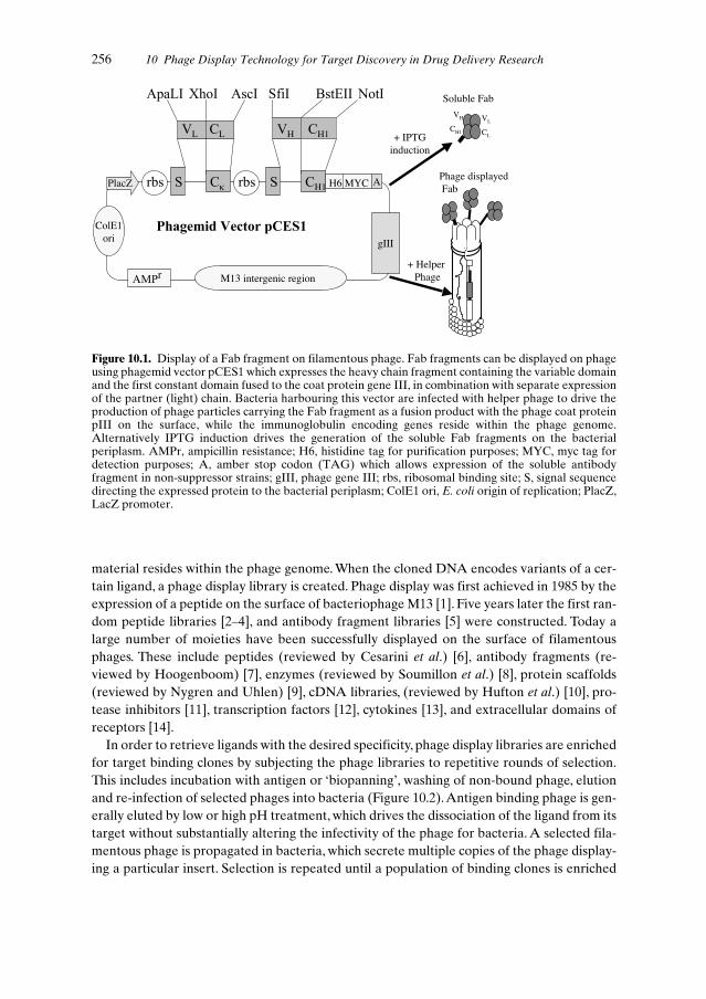
material resides within the phage genome. When the cloned DNA encodes variants of a cer-tain ligand, a phage display library is created. Phage display was first achieved in 1985 by theexpression of a peptide on the surface of bacteriophage M13 [1]. Five years later the first ran-dom peptide libraries [2–4], and antibody fragment libraries [5] were constructed. Today alarge number of moieties have been successfully displayed on the surface of filamentousphages. These include peptides (reviewed by Cesarini et al.) [6], antibody fragments (re-viewed by Hoogenboom) [7], enzymes (reviewed by Soumillon et al.) [8], protein scaffolds(reviewed by Nygren and Uhlen) [9], cDNA libraries, (reviewed by Hufton et al.) [10], pro-tease inhibitors [11], transcription factors [12], cytokines [13], and extracellular domains ofreceptors [14].
In order to retrieve ligands with the desired specificity, phage display libraries are enrichedfor target binding clones by subjecting the phage libraries to repetitive rounds of selection.This includes incubation with antigen or ‘biopanning’, washing of non-bound phage, elutionand re-infection of selected phages into bacteria (Figure 10.2).Antigen binding phage is gen-erally eluted by low or high pH treatment, which drives the dissociation of the ligand from itstarget without substantially altering the infectivity of the phage for bacteria. A selected fila-mentous phage is propagated in bacteria, which secrete multiple copies of the phage display-ing a particular insert. Selection is repeated until a population of binding clones is enriched
256 10 Phage Display Technology for Target Discovery in Drug Delivery Research
+ HelperPhage
Soluble Fab
Phage displayedFab
+ IPTGinduction
r M13 intergenic region
ColE1ori
L L
κ H1
H
H6 MYC
gIII
PlacZ A
H1
VH
CH1
VL
CL
Figure 10.1. Display of a Fab fragment on filamentous phage. Fab fragments can be displayed on phageusing phagemid vector pCES1 which expresses the heavy chain fragment containing the variable domainand the first constant domain fused to the coat protein gene III, in combination with separate expressionof the partner (light) chain. Bacteria harbouring this vector are infected with helper phage to drive theproduction of phage particles carrying the Fab fragment as a fusion product with the phage coat proteinpIII on the surface, while the immunoglobulin encoding genes reside within the phage genome.Alternatively IPTG induction drives the generation of the soluble Fab fragments on the bacterialperiplasm. AMPr, ampicillin resistance; H6, histidine tag for purification purposes; MYC, myc tag fordetection purposes; A, amber stop codon (TAG) which allows expression of the soluble antibodyfragment in non-suppressor strains; gIII, phage gene III; rbs, ribosomal binding site; S, signal sequencedirecting the expressed protein to the bacterial periplasm; ColE1 ori, E. coli origin of replication; PlacZ,LacZ promoter.

and eventually individual clones are screened for binding to the target. Any procedure thatefficiently separates binding clones from those that do not bind, can be used as a selection ap-proach. This has given rise to a large variety of selection methods (Figure 10.3).
Some phage display vectors allow ligands with desired specificity to be produced both assoluble or phage displayed molecules.Alternatively, selected ligands can be recloned for pro-duction of the soluble form, or synthesized in vitro as is commonly done for peptide ligands.Phage displayed moieties often exhibit the same or similar functional characteristics as theirnative counterparts in solution. Soluble forms of the ligand are commonly used to determinerelative specificity or affinity to the target. Genetic manipulation of the selected clones al-lows further optimization of the selected ligand to meet the requirements necessary for drugtargeting purposes.
10.2 Phage Display Technology 257
Select with antigen
E. coliAmplify in Bacteria
Rescue selected phagefor a new selection round
Phage library
Genetic Repertoire
pIII display
Display vector
TransfectE.coli
Rescuephage
Fusion Proteinencoding DNA
Wash unbound phage
Elute phages andreinfect E.coli
Cycle
Create secondary library
Figure 10.2. The phage display cycle, DNA encoding for millions of variants of a certain ligand (e.g.peptides, proteins or protein fragments) is batch cloned into the phage genome as part of the phage coatprotein pIII (coat proteins pVI and pVIII can also be used for display). From this repertoire, phagecarrying specific binding ligands can be isolated by a series of cycles of selections on the antigen, each ofwhich involves binding to antigen, washing unbound phage, elution of bound phage and re-amplificationin the bacterial cell.
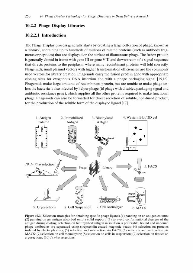
10.2.2 Phage Display Libraries
10.2.2.1 Introduction
The Phage Display process generally starts by creating a large collection of phage, known asa ‘library’, containing up to hundreds of millions of related proteins (such as antibody frag-ments or peptides) that are displayed on the surface of filamentous phage.The fusion proteinis generally cloned in frame with gene III or gene VIII and downstream of a signal sequencethat directs proteins to the periplasm, where many recombinant proteins will fold correctly.Phagemids, small plasmid vectors with higher transformation efficiencies, are the commonlyused vectors for library creation. Phagemids carry the fusion protein gene with appropriatecloning sites for exogenous DNA insertion and with a phage packaging signal [15,16].Phagemids make large amounts of recombinant protein, but are unable to make phage un-less the bacteria is also infected by helper phage (fd phage with disabled packaging signal andantibiotic resistance gene), which supplies all the other proteins required to make functionalphage. Phagemids can also be formatted for direct secretion of soluble, non-fused product,for the production of the soluble form of the displayed ligand [17].
258 10 Phage Display Technology for Target Discovery in Drug Delivery Research
Bio
Magnet
6. MACS
5. FACS
4. Western Blot/ 2D gel3. BiotinylatedAntigen
9. Cryosections
selection
2. ImmobilizedAntigen
8. Cell Suspension 7. Cell Monolayer
Phage Library
1. AntigenColumn
Magnet
Figure 10.3. Selection strategies for obtaining specific phage ligands.(1) panning on an antigen column;(2) panning on an antigen absorbed onto a solid support; (3) to avoid conformational changes of theantigen during coating, selection on biotinylated antigen in solution is preferable, bound and unboundphage antibodies are separated using streptavidin-coated magnetic beads; (4) selection on proteinsisolated by electrophoresis; (5) selection and subtraction via FACS; (6) selection and subtraction viaMACS; (7) selection on cell monolayers; (8) selection on cells in suspension; (9) selection on tissues oncryosections; (10) In vivo selections.

10.2.2.2 Peptide Display
Since many proteins exert their biological activity through relatively small regions of theirfolded surfaces, their interactions may in principle be reproduced by much smaller peptidesthat retain these localized bioactive surfaces. Peptides can provide information about themolecular interactions of protein binding sites [18], can be used as ligands for targeting pur-poses [19], are useful intermediates in the development and design of non-peptide drugs [20],and can act as small molecule drugs with biological activity [21]. Peptides are readily synthe-sized and are characterized by rapid tissue penetration having potentially improved proper-ties for certain in vivo applications [22] (Figure 10.4).
The first reported random peptide libraries were constructed in 1990, and fused to pIII[2–4]. These where followed by random libraries fused to pVIII [23]. The phage peptide li-braries are enriched on antigen, and clones analysed by sequencing to identify possible con-sensus residues. Once consensus amino acid sequences are found among selected peptides,the encoded peptides may be synthesized to confirm their specificity and determine affinityfor the target. The peptides themselves can be used as specific binding ligands in research ordiagnosis, for drug development, or they can form the starting point for the synthesis ofsmaller bioactive molecules.
10.2 Phage Display Technology 259
:Intermediaries for non peptide drugsMirror image phage displayPeptides with effector functions
Proteins with effector functions(immunoneutralizing Abs)De novo design & generation of proteinswith desired function & specificity
:Cell ELISAFACSWestern blotImmunohistochemistryIn vitro and in vivo studies
Western blotImmunoprecipitationMicrosequencingExpression cloning (cDNA libraries)
:From 2D gelsFrom expression cDNA librariesSelect binders on complex antigensPathfinderCell selectionsTissue selectionsIn vivo selections
Scaffolds with functional residuesAffinity maturationEngineering avidity & valencyTailoring effector functions
Figure 10.4. Application of phage display in drug and target discovery.

Phage peptide libraries may contain either constrained [24] or linear peptides. For the for-mer, the peptide is presented in a structurally constrained, thus more rigid form, for exampleusing cysteine bridging. Linear peptides can adopt more conformations in solution; they areparticularly useful for determining the consensus for the recognition and modification sitesof proteases, kinases etc. [25]. Constrained peptides often produce higher affinity ligands[26,27].
In general, relatively large peptides can be displayed by fusion to pIII, whereas for pVIIIdisplay, the maximum size of the peptide is usually limited to around 15 amino acid residues.This size restriction is less pronounced when using phagemids, in which wild-type coat pro-tein competes with the fusion product for insertion into the phage coat. In the case of pVIIIfusion, polyvalent display is achieved, since over 2000 copies of the coat protein are incorpo-rated into the viral particle. With phagemid systems lower numbers of fusion proteins perphage particle are obtained, but the phage coat protein may be engineered to allow more ef-ficient display [28]. Generally polyvalent display leads to selection of low (micromolar) affin-ity peptide binders [3]. On the other hand ‘monovalent’ display, obtained by using pIII as thefusion partner in a phagemid system, allows the selection of higher affinity (nanomolar) vari-ants [29].
Libraries can also be made from naturally occurring peptides (e.g. melanocortin [30] andsomatostatin [31]) and their variants, or be derived from protein-fragments.The former havebeen employed mainly for the identification of peptide variants with improved characteris-tics, and the latter for the identification of antigenic determinants present on targets, for ex-ample by selecting for binders to monoclonal antibodies specific for an antigen-fragmentpeptide library.
Naturally occurring peptides can be altered in size and structure, and randomization ofresidues can be introduced into libraries for the identification of peptides with improvedcharacteristics or improved binding specificity. Such an approach has been used to select re-ceptor-specific variants of atrial natriuretic peptide (ANP) with improved expression in E.coli and specificity for just one form of the ANP receptors [29].
Phage displayed peptide libraries have been successfully employed for the identificationof peptide ligands with a variety of applications. Recent reviews describe the developmentsin the generation and screening of peptide libraries [6], their application in the identificationof receptor ligands [32] and in drug development [33].
10.2.2.3 Antibody Display
One of the widest and most powerful applications of phage display technology has been inthe generation of recombinant antibodies. The first protein to be successfully displayed onphage was single-chain antibody variable fragment (scFv) [5].This was achieved by fusing thecoding sequence of the antibody variable regions to the amino terminus of the phage coatprotein pIII (Figure 10.1).
Large and highly diverse combinatorial antibody repertoires can be constructed on phage,displaying antibodies with diverse antigen combining sites, generated by the PCR amplifica-tion of antibody variable (V) genes [7,34]. Antibodies to any chosen antigen can be selectedfrom universal libraries made from non-immune sources of antibody genes. Human antibod-
260 10 Phage Display Technology for Target Discovery in Drug Delivery Research

ies may be generated from such non-immune ‘naïve’ libraries built with the V-genes of B cellsfrom human donors [35], or from synthetic libraries built with in vitro rearranged variablegermline genes [36].Alternatively, for some applications, antigen-biased antibody repertoirescan be created using V-genes from immune sources (reviewed by Hoogenboom et al.) [7].
Today several large single pot libraries with over 1010 independent clones have been gen-erated. From these libraries high affinity antibodies against virtually any antigen can be iso-lated [7]. The size of the library significantly improves the likelihood of identifying antibodyfragments with high specificity and high affinity due to the sampling of a larger diversity.
Phage displayed antibody libraries have been successfully used to isolate antibodiesagainst a variety of antigens such as self-antigens [37], haptens [38], carbohydrates [39], DNA[40] and RNA [41]. Selected antibodies may also have remarkable specificity for the antigen,for example, antibodies which are able to distinguish between two variant forms of a nativeantigen which differ in a single amino acid, have been selected [42].
The selected antibody fragments have been used in many applications, varying from anti-body-based biochemical assays to in vivo imaging of tumours. The relative ease with whichthey form multimers and fusion proteins, and the possibility of high levels of expression ofthese molecules in a variety of different hosts, makes them ideal protein-based diagnostic andpossibly future therapeutic reagents (reviewed by Hudson and Kortt) [43] (Figure 10.4).
10.2.2.4 Protein Scaffolds
Until the advent of the phage display era, antibodies were traditionally the sole source ofantigen binding molecules. By variation of surface residues of a protein, it is in principle pos-sible to build a library of proteins each with a different topological surface. From these li-braries, variants with a particular binding specificity can be selected using phage display. In-deed, protein display libraries based on proteins with very different folding patterns and thusvarying structural frameworks, have been constructed. Ideally protein scaffolds to be used indrug targeting should: (1) be small single domains, (2) be of human origin, (3) have pre-dictable pharmacokinetics (for human therapeutics), (4) have available sites or surfaces forthe introduction or transfer of functional sites, and, (5) have a sufficiently large antigen-bind-ing surface for affinity or specificity maturation. They should also be (6) suitable for engi-neering into multivalent, multi-specific molecules, or molecules with effector functions, and(7) yield high level expression in multiple hosts with the capacity to fold in vitro and in vivo.Although antibodies fulfil most of these requirements, there are applications where otherproteins may be more suitable.
Scaffold libraries have been created for several applications in affinity chromatography, orfor the generation of ligands to disease-related targets. The most developed scaffold is a twoalpha helix-containing variant of protein A from Staphylococcus, protein Z. so called ‘Affi-bodies’ [44], showing selective binding to respiratory syncytial virus (RSV) G protein, havebeen selected [45]. Some libraries have been used for the transfer of bioactive peptides orfunctional residues from one protein to a new protein scaffold. In our laboratory, human cy-totoxic T lymphocyte associated protein-4 (CTLA-4) has been used as a protein scaffold todisplay the 14mer somatostatin hormone, or RGD sequence-containing peptides. This hasproduced a series of ligands to the somatostatin receptor and αvβ3 integrin respectively, the
10.2 Phage Display Technology 261

latter being important in angiogenesis (see Chapters 9 and 7), with possible application as hu-man therapeutics [46,46a].
10.2.2.5 Engineering Proteins with Phage Libraries
Phage display technology offers a valuable tool for the in vitro evolution of molecules. By tar-geted or random mutagenesis, libraries containing variants of a protein can be constructed,and selection of these libraries under controlled conditions can result in the generation ofvariants with improved characteristics.A large variety of proteins have been successfully dis-played on phage with the purpose of selecting desired variants. Hormone variants with high-er affinity for their receptors [47], with similar biopotency and reduced size [48], or with im-proved specificity for one of its receptors [49] have been selected from phage libraries. Pro-teins with higher affinity or specificity for their receptor [50] or enzymes with higher or newcatalytic activities [51,52] have also been generated.
Minimizing proteins into significantly smaller polypeptides has also been achieved viaboth rational design processes and selection from vast combinatorial phage display libraries.Such ‘mini-proteins’ represent a potential intermediate step toward the development ofdrugs targeted to protein–protein interfaces [53]. Finally, binding ligands with desired func-tion and specificity can also be generated using a combination of phage display technologyand semi-rational protein design. In this way peptides that bind to the extracellular domainof the fibroblast growth factor (FGF) receptor, were selected from a peptide library. Guidedby the knowledge that agonist activity of FGF is conferred by the ability to cause receptordimerization, the selected peptide was expressed as a fused protein coupled to domains thatundergo spontaneous dimer formation, enhancing binding affinity to the receptor.
Furthermore, one of these fusion proteins also possessed agonist activity in vitro. This hasgenerated a small protein with no homology to FGF which binds to the FGF receptor repro-ducing the biological activity of FGF [54].
10.2.2.6 cDNA Expression Libraries
The display of cDNA libraries was first achieved on pIII by an indirect display method, basedon the interaction between the Jun and Fos leucine zippers [55,56]. This approach was suc-cessfully applied for the selection of allergenic proteins from Aspergillus fumigatus usingserum from affected patients [57].An alternative approach was later described where cDNAfusion occurred at the C-teminal domain of the minor coat protein pVI [58]. cDNA librariesare being used for the screening and selective isolation of genes by specific gene-product/lig-and interaction. For the identification of disease-related targets a cDNA fragment display li-brary can be selected against homogeneous ligands [59], such as natural ligands or ligands se-lected from antibody or peptide libraries against novel targets, or heterogeneous ligands suchas patient sera or polyclonal antibodies. The latter application has been validated by ourgroup for the selection of products from a colorectal carcinoma cDNA library using both ho-mogeneous and heterogeneous ligands [10]. Display of cDNA or cDNA fragment librarieson lambda phage is complementary to the filamentous phage display libraries. With M13
262 10 Phage Display Technology for Target Discovery in Drug Delivery Research

phage there will be a bias to display naturally secreted cDNAs or extracellular domains (in-tracellularly expressed cDNAs can also be expressed on phage, e.g. zinc finger proteins).Phage lambda display has the advantage of cytoplasmic expression of the cDNAs and high-er display levels [60]. Selection of cDNA or cDNA fragments using cells or tissues, and therapid cloning of cDNAs expressing proteins specific for these ligands should facilitate thesearch for novel targets and biologically important antigens (Figure 10.4).
10.3 Generation of Ligands Amenable for Targeting
In the following sections, procedures and approaches for retrieving ligands specific for well-defined targets will be discussed, followed by discussions relating to the application of re-fined selection methods for the identification of novel targets.
10.3.1 Selection of Ligands to Defined Targets
Most phage selection strategies rely on the availability of purified or recombinant antigen.This allows common selection strategies such as biopanning on immobilized antigen coatedonto solid supports [35], or on columns [38]. However, coating/immobilization of the antigenmay alter the conformational integrity of the antigen and as a consequence, antibodies thatdo not recognize the native form may be selected. To circumvent this problem, indirect coat-ing methods can be applied. Antibodies may be used to capture the antigen, and non-specif-ic Ig of the same species and isotype may be added during the selection procedure to avoidselection of antibodies to the capturing agent [61]. However, the most frequently usedmethod today, is based on selection with labelled soluble antigen, such as biotinylated anti-gen, followed by capture on a biotin-binding surface [62]. After incubation of phage with thebiotinylated antigen, unbound phage can be removed and the antigen-bound phage can beretrieved with streptavidin-coated paramagnetic beads and a magnet. This selection methodallows for the accurate control of selection parameters such as antigen concentration, whichmay result in selection according to the affinity for the antigen, e.g. decreasing the antigenconcentration to favour the selection of high affinity antibodies [63]. Phage libraries can alsobe incubated with biotinylated antigen and diluted into excess unlabelled antigen for vari-able times prior to capture on streptavidin-coated magnetic beads. This allows for selectionon the basis of the kinetics of dissociation (off-rate) from the antigen; e.g. longer incubationtimes with unlabelled antigen will favour selection of slow off-rate antibodies [62].
Phage display allows further variations to favour the selection of ligands directed towardsspecific epitopes present in the target antigen. Similar to the presence of immunodominantepitopes in vivo, selection-dominant epitopes exist in vitro [64]. Ligands directed to immun-odominant epitopes can be included during the panning procedure. By such ‘epitope block-ing panning’, a broader range of specific antibodies from combination libraries can be rescued. This approach has been used to isolate neutralizing human antibodies to weakly immunogenic epitopes of human immunodeficiency virus 1 (HIV-1) gp120 [65] and RSV[66].
10.3 Generation of Ligands Amenable for Targing 263

When the target molecules are not easily purified, or are cell surface antigens that requirethe presence of a lipid bilayer for maintaining their native form, selection on complex anti-gen mixtures is necessary. To avoid selection of non-relevant phage and to select antigen or,in the case of cell selections, to obtain cell type specific phage ligands, depletion and/or subtraction methods may be employed. Depletion is achieved by the incubation of the phagelibrary with non-target cell population(s), which do not display the target antigen, previousto the incubation with the target cell population. Subtraction, on the other hand, is performedwhen the phage library is incubated with both target and non-target cell populations simultaneously, and the target cell population with the bound phage is subsequently isolated.Competitive elution with an antibody or the antigen itself [67] can be used to elute only thoseantibodies homing to the desired antigen. Selection with alternating different sources of antigen to select only those ligands that bind to antigen(s) which are common to all sources[68], may also be employed.Alternatively, a method named ‘pathfinder’ has been devised for guided selection on complex antigen sources, using ligands directed towards the target molecule. These ligands, conjugated to horseradish peroxidase (HRP) can be used aspathfinders during the panning procedure. In the presence of biotin tyramine these mole-cules catalyse biotinylation of phage binding in close proximity to the target antigen, allow-ing specific recovery of ‘tagged’ phage from the total population using streptavidin. In thisway, phage binding to the target itself, or in its immediate proximity, are selectively recovered[69]. This technique has been applied for the selection of phage antibodies against antigensincluding carcinoembryonic antigen (CEA), E- and P-selectins, and for the selection of nov-el antibodies which recognize immobilized purified antigen [70].This technique could also beapplied in the future for the selection of ligands to novel targets such as unknown receptorsof known ligands or any molecules involved in other protein–protein interactions (Fig-ure 10.4).
10.3.2 Phage Display for Target Identification
10.3.2.1 In Vitro Selections on Complex Antigens
We have seen the versatility of phage display technology for the selection of ligands directedto known target molecules. However, one of the major advantages of this technology is basedon its applicability for the selection of ligands directed to novel targets present on certain tis-sues, cell types, or cells in a specific stage of differentiation or in disease-induced states. Theselection methods developed for defined targets may now be used to detect, by virtue of thephage, novel ligands or epitopes in the antigenic mixture. In addition, specific protocols havebeen developed to direct the selection process towards the isolation of tissue- or cell type-specific ligands (Figure 10.2).
In principle, whole live cells or crude preparations derived from cells or tissues can be usedas an antigen source to identify novel targets. For example, measles virus-infected cell lysateshave been used to select antibodies which reacted specifically with measles virus-infectedcells [71]. Protein mixtures from cells or tissues may first be separated to direct the selectionto specific cell compartments or antigens. By SDS-PAGE, the antigen(s) of interest can beidentified and isolated, e.g. antigens detected in 2-D gels of cell lysates of target cells but not
264 10 Phage Display Technology for Target Discovery in Drug Delivery Research
in lysates of non-relevant cells. The target antigen(s) can then be blotted onto a membrane,or eluted from the gel for later use as an antigen source for selection. This principle has beenproven by the selection of antibodies recognizing the native form of the ED-B domain of fi-bronectin, a marker of angiogenesis [72].
Selection techniques are often adapted to conditions where antigenic expression is as closeas possible to the in vivo situation.Antigen expression on cells is highly dependent on the cel-lular environment and on cell–cell and cell–matrix interactions. In order to maintain somecell–cell and partial cell–matrix interactions, cell panning can be carried out on in vitro cul-tured cell monolayers, where culture conditions can also be further modified in order to mim-ic the antigenic expression in vivo. Cell panning can also be carried out with cells in solution.Unbound phage can be removed after the cells and bound phage are separated from the su-pernatant by centrifugation or by magnetic retrieval with magnetically labelled beads whichbind to the cells (reviewed by Mutuberria et al.) [73].
A highly pure cell population, with its specific bound phages, can be isolated from complexcell mixtures by the use of magnetic activated cell sorting (MACS) [74] or by fluorescent ac-tivated cell sorting (FACS) [75,76]. Cells bearing the antigens of interest are magnetically orfluorescently labelled via cell-specific antibodies. Following incubation of the cell mixturewith the phage library, the antigen-positive cell population and bound phage is retrievedfrom the antigen-negative cells using MACS or FACS. When the phage library is incubatedwith the cell mixture prior to target cell isolation (subtraction), selection of target cell-spe-cific antibodies may be favoured, since negative non-target cells will compete for antibodiesthat bind to common antigens. MACS selections on a model system has shown clear advan-tages over other cell selection methods such as selections on monolayers [73].This may be at-tributed to the improved interaction between cells and phage, the efficient elimination ofnon-relevant phage by washing the cells immobilized on a magnetic column, and the high cellviability and integrity during this mild selection procedure. We have applied this selectionmethod using a large naive antibody library on mildly fixed endothelial cells immobilized ona magnetic separation column.Angiogenic factors and tumour-conditioned media were usedto induce tumour angiogenesis-associated antigen expression on cultured human umbilicalvein endothelial cells. A large number of endothelial cell binders have been selected by thisapproach, most of which bind preferentially to angiogenic vasculature, and therefore tumourvasculature. A further advantage of the MACS selection approach is that it is based on theisolation of positive target cells on magnetic columns, from which irrelevant phage can be si-multaneously washed off, using simple, efficient and gentle methods.
As an alternative to selections on cells, other antigen sources, which better maintain the invivo antigen expression profile and that allow appropriate in vitro selection procedures, areavailable. Selection on tissue cryosections may result in antibodies directed to intra- and ex-tracellular antigens on any cell type present in the tissue section, as well as antibodies bind-ing to matrix components [77].
Model selections, in which phage antibodies with defined specificity are mixed with non-specific phage and the enrichment and yield for a selection procedure is measured, providesa rapid experimental approach for studying such complex selection procedures. In order tocompare different selection approaches on complex preparations, and to determine the bestselection parameters for each approach, an extensive study of various in vitro and in vivomodel selections was recently carried out by our group [73].
10.3 Generation of Ligands Amenable for Targeting 265

10.3.2.2 In Vivo Selections and Selections for Functional Activity
In an original approach named ‘in vivo selection’ [19], phage capable of selective homing todifferent tissues, such as the vasculature of lung, skin, and pancreas [78], were recovered froma phage display peptide library following intravenous administration of the library to a livingmouse. In vivo selections have also been carried out in animals with a human tumourxenograft. Phage specifically bound to the murine tumour vasculature could then be recov-ered from the tumour tissue and amplified to yield tumour-specific endothelial cell bindingpeptides [79]. So far in vivo selections strategies have been limited to the selection of pep-tides directed to murine endothelial cell markers. This technique is producing ligands thatmay have extensive application in vasculature targeting. This was demonstrated with in vivoselected anti-integrin peptides coupled to the anti-cancer drug doxorubicin. The targeteddrug–peptide complex enhanced the efficacy of the drug against human breast cancerxenografts in nude mice with reduced toxicity [79].
All the selection methods described are applicable for the generation of ligands with bio-logical activity. Often ligands selected by conventional techniques can be screened for a for-tuitous biological function: immunoneutralizing antibodies, receptor agonists or antagonistscan be identified from a pool of selected ligands when screened for biological function. Al-ternatively, selections may be targeted towards biologically active sites of the antigens.
Some selection methods have been specifically designed for the selection of ligands with aparticular biological effector function. Such ‘selection for function’ has been used for the re-trieval of catalytic antibodies [80] and cell-internalizing phage antibodies [81], the latter be-ing highly useful molecules for the delivery of drugs, toxins, or DNA into the cytoplasm ofmammalian cells. Selection of antibody or peptide ligands for their function may, in the fu-ture, be directed towards cell survival or killing upon ligand binding, cell transfection, inhibi-tion of cell surface molecules such as drug transport molecules and inhibition of viral entryand receptor cross linking or triggering [7].
10.4 Engineering and Optimization for Targeting
Once the specific ligand has been selected, large arrays of possibilities are available in orderto reshape the ligand to obtain the best targeting results. In drug targeting applications, phar-macokinetics, biodistribution, penetration, and bioactivity are strictly governed by the char-acteristics of the ligand.
Certain therapeutic applications require high affinity ligands. Ligands such as peptideswith intrinsic low affinity for target antigens can be affinity matured. Secondary libraries ofthe selected peptide can be created by selectively incorporating mutations, and variants withhigher affinities can be selected. Often the display format will change from multi- to mono-valent display to aid a genuine affinity selection [82].
Affinity maturation of antibodies has been achieved by the introduction of diversity intothe V-genes, which then creates diversity within the antigen binding sites. This secondary li-brary is then subjected to a selection that will enrich high affinity variants. More or less ran-dom diversity may be introduced by altering variable domain pairings in a process called
266 10 Phage Display Technology for Target Discovery in Drug Delivery Research

chain shuffling [38], by error prone PCR [62], by using bacterial mutating strains [83], or byDNA shuffling [84].Alternatively, the antigen binding region may be mutated using oligonu-cleotide-directed mutagenesis or by the introduction, with limited frequency, of random mu-tations using PCR. The highest affinity increases in antibodies have been achieved when di-recting the mutations to the complementary determining region 3 (CDR3), yielding in somecases picomolar affinity antibodies [85,86]. Similar methodologies can be used with differentligands. In addition, it is not only library-derived ligands which can be affinity matured. Hy-bridoma-derived monoclonal antibodies, as well as other proteins or protein fragments maybe affinity matured by some of the methods mentioned above.Affinity maturation of the lig-and can also result in high affinity molecules with improved in vivo biodistribution. This wasdemonstrated by the anti-human CEA specific scFv, isolated and affinity matured by phagedisplay technology. Both the original and affinity-matured antibody showed targeting to tu-mour xenografts. However, although no difference was detected in tumour uptake, the affin-ity-matured antibody with improved off-rates, was retained in the tumour for a longer peri-od. [87]. Occasionally, maximizing the affinity in vitro may result in modification of the anti-body specificity which could complicate the use of the resulting ligands for therapeutic ap-plications [88].
The possibility of specifically tailoring antigen-binding properties can also be directed towards the engineering of avidity and valency. Antibody fragments are known to have more rapid tissue penetration than full antibody molecules, as demonstrated in tumour tar-geting [89]. Phage display has made easier the use of recombinant DNA technology for thedevelopment of multi-specific and multivalent molecules, previously generated using non-recombinant methods. Dimeric antibody fragments, or ‘diabodies,’ can be designed for bivalent or bispecific interactions [90]. Phage libraries displaying bivalent bispecific anti-body fragments have also been constructed. Diabody libraries enable the selection of themost appropriate bispecific molecules, with the highest affinity for binding, epitope recogni-tion and stability [91]. Multi-specific ligands can be used to direct targeted drugs to one ormore cellular antigens which may be present in either a particular cell type or diverse celltypes, or to stromal or secreted proteins found in the target tissue. Multivalent ligands havebeen exploited mainly for immunotherapy, in the stimulation of cytotoxic pathways in vivo to treat cancer [92]. The use of phage display in immunotherapy has been recently reviewed[93].
Ligands can be tailored to improve their in vivo stability. To improve the in vivo thermalstability of the tumour targeting monoclonal anti-epithelial glycoprotein-2 (EGP-2) anti-body, the antigen binding residues were grafted onto the framework of a highly stable scFvresulting in increased serum stability at 37°C [94].The stability of peptides for in vivo use hasbeen approached in a different manner, using selection of peptide libraries on the D-form ofthe antigen [95]. This technique named ‘mirror image phage display’ has recently been em-ployed in the generation of D-peptide inhibitors of HIV-1. Peptides directed against the mir-ror image (chemically synthesized with D-amino acids) of a pocket of a viral protein involvedin viral entry, were first selected. The D-peptide mirror images of the selected consensus se-quences, binding to the natural ‘L’ form of the target, were then chemically synthesized. Be-cause these D-peptides are not subject to degradation by naturally occurring enzymes, theycan be used as the starting point for the development of new drugs or as effective orally ad-ministered pharmaceuticals [96].
10.4 Engineering and Optimization for Targeting 267
A phage display-selected scFv directed against CEA has been designed to have effectorfunctions and tested for its potential in antibody-directed enzyme pro-drug therapy(ADEPT). A biologically active recombinant fusion protein containing anti-CEA scFv andthe enzyme pseudomonas carboxypeptidase (CPG2) has been produced as a recombinantprotein in E. coli and was shown to localize in human colon tumour xenografts. The tumourtargeting properties combined with the biological properties of the enzyme can be exploitedto induce tumour-specific pro-drug activation, in cases where a non-toxic pro-drug is con-verted by the action of the targeted enzyme into a highly cytotoxic drug [97].
Finally, phage display may help to address the issue of immunogenicity, particularly of non-human antibodies. Antibody humanization is often achieved by grafting CDR-loops into hu-man antibody fragments. However, humanization often requires the replacement of keyresidues involved in antigen binding in the framework regions, with corresponding residuesfrom the parent non-human antibody. A phage display method that allows selection offramework mutations which increase the binding of humanized antibodies has been de-scribed for the humanization of the anti-vascular endothelial growth factor (VEGF) murineantibody A4.6.1 [98]. The humanized version of this antibody is currently in phase II clinicaltrials to evaluate the inhibitory effect of this antibody-drug on tumour growth and neovas-cularization. Furthermore, by guided selection [99], a rodent antibody may be rebuilt into afully human antibody. In two consecutive rounds of chain shuffling, the rodent antibodygenes are replaced by human genes, which will mediate binding to a highly similar if not iden-tical epitope on the antigen, differing only in their chemistry of interaction [100].
10.5 Discovery of Novel Therapeutics Using Phage DisplayTechnology
Although phage display technology is becoming a widespread research tool, few ligands gen-erated by this technology have reached clinical trials. From the large array of ligands gener-ated by phage display with possible therapeutic applications, there are five antibodies select-ed from phage libraries currently undergoing clinical trials as drug candidates. Preliminarydata with a fully human anti-tumour necrosis factor alpha (TNFα)-neutralizing monoclonalantibody for the treatment of rheumatoid arthritis, demonstrated that the antibody was safeand effective in early clinical trials [101]. Other phage library-derived antibodies that are un-dergoing clinical trials include antibodies to interleukin 2 (IL-2) for the treatment of au-toimmune and inflammatory disorders, an anti-transforming growth factor beta-1 (TGFβ1)antibody as an anti-fibrotic drug, and an anti-TGFβ2 antibody for the treatment of prolifer-ative vitreoretinopathy and prevention of scarring in the eye following glaucoma therapy. Alarge series of ligands which may have possible therapeutic application are undergoing fur-ther characterization and optimization.
Cancer has become a major target in the application of this technology, and human anti-bodies against tumour antigens such as CEA, EGP-2 and Mucin-1 (MUC-1) are alreadyavailable. Also a series of peptides and antibodies directed against angiogenesis-relatedmarkers such as basic fibroblast growth factor (bFGF), VEGFs and their receptors, tumour-
268 10 Phage Display Technology for Target Discovery in Drug Delivery Research

associated fibronectin, tenascin isoforms, integrins or metalloproteases, have been selectedfor their potential application in tumour targeting, inhibition of tumour growth and in otherangiogenesis-related diseases (see Chapter 9 for drug targeting strategies aimed at angio-genesis-related molecules).
Identification of cell receptor ligands has generated a series of molecules with excellent re-ceptor specificity and occasionally with desired effector functions.This is the case for peptideantagonists of the human estrogen receptor, or cyclic peptides capable of activating the ery-thropoietin receptors (EPOR) isolated from phage display libraries. Other ligands with ef-fector functions include thrombin inhibitors, viral inhibitors and even intracellular targetssuch as a reverse transcriptase inhibitory antibody.Table 10.1 lists a number of ligands, whichmay in the future have widespread application as therapeutic agents.
10.5 Discovery of Novel Therapeutics Using Phage Display Technology 269
Table 10.1. Example of phage display ligands with therapeutic applications.
Name of ligand Type of ligand Target and applicability Reference
D2E7 scFv TNFα/Rheumatoid arthritis [101]
CAT-152 scFv TGFβ2/Glaucoma surgery, proliferative vitreo retinopathy http://www.catplc.co.uk
CAT-192 scFv TGFβ1/Antifibrogenic treatment http://www.catplc.co.uk
J695 scFv IL-2/Autoimmunity, Inflammation http://www.catplc.co.uk
Anti CEA CEA/Tumour targeting Reviewed in reference antibodies [102]
UBS-54 scFv Ep-CAM (EGP-2)/Colorectal carcinoma [103,104]
L19 scFv Fibronectin ED-B/Angiogenesis (ocular [105,106,107]neovasculature and tumour targeting)
TN11 scFv Tenascin C isoform/High grade [108]astrocytoma
10A scFv Mucin-1/Adenocarcinoma [109]PH1 Fab [110]
Human Fab Fab VEGF/Treatment of macular [111]Y0317 degeneration, angiogenesis inhibition http://www.gene.com/
and tumour growth. Pipeline/index.html
Anti GP 41 D- Peptide HIV GP41/Inhibition of HIV-1 [96]pep infection
RGD- Peptide αvβ3 integrin/Targeting angiogenic [79]Doxorubicin vasculature
PLAEIDGIELTY Peptide α9β1 integrin/Targeting lung epithelia [112](cystic fibrosis)
REA18 (rNAP) Peptide ANP-A receptor / Induction of [29,49]natriuresis and diuresis. Treatment of acute renal or heart failure
10.6 Conclusions
Phage display has become the most efficient and effective method developed to date forrapidly identifying peptides, antibodies and other proteins that bind to molecular targets.Some of these ligands have biological effector functions or are candidates for ligand-baseddrug targeting. Identification of the antigens targeted by phage-displayed ligands has beensimplified by this technology, by the use of DNA display libraries and by coupling gene iso-lation and clone identification with affinity selection, facilitating the search for novel targetsand biologically important antigens. Selection for function allows the retrieval of ligands witheffector functions for use in the generation of drugs. Selection for cellular internalization hasopened a new door for the delivery of macromolecular constructs and coupled drugs into thecytoplasm of mammalian cells.
The physical bond between the selected ligand and its encoding gene allows further ma-nipulation of the ligands to obtain optimal affinity and avidity, size, and valency. Further-more, the coupling of a drug to the targeting ligands can be readily engineered.
The design features required for an optimally-effective drug and/or targeting agent can bereadily obtained by phage display technology. Many features give phage display technologyclear advantages over conventional approaches for the generation of reagents for drug tar-geting purposes. These include diversity in protein type and sequence space of combinatori-al phage libraries, the power of filamentous phage-based selection, the possibilities of genet-ic manipulation to generate more effective ligands and specific effector functions, and theadaptability of the system for the production of therapeutic ligands derived from the li-braries.
In the near future phage display technology may have further application in the rapidanalysis and comparison of protein profiles of large proteomes, such as human cells and tis-sue.Arrays of phage-displayed proteins such as antibodies or peptides, which selectively bindto proteins from a complex mixture can be generated. Proteins absorbed by the antibody ar-rays can then be analysed by mass spectrometry. Such technologies will be especially usefulfor identifying differences between cell or tissue samples such as healthy versus diseasedstates, and may lead to the identification of drug targets. Once a protein of interest has beenidentified, its corresponding antibody or peptide ligand can be retrieved and used to monitorprotein expression or modification in a range of cell or tissue samples, and can also be usedfor cloning the target antigen.
Phage display has become a powerful method for the generation of protein-based bindingand biologically active reagents. In the forthcoming years an extensive application of thistechnique is predicted for the development of drugs and drug targeting entities.
References
[1] Smith GP, Science 1985, 228, 1315–1317.[2] Devlin JJ, Panganiban LC, Devlin PE, Science 1990, 249, 404–406.[3] Cwirla SE, Peters EA, Barrett RW, Dower WJ, Proc. Natl Acad. Sci. USA 1990, 87, 6378–6382.[4] Scott JK, Smith GP, Science 1990, 249, 386–390.[5] McCafferty J, Griffiths AD, Winter G, Chiswell DJ, Nature 1990, 348, 552–554.[6] Cesareni G, Castagnoli L, Cestra G, Comb. Chem. High Throughput Screen 1999, 2, 1–17.
270 10 Phage Display Technology for Target Discovery in Drug Delivery Research

[7] Hoogenboom HR, de Bruine AP, Hufton SE, Hoet RM, Arends JW, Roovers RC, Immunotech-nology 1998, 4, 1–20.
[8] Soumillion P, Jespers L, Bouchet M, Marchand-Brynaert J, Sartiaux P, Fastrez J, Appl. Biochem.Biotechnol. 1994, 47, 175–190.
[9] Nygren PA, Uhlen M, Curr. Opin. Struct. Biol. 1997, 7, 463–469.[10] Hufton SE, Moerkerk PT, Meulemans EV, de Bruine A, Arends J, Hoogenboom HR, J. Im-
munol. Methods 1999, 231, 39–51.[11] Dennis MS, Lazarus RA, J. Biol. Chem. 1994, 269, 22129–22136, [12] Wu H, Yang WP, Barbas CF, III, Proc. Natl Acad. Sci. USA 1995, 92, 344–348.[13] Gram H, Strittmatter U, Lorenz M, Gluck D, Zenke G, J. Immunol. Methods 1993, 161, 169–176.[14] Scarselli E, Esposito G, Traboni C, FEBS Lett. 1993, 329, 223–226.[15] Garrard LJ, Yang M, O’Connell MP, Kelley RF, Henner DJ, BioTechnology 1991, 9, 1373–1377.[16] Bass S, Greene R, Wells JA, Proteins 1990, 8, 309–314.[17] Hoogenboom HR, Griffiths AD, Johnson KS, Chiswell DJ, Hudson P, Winter G, Nucleic Acids
Res. 1991, 19, 4133–4137.[18] Geysen HM, Rodda SJ, Mason TJ, Tribbick G, Schoofs PG, J. Immunol. Methods 1987, 102,
259–274.[19] Pasqualini R, Ruoslahti E, Nature 1996, 380, 364–366.[20] Blackburn BK, Lee A, Baier M, Kohl B, Olivero AG, Matamoros R, Robarge KD, McDowell RS,
J. Med. Chem. 1997, 40, 717–729.[21] Buckley CD, Pilling D, Henriquez NV, Parsonage G, Threlfall K, Scheel-Toellner D, Simmons
DL, Akbar AN, Lord JM, Salmon M, Nature 1999, 397, 534–539.[22] Lister-James J, Moyer BR, Dean T, Q. J. Nucl. Med. 1996, 40, 221–233.[23] Felici F, Castagnoli L, Musacchio A, Jappelli R, Cesareni G, J. Mol. Biol. 1991, 222, 301–310.[24] Luzzago A, Felici F, Tramontano A, Pessi A, Cortese R, Gene 1993, 128, 51–57.[25] Matthews DJ, Wells JA, Science 1993, 260, 1113–1117.[26] Ladner RC, Trends Biotechnol. 1995, 13, 426–430.[27] Zang X, Yu Z, Chu YH, Bioorg. Med. Chem. Lett. 1998, 8, 2327–2332.[28] Sidhu SS, Weiss GA, Wells JA, J. Mol. Biol. 2000, 296, 487–495.[29] Cunningham BC, Lowe DG, Li B, Bennett BD, Wells JA, EMBO J. 1994, 13, 2508–2515.[30] Szardenings M, Tornroth S, Mutulis F, Muceniece R, Keinanen K, Kuusinen A, Wikberg JE, J.
Biol. Chem. 1997, 272, 27943–27948.[31] Rousch M, Lutgerink JT, Coote J, de Bruine A, Arends JW, Hoogenboom HR, Br. J. Pharmacol.
1998, 125, 5–16.[32] Koivunen E, Arap W, Rajotte D, Lahdenranta J, Pasqualini R, J. Nucl. Med. 1999, 40, 883–888.[33] Lowman HB, Annu. Rev. Biophys. Biomol. Struct. 1997, 26, 401–424.[34] Winter G, Griffiths AD, Hawkins RE, Hoogenboom HR, Ann. Rev. Immunol. 1994, 12, 433–455.[35] Marks JD, Hoogenboom HR, Bonnert TP, McCafferty J, Griffiths AD, Winter G, J. Mol. Biol.
1991, 222, 581–597.[36] Hoogenboom HR, Winter G, J. Mol. Biol. 1992, 227, 381–388.[37] Griffiths AD, Malmqvist M, Marks JD, Bye JM, Embleton MJ, McCafferty J, Baier M, Holliger
KP, Gorick BD, Hughes JN, Hoogenboom HR, Winter G, EMBO J. 1993, 12, 725–734.[38] Clackson T, Hoogenboom HR, Griffiths AD, Winter G, Nature 1991, 352, 624–628.[39] Deng SJ, MacKenzie CR, Sadowska J, Michniewicz J, Young NM, Bundle DR, Narang SA, J.
Biol. Chem. 1994, 269, 9533–9538.[40] Barbas SM, Ditzel HJ, Salonen EM, Yang WP, Silverman GJ, Burton DR, Proc. Natl Acad. Sci.
USA 1995, 92, 2529–2533.[41] Powers JE, Marchbank MT, Deutscher SL, Nucleic Acids Symp. Ser. 1995, 33, 240–243.[42] Griffin HM, Ouwehand WH, Blood 1995, 86, 4430–4436.[43] Hudson PJ, Kortt AA, J. Immunol. Methods 1999, 231, 177–189.[44] Nord K, Gunneriusson E, Ringdahl J, Stahl S, Uhlen M, Nygren PA, Nature Biotechnol. 1997, 15,
772-777.[45] Hansson M, Ringdahl J, Robert A, Power U, Goetsch L, Nguyen TN, Uhlen M, Stahl S, Nygren
PA, Immunotechnology 1999, 4, 237–252.[46] Nuttall SD, Rousch MJ, Irving RA, Hufton SE, Hoogenboom HR, Hudson PJ, Proteins 1999, 36,
217–227.[46a] Hufton SE, van Neer N, van den Beuken T, Desmet J, Sablon E, Hoogenboom HR, FEBS Lett.,
2000, 475, 225–231.[47] Lowman HB, Wells JA, J. Mol. Biol. 1993, 234, 564578.
References 271

[48] Li B, Tom JY, Oare D, Yen R, Fairbrother WJ, Wells JA, Cunningham BC, Science 1995, 270,1657–1660.
[49] Jin H, Li B, Cunningham B, Tom J, Yang R, Sehl P, Thomas GR, Ko A, Oare D, Lowe DG, J.Clin. Invest. 1996, 98, 969–976.
[50] Ballinger MD, Jones JT, Lofgren JA, Fairbrother WJ, Akita RW, Sliwkowski MX, Wells JA, J.Biol. Chem. 1998, 273, 11675–11684.
[51] Hansson LO, Widersten M , Mannervik B, Biochem. J. 1999, 344, 93–100.[52] Demartis S, Huber A, Viti F, Lozzi L, Giovannoni L, Neri P, Winter G, Neri D, J. Mol. Biol. 1999,
286, 617–633.[53] Cunningham BC, Wells JA, Curr. Opin. Struct. Biol. 1997, 7, 457–462.[54] Ballinger MD, Shyamala V, Forrest LD, Deuter-Reinhard M, Doyle LV, Wang J, Panganiban-
Lustan L, Stratton JR, Apell G, Winter JA, Doyle MV, Rosenberg S, Kavanaugh WM, NatureBiotechnol. 1999, 17, 1199–1204.
[55] Crameri R, Suter M, Gene 1993, 137, 69–75.[56] Crameri R, Jaussi R, Menz G, Blaser K, Eur. J. Biochem. 1994, 226, 53–58.[57] Crameri R, Faith A, Hemmann S, Jaussi R, Ismail C, Menz G, Blaser K, J. Exp. Med. 1996, 184,
265–270.[58] Jespers L, Messens JH, De Keyser A, Eeckhout D, Van den Brande J, Gandemans Y, Lauwereys
MJ, Vlasuk GP, Stanssens PE, Bio/Technol. 1995, 13, 378–382.[59] Sche PP, McKenzie KM, White JD, Austin DJ, Chem. Biol. 1999, 6, 707–716.[60] Santi E, Capone S, Mennuni C, Lahm A, Tramontano A, Luzzago A, Nicosia A, J. Mol. Biol.
2000, 296, 497–508.[61] Sanna PP, Williamson RA, De LA, Bloom FE, Burton DR, Proc. Natl Acad. Sci. USA 1995, 92,
6439–6443.[62] Hawkins RE, Russell SJ, Winter G, J. Mol. Biol. 1992b, 226, 889–896.[63] Hawkins RE, Russell SJ, Baier M, Winter G, J. Mol. Biol. 1993, 234, 958–964.[64] Hoogenboom HR, Lutgerink JT, Pelsers MM, Rousch MJ, Coote J, Van Neer N, De Bruine A,
Van Nieuwenhoven FA, Glatz JF, Arends JW, Eur. J. Biochem. 1999, 260, 774–784.[65] Ditzel HJ, Binley JM, Moore JP, Sodroski J, Sullivan N, Sawyer LS, Hendry RM, Yang WP, Bar-
bas CF, Burton DR, J. Immunol. 1995, 154, 893–906.[66] Ping T, Tornetta MA, Ames RS, Bankosky BC, Griego S, Silverman C, Porter T, Moore G, Sweet
RW, J. Immunol. 1996, 157, 772–780.[67] Meulemans EV, Slobbe R, Wasterval P, Ramaekers FC, van EG, J. Mol. Biol. 1994, 244, 353–360.[68] Andersen PS, Stryhn A, Hansen BE, Fugger L, Engberg J, Buus S, Proc. Natl Acad. Sci. USA
1996, 93, 1820–1824.[69] Osbourn JK, Earnshaw JC, Johnson KS, Parmentier M, Timmermans V, McCafferty J, Nature
Biotechnol. 1998, 16, 778–781.[70] Osbourn JK, Derbyshire EJ, Vaughan TJ, Field AW, Johnson KS, Immunotechnology 1998, 3,
293–302.[71] Burgoon MP, Williamson RA, Owens GP, Ghausi O, Bastidas RB, Burton DR, Gilden DH, J.
Neuroimmunol. 1999, 94, 204–211.[72] Pini A, Viti F, Santucci A, Carnemolla B, Zardi L, Neri P, Neri D, J. Biol. Chem. 1998, 273,
21769–21776.[73] Mutuberria R, Hoogenboom HR, van der Linden E, de Bruine AP, Roovers RC, J. Immunol.
Methods 1999, 231, 65–81.[74] Siegel DL, Chang TY, Russell SL, Bunya VY, J. Immunol. Methods 1997, 206, 73–85.[75] de Kruif J, Boel E, Logtenberg T, J. Mol. Biol. 1995, 248, 97–105.[76] Palmer DB, George AJ, Ritter MA, Immunology 1997, 91, 473–478.[77] Tordsson J, Abrahmsen L, Kalland T, Ljung C, Ingvar C, Brodin T, J. Immunol. Methods 1997,
210, 11–23.[78] Rajotte D, Arap W, Hagedorn M, Koivunen E, Pasqualini R, Ruoslahti E, J. Clin. Invest. 1998,
102, 430–437.[79] Arap W, Pasqualini R, Ruoslahti E, Science 1998, 279, 377–380.[80] Janda KD, Lo CH, Li T, Barbas CF, Wirsching P, Lerner RA, Proc. Natl Acad. Sci. USA 1994, 91,
2532–2536.[81] Becerril B, Poul MA, Marks JD, Biochem. Biophys. Res. Commun. 1999, 255, 386–393.[82] Wrighton NC, Farrell FX, Chang R, Kashyap AK, Barbone FP, Mulcahy LS, Johnson DL, Barrett
RW, Jolliffe LK, Dower WJ, Science 1996, 273, 458–464.[83] Low NM, Holliger PH, Winter G, J. Mol. Biol. 1996, 260, 359–368.
272 10 Phage Display Technology for Target Discovery in Drug Delivery Research

[84] Crameri A, Cwirla S, Stemmer WP, Nature Med. 1996, 2, 100–102.[85] Yang WP, Green K, Pinz SS, Briones AT, Burton DR, Barbas Cr, J. Mol. Biol. 1995, 254, 392–403.[86] Schier R, McCall A, Adams GP, Marshall KW, Merritt H, Yim M, Crawford RS, Weiner LM,
Marks C, Marks JD, J. Mol. Biol. 1996, 263, 551–567.[87] Jackson H, Bacon L, Pedley RB, Derbyshire E, Field A, Osbourn J, Allen D, Br. J. Cancer. 1998,
78, 181–188.[88] Casson LP, Manser T, J. Exp. Med. 1995, 182, 743–750.[89] Chester KA, Begent RH, Robson L, Keep P, Pedley RB, Boden JA, Boxer G, Green A, Winter
G, Cochet O, Lancet 1994, 343, 455–456.[90] Holliger P, Prospero T, Winter G, Proc. Natl Acad. Sci. USA 1993, 90, 6444–6448.[91] McGuinness BT, Walter G, FitzGerald K, Schuler P, Mahoney W, Duncan AR, Hoogenboom
HR, Nature Biotechnol. 1996, 14, 1149–1154.[92] Bauer S, Renner C, Juwana JP, Held G, Ohnesorge S, Gerlach K, Pfreundschuh M, Cancer Res.
1999, 59, 1961–1965.[93] de Haard H, Henderikx P, Hoogenboom HR, Adv. Drug Del. Rev. 1998, 31, 5–31.[94] Willuda J, Honegger A, Waibel R, Schubiger PA, Stahel R, Zangemeister-Wittke U, Pluckthun
A, Cancer Res. 1999, 59, 5758–5767.[95] Schumacher TN, Mayr LM, Minor Jr DL, Milhollen MA, Burgess MW, Kim PS, Science 1996, 271,
1854–1857.[96] Eckert DM, Malashkevich VN, Hong LH, Carr PA, Kim PS, Cell 1999, 99, 103–115.[97] Michael NP, Chester KA, Melton RG, Robson L, Nicholas W, Boden JA, Pedley RB, Begent RH,
Sherwood RF, Minton NP, Immunotechnology 1996, 2, 47–57.[98] Baca M, Presta LG, SJ OC, Wells JA, J. Biol. Chem. 1997, 272, 10678–10684.[99] Jespers LS, Roberts A, Mahler SM, Winter G, Hoogenboom HR, Biotechnology 1994, 12,
899–903.[100] Beiboer SH, Reurs A, Roovers RC, Arends JW, Whitelegg NR, Rees AR, Hoogenboom HR, J.
Mol. Biol. 2000, 296, 833–849.[101] Kempeni J, Ann. Rheum. Dis. 1999, 58 (Suppl. 1), I70–I72.[102] Mayer A, Chester KA, Flynn AA, Begent RH, J. Immunol. Methods 1999, 231, 261–273.[103] Huls G, Heijnen IA, Cuomo E, van der Linden J, Boel E, van de Winkel JG, Logtenberg T, Can-
cer Res. 1999, 59, 5778–5784.[104] Huls GA, Heijnen IA, Cuomo ME, Koningsberger JC, Wiegman L, Boel E, van der Vuurst de
Vries AR, Loyson SA, Helfrich W, van Berge Henegouwen GP, van Meijer M, de Kruif J, Logten-berg T, Nature Biotechnol. 1999, 17, 276–281.
[105] Carnemolla B, Neri N, Castellani P, Veirana N, Neri G, Pini A, Winter G ,Zardi L, Int. J. Cancer1996, 68, 397–405.
[106] Neri D, Carnemolla B, Nissim A, Leprini A, Querze G, Balza E, Pini A, Tarli L, Halin C, Neri P,Zardi L, Winter G, Nature Biotechnol. 1997, 15, 1271–1275.
[107] Birchler M, Viti F, Zardi L, Spiess B, Neri D, Nature Biotechnol. 1999, 17, 984–988.[108] Carnemolla B, Castellani P, Ponassi M, Borsi L, Urbini S, Nicolo G, Dorcaratto A, Viale G, Win-
ter G, Neri D, Zardi L, Am. J. Pathol. 1999, 154, 1345–1352.[109] Henderikx P, Kandilogiannaki M, Petrarca C, von Mensdorff-Pouilly S, Hilgers JH, Krambovitis
E, Arends JW, Hoogenboom HR, Cancer Res. 1998, 58, 4324–4332.[110] de Haard HJ, van Neer N, Reurs A, Hufton SE, Roovers RC, Henderikx P, de Bruine AP, Arends
JW, Hoogenboom HR, J. Biol. Chem. 1999, 274, 18218–18230.[111] Chen Y, Wiesmann C, Fuh G, Li B, Christinger HW, McKay P, de Vos AM, Lowman HB, J. Mol.
Biol. 1999, 293, 865–881.[112] Schneider H, Harbottle RP, Yokosaki Y, Jost P, Coutelle C, FEBS Lett. 1999, 458, 329–332.
References 273
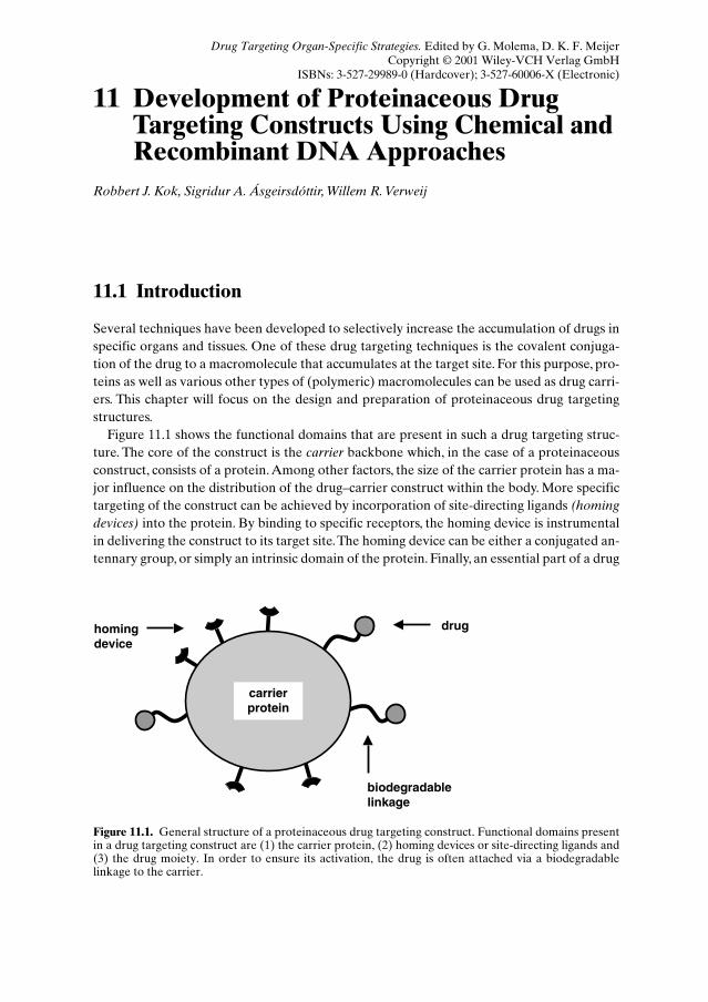
11 Development of Proteinaceous DrugTargeting Constructs Using Chemical andRecombinant DNA Approaches
Robbert J. Kok, Sigridur A. Ásgeirsdóttir, Willem R. Verweij
11.1 Introduction
Several techniques have been developed to selectively increase the accumulation of drugs inspecific organs and tissues. One of these drug targeting techniques is the covalent conjuga-tion of the drug to a macromolecule that accumulates at the target site. For this purpose, pro-teins as well as various other types of (polymeric) macromolecules can be used as drug carri-ers. This chapter will focus on the design and preparation of proteinaceous drug targetingstructures.
Figure 11.1 shows the functional domains that are present in such a drug targeting struc-ture. The core of the construct is the carrier backbone which, in the case of a proteinaceousconstruct, consists of a protein. Among other factors, the size of the carrier protein has a ma-jor influence on the distribution of the drug–carrier construct within the body. More specifictargeting of the construct can be achieved by incorporation of site-directing ligands (homingdevices) into the protein. By binding to specific receptors, the homing device is instrumentalin delivering the construct to its target site.The homing device can be either a conjugated an-tennary group, or simply an intrinsic domain of the protein. Finally, an essential part of a drug
biodegradablelinkage
homingdevice
drug
carrierprotein
Figure 11.1. General structure of a proteinaceous drug targeting construct. Functional domains presentin a drug targeting construct are (1) the carrier protein, (2) homing devices or site-directing ligands and(3) the drug moiety. In order to ensure its activation, the drug is often attached via a biodegradablelinkage to the carrier.
Drug Targeting Organ-Specific Strategies. Edited by G. Molema, D. K. F. MeijerCopyright © 2001 Wiley-VCH Verlag GmbH
ISBNs: 3-527-29989-0 (Hardcover); 3-527-60006-X (Electronic)
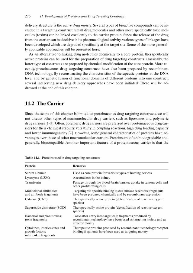
delivery structure is the active drug moiety. Several types of bioactive compounds can be in-cluded in a targeting construct. Small drug molecules and other more specifically toxic mol-ecules (toxins) can be linked covalently to the carrier protein. Since the release of the drugfrom the carrier can be decisive in its pharmacological activity, various types of linkages havebeen developed which are degraded specifically at the target site. Some of the more general-ly applicable approaches will be presented here.
As an alternative to linking drug molecules chemically to a core protein, therapeuticallyactive proteins can be used for the preparation of drug targeting constructs. Classically, thelatter type of constructs are prepared by chemical modification of the core protein. More re-cently, proteinaceous drug targeting constructs have also been prepared by recombinantDNA technology. By reconstructing the characteristics of therapeutic proteins at the DNAlevel and by genetic fusion of functional domains of different proteins into one construct,several interesting new drug delivery approaches have been initiated. These will be ad-dressed at the end of this chapter.
11.2 The Carrier
Since the scope of this chapter is limited to proteinaceous drug targeting constructs, we willnot discuss other types of macromolecular drug carriers, such as liposomes and polymericdrug carriers [1–3]. Often, polymeric drug carriers are preferred over proteinaceous drug car-riers for their chemical stability, versatility in coupling reactions, high drug loading capacityand lower immunogenicity [2]. However, some general characteristics of proteins have ad-vantages over those of other macromolecular carriers. Proteins are often biodegradable and,generally, biocompatible. Another important feature of a proteinaceous carrier is that the
276 11 Development of Proteinaceous Drug Targeting Constructs
Table 11.1. Proteins used in drug targeting constructs.
Protein Remarks
Serum albumin Used as core protein for various types of homing devices
Lysozyme (LZM) Accumulates in the kidney
Transferrin Passage through the blood–brain barrier; uptake in tumour cells andother proliferating cells
Monoclonal antibodies Targeting via specific binding to cell surface receptors; fragments and antibody fragments have been prepared chemically and by recombinant expression
Catalase (CAT) Therapeutically active protein (detoxification of reactive oxygenspecies)
Superoxide dismutase (SOD) Therapeutically active protein (detoxification of reactive oxygenspecies)
Bacterial and plant toxins; Toxic after entry into target cell; fragments produced by toxin fragments recombinant technology have been used as targeting moiety and as
effector moiety
Cytokines, interleukines and Therapeutic proteins produced by recombinant technology; receptorgrowth factors; binding fragments have been used as targeting moietyinterleukin fragments
carrier is an homogenous product. In contrast, polymeric carriers are non-homogeneous bynature. Finally, some proteins have unique characteristics that relate to their complex tertiaryor quaternary structures. For instance, the binding affinity of the protein to its natural recep-tor can be the driving force for the selective targeting of the construct. Other interesting car-rier proteins are those that are pharmacologically active themselves. Such intrinsically activeproteins can be used as active drug substances, or as carriers for small drug molecules in so-called dual active conjugates [4]. Table 11.1 lists examples of carrier proteins that will be dis-cussed in the following sections of this chapter.
11.2.1 Albumin
An important feature of the carrier protein is the size of the macromolecule. Small proteinswith a molecular weight lower than about 60 kDa, are rapidly cleared from the bloodstreamby glomerular filtration in the kidney [5]. By choosing a carrier protein with an adequatelyhigh molecular weight, renal filtration can be prevented. Being sufficiently large to preventrenal filtration, but at the same time small enough for efficient tissue penetration, the albu-min molecule has an ideal molecular weight (70 kDa). For this reason, serum albumins fromdifferent origins, such as human (HSA), bovine (BSA) or the albumin type autologous to thespecies in which the conjugate is being tested, have been the carrier of choice for many drugtargeting preparations.Two types of drug–albumin conjugates have been reported. First, sim-ple drug–albumin conjugates which accumulate in the target tissue by passive extravasationhave been described for the delivery of various anti-cancer drugs to solid tumours [6]. Due toseveral factors, such as elevated levels of vascular permeability factors and an impaired lym-phatic drainage, tumour blood vessels show an enhanced permeability and retention ofmacromolecules [7]. Preferably, a low number of drug molecules should be conjugated per al-bumin molecule, since otherwise the construct may undergo enhanced uptake by scavengerreceptors in the liver and spleen [8]. On the other hand, specific uptake by scavenger recep-tors has been exploited for the delivery of anti-inflammatory drugs to the liver [9–11].The second type of drug–albumin conjugate comprises those in which the albumin proteinfunctions as a backbone for both conjugated drug molecules and homing devices.This type ofstructure will selectively accumulate in the target tissue by binding to cell surface receptors,a process called active targeting. The binding of the construct to the target cells is primarilydriven by the qualities of the homing device, such as type and spatial orientation of the tar-geting moiety, rather than by the characteristics of the original carrier backbone. Therefore,similar structures can be prepared using carrier proteins other than albumin.
11.2.2 Low Molecular Weight Proteins
As stated earlier, proteins with a molecular weight lower than that of albumin are able topass through the glomerular membrane in the kidney. Consequently, such low molecularweight proteins (LMWPs) are rapidly removed from the bloodstream. Following glomerularfiltration, LMWPs are reabsorbed in the proximal tubular cells of the kidney. Since thisprocess makes the kidney the major catabolic site for these proteins, they can be used as car-
11.2 The Carrier 277

riers for renal drug delivery [12,13].A typical example of a protein that has been used for thistargeting purpose is lysozyme (LZM, 14 kDa) [14,15] (see Chapter 5 for a more detailed dis-cussion on the development of renal targeting preparations).
11.2.3 Monoclonal Antibodies
Monoclonal antibodies have been extensively reported on as carriers for targeted drug de-livery. Starting in the late 1970s, the production of monoclonal antibodies has now evolvedinto a routine technique that has yielded many potential carrier molecules. Particularly in thefield of cancer therapy, monoclonal antibodies are being used for the delivery of diagnosticand therapeutic agents [16] (see also Chapter 8). The original antibodies were of mouse ori-gin, evoking a human anti-mouse antibody (HAMA) immunological response when admin-istered in humans. The use of new recombinant techniques has enabled the preparation ofhumanized antibodies, in which the mouse recognition domain has been grafted onto a hu-man antibody structure [17].
Whole IgG antibodies with a molecular weight of 150 kDa, are often unable to penetratetumour tissue as efficiently as smaller molecules [18]. Therefore, smaller antibody fragmentsand genetically-engineered antibody derivatives have been investigated as drug carriers (seeFigure 11.5). These carrier molecules will be discussed in Section 11.8.1.
11.2.4 Transferrin
Some proteins are excellent carriers for drug targeting since they bind to more or less specif-ic receptors on the target cells. In addition to monoclonal antibodies, which in theory canbind to any kind of receptor, several natural ligands for cell-surface receptors have been ex-plored as carrier proteins. This approach is exemplified by constructs that have been devel-oped for targeting via the transferrin receptor (TfR).The transferrin receptor is expressed onmost proliferating cells, as well as in a few non-proliferating tissues, among which is the en-dothelium of brain capillaries. This distribution pattern has inspired the development oftransferrin-based constructs and anti-TfR antibodies as carriers for cancer therapy, as well asfor the delivery of compounds across the blood–brain barrier [19,20] (see also Chapter 2).With respect to the latter, the ability of the TfR to undergo transcytosis results in the releaseof the carrier complex in the brain, rather than in endocytosis by the endothelium of theblood–brain barrier. Once inside the central nervous system, the drug delivery construct canbind to TfR-positive cells, such as brain tumours, which can be regarded as a second step inthe delivery process.
As an alternative to targeting brain tumours which express the TfR, the transferrin ap-proach can be used for the delivery of fusion proteins which bind to pharmacological recep-tors inside the central nervous system.An example of this is the construct consisting of nervegrowth factor (NGF) and transferrin described in Section 11.8.2.3. The transferrin moiety inthis type of construct will enable it to enter the brain, upon which the drug moiety will act bybinding to its receptor. This approach seems especially suitable for compounds that cannotpass the blood–brain barrier, such as peptides and other hydrophilic substances.
278 11 Development of Proteinaceous Drug Targeting Constructs

11.3 The Homing Device
The binding of a protein to cell surface receptors can greatly enhance its selective accumula-tion and retention in the target tissue. However, most of the natural protein ligands are onlyavailable in limited amounts, insufficient for their application as carriers in drug deliverystudies. Therefore, many researchers have tried to develop non-natural receptor ligands. Fig-ure 11.2 shows some general approaches that have been followed to introduce receptor-bind-ing domains into a carrier protein. First, recognition domains, such as carbohydrates, peptidesor other molecules, can be covalently attached to side-chain residues of the core protein (Fig-ure 11.2a). Second, receptor binding domains can be introduced by recombinant DNA engi-neering of the protein backbone (Figure 11.2b). Third, the receptor binding properties of aprotein can be altered by the removal or modification of residues on the surface of the mol-ecule. Examples of such strategies are the partial deglycosylation of a protein (Figure 11.2c)or modifications in the surface-charge of the protein (Figure 11.2d).
A major advantage of coupling receptor binding ligands to a carrier protein is the multi-valent character of the construct obtained, which generally results in a drastic increase in the
11.3 The Homing Device 279
-
- - --
-
-
-+
++ +
+
+
+
+
(a) (b)
(d)(c)
Figure 11.2. Different approaches for the introduction of homing devices into proteinaceous drugtargeting constructs. (a) Site-directing ligands such as carbohydrates or short peptide sequences can bereacted to side-chain residues of the core protein. Typically, such homing devices are reacted to primaryamino groups of the protein. (b) Using recombinant DNA techniques, the receptor binding domain of aprotein can be grafted onto another protein. (c) Receptor binding domains can become available in acarrier protein by chemical or enzymatic (partial) deglycosylation. The removal of terminal sugarresidues results in the de-masking of the targeting groups. (d) Modification of charged groups on theprotein surface can result in increased affinity for specific receptors, e.g. the scavenger receptor whennegative charge is introduced into the protein.
overall binding constant compared to the single recognition domain. Particularly in the caseof low affinity ligands, such as sugar molecules, multivalent interactions may be essential forsufficiently strong binding to the target receptor to take place. In addition, multivalent bind-ing can contribute to receptor dimerization, a process often observed in the binding of nat-ural ligands to their receptors [21]. Dimerization and the subsequent intracellular signallingbetween the receptor molecules may be an essential step for the internalization of the recep-tor and its bound ligand, and thus for receptor-mediated endocytosis of the targeting con-struct. Apart from receptor internalization, intracellular signalling by the carrier may resultin a pharmacological response in the target cells. Such carrier-mediated pharmacological ac-tivity may be of therapeutic value or, inversely, may be counter-productive to the therapeuticactions of the coupled drug moiety.
11.3.1 Carbohydrate Ligands
Historically, the first proteinaceous carriers containing man-made site-directing ligands wereprepared by partial deglycosylation of natural glycoproteins [22]. For instance, desialylationof bovine fetuin produces asialofetuin, which contains clustered galactose residues at the endof its carbohydrate antennas. Asialofetuin, and other similarly prepared asiologlycoproteins,proved efficient carriers for hepatic delivery since the galactose residues are recognized bythe asialoglycoprotein receptor on hepatocytes [23]. Nevertheless, large scale preparation ofsuch modified proteins is cumbersome and may lead to heterogeneous and enzyme-contam-inated preparations [24].
Another approach to the preparation of glycoproteins is the derivatization of a non-glyco-sylated protein, such as serum albumin, with simple sugar residues such as galactose or man-nose. The synthetic preparation of such neoglycoproteins offers the advantages of a morepredictable composition of the carrier and the possibility of large-scale production [25].Many methods are available for the attachment of sugar groups to side-chain residues of theprotein, either at aromatic groups of tyrosine and phenylalanine side-chains, or at primaryamino groups of lysine residues [26].
Although neoglycoproteins show high affinity for carbohydrate-recognizing receptors(lectins), they demonstrate only moderate specificity for a specific receptor. This moderatespecificity relates to the relative simple orientation of the oligosaccharide residues comparedto those of the natural glycoprotein ligands which often display multibranched arrays of dif-ferent sugar molecules [27]. Consequently, these glycoprotein ligands show a much higher re-ceptor specificity.
Many approaches have been undertaken to obtain complex carbohydrate ligands with ahigh receptor specificity, either by de novo synthesis or by stripping the endogenous oligosac-charides from bulk amounts of natural glycoproteins [28]. In addition, carbohydrate mimet-ics are being developed in which part of the glycosyl antenna is substituted by other func-tional groups. For example, the modification of hydroxyl residues in fucose resulted in an in-creased specificity for either Kuppfer cells or for a tumour cell line, depending on the typeand position of the modified hydroxyl function [29].
As an alternative to chemical synthesis, complex glycoproteins can be prepared by molec-ular biology techniques [30,31]. Typically, the recombinant protein is expressed in mam-
280 11 Development of Proteinaceous Drug Targeting Constructs

malian cell lines that have acquired glycosyltransferases by genetic engineering. Biosynthesisof glycoproteins results in the natural linkage of the carbohydrate antenna to asparagineresidues of the protein backbone, rather than to lysine residues, which is the case with chem-ical conjugation of carbohydrates.
11.3.2 Folate
Apart from carbohydrate ligands, several other molecules can function as homing devices bybinding to cell-surface receptors.A well-known example is the folate vitamin, which has beenused as a targeting moiety in proteinaceous constructs, liposomes and low molecular weightprodrugs [32]. The folate receptor is expressed on a wide range of tumours, but also in theproximal tubule of the kidney. As a result of the latter, extensive renal accumulation of lowmolecular weight folate complexes may occur (see Chapter 5). This distribution can be pre-vented by using a carrier protein that is not filtered into urine, since the folate receptor is onlyexpressed on the luminal brush border, i.e. on the urinary side of the cells.
11.3.3 Peptide Ligands
Many natural protein ligands bind to their receptors via interactions of a specific area of theprotein backbone.The receptor binding domain of such a protein can be transferred into an-other protein, for instance a therapeutically active one. This technique is commonly appliedin the preparation of recombinant targeting constructs, and will be discussed in Section11.8.2.
When the receptor binding domain is encoded in a small peptide sequence, the peptide lig-and can also be synthesized and conjugated chemically to the carrier protein. This approachwas followed in our laboratory by Beljaars et al. for the development of carriers aimed at thehepatic stellate cell, a cell type involved in liver fibrosis [33] (see also Chapter 4). A peptidesequence derived from the receptor binding domains of collagen VI was incorporated into acyclic peptide homing device, and subsequently conjugated to lysine residues of HSA. Thiscarrier bound selectively to activated hepatic stellate cells and rapidly accumulated in thelivers of fibrotic rats.
A new technique in the field of drug delivery is the screening of phage display libraries forpeptide ligands that can be used as homing devices [1,34,35]. Briefly, these ligands are ob-tained by selective enrichment of a large library of different peptide sequences onto isolatedreceptors, whole cells or even in whole organs after in vivo administration (see also Chapter10). After several rounds of enrichment, a pool of ligands is obtained with increased affinityfor the target of interest.Table 11.2 lists some interesting studies on the identification of pep-tide homing devices and their corresponding (molecular) targets. Of special interest are thepeptide ligands in which the RGD (Arg-Gly-Asp) sequence is incorporated, since this motifis present in many receptor binding domains of protein ligands [43]. Specially constrainedconformations of the RGD motif are recognized by the αvβ3 and αvβ5 integrins, matrix-binding receptors upregulated in tumour blood vessels [34] (see Chapter 9 for a detailed de-scription of the use of these peptides in drug targeting strategies).
11.3 The Homing Device 281

11.3.4 Modifications of the Physicochemical Properties of the Protein
Apart from the incorporation of small functional groups in a protein that recognize spe-cific receptors, a more general modification of the physicochemical properties of the proteincan also lead to selective drug targeting. For instance, the coupling of lipophilic drug mole-cules to a carrier protein like serum albumin can result in cell-specific uptake of the conju-gate by the non-parenchymal cells of the liver, i.e. the Kupffer and endothelial cells [8,11].Competition experiments with poly-anionic substrates for the type B scavenger receptor,demonstrated that this receptor was involved in the clearance of such conjugates [8]. Twochanges in the physicochemical properties of the protein in particular conjugates could be re-sponsible for the affinity for the scavenger receptor: either the increase in net negativecharge, due to the removal of primary amino groups in the protein, or the increased hy-drophobicity of the proteinaceous structure [11].
Another approach that results in an altered surface-charge of the protein, is the derivati-zation of lysine residues with negatively charged substituents. Several carboxylic acids, suchas succinic acid (Suc), aconitic acid (Aco) or maleic acid (Mal), have been used for this pur-pose.Anionized proteins are preferentially accumulated in the liver endothelial cells, but up-take by macrophages in the liver and spleen has also been observed [9,44]. Interestingly,while this approach resulted in an increased scavenger receptor-mediated uptake in the liverof large proteins like albumin or catalase, the uptake of anionized LMWPs like superoxidedismutase (SOD) and LZM was only slightly affected [44]. Instead, Suc-LZM might be an in-teresting carrier for delivery of drug substances to the bladder, since its excretion into theurine was greater than that of drug–LZM conjugates [45]. Although liver targeting of SODcould not be achieved via succinylation of the LMWP, the attachment of negatively chargedpolymeric substituents like DIVEMA (DIVEMA: copolymer of divinyl ether and maleic an-hydride) did result in an increased liver accumulation, as did the attachment of galactose ormannose sugar residues [46,47].
Another target defined for anionized albumins are cells of the immune system that havebeen infected with the human immunodeficiency virus (HIV). Suc-HSA and Aco-HSA arepotent inhibitors of HIV-1 replication in vitro [48]. Thus, anionized albumins can be regard-ed as pharmacologically active proteins, that can be used either as such, or as dual-active con-
282 11 Development of Proteinaceous Drug Targeting Constructs
Table 11.2. Targets for peptide homing devices identified by phage display.
Target tissue Molecular target References
Tumour cells; angiogenic Melanoma- associated [36]endothelial cells antigen
Lung vasculature Membrane dipeptidase [37]
Endothelial cells in inflammatory E-selectin [38]lesions
Skeletal and cardiac muscle Unknown [35]
Tumour vasculature avβ3 and αvβ5 integrin [34] [39, 40]
Tumour vasculature Aaminopeptidase N [41]
Tumour cells Sialyl Lewis x antigen [42]

jugates for the delivery of other anti-HIV drugs such as azidothymidine-monophosphate(AZTMP) [49].
When high doses of anionized albumins are administered, the uptake by the scavenger re-ceptors in the liver and spleen becomes saturated [50]. Under these conditions, the negative-ly charged albumins were shown to distribute rapidly into the lymph. Furthermore, high con-centrations of the protein were sustained in the lymph, which may be advantageous in rela-tion to the antiviral effects of the drug, since the virus also resides in the lymphoid tissue [51].
11.4 The Active Drug
While being the main reason from a therapeutic point of view, for developing drug targetingstructures, the choice of active drug substance is often determined by rather opportunisticreasons such as the availability of the substance or its suitability for common coupling reac-tions. Many proof-of-principle studies have been carried out with the same model drug com-pounds, an example of this being doxorubicin. Whether those studies can be extrapolated toother drug compounds depends on factors related to the drug, target cell and targeting con-struct. The following considerations should offer some guidelines that may be of use in thedesign of new targeting constructs.
The first step in the selection of a drug substance is to determine whether the drug actual-ly exerts its action in the target cell or tissue.This question might seem obvious for drugs thatact by killing their target cells, such as anti-neoplastic drugs, which are toxic for proliferatingcells. Such drug molecules have frequently been incorporated into drug delivery prepara-tions. Since tumour therapy is associated with a high incidence of severe side-effects, thesedrug molecules make excellent candidates for targeted delivery. However, other categoriesof drugs exert their therapeutic effects by influencing multiple cell types or organs, whichmakes it difficult to define the therapeutic target site. Examples of this latter class include forinstance, cardiovascular-active compounds, which can be active in blood vessels, the heart,the kidney and even the central nervous system.Thus, it might well be that their beneficial ef-fects on a specific organ relate to multiple effects somewhere else in the body. Selective drugdelivery of such drug substances might even result in an impaired therapeutic response if anessential target is missed by the ‘delivered’ drug. Although such drug delivery constructs willprove to have poor therapeutic properties, they can however be important in elucidating thepotential therapeutic actions of a drug molecule in vivo. Many claims regarding the thera-peutic potential of drugs under investigation are based on in vitro experiments, often per-formed on non-relevant cell types or using therapeutically unattainable doses. The selectivedelivery of such compounds may corroborate such studies or, conversely, prove their irrele-vancy.
After a therapeutic agent and its target have been chosen, the next step is to ensure thatthe drug can reach its pharmacological target from the site of accumulation of the construct.As will be discussed in the next section on linkages, many drug targeting constructs will findtheir way into the lysosomal compartment, in which the complex will be degraded and theparent drug liberated from the carrier. Since most pharmacological targets are outside thelysosomal compartment, the liberated drug will have to traverse the lysosomal membrane inorder to exert its pharmacological activity. Little is known about this final step in the target-
11.4 The Active Drug 283
ing process. Most likely, the passage of the drug across the membrane is mediated by passivediffusion, but carrier-mediated transport via one of the many transporters in the lysosomalmembrane cannot be excluded [52]. Several characteristics of the drug can influence its pas-sive diffusion rate across the membrane, such as its lipophilicity and the presence of chargedfunctional groups.The acidic pH in the lysosomes favours the diffusion of weak acids into thecytosol, but can inhibit the transport of basic compounds such as amines. Particularly whenthe lysosomal conditions are hostile towards the drug (e.g. when the drug has a peptide-likestructure), this might result in a rapid degradation of the drug before it can exert its thera-peutic activity. For such compounds, an alternative method of targeting should be considered,or a membrane-translocating modality should be included in the targeting construct (see Sec-tion 11.9.2).
A final important factor that contributes to the therapeutic success of a drug targetingpreparation is the kinetics of processing of the delivered drug by the target cell.A rapid elim-ination of the free drug, either by metabolism at the target site or by (carrier-mediated) trans-port to other sites in the body, might prevent the drug from reaching effective therapeuticconcentrations. Although rapid metabolism of the drug at the target site seems disadvanta-geous, this process can contribute to a high selectivity of targeted versus non-targeted phar-macological actions. This principle has been applied in the so-called soft-drug approach,which uses pro-drugs that are activated into rapidly-metabolized compounds. Since the lattercompounds display very short elimination half-lives, their actions remain predominantly re-stricted to the site at which they are activated [53].
When the drug is cleared from the target site by redistribution to other tissues, the route ofelimination can be relevant in relation to possible side-effects of the drug.When a drug is de-livered into cells of the excretory organs, the liver and kidney, the elimination of the freecompound may take place directly via the bile and urine respectively, thus preventing sys-temic redistribution. In most other tissues, the delivered drug can be eliminated only via thesystemic circulation. Redistribution of the drug might eventually lead to effects on tissuesother than the target tissue, especially if the clearance of the drug from the body is low. Fur-thermore, relatively high concentrations of the drug will accumulate in the organs that are re-sponsible for the clearance of the drug.
As mentioned in Section 11.2, a special class of proteinaceous targeting constructs arethose in which a therapeutic protein is used as the active drug substance. In such a prepara-tion, the protein is redirected to the target tissue by the attachment of site-directing ligandssuch as those discussed in Section 11.3. For instance, interferon beta (IFN-β) can be redirect-ed to the liver by enzymatic desialylation in a procedure similar to that described earlier forfetuin (Section 11.3.1). The resultant asialo-IFN-β was found to have an in vivo anti-viral ef-fect when tested in a hepatitis B model in athymic nude mice [54].
A well-studied example of a proteinaceous drug is SOD. This enzyme is capable of deacti-vating reactive oxygen species such as superoxide radical O2
–, and has been proposed as a po-tential therapeutic for liver fibrosis. The attachment of liver-directing ligands such as galac-tose (Gal-SOD) or mannose (Man-SOD) resulted in an increased distribution of the proteinto hepatocytes and Kupffer cells respectively [47]. Disappointingly, Man-SOD showed noanti-inflammatory effect when tested in a rat model of liver disease, despite significant intra-hepatic accumulation [46]. A possible explanation is that Man-SOD is rapidly endocytosedand degraded, which limits its therapeutic effects.
284 11 Development of Proteinaceous Drug Targeting Constructs

11.5 The Linkage Between Drug and Carrier 285
Several other modifications have been explored for the targeted delivery of SOD. Chemi-cal modification with hydrophilic monomethoxypolyethyleneglycol polymers (MPEG) re-sulted in a derivative with an increased molecular weight of 130 kDa, and hence a reduced re-nal elimination rate. The MPEG-SOD preparations reduced arthritic lesions in a completeadjuvant arthritis model in the rat, while native SOD did not show an anti-inflammatory ef-fect [55].
Another antioxidative enzyme that has been targeted to the liver is catalase (CAT). Suc-cinylation and mannosylation resulted in an increased accumulation of the protein in thenon-parenchymal cells of the liver. Furthermore, the CAT derivatives reduced hepatic injuryin an ischaemia/reperfusion injury model [56].
The advances in recombinant DNA techniques can have a great impact on the preparationof (modified) therapeutic proteins. Technically, most recombinant preparations can be re-garded as proteins in which the pharmacological activity is encoded in the protein backbone.An illustrative example of a recombinant targeting construct is the fusion protein of SODand the non-toxic fragment of tetanus toxin which will be discussed in more detail in Section11.8.2.2 [57]. By means of the targeting properties of the toxin fragment, this hybrid proteinis selectively endocytosed by neuronal cells, and as such might be used for the treatment ofcerebral ischaemia/reperfusion injury [58].
11.5 The Linkage Between Drug and Carrier
Proteins contain many different functional groups that can be used for conjugation reactions.The more hydrophobic residues are normally situated in the core of the protein and only be-come available after disruption of the tertiary structure of the protein. Hydrophilic aminoacid residues are exposed at the protein surface and, consequently, these groups are normal-ly the target for the conjugation reaction. In addition, some carrier proteins, for instance IgGmolecules, contain surface-exposed glycosyl residues, which may also be used for the conju-gation of drug molecules.
Many conjugation procedures are based on nucleophilic substitution reactions, in which anactivated electrophilic group of the drug reacts with a nucleophilic group of the protein [59].This reaction is preferred over the activation of side-chain residues of the protein, since suchactivated groups might react with nucleophilic residues in the protein, resulting in internalcross-linking and polymerization of the carrier.
Of the hydrophilic side chain residues, the cysteinyl thiol group is the most reactive. How-ever, cysteine residues are involved in the formation of disulfide bridges between loops of theprotein backbone, and therefore only few proteins contain free thiol groups. For instance,serum albumin and SOD, both proteins with an uneven number of cysteine amino acids, havea single free thiol group [47,60]. Such a residue can be used for the site-specific conjugationof drug molecules, as was demonstrated for HSA. A thiol-derivative of the cytostatic drugmitomycin was conjugated via the cys34 residue of HSA in a 1 : 1 molar ratio [61].
Following cysteine residues, the second most reactive groups in a protein are the primaryamino groups, available in both lysine residues and at the N-terminus of the protein back-bone. Since primary amino groups are present in abundance in proteins, most conjugation re-

286 11 Development of Proteinaceous Drug Targeting Constructs
N H
X1
NH
X2
N H
X3
NH
X4
N H
dru
g
O
O
O
O
pro
tein
O
N H
X1
NH
X2
N H
X3
NH
X4
N HO
O
O
OO
dru
gp
rote
in
ONH
pro
tein
O
O
dru
g
HC
H3
NH
pro
tein
O
O
Od
rug
O
N H
pro
tein
CO
OH
O
NH
dru
g
N H
CO
OH
O
SNH
OO
NH
dru
gp
rote
in
PO
dru
g
O Op
rote
inNH
NH
O
CO
OH
NHO
N H
O
NH
O
dru
g
CO
OH
N H
O
NH
2 N HO
NH
O
O
N H
Np
rote
in
n
O
O
OH
OH
OH
OH
O
ON
O
dru
gO
OH
O
Np
rote
inn
NH
pro
tein
O
SS
dru
gN
SN H
O
O O
pro
tein
NHO
Nd
rug
(a)
(a)
(b)
(c)
(d)
(e)
(f)(g
)
(h)
(i)
(j)(k
)
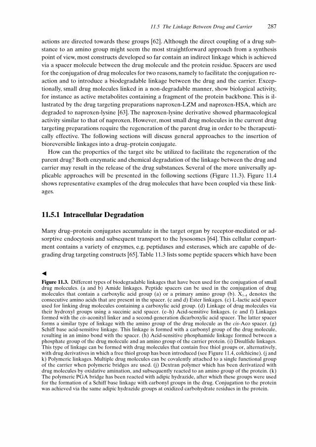
actions are directed towards these groups [62]. Although the direct coupling of a drug sub-stance to an amino group might seem the most straightforward approach from a synthesispoint of view, most constructs developed so far contain an indirect linkage which is achievedvia a spacer molecule between the drug molecule and the protein residue. Spacers are usedfor the conjugation of drug molecules for two reasons, namely to facilitate the conjugation re-action and to introduce a biodegradable linkage between the drug and the carrier. Excep-tionally, small drug molecules linked in a non-degradable manner, show biological activity,for instance as active metabolites containing a fragment of the protein backbone. This is il-lustrated by the drug targeting preparations naproxen-LZM and naproxen-HSA, which aredegraded to naproxen-lysine [63]. The naproxen-lysine derivative showed pharmacologicalactivity similar to that of naproxen. However, most small drug molecules in the current drugtargeting preparations require the regeneration of the parent drug in order to be therapeuti-cally effective. The following sections will discuss general approaches to the insertion ofbioreversible linkages into a drug–protein conjugate.
How can the properties of the target site be utilized to facilitate the regeneration of theparent drug? Both enzymatic and chemical degradation of the linkage between the drug andcarrier may result in the release of the drug substances. Several of the more universally ap-plicable approaches will be presented in the following sections (Figure 11.3). Figure 11.4shows representative examples of the drug molecules that have been coupled via these link-ages.
11.5.1 Intracellular Degradation
Many drug–protein conjugates accumulate in the target organ by receptor-mediated or ad-sorptive endocytosis and subsequent transport to the lysosomes [64]. This cellular compart-ment contains a variety of enzymes, e.g. peptidases and esterases, which are capable of de-grading drug targeting constructs [65]. Table 11.3 lists some peptide spacers which have been
11.5 The Linkage Between Drug and Carrier 287
Figure 11.3. Different types of biodegradable linkages that have been used for the conjugation of smalldrug molecules. (a and b) Amide linkages. Peptide spacers can be used in the conjugation of drugmolecules that contain a carboxylic acid group (a) or a primary amino group (b). X1-4 denotes theconsecutive amino acids that are present in the spacer. (c and d) Ester linkages. (c) L-lactic acid spacerused for linking drug molecules containing a carboxylic acid group. (d) Linkage of drug molecules viatheir hydroxyl groups using a succinic acid spacer. (e–h) Acid-sensitive linkages. (e and f) Linkagesformed with the cis-aconityl linker and a second-generation dicarboxylic acid spacer. The latter spacerforms a similar type of linkage with the amino group of the drug molecule as the cis-Aco spacer. (g)Schiff base acid-sensitive linkage. This linkage is formed with a carbonyl group of the drug molecule,resulting in an imino bond with the spacer. (h) Acid-sensitive phosphamide linkage formed between aphosphate group of the drug molecule and an amino group of the carrier protein. (i) Disulfide linkages.This type of linkage can be formed with drug molecules that contain free thiol groups or, alternatively,with drug derivatives in which a free thiol group has been introduced (see Figure 11.4, colchicine). (j andk) Polymeric linkages. Multiple drug molecules can be covalently attached to a single functional groupof the carrier when polymeric bridges are used. (j) Dextran polymer which has been derivatized withdrug molecules by oxidative amination, and subsequently reacted to an amino group of the protein. (k)The polymeric PGA bridge has been reacted with adipic hydrazide, after which these groups were usedfor the formation of a Schiff base linkage with carbonyl groups in the drug. Conjugation to the proteinwas achieved via the same adipic hydrazide groups at oxidized carbohydrate residues in the protein.
�

288 11 Development of Proteinaceous Drug Targeting Constructs
CH3
O
COOH
H
CH3
naproxen
methotrexate
NCH3
NH O
NH2
NH2
COOHHOOC
H
mitomycin C
O
O
NH2
CH3 NNH
O CH3
O
O
NH2
doxorubicin
N
OH
Cl
Cl
O
chlorambucil
O
NH
O
O
O
RO
OCH3
CH3
CH3
CH3
colchicine: R = CH3
coupled derivative: R = C2H4-SH
O
NH
N
CH3
O
OO
N3
P
OH
O
OH
AZTMP
captopril
NSH
O COOH
O
OO
O
OHOH
NH
OH
NH
NH
NH2
CH3
OH
CHO
OH
OH
NH
OH
CH3
NH
NH2
streptomycin
dexamethasoneO
OH
CH3
OH
H
CH3 OHCH3
F H
O
NH
NH
O
O
F
5-fluorouracil
O
O
O
OH
OH
O
OH
OH
CH3
OHNH2
OCH3
Figure 11.4. Overview of drug moleculesthat have been incorporated into drugtargeting that are discussed in Section11.5.1. The functional group of the drugmolecule that is used for conjugation tothe carrier has been high lighted.

developed for lysosomotropic drug delivery (i.e. delivery with constructs whose final desti-nation is the lysosomes). The enzymes involved in the degradation of the spacers shown inTable 11.3 are normally not present in extracellular areas. For this reason, and since peptidebonds are very resistant towards chemical degradation, the linkages that are formed withthese spacers are generally very stable in the circulation.
The peptide spacers shown in Figure 11.3a–b are susceptible to degradation by endopepti-dases, which either disrupt the bond between the spacer and drug molecule or, alternatively,one of the internal peptide bonds of the spacer. In a subsequent degradation step the re-maining part of the spacer is either attacked by other peptidases or is degraded via non-en-zymatic processes [68,69]. Peptide spacers have been used for the delivery of drug moleculesthat contain peptide carboxylic acid groups, an example of which is the cytostatic drugmethotrexate. It should be noted however that peptide spacers are ill-suited for the linkageof drug molecules which contain non-peptide carboxylic acid groups. Probably, the peptidas-es involved have high substrate specificities, resulting in the inability to degrade the amidebond that is formed with the drug molecule [63]. Peptide spacers have also been used for theconjugation of drug molecules to a primary amino group (Figure 11.3b). As can be conclud-ed from the listed drugs in Table 11.3, the peptidases involved in the release of these drugmolecules tolerate different types of non-peptide primary amino groups.
For drug molecules with carboxylic acid groups, ester bonds seem suitable linkages forlysosomal delivery.The anti-inflammatory drug naproxen has been conjugated to a proteina-ceous carrier via an ester linkage by means of an α-hydroxy acid spacer (Figure 11.3c) [70].Such a spacer, of which L-lactic acid is a typical example, contains in addition to the hydrox-yl group a carboxyl group that can be used to link the spacer to the protein. Similar types oflinkages can also be formed with a drug molecule containing an hydroxyl group and a spac-er with two carboxylic acid groups, such as the succinate spacer (Figure 11.3d). Examples in-clude the conjugates prepared with the corticosteroid dexamethasone and the taxoid pacli-taxel [11,71,71,72].
11.5 The Linkage Between Drug and Carrier 289
Table 11.3. Peptide sequences that have been used as spacers in lysosomotropic drug delivery.
Drug Spacer References
Conjugation at carboxylic acid group of the drug [66]
Methotrexate ala-leu-ala-leugly-gly-glygly-gly-phegly-phe-ala
Conjugation at amino group of the drug [67]
Doxorubicin, daunomycin ala-leu-ala-leugly-gly-gly-leugly-gly-phe-glygly-phe-leu-gly
5-Fluorouracil gly-phe-leu-gly-leu-gly
Mitomycin C ala-leu-ala-leugly-phe-ala-leugly-phe-leu-gly
Primaquine ala-leu-ala-leu
The relatively acidic pH of the lysosomes (pH 5) has led to the development of severallinkage types which are susceptible to acid-catalysed degradation. These acid-sensitive spac-ers are relatively stable at the neutral pH of the bloodstream, but become hydrolytically la-bile at lower pH values. Depending on the type of linkage and the functional group of thedrug molecule, three subtypes can be distinguished: cis-aconityl linkers (cis-Aco) (Fig-ure 11.3e–f), Schiff base hydrazones or imino linkages (Figure 11.3g), and phosphamideslinkages (Figure 11.3h), respectively. The cis-Aco linker can be used for the conjugation ofdrug molecules with a primary amino group [73,74]. The amide linkage that is formed be-tween the linker and the drug is chemically destabilized at lower pH values due to the flank-ing carboxylic acid group (anchiomeric assistance). When the cis-Aco spacer is conjugatedvia its flanking carboxylic acid group to the protein, the acid-sensitivity of the spacer is lost.In order to prevent such loss of pharmacological activity, several other cis-Aco spacers havebeen developed that are conjugated via a different chemistry to the protein (exemplified inFigure 11.3f) [75,76]. Often, these spacers contain maleimide and haloalkyl groups for thepurpose of the final coupling to the protein, since these groups can be reacted under mildconditions with thiol groups which have been previously inserted into the protein.
The Schiff base hydrazone linkers form an imino-linkage between a carbonyl group of thedrug and a hydrazine functionality of the spacer. Such linkages have been formed with drugmolecules containing either a keto group (doxorubicin) [77,78], an aldehyde group (strepto-mycin) [79] or a carboxylic acid group (chlorambucil) [80] (Figure 11.4). Recently, the use ofa branched spacer has been reported, which enables the attachment of multiple drug mole-cules to the spacer [81]. For the conjugation of the hydrazone spacer to a protein, strategiessimilar to those used in the case of the second generation cis-Aco spacers have been fol-lowed.
The third class of acid-catalysed linkages, the phosphamide linkages, are formed betweena phosphate group and a primary amino group (Figure 11.3h). This type of linkage has beenused for the lysosomotropic drug delivery of nucleotide analogues to the liver andmacrophages [22,82,83].
Another type of linkage that is degraded intracellularly is the disulfide bond (Fig-ure 11.3i). Disulfide linkages have been extensively used in the preparation of immunotoxins[84]. For instance, Ricin A immunotoxins are pharmacologically active only when they con-tain a biodegradable disulfide linkage [85]. With respect to the targeting of small drug mole-cules, disulfide linkages have been used in conjugates with the angiotensin converting en-zyme inhibitor captopril, a drug molecule that contains a free thiol group, and in conjugateswith derivatives of colchicine and methotrexate [14, 86–88].
A disadvantage of the disulfide linkage is its relative instability in the bloodstream. Disul-fide bonds can be degraded by reducing enzymes or disrupted chemically by thiol-disulfideexchange with free thiol compounds such as gluthathione [89]. Although the latter processwill proceed preferably intracellularly, due to the higher intracellular concentration of freethiol compounds, thiol-disulfide exchange can result in premature drug release. Disulfidespacers that contain sterically hindered disulfide bonds showed improved stability towardsthis non-enzymatic degradation [90,91].
One of the limitations of the use of a proteinaceous carrier is the relative small number ofdrug molecules that can be conjugated without gross alterations in the structure of the pro-tein. For example, extensive derivatization of an antibody carrier may lead to the loss of its
290 11 Development of Proteinaceous Drug Targeting Constructs

homing potential if the antigen recognition domain is affected. To circumvent this problem,dextran and poly-glutamic acid (PGA) polymers have been used as bridging molecules forthe conjugation of cytostatic drugs (Figure 11.3j–k) [92,93].These polymers were loaded withthe drug and subsequently reacted with amino groups or carbohydrate residues of the carri-er. This technique enabled the conjugation of about 80 doxorubicin molecules per protein inthe case of the dextran bridge, and up to 100 doxorubicin molecules as a result of the PGAlinkage. Efficacy studies with tumour cell lines and in vivo tumour xenograft models in micedemonstrated the potential of the above-described conjugates [93].
11.5.2 Extracellular Degradation
In addition to targeting constructs which are endocytosed and which release the active drugsubstance intracellularly, other constructs are activated outside the target cell. The latter ap-proach is not appropriate for target tissues in which the extracellular fluid is rapidly removedby perfusion, since this would result in a reduction of the time that the drug remained at thetarget site and consequently systemic redistribution of the drug would occur. Thus, extracel-lular drug release is preferred for use in compartments or tissues where the rate of perfusionis low. Conditions of slow perfusion are associated with most solid tumours due to their poorlymphatic drainage. In addition, many tumour cells secrete proteolytic enzymes that are ca-pable of degrading extracellular matrix in the process of tumour growth and metastasis. Ifsuch enzymes are present in the tumour and in minimal amounts in the extracellular fluid ofother tissues, their presence can be exploited for the selective release of the drug in the tu-mour tissue. Examples of such enzymes include cathepsins, that are normally only present inthe lysosomes, and matrix-degrading enzymes such as collagenase or plasminogen activators[67]. These enzymes are all peptidases, and therefore peptide linkers are feasible spacers foruse with this approach. In the case of a secreted lysosomal enzyme, the same spacer se-quences shown in Table 11.3 can be used for the linkers.
An attractive approach to drug targeting is the delivery of the drug-regenerating enzymeinstead of the actual drug substance to the target site. This approach, also referred to asADEPT (antibody directed enzyme pro-drug therapy), is based on a two-step targeting prin-ciple. In the first step, an enzyme is selectively delivered to the target site by means of an an-tibody–enzyme conjugate. In the second step, small-molecule pro-drugs are administered,which will subsequently be activated by the targeted enzyme [94,95].
A point to be noted regarding ADEPT relates to the plasma half-life of the enzyme–anti-body construct. Generally, antibody–enzyme conjugates, are slowly cleared from the centralcirculation.Their sustained presence in the bloodstream will lead to non-target site activationof the pro-drug.Thus, the enzyme and pro-drug should be consecutively administered after awell-chosen time interval when the concentration of the antibody-enzyme conjugate is stillhigh in the target tissue while being low in the central circulation and non-target tissues.
11.5 The Linkage Between Drug and Carrier 291

11.6 Recombinant DNA Approaches
The importance of recombinant DNA techniques for the synthesis of drug targeting con-structs is rapidly increasing. This approach offers, in theory, the possibility of generating allthree components of a drug targeting preparation as outlined in Figure 11.1.A carefully cho-sen cloning strategy results in a uniform end-product with optimum positioning of the differ-ent components. To obtain such a fully genetically engineered drug targeting construct, allthree components must be peptides or proteins.With respect to the active drug substance thisis likely to be an exception rather than the rule. Some constructs, such as immunotoxins andimmunocytokines, that do fulfil these requirements have been studied extensively in drugtargeting and will be described in detail.
By using recombinant DNA techniques, modifications in the protein backbone, such as ad-ditions, deletions and alterations of amino acids, are easily achieved. These modifications cancontribute to improved pharmacokinetic properties of the construct.Additions may consist ofthe introduction of residues that allow covalent conjugation of drug molecules. Deletions ofamino acids can employed to remove membrane-bound regions of a protein, thereby increas-ing its solubility. Single amino acid modifications can be used to minimize antibody responsesand alter the binding specificity and/or the three-dimensional structure of a certain protein.
The final requirement of a recombinant DNA approach to the preparation of a drug de-livery construct, is the ability to produce large amounts of the protein. This can by achievedby bacterial, fungal, insect and mammalian expression systems.The choice of system dependson how the expression of the protein is regulated, the required purity and yield of the pro-tein, and whether the protein is toxic to certain types of producer cells. Furthermore, individ-ual scientists may prefer a particular type of expressing system, depending on laboratory fa-cilities, safety considerations and production costs. The possibilities and limitations of differ-ent expression systems will be discussed and general guidelines which need to be taken intoaccount when choosing an appropriate strategy, will be mentioned briefly. Thereafter, withthe aid of several examples, the development and applications of drug delivery constructs ob-tained using recombinant DNA technology will be described.
11.7 Recombinant DNA Expression Systems
11.7.1 Heterologous Gene Expression in Escherichia coli
The Gram-negative bacterium E. coli is probably the most widely used host for heterologousprotein production. An obvious advantage of this system is its simplicity. The genetics arewell characterized, the cells grow fast allowing rapid production and analysis of the expressedprotein, and transformation is simple and requires minimal amounts of DNA.
In E. coli foreign genes are normally cloned using inducible promoters such as the lac pro-moter that is regulated by the lac repressor and induced by isopropyl β-D-thiogalactopyra-noside (IPTG). This controls gene expression and prevents loss of the gene in situationswhere production of the protein might be toxic to the cells. Stronger synthetic promoters, de-rived from the lac system, tac and trc promoters, are commercially available. Other common-
292 11 Development of Proteinaceous Drug Targeting Constructs

ly used promoters include T7 RNA polymerase promoter and promoters that are regulatedby temperature shift, such as the temperature sensitive λPL promoter and the cold shock pro-moter cspA [96].The latter promoter is especially beneficial for proteolytically-sensitive pro-teins since proteolysis is reduced at low temperature. Additionally, promoters that are acti-vated by a decrease in temperature may provide a partial solution to another frequently en-countered problem, namely misfolding and denaturation of proteins. Since cultivation underlow temperature favours correct protein folding, this problem is less likely to occur.
The (over)expression of proteins in the cytoplasm of E. coli often leads to the formation ofinsoluble aggregates known as inclusion bodies. In fact this can simplify the purification pro-tocol but at the same time often requires in vitro refolding of the protein into its active form,which can sometimes be difficult to achieve. Besides lowering the culture temperature (seeabove), the solubility of the expressed protein can be improved by constructing fusion pro-teins. Commercial systems suitable for fusion to maltose-binding protein (MBP), thioredox-in and glutathione S-transferase are available. Not only are fusion partners used to increasethe solubility of the protein of interest, but they can also facilitate its purification. Addition-ally, poly-histidine tags are commonly used for efficient purification via immobilized metalaffinity chromatography. Although the presence of the poly-His (and other) tags is accept-able in many cases because they rarely alter protein structure or function, their removal maybe required in some therapeutic applications.The liberation of the heterologous protein fromits fusion part (or affinity tag) is theoretically possible but needs expensive proteases and isvery seldom complete. This generally results in reduced yields of the active product.
A new insight into the problem of insolubility of heterologous proteins has evolved fromfurther information regarding the in vivo function of molecular chaperones in protein fold-ing.As reviewed by Baneyx, co-expression of chaperones in the bacterial system can improvethe folding and hence the yield of heterologous proteins [96].
An alternative approach is to direct secretion of proteins into the periplasmic space by us-ing a signal peptide sequence that is removed during the translocation process [97,98]. Vari-ous signal sequences, derived from naturally occurring secretory proteins, including PelB, β-lactamase and alkaline phosphatase can be used for secretion of heterologous proteins. Theperiplasm is an oxidizing environment, containing enzymes necessary for the formation andrearrangement of disulfide bonds. This is especially relevant for the recombinant productionof antibodies which require disulfide bonds for activity [99].
An important point which should be taken into account when expressing eukaryotic genesin E. coli, is the difference in codon usage between prokaryotes and eukaryotes. For instance,the arginine codons AGA and AGG are common in eukaryotic genes but rarely found in E.coli. This problem can be solved, either by site-directed mutagenesis or by co-overexpressionof the gene encoding tRNAArg(AGG/AGA). Another and more important limitation of E. colias an expression system for eukaryotic proteins, is its inability to glycosylate proteins. There-fore, if glycosylation is required, other expression systems should be used.
11.7.2 Fungal Expression Systems
Fungi, both filamentous fungi and yeast, are often the expression system of choice when ahigh yield of eukaryotic protein is desired. Fungi grow rapidly on cheap medium and gene
11.7 Recombinant DNA Expression Systems 293

manipulation is not difficult. In contrast to E. coli, fungi are able to carry out post-transla-tional modifications, such as glycosylation, proteolytic processing, folding and disulfidebridge formation. By applying fermentation technology, clinically and industrially importantproteins have been successfully expressed in fungi [100–102].
The most commonly used filamentous fungi for heterologous gene expression belong toAspergillus and Trichoderma species. The transformation system is based on complementa-tion of auxotrophic mutants or on dominant selection marker genes and results in the inte-gration of the foreign gene into the host genome [103]. Filamentous fungi have effective se-cretory machinery, allowing for accumulation of proteins in the culture medium. In As-pergillus, expression cassettes consisting of the foreign gene fused to an endogenous glu-coamylase and separated by a KEX-2 proteolytic site, have resulted in elevated expressionlevels [102,104]. The KEX-2 site is effectively cleaved by an endopeptidase in the endoplas-mic reticulum during secretion resulting in the correctly processed protein accumulating inthe medium.
Several yeast species have been engineered for heterologous protein production but themost commonly used for these purposes are the baker’s yeast Saccharomyces cerevisae andthe methylotropic yeast Pichia pastoris. Yeast systems utilize both integrated and extrachro-mosomal (non-integrated) vectors.
Strong inducible yeast promoters used for protein production in S. cerevisae, includeGAL1, GAL5, and GAL7 promoters which are induced by galactose, and repressed by glu-cose. The wealth of information on its genetics and molecular biology has made S. cerevisaean excellent model organism for protein–ligand and protein–protein interactions. However,for an abundant expression of heterologous proteins, other yeast systems such as P. pastoriswith its strong and highly regulated alcohol oxidase (AOX1) promoter, have been more suc-cessful. In the last 16 years expression of more than 300 foreign proteins have been reportedusing this system [105]. Examples of more than 100-fold higher protein yield of recombinantsingle chain antibodies in P. pastoris compared to E. coli have been reported [106,107].
In summary, fungal expression systems are often an excellent choice for eukaryotic proteinexpression. However, like any other system, fungi have their own limitations, including theinability to carry out the same types of glycosylation as higher eukaryotic organisms and, insome cases, problems related to accurate protein folding resulting in degradation of the pro-tein.
11.7.3 Baculovirus Expression Systems
Baculoviruses are members of a large group of double-stranded DNA viruses which only in-fect invertebrates, including insects.The restricted host range makes baculoviruses safer thanmammalian expression systems. The most widely used baculoviruses are Autographa califor-nica nuclear polyhedrosis virus and the Bombyx mori nuclear polyhedrosis virus. The hostcell most commonly used is Sf9, derived from the fall armyworm Spodoptera frugiperda.
Typically, the foreign gene is placed under the control of the extremely strong polyhedrinpromoter, allowing for a highly efficient secretion of the heterologous protein into the insectcell culture medium. Glycosylation and other post-translational modifications occur in theinsect cells. Up to 1998, more than 500 different heterologous proteins had been produced by
294 11 Development of Proteinaceous Drug Targeting Constructs

the baculovirus expression vector, of which more than 95% had the correct post-translation-al modifications [108]. No doubt, this number has rapidly increased since. However, a limita-tion of the baculovirus system is that optimal expression levels require high-quality growthmedia, careful culturing and the expression of the foreign protein during the phase in whichthe producing cells are dying.
11.7.4 Stable Transformations of Insect Cells
Although not as popular as the baculovirus-system, stable transformations of insect cells canbe used to circumvent the problems mentioned in the last paragraph. Common hosts includethe fruitfly and mosquito. The expressed genes are often under the control of the Drosophi-la metallothionein promoter. Genes coding for resistance to antibiotics such as hygromycinand neomycin are used as selection markers.
11.7.5 Expression Using Mammalian Cells
The mammalian cell expression system contains all the necessary regulatory machinery foraccurate and efficient processing and secretion of eukaryotic proteins, although there may bespecies differences. Foreign DNA is introduced into the cells either via virus infection or di-rectly, employing chemical (for instance lipocomplexes or calcium phosphate) and physical(electroporation or microinjection) methods. The transcriptional control elements (en-hancers and promoters) are complex and vary between mammalian cell types. However,simian virus 40 (SV40) and human cytomegalovirus (CMV) promoters are active in manycell types and are therefore commonly used. Obtaining stable transfected cell lines can betime consuming and therefore a transient expression system is often used for initial analysis.Typically COS (African green monkey kidney) and CHO (Chinese hamster ovary) cells areused for this purpose. An obvious advantage of mammalian cell expression is the possibilityof advanced glycosylation. Generally, the yield of heterologous proteins produced in a mam-malian cell system is much less than in other expression systems. However, for some proteins,the use of mammalian cells may solve the problems observed in prokaryotic and lower eu-karyotic organisms with regard to accurate folding and modifications.
11.7.6 Expression Systems: Concluding Remarks
The choice of an expression system for the production of a drug delivery construct is of vitalimportance but at the same time a difficult task. Several general considerations when choos-ing an appropriate expression system are outlined in Table 11.4. The use of microorganisms(bacteria and fungi) results in high yield of the product and they are therefore often pre-ferred by researchers. However, for highly specific therapeutic applications, the use of mi-croorganisms is less favourable since they are unable to carry out the post-translational mod-ifications necessary for activity of the protein. For instance, glycosylation is not possible in E.coli and although possible in fungi, it differs from that in mammalian systems. Use of insect
11.7 Recombinant DNA Expression Systems 295

and mammalian systems can, at least in part, overcome these limitations, but these systemsare more expensive and difficult to manipulate due to complex regulatory systems. Thechoice of appropriate expression organism depends on the individual protein and its appli-cations. In the past few years, the fundamental insights into the mechanisms of production,stability and cellular locations of proteins have increased greatly.This knowledge will help re-searchers working in the field of drug targeting to rationalize their choice of expression sys-tems.
11.8 Recombinant DNA Constructs
11.8.1 Antibody-based Constructs
As stated above, a coding sequence for carrier, homing device and active drug can be de-signed together in one fusion construct. However, even if this construct consisted of a smallcarrier, a very short recognition sequence as the homing device and a small proteinaceousdrug substance, the final design would encode for a relatively large protein. Due to the sizeof the construct, one could expect problems regarding the stable maintenance of the encod-ing gene in a certain expression system, in addition to problems with respect to accurate syn-thesis, export and folding of the recombinant protein. The smaller the total size of the re-combinant protein, and the smaller the changes made to the construct as compared to a nat-ural protein, the less likely it will be for these problems to occur.A significant reduction in re-
296 11 Development of Proteinaceous Drug Targeting Constructs
Table 11.4. Advantages and limitations of various expression systems.
Expression system Advantages Disadvantages
E. coli Economical, fast, easy, high yield, Insolubility and misfolding of pro-well characterized genetics, teins, no glycosylation possible,large number of cloning vectors difference in codon usage between
prokaryotes and eukaryotes
Fungi Economical, fast, easy, high yield, Glycosylation and other post-well characterized genetics (yeast), translational modifications are often glycosylation possible, able to different to mammalian systems secrete correctly folded and processed proteins
Insect cellsbaculovirus high yield, safe due to restricted Controlled culture conditions are
host range, able to perform most required, expression peaks when of the post-translational modifi- cells are dyingcations carried out by mammalian cells
stable transformants Stable Time consuming, relatively low yield
Mammalian cells Advanced post-translational Time consuming, relatively low yieldmodifications, signals for synthesis,processing and excretion are Complex regulatory systemcorrectly recognized

combinant protein size, and thereby in complexity of the construct can be achieved whenboth the carrier and homing device functions are intrinsic properties of one protein.
Antibodies make up a group of proteins which can be considered to have the properties ofboth a carrier and a homing device and, as a result, have been used in many drug targetingstudies [18,109]. However, the relatively large size (150 kDa) of whole IgG molecules ham-pers tissue penetration of these molecules. Several modifications of the original antibodystructure can be carried out to reduce the size of the IgG molecule. For instance, in naturalantibodies (Figure 11.5a) the Fc-region is necessary to activate T-cells of the immune system.Since this function of the antibody is not required in most drug targeting constructs, these do-mains have been removed by recombinant cloning techniques [110]. The resulting F(ab′)2,and Fab fragments, with molecular weight of around 100–110 and 50–55 kDa respectively, are
11.8 Recombinant DNA Constructs 297
2
(ScFv)3 (ScFv)4(ScFv)2
(ScFv)2
S
S
Leu-zipper(ScFv)2
Intact antibody F(ab’)2-fragment Fab-fragment(a) (b)
ScFv(d)(c)
(e)
(h)
(f)
(i) (j)
3
(g)
Streptavidin
Figure 11.5. Schematic representation of genetically engineered antibody constructs for drug targeting.Intact antibodies consisting of two heavy and two light chains (a) can be converted into divalent F(ab′)2
fragments (b) or to monovalent Fab fragments (c). These fragments are stabilized via disulfide bridges.Alternatively, the variable heavy and light chain fragments are linked via a flexible linker resulting in amonovalent ScFv (d). Di-, tri- and tetravalent scFv fragments can be constructed by connecting two,three or four scFv fragments with peptide linkers (e–g) or by introducing a S–S bridge between theindividual scFv fragments (h). Non-covalent interactions between scFv fragments are created byintroducing leucine zipper sequences into the construct (i) or via streptavidin–biotin interactions (j).

linked together by one or more disulfide bridges (Figure 11.5b, c). Even smaller antibody-de-rived fragments, the so-called single chain Fv (scFv) molecules (Mw around 30 kDa), built upof a VH and a VL region linked by a flexible peptide linker, have been constructed (Fig-ure 11.5d).
By fusion of a therapeutically active protein to a site other than the antigen recognitiondomain, these antibody fragments are able to function as carriers with intrinsic homing de-vices. However, a major drawback of these smaller fragments is the loss of the bivalent char-acter normally present in the antibody, which ensures high avidity (functional activity). Inmany cases the multivalent character has been restored by genetic engineering [17,18]. Bothcovalent and non-covalent interactions have been used to combine two or more ScFv mole-cules to so-called di-, tri- and tetravalent constructs (Figure 11.5e–j). Covalent interactionscan be achieved with totally genetically-engineered constructs consisting of two (or more)scFv moieties connected by a peptide linker, for instance repetitive sequences of the Gly4-Sermotif [111]. Alternatively, covalent interaction between the subunits is established by the in-troduction of a cysteine residue at the C-terminus of the monovalent molecules or via chem-ical cross-linking reagents [17]. Non-covalent interactions have been created with specific se-quences such as the leucine zipper domains (Fos- and Jun-fragments) which can interact witheach other [112]. Another approach is the assembly of multiple scFv molecules using strep-tavidin-scFv fusion proteins [113]. This strategy will result in the production of tetramericcomplexes due to the non-covalent assembly of four streptavidin moieties. In addition, bi-
298 11 Development of Proteinaceous Drug Targeting Constructs
Table 11.5. Immunotoxins, immunocytokines, cytotoxins and toxin-targeted constructs.
Type of construct Targeting moiety Effector moiety Examples References
Immunotoxins Antibody or antibody Bacterial or plant 3B3-PE [114]fragment (scFv, Fab, toxin αTac-DT [115]F(ab’)2) BerH2-SAP [116, 117]
OM124-PAP [117, 118]
Immunocytokines Antibody or antibody Cytokine, interleukin αEpCAM-IL-12 [119] fragment or growth factor αHer2/neu-IL-12 [120]
RM4-TNFα [121]RM4-IFNγ [122]
Cytotoxins Cytokine, interleukin, Toxin, toxic protein IL-2-DAB [123]growth factor or the or apoptosis-inducing TGFα-PE40 [124, 125] receptor binding do- protein IL-2-BAX [126]mains of these proteins bFGF-SAP [127]
bFGF-RNase [128]
Toxin-targeted Receptor binding ROS-scavenging pro- TT-SOD [57]constructs domain of toxin tein, CTL-epitopes LFn-Ova257-264 [129]
(e.g. gp120 HIV) LFn-LLO91-99 [130]LF254-gp120 [131]
Antibodies have been used as the targeting moiety for the delivery of active drug substances like toxins(immunotoxins) or cytokines and other immunomodulatory proteins (immunocytokines). For reviewson immunotoxins see references [132–135]. Cytokines or their receptor binding domains have alsobeen used as targeting moieties for toxins and other cell-killing proteins (cytotoxins). For reviews oncytotoxins see references [132, 134]. The receptor binding domain of toxins has been used to targetother effector molecules, such as enzymes or CTL epitopes, to the cells expressing receptors for the to-xin.

otin-labelled molecules can be attached to the recombinant protein via the biotin binding siteof the construct.
Recombinant drug targeting constructs utilizing the targeting moiety of antibodies havepredominantly involved toxins and cytokines as the active drug substance (Table 11.5). Thebacterial toxins most commonly used for immunotoxin constructs are diphtheria toxin (DT),pseudomonas exotoxin (PE) and to a lesser extent shiga(-like) toxin (ST/SLT). These toxinsall have the same overall composition: a receptor binding moiety, a fragment involved inmembrane translocation and a toxic or catalytic domain. In order to avoid interactions withcells that bear the ‘normal’ toxin receptor, the receptor-binding domain is removed, resultingin so-called truncated toxins. Genetic coupling of such chemical ‘bombs’ to an antibody orscFv, results in a highly selective and potent drug targeting construct.
A variant of the original immunotoxin approach is the so-called immunocytokines. Inthese constructs the antibody targeting moiety is maintained, but the toxin as the effectormolecule is replaced by a cytokine. In contrast to toxins, cytokines are often proteins en-dogenously produced in man. If both the antibody and cytokine are of human origin, then noforeign proteins are introduced which could provoke an antibody response from the host im-mune system when the drug targeting preparation is clinically applied.
Selective targeting of very potent cytokines may be an attractive approach to overcomethe many side-effects seen after general systemic administration of such compounds [122].Most cytokines are LMWPs and as such rapidly eliminated by renal glomerular filtration.Consequently, high doses are necessary to obtain locally effective concentrations.
The potential of immunocytokines was elegantly demonstrated in two separate studies us-ing IL-12 for anti-tumour therapy. IL-12, a potent stimulator of natural killer cells and cyto-toxic T-lymphocytes (CTL), activates the immune system to eradicate the cancer cells. IL-12
11.8 Recombinant DNA Constructs 299
P40P35 L chain H chain (Gly4Ser)3 linker
(b)
(a)
S-S bridge
Figure 11.6. Schematic diagram showing the assembly of IL-12 protein for antibody-based drugdelivery. (a) The mature sequences of the p35 subunit of IL-12 are fused to the C-terminus of the heavychain of a tumour-specific antibody and co-expressed with the antibody light chain and the p40 subunitof IL-12. Formation of the final immunocytokine requires the creation of disulfide bridges between theantibody chains and interactions of p35 and p40 subunits of IL-12 [119]. (b) Alternatively the IgG heavychain and both subunits of IL-12 can be linked via flexible linkers allowing for equimolar assembly of IL-12 [120].

is a heterodimeric protein composed of a p35 and a p40 subunit.A different approach for thefinal assembly of the IL-12 protein was followed in the two studies (Figure 11.6). Gillies et al.prepared a recombinant fusion construct of the p35 unit of IL-12 and a humanized anti-tu-mour antibody [119]. Co-expression of this construct with the p40 subunit yielded the finalimmunocytokine. In the second study, Peng et al. used recombinant single chain IL-12 (scIL-12), in which both subunits of the cytokine are linked via a flexible peptide linker, for thepreparation of an IL-12–antibody construct [120]. This latter approach ensures the correctequimolar assembly of IL-12, and may confer stability to the fusion protein. Both approach-es proved successful when tested in tumour xenograft models.
11.8.2 Receptor-targeted Constructs
Instead of utilizing the specific interaction between an antibody and its antigen epitope asthe homing mechanism of a drug targeting construct, one could choose from a whole varietyof specific interactions between structures on the target cell surface (receptors) and othermolecules (ligands). Here some examples of constructs or potential construct moieties fordrug targeting based on receptor–ligand interactions will be discussed.
11.8.2.1 Cytotoxins
The immunotoxins and immunocytokines have already been discussed.Another type of con-struct which makes use of both cytokines and toxins is the cytotoxin (Table 11.5). In theseconstructs the cytokine moiety is responsible for the targeting function, in contrast to the im-munocytokines in which the cytokine moiety is the active drug compound. An example of acytotoxin is the DT-IL-2 construct. The over-expression of the high affinity receptor for IL-2on activated T-cells, B-cells and macrophages was utilized to selectively kill these cells. Thereceptor-binding domain of DT was replaced by the N-terminal IL-2 fragment to form theDAB486-IL-2 construct [123,134]. Upon binding to the IL-2 receptor, the construct was in-ternalized by receptor-mediated endocytosis, and produced its toxic activity in the cytosol ofthe target cell.Moreover, given its safe and well-tolerated behaviour in phase I/II clinical studies, and basedon further information regarding the minimal structural requirements for the membranetranslocation moiety and proteolytic activation, a second generation construct DAB389-IL-2is currently being evaluated in a phase III clinical study [136]. These results have shown thatimmunotoxins and targeted cytotoxins can be used safely. However, the use of toxins is notalways without risks and side-effects. For example, DT390-anti-CD3sFv and DT390-IL-3showed, despite their selectivity, toxic side-effects in mouse models [137,138].
11.8.2.2 Toxin-targeted Constructs
As discussed in Section 11.8.1, many toxins of bacterial or plant origin are built up of differ-ent moieties or subunits which mediate binding, membrane translocation and catalytic or
300 11 Development of Proteinaceous Drug Targeting Constructs
toxic activity. Anthrax and tetanus toxin are examples of toxins whose binding and mem-brane translocation moieties, but not their toxic moiety, have been used in drug targetingconstructs.
Anthrax toxin is a bacterial toxin from Bacillus anthracis consisting of three parts: protec-tive antigen (PA), lethal factor (LF) and edema factor (EF). Both LF and EF compete forbinding sites on the PA protein.The PA protein binds with high affinity to an as yet unknownreceptor on macrophages and related cell types. When PA is internalized by the target cells,it functions as a shuttle protein for either EF or LF. Intracellularly, in the acidic environmentof the endosome, EF and LF are capable of entering the cytosol by pH-dependent pore for-mation [139].
The potential use of anthrax toxin as a delivery system aimed at antigen-presenting cells ismost clearly demonstrated in the delivery of HIV gp120-derived peptides [131]. The N-ter-minal domain of LF was genetically fused to the gp120 portion of the HIV envelope protein.When administered in combination with recombinant PA, this construct elicited a specific cy-totoxic T-lymphocyte immune response towards the HIV gp120 protein. This study and oth-ers, in which other peptide epitopes were delivered to antigen-presenting cells, imply a gen-eral application for the anthrax toxin as a peptide vaccine delivery vehicle [129,139].
A second example of a toxin that has been used as targeting device is tetanus toxin.Tetanus toxin is a potent neurotoxin, which can undergo uptake in the nerve endings of mo-tor neurones and subsequent retrograde transport into the central nervous system. The non-toxic C-fragment of tetanus toxin (TTC, 451 amino acids), has been used to increase the neu-ronal uptake of the therapeutic protein SOD [57]. Following intravenous infusion, the re-combinant hybrid protein reduced the occurrence of ischaemia-induced cerebral infarctionin rats [58].
11.8.2.3 TfR-directed Constructs
The most frequently reported target for translocation of proteinaceous drugs across theblood–brain barrier is the transferrin receptor [140] (see also Chapter 2). Recently, a system-atic study has reported on the genetic fusion of human transferrin and NGF [140]. This workclearly demonstrated the importance of controlling the positions of the different componentswithin a drug targeting construct, and how this could be obtained by recombinant DNA tech-nology. Only when NGF was cloned at the N-terminus of transferrin did the fusion proteinretain the activities of both component molecules. In addition, a relatively long flexible link-er between the two moieties, designed to promote dimerization, was required for functionalactivity. Direct attachment of NGF to transferrin probably prevented dimerization by sterichindrance.
The use of the TfR for drug targeting can be extended beyond the blood–brain barriersince all cells that have a high requirement for iron, such as actively proliferating tissues andtumour cells, express large numbers of transferrin receptors on their surface [19]. Cell acti-vation, induced for instance by HIV-1 replication, also upregulates the levels of TfR expres-sion. The delivery of anti-viral agents via the transferrin uptake pathway into HIV-infectedcells has been reported [141]. DNA sequences, encoding nine amino acid residues, cleavableby HIV-1 protease, were cloned into the human transferrin gene. After uptake of these con-
11.8 Recombinant DNA Constructs 301
structs, the recombinant protein could function as a competitive substrate for HIV-1 pro-tease.This approach made use of the fact that surface-exposed loops of globular proteins canoften tolerate insertions of additional amino acids without altering the function of the pro-tein [142]. Molecular modelling was used to select candidate insertion sites in surface-ex-posed loops of transferrin that were distant from the biologically active domains.
Evidently, resolution of the three-dimensional structures of proteins will aid in the designof rational approaches for constructing drug conjugates, as demonstrated by the above-men-tioned examples. Evaluation of molecular structure at this level may prove to be one of themore successful approaches used in the design of recombinant drug conjugates.
11.9 Recombinant Domains as Building Blocks for DrugTargeting Constructs
With the growing knowledge about protein structure–function relationships and the avail-ability of new techniques like phage display, we are now able to select small proteinaceous se-quences that could function as building blocks for recombinant targeting constructs. Suchbuilding blocks can function as a targeting moiety (homing device), a membrane transloca-tion moiety and/or an active drug substance. In order to facilitate the construction of recom-binant preparations, it seems reasonable to assemble the final construct from smaller sub-units.
11.9.1 Targeting Domain
Probably the smallest sequence known to be responsible for receptor recognition is theRGD-tripeptide, initially discovered in fibronectin [143]. However, the specificity of the in-teraction with different integrins, the counter receptors of RGD sequences on the cell sur-face, is established by the flanking sequences of the RGD motif and the conformation of thetripeptide. In other words, the presentation of the RGD sequence is important for specificrecognition by individual integrins.
Insertion of RGD sequences as targeting domains into protein carriers, is an attractive ap-proach for integrin-directed targeting. Studies with RGD sequences of viral origin clonedinto solvent-exposed regions of β-galactosidase demonstrated binding and internalization ofthe active chimeric enzyme into mammalian cells [144]. Likewise, introduction of an RGDmotif into the capsid protein of adenoviruses was shown to increase the cell-specific deliveryof adenoviral vectors as gene delivery vehicles [145]. Fusion proteins containing RGD se-quences are likely to be effective delivery systems but the clinical relevance thereof awaitsfurther analysis.The RGD motif can form the targeting domain, but at the same time can alsofunction as the active drug, since its binding to the receptor may result in prevention or dis-ruption of the natural ligand–integrin interaction, and consequently in a therapeutic re-sponse.
The use of phage-display techniques has identified peptide ligands with specific affinity tocell surface receptors or specific tissues (see Table 11.2). Such peptide homing devices can be
302 11 Development of Proteinaceous Drug Targeting Constructs

genetically inserted into a recombinant protein backbone. For some of these peptides, thesurface receptor has not yet been elucidated. Further investigation into the cellular process-ing of the receptor and its bound ligand is essential to ensure a rational design for the target-ing constructs that are being developed.
11.9.2 Membrane Translocation Domain
Most of the targeting domains mentioned above are aimed at cell surface molecules for theobvious reason of accessibility. In many cases these target receptors are able to internalize to-gether with the bound ligand. However, this process will deliver the construct to the lyso-somes, a compartment in which enzymes and low pH will result in degradation of proteins. Inorder to escape this aggressive environment, a membrane translocation domain might be in-troduced so that the drug delivery preparation can cross the cell membrane in a receptor-in-dependent manner.
Membrane translocation domains have been identified in toxins and viruses and derivedfrom signal sequences of secreted proteins.When derived from a signal sequence the translo-cation domain contains hydrophobic sequences [146–148] while the toxin and viral translo-cation domains contain mostly basic residues [149,150].
In terms of targeting, membrane translocation domains lack specificity for particular cellsor tissues. Therefore, these domains should be combined with targeting domains such asthose discussed in the previous section. In such a construct, the targeting domain will ensurea rapid accumulation at the surface of a specific cell type and the translocation domain willfacilitate entry into the cytosol of the target cells.
11.9.3 Assembly Domain
Although feasible and resulting in highly uniform end-products, the construction and syn-thesis of a complete drug targeting preparation as one genetic construct has one major dis-advantage: lack of flexibility. If a construct does not show the expected results, the wholeprocess of designing and production has to be repeated. Therefore, the use of assembly do-mains in the individual components of the drug targeting construct can be advantageous. Re-cently several studies have reported on the flexibility of such constructs containing anavidin/streptavidin moiety for non-covalent binding to biotinylated proteins. This approachwas followed for the delivery of biotinylated compounds across the blood–brain barrier us-ing a genetic fusion protein of avidin and an anti-TfR antibody [151].A similar approach wasused to engineer the RGD cell adhesion sequence into accessible surface regions of strepta-vidin without disrupting the biotin binding properties [152].
Even greater flexibility was achieved by genetic fusion of streptavidin with protein A[153,154]. Protein A specifically binds the Fc domain of IgG immunoglobulins of almost allmammals without inhibiting the antigen binding activity of the antibody. Thestreptavidin–protein A fusion construct was used for the assembly of complexes of biotiny-lated β-galactosidase and different monoclonal antibodies specific for tumour cell receptors.As a result these complexes were efficiently delivered into several cancer cell lines [154].
11.9 Recombinant Domains as Building Blocks for Drug Targeting Constructs 303

11.10 Concluding Remarks
This chapter has presented many approaches to the preparation of drug targeting constructs.In these constructs, a protein or part of it may function as a carrier for attached drug mole-cules, as a specific targeting moiety, or as a therapeutically active substance. Two entirely dif-ferent approaches have been followed in the preparation of proteinaceous drug targetingconstructs. First, chemical derivatization of existing proteins with site-directing ligandsand/or drug molecules, and second, the engineering and expression of recombinant DNAconstructs. Depending on the type of construct required, each approach has its own uniqueadvantages. For instance, the engineering of protein backbone structures is most accuratelyperformed by recombinant techniques. On the other hand, chemical approaches can be usedfor the attachment of small organic drug molecules, offering numerous opportunities fortherapeutic intervention which cannot be matched by proteinaceous drug substances. There-fore, with reference to the title of this chapter, proteinaceous drug targeting constructsshould preferably be prepared by chemical and recombinant DNA techniques, rather than bythe exclusive use of either one.
Acknowledgements
R. J. Kok and S. A. Ásgeirsdóttir are members of UNYPHAR, a network collaboration be-tween the universities of Groningen, Leiden and Utrecht and the pharmaceutical companyYamanouchi.
References
[1] Forssen E, Willis M, Adv. Drug Deliv. Rev 1998, 29, 249–271.[2] Seymour LW, Adv. Drug Deliv. Rev. 1994, 14, 89–111.[3] Brocchini S, Duncan R, In: Encyclopedia of Controlled Drug Delivery (Ed. Mathiowitz E), pp.
786–816. John Wiley and Sons, New York, 1999.[4] Meijer DKF, Molema G, Moolenaar F, De Zeeuw D, Swart PJ, J. Control. Rel. 1996, 39, 163–172.[5] Maack T, Johnson V, Kau ST, Figuereido J, Sigulem D, Kidney Int. 1979, 16, 251–270.[6] Stehle G, Wunder A, Schrenk HH, Hartung G, Heene DL, Sinn H, Anti-Cancer Drugs 1999, 10,
785–790.[7] Maeda H, Wu J, Sawa T, Matsumura Y, Hori K, J. Control. Rel. 2000, 65, 271–284[8] Stehle G, Sinn H, Wunder A, Schrenk HH, Schutt S, Maierborst W, Heene DL, Anti-Cancer
Drugs 1997, 8, 677–685.[9] Franssen EJF, Jansen RW, Vaalburg M, Meijer DKF, Biochem. Pharmacol. 1993, 45, 1215–1226.
[10] Melgert BN, Wartna E, Lebbe C, Albrecht C, Molema G, Poelstra K, Reichen J, Meijer DKF, J.Drug Target. 1998, 5, 329–342.
[11] Melgert BN, Olinga P, Jack VK, Molema G, Meijer DKF, Poelstra K, J. Hepatol. 2000, 32,603–611.
[12] Haas M, Meijer DKF, Moolenaar F, De Jong PE, De Zeeuw D, In: International Yearbook ofNephrology 1996 (Eds. Andreucci VE, Fine LG), pp. 3–11. Oxford University Press, Oxford, UK,1996.
[13] Franssen EJF, Moolenaar F, De Zeeuw D, Meijer DKF, Adv. Drug Deliv. Rev. 1994, 14, 67–88.[14] Kok RJ, Grijpstra F, Walthuis RB, Moolenaar F, De Zeeuw D, Meijer DKF, J. Pharmacol. Exp.
Ther. 1999, 288, 281–285.[15] Haas M, Kluppel ACA, Wartna ES, Moolenaar F, Meijer DKF, De Jong PE, De Zeeuw D, Kid-
ney Int. 1997, 52, 1693–1699.
304 11 Development of Proteinaceous Drug Targeting Constructs

[16] Milstein C, Waldmann H, Curr. Opin. Immunol. 1999, 11, 589–591.[17] Hudson PJ, Curr. Opin. Immunol. 1999, 11, 548–557.[18] Colcher D, Pavlinkova G, Beresford G, Booth BJ, Choudhury A, Batra SK, Q. J. Nucl. Med. 1998,
42, 225–241.[19] Wagner E, Curiel D, Cotten M, Adv. Drug Deliv. Rev. 1994, 14, 113–135.[20] Pardridge WM, Adv. Drug Deliv. Rev. 1999, 36, 299–321.[21] Feener EP, King GL, Adv. Drug Deliv. Rev. 1998, 29, 197–213.[22] Fiume L, Busi C, Di Stefano G, Mattioli A, Adv. Drug Deliv. Rev. 1994, 14, 51–65.[23] Schwartz AL, CRC. Crit. Rev. Biochem. 1984, 16, 207–223.[24] Van der Sluijs P, Meijer DKF, In: Liver Diseases. Targeted Diagnosis and Therapy Using Specific
Receptors and Ligands (Eds Wu GY, Wu CH), pp. 235–264. Marcel Dekker, Inc., New York, 1991.
[25] Wadhwa MS, Rice KG, J. Drug Target. 1995, 3, 111–127.[26] Stowell CP, Lee YC, Adv. Carbohydr. Chem. Biochem. 1980, 37, 225–281.[27] Monsigny M, Roche A-C, Midoux P, Mayer R, Adv. Drug Deliv. Rev. 1994, 14, 1–24.[28] Gabius S, Kayser K, Bovin NV, Yamazaki N, Kojima S, Kaltner H, Gabius H-J, Eur. J. Pharm.
Biopharm. 1996, 42, 250–261[29] Lerchen HG, Baumgarten J, Piel N, Kolb-Bachofen V, Angew. Chem. Int. Ed. Engl. 1999, 38,
3680–3683.[30] Weikert S, Papac D, Briggs J, Cowfer D, Tom S, Gawlitzek M, Lofgren J, Mehta S, Chisholm V,
Modi N, Eppler S, Carroll K, Chamow S, Peers D, Berman P, Krummen L, Nature Biotechnol.1999, 17, 1116–1121.
[31] Mulligan MS, Warner RL, Rittershaus CW, Thomas LJ, Ryan US, Foreman KE, Crouch LD, TillGO, Ward PA, J. Immunol. 1999, 162, 4952–4959.
[32] Sudimack J, Lee RJ, Adv. Drug Deliv. Rev. 2000, 41, 147–162.[33] Beljaars L, Molema G, Schuppan D, Geerts A, De Bleser PJ, Weert B, Meijer DKF, J. Biol.
Chem. 2000, 275, 12743–12751.[34] Koivunen E, Wang B, Ruoslahti E, Biotechnology (NY) 1995, 13, 265–270.[35] Samoylova TI, Smith BF, Muscle Nerve 1999, 22, 460–466.[36] Burg MA, Pasqualini R, Arap W, Ruoslahti E, Stallcup WB, Cancer Res. 1999, 59, 2869–2874.[37] Rajotte D, Ruoslahti E, J. Biol. Chem. 1999, 274, 11593–11598.[38] Martens CL, Cwirla SE, Lee RY, Whitehorn E, Chen EY, Bakker A, Martin EL, Wagstrom C,
Gopalan P, Smith CW, et al., J. Biol. Chem. 1995, 270, 21129–21136.[39] Arap W, Pasqualini R, Ruoslahti E, Science 1998, 279, 377–380.[40] Ellerby HM, Arap W, Ellerby LM, Kain R, Andrusiak R, Del Rio G, Krajewski S, Lombardo CR,
Rao R, Ruoslahti E, Bredesen DE, Pasqualini R, Nature Med. 1999, 5, 1032–1038.[41] Pasqualini R, Koivunen E, Kain R, Lahdenranta J, Sakamoto M, Stryhn A, Ashmun RA, Shapiro
LH, Arap W, Ruoslahti E, Cancer Res. 2000, 60, 722–727.[42] Mao S, Gao C, Lo CH, Wirsching P, Wong CH, Janda KD, Proc. Natl Acad. Sci. USA 1999, 96,
6953–6958.[43] Ruoslahti E, Annu. Rev. Cell Dev. Biol. 1996, 12, 697–715.[44] Furitsu H, Ogawara K, Fujita T, Yamashita F, Takakura Y, Sezaki H, Hashida M, Int. J. Pharm.
1997, 151, 15–26.[45] Kok RJ, Grijpstra F, Nederhoed KH, Moolenaar F, De Zeeuw D, Meijer DKF, Drug Delivery
1999, 6, 1–8.[46] Swart PJ, Hirano T, Kuipers ME, Ito Y, Smit C, Hashida M, Nishikawa M, Beljaars L, Meijer
DKF, Poelstra K, J. Hepatol. 1999, 30, 1034–1043.[47] Inoue M, Methods Enzymol. 1994, 233, 212–221.[48] Jansen RW, Molema G, Pauwels R, Schols D, De Clercq E, Meijer DKF, Mol. Pharmacol. 1991,
39, 818–823.[49] Kuipers ME, Swart PJ, Witvrouw M, Esté JA, Reymen D, De Clercq E, Meijer DKF, J. Drug Tar-
get. 1999, 6, 323–335.[50] Swart PJ, Beljaars E, Smit C, Pasma A, Schuitemaker H, Meijer DKF, J. Drug Target. 1996, 4,
109–116.[51] Swart PJ, Beljaars L, Kuipers ME, Smit C, Nieuwenhuis P, Meijer DKF, Biochem. Pharmacol.
1999, 58, 1425–1435.[52] Lloyd JB, Adv. Drug Deliv. Rev. 2000, 41, 189–200.[53] Bodor N, J. Control. Rel. 1999, 62, 209–222.[54] Eto T, Takahashi H, Nature Med. 1999, 5, 577–581.
References 305

[55] Conforti A, Caliceti P, Sartore L, Schiavon O, Veronese F, Velo GP, Pharmacol Res. 1991, 23,51–56.
[56] Yabe Y, Nishikawa M, Tamada A, Takakura Y, Hashida M, J. Pharmacol. Exp. Ther. 1999, 289,1176–1184.
[57] Francis JW, Hosler BA, Brown Jr RH, Fishman PS, J. Biol. Chem 1995, 270, 15434–15442.[58] Francis JW, Ren J, Warren L, Brown Jr RH, Finklestein SP, Exp. Neurol. 1997, 146, 435–443.[59] Brinkley M, Bioconjug. Chem. 1992, 3, 2–13.[60] Carter DC, Ho JX, Adv. Protein Chem 1994, 45, 153–203.[61] Yasuzawa T, Tomer KB, Bioconjug. Chem. 1997, 8, 391–399.[62] Means GE, Feeney RE, Bioconjug. Chem. 1990, 1, 2–12.[63] Franssen EJF, Koiter J, Kuipers CAM, Bruins AP, Moolenaar F, De Zeeuw D, Kruizinga WH,
Kellogg RM, Meijer DKF, J. Med. Chem. 1992, 35, 1246–1259.[64] McGraw TE, Maxfield FR, In: Targeted Drug Delivery (Ed. Juliano RL), pp. 11–41. Springer-Ver-
lag, Berlin, 1991.[65] Barrett AJ, Heath MF, Lysosomes: A Laboratory Handbook. Elsevier, Amsterdam, 1977.[66] Fitzpatrick JJ, Garnett MC, Anticancer Drug Des. 1995, 10, 1–9.[67] Soyez H, Schacht E, Van Der Kerken S, Adv. Drug Deliv. Rev. 1996, 21, 81–106.[68] Rejmanova P, Kopacek J, Pohl J, Baudys M, Kostka V, Makromol. Chem. 1983, 184, 2009–2020.[69] Duncan R, Cable HC, Lloyd JB, Rejmanová P, Kopacek J, Makromol. Chem. 1983, 184,
1997–2008.[70] Franssen EJF, Moolenaar F, De Zeeuw D, Meijer DKF, Pharm. Res. 1993, 10, 963–969.[71] Dosio F, Brusa P, Crosasso P, Arpicco S, Cattel L, J. Control. Rel. 1997, 47, 293–304.[72] Fiume L, Chiricolo M, Busi C, Mattioli A, Spinelli C, Spinosa G, Bartolini G, Minghetti L, Orlan-
di M, Tomasi V, Pharm. Acta Helv. 1989, 64, 351–352.[73] Shen W-C, Ryser J-P, Biochem. Biophys. Res. Commun. 1981, 102, 1048–1054.[74] Franssen EJF, Moolenaar F, De Zeeuw D, Meijer DKF, Int. J. Pharm. 1992, 80, R15–R19.[75] McIntyre GD, Scott Jr CF, Ritz J, Blattler WA, Lambert JM, Bioconjug. Chem. 1994, 5,
88–97.[76] Blattler WA, Kuenzi BS, Lambert JM, Senter PD, Biochemistry 1985, 24, 1517–1524.[77] Mueller BM, Wrasidlo WA, Reisfeld RA, Bioconjug. Chem. 1990, 1, 325–330.[78] Kruger M, Beyer U, Zahn H, Unger C, Kratz F, Chem. Pharm. Bull. 1997, 45, 399–401.[79] Coessens V, Schacht E, Domurado D, J. Control. Rel. 1996, 38, 141–150.[80] Beyer U, Roth T, Schumacher P, Maier G, Unold A, Frahm AW, Fiebig HH, Unger C, Kratz F, J.
Med. Chem. 1998, 41, 2701–2708.[81] King HD, Yurgaitis D, Willner D, Firestone RA, Yang MB, Lasch SJ, Hellstrom KE, Trail PA,
Bioconjug. Chem. 1999, 10, 279–288.[82] Midoux P, Negre E, Roche A-C, Mayer R, Monsigny M, Balzarini J, De Clercq E, Mayer E, Ghaf-
far A, Gangemi JD, Biochem. Biophys. Res. Commun. 1990, 167, 1044–1049.[83] Kuipers ME, Swart PJ, Hendriks MMWB, Meijer DKF, J. Med. Chem. 1995, 38, 883–889[84] Wong SS, In: Chemistry of Protein Conjugation and Cross-Linking, (Ed. Wong SS), pp. 147–194.
CRC Press, Boca Raton USA, 1993.[85] Cumber AJ, Westwood JH, Henry RV, Parnell GD, Coles BF, Wawrzynczak EJ, Bioconjug.
Chem. 1992, 3, 397–401.[86] Plourde R, Phillips AT, Wu CH, Hays RM, Chowdhury J, Chowdhury N, Wu GY, Bioconjug.
Chem. 1996, 7, 131–137.[87] Shen WC, Ryser HJP, La Manna L, J. Biol. Chem. 1985, 260, 10905–10908.[88] Umemoto N, Kato Y, Hara T, Cancer Immunol. Immunother. 1989, 28, 9–16.[89] Arunachalam B, Phan UT, Geuze HJ, Cresswell P, Proc. Natl Acad. Sci. USA 2000, 97, 745–750.[90] Thorpe PE, Wallace PM, Knowles PP, Relf MG, Brown AN, Watson GJ, Knyba RE,
Wawrzynczak EJ, Blakey DC, Cancer Res. 1987, 47, 5924–5931.[91] Arpicco S, Dosio F, Brusa P, Crosasso P, Cattel L, Bioconjug. Chem. 1997, 8, 327–337.[92] Galun E, Shouval D, Adler R, Shahaar M, Wilchek M, Hurwitz E, Sela M, Hepatology 1990, 11,
578–584.[93] Adler R, Hurwitz E, Wands JR, Sela M, Shouval D, Hepatology 1995, 22, 1482–1487.[94] Sherwood RF, Adv. Drug Deliv. Rev. 1996, 22, 269–288.[95] Niculescu-Duvaz I, Springer CJ, Adv. Drug Deliv. Rev. 1997, 26, 151–172.[96] Baneyx F, Curr. Opin. Biotechnol. 1999, 10, 411–421.[97] Ferenci T, Silhavy TJ, J. Bacteriol. 1987, 169, 5339–5342.[98] von Heijne G, Curr. Opin. Cell Biol. 1990, 2, 604–608.
306 11 Development of Proteinaceous Drug Targeting Constructs

[99] Verma R, Boleti E, George AJ, J. Immunol. Methods 1998, 216, 165–181.[100] Verdoes JC, Punt PJ, Van den Hondel CAMJJ, Appl. Microbiol. Biotechnol. 1995, 43, 195–205.[101] Archer DB, Peberdy JF, Crit. Rev. Biotechnol. 1997, 17, 273–306.[102] Ward PP, Piddington CS, Cunningham GA, Zhou X, Wyatt RD, Conneely OM, Biotechnology
(NY) 1995, 13, 498–503.[103] Turner G, Prog. Ind. Microbiol. 1994, 29, 641–665.[104] Mikosch T, Klemm P, Gassen HG, van den Hondel CA, Kemme M, J. Biotechnol. 1996, 52,
97–106.[105] Cereghino GP, Cregg JM, Curr. Opin. Biotechnol. 1999, 10, 422–427.[106] Ridder R, Schmitz R, Legay F, Gram H, Biotechnology (NY) 1995, 13, 255–260.[107] Eldin P, Pauza ME, Hieda Y, Lin G, Murtaugh MP, Pentel PR, Pennell CA, J. Immunol. Methods
1997, 201, 67–75.[108] Glick BR, Pasternak JJ, In: Molecular Biotechnology: Principles And Applications of Recombi-
nant DNA (Eds Glick BR, Pasternak JJ), pp. 145–169. American Society for Microbiology, Wash-ington DC, 1998.
[109] Kosmas C, Linardou H, Epenetos AA, J. Drug Target. 1993, 1, 81–91.[110] Segal DM, Weiner GJ, Weiner LM, Curr. Opin. Immunol. 1999, 11, 558–562.[111] Atwell JL, Breheney KA, Lawrence LJ, McCoy AJ, Kortt AA, Hudson PJ, Protein Eng. 1999, 12,
597–604.[112] Kostelny SA, Cole MS, Tso JY, J. Immunol. 1992, 148, 1547–1553.[113] Dubel S, Breitling F, Kontermann R, Schmidt T, Skerra A, Little M, J. Immunol. Methods 1995,
178, 201–209.[114] Bera TK, Kennedy PE, Berger EA, Barbas CF, Pastan I, Mol. Med. 1998, 4, 384–391.[115] Chaudhary VK, Gallo MG, Fitzgerald DJ, Pastan I, Proc. Natl Acad. Sci. USA 1990, 87,
9491–9494.[116] Grossbard ML, Fidias P, Kinsella J, O’Toole J, Lambert JM, Blattler WA, Esseltine D, Braman
G, Nadler LM, Anderson KC, Br. J. Haematol. 1998, 102, 509–515.[117] Terenzi A, Bolognesi A, Pasqualucci L, Flenghi L, Pileri S, Stein H, Kadin M, Bigerna B, Polito L,
Tazzari PL, Martelli MF, Stirpe F, Falini B, Br. J. Haematol. 1996, 92, 872–879.[118] Bolognesi A, Tazzari PL, Olivieri F, Polito L, Lemoli R, Terenzi A, Pasqualucci L, Falini B, Stirpe
F, Br. J. Haematol. 1998, 101, 179–188.[119] Gillies SD, Lan Y, Wesolowski JS, Qian X, Reisfeld RA, Holden S, Super M, Lo KM, J. Immunol.
1998, 160, 6195–6203.[120] Peng LS, Penichet ML, Morrison SL, J. Immunol. 1999, 163, 250–258.[121] Xiang J, Moyana T, Qi Y, J. Biotechnol. 1997, 53, 3–12.[122] Xiang J, Hum. Antibodies 1999, 9, 23–36.[123] Murphy JR, vanderSpek JC, Semin. Cancer Biol. 1995, 6, 259–267.[124] Chaudhary VK, Fitzgerald DJ, Adhya S, Pastan I, Proc. Natl Acad. Sci. USA 1987, 84, 4538–4542.[125] Siegall CB, Xu YH, Chaudhary VK, Adhya S, Fitzgerald D, Pastan I, FASEB J. 1989, 3,
2647–2652.[126] Aqeilan R, Yarkoni S, Lorberboum-Galski H, FEBS Lett. 1999, 457, 271–276.[127] Yu C, Cunningham M, Rogers C, Dinbergs ID, Edelman ER, J. Pharm. Sci. 1998, 87, 1300–1304.[128] Futami J, Seno M, Ueda M, Tada H, Yamada H, Protein Eng. 1999, 12, 1013-1019.[129] Ballard JD, Doling AM, Beauregard K, Collier RJ, Starnbach MN, Infect. Immun. 1998, 66,
615–619.[130] Doling AM, Ballard JD, Shen H, Krishna KM, Ahmed R, Collier RJ, Starnbach MN, Infect. Im-
mun. 1999, 67, 3290–3296.[131] Goletz TJ, Klimpel KR, Arora N, Leppla SH, Keith JM, Berzofsky JA, Proc. Natl Acad. Sci. USA
1997, 94, 12059–12064.[132] Strom TB, Anderson PL, Rubin-Kelley VE, Williams DP, Kiyokawa T, Murphy JR, Semin. Im-
munol. 1990, 2, 467–479.[133] Kreitman RJ, Pastan I, Adv. Drug Deliv. Rev. 1998, 31, 53–88.[134] Murphy JR, Curr. Opin. Struct. Biol. 1996, 6, 541–545.[135] Kreitman RJ, Curr. Opin. Immunol. 1999, 11, 570–578.[136] Frankel AE, Kreitman RJ, Sausville EA, Clin. Cancer Res. 2000, 6, 326–334.[137] Vallera DA, Panoskaltsis-Mortari A, Blazar BR, Protein Eng. 1997, 10, 1071–1076.[138] Vallera DA, Seo SY, Panoskaltsis-Mortari A, Griffin JD, Blazar BR, Protein Eng. 1999, 12,
779–785.[139] Ballard JD, Collier RJ, Starnbach MN, Proc. Natl Acad. Sci. USA 1996, 93, 12531–12534.
References 307

[140] Park E, Starzyk RM, McGrath JP, Lee T, George J, Schutz AJ, Lynch P, Putney SD, J. Drug Tar-get. 1998, 6, 53–64.
[141] Ali SA, Joao HC, Hammerschmid F, Eder J, Steinkasserer A, J. Biol. Chem. 1999, 274,24066–24073.
[142] Finkelstein AV, Curr. Opin. Struct. Biol. 1997, 7, 60–71.[143] Pierschbacher MD, Ruoslahti E, Nature 1984, 309, 30–33.[144] Villaverde A, Feliu JX, Aris A, Harbottle RP, Benito A, Coutelle C, Biotechnol. Bioeng. 1998, 59,
294–301.[145] Reynolds P, Dmitriev I, Curiel D, Gene Ther. 1999, 6, 1336–1339.[146] Lin YZ, Yao SY, Hawiger J, J. Biol. Chem. 1996, 271, 5305–5308.[147] Zhang L, Torgerson TR, Liu XY, Timmons S, Colosia AD, Hawiger J, Tam JP, Proc. Natl Acad.
Sci. USA 1998, 95, 9184–9189.[148] Rojas M, Donahue JP, Tan Z, Lin Y-Z, Nature Biotechnol. 1998, 16, 370–375.[149] Blanke SR, Milne JC, Benson EL, Collier RJ, Proc. Natl Acad. Sci. USA 1996, 93, 8437–8442.[150] Schwarze SR, Dowdy SF, Trends Pharmacol. Sci. 2000, 21, 45–48.[151] Penichet ML, Kang YS, Pardridge WM, Morrison SL, Shin SU, J. Immunol 1999, 163, 4421–4426.[152] McDevitt TC, Nelson KE, Stayton PS, Biotechnol. Prog. 1999, 15, 391–396.[153] Sano T, Cantor CR, Biotechnology (NY) 1991, 9, 1378–1381.[154] Ohno K, Levin B, Meruelo D, Biochem .Mol. Med. 1996, 58, 227–233.
308 11 Development of Proteinaceous Drug Targeting Constructs

12 Use of Human Tissue Slices in Drug Targeting Research
Peter Olinga, Geny M. M. Groothuis
12.1 Introduction
It has long been recognized that in vitro research can provide valuable information on basicmechanisms with respect to kinetics and efficacy of drug targeting concepts. Such in vitro re-search includes the use of isolated cells, cell lines and perfused organs. In this chapter the in-troduction of tissue slices into drug targeting research will be discussed.A brief history of theslice technique will first be given. Until now most research on the slice technique has been fo-cused on the metabolism and transport of drugs and this topic will therefore be summarizedbefore embarking on a discussion of the contribution of the tissue slice technique to the areaof drug targeting research.
In vitro research began with organ culture of embryonic organ rudiments [1]. The slicetechnique, using slices of tumour and liver tissue, was performed as early as 1923 by OttoWarburg [2] and in the following years by H. A. Krebs [3], who investigated the metabolismof amino acids in liver slices of cats, dogs and rats. These liver slices were prepared manuallywith limited reproducibility and viability [4].After a decline in the application of slices in liv-er research in favour of the use of isolated hepatocytes as well as isolated perfused liverpreparation, the development of the Krumdieck slicer in the 1980s led to a ‘comeback’ of thetechnique enabling the production of reproducible and viable liver slices [5].This technologyinduced a renaissance of the slice technology.The development of these in vitro preparationshas been of paramount importance for research on human liver function. As most of the re-search with tissue slices concerned the liver, this chapter will focus on the use of liver as thetarget tissue and only briefly mention the use of slices from other tissues in the concludingsection of the chapter.
The most abundant cell type in the liver is the hepatocyte, other cells in the liver are thenon-parenchymal cells: Kupffer cells, the resident macrophages of the liver, endothelial cellsand stellate cells. These cells have been discussed in more detail in Chapter 4.
Since a high yield isolation procedure of rat hepatocytes was described in 1969 [6], hepa-tocytes have become the model of choice for drug transport studies in the liver in vitro [7].With this procedure, isolated hepatocytes from many species have been prepared, includinghepatocytes from rat, mouse, chicken, dog, fish, hamster, pig, cow, sheep and monkey liver(for an extensive review see reference [8]). Before 1976, only relatively small numbers of hu-man hepatocytes could be isolated, due to the use of non-perfusion techniques [9]. Bojar et al.[10] were the first to use a perfusion technique on human livers which greatly enhanced theyield of hepatocytes. In principle the procedure that is now commonly used, is based on theone described by Seglen [6] for rat hepatocytes. Either a biopsy wedge with intact capsula on
Drug Targeting Organ-Specific Strategies. Edited by G. Molema, D. K. F. MeijerCopyright © 2001 Wiley-VCH Verlag GmbH
ISBNs: 3-527-29989-0 (Hardcover); 3-527-60006-X (Electronic)

three sides [11,12], or single lobes of liver [13] and even entire human livers [14] are used.One or more cannulas are inserted into (branches of) the portal vein(s) [12,13] and the tissueis perfused with collagenase. In general, the yield of human hepatocytes (5–20 x 106 hepato-cytes g–1 liver [12, 15–18]) is low compared to rat hepatocytes (60–70 x 106 hepatocytes g–1 liv-er, average yield in our laboratory).
The first studies with isolated human hepatocytes concentrated on the characterization ofthese hepatocytes, as well as on the improvement of the isolation procedure, and the possi-bilities of culturing these cells [16,19–24]. Thereafter studies were performed to investigatethe metabolism of drugs [25–28], in which emphasis was often put on the activity and con-centration of cytochrome P450 isoforms. Nowadays, hepatocytes are more generally used inmetabolic studies of specific compounds, in order to unravel potential species differences and
310 12 Use of Human Tissue Slices in Drug Targeting Research
Core tool Liver cylinder
Lobe
Recirculating Buffer
Slice
Blade
SLICER
Buffer
8 mm��
175-250 µm�
�
Weight: 13-17 mg�
Liver�
�
Figure 12.1. Production of precision-cutorgan slices using the Krumdieck slicer toachieve the preferred thickness and wetweight of a liver slice.

are also extensively used in a variety of other research fields including pharmacology andtoxicology.
Isolation from human liver of other cell types, such as Kupffer, endothelial and stellatecells has also been developed and extensively reviewed [29–31].
Although the isolated (human) liver cells have been shown to be valuable in the study ofmechanisms of drug transport in drug targeting research, the isolation procedures involvesdigestion to disrupt the cell-to-cell contacts. Clearly, enzymatic digestion may also damageplasma membranes and transport systems therein. In addition, for hepatocytes the normalpolarity is lost after isolation. For instance it has been reported that by using collagenase di-gestion to isolate hepatocytes, the amount of asialoglycoprotein receptor present on themembrane of the hepatocyte is reduced [32]. Recently, Ikejima et al. [33] showed that Kupf-fer cells isolated by the standard isolation procedure with collagenase and pronase had losttheir CD14 receptor, presumably an important receptor in the uptake of lipopolysaccharide.
In contrast to isolated liver cells, no enzymatic digestion is necessary for the isolated per-fused liver preparation.The isolated perfused liver preparation, extensively used to study ratliver functions [34], has also been employed with human liver tissue, but the application islimited by the fact that only pieces of tissue that are encapsulated with liver capsula can beused. The use of the isolated perfused human liver preparation has been discussed further inChapter 4.Another in vitro liver preparation that can be used without the need for enzymat-ic digestion is the liver slice (Figure 12.1). One of the main features of slices is that the origi-nal architecture of the organ is retained in the slice.Therefore, in liver slices, the different celltypes of the (human) liver; i.e. hepatocytes, Kupffer-, endothelial and stellate cells are stillpresent in contact with their original matrix environment, which enables the function of allcell types present and their normal intercellular communication to be studied. In addition,studies on cell selective distribution of carriers and drug–carrier conjugates can be per-formed in liver slices, which is of major importance in drug targeting research.The slice tech-nique itself can also be used on other solid organs such as kidney, lung, intestine and evenbrain; the use of slices from these organs will be described in the concluding section of thischapter, whilst the greater part of the chapter will describe the use of liver slices in drug tar-geting research.
12.2 Preparation of Liver Slices
Liver slices were initially prepared manually using razor blades or mechanical instrumentssuch as the Stadie Riggs tissue slicer [4]. The reproducibility of the thickness of the slices atthat time was largely dependent on the skills of the operator. Of note is the fact that the min-imal slice thickness that could be produced was about 0.5 mm. This dimension appeared tolimit the penetration of nutrients and oxygen into the inner cell layers: central necrosis in theslice occurred during incubation [35].
The introduction of the Krumdieck slicer enabled a more optimal and reproducible prepa-ration of liver slices (Figure 12.1).With this technique the thickness of the slices is adjustableto a value as low as 100 µm.The slicing procedure itself is performed in a buffer assuring min-imal trauma of the tissue. In addition, the Krumdieck slicer provides a rapid and automated
12.2 Preparation of Liver Slices 311

production of slices with reproducible thickness. Recently, the so-called ‘Brendel slicer’ wasintroduced, which has largely the same characteristics as the Krumdieck slicer, but offers theadvantage of more constant oxygenation. However, with this technique the slices have to beprepared manually [36]. Liver slices from both tissue slicers have been evaluated. No signifi-cant differences were observed in levels of protein, potassium, total glutathione (i.e. GSHand GSSG), reduced glutathione (GSH) and cytochrome P450 and activities of 7-ethoxyre-sorufin O-deethylase and 7-benzoxyresorufin O-debenzylase in freshly cut rat liver slicesproduced by either of the two tissue slicers [37].
To prepare liver slices with the Krumdieck slicer, cylindrical cores of tissue are first isolat-ed from the liver specimens (Figure 12.1). These tissue cores are prepared preferably by ad-vancing a sharp rotating metal tube into the liver tissue using a drilling press, thus assuringthe preparation of accurately cylindrical cores. If a biopsy punch is used to prepare the cores,it is difficult to obtain a uniform cylindrical shape.
The cores are subsequently placed in the slicer, and the slicing procedure is performed byadvancing the core over an oscillating knife in a controlled environment (Figure 12.1). Cold(4°C) Krebs–Henseleit buffer (pH = 7.4, saturated with 95% O2 and 5% CO2) supplement-ed with 25 mM glucose is commonly used in preparing the slices [35,38–40], but Williams’medium E [41], Earle’s balanced salt solutions [37], Sacks preservation medium [42] and V-7preservation buffer [43,44] are also used.
All these buffers have a glucose concentration of 25 mM, which seems to be essential forthe viability of the slices.
The optimal thickness for liver slices, in order to retain their viability during culture, is ap-proximately 175–250 µm. Price et al. [45] reported that the optimal thickness of liver slices fordrug metabolism studies should be 175 µm. In slices thicker than 250 µm the inner cell layerssuffer from a lack of oxygen and substrates, and in slices thinner than 175 µm the ratio ofdamaged cells in the outer cell layers to the living cell mass becomes unfavourable[36,43,44,46–48]. For cryopreservation slightly thicker slices were reported to give better re-sults [40], although recent developments show that slices of approximately 200–250 µm canalso be successfully used for cryopreservation (unpublished observation).
12.3 Incubation and Culture of Liver Slices
12.3.1 Incubation Systems
Previously, liver slices were incubated in static organ cultures [1]. Hart et al. [49] cultured ratliver slices for 24 h spread out on wet filter paper, floating on top of the incubation medium.Several slice-containing vessels were placed in a box with saturated 95% O2 and 5% CO2 at37°C. However, the slices employed were rather thick (approximately 0.3 mm) and only theupper cell layers (0.2 mm) in the slice contained viable cells. Together with the introductionof the Krumdieck slicer [5,46], a new incubation technique for slices, the dynamic organ cul-ture system (DOC), was introduced [35]. The main characteristic of this system is the inter-mittent exposure of the slice to incubation medium and the gas phase. The DOC is in fact amodified version of the Trowell incubation system [1].
312 12 Use of Human Tissue Slices in Drug Targeting Research

Meanwhile many incubation systems have been developed, mostly based on either DOCor culturing the slices in multi-well incubation systems [38,40–43,50–52] and all have beenused in pharmacological and toxicological research [36]. The most remarkable phenomenonemerging from these studies is the observation that the liver slices can be cultured for up to72 h with the maintenance of their biotransformation activities [43]. In contrast, the use ofprimary suspensions of hepatocytes for metabolic and transport studies is restricted to a fewhours [25]. Culturing of the hepatocytes allows experiments to last for a longer period of time(up to approximately 5–7 days). The hepatocytes form monolayers and develop bile canalic-ular-like spaces in between the cells [20]. However, specific liver functions such as albuminsecretion, transport activity and cytochrome P450 activity decrease considerably during in-cubation [53,54]. After 24 h of culturing, drug metabolism activity will already have de-creased by about 50%. This is very likely due to de-differentiation on the level of gene tran-scription [55]. In recent years much effort has been put into the improvement of the cultureconditions of hepatocytes by adding extracellular matrix components or by co-culturing withother cell types in order to maintain their differentiation status [54,56–63].Although survivaland functioning of these cells has been greatly improved, complete maintenance of differen-tiated isoenzymes patterns has not been achieved yet. In fact, the liver slices can be seen asthe most natural co-culture system within the original matrix.
12.3.2 Evaluation of Incubation Systems
There are only a few studies published in which the various incubation systems for liver sliceswere evaluated. Smith et al. [35] showed that slices in dynamic organ culture maintain theirviability, as measured by ATP and potassium concentration, up to 20 h. Connors et al. [64]used a 24-well incubation system, in which the medium was stirred with a magnetic stirrer. Inthis incubation system rat liver slices were cultured for 8 h and human liver slices for 9 h dur-ing which time a high potassium concentration was maintained in the slices. Connors et al.[65] reported that the 24-well incubation system and the dynamic organ culture gave similarmetabolite patterns after 24 h of incubation with a somatostatin analogue. Vickers et al. [66]also used the 24-well incubation system for 24 h, but no viability parameters were described.Dogterom et al. [51] showed that in a 12-well culture plate, which is put on a gyratory shaker,rat liver slices maintain their viability up to 11 h as determined by potassium concentrationand ATP content. However, an impairment of the rat liver slices in a 24-well incubation sys-tem on the gyratory shaker was seen after 11 h. This was explained by the insufficient agita-tion of the medium in the 24-well incubation system. Leeman et al. [41] described a modifi-cation of the dynamic organ culture: a netwell insert (200-µm polyester mesh carrier) placedin the wells of a six-well culture plate on a rocker platform. In this system, as with the DOC,the slices are intermittently exposed to the gas phase, which in this system is 40% O2/5%CO2/55% N2 and to the medium. Using this incubation system, the 3[4,5-dimethyl-thiazole-2-yl]-2,5-diphenyltetrazolium bromide (MTT) reduction, a test for the cellular reduction ca-pacity both in mitochondria and extramitochondrially involving NADH and NADPH [67],was maintained in the slices for up to 72 h [41]. Simple incubation of slices in a 25-ml Erlen-meyer flask in a shaking water bath was reported by de Kanter et al. to be successful over a24-h period [40].
12.3 Incubation and Culture of Liver Slices 313

314 12 Use of Human Tissue Slices in Drug Targeting Research
Figure 12.2. The five incubation systems for liver slices, divided into two groups: incubation systemscontinuously submerged in culture medium and dynamic organ culture-related incubation systems,where the liver slices are intermittently exposed to the medium and to the air.

Based on these various findings under a variety of conditions a thorough comparison offive incubation systems (Figure 12.2) was made by us in a collaborative study of four labora-tories [52]. The five systems that were evaluated included: the shaken flask (a 25-ml Erlen-meyer flask in a shaking water bath [40]), the stirred well (24-well culture plate equippedwith stainless steel grids and magnetic stirrers [38,64]), the rocker platform (a DOC systemusing six-well culture plates with Netwell inserts, rocked on a platform [41]), the roller system(dynamic organ culture rolled on an insert in a glass vial [35]) and the six-well shaker (six-well culture plates in a shaking water bath). In the rocker platform 40% O2/5% CO2/55% N2
was used whereas in the other four systems 95% O2/5% CO2 was used to oxygenate the tis-sue.The liver slices were incubated in these incubation systems for 0.5, 1.5 and 24.5 h and sub-sequently subjected to viability and metabolic function tests. The viability of the incubatedliver slices was evaluated by potassium content, MTT assay, energy charge, histomorphologyand lactate dehydrogenase (LDH) leakage.Their metabolic functions were studied by deter-mination of the metabolism of lidocaine (Figure 12.3), testosterone and antipyrine. Up to1.5 h of incubation, all five incubation systems gave similar results with respect to viabilityand metabolic function of the slices. However, after 24 h, the shaken flask, the rocker plat-form and the six-well shaker incubation systems, appeared to be superior to the stirred welland the roller incubator. It is notable that the cytochrome P450-dependent metabolism oftestosterone and lidocaine was retained at the same levels as found after 0.5 and 1.5 h of in-cubation in the shaken flask, rocker platform and six-well incubation systems. This suggeststhat the de-differentiation seen after 24 h in pure hepatocyte culture does not occur in slicesfor at least 24 h.
12.3 Incubation and Culture of Liver Slices 315
0
5
10
15
20
nmol
ME
GX
/mg
wet
wei
ght l
iver
slic
e
0.5 1.5 24.5
Incubation time (hours)
6 Well shaker
Roller system
Rocker platform
Stirred well
Shaken flask
**
*
**
Figure 12.3. Metabolism of lidocaine to MEGX (in nmol MEGX mg–1 wet weight liver slice) in liverslices after different incubation times (h). *p < 0.05 versus shaken flask, rocker platform, roller systemand six-well shaker. **p < 0.05 versus shaken flask, rocker platform and six-well shaker. Data are themean of three separate experiments ± SEM.

Brendel’s group compared two incubation systems, the roller system (dynamic organ cul-ture (Figure 12.2)) and the 12-well plate culture (the plates were put on a gyratory shaker),with respect to their ability to maintain the functionality of rat liver slices over 72 h of cul-turing. The slices were evaluated with respect to ATP concentration, potassium retention,MTT reduction and protein synthesis, in addition to alanine transaminase (ALT) and LDHleakage. Metabolic function was investigated by oxidative O-deethylation of 7-ethoxycoumarin(7-EC) [43]. It was concluded that dynamic organ culture was superior to multi-well plateculture [43]. Recently, another comparison has been made between the DOC-system and 12-well system, showing that the 12-well system was superior to the DOC-system with regard to themetabolism of xenobiotics following long-term incubations (> 24 h) [68]. However, this studywas performed in 95% air and 5% CO2, which may have influenced the results obtained. Ahigh oxygen percentage of at least 40% is essential for optimal incubation of liver slices, aswill be described in more detail below. In addition, both sets of experiments were carried outusing different incubation media, which also may have influenced the results obtained.
12.3.3 Incubation Systems for Human Liver Slices
Various incubation systems have also been tested using human liver slices, these include the24-well plates with magnetic stirrers [38,66,69], the six-well plates in a shaker [52] and theDOC roller system [70,71], but no direct comparison has been made as yet. Human liverslices can be cultured for 72 h in DOC and maintain their ability to respond to specific in-ducers of cytochrome P450 such asmethylclofenapate and Aroclor 1254 [71].
12.3.4 Oxygenation and Culture Media for Liver Slice Incubation
In addition to the incubation system itself, the oxygen and nutrient concentration of themedium are also important for the viability of liver slices [46,47]. It appeared that a nutrient-enriched medium containing bicarbonate maintained the slice viability better than a simplermedium such as Krebs–HEPES buffer [44]. It was shown that K+-retention, protein synthe-sis and LDH leakage was maintained in rat liver slices for 5 days in a Waymouth’s/bicarbon-ate medium in a dynamic organ culture system [44]. Oxygenation of the hepatocytes, espe-cially those in the centre of the slice, has been a major concern. In this respect it is importantto note that oxygen at too high a concentration may be toxic, due to tissue damage by oxygenradicals, whereas excessively low levels of oxygen may result in ischaemia. Both 95% air/5%CO2 and 95% O2/5% CO2 are commonly used. In long-term culture up to 5 days [43] theDOC system or DOC-related incubation systems are recommended, because of the inter-mittent exposure of the slice to culture medium and to the gas phase. This feature wasclaimed to be important for optimal gas exchange. In our experiments however, slices thatwere continuously submerged in medium performed equally well or even better than thosein the DOC system or DOC-related system (six-well culture plate on the rocker platform[72]). It can be calculated that liver slices consume only 0.3–1% of the dissolved O2 perminute. This implies that, provided that the medium is continuously oxygenated, the avail-ability of O2 is unlikely to be a limiting factor. In our laboratory we have carried out a study
316 12 Use of Human Tissue Slices in Drug Targeting Research

to investigate the effect of different oxygen percentages on ATP-levels and the rate of lido-caine metabolism in liver slices in the six-well incubation system. When rat liver slices wereincubated for up to 24 h with oxygen percentages between 50 and 95%, no differences wereobserved in either ATP levels or the rate of lidocaine biotransformation. However, if liverslices were incubated for up to 24 h with 20% O2 both ATP levels and the rate of lidocainebiotransformation were significantly decreased. Moreover, this also explains why the loweroxygen percentage (40%) used in the rocker platform system [72] did not seem to influencethe functionality or viability of rat liver slices [52]. Thus it seems that the agitation of themedium and a sufficient O2 supply is more influential with regard to the viability of the slicesthan their intermittent exposure to the medium and the gas phase.
12.3.5 Pre-incubation of Liver Slices
Another important issue in the incubation of liver slices is the potential benefits of pre-incu-bation. We showed that at least 1.5 h of incubation is necessary to restore K+ and ATP levels[52]. It has been suggested that a change of medium after pre-incubation is useful in remov-ing cell debris created by cells which were damaged during the slicing process. This is of spe-cial importance when leakage of cell components, such as LDH and ALT, is used as a mark-er of cell damage in toxicity studies. However, no conclusive evidence regarding the necessi-ty and duration of pre-incubation has been published as yet.
In conclusion, for short incubations the choice of incubation system is not critical for sliceviability which may be determined by other features, such as the volume of the medium, theduration of the sampling procedure and the costs. For studies where rapid sampling of theslices is necessary, for instance in studies on drug uptake, incubation in the shaken six-wellsystem is recommended. In the 24-well system the agitation of the medium appeared insuffi-cient, whereas in the DOC systems the uptake rate of the drug may be influenced by the lim-ited supply of substrate from the medium during exposure to the gas phase. For longer termincubations, the choice of incubation system and medium seems to be more critical and fur-ther basic studies on slice technology need to be carried out to assure optimal long-term cul-ture of liver slices. Among other conditions, incubation experiments should be undertakenusing different oxygen concentrations and concomitant measurement of oxygen consump-tion, in order to establish the optimal oxygen concentration. The agitation of the mediumshould also be studied in more detail. Care should be taken when extrapolating results ob-tained with rat liver slices to human liver slices, since considerable species differences withrespect to the influence of incubation systems on slice viability have been reported [43].
Finally, inter-laboratory standardization of incubation systems and culture media wouldincrease the validity of comparisons made between results from different laboratories.
12.4 Viability and Functionality of Liver Slices
For pharmacological, toxicological and transport studies it is of utmost importance to assessnot only the viability but also the functionality of the liver slices. This is essential both forend-point determination of toxic cell damage, and to assess the quality of the tissue during in-
12.4 Viability and Functionality of Liver Slices 317
cubation. Several viability tests have been developed for liver slices, in line with those for iso-lated hepatocytes: K+ retention, ATP content, energy charge, enzyme (LDH, ALT, AST)leakage, protein synthesis and MTT reduction [36,43,44,51,52,72,73]. Specific liver functiontests include urea synthesis, albumin synthesis, gluconeogenesis, biotransformation of testsubstrates (such as testosterone and 7-ethoxycoumarin) and GSH concentration [40,74–77]Potassium retention is generally used to assess the viability of liver slices [36,43]. However, inour studies on the comparison of incubation systems and on cold storage of slices, the potas-sium concentration in the slices was retained while their metabolic capacity had clearly de-creased [78].This illustrates that in drug metabolism studies the rate of metabolism of a stan-dard drug should be included as a viability test.
The determination of the energy charge (EC) is of limited value in assessing the viabilityof liver slices, because changes in ATP, ADP and AMP have to be quite large before a signif-icant variation in EC is observed. Fisher et al. [43] proposed the following ranking of sensi-tivity for tests aimed at the detection of cellular viability: ATP content > K+ retention > pro-tein synthesis > enzyme leakage > MTT reduction.
Because these different viability tests all reflect different aspects of cell viability, the choiceof test depends on the aim of the study. For toxicity studies where biotransformation is an im-portant bioactivation or detoxification step, metabolic function tests should be included tojudge the validity of the method, whereas viability tests are needed to assess toxic effects.Both positive and negative controls should be included in such studies. When human liver isused, the characterization of metabolic activity is especially important because of the largeinter-individual variability associated with this property [75].
The viability and function tests described above are used to evaluate the hepatocytes with-in the slice. Up to now, tests to measure the viability of the non-parenchymal cells have notbeen reported. The presence of the latter cell types is one of the conceptual advantages ofslices as compared to isolated hepatocytes. As some drug targeting devices are designed totarget non-parenchymal cells in the liver, the development of tests for the sinusoidal celltypes deserves more attention. For example, the uptake of substrates such as succinylated hu-man serum albumin (Suc-HSA, which is specifically endocytosed by endothelial cells [79]), orhyaluronic acid [80], can be used to assess the functionality of endocytotic pathways in theendothelial cells in the liver [81]. Other modified proteins that are specifically taken up byKupffer cells such as mannosylated HSA, may be used to assess the functionality of the en-docytotic pathway in Kupffer cells [79]. Another parameter which can be used to assess thefunctionality of these non-parenchymal liver cells, is the excretion of cytokines in response topro-inflammatory stimuli. Non-parenchymal cell function in liver slices will be described inmore detail in the Section 12.7.
12.5 In Vitro Transport Studies
12.5.1 Transport in Hepatocytes
In the liver drugs are predominantly taken up by the hepatocytes, e.g. by carrier-mediated up-take, metabolized in the hepatocyte and excreted either via the bile canaliculus into the bileor back into the bloodstream, e.g. by carrier-mediated excretion.
318 12 Use of Human Tissue Slices in Drug Targeting Research

The mechanisms of uptake and excretion of drugs by the liver has been widely studied us-ing isolated hepatocytes and isolated perfused livers of rodents. The subject was extensivelydiscussed by Oude Elferink et al. [82], summarized in a comprehensive special issue of theJournal of Hepatology in 1996 [83] and reviewed in several papers and chapters by our group[84,85]. In general, the rate and mechanism of drug uptake in isolated rat hepatocytes arevery similar to those found in vivo. Only a few studies have investigated transport of drugs inhuman hepatocytes, and even fewer have used liver slices. However, studies in human and rathepatocytes are hampered by the fact that the isolation procedures involve collagenase di-gestion for the disruption of cell-to-cell contacts. Clearly, this proteolytic enzyme may alsodamage plasma membranes and transport systems therein.Jansen et al. [86] published an example of transport of a drug targeting preparation in humanand rat hepatocytes. They found that the anti-viral drug ara-AMP coupled to lactosaminatedhuman serum albumin, was taken up to the same extent by human and rat hepatocytes. Thisis one of the few examples where an equal rate of transport was found in both species. In gen-eral the uptake of drugs in human hepatocytes is slower than that in rat hepatocytes. In vit-ro–in vivo scaling calculations [87,88] showed that these differences in the uptake rate ofdrugs between rat and human cells, actually reflect inter-species differences rather than be-ing due to differences in viability. Moreover, the differences described in rate and mechanismof drug transport in rat and man emphasize again that extrapolation to man of pharmacoki-netic data obtained in rat, is hazardous. Most of the current knowledge on drug transport car-riers is derived from experiments with rats. Therefore, more studies need to be carried out inhuman liver preparations to further elucidate the mechanisms of drug transport in the hu-man liver and hence the relevance of animal data.
These results also indicate that human hepatocytes are an appropriate model in which tostudy inter-species differences and the mechanisms of hepatic transport in man.
In contrast to isolated hepatocytes, liver slices retain the cellular architecture of the liverwithout prior digestion with collagenase. This makes a systematic comparison of the data re-lating to transport of free drugs as well as drug targeting moieties from isolated hepatocytesand liver slices, an attractive model for studying the potential and limitations of the liver slicemodel in this area of research.
12.5.2 Transport in Liver Slices
Mechanisms of drug uptake in liver slices were studied in vitro as early as in 1963 bySchanker and Solomon [89]. The results obtained in these experiments are still valuable andshow that the influence of temperature, anoxia, metabolic inhibitors and substrate inhibitioncan be successfully studied in this preparation. However, as mentioned before, at that timethe preparation of reproducible precision-cut slices was not feasible. Therefore, the slice in-cubation technique was virtually abandoned in transport studies after the introduction of thesuccessful isolation of rat hepatocytes.
In order to investigate the possibilities and limitations of the use of precision-cut liverslices prepared with a mechanical slicer in drug transport studies, different aspects of themechanism of uptake of several classes of drugs in human and rat liver slices were investi-gated in our laboratory. Four model compounds which enter hepatocytes via entirely differ-
12.5 In Vitro Transport Studies 319

ent membrane transport mechanisms were investigated: the fluorescent dyes rhodamine Band lucigenin, the cardiac glycoside digoxin and the neo-glycoprotein lactosylated albumin.Receptor-mediated endocytosis into endothelial cells was studied with succinylated andaconylated albumin. The rate of penetration into the rat liver slice was studied with thelipophilic cationic compound rhodamine B (RB) and the hydrophilic organic cation luci-genin (LU). RB, which enters hepatocytes by passive diffusion was homogeneously distrib-uted throughout the rat liver slice (250 µm thickness) within 5 min [81] (Figure 12.4). Theseresults indicate that for very lipophilic components both the penetration rate into the sliceand the diffusion rate into all the cells are rapid processes. In contrast, after incubation withLU, which is taken up by hepatocytes through adsorptive endocytosis, fluorescence in the in-ner cell layers could only be detected after 15 min. If the rates of uptake of drugs in liverslices are to be compared with parameters obtained in vivo, some difficulties may arise. Theuptake rate of compounds into the slice may not only reflect the uptake rate of the cells in-volved, but may also be influenced by the rate of penetration of the substrates (i.e. diffusionthrough sinusoidal spaces) into the slice. From our results with the lipophilic agent rho-damine B it is clear that the penetration process into the slice takes at least 5 min.Therefore,for substrates that are taken up into hepatocytes relatively fast, penetration into the slice maylimit the uptake of the drug by the inner cell layers in the slice.
Digoxin uptake into rat liver slices showed a temperature-dependent component, compat-ible with the involvement of carrier-mediated uptake mechanisms. Quinine markedly inhib-ited the uptake of digoxin, in contrast to its diastereomer quinidine, which only slightly in-hibited the digoxin uptake in rat liver slices. This stereoselective inhibition is in line with re-sults obtained in isolated rat hepatocytes and isolated perfused rat livers [90,91]. These re-sults were also found after cryopreservation of the slices, indicating that carrier-specific phe-nomena can be studied after cryopreservation [92].
From these results it can be concluded that liver slices are a powerful tool for studying themechanisms and specificity of carrier-mediated uptake of drugs and drug interactions whichoccur at the transport level.
320 12 Use of Human Tissue Slices in Drug Targeting Research
Figure 12.4. RhodamineB (25 µM) distribution inthe cross-section of a ratliver slice (± 250 µM) after5 min incubation. Fluo-rescence microscopy,bar = 100 µM.

Many drugs that are taken up and metabolized by hepatocytes are excreted via the bilecanaliculi into the bile. One of the remaining topics in liver slice research is the question ofwhether liver slices are capable of bile excretion via the bile canaliculus. Thompson et al.[93]showed that slices are capable of excreting bile acids, however, there is a need for more ex-periments to determine whether this excretion takes place across the bile canalicular mem-brane.
12.6 The Use of Liver Slices in Drug Targeting Research
12.6.1 Distribution and Transport of Drug Targeting Devices
In our Institute, drug targeting devices are predominantly developed for the treatment off in-flammatory diseases of the liver, such as fibrosis and cirrhosis. In order to increase the thera-peutic efficacy of drugs, human serum albumin is used as a protein backbone; modifying thisprotein by the attachment of different sugar groups or peptide molecules targets these mod-ified proteins to specific cell types in the liver as described in more detail in Chapter 4. Anti-inflammatory agents are subsequently coupled to the protein backbone to serve as effectormoieties.
Further studies are needed to determine whether these liver-directed drug targetingpreparations are actually delivered to the specific liver cells. In addition, experiments shouldbe carried out to ascertain whether the drug coupled to a drug targeting device is released inthe target cell in the liver, and to ensure that the drug is still active in these cells. Up until nowthe distribution of these modified proteins has been tested in vivo mainly in rats, and in vitroin the perfused rat liver or isolated liver cells (both parenchymal and non-parenchymalcells). Almost no studies have been performed in the target species, man. As the main aim ofdrug targeting research is the development of preparations for clinical use, we investigatedwhether precision-cut liver slices could be used to study the uptake of these modified pro-teins into target cells of the human liver.
The temperature-dependent uptake and immunohistochemical localisation of modifiedproteins, Lactose27-Human Serum Albumin (Lact27-HSA), Succinylated-Human Serum Al-bumin (Suc-HSA) and Aconylated Human Serum Albumin (Aco-HSA), in rat and humanliver slices showed that large molecules can enter the slice and are probably taken up by re-ceptor-mediated endocytosis (Figure 12.5). These large modified proteins were found dis-tributed all over the liver slice, as was determined by immuno-histochemistry [81] (Fig-ure 12.6). Recently, Beljaars et al. [94] showed that mannose-6-phosphate21-Bovine SerumAlbumin (M6P21-BSA) is taken up by the non-parenchymal cells of human liver using slicesfrom cirrhotic human livers. The non-parenchymal cells were identified as hepatic stellatecells (HSC) or endothelial cells. The involvement of hepatocytes, Kupffer cells, and bile ductepithelial cells was excluded. Until now, there has been little information concerning thepresence of the mannose-6-phosphate receptor in human adult livers. The accumulation ofM6P-modified albumin in human cirrhotic livers demonstrates, however, that M6P receptorsare present in the human liver and that M6P-modified albumin may be useful as a drug car-rier for the targeting of anti-fibrotic drugs in patients. In another study by Beljaars et al. [95],
12.6 The Use of Liver Slices in Drug Targeting Research 321

the internalization of modified human serum albumin (HSA) with 10 cyclic peptide moietiesrecognizing the collagen type VI receptor (C*GRGDSPC*, in which C* denotes the cycliz-ing cysteine residues) yielding pCVI-HSA, was studied in normal and cirrhotic rat liver slices.125I-pCVI-HSA was used to detect internalization in the liver slices. In contrast to 125I-HSA,an increase in the degradation products of 125I-pCVI-HSA was found over time, during theincubation of liver slices. These data show that pCVI-HSA is taken up and degraded in thecells of the liver slice. By immunohistochemistry it was shown that pCVI-HSA was specifi-cally bound to rat HSC, in particular to the activated cells.
These distribution studies show that liver slices can be used to assess the level of uptake ofa drug into the target cells in both healthy and diseased human liver. Isolation of non-
322 12 Use of Human Tissue Slices in Drug Targeting Research
0
1
2
3
4A
ccum
ulat
ion
fact
or
0 50 100 150
Time (min)
*
*
*
0
1
2
3
4
Acc
umul
atio
n fa
ctor
0 50 100 150
Time (min)
*
*
* Figure 12.5. Uptake of 125I-Suc-HSA in liverslices from humans and rats at 37°(❍) and 4°C(•). The accumulation factor is defined as theconcentration of the compound in the slicesdivided by the concentration in the medium.Each point is the mean of 5-6 separateexperiments ± SEM. n = number of livers.*p < 0.05 versus 4°C. The dotted line representsthe accumulation factor if 125I-Suc-HSA isexclusively distributed within the sinusoids.

12.7 Efficacy Testing of the Drug Targeting Device in the Liver 323
parenchymal cells from diseased livers is experimentally very difficult, therefore the liverslice technique offers a unique opportunity to study drug targeting preparations in diseasedliver.
12.7 Efficacy Testing of the Drug Targeting Device in the Liver
It is of paramount importance to ensure that the drug targeting device with the drug attachedor incorporated, is not only taken up by the target cells in the liver, but is also released fromthe device and remains active within the target cell. In the case of the development of drugtargeting strategies for inflammatory liver diseases, an in vitro system was needed that couldbe used to test the anti-inflammatory effect of the drug-targeting preparations in the humanliver.
To set up and validate the in vitro systems we initiated a study with rat liver slices. Stimu-lation by lipopolysaccharide (LPS) in liver slices was used to evoke a pro-inflammatory re-sponse in the liver. Lipopolysaccharide (LPS), a component of Gram-negative bacterial cellwalls (also called endotoxin), has been associated with tissue injury and sepsis. In the liverLPS activates the resident macrophages, the Kupffer cells, which results in cytokine release[96]. Furthermore, LPS is cleared by the liver, mainly by Kupffer cells [97]. One of the majorfeatures of endotoxic shock is the induction of nitric oxide synthase in the liver [98]. In-ducible nitric oxide synthase (iNOS), the expression of which is induced by LPS and cy-tokines, produces nitric oxide (NO) in large quantities [99].
The induction of iNOS by LPS, as observed in the hepatocytes in vivo [99], cannot beachieved in pure cultures of isolated hepatocytes. In fact, the induction of hepatocyte-associ-ated iNOS is found in pure cultures only after incubation with a mix of LPS and cytokines[100]. Induction of iNOS by LPS alone can be accomplished in co-cultures of hepatocytesand Kupffer cells with LPS [98]. These data indicate that induction of iNOS by LPS is medi-ated by cytokines released by the Kupffer cells in the liver and is thus a result of intercellular
Figure 12.6. Fluorescein-labelled aconylated humanserum albumin distribu-tion in the cross-section ofa rat liver slice (± 250 µM)after 120 min incubation.Bar = 100 µM.

communications. However, the establishment of such co-cultures is technically complexwhile the cellular organization as present in the intact liver cannot as yet be achieved in cul-ture. Therefore, to study the effects of LPS stimulation and the subsequent pharmacologicalintervention by targeted anti-inflammatory drugs in the whole liver in vitro, a system analo-gous to liver slices in which all cell types are present as in the original liver, would be the ideal.
Until recently only a few studies have focused on the activity or the viability of cell typesother than hepatocytes within the liver slice [101,102]. Therefore, we attempted to establishwhether non-parenchymal cells are still viable in the rat liver slice and whether they respond to LPS stimulation. Cytokine levels (tumour necrosis factor α (TNFα), interleukin1β (IL-1β) and interleukin-10 (IL-10)) were measured in the incubation medium as a marker of non-parenchymal cell function. We found that in liver slices stimulated by LPS,IL-1β, TNFα and IL-10 are formed. In addition, the study was designed to elucidate the in-teraction between non-parenchymal and parenchymal cells in the liver after LPS induction.After LPS activation of rat liver slices iNOS was upregulated in the hepatocytes as deter-mined by immunohistochemistry (Figure 12.7). This resulted in the production of NO, asmeasured by nitrate and nitrite (NOx) in the incubation medium (Figure 12.8). In addition to studying the pro-inflammatory response on a protein level, enzyme induction was studiedat the transcription level. By RT-PCR, changes in specific mRNA were studied during the LPS-induced pro-inflammatory response. In rat liver slices the mRNA of iNOS was already upregulated only 2–3 h after induction with LPS, whereas the enzyme iNOS wasfound after 5 h.
Anti-inflammatory drugs such as pentoxyfilline and dexamethasone inhibited the releaseof cytokines and thereby the induction of iNOS and the release of NO in the LPS-stimulatedliver slices.
Melgert et al. [103] used this in vitro system to determine whether the drug targetingpreparation containing the anti-inflammatory drug dexamethasone coupled to HSA, was stillable to manifest its anti-inflammatory properties in liver slices. Dexamethasone10–HSA anduncoupled dexamethasone showed effective inhibition of LPS-induced NO and TNFα pro-
324 12 Use of Human Tissue Slices in Drug Targeting Research
Figure 12.7. Cross-section of rat liver stained for iNOS. Left panel: control incubation after 24 h. Rightpanel: after 24 h incubation with 100 µg ml–1 LPS.

duction (Figure 12.9) in the liver slice model. These results show that the conjugate dexa-methasone10–HSA is taken up intracellularly and that active dexamethasone is released.
Studies similar to those described above are now being carried out in human liver slices.LPS induction in human liver slices also increased TNFα production to the same extent aswas found in rat liver slices [104] (Figure 12.10). Human liver slices also produced IL-6, IL-8and IL-1β, although the latter to a lesser extent than that observed in the liver slices of rat ori-gin. However, human liver slices produced less NO after LPS stimulation than those of therat. More experiments will be undertaken to elucidate this species difference.
Taken together these results indicate that non-parenchymal cells are still active in the slicepreparations and that intercellular communication is still intact. Furthermore, pharmacolog-ical intervention by anti-inflammatory drugs can be successfully studied in liver slices. To-gether with the results obtained in regard to drug transport, liver slices seem to be a verypromising in vitro system for studying intercellular distribution, cellular processing and ef-fectiveness of anti-inflammatory drugs coupled to a targeting device.
12.7 Efficacy Testing of the Drug Targeting Device in the Liver 325
0
20
40
60
80N
Ox
µM
0 5 10 15 20 25
Time (hours)
*
*
*
Figure 12.8. NO production in rat liver slices after incubation in the absence or presence of 100 µg ml–1
LPS for different time periods. The NO production is measured as nitrate/nitrite (NOx) concentrationsin the medium (µM). Control (�) and + 100 µg ml–1 LPS (�). Data are expressed as mean ± SEM of fourexperiments. *p < 0.005 represents a significant increase in NO production by liver slices due tostimulation by LPS.

326 12 Use of Human Tissue Slices in Drug Targeting Research
Figure 12.9. (a) TNFα production by rat liver slices (n = 9) after stimulation with 100 µg ml–1 LPS for24 h in either the presence or absence of dexamethasone (D). Vehicle consists of PBS. *p < 0.05 versusvehicle + LPS. (b) TNFα production by rat liver slices (n = 9) after LPS stimulation for 24 h with orwithout Dexa10-HSA (DH). Vehicle contains PBS and an equimolar amount of HSA. *p < 0.05 versusvehicle + LPS.
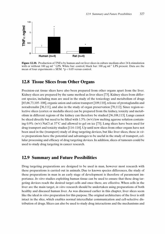
12.8 Tissue Slices from Other Organs
Precision-cut tissue slices have also been prepared from other organs apart from the liver.Kidney slices are prepared by the same method as liver slices [73]. Kidney slices from differ-ent species, including man are used in the study of the toxicology and metabolism of drugs[65,66,73,105–108], organic anion and cation transport [109,110], release of prostaglandin andnoradrenalin [36,111], and also in the study of organ preservation [70,112]. Since region-se-lective slices (cortex or medulla slices) can be prepared from the kidney, toxicity and metab-olism in different regions of the kidney can therefore be studied [36,108,113]. Lungs cannotbe sliced directly but need to be filled with 1.5% (w/v) low melting agarose solution contain-ing 0.9% (w/v) NaCl at 37°C and allowed to gel on ice [73]. Lung slices have been used fordrug transport and toxicity studies [114–118]. Up until now slices from other organs have notbeen used in the (transport) study of drug targeting devices, but like liver slices, these in vit-ro preparations have the potential and advantages to be useful in the study of transport, cel-lular processing and efficacy of drug targeting devices. In addition, slices of tumours could beused to study drug targeting in cancer research.
12.9 Summary and Future Possibilities
Drug targeting preparations are designed to be used in man, however most research withthese preparations is carried out in animals. Due to known species differences, the study ofthese preparations in man in an early stage of development is therefore of paramount im-portance. In vitro studies exploiting human tissue can be used to ensure that these drug tar-geting devices reach the desired target cells and once there, are effective. When cells in theliver are the main target, in vitro research should be undertaken using preparations of bothhealthy and diseased human liver. As was discussed earlier in this chapter, liver slices seemlike the ideal in vitro preparation for this purpose.The original architecture of the liver is stillintact in the slice, which enables normal intercellular communication and cell-selective dis-tribution of drugs. Slices can also be used to study drug interactions and the mechanisms and
12.9 Summary and Future Possibilities 327
0
0.5
1
1.5
Human (n=4) Rat (n=4)
*
*TN
Fα
ng
/ml
Figure 12.10. Production of TNFα by human and rat liver slices in culture medium after 24 h stimulationwith or without 100 µg ml–1 LPS. White bar: control; black bar: 100 µg ml–1 LPS present. Data are themean of four experiments ± SEM. *p < 0.05 versus control.

specificity of carrier-mediated uptake of drugs. In addition, the distribution of the drug intodifferent cell types in the tissue can easily be studied in preparations of organ slices, as canthe efficacy of the drug which is coupled to the targeting device. Furthermore, metabolismand toxicity of the drug targeting device or the released drug can be determined in the hu-man liver. And finally, an important aspect of this type of in vitro research in man is, that itwill ultimately lead to a reduction in the use of animal experiments.
In future, drug targeting devices aimed at other human organs may also be studied usingprecision-cut tissue slices.The latest data/literature on precision-cut tissue slices can be foundat http://www.farm.rug.nl/slice/open.html.
References
[1] Trowell OA, Expert Cell Res. 1959, 16, 118–147.[2] Warburg O, Biochem. Z, 1923, 142, 317–333.[3] Krebs HA, Hoppe-Seyl. Z, 1933, 217, 190–227[4] Stadie WC, Riggs BC, J. Biol. Chem. 1944, 154, 687.[5] Krumdieck CL, Anal. Biochem. 1980, 104, 118–123.[6] Seglen PO, Methods Cell Biol. 1976, 13, 29–83.[7] Skett P, Toxicol. In Vitro 1994, 3, 491–504.[8] Berry MN, Edwards AM, Barritt GJ, Isolated Hepatocytes Preparation, Properties and Applica-
tions, Vol. 21. Elsevier, Amsterdam, New York, Oxford, 1991.[9] Guillouzo A, Oudea P, Le-Guilly Y, Oudea MC, Lenoir P, Bourel M, Exp. Mol. Pathol. 1972, 16,
1–15.[10] Bojar H, Basler M, Fuchs F, Dreyfürst R, Staib W, Broelsch C, J. Clin. Chem. Clin. Biochem. 1976,
14, 527–532.[11] Reese JA, Byard JL, In Vitro 1981, 17, 935–940.[12] Groothuis GMM, Sandker GW, Pruim J, Weert B, Slooff MJH, Meijer DKF, Toxicol. In Vitro
1995, 9, 951–958.[13] Dorko K, Freeswick PD, Bartoli F, Cicalese L, Bardsley BA, Tzakis A, Nussler AK, Cell Trans-
plant. 1994, 3, 387–395.[14] Moshage HJ, Rijntjes PJ, Hafkenscheid JC, Roelofs HM, Yap SH, J. Hepatol. 1988, 7, 34–44.[15] Sandker GW, Isolated human hepatocytes: a research tool for investigations on drug transport, drug
metabolism and liver transplantation. PhD thesis, University of Groningen, 1993.[16] Ryan CM, Carter EA, Jenkins RL, Sterling LM, Yarmush ML, Malt RA, Tompkins RG, Surgery
1993, 113, 48–54.[17] Schröder AJ, Blaheta RA, Scholz M, Encke A, Markus BH, Zentralbl. Chir. 1994, 119, 127–138.[18] Takahashi M, Matsue H, Matsushita M, Nakajima Y, Uchino J, Artif. Organs 1993, 17, 653–659.[19] Trevisan A, Gudat F, Guggenheim R, Krey G, Durmuller U, Luond G, Duggelin M, Landmann J,
Tondelli P, Bianchi L, Hepatology 1982, 2, 832–835.[20] Ballet F, Bouma ME, Wang SR, Amit N, Marais J, Infante R, Hepatology 1984, 4, 849–854.[21] Houssin D, Capron M, Celier C, Cresteil T, Demaugre F, Beaune P, Life Sci. 1983, 33, 1805–1809.[22] Hsu IC, Lipsky MM, Cole KE, Su CH, Trump BF, In Vitro Cell Dev. Biol. 1985, 21, 154–160.[23] Miyazaki K, Takaki R, Nakayama F, Yamauchi S, Koga A, Todo S, Cell Tissue Res. 1981, 218,
13–21.[24] Strom SC, Jirtle RL, Jones RS, Novicki DL, Rosenberg MR, Novotny A, Irons G, McLain JR,
Michalopoulos G, J. Natl Cancer Inst. 1982, 68, 771–778.[25] Tee LB, Seddon T, Boobis AR, Davies DS, Br. J. Clin. Pharmacol. 1985, 19, 279––294.[26] Guillouzo A, Beaune P, Gascoin MN, Begue JM, Campion JP, Guengerich FP, Guguen-Guil-
louzo C, Biochem. Pharmacol. 1985, 34, 2991–2995.[27] Wiebkin P, Dees JH, Mathis JM, Prough RA, Drug Metab. Dispos. 1985, 13, 163–168.[28] Blaauboer BJ, van-Holsteijn I, van-Graft M, Paine AJ, Biochem. Pharmacol. 1985, 34, 2405–2408.[29] Friedman SL, Roll FJ, Anal. Biochem. 1987, 161, 207–218.[30] Alpini G, Phillips JO, Vroman B, LaRusso NF, Hepatology 1994, 20, 494–514.[31] Heuff G, Meyer S, Beelen RH, J. Immunol. Methods 1994, 174, 61–65.[32] Zeitlin PL, Hubbard AL, J. Cell. Biol. 1982, 92, 634–647.
328 12 Use of Human Tissue Slices in Drug Targeting Research

[33] Ikejima K, Enomoto N, Seabra V, Ikejima A, Brenner DA, Thurman RG, Am. J. Physiol. 1999,276, G591–G598.
[34] Meijer DKF, Keulemans K, Mulder GJ, Methods Enzymol. 1981, 77, 81–94.[35] Smith P, Krack G, McKee R, Johnson D, Gandolfi A, Hruby V, Krumdieck C, Brendel K, In Vit-
ro Cell Dev. Biol. 1986, 22, 706–712.[36] Parrish AR, Gandolfi AJ, Brendel K, Life Sci. 1995, 57, 1887–1901.[37] Price RJ, Ball SE, Renwick AB, Barton PT, Beamand JA, Lake BG, Xenobiotica 1998, 28,
361–371.[38] Olinga P, Merema MT, Meijer DKF, Slooff MJH, Groothuis GMM, ATLA 1993, 21, 466–468.[39] Ekins S, Murray GI, Burke MD, Williams JA, Marchant NC, Hawksworth GM, Drug Metab. Dis-
pos. 1995, 23, 1274–1279.[40] de Kanter R, Koster HJ, ATLA 1995, 23, 653–665.[41] Leeman W, van de Gevel I, Rutten A, Toxicol. In Vitro 1995, 9, 291–298.[42] Fisher RL, Putnam CW, Koep LJ, Sipes IG, Gandolfi AJ, Brendel K, Cryobiology 1991, 28,
131–142.[43] Fisher RL, Shaughnessy RP, Jenkins PM, Austin ML, Roth GL, Gandolfi AJ, Brendel K, Toxicol.
Methods 1995, 5, 99–113.[44] Fisher RL, Hasal SJ, Sanuik JT, Gandolfi AJ, Brendel K, Toxicol. Methods 1995, 5, 115–130.[45] Price RJ, Renwick AB, Barton PT, Houston JB, Lake BG, ATLA 1998, 26, 541–548.[46] Brendel K, Gandolfi A, Krumdieck C, Smith P, TIPS 1987, 8, 11–15.[47] Smith PF, McKee R, Gandolfi AJ, Krumdieck CL, Fisher R, Brendel K, Precision Cut Liver Slices:
A New In Vitro Tool In Toxicology. Telford Press, Caldwell, NJ, 1989.[48] Bach PH, Vickers AEM, Fisher R, Baumann A, Brittebo E, Carlile DJ, Koster HJ, Lake BG,
Salmon F, Sawyer TW, Skibinski G, ATLA 1996, 24, 893–923.[49] Hart A, Mattheyse J, Balinsky JB, In Vitro 1983, 19, 841–852.[50] Goethals F, Deboyser D, Lefebvre V, de Coster I, Roberfroid M, Toxicol. In Vitro 1990, 4,
435–438.[51] Dogterom P, Drug Metab. Dispos. 1993, 21, 699–704.[52] Olinga P, Groen K, Hof IH, de Kanter R, Koster HJ, Leeman WR, Rutten AAJJL, van Twillert
K, Groothuis GMM, J. Pharm. Toxicol. Methods 1997, 38, 59–69.[53] Kwekkeboom J, Princen H, Van Voorthuizen EM, Meijer P, Kempen H, Biochem. Biophys. Res.
Commun. 1989, 162, 619–625.[54] Paine AJ, Chem. Biol. Interact. 1991, 74, 1–31.[55] Padgham CR, Boyle CC, Wang XJ, Raleigh SM, Wright MC, Paine AJ, Biochem. Biophys. Res.
Commun. 1993, 197, 599–605.[56] Rogiers V, In: Human Cells In In Vitro Pharmaco-Toxicology (Eds Rogiers V, Sonck W, Shep-
hard E, Vercruysse A), pp. 77–115. VUB Press, Brussels, 1993.[57] Miyazaki M, Handa Y, Oda M, Yabe T, Miyano K, Sato J, Exp. Cell Res. 1985, 159, 176–190.[58] Dich J, Vind C, Grunnet N, Hepatology 1988, 8, 39–45.[59] Jefferson DM, Clayton DF, Darnell Jr J, Reid LM, Mol. Cell Biol. 1984, 4, 1929–1934.[60] Dunn JC, Tompkins RG, Yarmush ML, J. Cell Biol. 1992, 116, 1043–1053.[61] Koebe HG, Pahernik S, Eyer P, Schildberg FW, Xenobiotica 1994, 24, 95–107.[62] Saad B, Scholl FA, Thomas H, Schawalder H, Streit V, Waechter F, Maier P, Eur. J. Biochem.
1993, 213, 805–814.[63] Guillouzo A, Morel F, Fardel O, Meunier B, Toxicology 1993, 82, 209–219.[64] Connors S, Rankin DR, Gandolfi AJ, Krumdieck CL, Koep LJ, Brendel K, Toxicology 1990, 61,
171–184.[65] Connors MS, Larrauri A, Dannecker R, Nufer R, Brendel K, Vickers AEM, Xenobiotica 1996, 26,
133–141.[66] Vickers AEM, Fischer V, Connors S, Fisher RL, Baldeck JP, Maurer G, Brendel K, Drug Metab.
Dispos. 1992, 20, 802–809.[67] Berridge MV, Tan AS, Arch. Biochem. Biophys. 1993, 303, 474–482.[68] Hashemi E, Dobrota M, Till C, Ioannides C, Xenobiotica 1999, 29, 11–25.[69] Olinga P, Merema MT, Sandker GW, Slooff MJH, Meijer DKF, Groothuis GMM, In: The World
Congress on Alternatives and Animal Use in The Life Sciences: Education, Research, Testing (EdsGoldberg AM, Van Zutphen LFM), pp. 197–203. Liebert, New York, 1995.
[70] Fisher RL, Hasal SJ, Sanuik JT, Scott KS, Gandolfi AJ, Brendel K, Cryobiology 1993, 30, 250–261.[71] Lake BG, Charzat C, Tredger JM, Renwick AB, Beamand JA, Price RJ, Xenobiotica 1996, 26,
297–306.
References 329
[72] Leeman WR, van de Gevel IA, Rutten AAJJL, Toxicol. In Vitro 1995, 9, 291–298.[73] de Kanter R, Olinga P, de Jager MH, Merema MT, Meijer DKF, Groothuis GMM, Toxicol. In Vit-
ro 1999, 13, 737–744.[74] de Kanter R, Olinga P, Hof I, de Jager M, Verwillegen WA, Slooff MJ, Koster HJ, Meijer DK,
Groothuis GM, Xenobiotica 1998, 28, 225–234.[75] Olinga P, Merema M, Hof IH, de Jong KP, Slooff MJH, Meijer DKF, Groothuis GMM, Drug
Metab. Dispos. 1998, 26, 5–11.[76] Valentovic MA, Ball JG, Anestis DK, Rankin GO, Toxicol. In Vitro 1995, 9, 75–81.[77] Miller MG, Beyer J, Hall GL, Degraffenried LA, Adams PE, Toxicol. Appl. Pharmacol. 1993, 122,
108–116.[78] Olinga P, Merema MT, Hof IH, De Jager MH, De Jong KP, Slooff MJH, Meijer DKF, Groothuis
GMM, Xenobiotica 1998, 28, 349–360.[79] Jansen RW, Olinga P, Harms G, Meijer DKF, Pharm. Res. 1993, 10, 1611–1614.[80] Sutto F, Brault A, Lepage R, Huet P-M, J. Hepatol. 1994, 20, 611–616.[81] Olinga P, Hof IH, Smit M, de Jager MH, Nijenhuis E, Slooff MJH, Meijer DKF, Groothuis
GMM, J. Pharm. Toxicol. Methods, 2001 (in press).[82] Oude Elferink RPJ, Meijer DKF, Kuipers F, Jansen PLM, Groen AK, Groothuis GMM, Biochim.
Biophys. Acta 1995, 1241, 215–268.[83] Tiribelli C, Reichen J, Meijer DKF, J. Hepatol. 1996, 24, 11–34.[84] Meijer DKF, In: Handbook of Physiology. The Gastrointestinal System, Vol. III (Eds Schulz J,
Forte G, Rauner BB), pp. 717–758. Oxford University Press, New York, 1989.[85] Meijer DKF, Ziegler K, In: Biological Barriers to Protein Delivery (Eds Audus KL, Raub TJ), pp.
339–407. Plenum Press, New York and London, 1993.[86] Jansen RW, Kruijt JK, van Berkel TJ, Meijer DK, Hepatology 1993, 18, 146–152.[87] Olinga P, Merema MT, Hof IH, Slooff MJH, Proost JH, Meijer DKF, Groothuis GMM, J. Phar-
macol. Exp. Ther. 1998, 285, 506–510.[88] Sandker GW, Weert B, Olinga P, Wolters H, Slooff MJH, Meijer DKF, Groothuis GMM,
Biochem. Pharmacol. 1994, 47, 2193–2200.[89] Schanker LS, Solomon HM, Am. J. Physiol. 1963, 204, 829–832.[90] Hedman A, Meijer DKF, J. Pharm. Sci. 1998, 87, 457–461.[91] Hedman A, Meijer DK, J. Hepatol. 1998, 28, 240–249.[92] Olinga P, de Kanter R, Hof IH, de Jager MH, Slooff MJH, Koster HJ, Meijer DKF, Groothuis
GMM, Hepatology 1996, 24, 468A.[93] Thompson MB, Davis DG, Morris RW, J. Lipid Res. 1993, 34, 553–561.[94] Beljaars L, Molema G, Weert B, Bonnema H, Olinga P, Groothuis GMM, Meijer DKF, Poelstra
K, Hepatology 1999, 29, 1486–1493.[95] Beljaars L, Molema G, Schuppan D, Geerts A, De Bleser PJ, Weert B, Meijer DKF, Poelstra K,
J. Biol. Chem. 2000, 275, 12743–12751.[96] Aono K, Isobe K, Kiuchi K, Fan ZH, Ito M, Takeuchi A, Miyachi M, Nakashima I, Nimura Y, J.
Cell Biochem. 1997, 65, 349–358.[97] Van Bossuyt H, De Zanger RB, Wisse E, J. Hepatol. 1988, 7, 325–337.[98] Billiar TR, Curran RD, Ferrari FK, Williams DL, Simmons RL, J. Surg. Res. 1990, 48, 349–353.[99] Vos TA, Gouw AS, Klok PA, Havinga R, van Goor H, Huitema S, Roelofsen H, Kuipers F,
Jansen PL, Moshage H, Gastroenterology 1997, 113, 1323–1333.[100] Nussler AK, Di Silvio M, Liu ZZ, Geller DA, Freeswick P, Dorko K, Bartoli F, Billiar TR, Hepa-
tology 1995, 21, 1552–1560.[101] Luster MI, Germolec DR, Yoshida T, Kayama F, Thompson M, Hepatology 1994, 19, 480–488.[102] Sultan K, Hartung J, Bade EG, FEBS Lett. 1996, 394, 51–54.[103] Melgert BN, Olinga P, Jack VK, Molema G, Meijer DKF, Poelstra K, J. Hepatol. 2000, 32,
603–611.[104] Olinga P, Merema MT, de Jager MH, Slooff MJH, Limburg PC, Bijzet J, Poelstra K, Meijer DKF,
Groothuis GMM, Hepatology 1998, 28, 184A.[105] Stijntjes GJ, Commandeur JN, te Koppele JM, McGuinness S, Gandolfi AJ, Vermeulen NP, Tox-
icology 1993, 79, 67–79.[106] Vickers AEM, Fisher RL, Brendel K, Guertler J, Dannecker R, Keller B, Fischer V, Drug Metab.
Dispos. 1995, 23, 327–333.[107] Foulkes EC, Proc. Soc. Exp. Biol. Med. 1996, 211, 155–162.[108] Ruegg CE, Gandolfi AJ, Nagle RB, Krumdieck CL, Brendel K, J. Pharmacol. Methods 1987, 17,
111–123.
330 12 Use of Human Tissue Slices in Drug Targeting Research

[109] Dutt A, Priebe TS, Teeter LD, Kuo MT, Nelson JA, J. Pharmacol. Exp. Ther. 1992, 261,1222–1230.
[110] Drieman JC, Thijssen HH, Struyker-Boudier HA, Br. J. Pharmacol. 1993, 108, 204-208.[111] Rump LC, Schwertfeger E, Schaible U, Schuster MJ, Frankenschmidt A, Schollmeyer P, Eur. J.
Clin. Pharmacol. 1993, 44 (Suppl. 1), S47–S49.[112] Stubenitsky BM, Ametani M, Danielewicz R, Southard JH, Belzer FO, Transplant. Int. 1995, 8,
293–297.[113] Gonzalez-Flecha B, Evelson P, Sterin-Speziale N, Boveris A, Biochim. Biophys. Acta 1993, 1157,
155–161.[114] Hoet PH, Lewis CP, Demedts M, Nemery B, Biochem. Pharmacol. 1994, 48, 517–524.[115] Placke ME, Fisher GL, Toxicol. Appl. Pharmacol. 1987, 90, 284–298.[116] Price RJ, Renwick AB, Wield PT, Beamand JA, Lake BG, Arch. Toxicol. 1995, 69, 405–409.[117] Price RJ, Renwick AB, Beamand JA, Esclangon F, Wield PT, Walters DG , Lake BG, Food
Chem. Toxicol. 1995, 33, 233–237.[118] Fisher RL, Smith MS, Hasal SJ, Hasal KS, Gandolfi AJ, Brendel K, Hum. Exp. Toxicol. 1994, 13,
466–471.
References 331
13 Pharmacokinetic/PharmacodynamicModelling in Drug Targeting
Johannes H. Proost
13.1 Introduction
13.1.1 Drug Targeting and Effectiveness: The Role of Pharmacokinetics
The key issue in drug targeting is the improvement of the effectiveness of the intended drugtherapy in comparison to conventional drug administration. In the present context, effec-tiveness is defined as the net benefit of drug administration, that is, the balance of the thera-peutic drug effect and any harmful effect, including minor and major side-effects and toxici-ty. For the sake of simplicity, any harmful effect of the drug will be referred to as toxicitythroughout this chapter. Also, effectiveness may be defined in terms of the increased appar-ent potency and/or therapeutic effect of the administered drug. A drug targeting system pro-duces a larger and/or more prolonged pharmacologic effect than an equimolar dose of thefree drug, and a lower single dose and/or dosing rate is needed to reach the same effect.
To demonstrate an improvement in effectiveness, relevant and reliable measures of the ef-fect of drug administration should be available. Generally speaking, the best measure of theeffectiveness of drug therapy should be a measure of the ultimate goal: the benefit to the pa-tient. Although this approach is conceptually sound and logical, in practice the leap betweenthe experimental development of drug targeting preparations and the ultimate benefit to thepatient is extremely large. First, the experiments in the early development phase of a drug areusually carried out in small laboratory animals. Second, the pathogenesis in these animals is,in general, different from that in the patient for whom the therapy is intended. Third, themeasures of effectiveness and toxicity may differ between laboratory animals and man. Inlaboratory animals the range of potential parameters or end-points is practically not limited,since the complete animal can be analysed after sacrifice. On the other hand, the eventualgoal of the therapy, efficacy in the patient, cannot be readily assessed in objective terms.
Despite limitations, the measurement of the effectiveness of drug therapy in laboratoryanimals, for example, by the reduction in size of a solid tumour or decreased levels of surro-gate tumour markers, is indispensable for the development of drug targeting preparations.However, during rational drug development it is not sufficient to ascertain that one drugpreparation is more effective than another, it is important to find out the reasons why this isthe case.This will enable the introduction of further improvements in the process of optimiz-ing the design of the drug preparation. In the understanding of the effectiveness of drug ther-apy, pharmacokinetics (relating drug administration to drug concentration at the site of ac-tion) and pharmacodynamics (relating drug concentration to drug effect) are essential ele-ments [1,2]. It should be stressed that PK and PK/PD modelling (Section 13.2) are essential
Drug Targeting Organ-Specific Strategies. Edited by G. Molema, D. K. F. MeijerCopyright © 2001 Wiley-VCH Verlag GmbH
ISBNs: 3-527-29989-0 (Hardcover); 3-527-60006-X (Electronic)

tools in the evaluation of the effectiveness of any dosing strategy, including drug targeting. Insome cases, it is sufficient to measure the drug concentration in the target tissue, if the rela-tionship between concentration and drug effect is relatively simple (Section 13.2.5). In othercase, however, the complex relationship between concentration and effect, for example dueto dependence on time, dose, rate of drug administration, drug concentration, or rate of drugconcentration change, requires the application of PK/PD modelling [3]. A detailed review ofthe pharmacodynamic aspects of drug delivery has been published recently [4].
A second, and equally important, application of pharmacokinetics in the field of drug tar-geting is the evaluation of the potentials and limitations in the drug targeting approach in re-lation to the properties of the drug and the drug–carrier conjugate. The theoretical frame-work designed by Stella and Himmelstein [5], and explored further by Hunt et al. [6], Boddyet al. [7], and others, is a useful tool to investigate the desirable properties of the drug and thedrug–carrier conjugate, including the selection of therapeutic agents to be targeted by thechosen drug carrier.
Finally, pharmacokinetic and pharmacokinetic/pharmacodynamic modelling can be usedfor the purpose of prediction of the concentration–time profile of the drug and drug–carrierconjugate after repeated administration from single dose data, as well as for the prediction ofthe dose needed to maintain the concentration at the target site within a therapeutic window.
13.1.2 Pro-drugs and Drug–Carrier Conjugates
From the point of view of pharmacokinetics, there is no principal difference between pro-drugs and drug–carrier conjugates [5,6,8]. In both cases the active drug is administered as apart of a molecule that has pharmacokinetic and pharmacodynamic properties that are usu-ally largely different from that of the active moiety (‘drug’). The kinetics of pro-drugs whichare converted in the body to the active form by conjugation (for example, the formation ofthe phosphorylated active forms of nucleoside analogues) can be modelled using the sameapproach. In general, however, the pharmacokinetic properties of pro-drugs and drug–carri-er conjugates are quite different due to their different physicochemical properties, for exam-ple, with respect to their molecular weights, hydrophilic/hydrophobic character or exposedfunctional groups, among other structural features. The pharmacokinetic properties of pro-drugs and drug–carrier conjugates can be further optimized during the development phase ofa product, the objective of which is to improve the pharmacokinetic properties by conjuga-tion of the compounds to targeting devices which have a wide variety of physicochemicalproperties, without affecting the intrinsic potency of the coupled agent. In contrast, the phar-macokinetic properties of the active drug itself cannot usually be modified without affectingits pharmacologic profile. However, there are examples in which novel compounds were de-signed with improved pharmacokinetic properties without loss of potency or changes in thespectrum of activity.
Drug targeting technology may increase the drug concentration at the target site, may de-crease the drug concentration at the sites where toxicity may occur, may prolong the reten-tion time at the target site, and thus may improve the efficacy of drug administration.
For the sake of simplicity, in this chapter it is assumed that both the drug–carrier conjugateand the drug carrier itself, or the pro-drug, do not exert any pharmacologic or toxicologic ef-
334 13 Pharmacokinetic/Pharmacodynamic Modelling in Drug Targeting
fect, and that any therapeutic or toxic effect is due to the released or activated drug.This doesnot take into account the possibility of using intrinsically active carriers, which not only de-liver the coupled drug to the appropriate site, but also contribute to the overall therapeuticeffect, an approach known as ‘dual targeting’ [9].
13.1.3 Scope of this Chapter
The aim of this chapter is to provide an overview of the application of PK and PK/PD mod-elling and analysis to the field of drug targeting research. For those readers not familiar withthe general principles of pharmacokinetics and pharmacodynamics, modelling, simulation,and data analysis, these topics are described in some detail in Section 13.2. These methodscan be used for advanced PK and PK/PD modelling and analysis, as well as for conventionalanalysis of plasma concentration–time profiles of drugs, drug carriers, and drug–carrier con-jugates. Conventional pharmacokinetic approaches, including descriptive methods for theevaluation of the concentration or concentration ratio profiles in different tissues, are notdealt with in this chapter.
The particular models used in drug targeting research are dealt with in Section 13.3, andquantitative measures of the effectiveness of drug targeting are described in Section 13.4, fol-lowed by a discussion relating to their application in Section 13.5.
Drug targeting by direct regional drug administration, controlled drug release, and phar-macokinetic modelling and analysis of in vitro experimental data, are outside the scope ofthis chapter. For the sake of completeness, some references to relevant papers in these areasare given in Section 13.6. After a short section (13.7) on software for pharmacokinetic mod-elling and data analysis, the perspectives of the application of PK and PK/PD modelling arediscussed (Section 13.8).
13.2 Pharmacokinetics and Pharmacodynamics, Modelling,Simulation, and Data Analysis
13.2.1 Pharmacokinetics
13.2.1.1 Pharmacokinetic Processes
‘Pharmacokinetics’ (PK) can be defined as the study of the mechanisms and kinetics of drugdisposition in the body (acronym ‘LADME’), and includes the following:
• Liberation of drug from the dosage form. For example, the dissolution of drug from atablet;
• Absorption, the transport from the site of administration to the general circulation. For ex-ample the transport of a drug from the gastrointestinal lumen, via the portal vein and theliver to the central venous blood pool;
13.2 Pharmacokinetics and Pharmacodynamics, Modelling, Simulation, and Data Analysis 335

• Distribution of the drug throughout the body, characterized by the volume of distribution(V) which is defined as the amount of drug in the body divided by the drug concentrationin plasma;
• Metabolism, the biotransformation of the drug into metabolites, which may be inert, ac-tive, or toxic;
• Excretion of the intact drug, and its metabolites, into urine and faeces.
The term ‘elimination’ is used as a common term for the disappearance of the drug fromthe body by either metabolism or excretion. The term ‘clearance’ (CL) is used as a measureof the collective capacity of the eliminating organs to remove a certain drug, and is definedas the rate of drug elimination (amount/time) divided by the drug concentration in plasma,and indicates the volume of plasma that is cleared from the drug per unit of time (dimensionvolume/time). The elimination rate constant (k) is defined as the rate of drug elimination(amount/time) by the amount of drug in the body, and is equal to the clearance divided byvolume of distribution (CL/V). The (elimination) half-life (t1/2) is the time taken for the plas-ma concentration, as well as for the amount of drug in the body, to fall by 50%, and is ap-proximately equal to 0.7/k [10].
In the field of drug targeting, the LADME processes refer to both the drug–carrier conju-gate and the active drug. Liberation would refer to the release of the drug from a drug–carri-er conjugate or the conversion of a pro-drug to the active moiety.
13.2.1.2 Transport Mechanisms
The transport mechanisms that operate in distribution and elimination processes of drugs,drug–carrier conjugates and pro-drugs include convective transport (for example, by bloodflow), passive diffusion, facilitated diffusion and active transport by carrier proteins, and, inthe case of macromolecules, endocytosis. The kinetics of the particular transport processesdepend on the mechanism involved. For example, convective transport is governed by fluidflow and passive diffusion is governed by the concentration gradient, whereas facilitated dif-fusion, active transport and endocytosis obey saturable Michaelis–Menten kinetics.
13.2.1.3 Perfusion and Permeability
Both distribution of the drug within the body and elimination from the body require two se-quential steps: the transport of the drug by blood flow to the organ or tissue (perfusion), andtransport from the capillary to the tissue, and then to receptors on or in the cells of the tissue.The latter processes are governed by the permeability of the barriers between the capillarylumen and the receptor site, and may imply passive or carrier-mediated membrane passage.If there are hardly any barriers for the transport to the tissue, that is, if permeability is high,the supply of drug by the blood flow, that is, the perfusion of the organ or tissue may becomethe rate-limiting step of transport. In this case a large fraction of the drug present in blood istransported to the tissue, so the extraction ratio is high. On the other hand, if the perfusion ishigh, and the barriers for the transport within the tissue are considerable, permeability, may
336 13 Pharmacokinetic/Pharmacodynamic Modelling in Drug Targeting

become the rate-limiting step of transport. In this case a small fraction of the drug present inblood is transported to the tissue, and the extraction ratio is low. So, depending on the rate-limiting step, the net transport may be perfusion-limited or permeability-limited. In any case,the upper limit to the rate of delivery is provided by the product of blood flow and the bloodconcentration of the drug.
13.2.1.4 Plasma Protein Binding and Tissue Binding
Many drugs are partly bound to plasma proteins, primarily albumin for acidic drugs and α1-acid-glycoprotein for basic drugs, and to various macromolecular structures in the tissues[10].An extensive discussion of the influence of plasma protein binding and tissue binding onpharmacokinetics is beyond the scope of this chapter. However, it should be noted that thebinding of drugs has a major influence on pharmacokinetic and PK/PD modelling [11]. Theunbound drug concentration is the driving force for transport within the body, including dis-tribution, metabolism, and excretion, and for interaction with receptors, and thus for thepharmacologic effect. Unfortunately, the majority of PK and PK/PD models do not take intoaccount plasma protein binding and tissue binding, and describe only the total drug concen-tration. It should be noted that this approach may lead to erroneous interpretations of PKand PK/PD.
13.2.2 Pharmacodynamics
‘Pharmacodynamics’ (PD) can be defined as the study of the mechanisms of drug action, in-cluding the relationship between drug concentration at the site of action and the drug effect.In many cases drug action is the result of the interaction of the drug and a receptor. Howev-er, many PD models (Section 13.2.5) do not take into account the precise mechanism of ac-tion, and are applicable to both receptor-mediated drug effects and effects initiated by othermechanisms.
The effectiveness of drug targeting should be evaluated by taking into account not onlypharmacokinetic aspects, but also the pharmacodynamic aspects. The latter include the con-centration–effect relationship in the target tissue and at the sites where toxicity may occur[7,12]. The therapeutic effect of the drug and its toxic effect may be different with regard totheir mechanisms, and hence their concentration–effect relationship may also be different,both qualitatively (different PD models) and quantitatively (different model parameters).
13.2.3 Model and Modelling
The relationship between drug administration and the drug concentration at the site of ac-tion (PK) and the relationship between drug concentration at the site of action and the drugeffect (PD), may be quantified by mathematical models describing the PK and PD processesinvolved in the drug activity profile. Combining PK and PD models allows the quantificationof the relationship between drug administration and drug action (PK/PD models).
13.2 Pharmacokinetics and Pharmacodynamics, Modelling, Simulation, and Data Analysis 337

PK models (Section 13.2.4), PD models (Section 13.2.5), and PK/PD models (Section13.2.6) can be used in two different ways, that is, in simulations (Section 13.2.7) and in dataanalysis (Section 13.2.8). Simulations can be performed if the model structure and its under-lying parameter values are known. In fact, for any arbitrary dose or dosing schedule the drugconcentration profile in each part of the model can be calculated. The quantitative measuresof the effectiveness of drug targeting (Section 13.4) can also be evaluated. If actual measure-ments have been performed in in-vivo experiments in laboratory animals or man, the rele-vant model structure and its parameter values can be assessed by analysis of plasma disap-pearance curves, excretion rate profiles, tissue concentration data, and so forth (Section13.2.8).
13.2.4 Pharmacokinetic Models
There are many types of PK models, which can be divided in two classes.
13.2.4.1 Compartmental Models
These are relatively simple models describing drug transport between compartments whichare not necessarily specified in a physiological or anatomical context.The quantity of drug ineach compartment is assumed to be evenly distributed throughout the volume of the com-partment, and the rates of drug elimination and transport to other compartments are as-sumed to be proportional to the drug concentration in the original compartment. In pharma-cokinetic literature these compartments are called well-stirred compartments [10,13,14].
338 13 Pharmacokinetic/Pharmacodynamic Modelling in Drug Targeting
centralcompartment
V1
peripheralcompartment
V2
CL12
CL21
CL10 CL20
R1
Figure 13.1. Compartmental model based on clearance and volume (Section 13.2.4.1). The drug isadministered at a rate R1 into the central compartment, which is characterized by a volume ofdistribution V1. The drug is transported to and from the peripheral compartment with inter-compartmental clearance CL12 and CL21, respectively (usually it is assumed that there is no nettransport between the two compartments if the concentrations in both compartments are equal; in thiscase CL21 = CL12). The peripheral compartment is characterized by a volume of distribution V2.Elimination may take place from both compartments and is characterized by clearance CL10 and CL20,respectively.
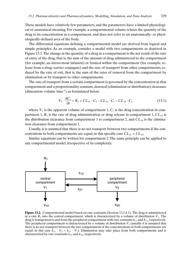
These models have relatively few parameters, and the parameters have a limited physiologi-cal or anatomical meaning. For example, a compartmental volume relates the quantity of thedrug to its concentration in a compartment, and does not refer to an anatomically- or physi-ologically-defined area of the body.
The differential equations defining a compartmental model are derived from logical andsimple principles. As an example, consider a model with two compartments as depicted inFigure 13.1.The change in the quantity of a drug in a compartment is the net result of the rateof entry of the drug, that is, the sum of the amount of drug administered to the compartment(for example, an intravenous infusion) or formed within the compartment (for example, re-lease from a drug–carrier conjugate) and the rate of transport from other compartments, re-duced by the rate of exit, that is, the sum of the rates of removal from the compartment byelimination or by transport to other compartments.
The rate of transport from a certain compartment is governed by the concentration in thatcompartment and a proportionality constant, denoted (elimination or distribution) clearance(dimension: volume time-1) as formulated below.
(13.1)
where V1 is the apparent volume of compartment 1, C1 is the drug concentration in com-partment 1, R1 is the rate of drug administration or drug release in compartment 1, CL12 isthe distribution clearance from compartment 1 to compartment 2, and CL10 is the elimina-tion clearance from compartment 1.
Usually, it is assumed that there is no net transport between two compartments if the con-centrations in both compartments are equal; in this specific case CL21 = CL12.
Similar equations can be written for compartment 2. The same principle can be applied toany compartmental model, irrespective of its complexity.
13.2 Pharmacokinetics and Pharmacodynamics, Modelling, Simulation, and Data Analysis 339
V1 ·dC1 = R1 + CL21 · C2 – CL12 · C1 – CL10 · C1dt
centralcompartment
V1
peripheralcompartment
V2
k12
k21
k10 k20
R1
Figure 13.2. Compartmental model based on rate constants (Section 13.2.4.1). The drug is administeredat a rate R1 into the central compartment, which is characterized by a volume of distribution V1. Thedrug is transported to and from the peripheral compartment with rate constants k12 and k21, respectively.The peripheral compartment is characterized by a volume of distribution V2 (usually it is assumed thatthere is no net transport between the two compartments if the concentrations in both compartments areequal; in this case k21 · V2 = k12 · V1). Elimination may take place from both compartments and ischaracterized by rate constants k10 and k20, respectively.

Usually, the differential equations are written in a different form, by relating the rate oftransport from a compartment to the quantity of the drug in that compartment and a rateconstant (dimension: time-1), as depicted in Figure 13.2. and formulated as follows.
(13.2)
where A1 is the quantity of the drug in compartment 1, k12 and k21 are distribution rateconstants and k10 is the elimination rate constant.
Comparing Eq. 13.1 and 13.2, it follows that a rate constant kxy is equal to CLxy / Vx.From the assumption that there is no net transport between two compartments if the con-
centrations in both compartments are equal, it follows that k21 · V2 = k12 · V1 (= CL21 = CL12).
Eq. 13.1 and 13.2 are mathematically equivalent, and thus may be used arbitrarily withoutaffecting the modelling results. However, Eq. 13.1 (and Figure 13.1) is preferred since it re-flects better the mechanistic basis, as drug transport is governed by drug concentration, bothfor passive diffusion according to Fick’s Law, and for carrier-mediated transport. In the caseof the latter, the terms referring to the rate of transport from compartment x to compartmenty,
CLxy · Cx (13.3)
should be replaced by their Michaelis–Menten equivalent
(13.4)
where Vmaxxy is the maximum transport rate between compartments x and y, and Kmxy isthe Michaelis–Menten constant of the transport between x and y.
An example of the use of Michaelis–Menten kinetics in a compartmental model is given inthe model of Stella and Himmelstein [5], depicted in Figure 13.3.
13.2.4.2 Physiologically-based Pharmacokinetic (PB-PK) Models
These are relatively complex models describing drug transport between blood and a series ofphysiological and/or anatomical entities, for example, organs, tissues, or cells [15–20]. PB-PKmodels are characterized by a relative large number of parameters. In many cases, several ofthese can be estimated from physiology or anatomy (for example, blood flow and volumes),others may be obtained from in vitro experiments (for example, partition coefficients be-tween water and tissue), or by experiments in isolated tissues (for example, binding and me-tabolism in isolated liver cells or slices; see Chapter 12). In principle, PB-PK models are welladapted to take into account the extracellular and/or intracellular events in the disposition ofthe targeting device.
The number of compartments in a physiologically-based pharmacokinetic model may varybetween two (in drug targeting: a target compartment and a non-target compartment) and 10or more, depending on the desired degree of differentiation. The more compartments, thegreater the ability of the model to define the true behaviour of the drug. However, the in-
340 13 Pharmacokinetic/Pharmacodynamic Modelling in Drug Targeting
dA1 = R1 + k21 · A2 – k12 · A1 – k10 · A1dt
Vmaxxy · CxKmxy + Cx
creased number of parameters increases the problem of assigning reliable values to these pa-rameters, both in simulation (Section 13.2.7) and in data analysis (Section 13.2.8). As a gen-eral rule, the number of compartments should be chosen carefully, according to the parsimo-ny principle: start the modelling with the simplest model that can discriminate the processesof interest. If the chosen model does not provide satisfactory results (in terms of credible pre-dictions or satisfactory goodness-of-fit), the model can be explored further by adding com-partments or connections in a step-by-step procedure.
On the other hand, PB-PK models are frequently used in toxicokinetics for a differentpurpose, that is, the model should be able to explain the drug distribution over a large num-ber of tissues as measured from in vivo animal studies, with the eventual goal of data extrap-olation to man. In this case, the starting point is a model including each organ and tissue fromwhich measurements are available. If necessary, the number of compartments can be reducedby combining compartments with similar properties. A detailed description of the process ofexplicit (or formal) combining has been given by Nestorov et al. [19] and Weiss [20].
The principles of PB-PK modelling will be explained using the model of Hunt [6], depict-ed in Figure 13.4, a PB-PK model suited for evaluation of drug targeting strategies (Sections13.3.2 and 13.4). For this model, the following set of differential equations describing the drugtransport (mass per unit of time) can be written according to mass balance:
(13.5)
(13.6)
(13.7)
(13.8)
where V is the volume of the compartment, C is the drug concentration, Q is the blood (orplasma, whichever is the reference fluid) flow, K is the tissue/blood partition coefficient, CLis the (elimination) clearance, and R is the rate of drug input; subscripts C, R, T and E referto the central compartment, response (or target) compartment, toxicity compartment, andelimination compartment, respectively (Note: Hunt et al. [6] did not include the partition co-efficient K as such, in their equations. Rather, their tissue concentrations refer to a blood orplasma concentration which is in equilibrium with the tissue concentration, equal to the ratioC/K; consequently, their tissue volumes refer to apparent volumes, equal to the product K · V).
13.2.4.3 Compartmental Models Versus Physiologically-based Models
Although compartmental models and physiologically-based models may at first, seem quitedifferent, and are usually treated as two different classes of models, both approaches are ac-tually similar [17].When appropriately defined, probably any PB-PK model can be written asa compartmental model and vice versa. This can be seen by comparing the models in Fig-ures 13.1 and 13.3, and their mathematical descriptions in Eq. 13.1 and 13.5.
13.2 Pharmacokinetics and Pharmacodynamics, Modelling, Simulation, and Data Analysis 341
VC · dCC = RC + QR ·
CR + QT · CT + QE ·
CE – (QR + QT + QE) · CCdt KR KT KE
VR · dCR = RR + QR · CC – QR ·
CR – CLR · CRdt KR
VT · dCT = RT + QT · CC – QT ·
CT – CLT · CTdt KT
VE · dCE = RE + QE · CC – QE ·
CE – CLE · CEdt KE
The major difference between both approaches is not in the mathematical or pictorial de-scription, but in the interpretation of the parameters. In compartmental modelling, the start-ing point is the parameters that do not necessarily have a particular anatomical or physio-logical meaning. This meaning, however, may become clear after a careful analysis of thedata, including measurements in different organs and tissues. On the other hand, PB-PKstarts with a model with physiologically meaningful parameters. It should be stated that,when applying a PB-PK model to real data, the identification of the parameters may becomea major problem in the interpretation (see Section 13.2.8.4).
13.2.4.4 Principles of Modelling
In both types of models, the quantity or concentration of the drug in various sites of the bodyis described by mathematical equations quantifying drug administration, drug transport anddrug elimination. These mathematical representations are usually in the form of differentialequations, which can be solved numerically. In some simple cases an explicit analytical solu-tion of the differential equations can be obtained, thus facilitating the calculations. The nu-merical procedure of solving the differential equations is more generally applicable, but iscomplicated by the necessity to find a compromise between accuracy and speed of execution.However, using modern, user-friendly software and fast-performing hardware, this is muchless of an issue today (see Section 13.7).
13.2.5 Pharmacodynamic Models
Pharmacodynamic (PD) models are used to describe the relationship between drug concen-tration and drug effect.An overview of various PD models can be found in the literature [21].The essential elements will be treated in the following sections.
13.2.5.1 Sigmoid Emax Model
For simplicity, a linear relationship between concentration and effect is often assumed, re-ducing the problem of PK/PD to the pharmacokinetics. However, the concentration–effectrelationship of any drug tends towards a plateau, and a sigmoidal model (sigmoid Emax mod-el or Hill equation) is more appropriate [21–24]:
(13.9)
where E is the drug effect (arbitrary unit; same unit as E0 and Emax), E0 is the drug effectin the absence of drug (typically zero, or baseline effect), Emax is the maximum achievabledrug effect, Ce is the drug concentration at the effector site, γ is a dimensionless value, indi-cating the gradient of the concentration–effect relationship, and EC50 is the drug concentra-tion at which the drug effect is 50% of the maximum effect Emax.
342 13 Pharmacokinetic/Pharmacodynamic Modelling in Drug Targeting
E = E0 + Emax ·Ce
γ
Ceγ + EC50
γ
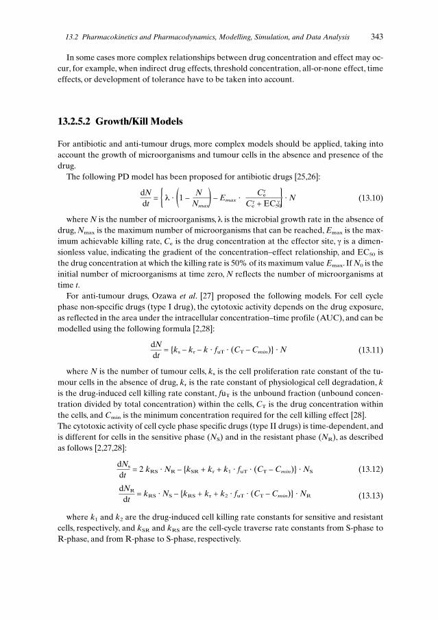
In some cases more complex relationships between drug concentration and effect may oc-cur, for example, when indirect drug effects, threshold concentration, all-or-none effect, timeeffects, or development of tolerance have to be taken into account.
13.2.5.2 Growth/Kill Models
For antibiotic and anti-tumour drugs, more complex models should be applied, taking intoaccount the growth of microorganisms and tumour cells in the absence and presence of thedrug.
The following PD model has been proposed for antibiotic drugs [25,26]:
(13.10)
where N is the number of microorganisms, λ is the microbial growth rate in the absence ofdrug, Nmax is the maximum number of microorganisms that can be reached, Emax is the max-imum achievable killing rate, Ce is the drug concentration at the effector site, γ is a dimen-sionless value, indicating the gradient of the concentration–effect relationship, and EC50 isthe drug concentration at which the killing rate is 50% of its maximum value Emax. If N0 is theinitial number of microorganisms at time zero, N reflects the number of microorganisms attime t.
For anti-tumour drugs, Ozawa et al. [27] proposed the following models. For cell cyclephase non-specific drugs (type I drug), the cytotoxic activity depends on the drug exposure,as reflected in the area under the intracellular concentration–time profile (AUC), and can bemodelled using the following formula [2,28]:
(13.11)
where N is the number of tumour cells, ks is the cell proliferation rate constant of the tu-mour cells in the absence of drug, kr is the rate constant of physiological cell degradation, kis the drug-induced cell killing rate constant, fuT is the unbound fraction (unbound concen-tration divided by total concentration) within the cells, CT is the drug concentration withinthe cells, and Cmin is the minimum concentration required for the cell killing effect [28].The cytotoxic activity of cell cycle phase specific drugs (type II drugs) is time-dependent, andis different for cells in the sensitive phase (NS) and in the resistant phase (NR), as describedas follows [2,27,28]:
(13.12)
(13.13)
where k1 and k2 are the drug-induced cell killing rate constants for sensitive and resistantcells, respectively, and kSR and kRS are the cell-cycle traverse rate constants from S-phase toR-phase, and from R-phase to S-phase, respectively.
13.2 Pharmacokinetics and Pharmacodynamics, Modelling, Simulation, and Data Analysis 343
dN= { λ · (1 –
N ) – Emax ·Ce
γ } · Ndt Nmax Ce
γ + EC50γ
dN= {ks – kr – k · fuT · (CT – Cmin)} · N
dt
dNs = 2 kRS · NR – {kSR + kr + k1 · fuT · (CT – Cmin)} · NSdt
dNR = kRS · NS – {kRS + kr + k2 · fuT · (CT – Cmin)} · NRdt
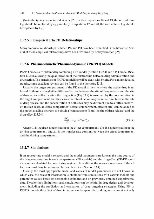
(Note the typing errors in Nakai et al. [28]; in their equations 16 and 19, the second termkSR should be replaced by kRS; similarly, in equations 17 and 20, the second term kRS shouldbe replaced by kSR).
13.2.5.3 Empirical PK/PD Relationships
Many empirical relationships between PK and PD have been described in the literature. Sev-eral of these empirical relationships have been reviewed by Kobayashi et al. [29].
13.2.6 Pharmacokinetic/Pharmacodynamic (PK/PD) Models
PK/PD models are obtained by combining a PK model (Section 13.2.4) and a PD model (Sec-tion 13.2.5), allowing the quantification of the relationship between drug administration anddrug action.The principles of PK/PD modelling will be dealt with briefly. For a more detailedtreatise, some excellent reviews can be found in the literature [21].
Usually, the target compartment of the PK model is the site where the active drug is re-leased. If there is a negligible diffusion barrier between the site of drug release and the siteof drug action (effector site), the drug action (Eq. 13.9) is governed by the concentration inthe target compartment. In other cases the site of action may be more remote from the siteof drug release, and the concentration at both sites may be different due to a diffusion barri-er. In such cases, an extra compartment (effect compartment, effector site) can be added tothe model as a link between the ‘driving’ compartment (here, the site of drug release) and thedrug effect [23,24]:
(13.14)
where Ce is the drug concentration in the effect compartment, C is the concentration in thedriving compartment, and ke0 is the transfer rate constant between the effect compartmentand the driving compartment.
13.2.7 Simulations
If an appropriate model is selected and the model parameters are known, the time course ofthe drug concentration in each compartment (PK models) and the drug effect (PK/PD mod-els) can be calculated for any dosing regimen. In addition, the relevant measures of the ef-fectiveness of drug targeting can be calculated (see Section 13.4).
Usually, the most appropriate model and values of model parameters are not known, inwhich case, the relevant information is obtained from simulations with various models andparameter values, based on reasonable estimates and on previously obtained experimentaldata. Despite their limitations, such simulations can be helpful in drug design and develop-ment, including the prediction and evaluation of drug targeting strategies. Using PK orPK/PD models, the effect of drug targeting can be quantified, taking into account not only
344 13 Pharmacokinetic/Pharmacodynamic Modelling in Drug Targeting
dCe = ke0 · (C – Ce)dt

the process modified in order to target the drug, but also the kinetics of the carrier–drug con-jugate and the active drug after liberation. Such a modelling process might precede the ex-perimentation process, in order to gain insight into the potential benefit of a drug targetingconcept in comparison to traditional administration of the drug.
13.2.8 Data Analysis by Modelling
Data analysis by modelling may be applied for various reasons, for example:
• To condense the data, thus obtaining a model with relatively few model parameters in-stead of one with a large number of measurements.
• To explore mechanisms involved in the process under investigation (for example, carrier-mediated transport).
• To make predictions (for example, to predict the dose needed to maintain the concentra-tion of the drug at the target site within a therapeutic window).
13.2.8.1 Model Building
The process of data analysis by modelling implies building a model from measurements,which involves two steps: (a) building the model structure, and (b) assessment of model pa-rameters.
First, the simplest model which includes the minimum number of compartments and mod-el parameters must be defined. For this model, the parameters are estimated from a set ofmeasurements obtained by non-linear regression or curve-fitting (Section 13.2.8.3). The pur-pose of this process is to find a set of model parameters which best fits the measurements(Section 13.2.8.2). If the goodness-of-fit is acceptable (Section 13.2.8.5), the model can beevaluated by comparison with other models (Section 13.2.8.6).
13.2.8.2 Defining the Objective Function
The first step in ‘curve fitting’ is to define the ‘best fit’. Usually, the criterion for ‘best fit’ is aweighted least-squares criterion, based on statistical grounds [30–34]. Assuming that the er-rors in the measured concentrations are normally distributed, the best fitting set of parame-ters is obtaining by minimization of the following objective function, OBJ:
(13.15)
where
n = total number of concentration measurementsCmeas,i = measured concentration at time point i (i = 1,2,...,n)Ccalc,i = calculated concentration at time point iσi = standard deviation of the measurement at time point i
13.2 Pharmacokinetics and Pharmacodynamics, Modelling, Simulation, and Data Analysis 345
OBJ = Σ {(Cmeas, i – Ccalc, i)2
+ ln (σi2)}σi
2
n
i = 1

In Eq. 13.15, the squared standard deviations (variances) act as ‘weights’ of the squaredresiduals.The standard deviations of the measurements are usually not known, and thereforean arbitrary choice is necessary. It should be stressed that this choice may have a large influ-ence of the final ‘best’ set of parameters. The scheme for appropriate weighting and, if ap-propriate, transformation of data (for example logarithmic transformation to fulfil the re-quirement of homoscedastic variance) should be based on reasonable assumptions with re-spect to the error distribution in the data, for example as obtained during validation of theplasma concentration assay. The choice should be checked afterwards, according to the pro-cedures for the evaluation of goodness-of-fit (Section 13.2.8.5).
Usually, the standard deviation of the measurement is dependent on the magnitude of theconcentration. The most commonly applied assumption is that the standard deviation is pro-portional to the concentration, which is either the measured concentration (also referred toas ‘data-based weighting’), or the calculated concentration (model-based weighting).
Many software packages provide only a limited selection of weighting procedures. Themost commonly applied weighting procedure is based on the assumption that the standarddeviation of the concentration is proportional to the measured concentration. In that case,the objective function may be simplified to:
(13.16)
Alternatively, the following objective function may be used, assuming that the errors in themeasured concentrations are log-normally distributed:
(13.17)
Since both Eq. 13.16 and 13.17 assume a constant coefficient of variation of the measure-ment error, these equations provide similar (but not identical) results.
13.2.8.3 Searching the Best-fitting Set of Parameters
The best-fitting set of parameters can be found by minimization of the objective function(Section 13.2.8.2). This can be performed only by iterative procedures. For this purpose sev-eral minimization algorithms can be applied, for example, Simplex, Gauss–Newton, and theMarquardt methods. It is not the aim of this chapter to deal with non-linear curve-fitting ex-tensively. For further reference, excellent papers and books are available [18].
The fitting procedure may be performed by any suitable minimization algorithm. In theo-ry, the final parameter set depends only on the objective criterion (Section 13.2.8.2) and isnot dependent on the minimization algorithm, nor on the initial set of parameter values (ex-cept for rounding-off errors). However, in practice, the minimization algorithm may fail toreach the minimum of the objective function, and may end in a local minimum. In this re-spect, minimization algorithms may vary widely. An algorithm which is insensitive to thechoice of the initial estimates, is said to be robust, which is a highly desirable property.To low-er the risk of convergence to a local minimum of the objective function, the convergence cri-terion (or stop criterion, for example the relative improvement of the objective function)
346 13 Pharmacokinetic/Pharmacodynamic Modelling in Drug Targeting
OBJ = Σ [ ln (Cmeas, i) – ln (Ccalc, i)]2
n
i = 1
OBJ = Σ (Cmeas, i – Ccalc, i)2
(Cmeas, i)2
n
i = 1

chosen should be sufficiently small, and the fitting procedure may be repeated with a differ-ent set of initial estimates, or a different minimizing algorithm.
13.2.8.4 Identification of Model Parameters
The procedure to obtain the best fitting set of model parameters (Section 13.2.8.3) can beperformed only if each model parameter is uniquely identifiable from the measurements[35–38]. This implies that the same set of model parameters is obtained, irrespective of theinitial set (Section 13.2.8.3). In some cases one or more model parameters cannot be identi-fied uniquely, because the measurement data do not contain enough ‘information’ on thatparticular parameter, for example:
• In the model depicted in Figure 13.1, the parameters CL10 and CL20 cannot be obtaineduniquely if only measurement data from compartment 1 are available. There is an infinitenumber of parameter sets yielding exactly the same concentration profile in compartment1. Only if certain constraints are imposed (for example, CL20 = 0 or CL20 = CL10), can themodel parameters be identified uniquely.
• In the case of Michaelis–Menten kinetics (Eq. 13.4), Vmaxxy and Kmxy cannot be assesseduniquely if the concentration Cx is far below the value of Kmxy; in that case, Eq. 13.4 re-duces to Eq. 13.3, and only one parameter CLxy, or the ratio Vmaxxy/Kmxy can be calcu-lated uniquely.
• In the model shown in Figure 13.1, if CL12 is very large compared to CL10, and if no datashortly after administration of a bolus dose are available, the model would behave as a sin-gle compartment model, and CL12 will not identifiable; also, only the sum of the volumesrather than both volumes separately can be assessed.
The problem of identification grows rapidly with increasing complexity of the model. Insome cases this problem can be solved by an appropriate experimental design. As a generalrule, the problem is reduced if the concentrations in compartments other than the centralplasma compartment can be measured. Also, simultaneous measurement of drug anddrug–carrier conjugate or pro-drug is a condition for identification of the models describedin Section 13.3.
Jacquez and Perry [37] developed the program IDENT to investigate the identification ofmodel parameters. In most cases, problems of identification can be detected by inspection ofthe standard errors of the model parameters (the standard error of a model parameter is ameasure of the credibility of the parameter value, which is provided by the most fitting pro-grams). A high standard error (for example, more than 50% of the parameter value) indi-cates that the parameter value cannot be assessed from the data, most likely due to an iden-tification problem. In that case, the parameter value itself is meaningless, and thus the para-meter set should be discarded (see Section 13.2.8.5).
13.2 Pharmacokinetics and Pharmacodynamics, Modelling, Simulation, and Data Analysis 347

13.2.8.5 Goodness-of-Fit
After fitting the parameters of a model to a set of measurement data, criteria for the good-ness-of-fit are required. There will always be some differences between the measured data and the values calculated from the model. These differences may be due to the followingcauses:
• Measurement errors in the data, for example, inevitable analytical errors implicit in thechosen analytical method. In general, measurement errors are random errors, and their or-der of magnitude may be known from the precision of the assay, as assessed during the val-idation of the assay. If the magnitude of the measurement errors is comparable to the pre-cision of the assay, the goodness-of-fit is acceptable.The possibility of problems in the caseof measurements close to the detection limit of the assay should be taken into account. Inthis case, the relative errors in the analysis may be significantly larger than over the usualrange.
• Model mis-specification. If an inappropriate model is chosen (for example, a model withtoo small a number of compartments, or an incorrect structure), it will not be able to de-scribe the measurements adequately, resulting in systematic deviations between the mea-surements (for example, plasma or tissue concentrations) and the values calculated fromthe model. Such systematic deviation can be detected by the visual methods described be-low.
• Other errors in the procedure, such as failure to distinguish between carrier-bound and un-bound drug, as well as errors in dosing, deviations in the time of measurement, incorrectsampling procedure, exchange of samples, mistakes in dilution during sample treatment, and soforth.These types of error are the most problematic, and no general solution can be given.
There are several methods available for the assessment of goodness-of-fit, however, there areno exact and objective criteria for its evaluation.This is due to the following: (1) goodness-of-fit is not a single property, and cannot be expressed in a single value, and (2) numerical mea-sures of goodness-of-fit do no have an absolute meaning.Therefore there is a dependence onsomewhat subjective criteria.To ensure maximal objectivity, the criteria for accepting a set ofmodel parameters obtained by the fitting procedure as a valid result should be defined ex-plicitly before the analysis is initiated. In practice, however, this condition is hardly applica-ble during the development of new drug targeting preparations, taking into account the com-plexity of the modelling procedure.
The following criteria could be used to ensure an acceptable goodness-of-fit:
• Visual inspection of the observed and calculated data should not reveal any significantlack of fit.
• Residuals (difference between observed and calculated data) or normalized residuals(residuals divided by the corresponding standard deviation) should be scattered random-ly around zero, by visual inspection.
• Normalized residuals should be neither diverging nor converging when plotted againsttime or plotted against (logarithm of) concentration, by visual inspection.
• Residuals should not be serially correlated, as identified by visual inspection or by an ap-propriate statistical test (for example a Run’s test).
348 13 Pharmacokinetic/Pharmacodynamic Modelling in Drug Targeting

• The standard error of each relevant parameter should be lower than a predefined value(for example 50% of the parameter value). High standard errors may reflect problems inthe identification (see Section 13.2.8.4).
• In the case of any of the calculated pharmacokinetic parameters being seen as physiologi-cally unfeasible, the analysis should be interpreted with care, and should not be presentedwithout comments.
• Outlying data points should be dealt with explicitly, and should not be discarded unless feltto be physiologically impossible. The impact of elimination of the outlying points on theparameter estimates should be investigated.
Non-compliance with one or more of these criteria may indicate that an inappropriate mod-el or an inappropriate weighting scheme was chosen.
13.2.8.6 Model Selection
There may be more than one plausible model structure that can be used to describe the data.In that case, any plausible model is analysed in a similar way. If the goodness-of-fit of morethan one model is acceptable (see Section 13.2.8.5), a procedure for selecting the ‘best’ mod-el is required.
It is common practice to compare the results of different models, each yielding an accept-able goodness-of-fit, according to the following procedure. First, the models are classified hi-erarchically in a tree structure.The more complex models are considered as extensions of thesimpler models, by adding extra parameters, for example, an extra compartment, a extradegradation step, or a time lag. It can be said that the simpler model is a special case of themore complex model, for example because one or more parameters have a fixed value (ingeneral a zero value). Then, starting with the simplest model (see Section 13.2.8.1), the mod-els are compared in pairs according to their hierarchical relationship. Such a comparison canbe based on statistical criteria, for example [39]:
(a) An F-test by which the following value is calculated:
(13.18)
where N is the number of measurements, P is the number of model parameters, and WRSSis the weighted residual sum of squares (see Section 13.2.8.2); the subscript s refers to thesimpler model and the subscript c to the more complex model.
If the value F exceeds the tabulated value of Fischer’s F-distribution for (Pc – Ps) and(N – Pc) degrees of freedom, and a confidence level of (usually) 95% (α = 0.05), then thecomplex model fits significantly better to the data than the simpler model. If not, the ‘parsi-mony’ principle dictates that the simpler model should be accepted as the ‘best’ model.(b) Akaike Information Criterion (AIC). For each model the AIC is calculated according tothe following equation:
AIC = N · ln (WRSS) + 2P (13.19)
The model with the lowest AIC value is accepted as the ‘best’ model.
13.2 Pharmacokinetics and Pharmacodynamics, Modelling, Simulation, and Data Analysis 349
F = WRSSS – WRSSC ·
N – PC
WRSSC PC – PS

13.3 Pharmacokinetic Models for Drug Targeting
In 1980, Stella and Himmelstein [5] introduced the principles of pharmacokinetic modellinginto the field of pro-drugs and site-specific delivery. In 1986, Hunt et al. [6] extended the mod-el of Stella and Himmelstein by taking into account a specific area where toxicity occurs.Their work may be considered as the frame of reference for later work in this area.
13.3.1 Model of Stella and Himmelstein
The model of Stella and Himmelstein [5], depicted in Figure 13.3, was originally derived forpro-drugs, and may be considered as a minimal model for evaluating the pharmacokinetics ofpro-drugs and drug targeting systems. Since the drug–carrier conjugate (DC) and the activedrug (D) are different entities, and since the model should be able to discriminate betweenthe target site and non-target sites, a pharmacokinetic model for drug–carrier conjugatesshould include, at least, the following compartments (Figure 13.3):
350 13 Pharmacokinetic/Pharmacodynamic Modelling in Drug Targeting
DCresponse (target)
compartmentVR(DR)
Dresponse (target)
compartmentVR(D)
Vmax R
Km R
CLC (DC)
DCcentral
compartmentVC(DR)
Dcentral
compartmentVC(D)
Vmax C
Km C
CLC (D)
RC(DC)
CLCR(DC) CLCR(D)
Figure 13.3. Model of Stella and Himmelstein, adapted from reference [5] (Section 13.3.1). Thedrug–carrier conjugate (DC) is administered at a rate RC(DC) into the central compartment of DC,which is characterized by a volume of distribution VC(DC). DC is transported with an inter-compartmental clearance CLCR(DC) to and from the response (target) compartment with volumeVR(DC), and is eliminated from the central compartment with a clearance CLC(DC). The active drug(D) is released from DC in the central and response compartments via saturable processes obeyingMichaelis–Menten kinetics defined by Vmax and Km values. D is distributed over the volumes VC(D)and VR(D) of the central and response compartment, respectively. D is transported with an inter-compartmental clearance CLCR(D) between the central compartment and response compartment, andis eliminated from the central compartment with a clearance CLC(D).

• The DC in non-target sites, including the plasma compartment (central compartment).Thedrug–carrier conjugate in the non-target sites may be modelled as a single compartment, ifDC distributes rapidly over its distribution volume.That is, the plasma compartment in thecase of large macromolecular carriers which cannot cross the endothelial lining of bloodvessels, or the extracellular volume in the case of smaller compounds which do have theopportunity to extravasate. Both administration (except for delivery to the target site) andelimination occur in this DC central compartment. If necessary, one or two peripheralcompartments may be added to improve the accuracy of predicting the distribution of theDC concentrations in the body.
• DC in target sites (response compartment). The drug–carrier conjugate is targeted bysome specific mechanism to the target site. The volume of this compartment depends onthe specificity of this mechanism, and can also be influenced by the disease process whichis being targeted. This value may range from a few milliliters (for a focal inflammation orinfection site) to several liters (for a widely spread disease) [40].
• D in target sites (response compartment). Ideally, the target site of the drug is located verynear to the DC target site, where the drug is released or activated. In that case, the targetsites of DC and D are identical, and any amount of drug released or activated may exertits effect immediately. However, if the drug is released or activated at sites distant from thetarget site of the drug, an additional compartment is required. This will have a detrimentaleffect on the efficacy of the targeting strategy [40].
• D in non-target sites, including the plasma compartment (central compartment). After re-lease or activation of the drug in or near the target site, the drug will be transported to theplasma compartment by diffusion and/or convection. Also, some drug may be released oractivated at non-target sites. The central compartment in which the drug distributes mayvary, depending on its physicochemical properties: from a minimum value equal to theplasma compartment to high values, exceeding the physical volume due to excessive bind-ing of the drug to tissue components [10]. Elimination of the drug occurs from this centralcompartment. If necessary, one or two peripheral compartments may be added to enable amore accurate prediction of the drug concentrations in the body to be made. If drug D isconverted to active metabolites, additional compartments to account for the fate of thesemetabolites may be required.
The central compartment may also include the sites where toxicity occurs. Alternatively,these sites may be modelled as compartments connected to the central compartment, analo-gous to the D target-site compartment. If the drug is released or activated within these toxi-city sites, and/or the drug carrier is targeted to some degree to these sites, an additional DCcompartment should be modelled, analogous to the DC target site.
Using the model shown in Figure 13.3, the main processes involved are the following.
13.3.1.1 Disposition of DC
This refers to the distribution and elimination of the DC, excluding transport to the targetsite (and toxicity site, as shown in Figure 13.4). In general, it is desirable that the DC is notrapidly eliminated from the circulation. This is necessary both to minimize the exposure of
13.3 Pharmacokinetic Models for Drug Targeting 351
eliminating organs to the targeted drug (in the case of release or activation of the drug in theeliminating organs) and to maximize the therapeutic availability, that is, to maximize the frac-tion of the dose that reaches the target site. Ideally, the elimination of the DC should takeplace exclusively in the target organ (see Sections 13.3.1.2 and 13.3.1.3). Elimination at othersites reduces the efficiency of targeting. For example, elimination of the DC without releaseof the active drug implies a waste of a sophisticated and expensive product, and eliminationof the DC with release of active drug in non-target tissue increases the concentration of D innon-target tissue, thus lowering the Drug Targeting Index (DTI, see Section 13.4.2) [12].
In general, optimization of the disposition of DC is the simplest part of the design of a drugtargeting system. There are many methods by which the retention time of the drug carrierwithin the target tissue can be prolonged (described in other chapters of this book), and the assessment of the disposition of the DC by measuring its plasma or blood concentra-tion–time profile is relatively simple.
13.3.1.2 Delivery of the DC to the Target Site
The focus of research in the field of drug targeting is mainly on the selectivity of the DC forthe target site (see other chapters in this book), undoubtedly because it is the essentialprocess in drug targeting.
Although the delivery of the DC to the target site is a critical step in the efficacy of drugtargeting, it is not necessarily the most critical step. In fact, each of the processes involvedmay be critical, and it is possible that in practice, given the sophisticated principles which areapplied to increase the selectivity of DC for the target site, one of the other processes in-volved (see Sections 13.3.1.1. and 13.3.1.3.6) is responsible for the eventual failures of drugtargeting systems.
The general principles of drug transport have been described in Sections 13.2.1.2 and13.2.1.3.The rate of delivery of the DC to the target site is not critical in absolute terms. Evenif the delivery rate is low, as long as the selectivity of the DC for the target site is high effi-cient targeting is assured. In this case, it will take more time to reach effective steady-statedrug levels in the target organ and the duration of the drug effect will be prolonged. Howev-er, if the selectivity of DC for the target site is less than 100%, the slow delivery of the DC tothe target site may decrease the efficacy of drug targeting [12].
If the rate of delivery of the DC to the target site is high compared to the release of the ac-tivated drug (see Section 13.3.1.3), accumulation of DC at the target site might result in a lossof DC from the target site either back into the blood, or via lymphatic drainage, and thus ina decrease of the efficiency of targeting. An example of this is the occurrence of retro-endo-cytosis in which the endocytosed DC is refluxed back into the systemic circulation before ithas released the drug to be targeted [41]. The possibility that receptor-mediated endocytosisis a bidirectional rather than a unidirectional process is often not taken into account in drugtargeting models. Yet, a secondary release of endocytosed material through reactivation ofendocytotic vesicles in the plasma membrane has been demonstrated in various cell types.This process may be of quantitative importance, especially if trafficking to and associationwith lysosomes is slow or if proteolytic degradation is rate-limiting.
352 13 Pharmacokinetic/Pharmacodynamic Modelling in Drug Targeting
13.3.1.3 Release or Activation of D at the Target Site
The release of the drug from the drug–carrier conjugate or the formation of the active moi-ety from the pro-drug, is also reported to be of major concern in many research papers ondrug targeting (see other chapters in this book). Although this process cannot be measuredin vivo in most cases, valuable information can be obtained from in vitro measurements, forexample, using cultured cell lines. The mechanism of release or activation may contribute tothe selectivity of the target site, for example, in case of the formation of the phosphorylatedactive forms of nucleoside analogues by viral enzymes. In the case of release or activation byenzymatic processes, the rate of release or activation may be limited by saturation of the en-zyme capacity.
In general, rapid release contributes to the efficiency of drug targeting, although in manycases the rate of release may not be critical as a result of rate limitation in the delivery of theDC to the target site. With respect to the selectivity of the target site for the release or acti-vation in comparison to non-target sites, it can be stated that the more selective the deliveryof the DC to the target site, the less important is the selectivity of the release or activation atthe target site.
13.3.1.4 Removal of D from the Target Site
Little attention has been paid to this aspect in most research papers on drug targeting, de-spite several reports on the importance of removal of the released drug from the target site[6,12,42]. Most likely, this lack of attention is related to at least three factors: removal of drugfrom the target site (1) cannot be manipulated by the design of the drug targeting system, (2)is difficult to measure, and (3) may be the bottleneck of the efficiency of drug targetingstrategies.
A striking example of the importance of the secondary removal of a drug from the targetsite are the multi-drug-resistance (MDR) processes of tumour cells. For instance antineo-plastic drugs can be extruded efficiently by P-glycoprotein [43]. Although this example isonly relevant for a limited category of drugs, it clearly demonstrates that the delivery of adrug into a cell does not guarantee an optimal retention time in the cell nor an optimal drugeffect. Levy [42] assumed that in many cases drug elimination from the target site will bemuch more rapid that drug elimination from the body, as exemplified for intracerebroven-tricular injections of barbiturates.
Removal of active drug from the target site can occur by two different routes: direct re-moval by a local elimination process (typically a metabolic process, causing inactivation ofthe drug), or removal by the blood flow following diffusion out of the target cell. In both cas-es removal reduces the efficiency of drug targeting by lowering the concentration of the ac-tive drug at the target site. However, in case of local removal, the active drug is not trans-ported to non-target tissues, and does not contribute to toxicity. In contrast, removal by thebloodstream causes an increase of concentration in the non-target tissue, thus lowering thetargeting efficiency further. It may be noted that a high blood flow to the target tissue favoursthe delivery of the DC to the target site, but also increases the rate of removal of the activedrug. Ideally, the active drug should be eliminated slowly from the target site, so that effec-
13.3 Pharmacokinetic Models for Drug Targeting 353

tive drug levels are maintained at the site after administration of relatively low maintenancedoses of the drug targeting system. Obviously, a low, but substantial, rate of active drug re-moval is a prerequisite for controlling the intensity and duration of the drug effect.
13.3.1.5 Release of D at Non-target Sites
The release or activation of a drug at non-target sites is the undesirable counterpart of re-lease at the target site (see Section 13.3.1.3), determining the selectivity of the release or ac-tivation of the drug, and thus of the drug targeting.
In principle, the release at non-target sites can be measured by the same methods as usedfor the target site (Section 13.3.1.3). In practice, however, measurement must be limited toone or more selected cell lines, which do not necessarily include all the non-target sites inwhich the drug may be released outside the intended target tissue.
13.3.1.6 Disposition of D
The distribution and elimination characteristics of the active drug have a profound influenceon the efficiency of drug targeting. This topic is dealt with in Section 13.5.
13.3.2 Model of Hunt
Hunt et al. [6] extended the model of Stella and Himmelstein by adding two compartments,that is, a specific area where toxicity occurs, and an elimination compartment consisting ofthe liver and kidney (Figure 13.4). Their model may be regarded as a simplified physiologi-cally-based pharmacokinetic model (Section 13.2.4.2). It is assumed that the drug concentra-tion in the blood or plasma (whichever is the reference fluid) exiting the compartments is afunction of the concentrations within the corresponding compartments. This implies that tis-sue perfusion rather than permeability of the drug between blood and tissue is assumed to bethe rate-limiting step of the transport between the central compartment and each of the oth-er compartments (Section 13.2.1.3).
The model does not use a pharmacokinetic model for the drug–carrier conjugate. Instead,the release of the active drug is modelled as an input function in each of the four compart-ments: ‘blood’ (central compartment), ‘response’ (target site), ‘toxicity’, and ‘elimination’.The aim of their model was the derivation of the Therapeutic Availability (TA, Section13.4.1) and the Drug Targeting Index (Section 13.4.2). For the particular derivations, severalsimplifications were made; for example, the aforementioned drug input replaces the modelfor DC. As a result, their simplified approach cannot be used for the prediction of the timecourse of the drug concentrations in the various compartments. This limitation does not af-fect the ability of the model to evaluate steady-state drug concentrations, which can also beused as an appropriate measure of the average concentration over a single dose interval af-ter repeated administration. However, their analysis does not give insight into the timecourse of drug action, that is, the time needed to reach steady state, and the duration of the
354 13 Pharmacokinetic/Pharmacodynamic Modelling in Drug Targeting

drug effect after the conclusion of drug administration.To evaluate the time course of action,a full model, including the pharmacokinetic behaviour of the drug–carrier conjugate, shouldbe used, for example the model of Stella and Himmelstein (Section 13.3.1) or the model ofBoddy et al. (Section 13.3.3).
13.3 Pharmacokinetic Models for Drug Targeting 355
centralcompartment
response (target)compartment
toxicitycompartment
eliminationcompartment
QR
QT
QC
QE
RC
RR
RT
RE
CLR
CLT
CLE
Figure 13.4. Model of Hunt, adapted from reference [6] (Section 13.3.2). The model describes the fateof the active drug (D) only. It consists of four compartments: A central compartment (VC) representingblood and all other tissues not accounted for by the other three compartments, a response (target)compartment (VR) representing all tissues containing target sites for the desired response, a toxicitycompartment (VT) representing tissues where the cascade of events leading to a toxic response isinitiated, and an elimination compartment (VE) representing the eliminating organs, excluding theelimination sites in the response and toxicity compartments. Each compartment is characterized by avolume of distribution V, a blood flow Q (where QC = QR + QT + QE), and, except for the centralcompartment, an elimination clearance CL. Drug delivery is designated by input functions R in the fourcompartments. Drug targeting and conventional drug administration are modelled by changing therelative contributions of RC, RR, RT, and RE. When RC = RT = RE = 0, the drug carrier is an ideal target-specific carrier.

Boddy and Aarons [44] used a simplified model, in which the toxicity sites are included in the systemic (non-target) tissues to allow derivation of the Drug Targeting Index which was less restricted by model assumptions.Their approach was criticized by Siegel et al.[45]. When some drug release from the carrier occurs at either the central, toxicity or elimi-nation regions, the DTI is affected by the specific fraction of the drug delivered to each re-gion. Since these regions are combined by Boddy and Aarons into a single systemic region,their model cannot account for this effect. Siegel et al. came to the conclusion that the modelof Hunt et al. and the model of Boddy and Aarons are only equivalent in the case of an idealcarrier.
356 13 Pharmacokinetic/Pharmacodynamic Modelling in Drug Targeting
CLC(DC)
RC(DC)
DCresponse (target)
compartmentVR
Dresponse (target)
compartmentVR
kR
DCcentral
compartmentVC
Dcentral
compartmentVC
kC
CLC (D)
QCR QCR
QCT QCT
DCtoxicity
compartmentVT
Dtoxicity
compartmentVT
kT
CLR (D)
CLT (D)
RC(D)
Figure 13.5. Model of Boddy, adapted from reference [7] (Section 13.3.3). The drug–carrier conjugate(DC) is administered at a rate RC(DC) into the central compartment, which is characterized by a volumeof distribution VC. DC is transported by blood flow QCR to and from the response (target) compartment,characterized by a volume of distribution VR, and by blood flow QCT to and from the toxicitycompartment, characterized by a volume of distribution VT. DC is eliminated from only the centralcompartment with a clearance CLC(DC). The active drug (D) is released from DC in the central,response and toxicity compartments with first-order rate constants kC, kR and kT, respectively. The D isdistributed over these compartments in a manner similar to the DC. The D is eliminated from thesecompartments with a clearance of CLC(D), CLR(D) and CLT(D), respectively. Conventional drugadministration can be simulated by the input of D at a rate RC(D) into the central compartment.

13.3.3 Model of Boddy
Boddy et al. [7] extended the model of Hunt by incorporating the pharmacokinetic behaviourof the drug–carrier conjugate, analogous to the model of Stella and Himmelstein. Thereforethis model, depicted in Figure 13.5, is suited to the evaluation of the time course profile ofdrug concentrations in each compartment, in contrast to the model of Hunt. Also, they re-duced the model by incorporating the elimination compartment within the central compart-ment. Furthermore, the model was simplified by assuming that the volumes of distributionare the same for the drug–carrier conjugate and the active drug. It is assumed that the bloodflowing from the compartments carries the DC and D at concentrations equal to those with-in the compartments. In this specific case, the inter-compartmental clearances for both DCand D can be equated to the blood flow to the response and toxicity compartments. The au-thors presented their model as a three-compartment model. However, since each compart-ment may contain both the drug and the drug–carrier conjugate (which are treated as differ-ent entities), it might be more appropriate to refer to it as a six-compartment model. Finally,Boddy et al. [7] extended the model to a PK/PD model by incorporation of the pharmacody-namic equations analogous to Eq. 13.9 describing the therapeutic and toxic effect. Thereforethis model is not only suitable for the evaluation of the effectiveness of drug targeting withrespect to drug concentrations, but also with regard to the balance between therapeutic andtoxic drug effects.
13.3.4 Model of Rowland and McLachlan
Rowland and McLachlan [46] also extended the model of Hunt to allow the evaluation of theeffect of a permeability barrier and plasma protein binding on drug uptake. The model ofRowland and McLachlan is based on the work of Aubrée-Lecat et al. [47], who investigatedthe influence of various parameters on the amount of a macromolecular drug taken up by thetarget tissue. In the model of Rowland and McLachlan the target site (target tissue) consistsof three distinct sites: blood, interstitium, and cells. Although the paper refers mainly to re-gional drug delivery (for example, intra-arterial injection), their model allows the evaluationof the effect of a permeability barrier and plasma protein binding on the removal of the drugfrom the target site, which may be a critical step in the effectiveness of drug targeting (seeSection 13.4). The influence of permeability and plasma protein binding is evaluated usingthe DTI (Section 13.4.2) as a measure of effectiveness of drug targeting.
13.4 Measures of Effectiveness of Drug Targeting
For practical purposes, measures of the effectiveness of drug targeting are required. Suchmeasures have been derived based on the pharmacokinetic profiles of the drug targeting sys-tem and that of the active drug.
13.3 Pharmacokinetic Models for Drug Targeting 357

13.4.1 Therapeutic Availability (TA)
Hunt et al. [6] introduced the term Therapeutic Availability as the ratio of the fraction of thedose reaching the target sites, if the dose is administered as the drug–carrier conjugate, to thefraction of the dose which reaches the same sites if an equal dose of the active drug is ad-ministered intravenously, as formulated below.
(13.20)
which is equivalent to
(13.21)
where AUC denotes the area under the curve (normalized for dose) after a single dose orover one dosing interval in the case of steady state; Css denotes the average steady-state con-centration; the subscript ‘target’ refers to the target site; DC refers to administration of thedrug–carrier conjugate, and D to the intravenous administration of the active drug.
In contrast to the parameter Bioavailability (the fraction of the administered dose reach-ing the systemic circulation), the Therapeutic Availability may exceed a value of 1. A valueexceeding 1 implies that the drug has been targeted successfully, the concentration at the tar-get site being higher than that following a conventional intravenous administration of thesame dose, and (see gueries) that a lower dose can be administered to reach the same con-centration. Thus, an increase in TA is effectively equivalent to an increase in potency.
Boddy et al. [7] defined the Therapeutic Availability as the ratio of the rate of input of freedrug divided by that of the drug carrier for the same degree of maximal therapeutic effect.When considering steady-state conditions, this definition is equivalent to that shown inEq. 13.20 and 13.21.
13.4.2 Drug Targeting Index (DTI)
Hunt et al. [6] also introduced the Drug Targeting Index, which was defined as the ratio ofdrug delivered to the target and toxicity sites when the drug–carrier conjugate is adminis-tered, divided by the same ratio when the active drug is administered intravenously and isformulated as follows.
(13.22)
which is equivalent to
(13.23)
where AUC denotes the area under the curve (normalized for dose) after a single dose orover one dosing interval in the case of steady state; Css denotes the average steady-state con-centration; subscripts ‘target’ and ‘tox’ refer to the target and toxicity sites, respectively; DC
358 13 Pharmacokinetic/Pharmacodynamic Modelling in Drug Targeting
TA = AUCtarget(DC)
AUCtarget(D)
TA = Csstarget(DC)
Csstarget(D)
DTI = AUCtarget(DC)/AUCtox(DC)
AUCtarget(D)/AUCtox(D)
DTI = Csstarget(DC)/Csstox(DC)
Csstarget(D)/Csstox(D)
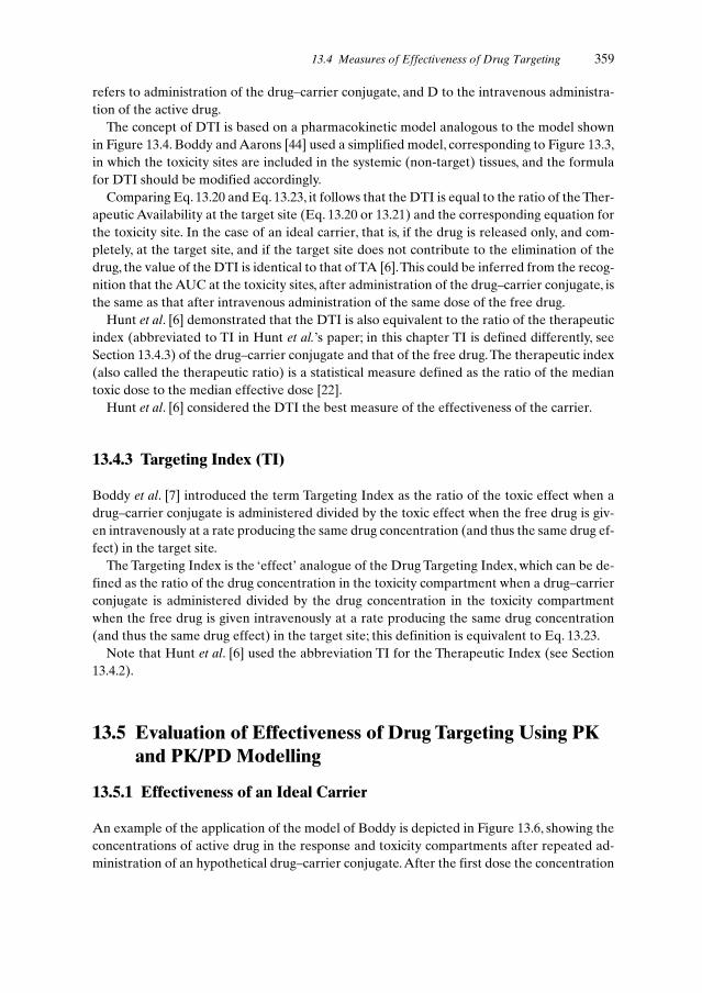
refers to administration of the drug–carrier conjugate, and D to the intravenous administra-tion of the active drug.
The concept of DTI is based on a pharmacokinetic model analogous to the model shownin Figure 13.4. Boddy and Aarons [44] used a simplified model, corresponding to Figure 13.3,in which the toxicity sites are included in the systemic (non-target) tissues, and the formulafor DTI should be modified accordingly.
Comparing Eq. 13.20 and Eq. 13.23, it follows that the DTI is equal to the ratio of the Ther-apeutic Availability at the target site (Eq. 13.20 or 13.21) and the corresponding equation forthe toxicity site. In the case of an ideal carrier, that is, if the drug is released only, and com-pletely, at the target site, and if the target site does not contribute to the elimination of thedrug, the value of the DTI is identical to that of TA [6].This could be inferred from the recog-nition that the AUC at the toxicity sites, after administration of the drug–carrier conjugate, isthe same as that after intravenous administration of the same dose of the free drug.
Hunt et al. [6] demonstrated that the DTI is also equivalent to the ratio of the therapeuticindex (abbreviated to TI in Hunt et al.’s paper; in this chapter TI is defined differently, seeSection 13.4.3) of the drug–carrier conjugate and that of the free drug.The therapeutic index(also called the therapeutic ratio) is a statistical measure defined as the ratio of the mediantoxic dose to the median effective dose [22].
Hunt et al. [6] considered the DTI the best measure of the effectiveness of the carrier.
13.4.3 Targeting Index (TI)
Boddy et al. [7] introduced the term Targeting Index as the ratio of the toxic effect when adrug–carrier conjugate is administered divided by the toxic effect when the free drug is giv-en intravenously at a rate producing the same drug concentration (and thus the same drug ef-fect) in the target site.
The Targeting Index is the ‘effect’ analogue of the Drug Targeting Index, which can be de-fined as the ratio of the drug concentration in the toxicity compartment when a drug–carrierconjugate is administered divided by the drug concentration in the toxicity compartmentwhen the free drug is given intravenously at a rate producing the same drug concentration(and thus the same drug effect) in the target site; this definition is equivalent to Eq. 13.23.
Note that Hunt et al. [6] used the abbreviation TI for the Therapeutic Index (see Section13.4.2).
13.5 Evaluation of Effectiveness of Drug Targeting Using PKand PK/PD Modelling
13.5.1 Effectiveness of an Ideal Carrier
An example of the application of the model of Boddy is depicted in Figure 13.6, showing theconcentrations of active drug in the response and toxicity compartments after repeated ad-ministration of an hypothetical drug–carrier conjugate.After the first dose the concentration
13.4 Measures of Effectiveness of Drug Targeting 359
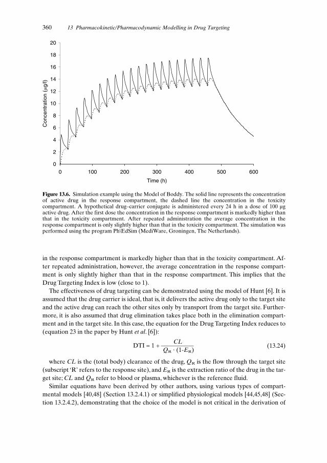
in the response compartment is markedly higher than that in the toxicity compartment. Af-ter repeated administration, however, the average concentration in the response compart-ment is only slightly higher than that in the response compartment. This implies that theDrug Targeting Index is low (close to 1).
The effectiveness of drug targeting can be demonstrated using the model of Hunt [6]. It isassumed that the drug carrier is ideal, that is, it delivers the active drug only to the target siteand the active drug can reach the other sites only by transport from the target site. Further-more, it is also assumed that drug elimination takes place both in the elimination compart-ment and in the target site. In this case, the equation for the Drug Targeting Index reduces to(equation 23 in the paper by Hunt et al. [6]):
(13.24)
where CL is the (total body) clearance of the drug, QR is the flow through the target site(subscript ‘R’ refers to the response site), and ER is the extraction ratio of the drug in the tar-get site; CL and QR refer to blood or plasma, whichever is the reference fluid.
Similar equations have been derived by other authors, using various types of compart-mental models [40,48] (Section 13.2.4.1) or simplified physiological models [44,45,48] (Sec-tion 13.2.4.2), demonstrating that the choice of the model is not critical in the derivation of
360 13 Pharmacokinetic/Pharmacodynamic Modelling in Drug Targeting
DTI = 1 + CL
QR · (1-ER)
0
2
4
6
8
10
12
14
16
18
20
0 100 200 300 400 500 600
Time (h)
Con
cent
ratio
n(µ
g/l)
Figure 13.6. Simulation example using the Model of Boddy. The solid line represents the concentrationof active drug in the response compartment, the dashed line the concentration in the toxicitycompartment. A hypothetical drug–carrier conjugate is administered every 24 h in a dose of 100 µgactive drug. After the first dose the concentration in the response compartment is markedly higher thanthat in the toxicity compartment. After repeated administration the average concentration in theresponse compartment is only slightly higher than that in the toxicity compartment. The simulation wasperformed using the program Ph\EdSim (MediWare, Groningen, The Netherlands).

the DTI. Therefore the criticism of Siegel et al. [45] does not seem relevant for the apprecia-tion of the DTI. It should be noted that the equations for the DTI in several references[7,44,48] are seemingly different from Eq. 31.24, by using the clearance from the non-targetsites instead of the total body clearance. In fact, these equations are identical after appropri-ate rearrangements.
Apart from drugs directed at hepatocytes and renal tubular cells, in the case of the major-ity of conventional drugs, it is likely that the drug is not eliminated directly from the targetsite, and therefore ER = 0, which further simplifies Eq. 13.24 to:
(13.25)
Eq. 13.25 is a more convenient form of Hunt’s equation (16) [6].
13.5.2 Implications of the DTI Concept
The simplicity of Eq. 13.25 is striking: the DTI is determined by only two parameters.This canbe understood qualitatively from the following reasoning. The total dose of the drug is deliv-ered at the target site. If the drug did not leave the target compartment (that is, QR = 0), thedrug concentration at the target site would remain constant, and the DTI would be infinitelyhigh. The faster the removal of the drug from the target site (increase of QR), the lower theAUC at the target site, and the higher the AUC at non-target sites, including the toxicity site,and thus the DTI is lowered. If there is no clearance of the active drug (that is, CL = 0), itwould eventually distribute evenly over the body, and there would be no net drug targetingeffect (DTI = 1). The faster the removal of the drug which is present outside the target site(increase of CL), the lower the concentration and the AUC in the non-target compartments,including the toxicity compartment, and thus the higher the DTI.
The impact of Eq. 13.25 can be rather impressive, as was demonstrated in the paper ofHunt et al. [6]. These authors evaluated Eq. 13.25 by expressing both CL and QR as percent-ages of the cardiac output (normal value for an adult man 5 l min-1). The total blood flowthrough the liver (1.5 l min-1) and kidneys (1.2 l min-1) is 54% of the cardiac output. Since ahigh extraction ratio in both the liver and the kidneys is unlikely, a clearance correspondingto 40% of the cardiac output may be considered as a maximum value for drugs eliminated byliver and kidney (for drugs eliminated by other mechanisms, a higher value might be possi-ble, however). If we postulate that a DTI value of 5 is a minimum for a successful targetingstrategy, it follows that for a drug with a clearance of 40% of the cardiac output, the bloodflow through the target organ must be lower than 10% of the cardiac output in order to ob-tain sufficient targeting efficiency. The upper limit for QR is even lower for drugs with a low-er clearance, and decreases further rapidly in the more realistic cases where the drug carrieris not ideal. For a detailed analysis see Hunt et al. [6].
In conclusion, drug targeting to tissues that receive a relatively large fraction of the cardiacoutput is unlikely to create effective targeting if transport of the free drug to and from thetarget cell can occur. Particularly after multiple dosing, a steady state will be reached in whichthe increase in free drug concentration in the target tissue compared to that in the plasmaand that in the toxicity related tissue, will be moderate.
13.5 Evaluation of Effectiveness of Drug Targeting Using PK and PK/PD Modelling 361
DTI = 1 + CLQR

It follows from Eq. 13.24 that the DTI may be higher than in the aforementioned analysisif ER > 0. However, for conventional drugs it is unlikely that there is a significant eliminationof the drug at the target site. Therefore the extensive evaluations of cases where ER > 0 [6, 7,46] do not seem applicable to targeting conventional drugs. On the other hand, for newertypes of drugs, including protein drugs, antisense oligonucleotides and plasmid DNA, a ma-jor fraction of the drug may be eliminated by lysosomal degradation at the target site [8].Therefore these types of drug are ideal candidates for drug targeting, even with the limitationof having to cross the cell membrane or other barriers (Section 13.5.3).
It should be noted that the conclusions inferred from the aforementioned pharmacokinet-ic models are valid only under the condition that the rather strict assumptions on which theparticular model is based, are justified. In general, however, it is not likely that deviation fromthese conditions will alter the general conclusions seriously. Despite the lack of knowledgeconcerning many of the parameters of the complete model (Figure 13.4), the concept of DTIallows an evaluation of the potential benefit of drug targeting, by providing insight into thecritical values of parameters which are of real concern in the design of drug targeting sys-tems.
The extended model of Boddy (Section 13.3.3) may be helpful for the investigation of thedependence of DTI, TI and TA on various model parameters. Boddy et al. [7] demonstratedthat the DTI is independent of the rate of drug release from the carrier (provided that istakes place only in the target site) or the rate of elimination of the drug–carrier conjugate byother mechanisms which do not lead to the release of the drug. Neither of these parametersinfluences the pharmacokinetics of the free drug in the steady state. However, the TA is af-fected by both parameters. TA increases either as the rate of drug release increases, or as therate of elimination of the DC decreases.
These authors also showed that an increase in the amount of drug eliminated directly fromthe target site (that is, an increase of ER) does not always increase the benefits of drug tar-geting, as in the case of the release of the free drug in the central or toxicity compartment.Levy [42] commented on some pharmacokinetic considerations of targeted drug delivery. Heassumed that drug elimination from the target site (see Section 13.3.1) will, in many case, bemuch more rapid that drug elimination from the body. Furthermore, he assumed that, fol-lowing targeted delivery, a drug will be eliminated from the site of action to the rest of thebody, which will act, at least initially, as an infinite sink. Consequently, it may be expectedthat, after a single dose, (1) the duration of action of a targeted bolus dose will be much short-er than the duration of action of a conventionally-administered, equipotent bolus dose, and(2) the elimination of a targeted drug from the site of action is not affected by changes in thedisposition of the active drug from the non-target compartments. During a steady state, how-ever, the situation is completely different, since the assumption of sink conditions in the sys-temic circulation is no longer valid, and thus systemic clearance becomes a major determi-nant of the effectiveness of drug targeting, as reflected by Eq. 13.24 or 13.25. Levy [42] cor-rectly stated that the selectivity of the drug targeting process will be lost as drug concentra-tions in the systemic circulation rise. Such a loss of selectivity can be minimized by increasingthe selectivity and specificity of targeting (that is, by using an ideal carrier) or by reducing therate of removal of drug from the target site. The latter is reflected in the increase of DTI byreducing QR in Eq. 13.24 or 13.25.
362 13 Pharmacokinetic/Pharmacodynamic Modelling in Drug Targeting
13.5.3 Drug Candidates for Effective Targeting
From a pharmacokinetic point of view, drug targeting strategies may be applied to increasethe Drug Targeting Index and/or Therapeutic Availability. Although drug targeting systemsmay increase DTI and TA for any drug, there are large differences between drugs with re-spect to the efficacy of drug targeting in comparison to conventional drug administration.Generally speaking, effective targeting can be achieved only if there is an explicit need fortargeting. If the active drug cannot reach the response sites, for example due to diffusion bar-riers, a targeting strategy is necessary, and is more likely to be effective than the active drug,even in the absence of barriers. Also, the efficacy of drug targeting is dependent on the phar-macodynamic properties of the drug (Section 13.2.2). For example, if the peak concentrationat the response site is more important for the therapeutic effect than the average concentra-tion, drug targeting may be more effective than predicted from Eq. 13.24 or 13.25, since theseequations refer to the AUC or average steady-state concentration (Eq. 13.22 and 13.23).Thissituation may occur for antibiotic and antineoplastic drugs if the drug effect is dependent onthe cell cycle phase (Section 13.2.5.2). The same applies to drugs which are used therapeuti-cally in a single dose or over a short period of time.
The theoretical analysis in Section 13.5.2 demonstrates that suitable candidates for combi-nation with a targeted drug carrier should exhibit a rapid elimination from the non-targetcompartments, or in other words, they should have a high plasma clearance rate. However,drugs exhibiting rapid elimination, for example due to a high extraction ratio in the liver, arepoor candidates for conventional therapy, and it is unlikely that such drugs will pass the con-ventional screening systems used during drug development [6].Therefore, currently availabledrugs used in conventional therapy, are, in general, poor candidates for effective drug target-ing with drug carrier systems.
From a pharmacokinetic point of view, the newer generation of therapeutic peptides maybe much better suited to drug targeting methods. This class of compounds tends to have ahigh clearance rate which probably occurs to a large extent at the target site (ER > 0). Therapid clearance rate implies the necessity for frequent dosing to maintain a therapeutic ef-fect, and the use of large doses to compensate for the high loss of drug. Also, transport of theactive drug to the response site may be hampered by diffusion barriers. These problems may be solved by applying these peptides in combination with an appropriate drug carriersystem.
13.5.4 Limitations of PK and PK/PD Modelling
Despite the promising applications of PK and PK/PD modelling in many fields of drug re-search [3,49] and drug utilization [29], awareness of their limitations is necessary.Among oth-ers, the following aspects may interfere with successful PK and PK/PD modelling and analy-sis:
• Inter-individual variability in pharmacokinetics, in particular with respect to clearance[10]. Little is known about the variability of transport to the target site and removal fromthe target site. Population analysis approaches in PK and PK/PD [50–52] may be helpful in
13.5 Evaluation of Effectiveness of Drug Targeting Using PK and PK/PD Modelling 363

identifying models and model parameters by analysing the data from a group of animals orsubjects simultaneously rather than individually.
• Inter-individual variability in pharmacodynamics. Although over the last 30 years it hasbeen the common view that the opposite holds true, Levy demonstrated that PD variabil-ity in humans is extensive, reproducible and usually more pronounced than PK variability[53,54].
• Intra-individual variability, for example due to up- or downregulation of various proteinsafter multiple dosing, including receptors, carriers and enzymes, resulting in, amongst oth-er effects, the development of tolerance.
• Complexity of the models, making selection of the most appropriate model difficult (Sec-tion 13.2.8.6).
• Difficulty in identifying model parameters (see Section 13.2.8.4).• Binding of drug or drug–carrier conjugate to plasma proteins and various other non-target
tissues [46,55], which potentially act as a slow release compartments (see Section 13.2.1.4).
13.6 Examples of PK Modelling in Drug Targeting
Until now, applications of drug targeting models to real data have been scarce. With the ex-ception of the aforementioned papers dealing with models and theoretical simulations, onlya few examples of PK modelling in the area of drug targeting were found in the literature.A brief description of the type of investigations reported in these papers will be given below.
13.6.1 In Vivo Studies
Nishikawa et al. [56] investigated the pharmacokinetic behaviour of dextran in mice. Dextranis a drug carrier with several suitable properties, including high solubility, low immunogenic-ity, and a long history as a plasma expander in clinical use. They applied it in a physiologicalmodel including Michaelis–Menten saturable processes for hepatic uptake and extra-hepat-ic elimination, and were able to demonstrate that dextran was taken up by the same receptoras galactosylated albumin.
Zhu et al. [57,58] applied PB-PK modelling to radiolabelled tumour-targeted monoclonalantibodies for radioimmunodetection and radioimmunotherapy.
Sakaeda et al. [59] described an example of physiologically-based modelling for a lung-specific pro-drug of the antibiotic drug ceftazidime.
Mishina and Jusko [60] studied the pharmacokinetics and pharmacodynamics of methyl-prednisolone encapsulated in liposomes in the rat. The data were analysed using PK andPK/PD models, taking into account the plasma concentration time profile, an effect com-partment analogous to that described by Sheiner [23,24], interaction of the drug with recep-tors, and a PD model analogous to that defined by Eq. 13.9, using the concentration of thedrug–receptor complex as the determinant of the drug effect, that is, the suppression of lym-phocyte proliferation.Although the authors succeeded in modelling the drug effect, their ap-
364 13 Pharmacokinetic/Pharmacodynamic Modelling in Drug Targeting

proach cannot be considered as a true drug targeting model, since it does not take into ac-count a target site as described in the models described in Section 13.3 (Figures 13.3–13.5).The authors state that their liposomes were taken up by macrophages of the RES, but theirmodel did not include a compartment to which the liposomes are targeted. Instead, their ef-fect compartment was directly linked to the plasma compartment, for both the liposome-encapsulated drug and the free drug. Therefore the specific drug targeting effects were nottaken into account. The major difference between the PK/PD properties of liposome-encap-sulated drugs and the free drug is the duration of their presence in plasma. Moreover,the authors did not evaluate the effectiveness of their targeting approach by using the mea-sures DTI or TA. From their results it can be concluded that the approach was successful inreducing the dosing rate needed to maintain a desired effect, implying that the TA was in-creased considerably. However, the authors did not provide any information regarding a pos-sible improvement in the DTI, and it cannot be excluded that the DTI was actually close tounity.
Swart et al. [61] investigated the pharmacokinetics of negatively-charged serum albuminswith a potent anti-HIV-1 activity in vitro, in relation to their potential applicability for dualtargeting [9], in monkeys. Using the program MW\Pharm [62], they evaluated the implica-tions of the pharmacokinetic behaviour for dosage regimens which would maintain the con-centration within a therapeutic range calculated from in vitro data.
13.6.2 In Vitro Studies
Sugiyama and Kato [63] described a model for the receptor-mediated endocytosis of thepolypeptide hormones epidermal growth factor (EGF) and hepatocyte growth factor (HGF)in isolated perfused rat liver and in isolated rat hepatocytes, to estimate the efficiency of drugtargeting using these polypeptide hormones as potential drug carriers.
Chan and Murphy [64] applied a mathematical model describing the kinetics of cellulartrafficking of monoclonal antibodies against melanoma cells, and of immunotoxins targetedby the antibodies. The model allowed the assessment of equilibrium and kinetic constants byfitting it to the data obtained from in vitro cultured cell experiments.
13.6.3 Regional Drug Administration
Gallo et al. [65] applied a physiologically-based pharmacokinetic model to the targeting ofanti-cancer drugs to the brain following intra-arterial administration in glioma-2 bearing rats.
Malhotra et al. [66] modelled the route of administration-based enhancement of deliveryof EAB 515, a hydrophilic N-methyl-D-aspartate antagonist, to the brain.
Stevens et al. [55] determined the relationship between the DTI and pharmacokinetic pa-rameters of three non-steroidal anti-inflammatory drugs using the rat air pouch inflamma-tion model.The derivation of the DTI was modified to take into account the simultaneous in-flux of plasma albumin, to which the drugs were extensively bound.
PK/PD evaluation of pulmonary administration has been described in several papers byHochhaus et al. [67,68].
13.6 Examples of PK Modelling in Drug Targeting 365

13.6.4 Controlled Drug Delivery
The application of PK and PD to controlled drug delivery has been promoted by, among oth-ers, Breimer and Danhof [3,49], and Hoffman [4].
Grass et al. [69] evaluated the performance of controlled release dosage forms of the anti-thrombic drug ticlopidine using computer simulations based on data from in vitro intestinalpermeability studies in various sections of the intestine of rabbit and monkey.
13.6.5 Pharmacokinetic Properties of Macromolecular Carrier Systems
The pharmacokinetic properties of macromolecular carrier systems for targeted drug deliv-ery were reviewed by Takakura and Hashida [8].
13.7 Software for PK and PK/PD Modelling
Where scientists working in drug development have not encountered PK and PK/PD model-ling, this lack of familiarity with these useful techniques may have limited their application.Also, the complexity of the models needed for predicting drug concentrations after adminis-tration of a drug targeting preparation, cannot be handled by many commercially-availablecomputer programs for ‘routine’ PK and PK/PD modelling.As a result, this field was investi-gated mainly by researchers with a broad experience in modelling and who wrote their owncomputer programs.
However, the recent availability of powerful, flexible, and user-friendly computer pro-grams have brought these techniques within the reach of every scientist. A major break-through was the introduction of graphics packages which facilitated structural model build-ing on screen, without the necessity for the user to write the mathematical model equation.Such graphics programs are included, among others, in the following packages:
• ACSL Tox (www.pharsight.com)• Ph\EdSim (MediWare, Groningen, The Netherlands; e-mail: [email protected])• SAAM II [70] (Department of Bioengineering, University of Washington, Seattle, WA;
http://courses.washington.edu/rfka)• ModelMaker (www.modelkinetix.com)
In many of the currently available computer programs, however, the user still has to write themodel equations in the case of more complex models such as that required for application todrug targeting. These programs include:
• Adapt II (Biomedical Simulations Resources, University of Southern California, Los An-geles, CA; www.usc.edu/dept/biomed/BMSR/index.html)
• Boomer (www.boomer.org)• Kinetica (www.innaphase.com)
366 13 Pharmacokinetic/Pharmacodynamic Modelling in Drug Targeting

• Simusolv (Dow Chemical Co., Midland MI; http://software-guide.com/cdprod1/swhrec/016/481.shtml)
• WinNonlin (www.pharsight.com)
A regularly updated list of pharmacokinetic software can be found atwww.boomer.org/pkin/soft.html.
13.8 Perspectives and Conclusions
A promising concept for the targeting of a particular drug to a target tissue is a prerequisite,but not sufficient on its own, for the development of an effective and successful drug target-ing system. During the early days of drug targeting, and perhaps even now, many researchersseemed to be focused primarily on the mechanism of targeting, for understandable reasons.To date however, the increase in the therapeutic repertoire is still disappointing, certainly inview of the enormous efforts which have been made in this area. It is tempting to ascribe this,at least in part, to the dearth of information in the area of pharmacokinetic and pharmaco-dynamic principles, despite the publication of several early papers [5,6,42].
On the other hand, it should be noted that many of the scientists who are experienced inthe field of PK and PK/PD modelling and analysis, did not enter the area of drug targetingwhich is a challenging new field, but also a treacherous quagmire of unwarranted simplifica-tions and over-dimensioned models. A literature survey shows that the contribution of PKand PK/PD to the area of drug targeting is mainly found in theoretical frameworks whichhave been developed to evaluate the potential benefit and limitations of drug targeting (Sec-tion 13.5). Until now, applications of drug targeting models to real data have been scarce(Section 13.6). It is not obvious whether this lack of successful PK and PK/PD analyses in theliterature reflects the unpredictable interactions of drug targeting systems and the living or-ganism or the lack of scientific sophistication. It may thus be argued that many challengesstill remain before the effectiveness of drug targeting systems by appropriate PK and PK/PDanalysis can be ultimately established. Population approaches in PK and PK/PD [50–52] maybe helpful in identifying models and model parameters by analysing data from groups of an-imals or subjects simultaneously rather than individually.
Nowadays, there can be no doubt that the application of pharmacokinetic and pharmaco-dynamic principles in the design of effective drug targeting systems is essential [1,2].This maybe illustrated by the following examples.
(a) Pharmacokinetic simulations have taught us to estimate quantitatively the potentialbenefits, and limitations, of drug targeting (Section 13.5).
(b) Pharmacokinetic simulations may identify the critical steps in the process (Section13.3). It has become clear that targeting of the drug to a specific tissue is not always the crit-ical step, as rapid removal of the active drug from the target organ may limit the beneficial ef-fects of the targeting.
(c) Pharmacokinetic simulations have made clear which drugs are suitable candidates fordrug targeting and which are not (Section 13.5.3). Drugs which are rapidly cleared from thebody when administered in their free form, are poor candidates for conventional drug thera-py, but are probably the best candidates for drug targeting.Although the latter aspect had al-
13.8 Perspectives and Conclusions 367

ready been stated by Hunt et al. in 1986 [6], their message did not seem to be understood bymany researchers, given the enormous volume of literature on the targeting of conventionaldrugs with a relatively low clearance rate. The fact that this basic premise does not seem tohave been acknowledged by research workers in this area, may well be related to the limitednumber of successful drug targeting procedures which have been reported thus far.
Looking to the future, the newer generation of therapeutic peptides that have been iden-tified using molecular biology technology rather than classical pharmacology, including pro-tein drugs, antisense oligonucleotides and plasmid DNA, are excellent candidates for drugtargeting, for at least three reasons. First, targeting may be necessary for this type of com-pound to reach the target sites, due to their physicochemical properties. Second, the clear-ance rate of these compounds is generally high, and therefore the DTI will be high.Third, thehigh clearance rate requires frequent dosing to maintain a therapeutic effect, and large dos-es to compensate for the high loss of the drug. Drug targeting may significantly increase theduration of action and the apparent potency.
In addition, the successful application of drug targeting to this new generation of drugsmay provide the impetus for further research and development, since well-designed drug tar-geting strategies may be used for the delivery of potentially therapeutic compounds whichcannot be utilized in the currently available conventional drug delivery systems. The resultsof future developments may challenge researchers in every discipline involved in drug de-velopment.
In the field of PK and PK/PD modelling, the newer generation of drugs provide opportu-nities to extend the area of research. These newer drugs may also raise many problems, forexample in the analysis of these compounds in tissues. This is particularly true in relation tothe selectivity of assays towards inactive or active metabolites, and with respect to the levelsof sensitivity required to detect very small amounts of highly active compounds. Also, PKmodelling may become more detailed with respect to the target and toxicity sites. Physiolog-ically-based PK/PD modelling may be necessary to evaluate the effectiveness under changed(patho-)physiological conditions (for example, changes in blood perfusion or pH of the tar-get site, changes in receptor density and the development of tolerance). PK and PK/PD mod-elling are valuable tools for quantifying the beneficial effects of changes in the constructionof drug–carrier conjugates, including the optimization of dosing schedules for such sophisti-cated drug targeting systems.
In conclusion, it may be expected that further development of PK and PK/PD will con-tribute to the successful development of new drug targeting products.
References
[1] Sugiyama Y, Adv. Drug Deliv. Rev. 1996, 19, 333–334.[2] Suzuki H, Nakai D, Seita T, Sugiyama Y, Adv. Drug Deliv. Rev. 1996, 19, 335–357.[3] Breimer DD, J. Drug Target 1996, 3, 411–415.[4] Hoffman A, Adv. Drug Deliv. Rev. 1998, 33, 185–199.[5] Stella VJ, Himmelstein KJ, J. Med. Chem. 1980, 23, 1275–1282.[6] Hunt CA, MacGregor RD, Pharm. Res. 1986, 3, 333–344.[7] Boddy A, Aarons L, Petrak K, Pharm. Res. 1989, 6, 367–372.[8] Takakura Y, Hashida M, Pharm. Res. 1996, 13, 820–831.[9] Meijer DKF, Jansen RW, Molema G, Antiviral Res. 1992, 18, 215–258.
368 13 Pharmacokinetic/Pharmacodynamic Modelling in Drug Targeting

[10] Rowland M, Tozer TN, Clinical Pharmacokinetics – Concepts and Applications. Williams andWilkins, Media PA, 1995.
[11] Proost JH, Wierda JMKH, Meijer DKF, J. Pharmacokinet. Biopharm. 1996, 24, 45–77.[12] Boddy A, Aarons L, Adv. Drug Deliv. Rev. 1989, 3, 155–163.[13] Rowland M, Benet LZ, Graham GG, J. Pharmacokinet. Biopharm. 1973, 1, 123–135.[14] Rowland M, Leitch D, Fleming G, Smith B, J. Pharmacokinet. Biopharm. 1984, 12, 129–147.[15] Rowland M, Drug Metab. Rev. 1984, 15, 55–74.[16] Bischoff KB, Bull. Math. Biol. 1986, 48, 309–322.[17] Balant LP, Gex-Fabry M, Xenobiot. 1990, 20, 1241–1257.[18] Bourne DWA, Mathematical Modeling of Pharmacokinetic Data. Technomic Publishing, Lancast-
er PA, 1995.[19] Nestorov IA, Aarons LJ, Arundel PA, Rowland M, J. Pharmacokinet. Biopharm. 1998, 26, 21–46.[20] Weiss M, Eur. J. Pharm. Sci. 1999, 7, 119–127.[21] Meibohm B, Derendorf H, Int. J. Clin. Pharmacol. Ther. 1997, 35, 401–413.[22] Goldstein A, Aronov L, Kalman SM, Principles of Drug Action: The Basis of Pharmacology. John
Wiley & Sons, New York, 1974.[23] Sheiner LB, Stanski DR, Vozeh S, Miller RD, Ham J, Clin. Pharmacol. Ther. 1979, 25, 358–371.[24] Holford NHG, Sheiner LB, Clin. Pharmacokinet. 1981, 6, 429–453.[25] Zhi J, Nightingale CH, Quintilliani R, J. Pharm. Sci. 1986, 75, 1063–1067.[26] Mouton JW, Vinks AATMM, Punt NC, Antimicrob. Agents Chemother. 1997, 41, 733–738.[27] Ozawa S, Sugiyama Y, Mitsuhashi J, Inaba M, Cancer Res. 1989, 49, 3823–3828.[28] Nakai D, Fuse E, Suzuki H, Inaba M, Sugiyama Y, J. Drug Target. 1996, 3, 443–453.[29] Kobayashi K, Jodrell DI, Ratain MJ, Cancer Surv. 1993, 17, 51–78.[30] Sheiner LB, J. Pharmacokinet. Biopharm. 1984, 12, 93–118.[31] Sheiner LB, J. Pharmacokinet. Biopharm. 1985, 13, 514–540.[32] Sheiner LB, J. Pharmacokinet. Biopharm. 1986, 14, 539–555.[33] Peck CC, Beal SL, Sheiner LB, Nichols AI, J. Pharmacokinet. Biopharm. 1984, 12, 545–558.[34] Sheiner LB, Beal SL, J. Pharmacokinet. Biopharm. 1985, 13, 185–201.[35] Jacquez JA, FASEB Fed. Proc. 1987, 46, 2477–2480.[36] Godfrey KR, Chapman MJ, J. Pharmacokinet. Biopharm. 1989, 17, 229–267.[37] Jacquez JA, Perry T, Am. J. Physiol. 1990, 258, E727–E736.[38] Godfrey KR, Chapman MJ, Vajda S, J. Pharmacokinet. Biopharm. 1994, 22, 229–251.[39] Ludden TM, Beal SL, Sheiner LB, J. Pharmacokinet. Biopharm. 1994, 22, 431–445.[40] McMartin C, Biotherapy 1991, 3, 9–23.[41] Magnusson S, Faerevik I, Berg T, In: Hepatic Endocytosis of Lipids and Proteins (Eds Windler
E,Greten H), pp. 189–195. W. Zuckschwerdt Verlag, München, 1992.[42] Levy G, Pharm. Res. 1987, 4, 3–4.[43] Zhu BT, Mol. Carcinog. 1999, 25, 1–13.[44] Boddy AV, Aarons LJ, J. Pharmacokinet. Biopharm. 1991, 19, 355–362.[45] Siegel RA, MacGregor RD, Hunt CA, J. Pharmacokinet. Biopharm. 1991, 19, 363–373.[46] Rowland M, McLachlan A, J. Pharmacokinet. Biopharm. 1996, 24, 369–387.[47] Aubrée-Lecat A, Duban MC, Demignot S, Domurado M, Fournié P, Domurado D, J. Pharma-
cokinet. Biopharm. 1993, 21, 75–98.[48] Stella VJ, Kearney AS, In: Targeted Drug Delivery (Ed. Juliano RL), pp. 71–103. Springer-Verlag,
Berlin, 1991.[49] Breimer DD, Danhof M, Clin. Pharmacokinet. 1997, 32, 259–267.[50] Sheiner LB, Ludden TM, Annu. Rev. Pharmacol. Toxicol. 1992, 32, 185–209.[51] Aarons L, Balant LP, Mentré F, Morselli PL, Rowland M, Steimer J-L, Vozeh S, Eur. J. Clin. Phar-
macol. 1996, 49, 251–254.[52] Vozeh S, Steimer J-L, Rowland M, Morselli P, Mentré F, Balant LP, Aarons L, Clin. Pharma-
cokinet. 1996, 30, 81–93.[53] Levy G, Clin. Pharmacokinet. 1998, 34, 323–333.[54] Levy G, Adv. Drug Deliv. Rev. 1998, 33, 201–206.[55] Stevens AJ, Martin SW, Brennan BS, McLachlan A, Gifford LA, Rowland M, Houston JB, Pharm.
Res. 1995, 12, 1987–1996.[56] Nishikawa M, Yamashita F, Takakura Y, Hashida M, Sezaki H, J. Pharm. Pharmacol. 1992, 44,
396–401.[57] Zhu H, Baxter LT, Jain RK, J. Nucl. Med. 1997, 38, 731–741.[58] Zhu H, Jain RK, Baxter LT, J. Nucl. Med. 1998, 39, 65–76.
References 369

[59] Sakaeda T, Fukumura K, Takahashi K, Matsumura S, Matsuura E, Hirano K, J. Drug Target. 1998,6, 261–272.
[60] Mishina EV, Jusko WJ, Pharm. Res. 1996, 13, 141–145.[61] Swart PJ, Schutten M, Smit C, Van Amerongen G, Proost JH, Osterhaus ADME, Meijer DKF,
Drug Del. 1996, 3, 165–171.[62] Proost JH, Meijer DKF, Comput. Biol. Med. 1992, 22, 155–163.[63] Sugiyama Y, Kato Y, Drug Dev. Ind. Pharm. 1994, 20, 591–614.[64] Chan MC, Murphy RM, Cancer Immunol. Immunother. 1999, 47, 312–319.[65] Gallo JM, Varkonyi P, Hassan EE, Groothuis DR, J. Pharmacokinet. Biopharm 1993, 21, 575–592.[66] Malhotra BK, Brundage RC, Lemaire M, Sawchuk RJ, J. Drug Target. 1997, 4, 277–288.[67] Hochhaus G, Schmidt E-W, Rominger KL, Möllmann H, Pharm. Res. 1992, 9, 291–297.[68] Hochhaus G, Mollmann H, Derendorf H, Gonzalez-Rothi RJ, J. Clin. Pharmacol. 1997, 37,
881–892.[69] Grass GM, Bozarth CA, Vallner JJ, J. Drug Target. 1994, 2, 23–33.[70] Barrett PHR, Bell BM, Cobelli C, Golde H, Schumitzky A, Vicini P, Foster DM, Metabolism 1998,
47, 484–492
370 13 Pharmacokinetic/Pharmacodynamic Modelling in Drug Targeting

14 Drug Targeting Strategy: Scrutinize theConcepts Before Screening the Constructs
Dirk K. F. Meijer
14.1 Introduction
The current problems in controlling cancer, severe infections and chronic degenerative dis-eases, as well as the lack of effective and safe pharmacotherapeutic measures for such disor-ders, have renewed interest in the options of targeting drugs, peptides, genes and anti-sensematerial to sites of disease.
Structure–activity relationship studies and rational drug design procedures have led to thesynthesis of many novel drugs that are highly potent. Yet, at the same time, they can exhibitsevere toxicity since they are also accessible to non-target cells. In fact, the physicochemicalfeatures of drugs that dictate their pharmacologic activity also determine their distributionpatterns in the body. To overcome the undesirable side-effects and to ‘uncouple’ the phar-macokinetic behaviour of the drug from its ‘pharmacodynamic profile’, the drug can be di-rected to its site of action and/or be diverted from sites where it will be potentially toxic bycoupling to macromolecular carriers. The chosen carrier is then supposed to determine thefate of the coupled drug in the body.
The design and development of potential carriers for cell-specific delivery of therapeuticsshould be based on a detailed knowledge of recognition sites on the surface of target cells aswell as on insight into the internalization and further cellular disposition of such macromol-ecules.
In the drug targeting approach, two different strategies can be distinguished: passive tar-geting and active targeting. In the case of passive targeting, the carrier-associated drug is, forinstance, delivered to macrophages, resulting in gradual degradation of the carrier and slowrelease of the liberated drug from the cells. Through size-restricted extravasation of the car-rier, the carrier–drug complex tends to stay in the systemic circulation and is, at least partly,prevented from distribution to sites where it may have a toxic effect. Active targeting, on theother hand, should lead to higher therapeutic concentrations at the site of action throughcell-specific delivery via the macromolecular carrier. In principle, in active targeting, the doseof the drug can be reduced and the side-effects will thus be decreased.
Drug delivery research in practice requires professional planning and careful avoidance ofintrinsic pitfalls. Some essential guidelines, derived from our own experience, are listed inTable 14.1. They should not only be taken into account before embarking on a drug deliveryproject, but should also be integrated during the developmental phases of the drug innova-tion process.
Drug Targeting Organ-Specific Strategies. Edited by G. Molema, D. K. F. MeijerCopyright © 2001 Wiley-VCH Verlag GmbH
ISBNs: 3-527-29989-0 (Hardcover); 3-527-60006-X (Electronic)

14.2 Receptor-based drug targeting
The success of drug targeting with macromolecular carriers is intimately dependent on theselectivity of the cellular targets in the body. Other crucial factors are the anatomical and/orpathological barriers that have to be passed en route to these recognition sites and the eventsfollowing receptor recognition and internalization of the drug conjugate: intracellular rout-ing encompassing carrier degradation and drug release.
Table 14.2 lists a number of receptors for macromolecules that have been identified so farand that are more or less specific for the organ/tissue or even the cell type indicated. Some ofthese receptors are lectins which recognize oligosaccharide chains in a specific geometricarrangement with a specific type of terminal sugar or otherwise clustered sugars and/or ran-domly exposed sugars with sufficient density. Others are receptors for cytokines, growth fac-tors and adhesion molecules which bind specific peptides that can be used as a homing de-vices. Such receptors can select their substrates on the basis of the specific conformation pre-sented by the functional groups and the charge density of such macromolecules. Selectivity inbinding can also be based on multivalency in sugar or peptide recognition.
Although such receptors often provide mechanisms for internalization followed by intra-cellular transport to compartments where degradation takes place, the rates of these process-es can be markedly different in various cell types. In some cases only external binding occursand consequently, the microclimate of the cell membrane at which local release of the drugfrom the carrier takes place, should provide sufficient driving force to ensure uptake of thedrug into the target cell.
372 14 Drug Targeting Strategy: Scrutinize the Concepts Before Screening the Constructs
Table 14.1. General guidelines in drug targeting research.
• It is preferable to test the effects of drug-targeting preparations on the whole body as early as possible
• It is advisable to test drug targeting preparations with regard to possible immunogenicity at an earlystage of development
• Cell-specific distribution of the drug-targeting preparations as well as the rate of drug release fromthe carrier should be studied both in the healthy and pathological situation
• Drug loading of the carrier should be carefully balanced: sufficient drug molecules should be inter-nalized to obtain therapeutic levels. However, excessive loading may corrupt the cell specificity ofthe carrier
• The chosen carrier should be non-toxic, as should its degradation products
• The chosen carrier should be capable of traversing anatomical barriers in the body en route to theparticular target tissues in the diseased state
• Since parenteral administration is required, drug targeting formulations should provide distinct ad-vantages in efficacy and safety compared with the non-targeted drug
• Special attention should be paid to the patenting of targeting constructs: the unique combination ofdrug, linker and carrier may provide options for product protection
• The large gap between pre-clinical and clinical research can be (at least partly) bridged by screeningof the disposition properties as well as the efficacy of drug delivery preparations in (diseased)human tissues in vitro
• Aspect of therapy costs of drug targeting preparations should be carefully weighed in relation to thepresent state of the art in the therapy and cost containment aspects of health care

With regard to the specificity of sugar–lectin interactions, it should be noted that interac-tions of sugar-based compounds with lectins on different cell types seem to be determined bythe recognition of either a randomly presented sugar with sufficient density on the protein ora particular sugar arranged in an antennary structure.Alternatively, high affinity binding mayinvolve recognition of a combination of different sugars. The use of one sugar type in inhibi-tion experiments may therefore give an false picture of the true recognition sites.
14.2 Receptor-based Drug Targeting 373
Table 14.2. Organ and tissue selective distribution of potential drug carriers based on receptorrecognizing principles: a few examples.
Organ/Tissue Carriers Species Diseases aimed at
Liver Hepatocytes Lactosaminated (H)SA Rat, man Hepatitis B and CArabinogalactan Man Liver cancerAsialoglycoproteins Rat, man
Kupffer cells Mannosylated (H)SA Rat Inflammation, sepsis
Endothelial cells Negatively charged (H)SA Rat, man Organ rejection, I/Rdamage
Stellate cells Man - 6 P (H)SA Rat, man Liver fibrosisRGD oligopeptides - Rat Liver fibrosis(H)SA
Cholangiocytes pol.IgA, alkaline Man, rat Peribiliary cirrhosisphosphatase
Kidney Tubular cells Low MW proteins Rat, man Nephrotic syndrome(Lysozyme) Renal cancer
Mesangial cells IgA (asialo) Rat Renal fibrosis Anti- Thy1-Ab Rat, man
Brain BBB endothelia Transferrin Rat, man CNS infectionsAnti-Transferrin-R Ab Rat Parkinson’s diseaseAnti-Insulin-R Ab Rat Alzheimer disease
Brain tumours
Lung Alveolar macrophages Glucosylated proteins Rat Lung cancerLung infections
Endothelial cells Anti-CD31 Ab Rat Lung inflammation/ cancer
Blood cells Monocytes/Macrophages ß-Glucans, Rat HIV infectionsMannosylated proteins Rat Ovarian cancer
T-lymphocytes HIV-gp120, IGF-I, sCD4 Rat, man HIV infectionsAnti-CD3 Ab Rejection transpl.
organs
B-lymphocytes Anti-CD20 Ab man B-cell cancer
Blood vessels Endothelia Lactoferrin, OxLDL Rat AtherosclerosisTumour vasculature Anti-VEGF-R Ab, VEGF Solid tumours
Intestines Enterocytes Dimeric IgG1 Rat Colitis, Crohn’s disease
Lactoferrin (enteral) Rat, man
Ab, Antibody; BBB, Blood Brain Barrier; CNS, Central Nervous System; HIV, Human Immunodefi-ciency Virus; HSA, Human Serum Albumin; IGF, Insulin Growth Factor; I/R, Ischaemia/Reperfusion;MW, Molecular Weight; OxLDL, Oxidized Low Density Lipoprotein; -R, -receptor; sCD4, solubleCD4; VEGF, Vascular Endothelial Growth Factor.

A general warning should be given with regard to the design of drug targeting prepara-tions for anti-infective drugs. If endocytosis is required for cellular delivery, infected cellsmay be less active in this respect, e.g. due to depletion of energy-rich metabolites or de-creased expression of cell surface receptors. The efficiency of the delivery process may thusbe decreased during infection, in particular after extensive transformation of cells leading togross changes in the surface molecules and/or the ability of the cell to degrade the drug–car-rier complex.
Both receptor density and affinity for a given substrate as well as the presence of compet-ing endogenous ligands, determine the extent of carrier–receptor occupation and thus the ex-traction of the carrier–drug complex by the target tissue. Endogenous ligands can include tu-mour antigens and soluble receptor molecules that are shed during the disease and its treat-ment, and may partly inactivate or neutralize the chosen drug carrier delivery system.
Finally, continuous exposure of certain receptors to their macromolecular ligands can leadto rapid downregulation of cell surface receptors, especially if receptor recycling within thecells is incomplete. Fortunately, expression of many receptors, for example for certain cy-tokines, growth hormones and adhesion factors, can be extensively upregulated in the diseaseprocess and this can result in disease-induced drug-targeting.
Down- and upregulation of receptors should therefore be taken into account in predictingthe pharmacokinetics of macromolecular carriers upon chronic administration. For instance,when the particular receptors to be targeted are present on more than one cell type in thebody, and up- or downregulation in these cells occurs at different rates, tissue specificity fordrug–carrier complexes in the body may change with time during chronic dosing. Also thetherapeutic effects attained may influence selective distribution through changes in receptorexpression and/or carrier degradation.
Some of the drug carriers which are currently being developed, provide intrinsic thera-peutic activity that may add to the effect of the coupled drug, a principle called dual target-ing. Such multi-active drug targeting preparations may offer the advantages of synergistic ef-fects and for instance, counteraction of drug resistance, in addition to improving the speci-ficity of distribution within the body.
The rate-limiting steps in the distribution of drug targeting preparations throughout thebody, can be elegantly simulated using appropriate (patho)physiology-based predictive mod-els. Computer-assisted modelling can give further insight and a more accurate prediction ofdrug levels at the target and non-target sites. Such simulations should certainly include mul-tiple dose regimens for obvious practical reasons. In general: drug delivery scientists shouldbe less attracted by superficial (in vitro) concepts but rather should look for a realistic pre-diction of the particular value of the chosen targeting procedures in the in vivo setting.
14.3 Concluding remarks
Although, so far, some promising results have been achieved in vitro in the ‘targeting’ of var-ious categories of drugs, it often remains unclear which fraction of the chosen carrier reallyenters the target cells in vivo. This was extensively studied for lactosaminated HSA in liver(hepatocyte) targeting, and for some monoclonal antibodies. However, much work remains
374 14 Drug Targeting Strategy: Scrutinize the Concepts Before Screening the Constructs
to be done with regard to carriers of the particle type, antibody preparations and (neo-)gly-coproteins as well as derivatized polyaminoacids and polymers. Rapid screening and struc-ture optimization for such potential carriers should be performed in various species, both inthe healthy and diseased state.
Coupling of drugs to macromolecular carrier systems a priori implies that parenteral for-mulations have to be used.Although parenteral dosing is quite acceptable for short-term andeven long-term clinical use (e.g. insulin and other peptide drugs), it is clear that drug target-ing preparations should have obvious advantages compared with the parent drug in order tojustify their development. These advantages could include much higher potency, shortertreatment periods, therapy of intracellular infections in the case of poorly penetrating drugs,and/or a major reduction in the dosing frequency and toxicity.
It should be emphasized that site-specific drug delivery does not prevent the build up ofsteady-state plasma concentrations of the parent drug: even if the release rate for the drug inthe target cells is slow, some of the targeted drug will tend to enter the general circulation.However, the plasma levels will be generally lower and the local concentration in the targettissue higher. It should also be taken into account that delivery procedures can lead to a shiftin toxicity patterns. For example, inclusion of daunomycin in pegylated liposomes may re-duce cardiac toxicity but, at the same time, may induce macrophage toxicity, since after mul-tiple dosing liposomes (even surface modified) will finally end up in the monocyte-phagocytesystem.
There is a current tendency to develop carriers on the basis of polypeptides and otherpolymeric carriers with rather simple structures. For instance, polylysines, polyhydrox-ymethyl-acrylamide and polylactic acid material with variations in charge and molecularweight can be tailor-made and equipped with clustered recognition sites. The biocompatibil-ity of such carrier systems with chronic dosing should, however, be more clearly established.
In conclusion, it can be stated that the opportunities for targeting drugs seem to be abun-dant. Nevertheless, the manipulation of drug distribution in the diseased state in humans willrequire a multidisciplinary effort on the part of cell biologists, biochemists, molecular biolo-gists, pharmacologists, pharmaceutical technologists and clinicians, before the many innova-tive technologies can be put into practice. In this respect, it is crucial that drug delivery tech-nology is more structurally integrated in the overall activity of industrial drug innovation.
It is unlikely that novel compounds with promising pharmacodynamic profiles will at thesame time possess completely adequate pharmacokinetic properties. Of note, attractive andpotent drugs which exhibit unfavourable kinetic properties or show severe side-effects andtoxicity, may be prematurely eliminated from the test bench, in spite of all the R & D moneyspent.
In general one should, in an early developmental phase, combine the available pharmaco-logical and drug delivery know-how to aim for novel therapeutic modalities that display highefficacy and selectively. In other words, drug targeting options should be considered more asa high-tech extension of the process of drug design and development and less as an art oftrouble-shooting in retrospect.
14.3 Concluding Remarks 375

aADEPT 217, 224, 268, 291adhesion molecules 93, 97, 172, 248adhesion molecule targeting 81, 181, 182,
191, 248, 249airways 55, 58albumin, see proteins, modified
(plasma)alkylglycoside targeting 126, 131alveoli 55, 58Alzheimer disease 23amino acid pro-drug approach 132angiogenesis 234– and inflammation 175, 186, 234– assays, in vitro 239– assays, in vivo 240– integrins, role of 236– VEGF, role of 235animal models (of disease)– angiogenesis 190, 240– atherosclerosis 190– brain diseases 45– brain uptake studies 31– colonic delivery 160– inflammation 189, 190– inflammatory bowel disease 160, 189– liver targeting 106, 109– renal targeting 150– rheumatoid arthritis 189– tumor vasculature targeting 247, 248,
250– tumor targeting 226antibodies, (monoclonal) 3, 205, 207, 256,
278, 297– affinity maturation 266– bispecific 207, 215, 224, 247– brain targeting 41– clinical applications 11, 15, 221, 268– diabodies 207, 267– drug conjugates 213– effector mechanisms 209– endothelial cell targeting 17, 180, 246,
247, 249– engineering 296– engineering in phage display 267– human(ized) antibodies 207, 211, 212,
222, 261, 268
Index
– immunocytokines 299– immunoliposomes, see liposomes– immunotoxins, see immunotoxins– phage display 256– pharmacokinetic modelling 364– radioimmunoconjugates 181, 215, 246,
249, 364– scFv 211, 260, 297– tumor (endothelial) cell targeting 205,
209, 246, 247, 249, 268antisense oligodeoxynucleotides– brain targeting 46– cellular handling 147– pharmacologic activity 145, 185– pharmacokinetics 146, 362– renal targeting 144, 148– stabilization 145apoptosis– in inflammation 173– in antibody therapy 210aprotinine 140arginine vasopressin targeting 126asialoglycoprotein receptors 91assembly domains 303asthma 53, 77, 175atherosclerosis, see inflammationavidin-biotin linker technology 43
bbaculovirus expression system 294biodegradable linkers 287blood-brain barrier 23, 26blood-cerebrospinal fluid barrier 26blood coagulation, targeted induction of
247brain 23– brain uptake index 31– architecture 26– CNS diseases 23pp– drug targeting to 34pp– pharmacologic drug delivery strategies
40– physical drug delivery strategies 36– in vitro models 34– in vivo models 31– receptor mediated uptake systems 29– transport systems 28
Drug Targeting Organ-Specific Strategies. Edited by G. Molema, D. K. F. MeijerCopyright © 2001 Wiley-VCH Verlag GmbH
ISBNs: 3-527-29989-0 (Hardcover); 3-527-60006-X (Electronic)

ccancer, see tumorcarbohydrate modifications 100, 113, 127,
182, 219, 280carrier 2, 275, 276see also– cellular carriers– liposomes– lipoproteins– microspheres– antibodies, monoclonal– micelles, polymeric– proteins, modified (plasma)– nanoparticles– polymers, solublecarrier development 275cationized albumin, see proteins, modified
(plasma)cellular carriers 7central nervous system, see brainchimeric peptides 41, 43cirrhosis, see fibrosisclinical applications 10– liposomes 11, 12, 114, 225– antibodies 11, 14, 15, 221, 268– polymers 13, 15, 225– proteins, modified (plasma) 15, 114coaguligand, see blood coagulation,
targeted induction ofCOER-24™ 162, 166colon 157– animal models, see animal models
(of disease)– inflammation 159– microflora 157, 159– motility 158– pH 158, 159– transit time 158colonic delivery 157, 158– absorption 161– animal models (of disease), see animal
models– degradable coatings 164– drug delivery index (DDI) 163– enteric coatings 161– enzyme controlled 161, 163– hydrogels 165– matrices 165– pH controlled 160, 161– pressure controlled 161, 167– pro-drug 163– time controlled 161, 166– visualization 168compartmental models 329conjugation techniques– chemical 43, 111, 112, 285– using recombinant DNA techniques 43,
292
378 Index
COPD 53, 77corticosteroids– in inflammation 183– in inflammatory bowel disease (IBD)
160– in liver fibrosis 99, 104, 112– in pulmonary diseases 53, 66cystic fibrosis 53, 83cytochrome-c 140cytokines 17, 40, 92pp, 104, 124, 172,
175pp, 210, 224, 298pp, 323, 372– in angiogenesis 235, 237cytotoxins 298, 300, 243, 245
ddiabodies, see antibodiesdosing schedules 368drug delivery index 163drug delivery to the target site 353drug release– at the target site 353– at non-target sites 355– in colonic delivery 160– in renal targeting 136, 140– extracellular release 291– lysosomal release 289drug removal from the target site 354drug targeting– active targeting 8, 371– passive targeting 8, 371– pharmacokinetics, PK/PD modelling
333, 351, 361, 364, 367– receptor based 372, 373– in drug innovation 375drug targeting index 355, 358, 362drugs 192, 283– see also corticosteroids and NSAIDs– anti-angiogenic 186, 251– antibiotics 83, 344– anti-HIV 267, 269– anti-inflammatory 103, 160, 182– cytostatic agents 48, 204, 214, 344– formulations for pulmonary delivery 67– JAK/STAT inhibitors 183– neuroactive drugs 24, 35– metabolism 310, 315– NFκB inhibitors 182– protein binding 337– selection for conjugation to carrier 283– selection for effective targeting 361, 363– for liver fibrosis 98, 103, 105– for pulmonary diseases 53– in renal targeting 137, 149– pharmacokinetics 333– transport 28, 35, 318, 319, 336, 340dry powder formulations 68dual targeting strategies 102, 250, 365

Index 379
eE. coli expression system 292effectiveness of drug targeting 141, 357,
363endocytosis (receptor mediated) 8, 108,
191– cytoplasmic delivery 9– in the brain 30– Kupffer cells 94, 108– mitochondrial targeting 10– nuclear targeting 9– sinusoidal endothelial cells 92, 108endoglin (targeting) 241, 245endothelial cells– activation 177– activation inhibition 182– angiogenic vasculature 181, 233, 265– brain endothelium 27, 34– inflammatory endothelial cells 172, 177,
179– in vitro assays 34, 187, 238– markers of 241– sources 237– sinusoidal endothelial cells 91, 93, 100– pulmonary endothelial cells 17, 181– tumor vasculature 17, 233, 241, 249, 265,
266expression systems 292, 296
ffibrosis– kidney 150– liver 96, 98folate– as homing device 281– pro-drug approach 134formulations for pulmonary delivery 67fungal expression system 293
ggalactose receptors 91, 94, 114gene therapy 9, 16, 221– brain targeting 48– nuclear targeting 9– pharmacokinetic considerations 362– pulmonary delivery/targeting 54, 81glomerulonephritis 150γ-glutamyl pro-drug approach 133
hHDL, see lipoproteinshepatic stellate cells 95hepatocytes 91, 309, 318, 365HIV, drug targeting to 46, 282, 301, 365HIV, infection 25, 373homing device 279HPMA, see polymers, soluble 5human tissue slices, see tissue slices
iimmunogenicity of drug targeting therapies
18, 210, 222, 268immunoliposomes, see liposomesimmune response 16, 25, 160, 171immunotherapy of cancer 209, 216, 224,
226, 227immunotoxins 213, 223, 245, 246, 298,
300inflammation 171– and angiogenesis 175, 186, 234– apoptosis in, see apoptosis– in brain diseases 25– inflammatory bowel disease 159, 174– rheumatoid arthritis 173, 177– kidney 124, 148– liver 89, 93, 94, 96, 323– role of NFκB 97, 105, 174, 178– atherosclerosis 174, 177inflammatory bowel disease (IBD), see also
inflammation 174, 177– animal models, see animal models
(of disease)– bowel pH 162– colonic delivery 160inhalation therapy 53– dry powder inhalation 73– formulations for 67inhaler– dry powder inhaler 63, 65– nebulizer 63, 64– (pressurized) metered dose inhaler 63,
65insulin receptor targeting 30, 42integrin targeting 181, 246integrins, see also angiogenesis 180, 236internalization, see endocytosisintestines, see colonintracelluar routing, see endocytosis
kkidney 121– animal models, see animal models
(of disease)– cells of the 123– diseases of the 124– drug targeting to 124, 135– in vitro models 149– tissue slices 327– renal catabolism of LMWP 139Krumdieck slicer 310, 312Kupffer cells 93, 100
lLDL, see lipoproteinsleukaemia 200, 222linkage between drug and carrier, see con-
jugation techniques
380 Index
lipoproteins 6, 32, 94, 102liposomes 3, 101, 220– clinical applications 11, 12, 114, 225– immunoliposomes 3, 47, 48, 181, 221– in brain targeting 47– in liver targeting 101– in lung targeting 81– in tumor targeting 220– pharmacokinetic modelling 364– stealth 220liver 89, 309– animal models, see animal models
(of disease)– cells of the 90, 107– drug targeting to 99, 106, 108, 114,
365– infectious diseases 114– inflammation 89, 93, 94, 96– fibrosis 89, 95, 96, 321– (human) liver slices 311low molecular weight proteins, see proteins,
modified (plasma)lung– aerosol generation 63– air flow resistance 74– cells of the 59, 60– drug targeting to 81– particle deposition 57– pulmonary delivery for systemic absorp-
tion 54, 58, 60, 82– inspiratory flow measurement 77– inspiratory pressure 75– in vitro deposition 78, 80– in vitro models for epithelial transport
62– in vitro particle size analysis 78– in vivo deposition 80– tissue slices 327– morphology, function 55lymphoma 200, 222lysosomes, see also drug release 8, 108lysozyme, see also low molecular weight
proteins– in renal targeting 137
mmammalian expression systems 295mannose receptors 92, 94micelles, polymeric 7microspheres 6monoclonal antibodies, see antibodiesmulti-drug resistance see tumor
nnanoparticles 6neoglycoproteins, see proteins, modified
(plasma)nephritis 150
nephropathy, diabetic 150NFκB, see inflammationNSAIDs 103, 111, 137, 184
oopsonization 101organ transplant rejection 53, 175organ perfusion 336, 361OROS-CT™ 162, 166pparenchymal cells, see hepatocyteParkinson´s disease 24PEG, modification with 2, 6, 43, 46, 47,
219, 220, 225peptides– as homing ligands 4, 103, 181, 246, 279,
281, 302, 322– engineering in phage display 267– formulations for pulmonary delivery 69– pulmonary delivery of 54– pulmonary delivery for systemic absorp-
tion 58, 82– phage display 259, 282– VIP analogues for brain targeting 43phage display 255– antibody libraries 212, 260– cDNA expression libraries 262– in vivo selection strategies 266– libraries 255, 258– ligands for drug targeting 263– peptide libraries 259– protein libraries 261– selection strategies 258, 264, 265, 266– target identification 264pharmacodynamics 333, 337pharmacokinetic considerations– drug – carrier conjugate 334– in drug targeting 351– pro-drug 334pharmacokinetic/pharmacodynamic model-
ling 333, 337– data analysis 346– effectiveness of drug targeting 357, 359,
363– pharmacodynamic models 344– pharmacokinetic models 338, 340– pharmacokinetic/pharmacodynamic mod-
els 345– for drug targeting 351, 364, 367– for regional drug administration 365– for controlled drug delivery 366– for macromolecular carriers 366– limitations 363– modelling 343, 359– simulations 346, 360– software 366pharmacokinetics 333, 335physiologically-based models 340

Index 381
polymers, soluble 4, 218– dendrimer 81– HPMA 5, 15, 219– SMANCS 5, 15, 219– clinical applications 15, 225pro-drug– amino acid pro-drug approach 126– folate pro-drug approach 134– in brain targeting 35– in colonic delivery 163– in renal delivery 126, 132– monotherapy 217– pharmacokinetic considerations 334– pharmacokinetic modelling 364protein engineering– chemical 279, 281, 282– in phage display 262, 266, 267– using rec. DNA techniques 292, 296,
302proteins, modified (plasma) 4, 280, 284– albumin (modified) 100, 182, 277, 282
– in liver targeting 100, 109, 110, 318,321
– naproxen conjugates 111, 324– corticosteroid conjugates 112– pharmacokinetic modelling 365
– charge modified 31, 41, 100, 282, 321– clinical applications 15, 114– formulations for pulmonary delivery 69– IFN 284– low molecular weight proteins 277, 282
– in renal targeting 135, 141– naproxen-lysozyme conjugates 137,141
– captopril-lysozyme conjugates 138,141, 142
– pulmonary delivery of 54– pulmonary delivery for systemic absorp-
tion 58, 82– SOD 102, 282, 284, 285– transferrin 278protein stabilization for pulmonary delivery
70proximal tubular cells 123, 125psoriasis 175pulmonary delivery/targeting, see lungpulmonary ventilation 55
rradioisotopes for therapy 215rat tissue slices, see tissue slicesreceptor mediated endocytosis, see endocy-
tosisrenal processing 128, 136renal targeting, see kidneyreporter assays 188
respiratory tract 55rheumatoid arthritis, see inflammation
sscavenger receptors 92, 94signal transduction 178, 179, 184sinusoidal endothelial cells, see endothelial
cellsslices, see tissue slicesSMANCS, see polymers, solubleSOD, see proteins, modified (plasma)soft drug approach 134software for PK/PD modelling 366
ttargeting index, see also drug targeting in-
dex 359therapeutic availability 355, 358, 362tissue slices 309, 327– drug transport 319– functionality 317– kidney slices 327– (human) liver slices 325– lung slices 327– preparation 311– in drug targeting research 321– incubation systems 312, 314– viability 317toxicokinetics 341toxins 213, 243, 299transcytosis 30transferrin receptor targeting 29, 41, 245,
278, 301transferrin, see proteins, modified (plasma)translocation domains 303tumor 199, 234– angiogenesis 202, 234– antigenic targets 206– barriers for therapy 203, 233– brain tumors 25– drug targeting to 48, 205, 223, 269– multi-drug resistance 203, 227, 354– pathology 199– structure 202– therapeutics 201– vasculature targeting, see endothelial
cells
vvaccination 1VEGF, see angiogenesis 235VEGF-Receptor (targeting) 243visualization– brain targeting/delivery 33, 46– colonic delivery 168– renal targeting 137
Related Documents