Drug loading, dispersion stability, and therapeutic efficacy in targeted drug delivery with carbon nanotubes Elena Heister 1,2 , Vera Neves 1,2 , Constanze Lamprecht 3 , S. Ravi P. Silva 2 , Helen M. Coley 1 , and Johnjoe McFadden 1,* 1 Faculty of Health and Medical Sciences, University of Surrey, Guildford, GU2 7XH, UK 2 Nanoelectronics Centre, Advanced Technology Institute, University of Surrey, Guildford, GU27XH, UK 3 Institute of Biophysics, Johannes Kepler University, Altenberger Strasse 69, A-4040 Linz, Austria Abstract We have designed a drug delivery system for the anti-cancer drugs doxorubicin and mitoxantrone based on carbon nanotubes, which is stable under biological conditions, allows for sustained release, and promotes selectivity through an active targeting scheme. Carbon nanotubes are particularly promising for this area of application due to their high surface area, allowing for high drug loading, and their unique interaction with cellular membranes. We have taken a systematic approach to PEG conjugation in order to create a formulation of stable and therapeutically effective CNTs. The presented drug delivery system may be a means of improving cancer treatment modalities by reducing drug-related side effects. 1. Introduction Cancer is amongst the top three killers in modern society, next to heart and cerebrovascular diseases. In 2009, approximately eight million people died from cancer worldwide according to the WHO. The two chemotherapeutic drugs examined in this study, doxorubicin and

Welcome message from author
This document is posted to help you gain knowledge. Please leave a comment to let me know what you think about it! Share it to your friends and learn new things together.
Transcript
Drug loading, dispersion stability, and therapeutic efficacy in targeted drug delivery
with carbon nanotubes
Elena Heister1,2
, Vera Neves1,2
, Constanze Lamprecht3, S. Ravi P. Silva
2, Helen M. Coley
1, and
Johnjoe McFadden1,*
1Faculty of Health and Medical Sciences, University of Surrey, Guildford, GU2 7XH, UK
2Nanoelectronics Centre, Advanced Technology Institute, University of Surrey, Guildford, GU27XH,
UK
3Institute of Biophysics, Johannes Kepler University, Altenberger Strasse 69, A-4040 Linz, Austria
Abstract
We have designed a drug delivery system for the anti-cancer drugs doxorubicin and
mitoxantrone based on carbon nanotubes, which is stable under biological conditions, allows
for sustained release, and promotes selectivity through an active targeting scheme. Carbon
nanotubes are particularly promising for this area of application due to their high surface
area, allowing for high drug loading, and their unique interaction with cellular membranes.
We have taken a systematic approach to PEG conjugation in order to create a formulation of
stable and therapeutically effective CNTs. The presented drug delivery system may be a
means of improving cancer treatment modalities by reducing drug-related side effects.
1. Introduction
Cancer is amongst the top three killers in modern society, next to heart and cerebrovascular
diseases. In 2009, approximately eight million people died from cancer worldwide according
to the WHO. The two chemotherapeutic drugs examined in this study, doxorubicin and
mitoxantrone, are frequently used in the clinic for the treatment of a range of solid tumors,
including breast and prostate cancers. However, both drugs are associated with severe,
sometimes fatal cardiotoxicity due to a lack of target specificity [1]. Several attempts have
been made to reduce this serious side effect, for example by liposomal encapsulation of
doxorubicin (Myocet®) or liposomal encapsulation plus additional PEGylation (Doxil®).
Additionally, active drug targeting strategies are being developed to further enhance
selectivity. In recent years, carbon nanotubes (CNTs) have emerged as promising candidates
for a multimodal drug delivery system. They not only allow for the attachment of multiple
copies of drug molecules, but can also be equipped with targeting agents and stealth
molecules to evade clearance by the immune system. Furthermore, they hold several potential
advantages over other nano-sized delivery systems, such as an exceptionally high drug
loading capacity due to their high surface area and the possibility for incorporating additional
therapeutic and diagnostic moieties, either on the surface or their inner cavity. In addition,
they interact with cellular membranes in a unique way: some types of CNTs have been
reported to enter mammalian cells by an endocytosis-independent, “needle-like” penetration
mechanism, which allows for direct cytoplasmic delivery of therapeutic payloads [2, 3]. A
number of studies have already reported successful delivery of anti-cancer drugs to human
cancer cells or tumor xenografts by means of carbon nanotubes [4-9]. Amongst these,
doxorubicin has emerged as a favored drug candidate due to its inherent fluorescence,
facilitating cellular imaging and quantitative assays based on absorption or fluorescence
spectroscopy. However, no study to date has reported the delivery of mitoxantrone by carbon
nanotubes, which is also a fluorescent anti-cancer drug with a similar mechanism of action to
that of doxorubicin.
In this article, we focus on the fine-tuning of the functionalisation chemistry of carbon
nanotubes for drug delivery applications, in particular with regard to therapeutic efficacy,
biocompatibility and stability. We first investigated how the binding conditions during the
drug loading step (e.g. pH, temperature, and light) influence the therapeutic activityof the
drugs and, in the case of pH, their binding onto nanotubes, and took a systematic approach to
PEG conjugation in order to achieve maximal dispersion stability. We then demonstrated pH-
dependent release of the drugs, which promoted their release inside cells upon internalisation.
We also studied drug release in cellular growth medium prior to cellular uptake, which is a
possible mechanism for premature drug release. Cellular uptake and fate of the designed drug
delivery vectors was visualised by confocal microscopy. Finally, the therapeutic efficacy of
the drug-CNT complexes was evaluated by in vitro cell viability assays. The results indicate
that interestingly, a stable dispersion is of minor importance for the therapeutic success of
the system. Furthermore, the attachment of folate as a targeting agent significantly enhanced
cytotoxicity, thereby achieving higher cell killing at lower drug concentrations combined
with a certain selectivity for cancerous cells, as folate receptors are significantly upregulated
by a broad spectrum of human cancers [10].
2. Material and Methods
2.1. Material
The applied single-walled CNTs (SWCNTs) were produced by SouthWest
NanoTechnologies (Norman, OK, U.S.A.) using the CoMoCAT® method (SWeNT® SG 65)
and were supplied by Sigma-Aldrich (Poole, UK), Order No. 70414, Lot No. MKBB63098.
Tube diameters were 0.8 ± 0.1 nm, carbon content was > 90% by weight, > 50% of tubes
were (6,5) chirality, >90% of tubes were semiconducting. RNA derived from baker’s yeast
was purchased from Sigma-Aldrich (Poole, UK), Order No. R6750. The various polyethylene
glycol (PEG) derivates used comprised polyoxyethylene bis(amine) Mw2000 (PEG 2000-
NH2), O-(2-Aminoethyl)-O′-(2-carboxyethyl)polyethylene glycol 3,000 hydrochloride (PEG
3000-COOH), and poly(ethylene oxide), 4-arm, amine terminated (branched PEG 2500-
NH2), purchased from Sigma-Aldrich (Poole, UK); furthermore carboxy-PEG8-amine (PEG
440-COOH), obtained from Thermo Fisher Scientific (Cramlington, UK), and 1,2-distearoyl-
sn-glycero-3-phosphoethanolamine-N-[amino(polyethylene glycol)-2000] (ammonium salt)
(PL-PEG 2000-NH2) from Avanti Polar Lipids Inc. (Alabaster, US). All other chemicals were
purchased from Sigma-Aldrich (Poole, UK) and used without further treatment.
2.2. Purification and acid oxidation
SWCNTs were first purified by incubation in concentrated nitric acid at 95 °C for 2 h. In a
second step, the sample was oxidised by incubation in a 3:1 mixture of concentrated nitric
and sulfuric acid at 95 °C for 2 h. Afterwards, the oxidised SWCNTs (oxSWCNTs) were
washed by vacuum-assisted filtration using 0.2 µm polycarbonate filters (Whatman Ltd.)
until the eluate was clear and of neutral pH. Oxidation debris was removed by washing with
sodium hydroxide (0.01 M) and three centrifugation steps at 75,000g were performed to
remove big agglomerates and bundles. The concentration of the resulting dispersion was
determined gravimetrically by filtering a known volume of oxSWCNT suspension using 0.2
µm polycarbonate filters (Whatman Ltd.), drying the filter for 1 h at 110 °C, and weighing
the amount of oxSWCNTs on the filter paper using a microbalance. Finally, the concentration
was adjusted to 200 µg/mL.
2.3. Sample characterisation
The obtained oxSWCNTs were then characterised by atomic force microscopy (AFM),
UV/vis absorption spectroscopy and Raman spectroscopy. For AFM sample preparation,
oxSWCNTs were diluted with water to obtain a concentration of 10 µg/mL and a droplet was
placed onto a freshly cleaved mica substrate (1 cm2) and dried in air. AFM measurements
were performed using a PicoPlus instrument (Agilent Technologies, Chandler, AZ, USA)
operated in contact mode at room temperature with a lateral scan rate of 1 - 1.5 Hz at 512
lines. Data analysis was carried out using “Gwyddion 2.9”, a free SPM data visualisation and
imaging tool released under the GNU General Public License. UV/vis absorption
spectroscopy was carried out using a Helios Alpha UV/Vis spectrophotometer from Thermo
Scientific. Raman spectroscopy measurements were performed using a Renishaw
microRaman instrument at an excitation wavelength of 785 nm. Acid-base titration to
determine the number of total acidic sites was carried out as described in the supporting
information.
2.4. Optimisation of the functionalisation procedure
2.4.1. Degradation of the two drugs as a function of pH, light, and temperature
0.5 mM doxorubicin or mitoxantrone solutions were prepared in 100 mM sodium phosphate
buffers at pH 5-9 and incubated in the fridge in darkness or at room temperature in daylight
for 3 days. Afterwards, Hela cells were incubated with various drug solutions at
concentrations ranging from 0.05 µM up to 10 µM for 72 h (doxorubicin samples) or 96 h
(mitoxantrone samples). Finally, the cytotoxicity of the drug solutions were tested by MTT
cell viability assays and IC50 values obtained by fitting using the four-parameter model.
Effect of pH on drug binding
5 µg oxSWCNTs were mixed with 0.5 mM doxorubicin or mitoxantrone and 5 mM sodium
phosphate buffer at pH 5, 6, 7, 8, or 9. Next, the samples were incubated for 24 h at 4 °C and
subsequently filtered using 300 kDa Pall Nanosep® centrifugal devices with OmegaTM
membranes. The filtrate and the flow-through of the first wash were collected for UV/vis
analyis (at 479 nm for doxorubicin or at 550 nm for mitoxantrone) in order to determine the
amount of unbound drug. The wavelength of 550 nm was chosen because the absorbance
values at the maxima (608 and 660 nm) were outside the linear range (> 0.8).
2.4.2. Effect of PEG type on drug loading and dispersion stability
To examine the effect of the PEG type on drug loading and dispersion stability, oxSWCNTs
were “PEGylated” with various PEG formulations as described in the following: 100 µg
oxSWCNTs were mixed with 5 mM sodium phosphate buffer pH 7.6 and 20 mM EDC/50
mM sulfo-NHS and incubated for 15 min at room temperature while stirring. Next, 1 mM
preparations of PEG 2000-NH2, PEG 440-COOH, PEG 3000-COOH, branched PEG 2500-
NH2, or PL-PEG 2000-NH2 were added and the samples incubated for 2 h at room
temperature while stirring. Note that in the case of PL-PEG no EDC/sulfo-NHS was used as
the functionalisation is non-covalent. Finally, unbound PEG was removed by filtration
through 0.2 µm polycarbonate filters (Whatman Ltd.) and the PEGylated nanotubes
(oxSWCNTsPEG) redispersed in water at a concentration of 50 µg/mL.
Next, drug binding and dispersion stability was investigated by mixing 10 µg oxSWCNTsPEG
with 100 µM doxorubicin or mitoxantrone and 5 mM sodium phosphate buffer pH 8
(doxorubicin samples) or pH 9 (mitoxantrone samples). After an incubation period of 24 h at
4 °C, all samples were split in half. The first portions were used to determine the amount of
unbound drug by filtration through 300 kDa Pall Nanosep® centrifugal devices with
OmegaTM
membranes. Both the filtrates and the flow-throughs of the first wash were
collected for UV/vis analysis (at 479 nm for doxorubicin or at 550 nm for mitoxantrone). A
sample of the same composition but without carbon nanotubes served as the 100% value. The
second portions were used to determine the dispersion stability (weight percentage of
dispersed tubes). Therefore, the samples were centrifuged at 12,000g for 5 min and the
supernatants analysed by UV/vis spectroscopy at 730 nm (doxorubicin samples) or at 850 nm
(mitoxantrone samples). Note that both wavelengths are outside the absorbance range of the
drugs to ensure measuring solely the absorbance signal of the SWCNTs.
2.4.3. Effect of PEG density on drug loading
100 µg oxSWCNTs were PEGylated in the same way as described above, except that instead
of various different PEG formulations, only branched PEG 2500-NH2 was used (amounts
added ranged from 5 µmol to 0.1 µmol). Considering that the amount of total acidic sites is
about 0.15 µmol/100 µg oxSWCNTs (as determined in the supporting information), this
corresponds to ratios of PEG molecules to acidic sites between 33.3 and 0.7. The
measurement of drug binding and dispersion stability was also carried out in the same way as
described above.
2.5. Drug release over time at different conditions
In order to measure drug release over time at different conditions, the two drugs doxorubicin
and mitoxantrone were loaded onto oxSWCNTs functionalised with branched PEG 2500-
NH2 at a drug/CNT weight ratio of 1:2, which guarantees 100% drug binding. After
incubation over night at 4 °C, the samples were mixed with 100 mM sodium phosphate
buffer pH 5.5/7.4 or cellular growth medium (MEM, phenol red-free) and incubated at 37 °C
for 24 h, 48 h, or 72 h. After each time point, 500 µL of each sample were filtered using 300
kDa Pall Nanosep® centrifugal devices and the amount of drug in the eluates analysed by
UV/vis absorption spectroscopy (at 479 nm for doxorubicin or at 608 nm for mitoxantrone).
2.6. Functionalisation of PEGylated oxSWCNTs with folic acid and fluorescein
200 µg folic acid was activated with 20 mM EDC/50 mM sulfo-NHS for 15 min at room
temperature. For attachment of folic acid only, 100 µg oxSWCNTsPEG were added and the

mixture stirred for 2 h at room temperature. For attachment of both folic acid and fluorescein,
200 µg NHS-fluorescein was added to the mixture prior to incubation, corresponding to a
molar ratio of approximately 1:1. Finally, unreacted folic acid and fluorescein were removed
by filtration through 0.2 µm polycarbonate filters (Whatman Ltd.) and the folate-targeted,
fluorescein labeled oxSWCNTsPEG redispersed in water at a concentration of 50 µg/mL.
Cell culture & cell viability studies
Hela cells were cultured in Eagle’s Minimal Essential Medium (MEM) supplemented with L-
glutamine (2 mM), 10% fetal bovine serum, non-essential amino acids (0.1 mM), and
penicillin/streptomycin (100 units/mL penicillin and 100 µg/mL streptomycin) for use in a
5% CO2 in air atmosphere. All cell culture reagents were obtained from Invitrogen (Paisley,
UK). For the targeting experiments, the cells were cultured in folate-free RPMI medium with
the same supplements for several cycles to induce overexpression of the folate receptor. In
order to perform a cell viability assay, cultured cells in single cell suspension were seeded at
a density of 104 cells per well and incubated for 24 h. The following day, dilution series of
the respective drug-SWCNT samples in cellular growth medium were prepared and
appropriate volumes added to the wells to obtain final drug concentrations of 5000, 2000,
1000, 500, 200, 100, 50, and 20 nM. After an incubation period of 96 hours, the old culture
medium containing the nanotube formulations was replaced with fresh medium to reduce
false positive results in cytotoxicity assays [11] and cell viability evaluated by means of the
MTT and WST assay. Each sample was tested in quadruplicate. Dose-response curves were
generated by plotting cell viability against drug concentration. In order to obtain IC50 values
(i.e. the drug concentrations, at which 50% of the cell population is killed), curves were fitted
using the four-parameter model, a widely-used model for fitting sigmoidal dose-response
curves.
2.7. Confocal microscopy
Cultured cells in single cell suspension were seeded into 24-well tissue culture plates
containing cover slips at a density of 75,000 cells per well. After an incubation period of 24 h
under standard tissue culture conditions, the medium in the wells was replaced with a 1:1
mixture of the respective nanotube sample in cell medium and the plate incubated for 1 h, 3
h, or 5 h at 37 °C. Afterwards, the medium was aspirated off, the cells washed twice with
PBS and incubated with 4% formaldehyde for 10 min. Next, ToPro3 nuclear stain in a
1:10,000 dilution (1 mL) was added to each well and the plate left to incubate for 10 min.
Finally, after abundant washing, the cover slips were mounted onto glass slides using
VECTASHIELD® fluorescent mounting medium and sealed with nail polish. Confocal
microscopy was carried out using a Zeiss LSM 510 confocal microscope in multichannel
mode. Fluorescein-labeled structures were excited at 488 nm and emission detected at 500-
550 nm, doxorubicin excited at 488 nm and detected at 650-710 nm, and TOPRO excited at
633 nm and detected at 650-710 nm.
3. Results and Discussion
Initially, the applied sample of oxidised, single-walled CNTs (oxSWCNTs) was characterised
by various techniques and we investigated how the binding conditions during the drug
loading step (e.g. pH, temperature, and light) influence the therapeutic activityof the drugs.
Next, binding of the drugs to oxSWCNTs was studied in respect to pH, and a systematic
approach to PEG conjugation was undertaken in order to achieve maximal dispersion
stability. After demonstrating pH-dependent drug release we visualized cellular uptake of
drug-loaded oxSWCNTsPEG by confocal microscopy and finally compared their therapeutic
efficacy to that of the free drugs and to drug-loaded oxSWCNTsPEG conjugated to the
targeting agent folate.
3.1. Preparation and characterisation of the applied carbon nanotubes
In a previous study we compared five different types of oxidised CNTs with regard to their
dispersion stability. Data suggested that oxSWCNTs produced by the CoMoCAT® method
exhibited the highest dispersion stability, attributed to their short aspect ratio after acid
oxidation [12]. We thus selected this nanotube sample as the most promising candidate for
the development of a CNT-based drug delivery system.
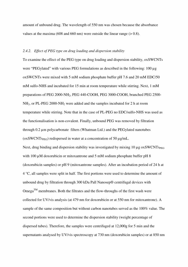
Figure 1 Characterisation of the used oxSWCNTs by a) atomic force microscopy, b) Raman
spectroscopy, and c) UV/vis absorption spectroscopy.
Figure 1 shows the characterisation results after analysis by atomic force microscopy (AFM),
Raman spectroscopy, and UV/vis absorption spectroscopy. The mean particle size of the
oxSWCNTs was about 126 ± 8 nm, as determined by phase analysis light scattering (PALS)
[12]. Considering that pristine SWCNTs are between 450 and 2000 nm long (mode at 900
nm) according to the manufacturer, this demonstrates that the acid treatment has shortened
the nanotubes by more than 90%, which is also evident in the AFM image (Figure 1a).
Further AFM analysis of this sample including higher magnification images can be found
elsewhere [12]. Their Raman spectrum (Figure 1b) featured three prominent peaks: the radial
breathing mode (RBM) at 263 cm-1
, which is inversely proportional to the tube diameter, the
disorder-related D-band at 1313 cm-1
, and the graphitic G-band at 1590 cm-1
. The intensity
ratio of the D- to the G-band is an indicator of the structural disorder originating from the
formation of in the graphitic structure. In the case of the oxSWCNTs examined in this study,
the ID/IG ratio was 0.19 for pristine SWCNTs and 0.55 for oxidised SWCNTs, which
indicated that the density of defects/functional groups was higher after oxidation. This also
explains why the typical UV/vis features of non-oxidised SWCNTs were absent after acid
oxidation (Figure 1c). It should furthermore be noted that the sample had been washed with
NaOH and HCl after acid oxidation and was thus free of oxidation debris [13].
3.2. Fine-tuning of the functionalisation procedure
3.2.1. Degradation of doxorubicin and mitoxantrone as a function of pH, light, and
temperature
The first experiment was designed to determine the optimal pH and temperature conditions
for the two drugs. It is well-known that both doxorubicin and mitoxantrone are not ideal
drugs from an analytical point of view. Doxorubicin is known to self-associate in
concentrated solutions, is liable to photolytic decomposition and degrades at unfavorable pH
conditions [14]. It is most stable at pH 4 and converts into the 7-hydroxyaglykone (detached
amino sugar residue) in more acidic conditions and destabilises at basic pH due to enolisation
of the C9 side chain. Similarly, mitoxantrone is also most stable at pH 4 and shows
accelerated degradation at higher pH, under UV irradiation, and at higher temperatures [15].
It furthermore forms precipitates upon refrigeration, which redissolve after warming to room
temperature without a loss of efficacy [16].
Drug delivery experiments are usually performed with a pH range of 5.5-7.5 to maintain cell
viability, yet we and others [5] found that the binding of doxorubicin and mitoxantrone to
CNTs works best at high pH, at which the drugs are deprotonated. This indicates that very
likely a compromise has to be found between achieving optimal drug loading, while
maintaining the activity of the drugs. We therefore incubated both drug molecules at a range
of pH (5-9) and temperatures (4 °C vs. 25 °C) for 3 days and then evaluated their cytotoxicity
by means of MTT cell viability assays.
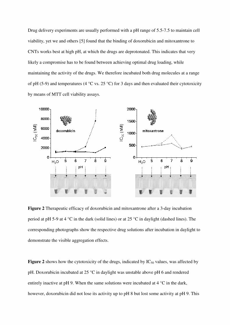
Figure 2 Therapeutic efficacy of doxorubicin and mitoxantrone after a 3-day incubation
period at pH 5-9 at 4 °C in the dark (solid lines) or at 25 °C in daylight (dashed lines). The
corresponding photographs show the respective drug solutions after incubation in daylight to
demonstrate the visible aggregation effects.
Figure 2 shows how the cytotoxicity of the drugs, indicated by IC50 values, was affected by
pH. Doxorubicin incubated at 25 °C in daylight was unstable above pH 6 and rendered
entirely inactive at pH 9. When the same solutions were incubated at 4 °C in the dark,
however, doxorubicin did not lose its activity up to pH 8 but lost some activity at pH 9. This

is in accordance with the literature [17] and shows that degradation of doxorubicin is
considerably accelerated by light and high pH, possibly due to the promotion of oxidation
reactions or the formation of drug polymers, which are incapable of penetrating the cellular
membrane [18]. The self-aggregation of neutral antracycline molecules by π-stacking is
consistent with our studies, which indicated that doxorubicin forms visible agglomerates
above pH 7, with the extent of agglomeration correlating with a loss of activity (photograph
beneath the diagram in Figure 2). In contrast, mitoxantrone was stable over the 3-day
incubation period for all pH values (Figure 2b), although some loss of activity was observed
at pH 7 when incubated in daylight at 25 °C in light, but not at 4 °C in darkness.
Interestingly, the drug also tended to precipitate at pH 7 (and, to a lesser extent, at pH 6 or 8),
which correlated with the loss of activity at this pH. However, compared to doxorubicin this
effect was much less marked, which is consistent with the fact that mitoxantrone solutions
form precipitates when kept at low temperatures, whilst maintaining the cytotoxic effect [16].
In conclusion, drug binding experiments should be performed at low temperatures and under
protection from light to maintain drug activity and, for doxorubicin, at pH 8 or lower.
3.2.2. Effect of pH on drug binding
Next, we investigated the influence of the pH on drug binding. Both doxorubicin and
mitoxantrone are able to attach to the surface of carbon nanotubes by π-π interactions due to
their inherent aromatic structure. In addition, both drug molecules contain pH-sensitive amine
groups, which makes their binding to nanotubes pH-dependent (enhanced at high pH and
reduced at low pH [5]). This potentially allows for drug release in acidic endosomes and the
microenvironment of tumors. To evaluate the pH dependence of drug binding, oxSWCNTs
were incubated with the two drugs at pH 5 to 9 for 18 hours, after which the samples were
filtered and the quantity of unbound drug in the filtrate measured by UV/vis absorption
spectroscopy. Samples without nanotubes (also from pH 5-9) served as controls.
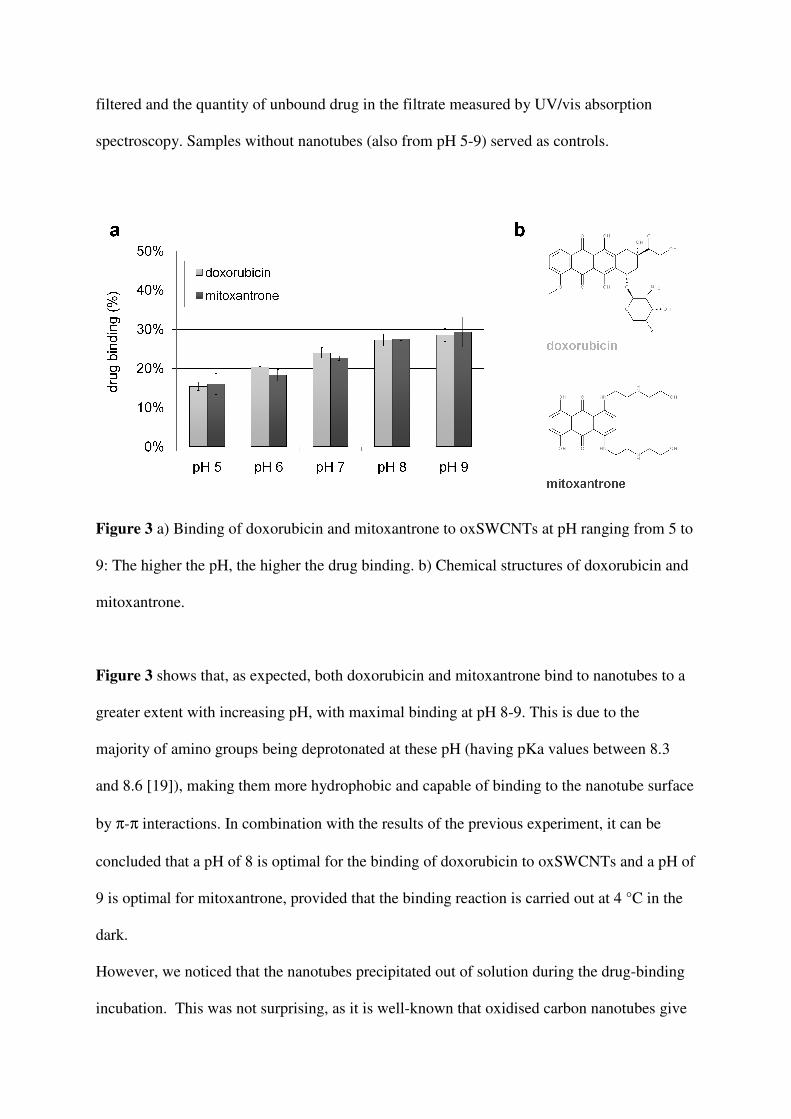
Figure 3 a) Binding of doxorubicin and mitoxantrone to oxSWCNTs at pH ranging from 5 to
9: The higher the pH, the higher the drug binding. b) Chemical structures of doxorubicin and
mitoxantrone.
Figure 3 shows that, as expected, both doxorubicin and mitoxantrone bind to nanotubes to a
greater extent with increasing pH, with maximal binding at pH 8-9. This is due to the
majority of amino groups being deprotonated at these pH (having pKa values between 8.3
and 8.6 [19]), making them more hydrophobic and capable of binding to the nanotube surface
by π-π interactions. In combination with the results of the previous experiment, it can be
concluded that a pH of 8 is optimal for the binding of doxorubicin to oxSWCNTs and a pH of
9 is optimal for mitoxantrone, provided that the binding reaction is carried out at 4 °C in the
dark.
However, we noticed that the nanotubes precipitated out of solution during the drug-binding
incubation. This was not surprising, as it is well-known that oxidised carbon nanotubes give
rise to electrostatic interactions with salts, such as the two anticancer drugs investigated here,
which can impair the dispersion stability due to charge neutralisation [20]. Precipitation did
not influence the efficacy of drug binding; but it is of course essential that any clinical
formulation remains stable in the circulation, so dispersion stability is critical. In a previous
study we have shown that the dispersion stability of oxidised nanotubes and also their
biocompatibility toward cancer cells can be improved by covalent conjugation to
polyethylene glycol (PEG) [12]. PEGylation is also a widely-used pharmaceutical
formulation strategy in clinical settings, as it reduces immunogenicity and non-specific
interactions with plasma proteins, while concomitantly prolonging blood circulation times.
We, therefore, attempted to optimise dispersion stability of PEGylated oxSWCNTs in the
presence of the drugs while monitoring the degree of drug loading.
3.2.3. Effect of the type of PEG on drug loading and dispersion stability
Five different types of PEG molecules were tested with regard to their effect on drug binding
and dispersion stability. This included a homobifunctional, amine-terminated PEG (PEG
2000-NH2), two heterobifunctional PEGs of different chain lengths (PEG 440-COOH and
PEG 3000-COOH), and a branched PEG with four amine-terminated arms (branched PEG
2500-NH2), all of which were covalently linked to oxSWCNTs by one of their amine groups
by carbodiimide-activated coupling. Note that this type of coupling reaction is not
quantitative and that amine-terminated PEG may also adsorb to oxidised nanotubes via acid-
base zwitterionic interactions, as shown by Huang et al. [21] and Marega et al. [22]. As a fifth
candidate, we investigated an amine-terminated phospholipid-PEG (PL-PEG2000-NH2),
which is widely used as a dispersion agent for carbon nanotubes [23, 24]. In this case the
functionalisation was based on non-covalent interactions promoted by the binding of the
phospholipid-moiety to the nanotube surface. The ideal PEG candidate should optimally

stabilise nanotube dispersions without impairing drug binding through steric hindrance or
repulsive electrostatic interactions.
Figure 4 Effect of the PEG type on drug binding and dispersion stability: Only one PEG
candidate – branched PEG 2500-NH2 – was able to promote a stable dispersion after addition
of the drugs, whereas in all other cases, the nanotubes precipitated out of solution.
Figure 4 shows the effect of the different PEG types on drug binding and dispersion stability.
PEGylation reduced the binding of doxorubicin by approximately 10%, whereas the binding
of mitoxantrone was only slightly reduced. Drug binding for each of the PEG molecules was
very similar; however, only the oxSWCNT sample functionalised with branched PEG

(branched PEG 2500-NH2) remained stable, whereas in all other cases the nanotubes had
precipitated out of solution. Based on these results, we thus decided to use branched PEG
2500-NH2 for all further experiments. The combined molecular weight of the PEG molecules
attached is 10 kDa (4 branches of 2.5 kDa each); a value which was also found optimal for
the prevention of nonspecific protein adsorption and favourable cellular responses in a study
by He et al. [25].
3.2.4. Effect of the degree of PEGylation on drug loading
The optimal PEG molecule (branched PEG 2500-NH2) was conjugated to oxSWCNTs at
various concentrations to investigate the effect of PEG density on drug loading and
dispersion stability. In this experiment, the nanotube concentration was kept constant, while
branched PEG was added at amounts ranging from 5 µmol to 0.1 µmol. After the conjugation
reaction, unreacted PEG molecules were removed by filtration through 0.2 µm polycarbonate
filters and repeated washing.
Figure 5 Impact of the PEG density on drug binding (a) and dispersion stability (b). Only the
highest initial amount of PEG added (5 µmol) resulted in sufficient dispersion stability after
addition of the drugs, although this reduced drug binding by about 40%. The numbers in

italics above the columns in (b) indicate the ratio of PEG molecules to acidic sites on the
oxSWCNTs.
As illustrated in Figure 5a, the more PEG was added initially, the lower was the drug
binding. Although we did not determine the actual amounts of PEG attached to the
oxSWCNTs after the coupling reaction, we presume that adding a higher amount of PEG
during the reaction leads to a higher PEG density of the final product, which may impair drug
binding due to sterical hindrance. Dispersion stability, on the other hand, significantly
improved with increasing initial amounts of PEG used (Figure 5b); in fact only the highest
amount of PEG added (5 µmol or a 33-fold molar excess) was able to maintain a sufficiently
stable dispersion after addition of the drugs. When comparing the two drugs, mitoxantrone-
loaded oxSWCNTsPEG (mito-oxSWCNTsPEG) were less stable than doxorubicin-loaded
oxSWCNTsPEG (dox-oxSWCNTsPEG); presumably because mitoxantrone is a bivalent salt,
whereas doxorubicin is monovalent. For subsequent experiments, branched PEG 2500-NH2
was used at an initial amount of 5 µmol to ensure maximal dispersion stability, although this
compromised drug binding by about 40%. Furthermore, we used a drug/oxSWCNTPEG
weight ratio of approximately 1:2 in order to obtain quantitative drug binding and thus
eliminating the need for a filtration step to remove unbound drug, which corresponded to a
final drug concentration of 25 µM (or 14.5 µg/mL doxorubicin and 12.9 µg/mL
mitoxantrone, respectively) and a final oxSWCNTPEG concentration of 25 µg/mL. The
absence of free drug (at zero time) is demonstrated in the next section.
3.3. Drug release over time at different conditions
The release of both doxorubicin and mitoxantrone should be promoted at low pH due to their
amine functionalities, which render them hydrophilic at low pH and hydrophobic at high pH.
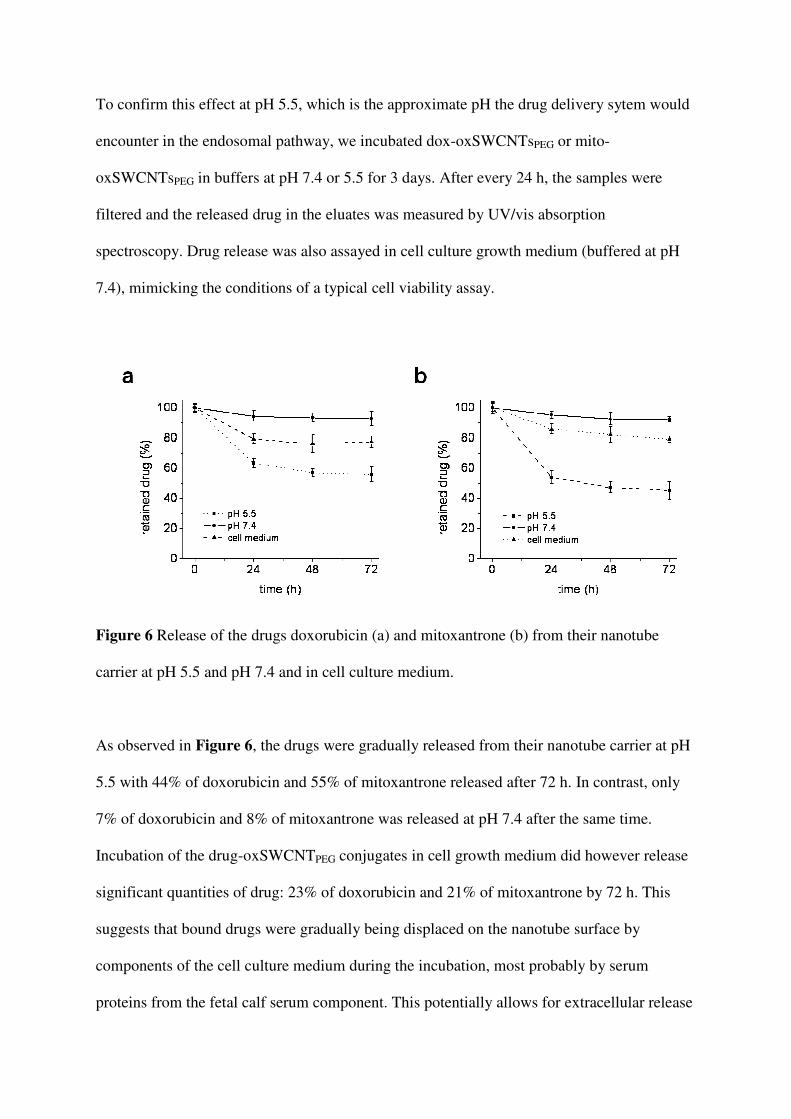
To confirm this effect at pH 5.5, which is the approximate pH the drug delivery sytem would
encounter in the endosomal pathway, we incubated dox-oxSWCNTsPEG or mito-
oxSWCNTsPEG in buffers at pH 7.4 or 5.5 for 3 days. After every 24 h, the samples were
filtered and the released drug in the eluates was measured by UV/vis absorption
spectroscopy. Drug release was also assayed in cell culture growth medium (buffered at pH
7.4), mimicking the conditions of a typical cell viability assay.
Figure 6 Release of the drugs doxorubicin (a) and mitoxantrone (b) from their nanotube
carrier at pH 5.5 and pH 7.4 and in cell culture medium.
As observed in Figure 6, the drugs were gradually released from their nanotube carrier at pH
5.5 with 44% of doxorubicin and 55% of mitoxantrone released after 72 h. In contrast, only
7% of doxorubicin and 8% of mitoxantrone was released at pH 7.4 after the same time.
Incubation of the drug-oxSWCNTPEG conjugates in cell growth medium did however release
significant quantities of drug: 23% of doxorubicin and 21% of mitoxantrone by 72 h. This
suggests that bound drugs were gradually being displaced on the nanotube surface by
components of the cell culture medium during the incubation, most probably by serum
proteins from the fetal calf serum component. This potentially allows for extracellular release
of the drug from the nanotube carrier and its subsequent diffusion through the cell membrane
without its nanocarrier, although this would only occur for long blood circulation times (1-3
days).
3.4. Intracellular distribution of CNTPEG-doxorubicin conjugates
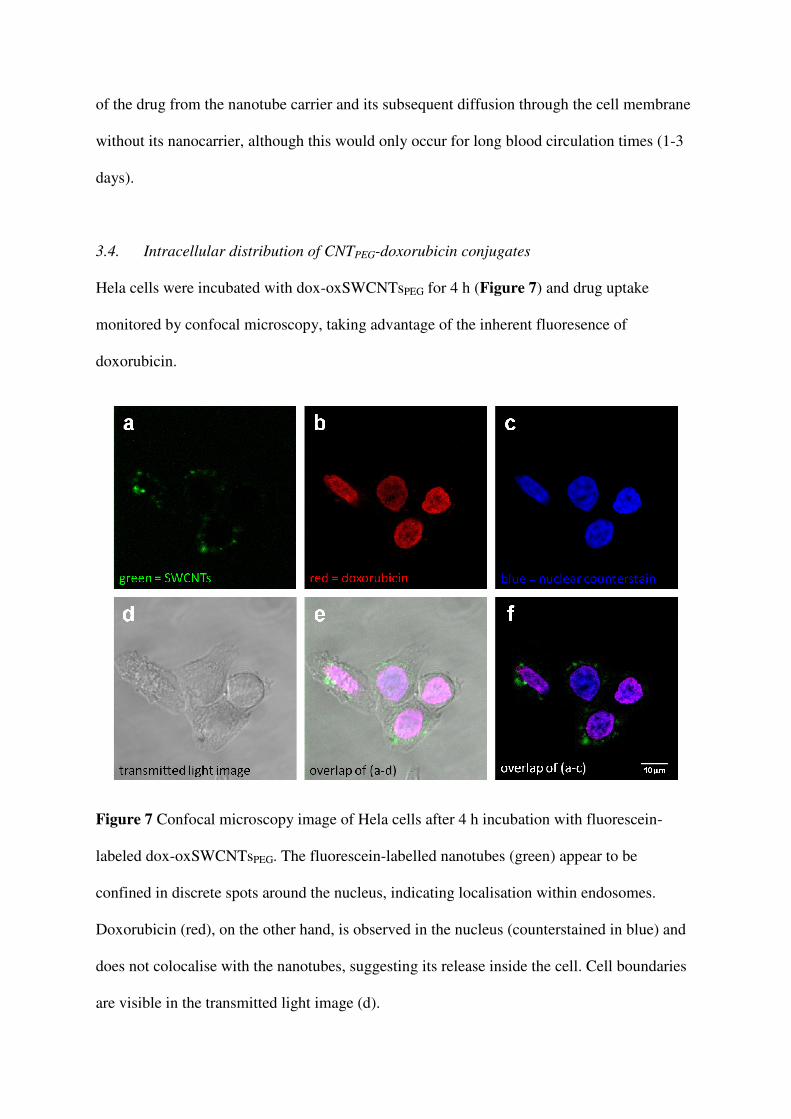
Hela cells were incubated with dox-oxSWCNTsPEG for 4 h (Figure 7) and drug uptake
monitored by confocal microscopy, taking advantage of the inherent fluoresence of
doxorubicin.
Figure 7 Confocal microscopy image of Hela cells after 4 h incubation with fluorescein-
labeled dox-oxSWCNTsPEG. The fluorescein-labelled nanotubes (green) appear to be
confined in discrete spots around the nucleus, indicating localisation within endosomes.
Doxorubicin (red), on the other hand, is observed in the nucleus (counterstained in blue) and
does not colocalise with the nanotubes, suggesting its release inside the cell. Cell boundaries
are visible in the transmitted light image (d).
The confocal images show that doxorubicin (red, Figure 7b) was entirely located in the cell
nucleus (counterstained with ToPro 3, in blue, Figure 7c), proving its release from the
nanotubes (labelled with fluorescein, in green), which were distributed as discrete spots in the
cytoplasm (Figure 7a), indicating localisation within endosomes. This suggests an
endocytotic, energy-dependent uptake mechanism, which would lead to the engulfment of the
drug-SWCNT-complexes in endosomes and subsequent release of the drug due to the lower
endo-/lysosomal pH (as also shown in Figure 6). The drug would then translocate to the cell
nucleus to exert its cytotoxic action; interference with DNA replication [26]. The exact
uptake mechanism and fate of CNTs inside cells is currently further investigated in our
laboratory and will be published separately [27, 28]. It is important to note that a small
amount of doxorubicin has been found to detach from its CNT carrier extracellularly during
incubation in cellular growth medium (Figure 6) and can potentially diffuse through the cell
membrane on its own.
3.5. Effect of dispersion stability on uptake and activity of the drug-SWCNT complexes
We first aimed to investigate whether the dispersion stability of drug-loaded oxSWCNTs
affects the drugs’ therapeutic activity on cultured cells. Two types of oxSWCNTs were used,
oxSWCNTs reacted with 5 mM PEG and “naked” oxSWCNTs, associated with dispersion
stabilities of 90-100% and 0%, respectively. In order to evaluate cytotoxicity, we used both
the MTT and the WST assay and compared the results of the two, as the former is known to
cause false positive test results due to interactions of the hydrophobic end product MTT-
formazan with the CNT surface, which can occur outside as well as inside cells [11]. The
WST assay follows the same assay principle as the MTT assay, but its end product is
hydrophilic and therefore not prone to attach to the surface of CNTs (please see the
supporting information for a full comparison between the two assays and the impact on this
experiment). In brief, we found that the MTT and WST assay lead to comparable results if
the CNT surface is fully occupied (e.g. in the case of drug-loaded, PEGylated oxSWCNTs),
but that the MTT assay leads to false positive results if parts or the whole CNT surface is
available to bind MTT-formazan.
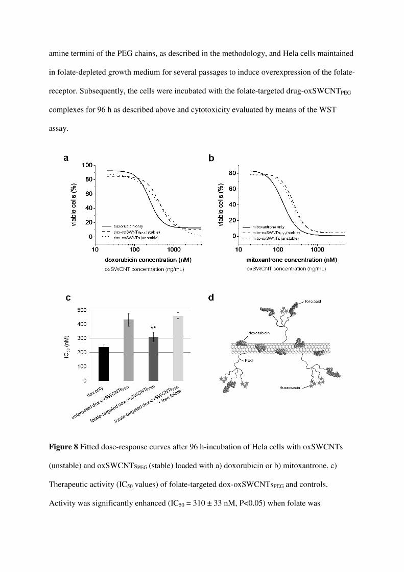
Figure 8a and b display the fitted dose-response curves obtained as a result of the performed
WST viability assays. Both oxSWCNTs (precipitated in cell medium) and oxSWCNTsPEG
(stable in cell medium) were non-toxic over the whole dose range (see Figure S2 in the
supporting information for the dose-response curve of oxSWCNTsPEG). When comparing the
drug-loaded samples, there was no significant difference between the stable, well-dispersed
sample (drug-loaded oxSWCNTsPEG) and the unstable sample (drug-loaded oxSWCNTs),
possibly due to the cells’ ability to take up precipitates of nanotubes [3] and premature drug
release in the cell medium (Figure 6). In comparison to the free drugs, drug-loaded nanotubes
were less cytotoxic (IC50 values of doxorubicin only/oxSWCNTsPEG = 239 ± 14 nM/432 ± 46
nM, IC50 values of mitoxantrone only/ mito-oxSWCNTsPEG = 133 ± 6 nM/248 ± 12 nM) .
This is likely due to drug molecules taken up by passive transport being free to translocate
directly to the nucleus, where they exert cytotoxicity, whereas drug molecules complexed to
carbon nantubes are released only after encountering the lower pH inside endosomes and
lysosomes and so lower amounts reach the nucleus to excert a cytotoxic effect. The next step
was to investigate, whether the attachment of a targeting agent could improve the complexes’
cytotoxicity and additionally provide specificity to cancerous cells. We chose folic acid as a
targeting agent, as the folate-receptor is significantly upregulated by a broad spectrum of
human cancers, in some cases by two orders of magnitude, facilitating cellular internalisation
of folate-conjugated nanocarriers by receptor-mediated endocytosis [10, 29]. Additionally,
non-cancerous cells only transport folate by this receptor but not folate conjugates of any type
[30], which adds an extra level of specificity. Folic acid was covalently attached folate to the
amine termini of the PEG chains, as described in the methodology, and Hela cells maintained
in folate-depleted growth medium for several passages to induce overexpression of the folate-
receptor. Subsequently, the cells were incubated with the folate-targeted drug-oxSWCNTPEG
complexes for 96 h as described above and cytotoxicity evaluated by means of the WST
assay.
Figure 8 Fitted dose-response curves after 96 h-incubation of Hela cells with oxSWCNTs
(unstable) and oxSWCNTsPEG (stable) loaded with a) doxorubicin or b) mitoxantrone. c)
Therapeutic activity (IC50 values) of folate-targeted dox-oxSWCNTsPEG and controls.
Activity was significantly enhanced (IC50 = 310 ± 33 nM, P<0.05) when folate was

conjugated to the PEG chains as a targeting agent. d) Schematic illustration of folate-targeted
and fluorescently labeled dox-oxSWCNTsPEG.
Figure 8c displays the resulting IC50 values: The system’s therapeutic efficacy was
significantly enhanced (IC50 = 310 ± 33 nM, P<0.05) when folate was conjugated to the PEG
chains as a targeting agent compared to the untargeted system (IC50 = 432 ± 46 nM), which
might be due to enhanced endocytotic uptake through receptor-mediated endocytosis (as
discussed above, drug release requires localisation of the drug-nanotube complexes inside
endo- or lysosomes). When the folate-receptors were saturated with free folic acid, however,
the efficacy became again similar to that of the untargeted system (IC50 = 460 ± 21 nM),
confirming the selectivity of the targeting approach and its advantage over using the free
drug.
4. Conclusions
We have developed a carbon nanotube-mediated drug delivery system for two different anti-
cancer drugs, affording high biological and chemical stability, high drug loading and
selective cancer treatment in an in vitro scenario by an active targeting scheme. After fine-
tuning the synthesis parameters to obtain maximal drug loading, while maintaining the
dispersion stability of the system, we demonstrated pH-dependent drug release in buffer
solutions and confirmed cellular uptake of the nanovectors and intracellular drug release, by
means of confocal microscopy. Finally, we evaluated the therapeutic efficacy of the system in
cell viability assays and found that while drug-loaded nanotubes were less effective than the
free drugs, attachment of the targeting agent folic acid significantly enhanced the efficacy and
selectivity of the system. Keeping in mind that free doxorubicin and mitoxantrone are both
highly effective in in vitro experiments due to their aqueous solubility and membrane
permeability, this is an encouraging result. The targeted, drug loaded nanotubes are selective
in contrast to the free drugs and additionally allow for sustained release, which is likely to
reduce drug-related side effects in animal models and clinical studies. Overall, our study
demonstrates the potential of carbon nanotubes as a multimodal drug delivery system and
presents a functionalisation scheme that is able to overcome many of the problems
encountered in this area of application.
Acknowledgements
This work has been performed in the framework of the FP6 Marie Curie Research Training
Network “CARBIO” (RTN-CT-2006-035616) funded by the European Union.
References
[1] Alderton PM, Gross J, Green MD. Comparative study of doxorubicin, mitoxantrone,
and epirubicin in combination with ICRF-187 (ADR-529) in a chronic cardiotoxicity
animal model. Cancer Research. 1992 Jan 1;52(1):194-201.
[2] Kostarelos K, Lacerda L, Pastorin G, Wu W, Wieckowski S, Luangsivilay J, et al.
Cellular uptake of functionalized carbon nanotubes is independent of functional group
and cell type. Nature Nanotechnology. 2007 Feb;2(2):108-13.
[3] Mu QX, Broughton DL, Yan B. Endosomal leakage and nuclear translocation of
multiwalled carbon nanotubes: developing a model for cell uptake. Nano Letters.
2009 Dec;9(12):4370-5.
[4] Heister E, Neves V, Tîlmaciu C, Lipert K, Sanz Beltrán V, Coley H, et al. Triple
functionalisation of single-walled carbon nanotubes with doxorubicin, a monoclonal
antibody, and a fluorescent marker for targeted cancer therapy. Carbon.
2009;47(9):2152-60.
[5] Liu Z, Sun XM, Nakayama-Ratchford N, Dai HJ. Supramolecular chemistry on water-
soluble carbon nanotubes for drug loading and delivery. ACS Nano. 2007
Aug;1(1):50-6.
[6] Ali-Boucetta H, Al-Jamal KT, McCarthy D, Prato M, Bianco A, Kostarelos K.
Multiwalled carbon nanotube-doxorubicin supramolecular complexes for cancer
therapeutics. Chemical Communications. 2008(4):459-61.
[7] Zhang XK, Meng LJ, Lu QG, Fei ZF, Dyson PJ. Targeted delivery and controlled
release of doxorubicin to cancer cells using modified single wall carbon nanotubes.
Biomaterials. 2009 Oct;30(30):6041-7.
[8] Liu Z, Fan AC, Rakhra K, Sherlock S, Goodwin A, Chen X, et al. Supramolecular
stacking of doxorubicin on carbon nanotubes for in vivo cancer therapy. Angew Chem
Int Ed Engl. 2009;48(41):7668-72.
[9] Li RB, Wu R, Zhao L, Wu MH, Yang L, Zou HF. P-glycoprotein antibody
functionalized carbon nanotube overcomes the multidrug resistance of human
leukemia cells. ACS Nano. 2010 Mar;4(3):1399-408.
[10] Russell-Jones G, McTavish K, McEwan J, Rice J, Nowotnik D. Vitamin-mediated
targeting as a potential mechanism to increase drug uptake by tumours. Journal of
Inorganic Biochemistry. 2004 Oct;98(10):1625-33.
[11] Wörle-Knirsch JM, Pulskamp K, Krug HF. Oops they did it again! Carbon nanotubes
hoax scientists in viability assays. Nano Letters. 2006 Jun;6(6):1261-8.
[12] Heister E, Lamprecht C, Neves V, Tilmaciu C, Datas L, Flahaut E, et al. Higher
dispersion efficacy of functionalized carbon nanotubes in chemical and biological
environments. ACS Nano. 2010 May 25;4(5):2615-26.
[13] Verdejo R, Lamoriniere S, Cottam B, Bismarck A, Shaffer M. Removal of oxidation
debris from multi-walled carbon nanotubes. Chemical Communications. 2007 Feb
7(5):513-5.
[14] Beijnen JH, Vanderhouwen OAGJ, Underberg WJM. Aspects of the degradation
kinetics of doxorubicin in aqueous solution. International Journal of Pharmaceutics.
1986 Oct;32(2-3):123-31.
[15] Wang DP, Liang GZ, Tu YH. Stability of mitoxantrone hydrochloride in solution.
Drug Development and Industrial Pharmacy. 1994;20(11):1895-903.
[16] Cancer Care Nova Scotia. Drug Monograph Mitoxantrone. Systemic Therapy Manual
for Cancer Treatment 2010:208-10.
[17] Bouma J, Beijnen JH, Bult A, Underberg WJM. Anthracycline antitumor agents - a
review of physicochemical, analytical and stability properties. Pharmaceutisch
Weekblad-Scientific Edition. 1986 Apr 25;8(2):109-33.
[18] Williams BA, Tritton TR. Photoinactivation of anthracyclines. Photochemistry and
Photobiology. 1981;34(1):131-4.
[19] Raghunand N, Mahoney BP, Gillies RJ. Tumor acidity, ion trapping and
chemotherapeutics I. pH-dependent partition coefficients predict importance of ion
trapping on pharmacokinetics of weakly basic chemotherapeutic agents. Biochemical
Pharmacology. 2003 Oct 1;66(7):1219-29.
[20] Peng XJ, Jia JJ, Gong XM, Luan ZK, Fan B. Aqueous stability of oxidized carbon
nanotubes and the precipitation by salts. Journal of Hazardous Materials. 2009 Jun
15;165(1-3):1239-42.
[21] Huang WJ, Fernando S, Allard LF, Sun YP. Solubilization of single-walled carbon
nanotubes with diamine-terminated oligomeric poly(ethylene glycol) in different
functionalization reactions. Nano Letters. 2003 Apr;3(4):565-8.
[22] Marega R, Aroulmoji V, Bergamin M, Feruglio L, Dinon F, Bianco A, et al. Two-
dimensional diffusion-ordered NMR spectroscopy as a tool for monitoring
functionalized carbon nanotube purification and composition. ACS Nano. 2010
Apr;4(4):2051-8.
[23] Prencipe G, Tabakman SM, Welsher K, Liu Z, Goodwin AP, Zhang L, et al. PEG-
branched polymer for functionalization of nanomaterials with ultralong blood
circulation. Journal of the American Chemical Society. 2009 Apr 8;131(13):4783-7.
[24] Lamprecht C, Danzberger J, Lukanov P, Tilmaciu CM, Galibert AM, Soula B, et al.
AFM imaging of functionalized double-walled carbon nanotubes. Ultramicroscopy.
2009 Jul;109(8):899-906.
[25] He QJ, Zhang JM, Shi JL, Zhu ZY, Zhang LX, Bu WB, et al. The effect of
PEGylation of mesoporous silica nanoparticles on nonspecific binding of serum
proteins and cellular responses. Biomaterials. 2010 Feb;31(6):1085-92.
[26] Momparler RL, Karon M, Siegel SE, Avila F. Effect of Adriamycin on DNA, Rna,
and Protein-Synthesis in Cell-Free Systems and Intact-Cells. Cancer Research.
1976;36(8):2891-5.
[27] Neves V, Heister E, Costa S, Tîlmaciu C, Borowiak-Palen E, Giusca C, et al. Uptake
and release of double-walled carbon nanotubes by mammalian cells. Advanced
Functional Materials. 2010;20(19):3272-9.
[28] Neves V. Carbon Nanotubes (CNT): Feasibility as nano-bio agents to target cancer.
PhD thesis, University of Surrey. 2011.
[29] Lu Y, Sega E, Leamon CP, Low PS. Folate receptor-targeted immunotherapy of
cancer: mechanism and therapeutic potential. Adv Drug Deliv Rev. 2004 Apr
29;56(8):1161-76.
[30] Byrne JD, Betancourt T, Brannon-Peppas L. Active targeting schemes for
nanoparticle systems in cancer therapeutics. Advanced Drug Delivery Reviews. 2008
Dec 14;60(15):1615-26.

List of Figures
Figure 9 Characterisation of the used oxSWCNTs by a) atomic force microscopy, b) Raman
spectroscopy, and c) UV/vis absorption spectroscopy.
Figure 10 Therapeutic efficacy of doxorubicin and mitoxantrone after a 3-day incubation
period at pH 5-9 at 4 °C in the dark (solid lines) or at 25 °C in daylight (dashed lines). The
corresponding photographs show the respective drug solutions after incubation in daylight to
demonstrate the visible aggregation effects.
Figure 11 a) Binding of doxorubicin and mitoxantrone to oxSWCNTs at pH ranging from 5
to 9: The higher the pH, the higher the drug binding. b) Chemical structures of doxorubicin
and mitoxantrone.
Figure 12 Effect of the PEG type on drug binding and dispersion stability: Only one PEG
candidate – branched PEG 2500-NH2 – was able to promote a stable dispersion after addition
of the drugs, whereas in all other cases, the nanotubes precipitated out of solution.
Figure 13 Impact of the PEG density on drug binding (a) and dispersion stability (b). Only
the highest initial amount of PEG added (5 µmol) resulted in sufficient dispersion stability
after addition of the drugs, although this reduced drug binding by about 40%. The numbers in
italics above the columns in (b) indicate the ratio of PEG molecules to acidic sites on the
oxSWCNTs.

Figure 14 Release of the drugs doxorubicin (a) and mitoxantrone (b) from their nanotube
carrier at pH 5.5 and pH 7.4 and in cell culture medium.
Figure 15 Confocal microscopy image of Hela cells after 4 h incubation with fluorescein-
labeled dox-oxSWCNTsPEG. The fluorescein-labelled nanotubes (green) appear to be
confined in discrete spots around the nucleus, indicating localisation within endosomes.
Doxorubicin (red), on the other hand, is observed in the nucleus (counterstained in blue) and
does not colocalise with the nanotubes, suggesting its release inside the cell. Cell boundaries
are visible in the transmitted light image (d).
Figure 16 Fitted dose-response curves after 96 h-incubation of Hela cells with oxSWCNTs
(unstable) and oxSWCNTsPEG (stable) loaded with a) doxorubicin or b) mitoxantrone. c)
Therapeutic activity (IC50 values) of folate-targeted dox-oxSWCNTsPEG and controls.
Activity was significantly enhanced (IC50 = 310 ± 33 nM, P<0.05) when folate was
conjugated to the PEG chains as a targeting agent. d) Schematic illustration of folate-targeted
and fluorescently labeled dox-oxSWCNTsPEG.
Related Documents