Molecular Biology of the Cell Vol. 12, 1671–1685, June 2001 Drosophila Heterochromatin Protein 1 (HP1)/Origin Recognition Complex (ORC) Protein Is Associated with HP1 and ORC and Functions in Heterochromatin- induced Silencing Mohammed Momin Shareef,* Chadwick King, Mona Damaj, RamaKrishna Badagu,* Da Wei Huang, and Rebecca Kellum ‡ *School of Biological Sciences, University of Kentucky, Lexington, Kentucky 40506-0225; and ² Department of Biological Sciences, McGill University, Montreal, Quebec H3A 1B1, Canada Submitted September 20, 2000; Revised February 16, 2001; Accepted March 19, 2001 Monitoring Editor: Joseph Gall Heterochromatin protein 1 (HP1) is a conserved component of the highly compact chromatin of higher eukaryotic centromeres and telomeres. Cytogenetic experiments in Drosophila have shown that HP1 localization into this chromatin is perturbed in mutants for the origin recognition complex (ORC) 2 subunit. ORC has a multisubunit DNA-binding activity that binds origins of DNA replication where it is required for origin firing. The DNA-binding activity of ORC is also used in the recruitment of the Sir1 protein to silence nucleation sites flanking silent copies of the mating-type genes in Saccharomyces cerevisiae. A fraction of HP1 in the maternally loaded cyto- plasm of the early Drosophila embryo is associated with a multiprotein complex containing Drosophila melanogaster ORC subunits. This complex appears to be poised to function in hetero- chromatin assembly later in embryonic development. Here we report the identification of a novel component of this complex, the HP1/ORC-associated protein. This protein contains similarity to DNA sequence-specific HMG proteins and is shown to bind specific satellite sequences and the telomere-associated sequence in vitro. The protein is shown to have heterochromatic localization in both diploid interphase and mitotic chromosomes and polytene chromosomes. Moreover, the gene encoding HP1/ORC-associated protein was found to display reciprocal dose-dependent variegation modifier phenotypes, similar to those for mutants in HP1 and the ORC 2 subunit. INTRODUCTION The eukaryotic nucleus undergoes dramatic morphological changes as it progresses through the cell cycle. The conden- sation of the chromatin upon entry into mitosis and its subsequent decondensation at the end of mitosis is one of the more stunning events. Certain regions of the chromo- somes, termed heterochromatin, fail to undergo these con- densation cycles but retain a compact appearance through- out the cell cycle (Heitz, 1928). Heterochromatin also has different functional properties from more typical chromo- somal regions (euchromatin). It often causes silencing of euchromatic genes that are juxtaposed to it by chromosomal rearrangement, is typically replicated later in S phase than euchromatin, and usually occupies a distinct subnuclear domain along the nuclear periphery (for review, see Brown, 1966). The centromeres and telomeres of higher eukaryotic chromosomes typically have a heterochromatic organiza- tion, and this structure is known to be important for proper chromosome mechanics during mitosis and meiosis (Allshire et al., 1995; Kellum et al., 1995; Dernburg et al., 1996; Karpen et al. 1996; Fanti et al., 1998; for review, see Dobie et al., 1999). The compact appearance of heterochromatin is likely to involve a unique nucleoprotein composition. It is known to be enriched with repetitive noncoding DNA, such as the AT-rich satellite sequences (Peacock et al., 1976, 1977; Pea- cock and Lohe, 1980), and to contain specific proteins. In the genetically tractable organisms of Saccharomyces cerevisiae, Schizosaccharomyces pombe, and Drosophila (Sinclair et al., 1983; Wustmann et al., 1989; Laurenson and Rine, 1992; Allshire et al., 1995), genetic assays have been used to iden- tify heterochromatin-associated proteins. Mutations in genes that are required to form Drosophila heterochromatin sup- press the variegated expression of a euchromatic gene caused by its placement next to heterochromatin by a chro- mosomal rearrangement. The product of the suppressor of variegation 2-5 gene, heterochromatin protein 1 (HP1; James ‡ Corresponding author. E-mail address: [email protected]. © 2001 by The American Society for Cell Biology 1671

Welcome message from author
This document is posted to help you gain knowledge. Please leave a comment to let me know what you think about it! Share it to your friends and learn new things together.
Transcript

Molecular Biology of the CellVol. 12, 1671–1685, June 2001
Drosophila Heterochromatin Protein 1 (HP1)/OriginRecognition Complex (ORC) Protein Is Associatedwith HP1 and ORC and Functions in Heterochromatin-induced SilencingMohammed Momin Shareef,* Chadwick King,† Mona Damaj,†RamaKrishna Badagu,* Da Wei Huang,† and Rebecca Kellum‡
*School of Biological Sciences, University of Kentucky, Lexington, Kentucky 40506-0225; and†Department of Biological Sciences, McGill University, Montreal, Quebec H3A 1B1, Canada
Submitted September 20, 2000; Revised February 16, 2001; Accepted March 19, 2001Monitoring Editor: Joseph Gall
Heterochromatin protein 1 (HP1) is a conserved component of the highly compact chromatin ofhigher eukaryotic centromeres and telomeres. Cytogenetic experiments in Drosophila have shownthat HP1 localization into this chromatin is perturbed in mutants for the origin recognitioncomplex (ORC) 2 subunit. ORC has a multisubunit DNA-binding activity that binds origins ofDNA replication where it is required for origin firing. The DNA-binding activity of ORC is alsoused in the recruitment of the Sir1 protein to silence nucleation sites flanking silent copies of themating-type genes in Saccharomyces cerevisiae. A fraction of HP1 in the maternally loaded cyto-plasm of the early Drosophila embryo is associated with a multiprotein complex containingDrosophila melanogaster ORC subunits. This complex appears to be poised to function in hetero-chromatin assembly later in embryonic development. Here we report the identification of a novelcomponent of this complex, the HP1/ORC-associated protein. This protein contains similarity toDNA sequence-specific HMG proteins and is shown to bind specific satellite sequences and thetelomere-associated sequence in vitro. The protein is shown to have heterochromatic localizationin both diploid interphase and mitotic chromosomes and polytene chromosomes. Moreover, thegene encoding HP1/ORC-associated protein was found to display reciprocal dose-dependentvariegation modifier phenotypes, similar to those for mutants in HP1 and the ORC 2 subunit.
INTRODUCTION
The eukaryotic nucleus undergoes dramatic morphologicalchanges as it progresses through the cell cycle. The conden-sation of the chromatin upon entry into mitosis and itssubsequent decondensation at the end of mitosis is one ofthe more stunning events. Certain regions of the chromo-somes, termed heterochromatin, fail to undergo these con-densation cycles but retain a compact appearance through-out the cell cycle (Heitz, 1928). Heterochromatin also hasdifferent functional properties from more typical chromo-somal regions (euchromatin). It often causes silencing ofeuchromatic genes that are juxtaposed to it by chromosomalrearrangement, is typically replicated later in S phase thaneuchromatin, and usually occupies a distinct subnucleardomain along the nuclear periphery (for review, see Brown,1966). The centromeres and telomeres of higher eukaryoticchromosomes typically have a heterochromatic organiza-
tion, and this structure is known to be important for properchromosome mechanics during mitosis and meiosis(Allshire et al., 1995; Kellum et al., 1995; Dernburg et al., 1996;Karpen et al. 1996; Fanti et al., 1998; for review, see Dobie etal., 1999).
The compact appearance of heterochromatin is likely toinvolve a unique nucleoprotein composition. It is known tobe enriched with repetitive noncoding DNA, such as theAT-rich satellite sequences (Peacock et al., 1976, 1977; Pea-cock and Lohe, 1980), and to contain specific proteins. In thegenetically tractable organisms of Saccharomyces cerevisiae,Schizosaccharomyces pombe, and Drosophila (Sinclair et al.,1983; Wustmann et al., 1989; Laurenson and Rine, 1992;Allshire et al., 1995), genetic assays have been used to iden-tify heterochromatin-associated proteins. Mutations in genesthat are required to form Drosophila heterochromatin sup-press the variegated expression of a euchromatic genecaused by its placement next to heterochromatin by a chro-mosomal rearrangement. The product of the suppressor ofvariegation 2-5 gene, heterochromatin protein 1 (HP1; James‡ Corresponding author. E-mail address: [email protected].
© 2001 by The American Society for Cell Biology 1671

and Elgin, 1986; Eissenberg et al., 1990), is a heterochromaticprotein that is conserved from fission yeast to humans(Saunders et al., 1993; Allshire et al., 1995; Pak et al., 1997).
Because direct DNA-binding activity had not been attrib-uted to HP1, its localization into heterochromatin has longbeen thought to involve the DNA-binding activities of otherproteins. Mutations in the origin recognition complex (ORC)2 subunit of the Drosophila melanogaster ORC (DmORC) wererecently shown to perturb HP1 localization (Pak et al., 1997;Huang et al., 1998). This highly conserved protein complexwas purified from S. cerevisiae as in vitro binding activity forsequences that permit autonomous replication of plasmidsin vivo (Bell and Stillman, 1992). It is also known to berequired for replication of chromosomal DNA (Bell et al.,1993; Rowles et al., 1996; Landis et al., 1997) and to beassociated with specific replicator sequences in S. cerevisiaeand metazoans (Aparicio et al., 1997; Austin et al., 1999;Royzman et al., 1999). The DNA-binding activity of ORCserves a second function in S. cerevisiae in the recruitment ofthe Sir1 protein to ORC-binding sequences within a pair ofsilencing nucleation sites flanking silent copies of the mat-ing-type genes (Bell et al., 1993; Chien et al., 1993; Fox et al.,1995; Triolo and Sternglanz, 1996). The Sir1 protein then actsin conjunction with the DNA-binding proteins repressor–activator protein 1 (Rap1) and ARS-binding factor 1 (Abf1),which also bind sequences in the silencing nucleator, torecruit other members of the Sir protein complex to the sitethrough heterotypic and homotypic protein–protein interac-tions (Pillus and Rine, 1989; Kurtz and Shore, 1991; Hecht etal., 1995; Moazed et al., 1997). The observation that DrosophilaORC subunits are associated with HP1 in interphase hetero-chromatin and cause a perturbation in the localization ofHP1 into heterochromatin when mutated suggests a similarrole for ORC in Drosophila heterochromatin assembly (Pak etal., 1997; Huang et al., 1998).
It is not understood how the heterochromatin assemblyfunction of ORC might be specified in heterochromatin,whereas its more general role in DNA replication is carriedout throughout the genome. Studies in S. cerevisiae with asynthetic silencer that contains a consensus ARS sequencecombined with Rap1 and Abf1 binding sites from nonsi-lenced chromosomal regions showed the silencing activityof an ARS sequence to be DNA context dependent (McNallyand Rine, 1991). A similar set of protein-binding sequencesand associated factors might also cooperate with ORC inDrosophila heterochromatin assembly. Here we report theidentification of a 55-kDa protein that copurifies with anORC-containing complex of HP1 from the cytoplasm of theearly Drosophila embryo that appears to be poised for func-tion in heterochromatin assembly later in embryonic devel-opment (Huang et al., 1998). The protein contains an HMGbox and is proposed to serve a role in Drosophila heterochro-matin assembly that is similar to those of the Rap1 and Abf1proteins in budding yeast.
MATERIALS AND METHODS
Peptide Sequence Identification of HP1/ORC-associated ProteinThe 55-kDa HP1/ORC-associated protein (HOAP) was coimmuno-affinity purified from a cytoplasmic extract of early Drosophila em-bryos as previously described (Huang et al., 1998). The Coomassie
brilliant blue-stained band for a 55-kDa copurifying protein (p55)was excised from an SDS-polyacrylamide gel and subjected to insitu proteolytic cleavage and peptide sequence determination byEdman degradation at Harvard Microchemistry (Cambridge, MA).The National Center for Biotechnology Information database wassearched with sequences from 2 different peptides of the protein(QMSAFLRKYLAD and TRITEEDLARPYTED), and both sequenceswere found to match the sequence in the hypothetical protein-coding sequence for the Drosophila anonymous fast-evolving 1G5 (anonfe 1G5) gene (Schmid and Tautz, 1997).
Antibody PreparationAntibodies were increased against a fusion protein expressed froman anon fe 1G5 cDNA PCR amplified from an ovarian cDNA library(Stroumbakis et al., 1994) with the use of oligonucleotide primersdesigned from the 59 (ATGTCGGGGACGCAAATGTCTG) and 39(CAATCGGGTGATGTTCTGCTTC) ends of the gene. The anon fe1G5 cDNA was ligated into the XbaI and XhoI sites of the pET20bvector to produce the complete hypothetical anon fe 1G5 gene prod-uct with the pelB leader (19 amino acids) fused to its amino termi-nus and a 6-histidine tag fused to its carboxyl terminus. The taggedfusion protein was expressed in bacteria, purified by Ni-nitriletriacetic acid-agarose chromatography and PAGE, and used in rab-bit immunoinjections (4 biweekly subcutaneous injections of 1 ml).The antiserum was affinity purified over an Affigel-10 column (Bio-Rad, Hercules, CA) containing the expressed anon fe 1G5 gene fusionprotein.
The anti-DmORC 2 antibody was increased against a syntheticpeptide of the DmORC 2 protein (KRSVDGSEQLTIPID-GALLQQFLEEQ) (Berkeley Drosophila Genome Project [BDGP] ac-cession number AAC46955) covalently linked to keyhole lymphethemocyanin and was affinity purified over a column containing thesynthetic peptide (Research Genetics, Huntsville, AL).
The anti-HP1 antibody used in all experiments was increasedagainst an amino-terminal peptide of HP1 (CIDNPESSAKVS-DAEEE) in rabbits and immunoaffinity purified over a 6-histidineHP1 affinity chromatography column as previously described(Huang et al., 1998).
Immunoprecipitation ExperimentsAn antibody directed against the HP1 peptide (Huang et al., 1998)was used to immunoprecipitate the large ORC-containing HP1 com-plex from sized fractions of an embryonic cytoplasm extract asdescribed by Huang et al. (1998). The antibody prepared against theanon fe 1G5 gene product was used in the reciprocal experiment ofimmunoprecipitating HOAP complexes from the unfractionatedcytoplasmic complex. Antibodies that recognize HP1, HOAP, andDrosophila ORC subunits 2 and 6 (gifts from M. Botchan, Universityof California at Berkeley, Berkeley, CA) were used in immunoblotanalyses of the fractions from the immunoprecipitation as describedby Huang et al. (1998).
Chromatin ImmunoprecipitationsCross-linked chromatin was prepared from salt-extracted cycle 14interphase nuclei (embryos collected 2.3–3.3 h after oviposition)with the use of a modification of the methods of Kuo and Allis(1999) and Orlando et al. (1997). Nuclei were first extracted with 50mM HEPES, pH 7.6, containing 0.5 M KCl and 0.1% Triton-X 100 toremove the salt-sensitive fractions of HP1 that are not ORC associ-ated (Huang et al., 1998). The salt-resistant chromatin pellet wasresuspended in 50 mM HEPES, pH 7.6, and 60 mM KCl (5 mgDNA/ml), and formaldehyde, pH 7.0, was added to a final concen-tration of 0.9% for 10 min at room temperature. The cross-linkingreaction was terminated by pelleting the chromatin and resuspend-ing it once in ice-cold wash buffer (0.5 M EGTA and 10 mM Tris-HCl, pH 8.0) plus 10 mM EDTA and 0.25% Triton X-100, once in
M.M. Shareef et al.
Molecular Biology of the Cell1672

wash buffer plus 0.2 M NaCl and 1.0 mM EDTA, and once in washbuffer plus 1.0 mM EDTA and 1.0% Triton X-100. The chromatinpellet was then resuspended in 1% Triton X-100, 1.0 mM EDTA, 0.5M EGTA, and 10 mM HEPES, 7.5, and sonicated (four 10-s bursts onoutput setting 5) to an average length of 500 bp DNA. Glycerol wasadded to a final concentration of 5%, and the samples were stored at270°C. Where indicated, samples were digested with micrococcalnuclease I (Mnase I; 0.02 U/OD260 unit) for 0–10 min at roomtemperature after dialysis into Tris-EDTA and the addition ofMgCl2 to 5 mM.
Cross-linked chromatin samples (200 ml) were diluted 10-fold inradioimmunoprecipitation assay (RIPA) buffer (1% Triton X-100,0.1% Na deoxycholate, 0.1% SDS, 140 mM NaCl, and 1 mM PMSF)and preincubated with protein A-agarose for 1 h before they wereincubated in parallel with a-HP1, a-HOAP, and nonimmune im-munoglobulin G (IgG) immunoresins for 3 h at 4°C (antibodies werecovalently linked to resin with dimethylpimelimidate; Harlow andLane, 1988). The immunoresins were washed 2 times with 10 mlRIPA buffer and 1 time with LiCl-RIPA (0.25 M LiCl, 0.5% Triton-X,0.5% Na-deoxycholate, 1 mM Na-EDTA, and 10 mM Tris-HCl) andthen boiled for 5 min in SDS loading buffer (0.25 M Tris, pH 6.8, 10%glycerol, 2% SDS, 2.5% b-mercaptoethanol, and 0.2 mM bromphenolblue) to reverse chromatin cross-links and release solubilized pro-teins. Antibodies that recognize HP1, HOAP, and DmORC 2 (de-scribed above), DmORC 6 (a gift from M. Botchan; Pak et al., 1997),and DmLamin (a gift from H. Saumweber and J. Sedat (HowardHughes Medical Institute, San Francisco, CA); Saumweber et al.,1980; Frasch et al., 1988) were used to immunoblot solubilizedproteins.
ImmunostainingFor embryo immunostaining, Drosophila embryos were fixed informaldehyde and immunostained as previously described (Kellumand Alberts, 1995). Larval brain squashes were prepared by dissect-ing larval brains from third instar larvae, incubating them in ahypotonic solution (0.5% sodium citrate) for 10 min, and then fixingthem in a drop of 5 methanol:5 acetic acid:0.5 distilled water for 2min before they were squashed under a siliconized coverslip over amicroscope slide (Pimpinelli et al., 2000). Polytene chromosomesquashes were prepared from salivary glands dissected from thirdinstar larvae by the method of Alfageme et al. (1980) with the use ofCohen’s nuclear medium and fixatives of 2% formaldehyde in PBSfor 20 s followed by 50% acetic acid. The coverslips were removed,and the slides were immersed in cold PBS before they were immu-nostained with affinity-purified antibodies that recognize HP1 (1:500 dilution), HOAP (1:200), or ORC2 (1:100) antibodies (describedin Antibody Preparation). In double-immunostaining experiments,the HP1 antibody was directly labeled with N-hydroxysuccinimide-rhodamine (Pierce, Rockford, IL; pamphlet 46102); HOAP and ORC2 antibodies were indirectly labeled with a fluorescein-labeled an-tibody that recognizes rabbit IgG (1:1000; Jackson ImmunoResearch,West Chester, PA). Slides were incubated with antibodies for 1 h atroom temperature and washed (3 times for 15 min each) betweenprimary and secondary antibody incubations and after the second-ary antibody incubation. Immunostained specimens were mountedin PBS containing 85% glycerol, 10 mM p-phenylenediamine, and0.01 mg/ml DAPI. Images were acquired with a Leica (Nussloch,Germany) TCSNT confocal microscope (final magnification, 2523).
Electrophoretic Mobility Shift AssaysThe pelB/6-histidine-tagged recombinant HOAP protein was ex-pressed from the pET20b vector in Escherichia coli strain BL21 (DE3)and purified by Ni-NT-agarose chromatography. The column waswashed with 250 mM imidizole to remove contaminating proteinsand proteolytically cleaved HOAP protein before eluting the full-length fusion protein with 500 mM imidizole. The eluted proteinwas dialyzed into PBS and concentrated to 200 mg/ml.
Binding reactions (10 ml) were carried out with 0.5 ng (;30 fmol,3000 cpm) end-labeled probe and 1.2 mg 6-histidine-tagged HOAPprotein (;35 fmol) for 20 min at 30°C in a buffer of 12 mM HEPES,pH 7.9, 4 mM Tris Cl, pH 7.9, 60 mM KCl, 1 mM EDTA, 1 mM DTT,0.2 mg/ml BSA, 2% Tween 20, 12% glycerol, and 250 ng poly[dG-C][d(G-C)] carrier DNA. The reactions were then electrophoresedthrough a 4% polyacrylamide gel containing Tris acetate-EDTAbuffer (35 mA for 1.5 h). The labeled probes were prepared fromsingle-stranded oligonucleotides containing 4 tandem repeats ofsatellite sequences AATAT, AATAG, AATAC, AAGAC, AAGAG,and AACAA and 3 tandem repeats of satellite sequences AATA-AAC, AATAGAC, AAGAGAG, and AATAACATAG. Four hun-dred fifty-seven base pairs of the subtelomeric minisatellite te-lomere-associated sequence (TAS) from chromosome arm 2L(Walter et al., 1995) were used to examine binding of HOAP to theTAS sequence. The Drosophila virilis satellite sequence probes wereprepared from single-stranded oligonucleotides containing 4 tan-dem repeats of satellite sequences ACAAACT, ATAAACT, andACAAATT. All probes were end labeled to equivalent specificactivities (6500 6 10% cpm/ng) with the use of the filling-in activityof Klenow I. Competition experiments were performed with 50, 100,200, and 4003 excesses of cold competitor to labeled probe. A 63-bpfragment of the scs chromatin boundary element (Kellum andSchedl, 1991) was used as a nonspecific competitor DNA fragment.
Phenotypic Analyses of the anon fe 1G5 GeneThe cytological map position of the anon fe 1G5 gene was deter-mined by in situ hybridization to polytene chromosome squashes(fee for service by the Department of Biological Sciences [E.Woloshyn]), University of Alberta, Edmonton, Alberta, Canada).The Df(3R)F89-4 deficiency stock (a gift from E. Knust and theBloomington Stock Center at Indiana University, Bloomington, IN;Tepass and Knust, 1990) was used to determine the effect of reduc-ing the dose of the anon fe 1G5 gene on position effect variegation.An hs-anon fe 1G5 transgene was used to determine the effect ofincreasing the dose of the anon fe 1G5 gene on position effect varie-gation. The hs-anon fe 1G5 transgenic animals were obtained by Pelement-mediated germ line transformation of a P{w1-CaSpeR-hs-anon fe 1G5} vector (fee for service by the Department of Biology [D.Hickey], University of Ottawa, Ottawa, Ontario, Canada).
Two different reporter genes were used to monitor the effect of theanon fe 1G5 gene on position effect variegation. The whitem4 allele, inwhich the white gene is juxtaposed to pericentric heterochromatin ofthe X chromosome, was used to monitor the effect of the Df(3R)F89-4deficiency. To this end, wm4 females were crossed to w118/Y;Df(3R)F89-4/TM3Sb males, and the eye color phenotypes of theDf(3R)F89-4/1 progeny were compared with those of theirTM3Sb/1 siblings. The eye color phenotypes of the TM3Sb/1sibling animals were also compared with those of the wm4 animalslacking the TM3Sb chromosome, and the TM3Sb chromosome wasfound not to affect the variegated phenotype. The effect of theDf(3R)F89-4 deficiency and 2 additional copies of the gene from theP{w1}[hsHOAP] transgene on expression of the In(3L)BL1 hs-lacZreporter (Lu et al., 1996) was also determined. Adults of the geno-types 1) In(3L)BL1/TM3Ser, 2) In(3L)BL1/Df(3R)f89-4, 3)P{w1}[hsHOAP]; In(3L)BL1/TM3Ser, and 4) P{w1}[hsHOAP]/CyO;In(3L)BL1/TM3Ser were first subjected to heat shock at 37°C for 30min to induce expression of the P{w1}[hsHOAP] transgene. Embryoswere then collected for 3 h at room temperature, heat shocked at37°C for 30 min to induce expression of the In(3L)BL1 hs-lacZ re-porter, and allowed to recover for 1 h at room temperature beforethey were stained for b-galactosidase activity as described by Lu etal. (1996). Collection and staining of embryos from all genotypeswere performed side by side. b-Galactosidase enzymatic activity ineach genotype was quantitated with the use of protein extractsprepared from adults (100 males and 100 females) of each genotypeand chlorophenol red-b-d-galactopyranoside as substrate, as de-scribed by Simon and Lis (1987).
HP1/ORC-associated Protein in Silencing
Vol. 12, June 2001 1673
RESULTS
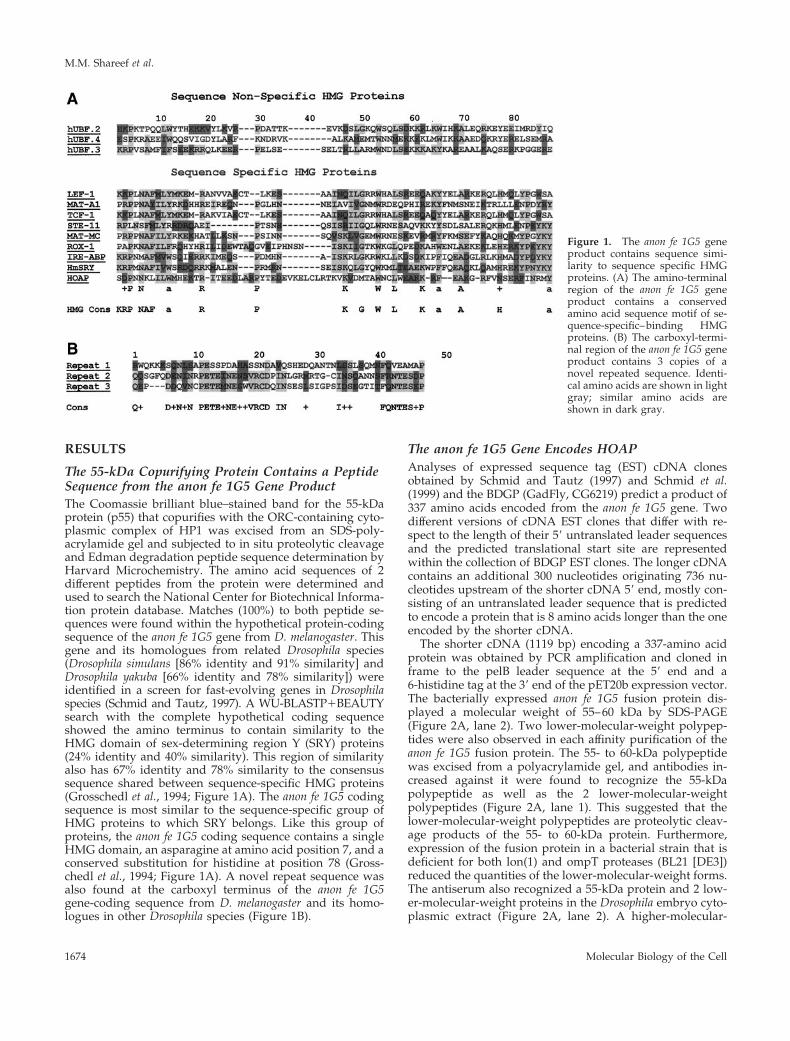
The 55-kDa Copurifying Protein Contains a PeptideSequence from the anon fe 1G5 Gene ProductThe Coomassie brilliant blue–stained band for the 55-kDaprotein (p55) that copurifies with the ORC-containing cyto-plasmic complex of HP1 was excised from an SDS-poly-acrylamide gel and subjected to in situ proteolytic cleavageand Edman degradation peptide sequence determination byHarvard Microchemistry. The amino acid sequences of 2different peptides from the protein were determined andused to search the National Center for Biotechnical Informa-tion protein database. Matches (100%) to both peptide se-quences were found within the hypothetical protein-codingsequence of the anon fe 1G5 gene from D. melanogaster. Thisgene and its homologues from related Drosophila species(Drosophila simulans [86% identity and 91% similarity] andDrosophila yakuba [66% identity and 78% similarity]) wereidentified in a screen for fast-evolving genes in Drosophilaspecies (Schmid and Tautz, 1997). A WU-BLASTP1BEAUTYsearch with the complete hypothetical coding sequenceshowed the amino terminus to contain similarity to theHMG domain of sex-determining region Y (SRY) proteins(24% identity and 40% similarity). This region of similarityalso has 67% identity and 78% similarity to the consensussequence shared between sequence-specific HMG proteins(Grosschedl et al., 1994; Figure 1A). The anon fe 1G5 codingsequence is most similar to the sequence-specific group ofHMG proteins to which SRY belongs. Like this group ofproteins, the anon fe 1G5 coding sequence contains a singleHMG domain, an asparagine at amino acid position 7, and aconserved substitution for histidine at position 78 (Gross-chedl et al., 1994; Figure 1A). A novel repeat sequence wasalso found at the carboxyl terminus of the anon fe 1G5gene-coding sequence from D. melanogaster and its homo-logues in other Drosophila species (Figure 1B).
The anon fe 1G5 Gene Encodes HOAPAnalyses of expressed sequence tag (EST) cDNA clonesobtained by Schmid and Tautz (1997) and Schmid et al.(1999) and the BDGP (GadFly, CG6219) predict a product of337 amino acids encoded from the anon fe 1G5 gene. Twodifferent versions of cDNA EST clones that differ with re-spect to the length of their 59 untranslated leader sequencesand the predicted translational start site are representedwithin the collection of BDGP EST clones. The longer cDNAcontains an additional 300 nucleotides originating 736 nu-cleotides upstream of the shorter cDNA 59 end, mostly con-sisting of an untranslated leader sequence that is predictedto encode a protein that is 8 amino acids longer than the oneencoded by the shorter cDNA.
The shorter cDNA (1119 bp) encoding a 337-amino acidprotein was obtained by PCR amplification and cloned inframe to the pelB leader sequence at the 59 end and a6-histidine tag at the 39 end of the pET20b expression vector.The bacterially expressed anon fe 1G5 fusion protein dis-played a molecular weight of 55–60 kDa by SDS-PAGE(Figure 2A, lane 2). Two lower-molecular-weight polypep-tides were also observed in each affinity purification of theanon fe 1G5 fusion protein. The 55- to 60-kDa polypeptidewas excised from a polyacrylamide gel, and antibodies in-creased against it were found to recognize the 55-kDapolypeptide as well as the 2 lower-molecular-weightpolypeptides (Figure 2A, lane 1). This suggested that thelower-molecular-weight polypeptides are proteolytic cleav-age products of the 55- to 60-kDa protein. Furthermore,expression of the fusion protein in a bacterial strain that isdeficient for both lon(1) and ompT proteases (BL21 [DE3])reduced the quantities of the lower-molecular-weight forms.The antiserum also recognized a 55-kDa protein and 2 low-er-molecular-weight proteins in the Drosophila embryo cyto-plasmic extract (Figure 2A, lane 2). A higher-molecular-
Figure 1. The anon fe 1G5 geneproduct contains sequence simi-larity to sequence specific HMGproteins. (A) The amino-terminalregion of the anon fe 1G5 geneproduct contains a conservedamino acid sequence motif of se-quence-specific–binding HMGproteins. (B) The carboxyl-termi-nal region of the anon fe 1G5 geneproduct contains 3 copies of anovel repeated sequence. Identi-cal amino acids are shown in lightgray; similar amino acids areshown in dark gray.
M.M. Shareef et al.
Molecular Biology of the Cell1674

weight polypeptide in the Drosophila embryo extract wasalso weakly recognized by this antibody (Figure 2A, lane 2)but not by subsequent batches of antibodies produced inother animals (Figure 2A, lane 3).
The affinity-purified antiserum was then used to immu-noblot gel filtration fractions of the cytoplasmic extract (Fig-ure 2B). The fractionation of the endogenous anon fe 1G5gene product was found to overlap with those of the ORC 2subunits and the ORC-containing complexes of HP1. Thelarge HP1 complexes were then immunoaffinity purifiedfrom the gel filtration fractions containing the HP1/ORCcomplexes as described by Huang et al. (1998), and antibod-ies increased against the anon fe 1G5 gene product were usedto determine the presence of this gene product in the HP1complexes. These analyses showed both HP1 and the endog-enous anon fe 1G5 gene product to be quantitatively depleted
from the input fraction (Figure 2C, lanes 1 and 2) andspecifically retained on the HP1 immunoaffinity resin (Fig-ure 2C, lanes 4 and 5).
The antiserum was then used in the reciprocal experimentof immunoprecipitating the endogenous anon fe 1G5 geneproduct from the unfractionated cytoplasmic extract to de-termine by immunoblot analyses whether HP1 and ORCsubunits coimmunoprecipitate with the anon fe 1G5 geneproduct (Figure 2D). A comparison of the immunoblot sig-nal for the HOAP protein in the input and unretained frac-tions showed that the immunoaffinity purification was car-ried out under near-saturating conditions (Figure 2D,compare lanes 1 and 3). HP1 and ORC proteins were alsofound to immunoprecipitate with the endogenous anon fe1G5 gene product, although a comparison of the immuno-blot signals for each of these proteins in the input, unre-tained, and retained fractions (Figure 2D, lanes 1, 2, and 4,respectively) showed that only a small fraction of each pro-tein (2–10%) was retained in the immunoprecipitation.These data are consistent with the fraction of HP1 and ORCsubunits in the cytoplasmic extract that were previouslydetermined to exist in the large HP1 complexes (Pak et al.,1997; Huang et al., 1998) and the gel filtration profiles forHP1 and the ORC 2 subunit in comparison with that ofHOAP (Figure 2B). These reciprocal immunoprecipitationdata confirmed the identity of the 55-kDa copurifying HP1/ORC-associated protein as the product of the anon fe 1G5gene; hence, we will refer to the protein as HP1/ORC-associated Protein (HOAP).
HOAP Is Associated with HP1 and ORC In Salt-resistant ChromatinThe immunoprecipitation experiments described abovedemonstrate an association of the anon fe 1G5 gene product(HOAP) with ORC-containing HP1 complexes in the mater-nally loaded cytoplasm of the early embryo. For determin-ing whether HOAP is a nuclear protein that is associatedwith HP1 and ORC subunits in interphase chromatin, theantiserum increased against the anon fe 1G5 gene productwas first used to immunoblot fractions of proteins that weresalt extracted from interphase nuclei (Figure 3A). The inter-phase nuclei for these experiments were isolated from cycle14 embryos, the stage at which a rapid series of synchronousnuclear divisions in early embryogenesis ends. During thisdevelopmental stage, the nuclei acquire a G2 phase, and theheterochromatin first becomes a distinct cytological entity.The prolonged interphase of this nuclear cycle allows one toobtain a nearly homogeneous collection of embryos withnuclei that are synchronized in interphase and contain fullyformed heterochromatin. Nuclei were isolated from em-bryos of this stage and sequentially extracted with succes-sively increasing concentrations of salt. It was previouslyshown that each differentially extracted fraction containsdistinct populations of HP1 phosphoisoforms (Huang et al.,1998; Figure 3A). The fraction of HP1 that requires thehighest concentration of salt to be extracted (1 M KCl) ischaracteristically underphosphorylated and specifically as-sociated with Drosophila ORC subunits (Huang et al., 1998).To determine whether HOAP is a nuclear protein and thestrength of its association with interphase chromatin, theHOAP antibodies were used to immunoblot the differen-tially extracted nuclear fractions. These analyses showed
Figure 2. HOAP is associated with cytoplasmic complexes con-taining HP1 and ORC subunits. Antibodies were increased againstan anon fe 1G5 fusion protein expressed in bacteria in 2 differentrabbits. Lanes 1–3, immunoblotting of the bacterially expressed anonfe 1G5 fusion protein and the endogenous anon fe 1G5 gene productin the embryo cytoplasmic extract (lane 2) with the use of antibodiesproduced by 1 rabbit and the endogenous anon fe 1G5 gene productin the embryo cytoplasmic extract with the use of antibodies pro-duced by a second rabbit (lane 3). The antibody increased againstthe DmORC 2 peptide was used to immunoblot the endogenousDmORC 2 protein in the embryo cytoplasmic extract (lane 4). (B)Antibodies that recognize HP1, HOAP (anon fe 1G5 gene product),and DmORC 2 were used to immunoblot gel filtration fractions ofthe embryo cytoplasmic extract. (C) The immunoprecipitate of HP1from the embryo cytoplasm extract was immunoblotted with anti-bodies that recognize the anon fe 1G5 gene product (HOAP) andHP1. Lane 1, input protein (5% total); lane 2, protein not retained ona-HP1 beads (5% total); lane 3, protein not retained on nonimmuneIgG beads (5% total); lane 4, protein retained on a-HP1 beads (100%total); lane 5, protein retained on nonimmune IgG beads. (D) Im-munoprecipitates of the endogenous anon fe 1G5 gene product(HOAP) from fractions of the cytoplasmic extract containing a largeHP1 complex were immunoblotted with antibodies that recognizeDmORC 6, HP1, the anon fe 1G5 gene product (HOAP), and DmORC2. Lane 1, input protein (5% total); lane 2, protein not retained ona-anon fe 1G5 beads (5% total); lane 3, protein not retained onnonimmune IgG beads (5% total); lane 4, protein retained on a-anonfe 1G5 beads (100% total); lane 5, protein retained on nonimmuneIgG beads.
HP1/ORC-associated Protein in Silencing
Vol. 12, June 2001 1675

HOAP to be a nuclear protein and to be even more tightlyassociated with interphase chromatin than the most salt-resistant fraction of HP1 (Figure 3A, lane 4). A significantfraction of DmORC subunits 2 and 6 is also found in thesalt-resistant chromatin (Figure 3A).
A chromatin immunoprecipitation assay was then used todetermine whether the anon fe 1G5 gene product is present inchromatin fragments containing HP1 and ORC (Figure 3B).The chromatin samples used for these experiments wereprepared from interphase nuclei of cycle 14 embryos thathad first been extracted with 0.5 M KCl to remove all but themost salt-resistant fraction of HP1 that is associated withORC subunits. The chromatin was cross-linked by treatmentwith formaldehyde and then sonicated to an average lengthof 500 bp. Aliquots of the chromatin fragments were thenincubated in parallel with a-HP1, a-HOAP, and nonimmuneIgG immunoresins. After an extensive wash, each immu-noresin was resuspended in SDS loading buffer and boiledfor 5 min to reverse the cross-links in the retained chromatin.The protein released from each immunoresin was then im-munoblotted with antibodies that recognize HP1, HOAP,and DmORC subunits 2 and 6 (Figure 3B). The immunoblotanalyses showed each protein to be present in the a-HP1chromatin immunoprecipitate (Figure 3B, lane 3) as well asthe a-HOAP chromatin immunoprecipitate (Figure 3B, lane2). In contrast, Drosophila lamin, which was also present inthe salt-resistant chromatin fraction, did not coimmunopre-
cipitate with either HP1 or HOAP (Figure 3B, lanes 1–3,Lamin). Visual comparisons of the immunoblot signal foreach protein in the input (Figure 3B, lane 1, 10% total) andthe immunoresin-retained fraction (Figure 3B, lane 3, 50%total) indicated that HP1, HOAP, and the 2 DmORC sub-units were largely depleted from the input fraction in thea-HP1 immunoprecipitation. The DmORC subunits andHOAP were also depleted from the input chromatin in theHOAP immunoprecipitation; however; only a small fraction(;10%) of HP1 appeared to be associated with HOAP in thisfraction of chromatin (Figure 3B, compare lanes 1 and 2).The exact nature of the associations among HP1, HOAP, andDmORC subunits in this chromatin fraction is not clear,although the ability of HOAP antibodies to immunodepletethe ORC subunits suggests a close association among theseproteins in this fraction. To further examine the possibilitythat HP1, HOAP, and ORC proteins coprecipitate in chro-matin as a result of protein–protein interactions betweendifferent chromatin fragments or the mere proximity of theirbinding sites on DNA rather than their being in a complexon chromatin, the cross-linked chromatin was treated withMnase I before immunoprecipitation. As shown in Figure3B, lanes 5–8, the results of the chromatin immunoprecipi-tation with HP1 antibodies were unaffected by this treat-ment.
HOAP Colocalizes with Subfractions of HP1 andORC in HeterochromatinImmunostaining experiments were then used to determinewhere in interphase chromatin the HOAP protein is located.The antibody increased against the anon fe 1G5 gene productwas first used to immunostain cycle 14 Drosophila embryos.Pericentric heterochromatin is known to occupy a distinctdomain at the nuclear apical surface in embryos of thisdevelopmental stage (Foe and Alberts, 1983). HP1 is en-riched approximately fourfold in this domain (James et al.,1989; Kellum et al., 1995). A side view of the nuclei of anembryo of this stage shows HOAP localized in a pattern ofbrightly stained spots at the apical surface where the in-tensely DAPI-stained sequences are clustered (Figure 4A).The embryos were coimmunostained with a-HP1 antibod-ies, and the spots of HOAP immunostaining were found tolie within the larger domain of enriched HP1 staining inthese nuclei (Figure 4A, HOAP [green] and HP1 [red],merged).
A similar punctate pattern of HOAP immunostaining wasalso observed in the diploid nuclei of wild-type larval brainsquashes (Figure 4, B and C) but absent in squashes fromlarvae that were homozygous for a deficiency removing theanon fe 1G5 gene [Figure 4D, (Def)]. A double-immunostain-ing experiment was carried out with a-HOAP and a-HP1antibodies to determine whether the spots of HOAP immu-nostaining overlap with the domain of HP1 enrichment inheterochromatin of these nuclei. As had been observed inembryos, the spots of HOAP immunostaining were clus-tered within the domain of enriched HP1 overlapping theintensely DAPI-stained satellite sequences of these nuclei[Figure 4B, (DAPI)]. HOAP was also diffusely localizedthroughout the mitotic chromosomes of larval brainsquashes with a slight enrichment throughout pericentricheterochromatin [Figure 4D, (mitotic)].
Figure 3. HOAP is in a tightly bound interphase chromatin frac-tion containing HP1 and DmORC subunits. (A) Equal volumes ofprotein that were sequentially extracted from interphase nuclei with60 mM and 0.5 and 1.0 M KCl and in the remaining pellet fractionwere immunoblotted with antibodies that recognize ORC 2, HOAP,HP1, and ORC 6. (B) Formaldehyde cross-linked fractions of salt-resistant chromatin were immunoprecipitated with a-HOAP anda-HP1 antibodies and immunoblotted with antibodies that recog-nize DmORC 2, HOAP, HP1, DmORC 6, and DmLamin B. Lane 1,input chromatin (10% total); lane 2, chromatin retained on a-HOAPbeads (50% total); lane 3, chromatin retained on a-HP1 beads (50%total); lane 4, chromatin retained on nonimmune IgG beads (50%total). The input chromatin in lanes 5–8 was treated with MNase Ibefore immunoprecipitation with a-HP1 antibodies. Immunopre-cipitated chromatin fractions were immunoblotted with antibodiesthat recognize DmORC 2, HOAP, and HP1. Lane 5, input chromatintreated with MNase I for 10 min (10% total); lanes 6–8, chromatintreated with MNase I for 0 (lane 6), 1 (lane 7), and 10 (lane 8) minthat was retained on a-HP1 beads (50% total each).
M.M. Shareef et al.
Molecular Biology of the Cell1676

We also wished to examine the localization of HOAPrelative to that of the DmORC 2 subunit in heterochromatin.To this end, a double-immunolocalization experiment wasundertaken with HOAP antibodies and antibodies increasedagainst a peptide of the DmORC 2 subunit (Figure 4C). TheDmORC 2 antibody recognizes a single polypeptide of themolecular weight expected for the DmORC 2 subunit in theembryonic cytoplasm extract (Figure 2A, lane 3). When theseantibodies were used in conjunction with antibodies thatrecognize HOAP in larval brain squash immunostainingexperiments, HOAP was found to overlap or to be closelyapposed to spots of enriched ORC 2 immunostaining in theheterochromatic domain of the nucleus (Figure 4C). Thesespots were also located near the intensely DAPI-stainedsatellite sequences in heterochromatin [Figure 4C, (DAPI)],which were shown in separate experiments to lie within thedomain of enriched HP1 in heterochromatin as described byPak et al. (1997) (our unpublished results).
HOAP Is Localized Predominantly at Telomeres andDiffusely throughout Regions of PericentricHeterochromatin of Salivary Gland PolyteneChromosomesTo examine the distribution of HOAP protein at higherresolution, polytene chromosomes from D. melanogaster sal-
ivary glands were coimmunostained with a-HOAP anda-HP1 antibodies (Figure 5). These experiments showed aprominent HOAP localization at the telomere of each chro-mosome [Figure 5A, arrows, (t)]. Staining at the end of 2Lwas weaker and observed less consistently than at otherchromosomes. HP1 has also been frequently observed at thetelomeres of polytene chromosomes, most consistently at theends of the X, 3R, and 2R chromosomes (James et al., 1989;Fanti et al., 1998). More diffuse HOAP immunostaining wasalso observed throughout a region of pericentric heterochro-matin that stains intensely with the a-HP1 antibody (Figure5, B [HP1] and C [HP1 and HOAP merged]) on the left andright arms of the third chromosome. The HOAP immuno-staining in the pericentric heterochromatin of the third chro-mosome was flanked by prominent bands of staining at 80Fat the bases of 3R and 3L (Figure 5D, bracket 80F). It is ofinterest that prominent DmORC 2 immunostaining at thechromocenter of polytene chromosomes is also restricted tothe fourth chromosome and the base of 3R (Pak et al., 1997).Diffuse staining was also consistently found throughout thepericentric heterochromatin at the base of the fourth chro-mosome (Figure 5D, arrowhead 4). The localization patternfor HOAP resembles that reported for the TAS element,which is found predominantly at the telomeres of 2R and 3R,but weaker staining is also observed throughout the chro-
Figure 4. HOAP colocalizes with subfrac-tions of HP1 and DmORC2 in interphase nu-clei. (A) Immunolocalization of HOAP(green), HP1 (red), and HOAP (green) andHP1 (red) merged in DAPI-stained interphasenuclei (blue) of a cycle 14 Drosophila embryo(side view). (B) Immunolocalization of HOAP(green), HP1 (red), and HOAP (green) andHP1 (red) merged in DAPI-stained interphasenuclei (blue) of a larval brain squash. (C) Im-munolocalization of HOAP (green), DmORC2subunit (red), and HOAP (green) andDmORC2 (red) merged in DAPI-stained inter-phase nuclei (blue) of a larval brain squash.(D) Immunolocalization of HOAP [HOAP(Def)] in DAPI-stained interphase nuclei[DAPI (Def)] of a brain squash from homozy-gous Df(3R)crb-F89-4 larvae and HOAP[HOAP (mitotic)] in DAPI-stained metaphasechromosomes [DAPI (mitotic)] of a wild-typelarval brain squash.
HP1/ORC-associated Protein in Silencing
Vol. 12, June 2001 1677

mocenter of polytene chromosomes (Karpen and Spradling,1992).
The HOAP Localization Pattern Is Conserved inDrosophila Species Containing Similar SatelliteSequence CompositionsThe punctate pattern of localization for HOAP in the in-tensely DAPI-stained regions of the nucleus suggests that itmay be associated with 1 or more repetitive DNA sequencesenriched in these regions. One approach used to investigatewhich satellite sequence might serve as the binding site fora heterochromatin protein has been to compare its distribu-tion in Drosophila species containing similar satellite compo-sitions with that in species containing dissimilar composi-tions (Raff et al., 1994; Platero et al., 1998). The anon fe 1G5gene was identified in a molecular evolutionary study aimedat identifying fast evolving genes that are conserved inspecies that are closely related to D. melanogaster (D. simu-lans, Drosophila mauritiana, and D. yakuba) but are absentfrom the more distantly related D. virilis. Interestingly, theclosely related species containing an anon fe 1G5 gene ho-mologue are also known to have similar satellite sequencecompositions (Lohe and Roberts, 1988), whereas the moredistantly related D. virilis lacks an anon fe 1G5 gene homo-logue and has a very different satellite sequence compositionfrom that of D. melanogaster and its close relatives (Gall andAtherton, 1974).
It has been suggested that the conservation of heterochro-matin-binding proteins is driven by the sequence bias ofheterochromatin DNA repeats in that species (Csink andHenikoff, 1998). According to this view, the spots of HOAPstaining observed in D. melanogaster might also be expected
in its closely related species. A similar punctate pattern ofstaining in these species would also support binding ofHOAP to satellite sequences that are shared between thespecies. To each of these ends, HOAP immunostaining ex-periments were carried out on larval brain squashes from 2closely related species (D. simulans and D. mauritiana) andfrom D. virilis (Figure 6). Spots of HOAP immunostainingwere observed in the region of intensely DAPI-stained sat-ellite sequences in interphase nuclei from each closely re-lated species (Figure 6, A–C, HOAP) but not in the nucleifrom D. virilis (Figure 6D, HOAP). Coimmunostaining ofthese nuclei with HP1 antibodies showed the spots of HOAPimmunostaining to be located within or closely juxtaposedto the larger domain of HP1 immunostaining (Figure 6,A–C).
HOAP Protein Binds Specific D. melanogasterSatellite- and Telomere-associated Sequences InVitroThe similarity of the anon fe 1G5 protein-coding sequence tothat of HMG proteins suggests that it possesses DNA-bind-ing activity. The immunostaining experiments describedabove indicate a binding specificity for 1 or more repetitiveDNA found in heterochromatin. Gel mobility shift assayswere used to determine whether HOAP is capable of bind-ing to each of 10 different satellite DNA sequences enrichedin D. melanogaster heterochromatin (Brutlag, 1980) and to theTAS (Karpen and Spradling, 1992; Walter et al., 1995).
A bacterially expressed hexahistidine-tagged recombinantform of the HOAP protein was used in the gel mobility shiftassays. The recombinant protein was affinity purified over aNi-NT agarose column with the use of an elution profile that
Figure 5. HOAP is localized predominantly at telo-meres and weakly throughout regions of pericentricheterochromatin of polytene chromosomes. Shownare immunostaining of HOAP (green; A), HP1 (red;B), and HOAP (green) and HP1 (red) merged (C)and an enlargement of HOAP (green) and HP1 (red)merged at the chromocenter (D). The HP1 signalappears faded when superimposed over the brightsignal from the intensely DAPI-stained chromo-center. (B) Inset, HP1 signal alone.
M.M. Shareef et al.
Molecular Biology of the Cell1678

yielded .90% full-length recombinant HOAP protein (Fig-ure 7A). The DNA probes for the experiments were pre-pared by annealing single-stranded oligonucleotides of eachD. melanogaster satellite sequence. The 5-bp satellite se-quences (AATAT, AATAG, AATAC, AAGAC, AAGAG,and AACAA) were repeated in tandem 4 times, whereas the7- and 10-bp satellite sequences (AATAAAC, AATAGAC,AAGAGAG, and AATAACATAG) were repeated in tandem3 times. The double-stranded oligonucleotides and the457-bp TAS isolated from the telomere of 2L (Walter et al.,1995) were then labeled to equivalent specific activities withthe use of Klenow I to fill in a single-stranded overhang.
In the gel mobility shift assays that were then used tomonitor binding of the recombinant HOAP protein to eachlabeled satellite sequence and to a nonspecific DNA frag-ment, the HOAP protein displayed the strongest bindingaffinity for 3 specific satellite sequences (AATAT, AATAG,and AATAACATAG; Figure 7B), although minimal bindingto the other sequences could also be observed with pro-longed autoradiographic exposures (our unpublished re-sults). The recombinant HOAP protein was also able to bindthe 457-bp TAS isolated from the telomere of 2L (Walter etal., 1995) in a gel mobility shift assay (Figure 7C). To deter-mine which of the satellite sequences HOAP binds with the
greatest affinity, competitive binding assays were performedwith the 3 strongest binding satellite sequences and the TASelement (Figure 7C). Each sequence was used as a compet-
Figure 6. Punctate localization of HOAP in heterochromatin isconserved in closely related Drosophila species that contain an anonfe 1G5 gene and similar satellite sequence compositions. Larvalbrain squashes from D. melanogaster (A), D. simulans (B), D. mauri-tiana (C), and D. virilis (D) were stained with DAPI and a-HP1 anda-HOAP antibodies.
Figure 7. HOAP binds D. melanogaster satellite and TAS in vitro.(A) Coomassie brilliant blue staining of the bacterially expressedhexahistidine-tagged recombinant HOAP protein used in the elec-trophoretic mobility shift assays. (B) Electrophoretic mobility shiftassays were carried out with the recombinant HOAP protein andDrosophila satellite sequences AATAT (lanes 1 and 2), AATAG(lanes 3 and 4), AATAC (lanes 5 and 6), AAGAC (lanes 7 and 8),AAGAG (lanes 9 and 10), AACAA (lanes 11 and 12), AATAAAC(lanes 13 and 14), AATAGAC (lanes 15 and 16), AAGAGAG (lanes17 and 18), and AATACATAG (lanes 19 and 20). For each pair oflanes, the odd-numbered lane contains a given satellite sequencefree probe, and the even-numbered lane contains that same probe inthe presence of HOAP protein. (C) Competitive binding studieswith recombinant HOAP protein, satellite sequences AATAT,AATAG, and AATAACATAG, and TAS element. For each labeledprobe (AATAT, AATAG, AATAACATAG, or TAS, as indicated onthe left) lanes are as follows: lane 1, free labeled probe; lane 2,labeled probe in the presence of HOAP protein; lanes 3–22, bindingof HOAP protein to the probe indicated to the left competed with anincreasing (50, 100, 200, and 4003) molar excess of cold AATATcompetitor (lanes 3–6), AATAG competitor (lanes 7–10), AATAA-CATAG competitor (lanes 11–14), TAS competitor (100, 200, and3003 molar excess; lanes 15–18), and nonspecific DNA competitor(scs sequence; lanes 19–22). (D) Electrophoretic mobility shift assayswere carried out with the labeled D. melanogaster satellite sequenceAATAACATAG (lanes 1 and 2) and D. virilis satellite sequencesACAAACT (lanes 3 and 4), ATAAACT (lanes 5 and 6), andACAAATT (lanes 7 and 8) in the absence and presence of HOAPprotein, respectively.
HP1/ORC-associated Protein in Silencing
Vol. 12, June 2001 1679

itor to binding of HOAP to itself and to each of the otherbinding sequences. In these experiments, the binding ofHOAP to each sequence was most effectively competed by acold excess of that same sequence (e.g., cold AATAT withlabeled AATAT [AATAT; Figure 7C, lanes 3–6], coldAATAG with labeled AATAG [AATAG; lanes 7–10], coldAATAACATAG with labeled AATAACATAG [AATAA-CATAG; lanes 11–14], and cold TAS with labeled TAS [TAS,lanes 15–18]). The binding of HOAP to the AATAT probecould also be effectively competed by an excess of coldAATAG (AATAT; Figure 7C, lanes 7–10), because binding tothe AATAG probe could be competed with an excess of coldAATAT (AATAG; Figure 7C, lanes, 3–6). A cold excess ofthe 10-bp repeat sequence (AATAACATAG) was able tocompete with HOAP binding to itself and to the two 5-bpsequences almost as effectively as each 5-bp sequence toitself (Figure 7C, lanes 11–14). In contrast, the two 5-bpsequences were only able to compete for binding of HOAPto AATAACATAG at the highest molar excess of cold com-petitor used (AATAACATAG; Figure 7C, lanes 3–6 and7–10). HOAP binding to the TAS element could be competedby itself but not by any of the 3 satellite sequences (even ata 400-fold molar excess). However, HOAP binding to each ofthe 3 satellite sequences could be competed by a 300-foldmolar excess of the TAS element, possibly indicating a pref-erence for HOAP binding to the TAS element. An unrelatednonspecific DNA sequence was found to be an ineffectivecompetitor for HOAP binding to the 3 satellite sequences orthe TAS element even at a 400-fold molar excess (Figure 7C,lanes 15–19).
The punctate heterochromatic localization of HOAP isconserved in closely related Drosophila species with similarsatellite sequence compositions [(RRN)m(RN)n rule]. Themore distantly related D. virilis lacks an anon fe 1G5 gene andalso contains satellite sequences that follow a different rule[(AAN)m(NA)n; Gall and Atherton, 1974; Lohe and Roberts,1988]. It has been suggested that the conservation of hetero-chromatin-binding proteins might be driven by the sequencebias of satellite sequence repeats present in a given species(Csink and Henikoff, 1998). To test this idea with regard toconservation of the anon fe 1G5 gene, each of the 3 differentD. virilis satellite sequences (ACAAACT, ATAAACT, andACAAATT) was tested as a binding site for the HOAPprotein. HOAP displayed minimal binding to each of the D.virilis sequences in comparison with binding to the D. mela-nogaster AATAACATAG satellite repeat labeled to equiva-lent specific activity (Figure 7D).
The anon fe 1G5 Gene Displays a ReciprocalModifier of Variegation PhenotypesThe immunoprecipitation and immunostaining experimentsdescribed above demonstrate an association between HOAPand the salt-resistant fractions of HP1 and ORC in inter-phase nuclei. Mutants for the DmORC 2 subunit display aSuppressor of variegation [Su(var)] phenotype (Pak et al., 1997)and also exhibit defects in HP1 localization into heterochro-matin (Huang et al., 1998). To determine whether HOAP alsofunctions in heterochromatin assembly, we undertook ge-netic analyses of a Drosophila mutant that is deficient for thechromosomal region containing the anon fe 1G5 gene encod-ing HOAP.
The cytological position of the anon fe 1G5 gene was de-termined by DNA in situ hybridizations to polytene chro-mosomes from larval salivary glands (our unpublished re-sults). The cytological location determined by theseexperiments (95D–E) was later confirmed by a DNA se-quence from the BDGP (unpublished results, accession num-ber AC008203) and by in situ hybridizations in the indepen-dent study of Schmid et al. (1999). A mutant stock in whichthe chromosomal interval from 95D7-11-95F15 (Df(3R)crb-F89-4) has been removed was obtained through the Bloom-ington Stock Center. This stock was used to determine theeffect of removing 1 copy of the anon fe 1G5 gene on theposition effect–variegated phenotype of the whitem4 allele, inwhich the white gene has undergone a chromosomal rear-rangement juxtaposing it to centric heterochromatin. Thevariegated phenotype associated with the whitem4 rearrange-ment (Figure 8A) was found to be suppressed by theDf(3R)crb-F89-4 chromosome (Figure 8B).
The chromosomal interval removed in the Df (3R)crb-F89-4 stock is rather large; information from BDGP GadFlyindicates that ;60 genes in addition to anon fe 1G5 areremoved. We therefore wished to determine whether thereduced dose of the anon fe 1G5 gene in the Df(3R)crb-F89-4stock was at least partially responsible for this Su(var) phe-notype. To this end, we determined whether expression ofthe anon fe 1G5 gene from a transgene in the Df(3R)crb-F89-4stock could suppress the Su(var) phenotype of the deficiencystock. The ability of the anon fe 1G5 transgene to suppress theSu(var) phenotype would be supportive of the idea that lossof the anon fe 1G5 gene in this deficiency is at least partlyresponsible for the Su(var) phenotype. Because the whitegene served as the reporter for identifying anon fe 1G5 trans-genic animals for the transformation vector we were using,it was necessary to use a different assay that is unrelated to
Figure 8. The anon fe 1G5 gene displays a reciprocal modifier ofvariegation phenotypes. The variegated phenotype of whitem4 wasobserved in a wild-type genetic background (A) and a Df(3R)F89-4/TM3Ser genetic background (B). Variegated expression of theIn(3L)BL hs-LacZ reporter was observed in a wild-type genetic back-ground (C), a Df(3R)F89-4/TM3Ser genetic background (D), and thepresence of 2 copies of a heat shock–induced hs-anon fe 1G5 trans-gene (E).
M.M. Shareef et al.
Molecular Biology of the Cell1680

eye pigmentation to monitor rescue of the Su(var) phenotypeby the anon fe 1G5 transgene. We chose to use the In(3L)BL1stock of Lu et al. (1996), which carries a variegating hsp70-LacZ transgene juxtaposed to pericentric heterochromatin bya chromosomal rearrangement. Embryos were collectedfrom the In(3L)BL1 stock carrying 2 wild-type copies of theanon fe 1G5 gene and compared with those collected simul-taneously from the same stock carrying either the Df(3R)crb-F89-4 deficiency chromosome or 2 additional copies of theanon fe 1G5 gene as a transgene. Each parental genotype wassimultaneously subjected to heat shock to induce expressionof the hs-anon fe 1G5 transgene in the transgenic stock. Thistreatment also induces the hs-lacZ reporter in each parentalgenotype, having the effect of neutralizing rather than en-hancing anon fe 1G5 transgene rescue. Embryos collectedfrom each genotype were then heat shocked to induce ex-pression of the In(3L)BL1hs-lacZ reporter (as well as thehs-anon fe 1G5 transgene) before they were fixed and stainedfor b-galactosidase activity from expression of the hs-lacZreporter (Figure 8, C–E). Embryos from animals carrying 2copies of the endogenous anon fe 1G5 gene displayed mod-erate levels of variegated staining (Figure 8C). In contrast,embryos from animals carrying the Df(3R)crb-F89-4 chromo-some contained high levels of b-galactosidase stainingthroughout the embryo (Figure 8D). The reciprocal pheno-type was observed in embryos collected from animals witheither 1 or 2 copies of the anon fe 1G5 transgene. The majorityof embryos collected from these animals lacked b-galactosi-dase staining altogether or contained reduced levels of var-iegated staining (Figure 8E).
Quantitative measurements of b-galactosidase activities inadults of each genotype were also obtained with the use ofchlorophenol red-b-d-galactopyranoside as substrate (Si-mon and Lis, 1987). The level of b-galactosidase activity inextracts from animals carrying the Df(3R)crb-F89-4 defi-ciency chromosome (Table 1, Df(3R)F89-4) was approxi-mately twofold higher than that measured in extracts fromwild-type animals (Table 1, wild-type). The Su(var) pheno-type associated with the Df(3R)crb-F89-4 was partially re-versed by expression of the anon fe 1G5 transgene from aheat shock–inducible promoter (Table 1, Df(3R)F89-4;P{hsHOAP}). The level of b-galactosidase activity in extractsfrom animals carrying 2 copies of the anon fe 1G5 transgenein addition to the 2 endogenous copies of the gene (Table 1,P{hsHOAP}/P{hsHOAP}) was almost twofold lower than
that measured in extracts from wild-type animals. A moremoderate reduction in activity was measured in animalscarrying a single extra copy of the gene as a transgene (Table1, P{hsHOAP}/CyO).
DISCUSSION
Interphase nuclei contain multiple fractions of HP1, contain-ing different levels of phosphorylation and having differentstrengths of chromatin association. The most tightly boundchromatin fraction is underphosphorylated and specificallyassociated with Drosophila ORC subunits (Huang et al.,1998). A specific fraction of the protein in the maternallyloaded cytoplasm of the early embryo is also underphos-phorylated and specifically associated with a multiproteincomplex containing DmORC subunits. This cytoplasmicfraction is thought to be poised for assembly into the tightlybound chromatin fraction during heterochromatin assemblylater in embryonic development. Mutants for the ORC2 sub-unit display a perturbation in HP1 localization into Drosoph-ila heterochromatin. This observation has led to the postu-lation that ORC plays a role in the recruitment of HP1 intoDrosophila heterochromatin that may be analogous to its rolein recruiting the SIR1 protein to silencing nucleation sites inS. cerevisiae (Huang et al., 1998). Here we report the identi-fication of an unknown protein component (p55) of theHP1/ORC complex of the maternally loaded cytoplasm. Thepeptide sequence from this protein matches the sequencefrom the hypothetical protein product of the Drosophila anonfe 1G5 gene (Schmid and Tautz, 1997). Antibodies increasedagainst a recombinant protein produced from this gene wereused in reciprocal immunoprecipitation experiments toshow that the HOAP p55 protein is the product of this gene.
HOAP Contains a Region of Similarity to HMGProteinsThe amino terminus of the anon fe 1G5 gene-coding sequencecontains similarity to the HMG domain of the SRY proteinand the group of DNA sequence-specific HMG proteins towhich it belongs. This similarity to HMG proteins is ofinterest in view of reports of interactions of a human homo-logue of HP1 with the SP100-HMG protein (Lehming et al.1998; Seeler et al., 1998). This HMG protein displays a punc-tate localization similar to that of HOAP in normal cells thatbecomes disrupted in some cancer cells (Szostecki et al.,1990; Ascoli and Maul, 1991). The HMG sequence motif inthe HOAP protein suggested that it may possess DNA-binding activity. We have, indeed, demonstrated sequence-specific binding of HOAP to 3 D. melanogaster satellite DNAsequences (AATAT, AATAG, and AATAACATAG) and tothe TAS.
The protein was also found to have a punctate pattern ofdistribution in the region where pericentric satellite se-quences are clustered in interphase nuclei. The spots ofHOAP staining lie within the domain of HP1 enrichmentand are closely apposed to sites of enriched ORC 2 immu-nostaining in this domain. This enrichment of HOAP inpericentric heterochromatin was also observed in mitoticchromosomes of diploid cells. The distribution of HOAP inpolytene chromosomes was also heterochromatic; it waspredominantly found at telomeres, but weaker staining was
Table 1. Quantitation of b-galactosidase activities fromIn(3L)BL1hs-lacZ reporter in HOAP-overexpressing and -underex-pressing lines
A574a Mut/wtb
Wild type 0.869 1.00Df(3R)F89-4 2.02 2.32Df(3R)F89-4; P{hsHOAP} 1.10 1.27P{hsHOAP}/CyO 0.696 0.616P{hsHOAP}/P{hsHOAP} 0.489 0.563
a A574 units measured upon conversion of CPRG (chlorophenolred-b-D-galactoside) substrate to product (A574).b Mut, mutant; wt, wild type.
HP1/ORC-associated Protein in Silencing
Vol. 12, June 2001 1681
also observed throughout regions of pericentric heterochro-matin. These results indicate an association of HOAP with 1or more repeated sequences in telomeric and pericentricheterochromatin. The TAS is 1 repetitive sequence that isfound in both regions (Karpen and Spradling, 1992). HOAPwas able to bind this sequence in vitro; however, furtherstudies will be required to determine the actual in vivobinding site for the HOAP protein.
The pattern of distribution for HOAP in diploid and poly-tene tissues and its dynamics during the cell cycle differfrom those of any previously characterized heterochroma-tin-associated protein in Drosophila. HP1 displays prominentenrichment throughout pericentric heterochromatin and ateach telomere of salivary gland polytene chromosomes(James et al., 1989; Fanti et al., 1998). The association of HP1with pericentric heterochromatin, however, appears to beless stable during mitosis (Kellum et al., 1995; Platero et al.,1998). Two other heterochromatin-associated proteins(GAGA factor and Prod) have prominent binding sites inboth heterochromatin and euchromatin. Binding to hetero-chromatic satellite repeat sequences (AAGAG and AATAA-CATAG, respectively) is observed in mitotic chromosomespreads, but both proteins become redistributed to sites ofeuchromatic gene regulation during interphase (Raff et al.,1994; Torok et al., 1997; Platero et al., 1998). This redistribu-tion and differences in the representation of satellite se-quences in polytene and diploid tissues (Miklos and Cotsell,1990) contribute to a euchromatic pattern of distribution forboth proteins in interphase polytene chromosomes. HOAP,by contrast, is found predominantly at heterochromatic re-gions of both diploid and polytene nuclei and appears toretain its association with pericentric heterochromatinthroughout the diploid cell cycle. Its localization predomi-nantly at telomeres of polytene chromosomes but in peri-centric regions of diploid chromosomes probably reflectsdifferences in the representation of particular heterochro-matic sequences in the 2 cell types. These distinct character-istics of HOAP distribution in heterochromatin are likely toreflect a unique role for it in these regions.
A Role for HOAP in Heterochromatin AssemblyGenetic screens in Drosophila have been used to identifychromosomal regions that modify the position effect–varie-gated phenotype of the whitemot4 allele when either dupli-cated or deleted (Locke et al., 1988; Wustmann et al., 1989).The proteins encoded by these genes are thought to play arole in heterochromatin assembly. We have used a defi-ciency for the chromosomal region containing the anon fe1G5 gene [Df(3R)F89-4] to determine whether the HOAPprotein has a role in heterochromatin assembly. This defi-ciency caused a suppression of the position effect–variegatedphenotype of the whitem4 allele as well as the hs-lacZ reporterof the In(3L)BL1 chromosomal rearrangement (Lu et al.,1996). Suppression of the Su(var) phenotype with an anon fe1G5 transgene suggests that loss of the anon fe 1G5 gene is atleast partially responsible for the Su(var) phenotype associ-ated with this deficiency.
The chromatin immunoprecipitation experiments withHP1 and HOAP antibodies showed HOAP to be located inclose proximity to HP1 and ORC proteins in the salt-resis-tant fraction of interphase chromatin. We speculate that thisfraction of chromatin contains binding sequences for ORC
and HOAP from which heterochromatin assembly may benucleated by a process resembling the recruitment of the Sircomplex to silencing nucleation sites in S. cerevisiae. Thedemonstration that HOAP binds specific satellite sequencessuggests that it may play a role in Drosophila heterochroma-tin that is analogous to the roles of the DNA-binding Rap1and Abf1 proteins in budding yeast silencing. Indeed, thelimited degree of overlap between the HP1-containing chro-matin fractions and those containing ORC or HOAP in thechromatin immunoprecipitation experiments may be indic-ative of a heterochromatin assembly nucleation activity forHOAP rather than as a bona fide structural component ofheterochromatin.
A repeated DNA sequence of any type has been shown, insome cases, to induce heterochromatin formation in euchro-matic regions of Drosophila chromosomes (Dorer and Heni-koff, 1994). It has been proposed that heterochromatin as-sembly is induced in these situations as a result ofheterochromatin proteins recognizing unusual folded struc-tures at sites of paired repeats. HMG proteins constitute 1class of protein that is likely to play a role in such a mech-anism; the sequence recognition motif of even the sequence-nonspecific class of HMG proteins involves recognition of analtered DNA secondary structure (Lilley, 1988; Pohler et al.,1998). However, HOAP was found to display preferredbinding to specific satellite repeats as well as to the mini-satellite TAS. Interestingly, TASs have been found flankinga number of variegating transgenes inserted into both telo-meres (of X, 2R, and 3R) and pericentric heterochromatin (of2R and 2L; Karpen and Spradling, 1992; Zhang and Spra-dling, 1995; Cryderman et al., 1998). They have also beenimplicated in the mediation of the P cytotype by a singlestrongly repressed autonomous P element [Lk-P(1A)] in-serted next to a block of TAS repeats in the telomere of theX chromosome (Ronsseray et al., 1996; Roche and Rio, 1998).The variegated expression of telomeric transgenes flankedby TAS elements has been found to be unaffected by areduced dose of the HP1 gene [Su(var)2-5] (Cryderman et al.,1998), although the ability of Lk-P(1A) to repress P dysgen-esis is strongly reduced (Ronsseray et al., 1996, 1998).
HOAP Is Encoded by a Fast-evolving GeneThe anon fe 1G5 gene was identified in a screen for fast-evolving Drosophila genes on the basis of their conservationin species closely related to D. melanogaster (D. simulans, D.yakuba, and D. mauritiana) but not in the more distantlyrelated D. virilis (Schmid and Tautz, 1997). It is of interestthat an anon fe 1G5 gene homologue is present in each of theclosely related species that contain satellite sequences con-stituting in vitro HOAP binding sites but not in the distantlyrelated D. virilis containing different satellite sequences thatdo not serve as in vitro binding sites for the HOAP protein(Gall and Atherton, 1974; Brutlag, 1980; Schmid et al., 1999).One of these satellite sequences (AATAT) is even known tohave a conserved chromosomal location in each of the spe-cies containing an anon fe 1G5 gene (Lohe and Roberts, 1988).HOAP immunostaining in interphase and mitotic diploidcells of D. melanogaster is consistent with an association ofHOAP with 1 or more of these sequences in vivo. Moreover,the punctate pattern of HOAP distribution in the hetero-chromatic regions is conserved in the closely related species.The localization pattern of HOAP on polytene chromosome
M.M. Shareef et al.
Molecular Biology of the Cell1682
also suggests a relationship to the TAS first described byKarpen and Spradling (1992). It is not currently knownwhether TASs are present in other Drosophila species, but adifferent subtelomeric satellite has been found in D. virilisthat is not yet known to exist in D. melanogaster (Biessmannet al., 2000).
The conservation of an anon fe 1G5 gene only in speciescontaining HOAP-binding sequences is consistent withthe ideas put forth by Csink and Henikoff (1998) of arelationship between conservation of heterochromatin-binding proteins and the sequence bias of repetitive het-erochromatin sequences in a given species. Csink andHenikoff (1998) also speculated that the enrichment ofsatellite sequences at centromeres might play a passiverole in centromere formation by providing a mechanismfor excluding efficient origins of replication, and, thereby,might determine these regions as centromeres as a conse-quence of their late replication. ORC 2 immunostainingexperiments indicate that ORC is enriched rather thanexcluded from pericentric heterochromatin in Drosophila(Pak et al., 1997). Moreover, ORC binding does not appearto play the critical role in determining the firing time of anorigin; rather, other factors such as chromatin organiza-tion and Cdc45p binding are thought to be importantfactors (Wintersberger, 2000). ORC proteins may functionin concert with HP1 and DNA-binding proteins such asHOAP to establish a heterochromatin structure in peri-centric regions that then determines the late-replicatingproperties of those regions. According to this model, therepetitive sequences in heterochromatin play an activerather than a passive role in centromere formation. Al-though a conserved feature of pericentric heterochroma-tin is its repetitive sequence composition, there is littleconservation at the level of the primary DNA sequence.The collection of DNA-binding proteins participating inheterochromatin formation in a given species is, therefore,likely to be determined by the satellite sequence compo-sition of that species.
A number of fast-evolving genes, including those of SRYproteins, are known to have roles in sexual selection orspecies-specific differentiation (Whitfield et al., 1993; Schmidand Tautz, 1997). Functions for heterochromatin in organiz-ing nuclear architecture, chromosome pairing, and centro-mere function (Allshire et al., 1995; Kellum et al., 1995; Dern-burg et al., 1996; Karpen et al. 1996; Fanti et al., 1998; forreview, see Dobie et al. 1999) are likely to be relevant toprocesses that drive species differentiation. As such, it maynot be surprising that a heterochromatin sequence-bindingprotein might play a role in evolutionary processes drivingspecies differentiation.
ACKNOWLEDGMENTS
We thank M. Botchan for the DmORC 2 and 6 antibodies, H.Saumweber and J. Sedat for the DmLamin antibodies, J. Eissenbergfor the In(3L)BL1 stock, E. Knust for the Df(3R)89-4 stock, L. Wall-rath and H. Biessmann for the TAS sequence clone, and P. Tolias forthe ovarian gt22A cDNA library. This work was supported bygrants from the Canadian Medical Research Council and NationalInstitutes of Health.
REFERENCES
Alfageme, C.R., Rudkin, G.T., and Cohen, L.H. (1980). Isolation,properties, and cellular distribution of D1, a chromosomal proteinof Drosophila. Chromosoma 78, 1–31.
Allshire, R.C., Nimmo, E.R., Ekwall, K., Javerzat, J.P., and Cranston,G. (1995). Mutations derepressing silent centromeric domains infission yeast disrupt chromosome segregation. Genes Dev. 9, 218–233.
Aparicio, O.M., Weinstein, D.M., and Bell, S.P. (1997). Componentsand dynamics of DNA replication complexes in S. cerevisiae: redis-tribution of MCM proteins and Cdc45p during S phase. Cell 91,59–69.
Ascoli, C.A., and Maul, G.G. (1991). Identification of a novel nucleardomain. J. Cell Biol. 112, 785–795.
Austin, R.J., Orr-Weaver, T.L., and Bell, S.P. (1999). Drosophila ORCspecifically binds to ACE3, an origin of DNA replication controlelement. Genes Dev. 13, 2639–2649.
Bell, S.P., Kobayashi, R., and Stillman, B. (1993). Yeast origin recog-nition complex functions in transcription silencing and DNA repli-cation. Science 262, 1844–1849.
Bell, S.P., and Stillman, B. (1992). ATP-dependent recognition ofeucaryotic origins of DNA replication by a multiprotein complex.Nature 357, 128–134.
Biessmann, H., Zurovcova, M., Yao, J.G., Lozovskaya, E., andWalter, M.F. (2000). A telomeric satellite in Drosophila virilis and itssibling species. Chromosoma 109, 372–380.
Brown, S.W. (1966). Heterochromatin. Science 151, 417–425.
Brutlag, D.L. (1980). Molecular arrangement and evolution of het-erochromatic DNA. Annu. Rev. Genet. 14, 121–144.
Chien, C.T., Buck, S., Sternglanz, R., and Shore, D. (1993). Targetingof SIR1 protein establishes transcriptional silencing at HM loci andtelomeres in yeast. Cell 75, 531–541.
Cryderman, D.E., Cuaycong, M.H., Elgin, S.C.R., and Wallrath, L.L.(1998). Characterization of sequences associated with position-effectvariegation at pericentric sites in Drosophila heterochromatin. Chro-mosoma 107, 277–285.
Csink, A.K., and Henikoff, S. (1998). Something from nothing: theevolution and utility of satellite repeats. Trends Genet. 14, 200–204.
Dernburg, A.F., Sedat, J.W., and Hawley, R.S. (1996). Direct evi-dence of a role for heterochromatin in meiotic chromosome segre-gation. Cell 86, 135–146.
Dobie, K.W., Hari, K.L., Maggert, K.A., and Karpen, G.H. (1999).Centromere proteins and chromosome inheritance: a complex affair.Curr. Opin. Genet. Dev. 9, 206–217.
Dorer, D.R., and Henikoff, S. (1994). Expansions of transgene repeatscause heterochromatin formation and gene silencing in Drosophila.Cell 77, 993–1002.
Eissenberg, J.C., James, T.C., Foster-Hartnett, D.M., Hartnett, T.,Ngan, V., and Elgin, S.C.R. (1990). Mutation in a heterochromatin-specific protein is associated with suppression of position-effectvariegation in Drosophila melanogaster. Proc. Natl. Acad. Sci. USA 87,9923–9927.
Fanti, L., Giovinazzo, G., Berloco, M., and Pimpinelli, S. (1998). Theheterochromatin protein 1 prevents telomere fusions in Drosophila.Mol. Cell 2, 527–538.
Foe, V.E., and Alberts, B.M. (1983). Studies of nuclear and cytoplas-mic behavior during the five mitotic cycles that precede gastrulationin Drosophila embryogenesis. J. Cell Sci. 61, 31–70.
HP1/ORC-associated Protein in Silencing
Vol. 12, June 2001 1683

Fox, C.A., Loo, S., Dillin, A., and Rine, J. (1995). The origin recog-nition complex has essential functions in transcriptional silencingand chromosomal replication. Genes Dev. 9, 911–924.
Frasch, M., Paddy, M., and Saumweber, H. (1988). Developmentaland mitotic behavior of two novel groups of nuclear envelopeantigens of Drosophila melanogaster. J. Cell Sci. 90, 247–263.
Gall, J.G., and Atherton, D.D. (1974). Satellite DNA sequences inDrosophila virilis. J. Mol. Biol. 85, 633–664.
Grosschedl, R., Giese, K., and Pagel, J. (1994). HMG domain pro-teins: architectural elements in the assembly of nucleoprotein struc-tures. Trends Genet. 10, 94–100.
Harlow, E., and Lane, D. (1988). Storing and purifying antibodies.In: Antibodies: A Laboratory Manual, Cold Spring Harbor, NY:Cold Spring Harbor Laboratory Press, 726.
Hecht, A., Laroche, T., Strahl-Bolsinger, S., Gasser, S.M., and Grun-stein, M. (1995). Histone H3 and H4 N-termini interact with SIR3and SIR4 proteins: a molecular model for the formation of hetero-chromatin in yeast. Cell 80, 583–592.
Heitz, E. (1928). Das Heterochromatin der Moose. I Jahrb. Wissen-sch. Bot. 69, 762–818.
Huang, D.W., Fanti, L., Pak, D.T.S., Botchan, M.R., Pimpinelli, S.,and Kellum, R. (1998). Distinct cytoplasmic and nuclear fractions ofDrosophila heterochromatin protein 1: their phosphorylation levelsand associations with origin recognition complex proteins. J. CellBiol. 142, 307–318.
James, T.C., Eissenberg, J.C., Craig, C., Dietrich, V., Hobson, A., andElgin, S.C.R. (1989). Distribution patterns of HP1, a heterochroma-tin-associated nonhistone chromosomal protein of Drosophila. Eur.J. Cell Biol. 50, 170–180.
James, T.C., and Elgin, S.C.R. (1986). Identification of a nonhistonechromosomal protein associated with heterochromatin in Drosophilamelanogaster and its gene. Mol. Cell. Biol. 6, 3862–3872.
Karpen, G.H., Le, M.H., and Le, H. (1996). Centric heterochromatinand the efficiency of achiasmate disjunction in Drosophila femalemeiosis. Science 273, 118–122.
Karpen, G.H., and Spradling, A.C. (1992). Analysis of subtelomericheterochromatin in the Drosophila minichromosome Dp1187 by sin-gle P element insertional mutagenesis. Genetics 132, 737–753.
Kellum, R., and Alberts, B.M. (1995). Heterochromatin protein 1 isrequired for correct chromosome segregation in Drosophila embryos.J. Cell Sci. 108, 1419–1431.
Kellum, R., Raff, J., and Alberts, B.M. (1995). Heterochromatin pro-tein 1 distribution during development and during the cell cycle inDrosophila embryos. J. Cell Sci. 108, 1407–1418.
Kellum, R., and Schedl, P. (1991). A position-effect assay for bound-aries of higher order chromosomal domains. Cell. 64, 941–950.
Kuo, M.-H., and Allis, C.D. (1999). In vivo cross-linking and immu-noprecipitation for studying dynamic protein: DNA associations ina chromatin environment. Methods 19, 425–433.
Kurtz, S., and Shore, D. (1991). RAP1 protein activates and silencestranscription of mating-type genes in yeast. Genes Dev. 5, 616–628.
Landis, G., Kelley, R., Spradling, A.C., and Tower, J. (1997). The k43gene, required for chorion gene amplification and diploid cell chro-mosome replication, encodes the Drosophila homolog of yeast originrecognition complex subunit 2. Proc. Natl. Acad. Sci. USA 94, 3888–3892.
Laurenson, P., and Rine, J. (1992). Silencers, silencing, and heritabletranscriptional states. Microbiol. Rev. 56, 543–560.
Lehming, N., Le Saux, A., Schuller, J., and Ptashne, M. (1998).Chromatin components as part of a putative transcriptional repress-ing complex. Proc. Natl. Acad. Sci. USA 95, 7322–7326.
Lilley, D.M. (1988). DNA opens up—supercoiling and heavy breath-ing. Trends Genet. 4, 111–114.
Locke, J., Kotarski, M.A., and Tartof, K.D. (1988). Dosage-dependentmodifiers of position effect variegation in Drosophila and a massaction model that explains their effect. Genetics 120, 181–198.
Lohe, A., and Roberts, P. (1988). Evolution of satellite DNA se-quences in Drosophila. In: Heterochromatin: Molecular and Struc-tural Aspects, ed. R.S. Verma, Cambridge, United Kingdom: Cam-bridge University Press, 148–186.
Lu, B.Y., Bishop, C.P., and Eissenberg, J.C. (1996). Developmentaltiming and tissue specificity of heterochromatin-mediated silencing.EMBO J. 15, 1323–1332.
McNally, F.J., and Rine, J. (1991). A synthetic silencer mediatesSIR-dependent functions in Saccharomyces cerevisiae. Mol. Cell. Biol.11, 5648–5659.
Moazed, D., Kistler, A., Axelrod, A., Rine, J., and Johnson, A.D.(1997). Silent information regulator protein complexes in Saccharo-myces cerevisiae: a SIR2/SIR4 complex and evidence for a regulatorydomain in SIR4 that inhibits its interaction with SIR3. Proc. Natl.Acad. Sci. USA 94, 2186–2191.
Miklos, G.L., and Cotsell, J.N. (1990). Chromosome structure atinterfaces between major chromatin types: alpha- and beta-hetero-chromatin. Bioessays 12, 1–6.
Orlando, V., Strutt, H., and Paro, R. (1997). Analysis of chromatinstructure by in vivo formaldehyde cross-linking. Methods 11, 205–214.
Pak, D.T.S., Pflumm, M., Chesnokov, I., Huang, D.W., Kellum, R.,Marr, J., Romanowski, P., and Botchan, M.R. (1997). Association ofthe origin recognition complex with heterochromatin and HP1 inhigher eukaryotes. Cell 91, 311–323.
Peacock, W.J., Appels, R., Dunsmuir, P., Lohe, A.R., and Gerlach,W.L. (1976). Highly repeated DNA sequences: chromosomal loca-tion and evolutionary conservation. In: International Cell Biology,ed. B.R. Brinkley and K.R. Porter, New York: Rockefeller UniversityPress, 494–506.
Peacock, W.J., and Lohe, A.R. (1980). Repeated sequences in thechromosomes of Drosophila. Symp. R. Entomol. Soc. Lond. 10, 1–12.
Peacock, W.J., Lohe, A.R., Gerlach, W.L., Dunsmuir, P., Dennis, E.S.,and Appels, R. (1977). Fine structure and evolution of DNA inheterochromatin. Cold Spring Harbor Symp. Quant. Biol. 42, 1121–1135.
Pillus, L., and Rine, J. (1989). Epigenetic inheritance of transcrip-tional states in S. cerevisiae. Cell 59, 637–647.
Pimpinelli S., Bonaccorsi, S., Fanti. L., and Gatti. M. (2000). Prepa-ration and analyses of Drosophila mitotic chromosomes. In: Drosoph-ila Protocols, ed. W. Sullivan, M. Ashburner, and R.S. Hawley, ColdSpring Harbor, NY: Cold Spring Harbor Laboratory Press, 3–23.
Platero, J.S., Csink, A.K., Quintanilla, A., and Henikoff, S. (1998).Changes in chromosomal localization of heterochromatin-bindingproteins during the cell cycle in Drosophila. J. Cell Biol. 140, 1297–1306.
Pohler, J.R., Norman, D.G., Bramham, J., Bianchi, M.E., and Lilley,D.M. (1998). HMG box proteins bind to four-way DNA junctions intheir open conformation. EMBO J. 17, 817–826.
Raff, J.R., Kellum, R., and Alberts, B. (1994). The Drosophila GAGAtranscription factor is associated with specific regions of heterochro-matin throughout the cell cycle. EMBO J. 13, 5977–5983.
Roche, S.E., and Rio, D.C. (1998). Trans-silencing of P elements insubtelomeric heterochromatin involves the Drosophila polycombgroup gene, enhancer of zeste. Genetics 149, 1839–1855.
M.M. Shareef et al.
Molecular Biology of the Cell1684

Ronsseray, S., Lehmann, M., Nouaud, D., and Anxolabehere, D.(1996). The regulatory properties of autonomous subtelomeric Pelements are sensitive to a suppressor of variegation in Drosophilamelanogaster. Genetics 143, 1663–1674.
Ronsseray, S., Marin, L., Lehmann, M., and Anxolabehere, D. (1998).Repression of hybrid dysgenesis in Drosophila melanogaster by com-binations of telomeric P-element reporters and naturally occurring Pelements. Genetics 149, 1857–1866.
Rowles, A., Chong, J.P., Brown, L., Howell, M., Evan, G.I., and Blow,J.J. (1996). Interaction between the origin recognition complex andthe replication licensing system in Xenopus. Cell 87, 287–296.
Royzman, I., Austin, R.J., Bosco, G., Bell, S.P., and Orr-Weaver, T.L.(1999). ORC localization in Drosophila follicle cells and the effects ofmutations in dE2F and dDP. Genes Dev. 13, 827–840.
Saumweber, H., Symmons, P., Kabisch, R., Will, H., and Bonhoeffer,F. (1980). Monoclonal antibodies against chromosomal proteins ofDrosophila melanogaster: establishment of antibody producing celllines and partial characterization of corresponding antigens. Chro-mosoma 80, 253–275.
Saunders, W.S., Chue, C., Goebl, M., Craig, C., Clark, R.F., Powers,J.A., Eissenberg, J.C., Elgin, S.C.R., Rothfield, N.F., and Earnshaw,W.C. (1993). Molecular cloning of a human homolog of Drosophilaheterochromatin protein HP1 using anti-centromere autoantibodieswith anti-chromo specificity. J. Cell Sci. 104, 573–582.
Schmid, K.J., Nigro, L., Aquadro, C.F., and Tautz, D. (1999). Largenumber of replacement polymorphisms in rapidly evolving genes ofDrosophila. Implications for genome-wide surveys of DNA polymor-phism. Genetics 153, 1717–1729.
Schmid, K.J., and Tautz, D. (1997). A screen for fast evolving genesfrom Drosophila. Proc. Natl. Acad. Sci. USA 94, 9746–9750.
Seeler, J.-S., Marchio, A., Sitterlin, D., Transby, C., and Dejean, A.(1998). Interaction of SP100 with HP1 proteins: a link between thepromyelocytic leukemia-associated nuclear bodies and the chroma-tin compartment. Proc. Natl. Acad. Sci. USA 95, 7316–7321.
Simon, J.A., and Lis, J.T. (1987). A germline transformation analysisreveals flexibility in the organization of heat shock consensus ele-ments. Nucleic Acids Res. 15, 2971–2988.
Sinclair, D.A.R., Mottus, R.C., and Grigliatti, T.A. (1983). Geneswhich suppress position-effect variegation in Drosophila melanogasterare clustered. Mol. Gen. Genet. 191, 326–333.
Stroumbakis, N.D., Li, Z., and Tolias, P.P. (1994). RNA- and single-stranded DNA-binding (SSB) proteins expressed during Drosophilamelanogaster oogenesis: a homolog of bacterial and eukaryotic mi-tochondrial SSBs. Gene 143, 171–177.
Szostecki, C., Guldner, H.H., Netter, H.J., and Will, H. (1990). Iso-lation and characterization of cDNA encoding a human nuclearantigen predominantly recognized by autoantibodies from patientswith primary biliary cirrhosis. J. Immunol. 145, 4338–4347.
Tepass, U., and Knust, E. (1990). Phenotypic and developmentalanalysis of mutations at the crumbs locus, a gene required for thedevelopment of epithelia in Drosophila melanogaster. Roux Arch.Dev. Biol. 199, 189–206.
Torok, T., Harvie, P.D., Buratovich, M., and Bryant, P.J. (1997). Theproduct of proliferation disrupter is concentrated at centromeresand required for mitotic chromosome condensation and cell prolif-eration in Drosophila. Genes Dev. 11, 213–226.
Triolo, T., and Sternglanz, R. (1996). Role of interactions between theorigin recognition complex and SIR1 in transcriptional silencing.Nature 381, 251–253.
Walter, M.F., Jang, C., Kasravi., B., Donath, J., Mechler, B.M., Mason,J.M., and Biessmann, H. (1995). DNA organization and polymor-phism of a wild-type Drosophila telomere region. Chromosoma 104,229–241.
Whitfield, L.S., Lovell-Badge, R., and Goodfellow, P.N. (1993).Rapid sequence evolution of the mammalian sex-determining geneSRY. Nature 364, 713–715.
Wintersberger, E. (2000). Why is there late replication? Chromo-soma 109, 300–307.
Wustmann, G., Szidonya, J., Taubert, H., and Reuter, G. (1989). Thegenetics of position-effect variegation modifying loci in Drosophilamelanogaster. Mol. Gen. Genet. 217, 520–527.
Zhang, P., and Spradling, A.C. (1995). The Drosophila salivary glandchromocenter contains highly polytenized subdomains of mitoticheterochromatin. Genetics 139, 659–670.
HP1/ORC-associated Protein in Silencing
Vol. 12, June 2001 1685
Related Documents