Drosophila Genome-Wide RNAi Screen Identifies Multiple Regulators of HIF–Dependent Transcription in Hypoxia Andre ´ s Dekanty 1 , Nuria M. Romero 1,2 , Agustina P. Bertolin 1,3 , Marı ´a G. Thomas 1,3 , Claudia C. Leishman 1 , Joel I. Perez-Perri 1 , Graciela L. Boccaccio 1,3 , Pablo Wappner 1,2,3 * 1 Instituto Leloir, Universidad de Buenos Aires, Buenos Aires, Argentina, 2 Departamento de Fisiologı ´a, Biologı ´a Molecular, y Celular, Facultad de Ciencias Exactas y Naturales, Universidad de Buenos Aires, Buenos Aires, Argentina, 3 Consejo Nacional de Investigaciones Cientı ´ficas y Te ´ cnicas, Buenos Aires, Argentina Abstract Hypoxia-inducible factors (HIFs) are a family of evolutionary conserved alpha-beta heterodimeric transcription factors that induce a wide range of genes in response to low oxygen tension. Molecular mechanisms that mediate oxygen-dependent HIF regulation operate at the level of the alpha subunit, controlling protein stability, subcellular localization, and transcriptional coactivator recruitment. We have conducted an unbiased genome-wide RNA interference (RNAi) screen in Drosophila cells aimed to the identification of genes required for HIF activity. After 3 rounds of selection, 30 genes emerged as critical HIF regulators in hypoxia, most of which had not been previously associated with HIF biology. The list of genes includes components of chromatin remodeling complexes, transcription elongation factors, and translational regulators. One remarkable hit was the argonaute 1 (ago1) gene, a central element of the microRNA (miRNA) translational silencing machinery. Further studies confirmed the physiological role of the miRNA machinery in HIF–dependent transcription. This study reveals the occurrence of novel mechanisms of HIF regulation, which might contribute to developing novel strategies for therapeutic intervention of HIF–related pathologies, including heart attack, cancer, and stroke. Citation: Dekanty A, Romero NM, Bertolin AP, Thomas MG, Leishman CC, et al. (2010) Drosophila Genome-Wide RNAi Screen Identifies Multiple Regulators of HIF– Dependent Transcription in Hypoxia. PLoS Genet 6(6): e1000994. doi:10.1371/journal.pgen.1000994 Editor: Eric Rulifson, University of California San Francisco, United States of America Received January 29, 2010; Accepted May 19, 2010; Published June 24, 2010 Copyright: ß 2010 Dekanty et al. This is an open-access article distributed under the terms of the Creative Commons Attribution License, which permits unrestricted use, distribution, and reproduction in any medium, provided the original author and source are credited. Funding: This work was supported by a PICT 2007 ANPCyT grant, the HHMI grant 55005973, and the Wellcome Trust grant WT087675MA. The funders had no role in study design, data collection and analysis, decision to publish, or preparation of the manuscript. Competing Interests: The authors have declared that no competing interests exist. * E-mail: [email protected] Introduction The cellular response to low oxygen tension (hypoxia) involves changes in gene expression that mediate adaptation to this condition. The hypoxic response is primarily mediated by a family of highly conserved transcription factors named Hypoxia Induc- ible Factors (HIFs) [1]. HIFs are a/b heterodimers, in which the common b subunit is constitutive and a subunits are negatively regulated by O 2 through several concurrent mechanisms that include oxygen-dependent proteasomal degradation [2], blockage of transcriptional co-activator recruitment [3,4] and subcellular localization [5,6]. HIFa proteolysis requires polyubiquitination, which in turn depends on the hydroxylation of two key prolyl residues localized in the so-called oxygen-dependent degradation domain (ODDD) [7,8]. Hydroxylation is mediated by specific HIF prolyl-4-hydroxylases, named PHDs that utilize dioxygen as a co- substrate, and hence, are considered bonafide cellular oxygen sensors [9,10]. The machinery that mediates the transcriptional response to hypoxia is conserved in Drosophila melanogaster [11], being Sima and Tango the fly orthologues of HIFa and HIFb [12] respectively, and Fatiga, the single Drosophila PHD [13]. As in mammalian cells, Sima is stable in hypoxia but rapidly degraded in normoxic conditions; its degradation requires Fatiga-dependent hydroxyl- ation of a specific prolyl residue localized in the Sima ODDD [12,14]. The fatiga gene is in turn transcriptionally activated by HIF, defining a negative feedback loop [12,15]. HIF plays a crucial role in several human pathologies, including coronary heart disease, stroke and cancer [16,17], and thus, considerable effort has been devoted to the characterization of the cellular response to hypoxia, and to the identification of HIF regulators that may contribute to developing novel strategies for therapeutic intervention. Various small molecule screens searching for HIF regulators have been conducted using high-throughput approaches (see [18] for a review). Although these strategies have been instrumental for manipulating HIF-dependent transcription, they have resulted less informative for the identification of the molecular targets involved. In this work, we have carried out a genome-wide RNAi screen in Drosophila Schneider (S2) cells, aimed to the identification of genes required for HIF activity in hypoxic conditions. We have identified 30 regulators of the HIF response, including some previously reported genes, such as members of the phosphoino- sitide 3-kinase (PI3K) and Target of Rapamycin (TOR) signaling pathways [19], subunits of the COP9 signalosome complex [20,21], and components of the Brahma chromatin-remodeling complex [22]. Among the genes identified as novel regulators of HIF-dependent transcription, we found the chromatin modifying elements Reptin and Pontin, several transcriptional and transla- tional regulators, and the miRNA pathway component Argonaute PLoS Genetics | www.plosgenetics.org 1 June 2010 | Volume 6 | Issue 6 | e1000994

Welcome message from author
This document is posted to help you gain knowledge. Please leave a comment to let me know what you think about it! Share it to your friends and learn new things together.
Transcript
Drosophila Genome-Wide RNAi Screen IdentifiesMultiple Regulators of HIF–Dependent Transcription inHypoxiaAndres Dekanty1, Nuria M. Romero1,2, Agustina P. Bertolin1,3, Marıa G. Thomas1,3, Claudia C. Leishman1,
Joel I. Perez-Perri1, Graciela L. Boccaccio1,3, Pablo Wappner1,2,3*
1 Instituto Leloir, Universidad de Buenos Aires, Buenos Aires, Argentina, 2 Departamento de Fisiologıa, Biologıa Molecular, y Celular, Facultad de Ciencias Exactas y
Naturales, Universidad de Buenos Aires, Buenos Aires, Argentina, 3 Consejo Nacional de Investigaciones Cientıficas y Tecnicas, Buenos Aires, Argentina
Abstract
Hypoxia-inducible factors (HIFs) are a family of evolutionary conserved alpha-beta heterodimeric transcription factors thatinduce a wide range of genes in response to low oxygen tension. Molecular mechanisms that mediate oxygen-dependentHIF regulation operate at the level of the alpha subunit, controlling protein stability, subcellular localization, andtranscriptional coactivator recruitment. We have conducted an unbiased genome-wide RNA interference (RNAi) screen inDrosophila cells aimed to the identification of genes required for HIF activity. After 3 rounds of selection, 30 genes emergedas critical HIF regulators in hypoxia, most of which had not been previously associated with HIF biology. The list of genesincludes components of chromatin remodeling complexes, transcription elongation factors, and translational regulators.One remarkable hit was the argonaute 1 (ago1) gene, a central element of the microRNA (miRNA) translational silencingmachinery. Further studies confirmed the physiological role of the miRNA machinery in HIF–dependent transcription. Thisstudy reveals the occurrence of novel mechanisms of HIF regulation, which might contribute to developing novel strategiesfor therapeutic intervention of HIF–related pathologies, including heart attack, cancer, and stroke.
Citation: Dekanty A, Romero NM, Bertolin AP, Thomas MG, Leishman CC, et al. (2010) Drosophila Genome-Wide RNAi Screen Identifies Multiple Regulators of HIF–Dependent Transcription in Hypoxia. PLoS Genet 6(6): e1000994. doi:10.1371/journal.pgen.1000994
Editor: Eric Rulifson, University of California San Francisco, United States of America
Received January 29, 2010; Accepted May 19, 2010; Published June 24, 2010
Copyright: � 2010 Dekanty et al. This is an open-access article distributed under the terms of the Creative Commons Attribution License, which permitsunrestricted use, distribution, and reproduction in any medium, provided the original author and source are credited.
Funding: This work was supported by a PICT 2007 ANPCyT grant, the HHMI grant 55005973, and the Wellcome Trust grant WT087675MA. The funders had norole in study design, data collection and analysis, decision to publish, or preparation of the manuscript.
Competing Interests: The authors have declared that no competing interests exist.
* E-mail: [email protected]
Introduction
The cellular response to low oxygen tension (hypoxia) involves
changes in gene expression that mediate adaptation to this
condition. The hypoxic response is primarily mediated by a family
of highly conserved transcription factors named Hypoxia Induc-
ible Factors (HIFs) [1]. HIFs are a/b heterodimers, in which the
common b subunit is constitutive and a subunits are negatively
regulated by O2 through several concurrent mechanisms that
include oxygen-dependent proteasomal degradation [2], blockage
of transcriptional co-activator recruitment [3,4] and subcellular
localization [5,6]. HIFa proteolysis requires polyubiquitination,
which in turn depends on the hydroxylation of two key prolyl
residues localized in the so-called oxygen-dependent degradation
domain (ODDD) [7,8]. Hydroxylation is mediated by specific HIF
prolyl-4-hydroxylases, named PHDs that utilize dioxygen as a co-
substrate, and hence, are considered bonafide cellular oxygen
sensors [9,10].
The machinery that mediates the transcriptional response to
hypoxia is conserved in Drosophila melanogaster [11], being Sima and
Tango the fly orthologues of HIFa and HIFb [12] respectively,
and Fatiga, the single Drosophila PHD [13]. As in mammalian cells,
Sima is stable in hypoxia but rapidly degraded in normoxic
conditions; its degradation requires Fatiga-dependent hydroxyl-
ation of a specific prolyl residue localized in the Sima ODDD
[12,14]. The fatiga gene is in turn transcriptionally activated by
HIF, defining a negative feedback loop [12,15].
HIF plays a crucial role in several human pathologies, including
coronary heart disease, stroke and cancer [16,17], and thus,
considerable effort has been devoted to the characterization of the
cellular response to hypoxia, and to the identification of HIF
regulators that may contribute to developing novel strategies for
therapeutic intervention. Various small molecule screens searching
for HIF regulators have been conducted using high-throughput
approaches (see [18] for a review). Although these strategies have
been instrumental for manipulating HIF-dependent transcription,
they have resulted less informative for the identification of the
molecular targets involved.
In this work, we have carried out a genome-wide RNAi screen
in Drosophila Schneider (S2) cells, aimed to the identification of
genes required for HIF activity in hypoxic conditions. We have
identified 30 regulators of the HIF response, including some
previously reported genes, such as members of the phosphoino-
sitide 3-kinase (PI3K) and Target of Rapamycin (TOR) signaling
pathways [19], subunits of the COP9 signalosome complex
[20,21], and components of the Brahma chromatin-remodeling
complex [22]. Among the genes identified as novel regulators of
HIF-dependent transcription, we found the chromatin modifying
elements Reptin and Pontin, several transcriptional and transla-
tional regulators, and the miRNA pathway component Argonaute
PLoS Genetics | www.plosgenetics.org 1 June 2010 | Volume 6 | Issue 6 | e1000994
1. Further analysis confirmed an absolute requirement of core
components of the miRNA machinery for the hypoxic response,
both in cell culture and in vivo, suggesting a physiological role of
miRNAs in HIF activity.
Results/Discussion
Genome-wide RNAi screen for HIF regulatorsThe genomic screen was carried out in Drosophila S2 cells
bearing a stably-transfected hypoxia inducible reporter, in which a
HIF-Responsive-Element (HRE) derived from the murine lactate
dehydrogenase-A (ldh-A) enhancer drives the expression of firefly
luciferase (Figure S1A; [15]). The HRE-Luc reporter was strongly
induced upon exposure of the cells to hypoxia or to the iron
chelating agent desferrioxamine (DFO), a compound that mimics
the effect of hypoxia (Figure S1B) [15]. RNAi pilot experiments
demonstrated that induction of the HRE-Luc reporter was
dependent on Sima and Tango (Figure 1A) [15], and therefore,
served as a reliable assay for testing the genomic double stranded
RNA (dsRNA) library of the RNAi Screening Center (DRSC;
http://flyrnai.org) that corresponds to more than 90% of the
Drosophila transcriptome [23].
The screen was divided in 3 sequential phases (Table S1; see
also Materials and Methods): I) a primary screen carried out in
cells exposed to DFO, using a first-generation genomic library
(DRSC 1.0 library) [23]; II) a secondary screen in which the genes
that scored as positives in the primary screen were re-tested in cells
also exposed to DFO, using a second generation library (DRSC
Validation library) [24,25], and normalizing the results with a
constitutive transcriptional reporter (see below); and finally, III) a
tertiary screen in which genes that scored as positives in the two
previous phases were tested in hypoxia (1% O2).
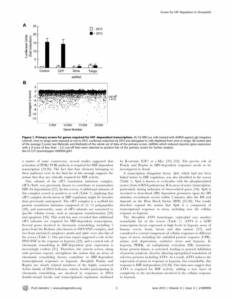
I) The results of the primary screen were highly reproducible
with Z score values (see Materials and Methods) showing a
correlation coefficient of 0.6 between duplications (Figure S1C). A
few dsRNAs rendered less reproducible results (i.e. the duplicates
were more divergent), but nevertheless, were included in the
secondary screen to avoid loosing potentially relevant hits. As
shown in Figure 1B, approximately 97% of the dsRNAs rendered
Z score values of around zero, indicating that, as expected, the
majority of them do not affect HIF-dependent transcription. The
screen was carried out in cells exposed to DFO and therefore, set
up for the identification of positive regulators only. Thus, a
substantial number of genes rendered negative Z score values
(putative activators) but no genes with significant positive Z-score
values (putative inhibitors) were obtained. We decided to define a
Z score cut-off value of 22.5 for a gene to be considered a hit of
the screen (Figure 1B) and, based on this criterion, 603 genes were
initially selected for further analysis (Table S2). Noteworthy, both
sima and tango -the Drosophila HIF-alpha and beta subunits
respectively- scored as positives in this primary selection, with Z
scores of 26.4 and 24.1 respectively, suggesting that the screen is
reliable and has the potential to identify novel genes required for
HRE-dependent transcription. Next, in order to eliminate genes
that presumably interfere with basic cellular functions and prevent
cell viability, the 603 hits were filtered against the results of a
RNAi genome-wide screen for genes required for cell viability,
previously carried out in the same cellular system with the same
library [23]; 311 genes fell in the ‘‘cell viability’’ category, so they
were not pursued further. Open reading frames that have been
predicted but never demonstrated (the ‘‘Sanger collection’’: 67
genes) were also eliminated from the analysis. Thus, after filtration,
the number of positive genes from the primary screen was reduced
to 225 (Table S3).
II) For the secondary screen, we developed a stably transfected
cell line, which contained, along with the HRE-firefly luciferase
reporter, a constitutive actin-Renilla-luciferase element, which was
used to normalize the results (see Materials and Methods). This
phase of the screen was carried out with a second-generation
library (DRSC Validation Library; http://flyrnai.org), which was
designed to eliminate false positives that arise from off-target
effects of the original library [24,25]; this new library includes
more than one dsRNA for most genes (Table S4). Like in the
primary screen, DFO was used as the hypoxic-mimetic agent
(Table S1).
At the secondary screen, those genes that provoked a reduction
of HRE-Luc reporter activity of more than 50% with at least one
of the two dsRNAs were considered as hits. On this basis, 66 genes
scored as positives, and based on the strength of the effect, this set
of genes was further classified into two categories: Group A) Genes
that rendered -with at least one of the corresponding dsRNAs-
over 75% inhibition of HRE-luciferase activity (23 genes), and
group B) Genes that provoked an inhibition of 50–75% of the
activity -with at least one of the corresponding dsRNAs- (43 genes)
(Table S4). As expected, sima and tango were among the hits of the
secondary screen with approximately 96% inhibition.
III) Finally, we carried out a tertiary screen, in which genes that
scored as positives in the secondary screen were tested in cells
exposed to hypoxia (1% O2). All 23 genes that scored in group A
(strong inhibition) were included in the tertiary screen, along with
a selected set of genes from group B (12 genes) that are functionally
related to those from group A. Thus, a total number of 35 genes
were analyzed in hypoxia (Table 1). In this final screen 30 genes,
including sima and tango, scored as positives with at least one of the
two dsRNAs provoking more than 50% inhibition of HRE
reporter activity (Table 1). Genes already known to be required for
the HRE response, such as elements from the PI3K/TOR
pathway [19] and the COP9 signalosome complex [20,21], as well
as genes that were not previously linked to HIF (see below), were
among the hits in this final phase of the screen (Table 1).
Four genes of the PI3K and TOR pathways -PDK1, TOR,
Rheb and Raptor- were among the positive hits. Although it is still
Author Summary
Adaptation of cells to low oxygen (hypoxia) is aphysiological response related to important diseases,including heart attacks, stroke, cancer, and diabetes. Themechanisms that mediate adaptation to hypoxia inhumans are almost identical to those operating in diverseanimal species, including mice, worms, and insects. Themaster regulator of cellular responses to hypoxia is atranscription factor named HIF, which induces a set ofgenes that mediate adaptation to oxygen starvation.Although it is known that regulation of HIF occurs mainlyat the level of protein degradation and transcriptionalcoactivator recruitment, a comprehensive screen for HIFregulators has not been performed before. In this work, wehave conducted an RNAi-based screen of the genome ofthe fruit fly Drosophila melanogaster, searching for genesthat are required for HIF activity. This screen carried out ina cell culture system led to the definition of 30 criticalregulators of HIF, most of which have not been associatedwith hypoxia biology before. The hits of the screenincluded components of chromatin remodeling complex-es, transcription elongation factors, and translationalregulators. Our results open the possibility of performingdetailed studies on HIF regulation that may lead to noveltherapeutic strategies for important human diseases.
Screen for HIF Regulators in Drosophila
PLoS Genetics | www.plosgenetics.org 2 June 2010 | Volume 6 | Issue 6 | e1000994
a matter of some controversy, several studies suggested that
activation of PI3K/TOR pathway is required for HIF-dependent
transcription [19,26]. The fact that four elements belonging to
these pathways were in the final list of hits strongly supports the
notion that they are critically required for HIF activity.
One subunit of the eIF3 translation initiation complex,
eIF3e/Int6, was previously shown to contribute to mammalian
HIF-2a degradation [27]. In this screen, 4 additional subunits of
this complex scored as positives as well (Table 1), implying that
eIF3 complex involvement in HIF regulation might be broader
than previously anticipated. The eIF3 complex is a scaffold for
protein translation initiation composed of 12–13 polypeptides
[28], and noteworthy, some of eIF3 subunits are associated to
specific cellular events, such as oncogenic transformation [29]
and apoptosis [30]. This work has now revealed that additional
eIF3 subunits are required for HIF-dependent transcription.
Several genes involved in chromatin remodeling, including 5
genes from the Brahma (also known as SWI/SNF) complex, and
two from unrelated complexes -pontin and reptin- were also hits of
the screen (Table 1). One previous report suggested a role of the
SWI/SNF in the response to hypoxia [22], and a central role of
chromatin remodeling in HIF-dependent gene expression is
increasingly evident [31]. Therefore, the current screen, along
with previous reports, strengthens the notion that an array of
chromatin remodeling factors contribute to HIF-dependent
transcriptional responses to hypoxia. Drosophila Pontin and
Reptin are closely related members of the highly conserved
AAA+ family of DNA helicases, which, besides participating in
chromatin remodeling, are involved in responses to DNA
double-strand breaks and transcriptional regulation mediated
by b-catenin, E2F1 or c-Myc [32] [33]. The precise role of
Pontin and Reptin in HIF-dependent responses needs to be
investigated in detail.
A transcription elongation factor, Spt6, which had not been
linked before to HIF regulation, was also identified in the screen
(Table 1). Spt6 is known to co-localize with the phosphorylated
(active) form of RNA polymerase II in areas of active transcription,
particularly during induction of stress-related genes [34]. Spt6 is
recruited to heat-shock (HS) dependent promoters upon the HS
stimulus; recruitment occurs within 2 minutes after the HS and
depends on the Heat Shock Factor (HSF) [35,36]. Our results
therefore expand the notion that Spt6 is a component of
transcriptional responses to stress, including now the cellular
response to hypoxia.
The Drosophila ATF4 homologue cryptocephal was another
remarkable hit of the screen (Table 1). ATF4 is a bZIP
transcription factor expressed at high levels in hypoxic areas of
human cervix, brain, breast and skin tumors [37], and
considered a central component of cellular responses to different
types of stress, including the unfolded protein response (UPR),
amino acid deprivation, oxidative stress and hypoxia. In
hypoxia, PERK, an endoplasmic reticulum (ER) transmem-
brane protein kinase, is activated, leading to general inhibition
of protein synthesis, thereby allowing upregulated translation of
selective proteins including ATF4. As a result, ATF4 induces the
expression of genes in response to hypoxia, but remarkably, this
response is HIF-independent [19,38]. Our data now suggest that
ATF4 is required for HIF activity, adding a new layer of
complexity to the mechanisms involved in the cellular response
to hypoxia.
Figure 1. Primary screen for genes required for HIF–dependent transcription. (A) S2-HRE-Luc cells treated with dsRNA against gfp (negativecontrol), sima or tango were exposed or not to DFO. Luciferase induction by DFO was abrogated in cells depleted from sima or tango. (B) Scatter plotof the average Z-score (see Materials and Methods) of the whole set of data of the primary screen. dsRNAs which reduced reporter gene expressionwith a Z-score of less than 22.5 (cut-off line) were selected as positive hits of the primary screen for further analysis.doi:10.1371/journal.pgen.1000994.g001
Screen for HIF Regulators in Drosophila
PLoS Genetics | www.plosgenetics.org 3 June 2010 | Volume 6 | Issue 6 | e1000994
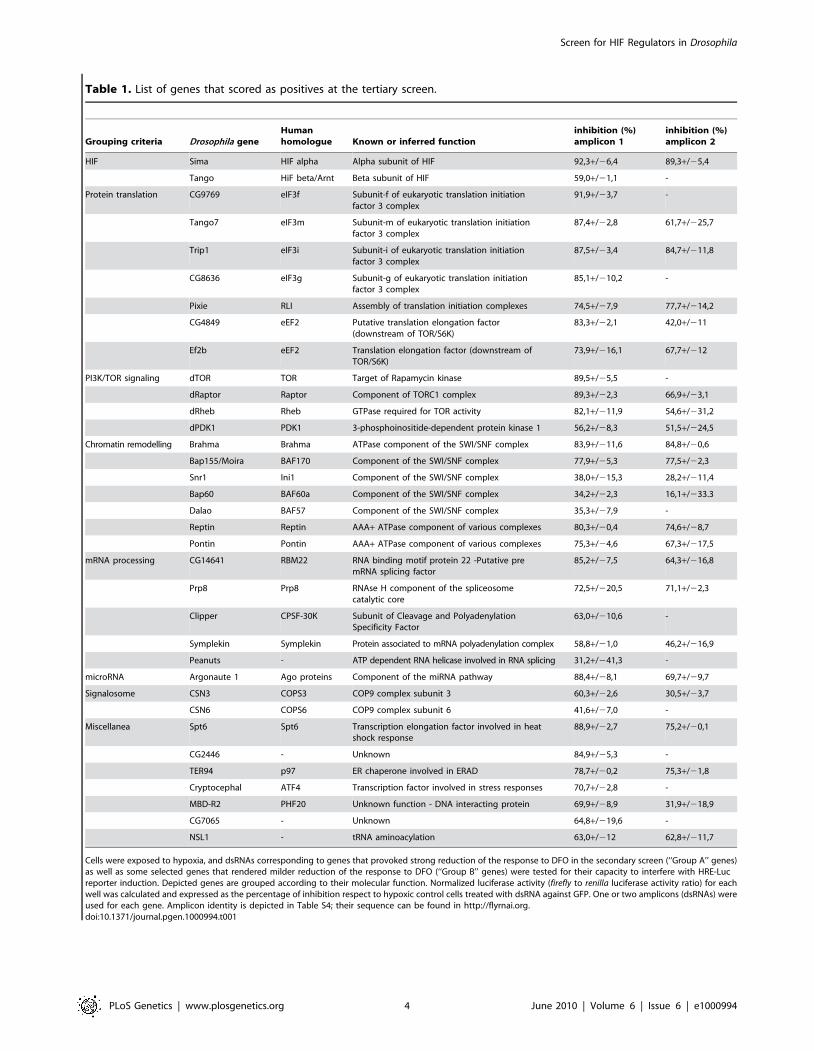
Table 1. List of genes that scored as positives at the tertiary screen.
Grouping criteria Drosophila geneHumanhomologue Known or inferred function
inhibition (%)amplicon 1
inhibition (%)amplicon 2
HIF Sima HIF alpha Alpha subunit of HIF 92,3+/26,4 89,3+/25,4
Tango HiF beta/Arnt Beta subunit of HIF 59,0+/21,1 -
Protein translation CG9769 eIF3f Subunit-f of eukaryotic translation initiationfactor 3 complex
91,9+/23,7 -
Tango7 eIF3m Subunit-m of eukaryotic translation initiationfactor 3 complex
87,4+/22,8 61,7+/225,7
Trip1 eIF3i Subunit-i of eukaryotic translation initiationfactor 3 complex
87,5+/23,4 84,7+/211,8
CG8636 eIF3g Subunit-g of eukaryotic translation initiationfactor 3 complex
85,1+/210,2 -
Pixie RLI Assembly of translation initiation complexes 74,5+/27,9 77,7+/214,2
CG4849 eEF2 Putative translation elongation factor(downstream of TOR/S6K)
83,3+/22,1 42,0+/211
Ef2b eEF2 Translation elongation factor (downstream ofTOR/S6K)
73,9+/216,1 67,7+/212
PI3K/TOR signaling dTOR TOR Target of Rapamycin kinase 89,5+/25,5 -
dRaptor Raptor Component of TORC1 complex 89,3+/22,3 66,9+/23,1
dRheb Rheb GTPase required for TOR activity 82,1+/211,9 54,6+/231,2
dPDK1 PDK1 3-phosphoinositide-dependent protein kinase 1 56,2+/28,3 51,5+/224,5
Chromatin remodelling Brahma Brahma ATPase component of the SWI/SNF complex 83,9+/211,6 84,8+/20,6
Bap155/Moira BAF170 Component of the SWI/SNF complex 77,9+/25,3 77,5+/22,3
Snr1 Ini1 Component of the SWI/SNF complex 38,0+/215,3 28,2+/211,4
Bap60 BAF60a Component of the SWI/SNF complex 34,2+/22,3 16,1+/233.3
Dalao BAF57 Component of the SWI/SNF complex 35,3+/27,9 -
Reptin Reptin AAA+ ATPase component of various complexes 80,3+/20,4 74,6+/28,7
Pontin Pontin AAA+ ATPase component of various complexes 75,3+/24,6 67,3+/217,5
mRNA processing CG14641 RBM22 RNA binding motif protein 22 -Putative premRNA splicing factor
85,2+/27,5 64,3+/216,8
Prp8 Prp8 RNAse H component of the spliceosomecatalytic core
72,5+/220,5 71,1+/22,3
Clipper CPSF-30K Subunit of Cleavage and PolyadenylationSpecificity Factor
63,0+/210,6 -
Symplekin Symplekin Protein associated to mRNA polyadenylation complex 58,8+/21,0 46,2+/216,9
Peanuts - ATP dependent RNA helicase involved in RNA splicing 31,2+/241,3 -
microRNA Argonaute 1 Ago proteins Component of the miRNA pathway 88,4+/28,1 69,7+/29,7
Signalosome CSN3 COPS3 COP9 complex subunit 3 60,3+/22,6 30,5+/23,7
CSN6 COPS6 COP9 complex subunit 6 41,6+/27,0 -
Miscellanea Spt6 Spt6 Transcription elongation factor involved in heatshock response
88,9+/22,7 75,2+/20,1
CG2446 - Unknown 84,9+/25,3 -
TER94 p97 ER chaperone involved in ERAD 78,7+/20,2 75,3+/21,8
Cryptocephal ATF4 Transcription factor involved in stress responses 70,7+/22,8 -
MBD-R2 PHF20 Unknown function - DNA interacting protein 69,9+/28,9 31,9+/218,9
CG7065 - Unknown 64,8+/219,6 -
NSL1 - tRNA aminoacylation 63,0+/212 62,8+/211,7
Cells were exposed to hypoxia, and dsRNAs corresponding to genes that provoked strong reduction of the response to DFO in the secondary screen (‘‘Group A’’ genes)as well as some selected genes that rendered milder reduction of the response to DFO (‘‘Group B’’ genes) were tested for their capacity to interfere with HRE-Lucreporter induction. Depicted genes are grouped according to their molecular function. Normalized luciferase activity (firefly to renilla luciferase activity ratio) for eachwell was calculated and expressed as the percentage of inhibition respect to hypoxic control cells treated with dsRNA against GFP. One or two amplicons (dsRNAs) wereused for each gene. Amplicon identity is depicted in Table S4; their sequence can be found in http://flyrnai.org.doi:10.1371/journal.pgen.1000994.t001
Screen for HIF Regulators in Drosophila
PLoS Genetics | www.plosgenetics.org 4 June 2010 | Volume 6 | Issue 6 | e1000994
Argonaute 1 and the miRNA machinery are necessary forthe transcriptional response to hypoxia
Argonaute 1 (Ago1), a central component of the microRNA
silencing machinery [39] also scored as positive in the screen
(Table 1). Given that little is known about the participation of the
miRNA machinery in HIF regulation, we sought to further
characterize Ago1 involvement in this process. We began by
checking that dsRNA treatments were effective in reducing Ago1
protein levels (Figure 2A), and consistent with this, we confirmed
that the function of the miRNA machinery was impaired (Figure
S2A). To determine if inhibition of HRE-Luc reporter activity
after Ago1 depletion reflects the behavior of endogenous hypoxia-
inducible genes, we examined transcript levels of two well-
established Sima downstream targets: ldh and PHD/fatiga [15].
The two transcripts were strongly upregulated in cells exposed to
hypoxia, and this induction was dramatically reduced in cells
treated with ago1 dsRNA (Figure 2B). In order to assess if Ago1 is
required in the hypoxic response as part of the miRNA pathway,
we silenced other components of the miRNA machinery. dsRNAs
for dicer-1, drosha or gw182 strongly reduced luciferase reporter
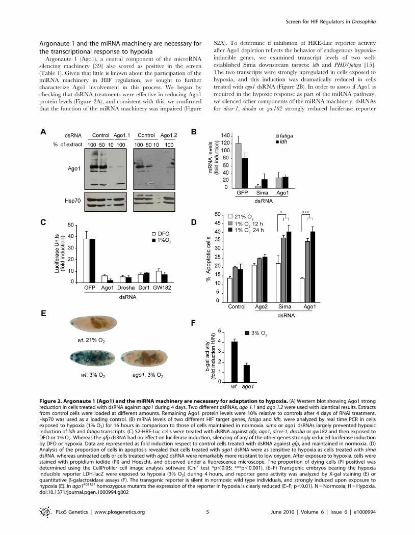
Figure 2. Argonaute 1 (Ago1) and the miRNA machinery are necessary for adaptation to hypoxia. (A) Western blot showing Ago1 strongreduction in cells treated with dsRNA against ago1 during 4 days. Two different dsRNAs, ago 1.1 and ago 1.2 were used with identical results. Extractsfrom control cells were loaded at different amounts. Remaining Ago1 protein levels were 10% relative to controls after 4 days of RNAi treatment.Hsp70 was used as a loading control. (B) mRNA levels of two different HIF target genes, fatiga and ldh, were analyzed by real time PCR in cellsexposed to hypoxia (1% O2) for 16 hours in comparison to those of cells maintained in normoxia. sima or ago1 dsRNAs largely prevented hypoxicinduction of ldh and fatiga transcripts. (C) S2-HRE-Luc cells were treated with dsRNA against gfp, ago1, dicer-1, drosha or gw182 and then exposed toDFO or 1% O2. Whereas the gfp dsRNA had no effect on luciferase induction, silencing of any of the other genes strongly reduced luciferase inductionby DFO or hypoxia. Data are represented as fold induction respect to control cells treated with dsRNA against gfp, and maintained in normoxia. (D)Analysis of the proportion of cells in apoptosis revealed that cells treated with ago1 dsRNA were as sensitive to hypoxia as cells treated with simadsRNA, whereas untreated cells or cells treated with ago2 dsRNA were remarkably more resistant to low oxygen. After exposure to hypoxia, cells werestained with propidium iodide (PI) and Hoescht, and observed under a fluorescence microscope. The proportion of dying cells (PI positive) wasdetermined using the CellProfiler cell image analysis software (Chi2 test *p,0.05; ***p,0.001). (E–F) Transgenic embryos bearing the hypoxiainducible reporter LDH-lacZ were exposed to hypoxia (3% O2) during 4 hours, and reporter gene activity was analyzed by X-gal staining (E) orquantitative b-galactosidase assays (F). The transgenic reporter is silent in normoxic wild type individuals, and strongly induced upon exposure tohypoxia (E). In ago1k08121 homozygous mutants the expression of the reporter in hypoxia is clearly reduced (E–F; p,0.01). N = Normoxia; H = Hypoxia.doi:10.1371/journal.pgen.1000994.g002
Screen for HIF Regulators in Drosophila
PLoS Genetics | www.plosgenetics.org 5 June 2010 | Volume 6 | Issue 6 | e1000994
induction in cells exposed to DFO or hypoxia (Figure 2C),
suggesting that the miRNA pathway is required for the
transcriptional response to hypoxia. Other genes related to ago1,
which have no reported function in the miRNA pathway, were
also evaluated: Depletion of argonaute2, piwi or dicer-2, did not affect
the HRE-response in S2 cells (Figure S2B). It is well known that
HIF play a crucial role in the adaptive response to hypoxia by
controlling the expression of genes that eventually promote cell
survival. Thus, we studied if Ago1 does indeed contribute to cell
viability in hypoxia. As depicted in Figure 2D, cells treated with
ago1 dsRNA and exposed to hypoxia enter apoptosis at a higher
proportion than untreated cultures, or cells treated with control
ago2 dsRNA, suggesting a physiological requirement of Ago1 in
the response to hypoxia. We next sought to test whether Ago1 is
required for the HRE response in vivo. We analyzed the expression
of the hypoxia-responsive transgenic reporter LDH-LacZ [12] in
ago1k08121 mutant embryos. As previously reported, in wild type
embryos LDH-LacZ is silent in normoxia and induced in hypoxia
in a characteristic expression pattern that corresponds to some of
the developing tracheal branches [12] (Figure 2E). In contrast, in
ago1k08121 homozygous embryos, induction of the LDH-LacZ
reporter in hypoxia was much weaker, indicating that Ago1
contributes to HIF/Sima dependent transcription in vivo (Figure 2E
and 2F).
We have recently shown that oxygen-dependent subcellular
localization is an important mechanism of Sima regulation: Sima
shuttles continuously between the nucleus and cytoplasm, and
nuclear export is inhibited in hypoxia [6,40]. To get insights into how
Ago1 depletion affects the transcriptional response to hypoxia, we
studied Sima subcellular localization, and found no differences
between Ago1 mutant embryos and wild type controls (Figure S3).
Next we sought to study if Sima protein accumulation in hypoxic cells
is affected upon Ago1 depletion. As depicted in Figure 3A, hypoxic
induction of Sima protein is clearly reduced in S2 cells treated with
ago1 dsRNA. The next step was to analyze sima transcript levels. Real
time PCR analysis revealed a striking upregulation of sima mRNA
levels upon exposure of the cells to hypoxia, and that ago1 RNAi
treatment inhibited this induction (Figure 3B). These results indicate
that HIF transcriptional induction or mRNA stabilization plays a role
in the Drosophila hypoxic response, and suggests that the miRNA
machinery participates in this regulation.
The above results prompted us to analyze possible changes in the
miRNA machinery in hypoxia: a well known effect of miRNA
dependent translational silencing is the accumulation of Processing
Bodies (PBs), which are cytosolic foci that contain untranslated
mRNAs and proteins, as well as small RNAs involved in translational
silencing [41]. Thus, we explored whether exposure to hypoxia
stimulates accumulation of PBs. As shown in Figure 4A and 4B, a
clear increase of PBs was observed after exposing the cells to hypoxia,
as revealed by anti-DCP1 or anti-Hedls antibody staining [42]. The
accumulation of PBs in hypoxia was transient, reaching a peak
6 hours after transferring the cells to 1% O2 (Figure 4A). To explore if
this effect is related to the miRNA pathway, we analyzed PB
formation in cells depleted of Ago1 or GW182 and exposed to
hypoxia. As shown in Figure 4B, both basal PB levels and induction of
PBs by hypoxia were strongly reduced in these cells. It is unclear
whether PB accumulation per se is required for HIF-dependent
transcription or if alternatively, PB accumulation only reflects the
activity of the miRNA machinery in the hypoxic response.
Taken together, our results suggest that the miRNA pathway
plays a physiological role in cellular responses to hypoxia. Why
does Ago1 depletion prevent sima mRNA induction? Although the
identity of the target molecules that are controlled by the miRNA
machinery is unknown, we can speculate that such unknown
regulators directly or indirectly prevent sima transcriptional
induction or alternatively contribute to sima messenger degrada-
tion (Figure 5). In mammalian cells, HIFa mRNAs are induced by
NF-kB, so that NF-kB regulation plays an important role in the
response to hypoxia [43]. It is not known if in Drosophila an NF-kB
protein is required for sima transcriptional induction. If this was the
case, it should be investigated if an inhibitor of the NF-kB pathway
(i.e. IkB/Cactus) is subjected to miRNA-dependent translational
regulation during adaptation to hypoxia.
Most major HIF regulators including PHDs, the von Hippel-
Lindau tumour suppressor protein (VHL) [2] and factor inhibiting
HIF (FIH) [4] are all inhibitors of the hypoxic response. The
screen we have carried out here was instead focused on positive
regulators of HIF. One remarkable feature of the results we have
obtained is that most of the hits belong to just a few multiprotein
complexes or signaling pathways. These include the PI3K/TOR
pathway (translational regulation), eIF3 and eEF2 complexes
(translational regulation), the COP9 signalosome (protein degra-
dation/translational regulation), and the Brahma/SWI/SNF
complex (chromatin remodeling). Noteworthy, besides genes
belonging to these complexes, other hits of the screen are also
linked to translational control (Ago1) or chromatin remodeling
(Reptin, Pontin). Thus, one central conclusion of the results of this
screen is that translational control and chromatin remodeling are
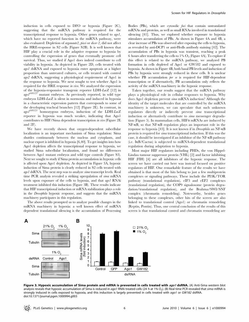
Figure 3. Hypoxic accumulation of Sima protein and mRNA is prevented in cells treated with ago1 dsRNA. (A) Anti-Sima western blotanalysis reveals that hypoxic accumulation of Sima is reduced in ago1 RNAi treated cells (24 h at 1% O2). (B) Real time PCR revealed that sima mRNA isstrongly induced in cells exposed to hypoxia, and this induction is largely prevented in cells treated with ago1 or GW182 dsRNA.doi:10.1371/journal.pgen.1000994.g003
Screen for HIF Regulators in Drosophila
PLoS Genetics | www.plosgenetics.org 6 June 2010 | Volume 6 | Issue 6 | e1000994
two important mechanisms of HIF regulation, whose character-
ization in detail will broaden our understanding of HIF regulation
and the cellular response to hypoxia.
Materials and Methods
Vectors, reporters, and cell cultureThe reporter plasmids HRE- firefly luciferase (HRE-Luc) and
act-renilla luciferase were previously described [15,44]. The
miRNA reporter pAC-miR-12 and CG10011-luc were a gift from
E. Izaurralde [45]. pBLAST (Blasticidine resistance) and pPUR
(Puromycin resistance) vectors were used to generate S2 stable cell
lines. Drosophila Schneider’s lines S2 or S2R+ cells were maintained
at 25uC in Schneider or M3 medium (Sigma), supplemented
with 10% fetal bovine serum (Gibco), 50 units/ml penicillin and
50 mg/ml streptomycin in 25 or 75 cm2 T-flasks (Greiner). Cells
were grown in 12, 24, 96 or 384-well plates (Greiner), during 3
days and treated with 100 mM of DFO (Sigma) or exposed to
hypoxia in a Forma Scientific 3131 incubator.
Synthesis of dsRNA and RNAi treatmentsFor dsRNAs not obtained from the Drosophila RNAi Screening
Center (DRSC), fragments of the genes were amplified by PCR
from cDNA or genomic DNA using T7-tailed oligonucleotides as
primers. dsRNA synthesis was carried out with a T7 Megascript
kit (Ambion) following manufacturer’s instructions. The ‘‘bathing’’
method was utilized to introduce dsRNAs into S2 or S2R+ cells as
previously described [46].
RNAi ScreensFor screening experiments Drosophila S2 cells were maintained at
25uC in Schneider’s medium. The primary screen was carried out
at the Drosophila RNAi Screening Center (DRSC), the secondary and
tertiary screens were performed in our laboratory with dsRNAs
obtained from the DRSC. Primer and amplicon information can
be found at http://flyrnai.org.
Primary screen. Two sets of 58 384-well screening plates
(Costar) containing approximately 0.2 mg of dsRNA per well were
provided by the DRSC (DRSC 1.0 library). Sima or GFP were used as
positive and negative controls, respectively. Three days after plating,
the cells were stimulated with DFO (100 mM) for 20 h and then firefly
luciferase activity was determined using the SteadyGlo reagent
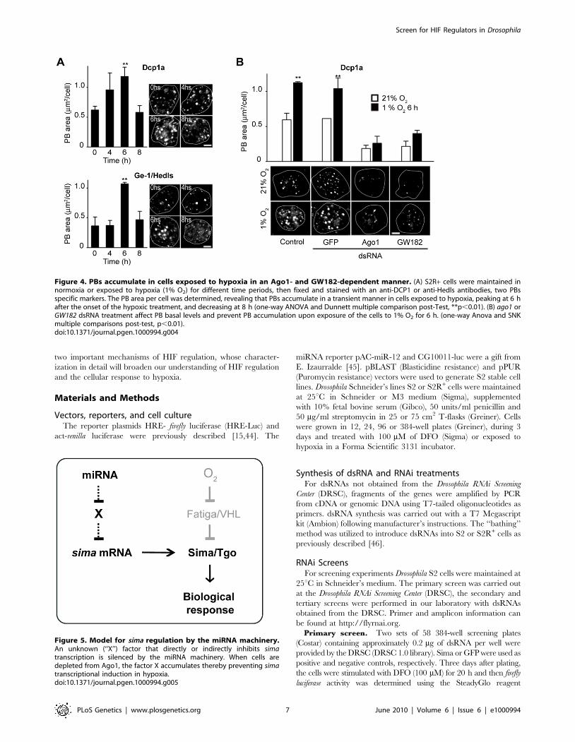
Figure 4. PBs accumulate in cells exposed to hypoxia in an Ago1- and GW182-dependent manner. (A) S2R+ cells were maintained innormoxia or exposed to hypoxia (1% O2) for different time periods, then fixed and stained with an anti-DCP1 or anti-Hedls antibodies, two PBsspecific markers. The PB area per cell was determined, revealing that PBs accumulate in a transient manner in cells exposed to hypoxia, peaking at 6 hafter the onset of the hypoxic treatment, and decreasing at 8 h (one-way ANOVA and Dunnett multiple comparison post-Test, **p,0.01). (B) ago1 orGW182 dsRNA treatment affect PB basal levels and prevent PB accumulation upon exposure of the cells to 1% O2 for 6 h. (one-way Anova and SNKmultiple comparisons post-test, p,0.01).doi:10.1371/journal.pgen.1000994.g004
Figure 5. Model for sima regulation by the miRNA machinery.An unknown (‘‘X’’) factor that directly or indirectly inhibits simatranscription is silenced by the miRNA machinery. When cells aredepleted from Ago1, the factor X accumulates thereby preventing simatranscriptional induction in hypoxia.doi:10.1371/journal.pgen.1000994.g005
Screen for HIF Regulators in Drosophila
PLoS Genetics | www.plosgenetics.org 7 June 2010 | Volume 6 | Issue 6 | e1000994
(Promega). The Z-score value for each well was calculated as the
luciferase activity of the well minus the average of the luciferase activity
of the whole plate divided by the standard deviation of the plate.
Secondary screen. The secondary screen was carried out in
96-well plates. Firefly and Renilla luciferase activities were
determined using the DualGlo reagent (Promega) in a Veritas
Luminometer. Normalized luciferase activity (firefly to renilla
luciferase activity ratio) for each well was calculated as a
percentage of the control wells treated with GFP dsRNA.
Tertiary screen in hypoxia. Fifty-nine dsRNAs from the
DRSC Validation library were used to cover the 35 selected genes.
S2-HRE-Luc cells were incubated with dsRNAs in 96-well plates
as described above and then exposed to hypoxia (1% O2) in a
Forma Scientific 3131 incubator for 20 hours. Firefly and Renilla
luciferase activities were determined and normalized as above.
Real-time PCRTotal RNAs from cells exposed to different treatments were
isolated using the Trizol reagent (Invitrogen). One to 5 mg of total
RNA were used as a template for cDNA synthesis, using the
SuperScript III First Strand Synthesis System for RT-PCR
(Invitrogen). Quantitative real time PCR was performed in the
MX3005P real time PCR instrument (Stratagene, La Jolla, CA)
with Syber Green, the hot start Platinum Taq DNA polymerase
(Invitrogen) and the ROX reference dye (Invitrogen). Primers for
amplifying 100–300 bp of each PCR product were used. PCR
reactions were carried out for 5 min at 95uC followed by 35 cycles
of three-step PCR for 30 seconds at 95uC, 1 min at 60uC, and
1 min at 72uC. Each sample was analyzed in triplicate. The data
were normalized by subtracting the difference of the CT values
between the target gene of interest (Tgene) and that of tubulin
mRNA, thereby obtaining a DCT (Tgene CT 2Tubulin CT).
Relative expression (fold induction) was calculated as
22(SDCT
2CDCT
) where SDCT2CDCT is the difference between
the sample DCT (treated cells) and the control DCT (RNAi GFP
cells). Both target gene and tubulin reactions approached 100%
efficiency as determined by standard curves. PCR products were
analyzed on agarose gels to check that a single band was amplified.
Fly stocksFlies used were yw, ldh- LacZ [12] and yw, ago1k08121/CyO.
b-galactosidase activityWild type or ago1k08121 embryos were exposed to 3% or 21% O2
for 4 h, homogenized in lysis buffer (50 mM Tris HCl [pH 7.8],
2 mM EDTA, 10% glycerol, 2 mM DTT, 1% Triton X-100,
1 mM PMSF) and centrifuged at 2,5006g for 3 min at 4uC.
Enzymatic reactions were carried out by incubating 20 to 50 mg of
protein extract in 180 ml buffer, containing 80 mM Na3PO4
(pH 7.3), 102 mM b-mercaptoethanol, 9 mM MgCl2, and 4 mM
CPRG (Chlorophenol Red b-d-galactopyranoside; Roche Diag-
nostics) at 37uC, and absorbance at 574 nm was recorded at 10,
30, 60, 120, and 180 min time points; color development was
linear throughout this time period. Endogenous background was
subtracted using a heat-inactivated sample.
ImmunofluorescenceFor PB staining either a mouse monoclonal anti-DCP1 antibody
(Abnova) was utilized at a 1:1000 dilution were used or a rabbit
anti-HEDLS antiserum (Bethyl) was used at 1:500 dilution. Images
were acquired in LSM510 Meta confocal microscopes (Carl Zeiss),
using a Plan-Apochromat 636/1.4 oil objective. Equipment
adjustment was assessed by using 1mm Focal Check fluorescent
microspheres (Molecular Probes). Pictures were exported to Adobe
Photoshop software for cropping. Neither filters nor gamma-
adjustments were applied. PB number and size in mm2 were
determined with the ‘‘Analyze Particles’’ tool of the Image J
software (NIH) in randomly selected micrographs.
Supporting Information
Figure S1 HRE-luciferase reporter induction in cells exposed to
hypoxia or DFO. (A) Schematic representation of the HIF-responsive
firefly luciferase reporter element used in this study (HRE-Luc). A
dimerized regulatory sequence derived from the murine lactate
dehydrogenase enhancer was cloned upstream of a firefly luciferase
gene in a pGL3 plasmid bearing a fly hsp70 minimal promoter. Each
51 bp sequence contains two HIF responsive elements (HREs) and
one cyclic AMP responsive element (CRE). (B) S2-HRE-luc cells were
seeded in 96-wells tissue culture plates (16104 cells per well), grown
for 3 days, and stimulated with DFO (100 mM), or exposed to
hypoxia (1% O2) for 20 hours. Strong induction of luciferase activity
was observed in cells stimulated with DFO or hypoxia. Results are
expressed as fold induction of luciferase activity respect to normoxic
untreated cells. (C) Scatter plot of the duplicate results (Z-scores; see
Materials and Methods) of the primary screen, showing the overall
reproducibility of the data.
Found at: doi:10.1371/journal.pgen.1000994.s001 (0.03 MB PDF)
Figure S2 miRNAs and the response to hypoxia. (A) Upper
panel, schematic representation of the miRNA reporter CG10011-
luc; the miR-12 miRNA binds to the 39 UTR of the luciferase
mRNA, thereby inhibiting translation. Over-expression of miR-12
is therefore expected to provoke strong inhibition of translation.
Lower panel, S2 cells were co-transfected with the CG10011-luc
reporter and the pAC-miR-12 over-expression plasmid, or with an
empty vector (pAC) as a control, and exposed to ago1 or gfp dsRNA
treatments during 4 days. miR-12 over-expression inhibits 80% of
luciferase expression in the control cells treated with gfp dsRNA,
whereas in cells depleted from ago1 (ago1.1 or ago1.2 dsRNAs) miR-
12 over-expression failed to inhibit luciferase expression to a large
extent. (B) S2-HRE-luc cells were treated with dsRNA against gfp
(control), sima, ago1, ago2, piwi, or dicer-2, grown during 4-8 days,
and stimulated with DFO (100 mM). Cells depleted from ago1 or
sima showed strong reduction of reporter activity, whereas cells
depleted from ago2, dicer-2, or piwi exhibited normal induction of
the reporter upon DFO exposure.
Found at: doi:10.1371/journal.pgen.1000994.s002 (0.01 MB PDF)
Figure S3 Regulation of Sima subcellular localization is not
affected in Ago1 homozygous mutant embryos. We have analyzed
Sima subcellular localization in en-Gal4/UAS-sima transgenic
embryos carrying a homozygous mutation in the Ago1 locus
(ago1k0208), and compared with Sima localization in en-Gal4/UAS-
sima wild type individuals. The analysis was carried out as we
reported previously (Dekanty et al., 2005) [15]. Three categories of
Sima subcellular localization were defined for quantitative
purposes: ‘‘Nuclear’’ (black color), ‘‘Ubiquitous’’ (grey) and
‘‘Cytoplasmic’’ (white). The Ago1 mutation does not impinge on
Sima subcellular localization neither in normoxia nor in hypoxia.
Found at: doi:10.1371/journal.pgen.1000994.s003 (0.01 MB PDF)
Table S1 Summary of the three phases of the overall screen for
genes required for HIF activity.
Found at: doi:10.1371/journal.pgen.1000994.s004 (0.04 MB PDF)
Table S2 Results of the primary screen carried out in cells
exposed to DFO are shown. The screen was performed in
duplicate; genes in which at least one of the two Z scores values
Screen for HIF Regulators in Drosophila
PLoS Genetics | www.plosgenetics.org 8 June 2010 | Volume 6 | Issue 6 | e1000994
was below 22.5 are depicted in the table. Under this criterion, 603
genes scored as positives in this initial phase of the screen.
Found at: doi:10.1371/journal.pgen.1000994.s005 (0.43 MB PDF)
Table S3 The data obtained at the primary screen (Table S2)
were filtered against the results of a cell viability screen previously
carried out at the DRSC (Boutros et al. 2004) [23]. Sequences
from the ‘‘Sanger collection’’ were also eliminated from the study;
the 225 genes that remained as positive hits of the primary screen
are depicted.
Found at: doi:10.1371/journal.pgen.1000994.s006 (0.27 MB PDF)
Table S4 The secondary screen was also carried out in cells
exposed to DFO. A second-generation library (DRSC 2.0 library)
was used, in which most genes are represented by more than one
dsRNA. Normalized luciferase activity (firefly/renilla luciferase
activity ratio) for each well was calculated and expressed as a
percentage of the inhibition respect to control cells treated with
dsRNA against GFP that were exposed to DFO. The screen was
carried out in duplicate and the mean percentage of inhibition is
depicted.
Found at: doi:10.1371/journal.pgen.1000994.s007 (0.26 MB PDF)
Acknowledgments
We wish to thank the Drosophila RNAi screening center at Harvard Medical
School, where the primary screen was conducted, Elisa Izaurralde for
reagents, the Bloomington stock center for fly stocks, and the Wappner lab
for discussions.
Author Contributions
Conceived and designed the experiments: AD GLB PW. Performed the
experiments: AD NMR APB MGT CCL JIPP. Analyzed the data: AD
NMR APB MGT CCL JIPP GLB PW. Wrote the paper: AD GLB PW.
References
1. Semenza GL (2007) Hypoxia-inducible factor 1 (HIF-1) pathway. Sci STKE2007: cm8.
2. Maxwell PH, Wiesener MS, Chang GW, Clifford SC, Vaux EC, et al. (1999)
The tumour suppressor protein VHL targets hypoxia-inducible factors foroxygen-dependent proteolysis. Nature 399: 271–275.
3. Hewitson KS, McNeill LA, Riordan MV, Tian YM, Bullock AN, et al. (2002)
Hypoxia-inducible factor (HIF) asparagine hydroxylase is identical to factor
inhibiting HIF (FIH) and is related to the cupin structural family. J Biol Chem277: 26351–26355.
4. Lando D, Peet DJ, Gorman JJ, Whelan DA, Whitelaw ML, et al. (2002) FIH-1 is
an asparaginyl hydroxylase enzyme that regulates the transcriptional activity ofhypoxia-inducible factor. Genes Dev 16: 1466–1471.
5. Kallio PJ, Okamoto K, O’Brien S, Carrero P, Makino Y, et al. (1998) Signal
transduction in hypoxic cells: inducible nuclear translocation and recruitment ofthe CBP/p300 coactivator by the hypoxia-inducible factor-1alpha. Embo J 17:
6573–6586.
6. Romero NM, Irisarri M, Roth P, Cauerhff A, Samakovlis C, et al. (2008)
Regulation of the Drosophila hypoxia-inducible factor alpha Sima by CRM1-dependent nuclear export. Mol Cell Biol 28: 3410–3423.
7. Jaakkola P, Mole DR, Tian YM, Wilson MI, Gielbert J, et al. (2001) Targeting
of HIF-alpha to the von Hippel-Lindau ubiquitylation complex by O2-regulatedprolyl hydroxylation. Science 292: 468–472.
8. Ivan M, Kondo K, Yang H, Kim W, Valiando J, et al. (2001) HIFalpha targeted
for VHL-mediated destruction by proline hydroxylation: implications for O2sensing. Science 292: 464–468.
9. Epstein AC, Gleadle JM, McNeill LA, Hewitson KS, O’Rourke J, et al. (2001)
C. elegans EGL-9 and Mammalian Homologs Define a Family of Dioxygenases
that Regulate HIF by Prolyl Hydroxylation. Cell 107: 43–54.
10. Bruick RK, McKnight SL (2001) A Conserved Family of Prolyl-4-HydroxylasesThat Modify HIF. Science 294: 1337–1340.
11. Romero NM, Dekanty A, Wappner P (2007) Cellular and developmental
adaptations to hypoxia: a Drosophila perspective. Methods Enzymol 435:123–144.
12. Lavista-Llanos S, Centanin L, Irisarri M, Russo DM, Gleadle JM, et al. (2002)
Control of the Hypoxic Response in Drosophila melanogaster by the BasicHelix-Loop-Helix PAS Protein Similar. Mol Cell Biol 22: 6842–6853.
13. Centanin L, Ratcliffe PJ, Wappner P (2005) Reversion of lethality and growth
defects in Fatiga oxygen-sensor mutant flies by loss of Hypoxia-Inducible Factor-
alpha/Sima. EMBO Rep 6: 1070–1075.
14. Arquier N, Vigne P, Duplan E, Hsu T, Therond PP, et al. (2006) Analysis of thehypoxia-sensing pathway in Drosophila melanogaster. Biochem J 393: 471–
480.
15. Dekanty A, Lavista-Llanos S, Irisarri M, Oldham S, Wappner P (2005) Theinsulin-PI3K/TOR pathway induces a HIF-dependent transcriptional response
in Drosophila by promoting nuclear localization of HIF-{alpha}/Sima. J CellSci 118: 5431–5441.
16. Keith B, Simon MC (2007) Hypoxia-inducible factors, stem cells, and cancer.
Cell 129: 465–472.
17. Rankin EB, Giaccia AJ (2008) The role of hypoxia-inducible factors in
tumorigenesis. Cell Death Differ 15: 678–685.
18. Melillo G (2007) Hypoxia-inducible factor 1 inhibitors. Methods Enzymol 435:385–402.
19. Wouters BG, Koritzinsky M (2008) Hypoxia signalling through mTOR and the
unfolded protein response in cancer. Nat Rev Cancer 8: 851–864.
20. Miyauchi Y, Kato M, Tokunaga F, Iwai K (2008) The COP9/signalosomeincreases the efficiency of von Hippel-Lindau protein ubiquitin ligase-mediated
hypoxia-inducible factor-alpha ubiquitination. J Biol Chem 283: 16622–16631.
21. Bemis L, Chan DA, Finkielstein CV, Qi L, Sutphin PD, et al. (2004) Distinctaerobic and hypoxic mechanisms of HIF-alpha regulation by CSN5. Genes Dev
18: 739–744.
22. Wang F, Zhang R, Beischlag TV, Muchardt C, Yaniv M, et al. (2004) Roles ofBrahma and Brahma/SWI2-related gene 1 in hypoxic induction of the
erythropoietin gene. J Biol Chem 279: 46733–46741.
23. Boutros M, Kiger AA, Armknecht S, Kerr K, Hild M, et al. (2004) Genome-wide
RNAi analysis of growth and viability in Drosophila cells. Science 303: 832–835.
24. Kulkarni MM, Booker M, Silver SJ, Friedman A, Hong P, et al. (2006) Evidenceof off-target effects associated with long dsRNAs in Drosophila melanogaster
cell-based assays. Nat Methods 3: 833–838.
25. Flockhart I, Booker M, Kiger A, Boutros M, Armknecht S, et al. (2006)FlyRNAi: the Drosophila RNAi screening center database. Nucleic Acids Res
34: D489–494.
26. Bernardi R, Guernah I, Jin D, Grisendi S, Alimonti A, et al. (2006) PML inhibitsHIF-1alpha translation and neoangiogenesis through repression of mTOR.
Nature 442: 779–785.
27. Chen L, Uchida K, Endler A, Shibasaki F (2007) Mammalian tumor suppressor
Int6 specifically targets hypoxia inducible factor 2 alpha for degradation byhypoxia- and pVHL-independent regulation. J Biol Chem 282: 12707–
12716.
28. Hinnebusch AG (2006) eIF3: a versatile scaffold for translation initiationcomplexes. Trends Biochem Sci 31: 553–562.
29. Zhang L, Smit-McBride Z, Pan X, Rheinhardt J, Hershey JW (2008) An
oncogenic role for the phosphorylated h-subunit of human translation initiationfactor eIF3. J Biol Chem 283: 24047–24060.
30. Lin YM, Chen YR, Lin JR, Wang WJ, Inoko A, et al. (2008) eIF3k regulates
apoptosis in epithelial cells by releasing caspase 3 from keratin-containing
inclusions. J Cell Sci 121: 2382–2393.
31. Fish JE, Yan MS, Matouk CC, St Bernard R, Ho JJ, Jr., et al. (2010) Hypoxicrepression of endothelial nitric-oxide synthase transcription is coupled with
eviction of promoter histones. J Biol Chem 285: 810–826.
32. Bauer A, Chauvet S, Huber O, Usseglio F, Rothbacher U, et al. (2000) Pontin52and reptin52 function as antagonistic regulators of beta-catenin signalling
activity. Embo J 19: 6121–6130.
33. Gallant P (2007) Control of transcription by Pontin and Reptin. Trends Cell Biol17: 187–192.
34. Kaplan CD, Morris JR, Wu C, Winston F (2000) Spt5 and spt6 are associated
with active transcription and have characteristics of general elongation factors in
D. melanogaster. Genes Dev 14: 2623–2634.
35. Andrulis ED, Guzman E, Doring P, Werner J, Lis JT (2000) High-resolutionlocalization of Drosophila Spt5 and Spt6 at heat shock genes in vivo: roles in
promoter proximal pausing and transcription elongation. Genes Dev 14:2635–2649.
36. Saunders A, Werner J, Andrulis ED, Nakayama T, Hirose S, et al. (2003)
Tracking FACT and the RNA polymerase II elongation complex throughchromatin in vivo. Science 301: 1094–1096.
37. Ameri K, Lewis CE, Raida M, Sowter H, Hai T, et al. (2004) Anoxic induction
of ATF-4 through HIF-1-independent pathways of protein stabilization in
human cancer cells. Blood 103: 1876–1882.
38. Koumenis C (2006) ER stress, hypoxia tolerance and tumor progression. CurrMol Med 6: 55–69.
39. Sontheimer EJ, Carthew RW (2004) Molecular biology. Argonaute journeys into
the heart of RISC. Science 305: 1409–1410.
40. Irisarri M, Lavista-Llanos S, Romero NM, Centanin L, Dekanty A, et al. (2009)Central role of the oxygen-dependent degradation domain of Drosophila
HIFalpha/Sima in oxygen-dependent nuclear export. Mol Biol Cell 20:3878–3887.
Screen for HIF Regulators in Drosophila
PLoS Genetics | www.plosgenetics.org 9 June 2010 | Volume 6 | Issue 6 | e1000994

41. Eulalio A, Huntzinger E, Izaurralde E (2008) GW182 interaction with
Argonaute is essential for miRNA-mediated translational repression and mRNAdecay. Nat Struct Mol Biol 15: 346–353.
42. Eulalio A, Behm-Ansmant I, Schweizer D, Izaurralde E (2007) P-body formation
is a consequence, not the cause, of RNA-mediated gene silencing. Mol Cell Biol27: 3970–3981.
43. Belaiba RS, Bonello S, Zahringer C, Schmidt S, Hess J, et al. (2007) Hypoxiaup-regulates hypoxia-inducible factor-1alpha transcription by involving phos-
phatidylinositol 3-kinase and nuclear factor kappaB in pulmonary artery smooth
muscle cells. Mol Biol Cell 18: 4691–4697.
44. Darlington TK, Wager-Smith K, Ceriani MF, Staknis D, Gekakis N, et al.
(1998) Closing the circadian loop: CLOCK-induced transcription of its owninhibitors per and tim. Science 280: 1599–1603.
45. Rehwinkel J, Behm-Ansmant I, Gatfield D, Izaurralde E (2005) A crucial role for
GW182 and the DCP1:DCP2 decapping complex in miRNA-mediated genesilencing. Rna 11: 1640–1647.
46. Clemens JC, Worby CA, Simonson-Leff N, Muda M, Maehama T, et al. (NatlAcad Sci U S A 2000 Jun 6) Use of double-stranded RNA interference in
Drosophila cell lines to dissect signal transduction pathways. Proc 97:
6499–6503.
Screen for HIF Regulators in Drosophila
PLoS Genetics | www.plosgenetics.org 10 June 2010 | Volume 6 | Issue 6 | e1000994
Related Documents