Dramatic Reversal of Derangements in Muscle Metabolism and Diastolic Left Ventricular Function after Bariatric Surgery Joshua G. Leichman, MD 1 , Erik B. Wilson, MD 2 , Terry Scarborough, MD 2 , David Aguilar, MD 1,3 , Charles C. Miller III, PhD 4 , Sherman Yu, MD 2 , Mohamed F. Algahim, BSc 1 , Manuel Reyes, MD 1 , Frank G. Moody, MD 2 , and Heinrich Taegtmeyer, MD, DPhil 1 1The University of Texas Health Science Center at Houston, School of Medicine, Department of Internal Medicine 2The University of Texas Health Science Center at Houston, School of Medicine, Department of Surgery 3Baylor College of Medicine, Department of Internal Medicine 4The University of Texas Health Science Center at Houston, School of Medicine, Department of Cardiovascular and Thoracic Surgery Abstract Objective—To define muscle metabolic and cardiovascular changes following surgical intervention in clinically severe obese patients. Background—Obesity is a state of metabolic dysregulation which may lead to maladaptive changes in heart and skeletal muscle, including insulin resistance and heart failure. In a prospective longitudinal study 43 consecutive patients were subjected to metabolic profiling, skeletal muscle biopsies and resting echocardiograms at baseline as well as three and nine months after bariatric surgery. Results—Body mass index (BMI) decreased [mean changes (95% CI): 7.7 kg/m 2 (6.70–8.89) at 3 months and 5.6 kg/m 2 (4.45–6.80), p<0.0001 at 9 months after surgery], with restoration of insulin sensitivity and decreases in plasma leptin at the same time points. Concurrent with these changes were dramatic decreases in skeletal muscle transcript levels of stearoyl CoA desaturase (SCD) and pyruvate dehydrogenase kinase-4 (PDK4) at three and nine months (p<0.0001, for both), and a significant decrease in peroxisome proliferation activated receptor alpha (PPAR-α) regulated genes at nine months. Left ventricular relaxation impairment, assessed by tissue Doppler imaging, normalized nine months after surgery. Conclusions—Weight loss results in the reversal of systemic and muscle metabolic derangements and is accompanied by a normalization of left ventricular diastolic function. Address correspondence and request for reprints to: Heinrich Taegtmeyer, MD, DPhil, Professor of Medicine, University of Texas Houston Medical School, Department of Medicine/Cardiology, 6431 Fannin, MSB 1.246, Houston, TX 77030, Tel: 713 500-6569, Fax: 713 500-0637, E-mail: [email protected]. Publisher's Disclaimer: This is a PDF file of an unedited manuscript that has been accepted for publication. As a service to our customers we are providing this early version of the manuscript. The manuscript will undergo copyediting, typesetting, and review of the resulting proof before it is published in its final citable form. Please note that during the production process errors may be discovered which could affect the content, and all legal disclaimers that apply to the journal pertain. Authors Verifications: None of the authors of this work have any financial conflicts of interest to disclose. All authors had access to the data and a role in writing the manuscript. Clinical Trials Registry: ClinicalTrials.gov Identifier: NCT00178633; http:/www.clinicaltrials.gov NIH Public Access Author Manuscript Am J Med. Author manuscript; available in PMC 2009 November 1. Published in final edited form as: Am J Med. 2008 November ; 121(11): 966–973. doi:10.1016/j.amjmed.2008.06.033. NIH-PA Author Manuscript NIH-PA Author Manuscript NIH-PA Author Manuscript

Welcome message from author
This document is posted to help you gain knowledge. Please leave a comment to let me know what you think about it! Share it to your friends and learn new things together.
Transcript
Dramatic Reversal of Derangements in Muscle Metabolism andDiastolic Left Ventricular Function after Bariatric Surgery
Joshua G. Leichman, MD1, Erik B. Wilson, MD2, Terry Scarborough, MD2, David Aguilar,MD1,3, Charles C. Miller III, PhD4, Sherman Yu, MD2, Mohamed F. Algahim, BSc1, ManuelReyes, MD1, Frank G. Moody, MD2, and Heinrich Taegtmeyer, MD, DPhil11The University of Texas Health Science Center at Houston, School of Medicine, Department of InternalMedicine
2The University of Texas Health Science Center at Houston, School of Medicine, Department of Surgery
3Baylor College of Medicine, Department of Internal Medicine
4The University of Texas Health Science Center at Houston, School of Medicine, Department ofCardiovascular and Thoracic Surgery
AbstractObjective—To define muscle metabolic and cardiovascular changes following surgicalintervention in clinically severe obese patients.
Background—Obesity is a state of metabolic dysregulation which may lead to maladaptivechanges in heart and skeletal muscle, including insulin resistance and heart failure. In a prospectivelongitudinal study 43 consecutive patients were subjected to metabolic profiling, skeletal musclebiopsies and resting echocardiograms at baseline as well as three and nine months after bariatricsurgery.
Results—Body mass index (BMI) decreased [mean changes (95% CI): 7.7 kg/m2 (6.70–8.89) at 3months and 5.6 kg/m2 (4.45–6.80), p<0.0001 at 9 months after surgery], with restoration of insulinsensitivity and decreases in plasma leptin at the same time points. Concurrent with these changeswere dramatic decreases in skeletal muscle transcript levels of stearoyl CoA desaturase (SCD) andpyruvate dehydrogenase kinase-4 (PDK4) at three and nine months (p<0.0001, for both), and asignificant decrease in peroxisome proliferation activated receptor alpha (PPAR-α) regulated genesat nine months. Left ventricular relaxation impairment, assessed by tissue Doppler imaging,normalized nine months after surgery.
Conclusions—Weight loss results in the reversal of systemic and muscle metabolic derangementsand is accompanied by a normalization of left ventricular diastolic function.
Address correspondence and request for reprints to: Heinrich Taegtmeyer, MD, DPhil, Professor of Medicine, University of TexasHouston Medical School, Department of Medicine/Cardiology, 6431 Fannin, MSB 1.246, Houston, TX 77030, Tel: 713 500-6569, Fax:713 500-0637, E-mail: [email protected]'s Disclaimer: This is a PDF file of an unedited manuscript that has been accepted for publication. As a service to our customerswe are providing this early version of the manuscript. The manuscript will undergo copyediting, typesetting, and review of the resultingproof before it is published in its final citable form. Please note that during the production process errors may be discovered which couldaffect the content, and all legal disclaimers that apply to the journal pertain.Authors Verifications: None of the authors of this work have any financial conflicts of interest to disclose. All authors had access tothe data and a role in writing the manuscript.Clinical Trials Registry: ClinicalTrials.gov Identifier: NCT00178633; http:/www.clinicaltrials.gov
NIH Public AccessAuthor ManuscriptAm J Med. Author manuscript; available in PMC 2009 November 1.
Published in final edited form as:Am J Med. 2008 November ; 121(11): 966–973. doi:10.1016/j.amjmed.2008.06.033.
NIH
-PA Author Manuscript
NIH
-PA Author Manuscript
NIH
-PA Author Manuscript
Keywordsobesity; metabolism; bariatric surgery; leptin; stearoyl Co-A desaturase; pyruvatev dehydrogenasekinase-4; echocardiography
IntroductionObesity is a state of metabolic dysregulation with an increased risk for premature death anddisability.1 The maladaptive response of obesity has been attributed to metabolic changescaused by either increased energy substrate supply or decreased energy substrate utilization,or both. The body’s initial response is to store the energy excess in adipose tissue, to increasecardiac mass in response to the hemodynamic load associated with obesity, and to upregulategene expression to account for changing metabolic demands. It has been suggested that, whenthe storage capacity of the adipocyte is exhausted there is “spillover” to other organs of thebody.2 Lipid accumulation in non-adipose tissue is a hallmark of dysregulated local andsystemic metabolism,3, 4 of insulin resistance,5 and possibly also of the development of heartfailure.6, 7
Obesity has reached epidemic proportions and obesity-related illness consumes billions ofdollars in health care.8 In addition to premature heart disease, obesity is linked to cancer, sleepapnea and birth defects. Treatment of obesity should therefore be an important determinant fora normal life expectancy and quality of life. Weight reduction can ameliorate many of the co-morbid conditions associated with obesity.9 Indeed, bariatric surgery for severe obesity isassociated with survival benefits.10–12 The metabolic changes that accompany significantweight loss are paramount for these improved outcomes. Here we proposed that there is apotential for reversal of the maladaptive processes of obesity with sustained weight loss afterbariatric surgery.
Material and MethodsSubject Selection
We offered participation to patients of any race/ethnicity from the University of Texas HoustonBariatric Surgery Center (UTHBSC), who met the inclusion criteria for bariatric surgeryoutlined previously.13 Exclusion criteria were coronary artery disease, ischemiccardiomyopathy, severe peripheral vascular disease, a current history of smoking, pregnancy,or an age of less than 18 years. The study was approved by the Committee for the ProtectionHuman Subjects at The University of Texas Health Science Center – Houston. All patientssigned an informed consent form prior to enrollment in the study.
Study ProtocolWe prospectively enrolled 43 consecutive patients. The study protocol has been publishedpreviously.13 Briefly, patients underwent a physical examination, anthropometricmeasurements, a 12-lead electrocardiogram, and resting echocardiogram. At the time ofsurgery a skeletal muscle biopsy was performed. All studies were repeated at three and ninemonths post-operatively.
SurgeryPatients were offered two types of bariatric surgery. The patients chose which surgery theypreferred to have, as this is the current standard practice in the United States.14 The majority(n=30) chose laparoscopic small pouch gastric bypass (SPGB) with a Roux-en Y procedure;
Leichman et al. Page 2
Am J Med. Author manuscript; available in PMC 2009 November 1.
NIH
-PA Author Manuscript
NIH
-PA Author Manuscript
NIH
-PA Author Manuscript
the others (n=13) chose a laparoscopic adjustable gastric banding (LAGB) procedure. Bothprocedures are well described in the literature.15, 16
Patient Enrollment and Follow-upOf the 43 patients enrolled six were lost to follow-up at nine months. Reasons for the loss tofollow-up were either inability to contact the patients or the patients’ stated unwillingness tocontinue with the study. Of the 6 patients who were lost to follow-up, 5 underwent SPGB and1 underwent LAGB. Of the remaining 37 patients (86%) there were six individuals withinadequate baseline echocardiograms and four other patients who had limited studies becauseof technical difficulties due to body habitus. All patients had skeletal muscle biopsies atbaseline but only 25 completed the follow-up at three and nine months. Reasons for the lowercompletion rate of the skeletal muscle biopsies were fear of pain, a stated discomfort duringthe procedure, or (in two instances) our inability to obtain an adequate sample for analysis.
BiopsiesAt the time of surgery, and at three and nine months after surgery, a percutaneous biopsy ofthe vastus lateralis was obtained using a 6 G × 4.75-inch biopsy needle (Popper and Sons, NewHyde Park, NY). Tissue samples were immediately placed in liquid nitrogen and stored at −80°C until analyzed.
HistologyOil-red-O staining was performed on skeletal muscle sections by the Department of Pathologyat The University of Texas Medical School at Houston using standard procedures.Photomicrographs of (10x) stained sections were taken on a Zeiss Axiophoto microscope usinga Leitz Microlumina digital camera. Oil-red-O staining was quantified using Image Pro Plussoftware with color cube-based selection criteria to ensure that only stained regions werecounted as described previously.6 We examined four sections for each patient at each of thetime points. Results are expressed as a percentage of stained area [arbitrary units (AU)].
Quantitative RT-PCRRNA was extracted from skeletal muscle biopsies by standard methods, and RNAconcentrations were measured spectrophotometrically.17 Transcript levels were measured byreverse transcription followed by real-time quantitative PCR as described before.17 Wefocused on enzymes of fatty acid metabolism, especially those regulated by the peroxisomeproliferation activated receptor alpha (PPAR-α), including: carnityl palmitoyl transferase 1(CPT-1), medium chain acetyl CoA dehydrogenase (MCAD), uncoupling protein 3 (UCP3),pyruvate dehydrogenase kinase 4 (PDK4). We also analyzed gene transcript levels for humanstearoyl CoA desaturase (SCD). The nucleotide sequences for probes as well as forward andreverse primers for the quantitative PCR assays have been published previously 18 with theexceptions of SCD (forward primer, 5′-TGGTGATGTTCCAGAGGAGGTACT-3′; reverseprimer, 5′-AACGAACACACTGTTTTGAAAAGTTT-3′; and probe, 5′-FAMCCTGGCTTGCTGATGATGTGCTTCA-TAMRA3). Transcript levels werenormalized to two internal controls and referenced to total RNA content.
Statistical AnalysesStatistical analyses were performed with SPSS 14.0 (SPSS Inc., Chicago, IL). Significancelevels were set at α=0.05. We evaluated all of the study variables for conformation to normalityusing Q-Q plots, skewness and kurtosis statistics. Significantly nonnormal variables weretransformed prior to analysis. Independent sample t-tests were performed to evaluatedifferences in outcomes between the patients who underwent laparoscopic adjustable gastricbanding and those patients who underwent laparoscopic small pound gastric bypass. Repeated
Leichman et al. Page 3
Am J Med. Author manuscript; available in PMC 2009 November 1.
NIH
-PA Author Manuscript
NIH
-PA Author Manuscript
NIH
-PA Author Manuscript
measures ANOVA were performed to evaluate the effects at three and nine months post-operatively. Effect of surgery was assessed as a variable between subjects with the repeatedmeasures ANOVA. Data are expressed as mean values plus or minus the standard error of themean and as the change in mean values from baseline to three months post-operatively andfrom three months to nine months post-operatively with 95% confidence intervals. Pearsoncorrelation coefficients were prepared to evaluate the univariate relationships.
ResultsPre-operative Findings
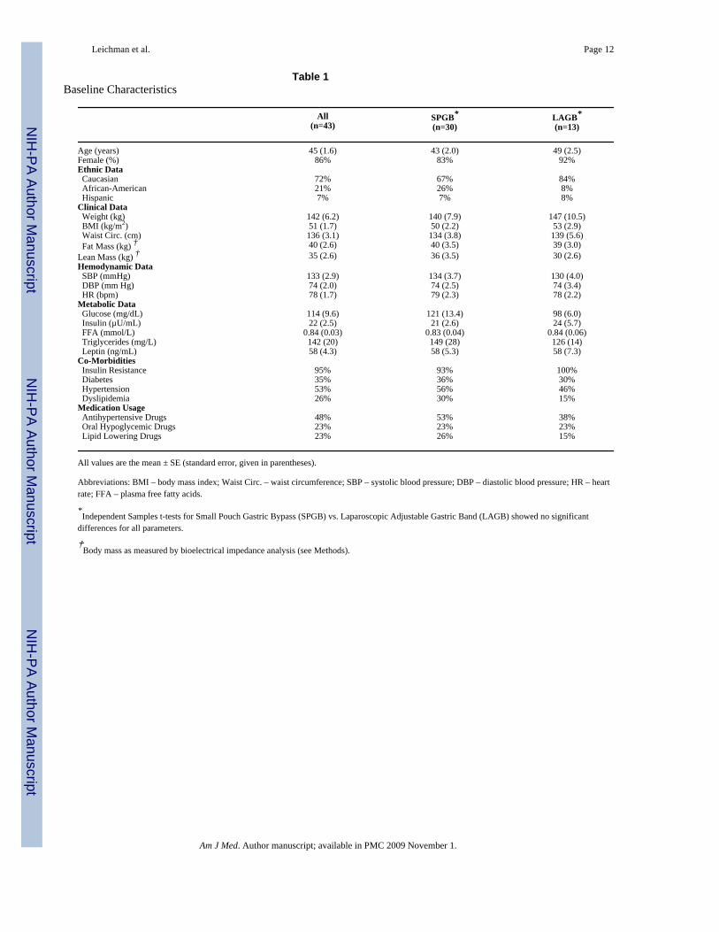
Table 1 lists the baseline characteristics of all patients, for the patients undergoing small pouchgastric bypass and for the patients undergoing laparoscopic adjustable gastric banding. Therewere no significant differences in the baseline characteristics between the two surgical groupsfor any of the variables measured. Patients had more fat mass than lean body mass at baseline,as measured by bioelectrical impedance analysis (Table 1). Mean blood pressure and heartrates were in the normal range. Almost all of the patients met criteria for insulin resistance, butonly 35% of the patients had frank diabetes mellitus. Fasting plasma free fatty acid levels wereelevated, and leptin levels were almost three times higher than the reference range.
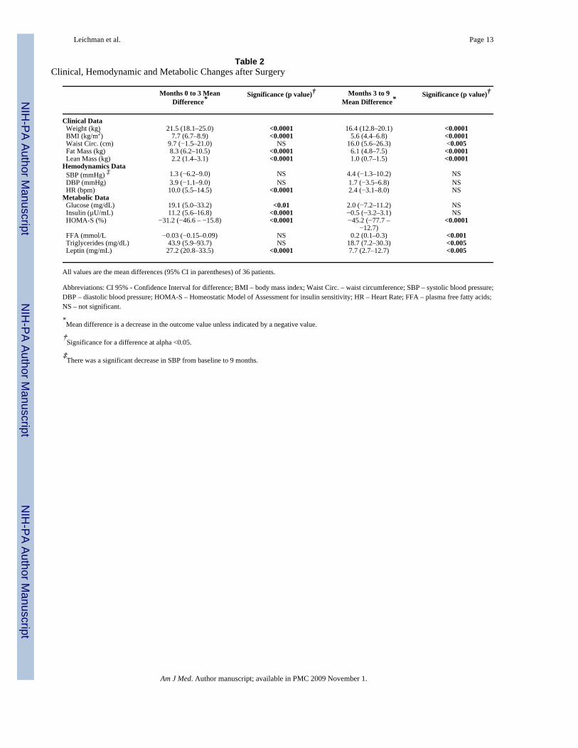
Post-operative ChangesWeight and Hemodynamic Parameters—For practical purposes we chose to analyzepatients who underwent laparoscopic small pouch gastric bypass and those who selectedlaparoscopic adjustable gastric banding as one group. Table 2 shows parameters at three andnine months after surgery. Weight, BMI, waist circumference and fat mass decreased at a fasterrate during the first three months than during the subsequent six months. Fat mass changed toa greater extent than lean mass (Table 2). Lean mass decreased to a greater extent during thefirst three months compared to the subsequent six months (0.73 kg/month in the first threemonths vs 0.16 kg/month during the last six months).
Hemodynamics and Systemic Metabolism—Table 2 also lists the changes inhemodynamic measurements and metabolism after surgery. Concurrent with the decrease inblood pressure was a 33% reduction in the use of anti-hypertensive drugs at three and ninemonths compared to baseline. A decrease in glucose, insulin, and leptin concentrations wasobserved early in the weight loss period, whereas the decrease in plasma FFA was apparentonly at nine months after surgery. The decrease in leptin correlated with the loss of weightfrom baseline to three months and three months to nine months post-operatively (r=0.3, p=0.03and r=0.64, p<0.0001, respectively). Despite the greater decreases in leptin and fat mass earlyon, the correlation between the changes in leptin and fat mass was stronger later on (r=0.53,p=0.001). Glucose and insulin concentrations rapidly decreased after surgery and thus insulinsensitivity (HOMA-S) began to normalize at three months or earlier (three months was the firsttime point we assessed). HOMA-S continued to improve and was in the normal range by ninemonths post-operatively (Table 2). This improvement in insulin sensitivity occurred despitethe relatively small decreases in glucose and insulin concentrations at nine months. It is afunction of the non-linear nature of the HOMA model.19
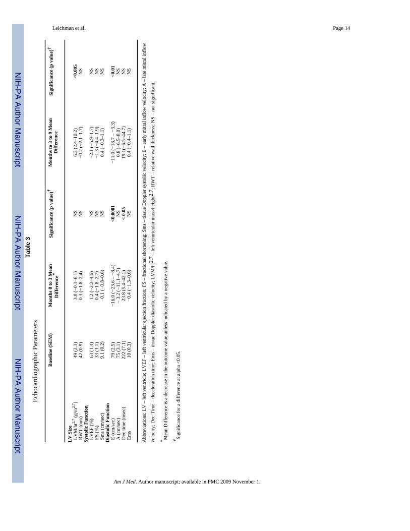
Echocardiography—Left ventricular mass was increased compared to normal 20 anddecreased after three months of weight loss (Table 3). At baseline, i.e. before surgery, 42% ofthe cohort exhibited left ventricular relaxation impairment based on tissue Doppler imaging(Ems) 21 with a mean velocity of 8.3 cm/sec (data not shown). There were no significantdifferences in any other baseline characteristics between those with diastolic dysfunction andthose without (data not shown). Compared to baseline velocities Ems increased at three monthsand nine months after surgery [Mean change (CI 95%): 1.9 cm/sec (0.52 – 3.4), p =0.011 and
Leichman et al. Page 4
Am J Med. Author manuscript; available in PMC 2009 November 1.
NIH
-PA Author Manuscript
NIH
-PA Author Manuscript
NIH
-PA Author Manuscript
1.2 cm/sec (0.32–2.1)]. Similar trends were seen with more load dependent variables ofdiastolic function such as early mitral inflow (E) and deceleration time (DecT) in the diastolicdysfunction group (Table 3).
Histology—Figure 1 shows oil-red-O stains of vastus latralis biopsy samples at 0, 3, and 9months after surgery. There was a significant decrease in oil-red-O staining at nine months.[4.2 AU (−2.4 – 10.9), p<0.2, and 8.7 AU (2.4 – 15.1), p<0.009, comparing baseline to threemonths, and three months to nine months, respectively]. The decrease in oil-red-O stainingbetween three and nine months correlated significantly with a decrease in weight and fat mass(r=0.57, p<0.004; and r=0.44, p<0.03, respectively). There was also a borderline associationbetween oil-red-O intensity and plasma free fatty acid levels (r=0.39, p<0.059).
Skeletal Muscle Gene Expression—Transcript levels for metabolic enzymes are shownin Figure 2. SCD transcript levels decreased dramatically at three months after surgery (Figure2A) and remained so at nine months. PDK4 transcript levels also decreased rapidly andsignificantly at three months after surgery and continued to decrease at nine months aftersurgery (Figure 2B). The expression of the PPAR-α regulated genes (with the exception ofPDK4) only decreased nine months post-operatively (Figure 2C), and the changes in the PPAR-α regulated genes were highly correlative with each other (data not shown). These changeswere concurrent with the decrease in the plasma FFA concentrations between three and ninemonths post-operatively.
Impact of the Type of Surgery on Outcomes—The two types of surgery, laparoscopicsmall pound gastric bypass and laparoscopic adjustable gastric banding, included in this studyproduce weight loss by different mechanisms; laparoscopic small pound gastric bypass is arestrictive and malabsorptive process whereas laparoscopic adjustable gastric banding is arestrictive process. We analyzed the outcomes for each group in this study in order to ascertainwhether there was an effect produced by the surgery type. Not surprisingly there was asignificant difference in the rate of weight loss between the two groups at both three and ninemonths [Mean weight loss at three and nine months (SEM): laparoscopic small pound gastricbypass 23 kg (1.9) and 20 kg (1.8), laparoscopic adjustable gastric banding 15 kg (2.0) and 9kg (2.8), p = 0.01 and 0.003, respectively for three and nine months]. However there was nosignificant contribution of the type of surgery to the overall improvement in weight by analysisof variance. Furthermore, there was no overall effect of the type of surgery on other outcomesmeasured, such as systemic metabolism, cardiac function, hemodynamics or skeletal musclegene expression (data not shown). These results suggest that outcomes in the early phase ofweight loss are independent of the type of surgery.
DiscussionThe main findings of our study are that weight loss induced by surgery is accompanied by areversal of insulin resistance and dramatic changes in skeletal muscle metabolism. The formerfindings were expected but the latter findings are new. Adipose tissue is an active endocrineorgan and an increased adipose mass is associated with insulin resistance and alterations offatty acid oxidation by complex mechanisms.22, 23 These effects are mediated by adiposederived hormones and cytokines (e.g. leptin and TNF-α) which exert control over skeletalmuscle metabolic gene expression. We speculate that the decrease in fat mass led to a decreasein leptin concentrations and to improvements in systemic insulin sensitivity. The mostsurprising findings were profound changes in skeletal muscle metabolic gene expression.Expression of SCD and PDK4 decreased very early in the weight loss process, while the PPAR-α regulated gene expression decreased later in weight loss, at the same time plasma FFAconcentrations decreased. The changes may be responsible for the reversal of the maladaptiveprocesses associated with obesity.
Leichman et al. Page 5
Am J Med. Author manuscript; available in PMC 2009 November 1.
NIH
-PA Author Manuscript
NIH
-PA Author Manuscript
NIH
-PA Author Manuscript
Skeletal muscle histologyOil-red-O stains intra-myocellular triglycerides and increased intensity suggests increased lipidstorage in non-adipose tissue, most likely as the result of increased fatty acid supply, decreasedrates of triglyceride export or hydrolysis, and/or decreased rates of fatty acid oxidation. Thedecrease in oil-red-O staining is associated with a normalization of substrate and hormonelevels in the circulation as well as transcript levels in the tissue, suggesting a concerted programof reversed lipotoxicity.
Skeletal muscle gene expressionOther surprising findings were profound changes in skeletal muscle metabolic gene expression.Expression of SCD and PDK4 decreased very early in the weight loss process, while the PPAR-α regulated gene expression decreased later in weight loss, at the same time plasma FFAconcentrations decreased. This is the first demonstration of gene expression changes early afterbariatric surgery. These changes have important implications for the possible mechanisms forthe reversal of the maladaptive processes in non-adipose tissue.
The dramatic changes in transcript levels of steroyl-CoA desaturase (SCD) are especiallyrelevant because SCD is the rate limiting enzyme responsible for converting saturated fattyacids into monounsaturated fatty acids, the main precursors of triglycerides.24 SCD is knownto be increased in the skeletal muscle of obese individuals and may lead to abnormal lipidpartitioning.25 The marked decrease in SCD gene expression post-operatively suggests achange in the flux of fatty acid metabolism, moving from esterification towards beta-oxidation.This result is supported by a sustained expression of the key genes responsible for fatty acidoxidation, the PPAR-α regulated genes and the increased concentrations of plasma FFA. Leptinmediates lipid oxidation through the inhibition of SCD,26 which may result in an increase insaturated fatty acids and their oxidation.
Systemic glucose and insulin concentrations also decreased resulting in improved systemicinsulin sensitivity early in weight loss. As weight loss continued at nine months after surgeryinsulin sensitivity normalized in a majority of the cohort. Skeletal muscle gene expression ofpyruvate dehydrogenase kinase-4 (PDK-4) decreased by 83% from the baseline expression atthree months and 92% at nine months. PDK-4 phosphorylates the pyruvate dehydrogenasecomplex, and is an important inhibitory regulator of glucose oxidation.27 PDK-4 is regulatedby several factors including insulin and fatty acid concentrations.28, 29
These findings are interesting because in the state increased FFA concentrations or insulinresistance, PDK-4 expression is generally increased, limiting glucose oxidation. Our data atthree months suggest a mechanism independent of systemic concentrations of FFA or insulin,as the cause for the decreased expression of PDK-4. We suspect that the change is linked toimproved insulin signaling, because there was no change in PPAR-α gene expression at thistime point and PDK-4 expression has been shown to act independently of PPAR-α expression.30 Our findings are corroborated with another study demonstrating a decrease in PDK-4expression three years after bilio-pancreatic diversion for weight loss.31
Left ventricular diastolic functionWe speculate that the improvement in dysregulation of skeletal muscle metabolism may alsohave a correlate in improved function of cardiac muscle. Obesity is associated withderangements in left ventricular diastolic function.32 More importantly, diastolic dysfunctionis an independent risk for increased morbidity and mortality.33, 34 We demonstrated animprovement in a load independent measure of diastology, the septal mitral annular velocity,as measured by tissue Doppler imaging. Although we have previously demonstrated an inverserelationship between plasma FFA and diastolic function in obese individuals,35 in this study
Leichman et al. Page 6
Am J Med. Author manuscript; available in PMC 2009 November 1.
NIH
-PA Author Manuscript
NIH
-PA Author Manuscript
NIH
-PA Author Manuscript
we were not able to demonstrate any significant correlations between the change in Ems andother measured outcomes. The inability to detect a relationship may be due to the small numberof subjects in this cohort.
However, there are experimental data to suggest a role in FFA metabolism affecting leftventricular contractile function. Animal models of PPAR-α over-expression36 as well asanimals treated with PPAR agonists37, 38 show evidence of cardiomyopathy and worseningheart failure. Furthermore, animal models of PPAR-α knockout demonstrate a cardioprotectivephenotype compared to overexpression of PPAR-α.39 Diastolic function in our cohort showsan early and sustained improvement in those with dysfunction and it is tempting to speculatethat this change may be influenced by the improved FFA metabolism, influenced by leptin,SCD, and PPAR-α gene expression.
LimitationsWe are excited about the findings but we are also aware of the following limitations. First, thisis a small prospective, longitudinal observational study and we can only infer a relationshipbetween the outcomes, but not prove a causal relationship. Larger studies using externalcontrols need to be done to help define true relationships between gene skeletal geneexpression, systemic metabolism, and contractile function of the heart. Secondly, geneexpression does not necessarily reflect protein content or activity. This is a shortcomingencountered in all gene expression studies and further work is needed to establish metabolic,structural and functional correlates.
ConclusionsNon-pharmacologic weight loss induced by bariatric surgery results in an early reversal of themaladaptive responses to obesity. Leptin resistance and insulin resistance reverse, leading toimproved systemic metabolism as well as skeletal muscle gene expression. It is possible thatthese mechanisms may also exert a positive effect on left ventricular diastolic function. Theimplications of these findings for freedom from co-morbidities of obesity remain to besubstantiated by the long-term follow-up of the post-surgical patients.
AcknowledgmentsThe study was supported by the National Heart, Lung, and Blood Institute of the US Public Health Service(R01HL73162). None of the authors of this work have any financial conflicts of interest to disclose. The authors alsoacknowledge the following contributors: Carol Wolin-Riklin, RN, University of Texas at Houston General ClinicalResearch Center, and Charles Majka, BS, University of Texas Houston Medical School for data collection; RebeccaL. Salazar, BS, University of Texas Houston Medical School Department of Medicine, for technical assistance. Wethank Roxy A. Tate for expert editorial help.
REFERENCES1. Adams KF, Schatzkin A, Harris TB, et al. Overweight, obesity, and mortality in a large prospective
cohort of persons 50 to 71 years old. N Engl J Med 2006;355:763–778. [PubMed: 16926275]2. Friedman JJ. Fat in all the wrong places. Nature 2002;415:268–269. [PubMed: 11796987]3. Unger R, Zhou Y, Orci L. Regulation of fatty acid homeostasis in cells: novel role of leptin. Proc Natl
Acad Sci U S A 1999;96:2327–2332. [PubMed: 10051641]4. Young ME, Guthrie PH, Razeghi P, et al. Impaired long-chain fatty acid oxidation and contractile
dysfunction in the obese Zucker rat heart. Diabetes 2002;51:2587–2595. [PubMed: 12145175]5. Shulman GI. Cellular mechanisms of insulin resistance. J Clin Invest 2000;106:171–176. [PubMed:
10903330]6. Sharma S, Adrogue JV, Golfman L, et al. Intramyocardial lipid accumulation in the failing human
heart resembles the lipotoxic rat heart. FASEB J 2004;18:1692–1700. [PubMed: 15522914]
Leichman et al. Page 7
Am J Med. Author manuscript; available in PMC 2009 November 1.
NIH
-PA Author Manuscript
NIH
-PA Author Manuscript
NIH
-PA Author Manuscript
7. Kenchaiah S, Evans JC, Levy D, et al. Obesity and the risk of heart failure. N Engl J Med 2002;347:305–313. [PubMed: 12151467]
8. Finkelstein EA, Fiebelkorn IC, Wang G. National medical spending attributable to overweight andobesity: how much, and who's paying? Health Aff (Millwood). 2003Suppl WebExclusives:W3-219-226.
9. Sjostrom L, Lindroos AK, Peltonen M, et al. Lifestyle, diabetes, and cardiovascular risk factors 10years after bariatric surgery. N Engl J Med 2004;351:2683–2693. [PubMed: 15616203]
10. Sjostrom L, Narbro K, Sjostrom CD, et al. Effects of bariatric surgery on mortality in Swedish obesesubjects. N Engl J Med 2007;357:741–752. [PubMed: 17715408]
11. Adams TD, Gress RE, Smith SC, et al. Long-term mortality after gastric bypass surgery. N Engl JMed 2007;357:753–761. [PubMed: 17715409]
12. Bray GA. The missing link - lose weight, live longer. N Engl J Med 2007;357:818–820. [PubMed:17715415]
13. Leichman JG, Aguilar D, King TM, et al. Improvements in systemic metabolism, anthropometrics,and left ventricular geometry 3 months after bariatric surgery. Surg Obes Relat Dis 2006;2:592–599.[PubMed: 17138229]
14. Buchwald H. Bariatric surgery for morbid obesity: health implications for patients, healthprofessionals, and third-party payers. J Am Coll Surg 2005;200:593–604. [PubMed: 15804474]
15. Higa KD, Boone KB, Ho T, Davies OG. Laparoscopic Roux-en-Y gastric bypass for morbid obesity:technique and preliminary results of our first 400 patients. Arch Surg 2000;135:1029–1033.[PubMed: 10982506]discussion 1033-1024.
16. Fielding GA, Allen JW. A step-by-step guide to placement of the LAP-BAND adjustable gastricbanding system. Am J Surg 2002;184:26S–30S. [PubMed: 12527347]
17. Razeghi P, Young ME, Alcorn JL, Moravec CS, Frazier OH, Taegtmeyer H. Metabolic geneexpression in fetal and failing human heart. Circulation 2001;104:2923–2931. [PubMed: 11739307]
18. Razeghi P, Sharma S, Ying J, et al. Atrophic remodeling of the heart in vivo simultaneously activatespathways of protein synthesis and degradation. Circulation 2003;108:2536–2541. [PubMed:14610007]
19. Levy JC, Matthews DR, Hermans MP. Correct homeostasis model assessment (HOMA) evaluationuses the computer program. Diabetes Care 1998;21:2191–2192. [PubMed: 9839117]
20. Liu JE, Roman MJ, Pini R, Schwartz JE, Pickering TG, Devereux RB. Cardiac and arterial targetorgan damage in adults with elevated ambulatory and normal office blood pressure. Ann Intern Med1999;131:564–572. [PubMed: 10523216]
21. Munagala VK, Jacobsen SJ, Mahoney DW, Rodeheffer RJ, Bailey KR, Redfield MM. Associationof newer diastolic function parameters with age in healthy subjects: a population-based study. J AmSoc Echocardiogr 2003;16:1049–1056. [PubMed: 14566298]
22. Kahn BB, Flier JS. Obesity and insulin resistance. J Clin Invest 2000;106:473–481. [PubMed:10953022]
23. Lelliott C, Vidal-Puig AJ. Lipotoxicity, an imbalance between lipogenesis de novo and fatty acidoxidation. Int J Obes Relat Metab Disord 2004;28:S22–S28. [PubMed: 15592482]
24. Enoch HG, Strittmatter P. Role of tyrosyl and arginyl residues in rat liver microsomal stearylcoenzymeA desaturase. Biochemistry 1978;17:4927–4932. [PubMed: 31169]
25. Hulver MW, Berggren JR, Carper MJ, et al. Elevated stearoyl-CoA desaturase-1 expression in skeletalmuscle contributes to abnormal fatty acid partitioning in obese humans. Cell Metab 2005;2:251–261.[PubMed: 16213227]
26. Cohen P, Miyazaki M, Socci ND, et al. Role for stearoyl-CoA desaturase-1 in leptin-mediated weightloss. Science 2002;297:240–243. [PubMed: 12114623]
27. Bowker-Kinley MM, Davis WI, Wu P, Harris RA, Popov KM. Evidence for existence of tissue-specific regulation of the mammalian pyruvate dehydrogenase complex. Biochem J 1983;329(Pt 1):191–196. [PubMed: 9405293]
28. Wu P, Sato J, Zhao Y, Jaskiewicz J, Popov KM, Harris RA. Starvation and diabetes increase theamount of pyruvate dehydrogenase kinase isoenzyme 4 in rat heart. Biochem J 1998;329:197–201.[PubMed: 9405294]
Leichman et al. Page 8
Am J Med. Author manuscript; available in PMC 2009 November 1.
NIH
-PA Author Manuscript
NIH
-PA Author Manuscript
NIH
-PA Author Manuscript
29. Fuller SJ, Randle PJ. Reversible phosphorylation of pyruvate dehydrogenase in rat skeletal-musclemitochondria. Effects of starvation and diabetes. Biochem J 1984;219:635–646. [PubMed: 6331393]
30. Holness MJ, Smith ND, Bulmer K, Hopkins T, Gibbons GF, Sugden MC. Evaluation of the role ofperoxisome-proliferator-activated receptor alpha in the regulation of cardiac pyruvate dehydrogenasekinase 4 protein expression in response to starvation, high-fat feeding and hyperthyroidism. BiochemJ 2002;364:687–694. [PubMed: 12049632]
31. Rosa G, Di Rocco P, Manco M, et al. Reduced PDK4 expression associates with increased insulinsensitivity in postobese patients. Obes Res 2003;11:176–182. [PubMed: 12582211]
32. Peterson LR, Waggoner AD, Schechtman KB, et al. Alterations in left ventricular structure andfunction in young healthy obese women: assessment by echocardiography and tissue Dopplerimaging. J Am Coll Cardiol 2004;43:1399–1404. [PubMed: 15093874]
33. Bella JN, Palmieri V, Roman MJ, et al. Mitral ratio of peak early to late diastolic filling velocity asa predictor of mortality in middle-aged and elderly adults: the Strong Heart Study. Circulation2002;105:1928–1933. [PubMed: 11997279]
34. Smith GL, Masoudi FA, Vaccarino V, Radford MJ, Krumholz HM. Outcomes in heart failure patientswith preserved ejection fraction: mortality, readmission, and functional decline. J Am Coll Cardiol2003;41:1510–1518. [PubMed: 12742291]
35. Leichman JG, Aguilar D, King TM, Vlada A, Reyes M, Taegtmeyer H. Association of plasma freefatty acids and left ventricular diastolic function in patients with clinically severe obesity. Am J ClinNutr 2006;84:336–341. [PubMed: 16895880]
36. Finck BN, Han X, Courtois M, et al. A critical role for PPARalpha-mediated lipotoxicity in thepathogenesis of diabetic cardiomyopathy: modulation by dietary fat content. Proc Natl Aad Sci U.S.A2003;100:1226–1231.
37. Young ME, Laws FA, Goodwin GW, Taegtmeyer H. Reactivation of peroxisome proliferator-activated receptor alpha is associated with contractile dysfunction in hypertrophied rat heart. J BiolChem 2001;276:44390–44395. [PubMed: 11574533]
38. Vikramadithyan RK, Hirata K, Yagyu H, et al. Peroxisome proliferator-activated receptor agonistsmodulate heart function in transgenic mice with lipotoxic cardiomyopathy. J Pharmacol Exp Ther2005;313:586–593. [PubMed: 15671204]
39. Sambandam N, Morabito D, Wagg C, Finck BN, Kelly DP, Lopaschuk GD. Chronic activation ofPPARalpha is detrimental to cardiac recovery after ischemia. Am J Physiol Heart Circ Physiol2006;290:H87–H95. [PubMed: 16155108]
Leichman et al. Page 9
Am J Med. Author manuscript; available in PMC 2009 November 1.
NIH
-PA Author Manuscript
NIH
-PA Author Manuscript
NIH
-PA Author Manuscript
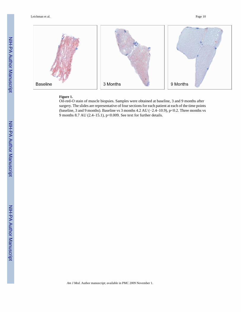
Figure 1.Oil-red-O stain of muscle biopsies. Samples were obtained at baseline, 3 and 9 months aftersurgery. The slides are representative of four sections for each patient at each of the time points(baseline, 3 and 9 months). Baseline vs 3 months 4.2 AU (−2.4–10.9), p<0.2. Three months vs9 months 8.7 AU (2.4–15.1), p<0.009. See text for further details.
Leichman et al. Page 10
Am J Med. Author manuscript; available in PMC 2009 November 1.
NIH
-PA Author Manuscript
NIH
-PA Author Manuscript
NIH
-PA Author Manuscript
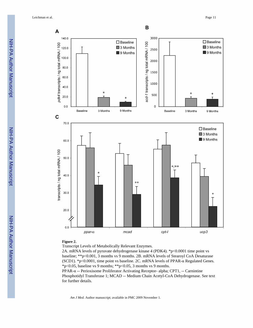
Figure 2.Transcript Levels of Metabolically Relevant Enzymes.2A. mRNA levels of pyruvate dehydrogenase kinase 4 (PDK4). *p<0.0001 time point vsbaseline; **p<0.001, 3 months vs 9 months. 2B. mRNA levels of Stearoyl CoA Desaturase(SCD1). *p<0.0001, time point vs baseline. 2C. mRNA levels of PPAR-α Regulated Genes.*p<0.05, baseline vs 9 months; **p<0.05, 3 months vs 9 months.PPAR-α -- Perioxisome Proliferator Activating Receptor- alpha; CPT1, -- CarnintinePhosphotidyl Transferase 1; MCAD -- Medium Chain Acetyl-CoA Dehydrogenase. See textfor further details.
Leichman et al. Page 11
Am J Med. Author manuscript; available in PMC 2009 November 1.
NIH
-PA Author Manuscript
NIH
-PA Author Manuscript
NIH
-PA Author Manuscript
NIH
-PA Author Manuscript
NIH
-PA Author Manuscript
NIH
-PA Author Manuscript
Leichman et al. Page 12
Table 1Baseline Characteristics
All(n=43)
SPGB*(n=30)
LAGB*(n=13)
Age (years) 45 (1.6) 43 (2.0) 49 (2.5)Female (%) 86% 83% 92%Ethnic Data Caucasian 72% 67% 84% African-American 21% 26% 8% Hispanic 7% 7% 8%Clinical Data Weight (kg) 142 (6.2) 140 (7.9) 147 (10.5) BMI (kg/m2) 51 (1.7) 50 (2.2) 53 (2.9) Waist Circ. (cm) 136 (3.1) 134 (3.8) 139 (5.6) Fat Mass (kg) † 40 (2.6) 40 (3.5) 39 (3.0)Lean Mass (kg) † 35 (2.6) 36 (3.5) 30 (2.6)Hemodynamic Data SBP (mmHg) 133 (2.9) 134 (3.7) 130 (4.0) DBP (mm Hg) 74 (2.0) 74 (2.5) 74 (3.4) HR (bpm) 78 (1.7) 79 (2.3) 78 (2.2)Metabolic Data Glucose (mg/dL) 114 (9.6) 121 (13.4) 98 (6.0) Insulin (µU/mL) 22 (2.5) 21 (2.6) 24 (5.7) FFA (mmol/L) 0.84 (0.03) 0.83 (0.04) 0.84 (0.06) Triglycerides (mg/L) 142 (20) 149 (28) 126 (14) Leptin (ng/mL) 58 (4.3) 58 (5.3) 58 (7.3)Co-Morbidities Insulin Resistance 95% 93% 100% Diabetes 35% 36% 30% Hypertension 53% 56% 46% Dyslipidemia 26% 30% 15%Medication Usage Antihypertensive Drugs 48% 53% 38% Oral Hypoglycemic Drugs 23% 23% 23% Lipid Lowering Drugs 23% 26% 15%
All values are the mean ± SE (standard error, given in parentheses).
Abbreviations: BMI – body mass index; Waist Circ. – waist circumference; SBP – systolic blood pressure; DBP – diastolic blood pressure; HR – heartrate; FFA – plasma free fatty acids.
*Independent Samples t-tests for Small Pouch Gastric Bypass (SPGB) vs. Laparoscopic Adjustable Gastric Band (LAGB) showed no significant
differences for all parameters.
†Body mass as measured by bioelectrical impedance analysis (see Methods).
Am J Med. Author manuscript; available in PMC 2009 November 1.
NIH
-PA Author Manuscript
NIH
-PA Author Manuscript
NIH
-PA Author Manuscript
Leichman et al. Page 13
Table 2Clinical, Hemodynamic and Metabolic Changes after Surgery
Months 0 to 3 MeanDifference* Significance (p value)† Months 3 to 9
Mean Difference* Significance (p value)†
Clinical Data Weight (kg) 21.5 (18.1–25.0) <0.0001 16.4 (12.8–20.1) <0.0001 BMI (kg/m2) 7.7 (6.7–8.9) <0.0001 5.6 (4.4–6.8) <0.0001 Waist Circ. (cm) 9.7 (−1.5–21.0) NS 16.0 (5.6–26.3) <0.005 Fat Mass (kg) 8.3 (6.2–10.5) <0.0001 6.1 (4.8–7.5) <0.0001 Lean Mass (kg) 2.2 (1.4–3.1) <0.0001 1.0 (0.7–1.5) <0.0001Hemodynamics Data SBP (mmHg) ‡ 1.3 (−6.2–9.0) NS 4.4 (−1.3–10.2) NS DBP (mmHg) 3.9 (−1.1–9.0) NS 1.7 (−3.5–6.8) NS HR (bpm) 10.0 (5.5–14.5) <0.0001 2.4 (−3.1–8.0) NSMetabolic Data Glucose (mg/dL) 19.1 (5.0–33.2) <0.01 2.0 (−7.2–11.2) NS Insulin (µU/mL) 11.2 (5.6–16.8) <0.0001 −0.5 (−3.2–3.1) NS HOMA-S (%) −31.2 (−46.6 – −15.8) <0.0001 −45.2 (−77.7 –
−12.7)<0.0001
FFA (mmol/L −0.03 (−0.15–0.09) NS 0.2 (0.1–0.3) <0.001 Triglycerides (mg/dL) 43.9 (5.9–93.7) NS 18.7 (7.2–30.3) <0.005 Leptin (mg/mL) 27.2 (20.8–33.5) <0.0001 7.7 (2.7–12.7) <0.005
All values are the mean differences (95% CI in parentheses) of 36 patients.
Abbreviations: CI 95% - Confidence Interval for difference; BMI – body mass index; Waist Circ. – waist circumference; SBP – systolic blood pressure;DBP – diastolic blood pressure; HOMA-S – Homeostatic Model of Assessment for insulin sensitivity; HR – Heart Rate; FFA – plasma free fatty acids;NS – not significant.
*Mean difference is a decrease in the outcome value unless indicated by a negative value.
†Significance for a difference at alpha <0.05.
‡There was a significant decrease in SBP from baseline to 9 months.
Am J Med. Author manuscript; available in PMC 2009 November 1.
NIH
-PA Author Manuscript
NIH
-PA Author Manuscript
NIH
-PA Author Manuscript
Leichman et al. Page 14Ta
ble
3Ec
hoca
rdio
grap
hic
Para
met
ers
Bas
elin
e (S
EM
)M
onth
s 0 to
3 M
ean
Diff
eren
ce*
Sign
ifica
nce
(p v
alue
)†M
onth
s to
3 to
9 M
ean
Diff
eren
ceSi
gnifi
canc
e (p
val
ue)†
LV
Siz
e L
VM
/ht2.
7 (g/m
2.7 )
49 (2
.3)
3.0
(−0.
1–6.
1)N
S6.
3 (2
.4–1
0.2)
<0.0
05 R
WT
(mm
)42
(0.9
)0.
3 (−
1.8–
2.4)
NS
−0.2
(−2.
1–1.
7)N
SSy
stol
ic F
unct
ion
LV
EF (%
)63
(1.4
)1.
2 (−
2.2–
4.6)
NS
−2.1
(−5.
9–1.
7)N
S F
S (%
)33
(1.1
)0.
4 (−
1.8–
2.7)
NS
−1.3
(−4.
4–1.
9)N
S S
ms (
cm/s
ec)
9.1
(0.2
)−0
.1 (−
0.8–
0.6)
NS
0.4
(−0.
3–1.
1)N
SD
iast
olic
Fun
ctio
n E
(cm
/sec
)79
(2.5
)−1
6.0
(−23
.6 –
−8.
4)<0
.000
1−1
1.0
(−18
.7 –
−3.
3)<0
.01
A (c
m/s
ec)
75 (3
.1)
− 3.
2 (−
11.1
–4.7
)N
S0.
8 (−
6.5–
8.0)
NS
Dec
tim
e (m
sec)
222
(7.1
)23
.8 (5
.4–4
2.1)
< 0.
0519
.1(−
6.5–
44.7
)N
S E
ms
10 (0
.3)
−0.4
(−1.
3–0.
6)N
S0.
4 (−
0.4–
1.1)
NS
Abb
revi
atio
ns: L
V –
left
vent
ricle
; LV
EF –
left
vent
ricul
ar e
ject
ion
frac
tion;
FS
– fr
actio
nal s
horte
ning
; Sm
s – ti
ssue
Dop
pler
syst
olic
vel
ocity
; E –
ear
ly m
itral
inflo
w v
eloc
ity; A
– la
te m
itral
inflo
w
velo
city
; Dec
Tim
e –
dece
lera
tion
time;
Em
s – ti
ssue
Dop
pler
dia
stol
ic v
eloc
ity; L
VM
/ht2
.7 –
left
vent
ricul
ar m
ass/
heig
ht2.
7 ; R
WT
– re
lativ
e w
all t
hick
ness
; NS
– no
t sig
nific
ant.
* Mea
n D
iffer
ence
is a
dec
reas
e in
the
outc
ome
valu
e un
less
indi
cate
d by
a n
egat
ive
valu
e.
† Sign
ifica
nce
for a
diff
eren
ce a
t alp
ha <
0.05
.
Am J Med. Author manuscript; available in PMC 2009 November 1.
Related Documents