Introduction to Forensic Science Chapter 7, Page 1 Draft 2/8/12 J. T. Spencer DRAFT An Introduction to Forensic Science: The Science of Criminalistics James T. Spencer, Ph.D. Professor of Chemistry and Forensic Science Syracuse University CHAPTER 7 Anatomical Evidence: The Outside Story Confidential Correspondence Copyright © 2007-2012, James T. Spencer

Welcome message from author
This document is posted to help you gain knowledge. Please leave a comment to let me know what you think about it! Share it to your friends and learn new things together.
Transcript
Introduction to Forensic Science Chapter 7, Page 1 Draft 2/8/12 J. T. Spencer
DRAFT
An Introduction to Forensic Science:
The Science of Criminalistics
James T. Spencer, Ph.D. Professor of Chemistry and Forensic Science
Syracuse University
CHAPTER 7 Anatomical Evidence: The Outside Story
Confidential Correspondence
Copyright © 2007-2012, James T. Spencer

Introduction to Forensic Science Chapter 7, Page 2 Draft 2/8/12 J. T. Spencer
An Introduction to Forensic Science Prof. James T. Spencer, Syracuse University
II. Biological Evidence
Chapter 7: Anatomical Evidence: The Outside Story 7.1. Anatomical Evidence Introduction 7.2. Fingerprints Background and Introduction Skin: the Amazing Organ Development and Structures of Fingerprints Fingerprint Patterns Comparing Fingerprints Computerized Methods: IAFIS, NGI, and Beyond Uses of Fingerprints: Identification vs. Authentication Observing Fingerprint Patterns Preserving Visualized Fingerprints Legal Challenges to Fingerprint Evidence Palm and Footprint Evidence Ear and Lip Pattern Evidence 7.3. Hair Analysis Introduction Hair and Fur Composition of Hair Hair Structure How Hair Grows Sex and Ethnic Differences in Hair Structure Hair Treatment Diseases of the Hair Hair Toxicology Hair Comparison and Identification Nails 7.4 Fiber Analysis Introduction What Are Fibers? Natural Fibers Regenerated Fibers Synthetic Fibers Polymers Forensic Analysis of Fibers Collection of Fibers in Larger Pieces 7.5 Biometrics History of Biometrics Biometrics Basics Biometric Methods Types of Biometric Traits Automated Biometric Identification System (IDENT) References and Bibliography Glossary of Terms Questions for Further Practice and Mastery
Introduction to Forensic Science Chapter 7, Page 3 Draft 2/8/12 J. T. Spencer
7.1. Anatomical Evidence: The Outside Story Learning Goals and Objectives Forensic medicine and anatomical evidence can provide critical forensic information. In order to have a deeper appreciation of how external anatomical structures provide essential forensic information, you will need to demonstrate an understanding of:
Ø What makes up our skin and how does it function; Ø What are ridge patters in skin; Ø How can ridge patterns can be transferred and detected as fingerprints; Ø How fingerprints can be compared and what information they contain; Ø What is the chemical and physical structure of hair and fibers; Ø How can hair and fibers be identified; Ø What is biometrics and how can biometric information be used; Ø What are the limitations and strengths of biometric information.
INTRODUCTION An understanding of the key physical characteristics of the human body through the medical arts as part of forensic investigations has had a long and interesting history. For more than well over 2,000 years, people have looked to the bodies of suspects and victims to reveal the sequence of actions that occurred as part of the commission of a crime. Today, information from our bodies can ideally provide a unique and unambiguous identification of a person for both forensic and security applications alike. Thus, if we know where and how to look for it, Locard's Principle tells us that we should be able to find important evidence left behind by our physical bodies themselves. In previous chapters, we have focused primarily upon the smallest types of biological evidence: the molecular, cellular and chemical components of biological systems. This approach has provided us with valuable insights for identifying the origin of the biological samples on the smallest scales. In this and the following two chapters, our perspective now shifts to the examination of the larger, multi-cellular arrays of tissues and structures of our bodies: our organs and anatomical structures. We will specifically look at how the evidence of our bodies, along items closely associated with them (fibers, cloth, and others), can be used to either uniquely connect a particular person with the evidence or identify a person for security applications. The types of evidence that we’ll consider in this chapter also typically fall within a broad definition of evidence called trace evidence. Typical types of trace evidence include fingerprints, hair, fiber, glass, soil, and explosives, among others. One definition of trace analysis involves the comparison of small pieces of evidence with a standard (often called an exemplar) in an attempt to see if the origin or use of the evidence can be
Introduction to Forensic Science Chapter 7, Page 4 Draft 2/8/12 J. T. Spencer
identified. Examples of trace analysis and evidence might include a small glass chip to be identified as coming from a particular headlight, a fingerprint connected with a particular person, or a bone chip arising from a particular bone of the body. Looking carefully at the human body provides an amazing array individual characteristics that identify us uniquely from all other humans. Patterns on our hands and feet, in our eyes, and on our faces can be used to identify us. The fast-growing field of biometrics tries to find rapid and highly reliable methods to correlate these features with a person known identity. The area of biometric identification is rapidly becoming an integral part of both traditional forensic investigations and security analysis. In fact, this chapter could well be subtitle “Biometrics in forensic and security applications” since this is the direction that both fields and forensic agencies, such as the FBI, are rapidly moving.
We will begin in this chapter on the outside of our bodies and consider several seemingly quite unrelated topics: fingerprints, hair and fiber analysis, and several others. But each of these types of forensic evidence bear in common that they involve larger groups of soft tissues and organs of body itself, rather than from the actions of our bodies or based primarily at the cellular level. In addition, they share a close development connection as well. Very early in our embryonic development, three germinal layers form that eventually give rise to all of the organs and structures in our bodies. The outmost of these germ layers, called the ectoderm, specifically gives rise to our epidermis (skin), hair, eyes and nervous system. These “outermost” tissues are particularly important in biometrics and are, therefore, grouped together in this chapter: skin, hair, eyes and the closely associated topic of fibers. Finally, they also share the common trait that they are found on the part of our bodies that directly faces the environment – the outside. In the following chapter, we will explore the internal structures and organs of the body and look at how medicine can tell us about the history of a person and what these internal organs can tell us in a forensic examination.
Introduction to Forensic Science Chapter 7, Page 5 Draft 2/8/12 J. T. Spencer
7.2. Fingerprints Learning Goals and Objectives Fingerprints have been used for centuries as a unique mark of a particular person. We now recognize that each person has a set of ridges on their fingers that sets them apart from all others and, therefore, allows their fingerprints to be used to potentially identify their involvement is crimes. In order to understand how fingerprints are used, you will need to develop an understanding of:
Ø the origins and development of fingerprints as unique personal identifiers; Ø the biological formation of “friction” ridges and their structures and features; Ø the main patterns of fingerprints: loops, whorls and arches; Ø the identification and use of minutiae and other fine features for identification; Ø the meaning of visible, latent and plastic fingerprints; Ø the methods for visualizing, lifting, preserving, and comparing latent fingerprints; Ø the development of IAFIS and similar systems; Ø The use of other biological structures for identification (e.g., lips, ears, skin);
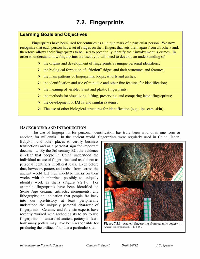
BACKGROUND AND INTRODUCTION The use of fingerprints for personal identification has truly been around, in one form or another, for millennia. In the ancient world, fingerprints were regularly used in China, Japan, Babylon, and other places to certify business transactions and as a personal sign for important documents. By the 3rd century BC, the evidence is clear that people in China understood the individual nature of fingerprints and used them as personal identifiers in official seals. Even before that, however, potters and artists from across the ancient world left their indelible marks on their works with thumbprints, possibly to uniquely identify work as theirs (Figure 7.2.1). For example, fingerprints have been identified on Stone Age ceramic artifacts, monuments, and lithographs; an indication that people far back into our pre-history at least peripherally understood the uniquely personal character of fingerprints. Ceramic and forensic experts have recently worked with archeologists to try to use fingerprints on unearthed ancient pottery to learn how many potters may have been responsible for producing the artifacts found at a particular site.
Figure 7.2.1. Ancient fingerprints from ceramic pottery (J. Ancient Fingerprints 2007, 1, 4-15).
Introduction to Forensic Science Chapter 7, Page 6 Draft 2/8/12 J. T. Spencer
Possibly the first recorded case of the true forensic use of fingerprints, however, comes from medieval Rome where the 10th century Roman attorney Quintilian was able to show that bloody hand prints found at a crime scene were meant to frame a blind man for the murder of his mother by the true murderer. In the 1600s, however, there were several important fundamental developments in
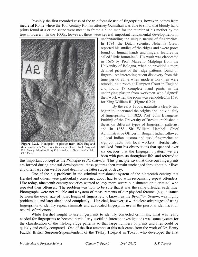
understanding the unique nature of fingerprints. In 1684, the Dutch scientist Nehemia Grew, reported his studies of the ridges and sweat pores found on human hands and fingers, features he called "little fountains". His work was elaborated in 1686 by Prof. Marcello Malphigi from the University of Bologna, when he provided a more detailed picture of the ridge patterns found on fingers. An interesting recent discovery from this time period came when modern workmen were remodeling a room at Hampton Court in England and found 17 complete hand prints in the underlying plaster from workmen who "signed" their work when the room was remodeled in 1690 for King William III (Figure 6.2.2). By the early 1800s, naturalists clearly had begun to understand the origins and individuality of fingerprints. In 1823, Prof. John Evangelist Purkinji of the University of Breslau, published a thesis on different types of fingerprint patterns, and in 1858, Sir William Hershel, Chief Administrative Officer in Bengal, India, followed a local Indian custom and used fingerprints to sign contracts with local workers. Hershel also realized from his observations that spanned over six decades that the fingerprint pattern we are born with persists throughout life, and referred to
this important concept as the Principle of Persistency. This principle says that once our fingerprints are formed during prenatal development, these patterns then remain unchanged throughout our lives and often last even well beyond death to the latter stages of decay. One of the big problems in the criminal punishment system of the nineteenth century that Hershel and others were particularly concerned about had to do with recognizing repeat offenders. Like today, nineteenth century societies wanted to levy more severe punishments on a criminal who repeated their offenses. The problem was how to be sure that it was the same offender each time. Photographs were not reliable and a system of measurements of our physical features (e.g., distance between the eyes, size of nose, length of fingers, etc.), known as the Bertillion System, was highly problematic and later abandoned completely. Herschel, however, saw the clear advantages of using fingerprints to identify repeat criminals and advocated fingerprint use in the personal identification records of prisoners. While Hershel sought to use fingerprints to identify convicted criminals, what was really needed for fingerprints to become particularly useful in forensic investigations was some system for the classification of the lifelong ridge patterns so that large numbers of prints and files could be quickly and easily compared. One of the first attempts at this task came from the work of Dr. Henry Faulds, British Surgeon-Superintendent of the Tsukiji Hospital in Tokyo, who developed the first
Figure 7.2.2. Handprint in plaster from 1690 England (from Advances in Fingerprint Technology, Chapt. 1 by J. Berry and D.A. Stoney; Edited by Henry C. Lee and R. E. Gaensslen (2nd Ed.), CRC Press).
Introduction to Forensic Science Chapter 7, Page 7 Draft 2/8/12 J. T. Spencer
systematic method of classification. Dr. Faulds also clearly recognized the use of fingerprints in forensic investigations and wrote "When bloody fingerprints or impressions on clay, glass, etc. exist, they may lead to the scientific identification of criminals..... There can be no doubt as to the advantage of having, beside their photograph, a copy of the forever unchangeable finger furrows of important criminals" (Nature, October 28, 1880). He recognized the value of latent prints (prints not visible to the naked eye) and used his expertise to exonerate a staff member at his hospital who was incorrectly charged with robbery. Dr. Faulds is today recognized as the "father of fingerprinting", although this recognition didn't come until nearly half a century after his death.
Will West
THE CASE OF THE WILLS WEST
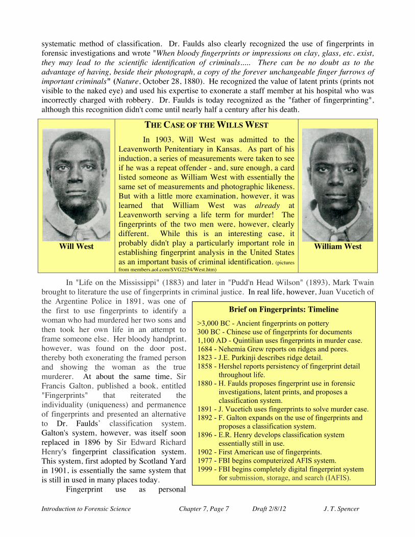
In 1903, Will West was admitted to the Leavenworth Penitentiary in Kansas. As part of his induction, a series of measurements were taken to see if he was a repeat offender - and, sure enough, a card listed someone as William West with essentially the same set of measurements and photographic likeness. But with a little more examination, however, it was learned that William West was already at Leavenworth serving a life term for murder! The fingerprints of the two men were, however, clearly different. While this is an interesting case, it probably didn't play a particularly important role in establishing fingerprint analysis in the United States as an important basis of criminal identification. (pictures from members.aol.com/SVG2254/West.htm)
William West
In "Life on the Mississippi" (1883) and later in "Pudd'n Head Wilson" (1893), Mark Twain brought to literature the use of fingerprints in criminal justice. In real life, however, Juan Vucetich of the Argentine Police in 1891, was one of the first to use fingerprints to identify a woman who had murdered her two sons and then took her own life in an attempt to frame someone else. Her bloody handprint, however, was found on the door post, thereby both exonerating the framed person and showing the woman as the true murderer. At about the same time, Sir Francis Galton, published a book, entitled "Fingerprints" that reiterated the individuality (uniqueness) and permanence of fingerprints and presented an alternative to Dr. Faulds’ classification system. Galton's system, however, was itself soon replaced in 1896 by Sir Edward Richard Henry's fingerprint classification system. This system, first adopted by Scotland Yard in 1901, is essentially the same system that is still in used in many places today. Fingerprint use as personal
Brief on Fingerprints: Timeline
>3,000 BC - Ancient fingerprints on pottery 300 BC - Chinese use of fingerprints for documents 1,100 AD - Quintilian uses fingerprints in murder case. 1684 - Nehemia Grew reports on ridges and pores. 1823 - J.E. Purkinji describes ridge detail. 1858 - Hershel reports persistency of fingerprint detail
throughout life. 1880 - H. Faulds proposes fingerprint use in forensic
investigations, latent prints, and proposes a classification system.
1891 - J. Vucetich uses fingerprints to solve murder case. 1892 - F. Galton expands on the use of fingerprints and
proposes a classification system. 1896 - E.R. Henry develops classification system
essentially still in use. 1902 - First American use of fingerprints. 1977 - FBI begins computerized AFIS system. 1999 - FBI begins completely digital fingerprint system
for submission, storage, and search (IAFIS).
Introduction to Forensic Science Chapter 7, Page 8 Draft 2/8/12 J. T. Spencer
identifiers in the United States really began in 1902 when the New York Civil Service Commission and, in 1903, the New York State Prison system began using fingerprints for the identification of convicted criminals. At about the same time (1902) in a related development, R. Fischer presented his related work on the furrows of the human lips for individual identification, a field known as cheiloscopy. This work culminated in 1968 when the lip prints of over 1,300 people were examined at Tokyo University with the conclusion that lip prints, like fingerprints, are also unique to an individual. Finally, in 1977 the FBI began the use of its Automated Fingerprint Identification System (AFIS) using digital scans of fingerprints. This system was upgraded in 1996 to allow for the computerized searches of the entire AFIS fingerprint database, and then modified again in 1999 with the formation of the Integrated Automated Fingerprint Identification System (IAFIS), that provided for the automated digital computer submission, storage, and search of the national FBI fingerprint database. Today, federal and state agencies can receive answers to requests for matching criminal fingerprint patterns with well over 55 million fingerprint records on file within two hours of submission. SKIN: THE AMAZING ORGAN Our skin is the largest organ system in the human body weighting, on average, 25 pounds and covering about 20 ft2 in area. Our skin is part of a larger system, called the integumentary system, that forms the outer “boundary” of our bodies and includes our skin, hair and nails (the latter two are considered “derivatives” of our epidermis). It consists of an array of various tissues and structures that function together for the protection and regulation of underlying organs and gets about one-third of all the oxygenated blood that heaves the heart. In particular, the skin helps to regulate the temperature of our bodies in the face of constantly changing thermal environments, controls moisture loss, protects us from physical impact and wear, provides a barrier to the entry of unwanted substances and agents into our bodies from a hostile environment (such as bacteria and viruses), and serves as a highly sensitive sensory organ for the body. It allows us to feel the lightest touch and yet withstand some pretty significant impact and abrasion forces. It’s tough, durable and constantly being repaired and replaced.
The skin contains a variety of specialized cells and structures (Figure 7.2.3). Our skin has three major layers, although each is often broken into smaller sublayers. The lowest layer is referred to as the subcutaneous layer (or more accurately called the hypodermis) and is composed largely of fat and connective tissue that contains larger blood vessels and nerves. The middle layer, referred to as the dermis, is composed mostly collagen (protein) fibers, elastic tissue, and reticular fibers (crosslinked fibers that form a fine supporting meshwork). The dermis is also the place where the hair follicles, sebaceous (oil) glands, eccrine (sweat) glands, apocrine (scent) glands, and hair erector muscles are found. Additionally, nerves and smaller blood vessels run through this layer and transmit information about temperature, touch, pressure, and sometimes pain to our brains. More will be presented later on hair and how it grows from the follicles located in these dermal layers. The main structural function of the dermis is to support and nurture the layer lying above it. The outermost layer of our skin is called the epidermis and ranges in thickness from very thin on our eyelids (about 0.05 mm) to rather thick on the palms of our hands and the soles of our feet (around 1.5 mm thick). It is this layer that also contains melanin, the pigment responsible for skin coloration. At the lowest portion of the epidermis, often referred to as the “generating layer” (stratum basale), column-like cells constantly divide and push previously formed cells towards the surface, causing these cells to flatten out and ultimately die in the process. The very top layer of the epidermis (stratum corneum), the part directly in contact with the outside world, is composed entirely of 25 to 30 layers of dead cells that stay at the surface for about two weeks before being shed and replaced from layers below.
Introduction to Forensic Science Chapter 7, Page 9 Draft 2/8/12 J. T. Spencer
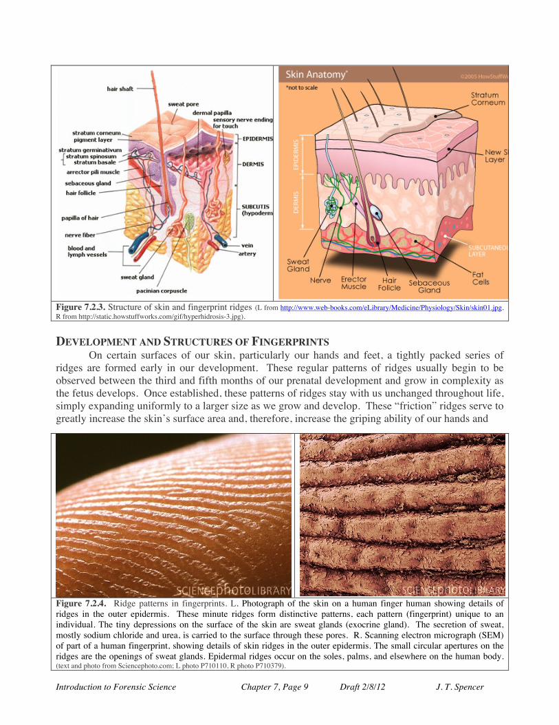
Figure 7.2.3. Structure of skin and fingerprint ridges (L from http://www.web-books.com/eLibrary/Medicine/Physiology/Skin/skin01.jpg, R from http://static.howstuffworks.com/gif/hyperhidrosis-3.jpg). DEVELOPMENT AND STRUCTURES OF FINGERPRINTS
On certain surfaces of our skin, particularly our hands and feet, a tightly packed series of ridges are formed early in our development. These regular patterns of ridges usually begin to be observed between the third and fifth months of our prenatal development and grow in complexity as the fetus develops. Once established, these patterns of ridges stay with us unchanged throughout life, simply expanding uniformly to a larger size as we grow and develop. These “friction” ridges serve to greatly increase the skin’s surface area and, therefore, increase the griping ability of our hands and
Figure 7.2.4. Ridge patterns in fingerprints. L. Photograph of the skin on a human finger human showing details of ridges in the outer epidermis. These minute ridges form distinctive patterns, each pattern (fingerprint) unique to an individual. The tiny depressions on the surface of the skin are sweat glands (exocrine gland). The secretion of sweat, mostly sodium chloride and urea, is carried to the surface through these pores. R. Scanning electron micrograph (SEM) of part of a human fingerprint, showing details of skin ridges in the outer epidermis. The small circular apertures on the ridges are the openings of sweat glands. Epidermal ridges occur on the soles, palms, and elsewhere on the human body. (text and photo from Sciencephoto.com; L photo P710110, R photo P710379).
Introduction to Forensic Science Chapter 7, Page 10 Draft 2/8/12 J. T. Spencer
feet, especially on smooth and wet surfaces, and to increase the sensitivity of our touch sense (Figure 7.2.4). These ridges are believed to originate during our prenatal development from the buckling of the basal cell layer of the fetal epidermis as the cells in this layer grow rapidly and do not have sufficient space to spread out so the layer end up permanently bending and buckling to form the ridges that we see at the surface of the skin.
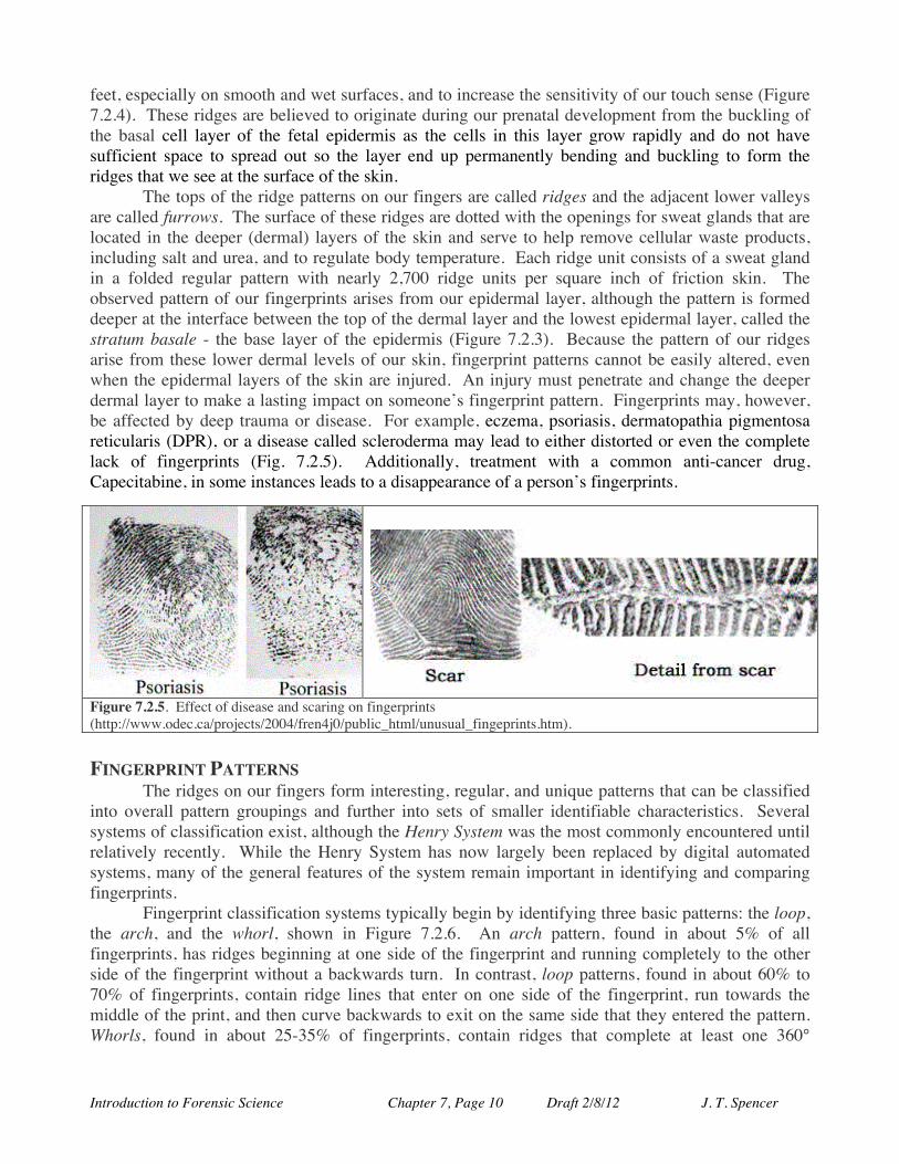
The tops of the ridge patterns on our fingers are called ridges and the adjacent lower valleys are called furrows. The surface of these ridges are dotted with the openings for sweat glands that are located in the deeper (dermal) layers of the skin and serve to help remove cellular waste products, including salt and urea, and to regulate body temperature. Each ridge unit consists of a sweat gland in a folded regular pattern with nearly 2,700 ridge units per square inch of friction skin. The observed pattern of our fingerprints arises from our epidermal layer, although the pattern is formed deeper at the interface between the top of the dermal layer and the lowest epidermal layer, called the stratum basale - the base layer of the epidermis (Figure 7.2.3). Because the pattern of our ridges arise from these lower dermal levels of our skin, fingerprint patterns cannot be easily altered, even when the epidermal layers of the skin are injured. An injury must penetrate and change the deeper dermal layer to make a lasting impact on someone’s fingerprint pattern. Fingerprints may, however, be affected by deep trauma or disease. For example, eczema, psoriasis, dermatopathia pigmentosa reticularis (DPR), or a disease called scleroderma may lead to either distorted or even the complete lack of fingerprints (Fig. 7.2.5). Additionally, treatment with a common anti-cancer drug, Capecitabine, in some instances leads to a disappearance of a person’s fingerprints.
Figure 7.2.5. Effect of disease and scaring on fingerprints (http://www.odec.ca/projects/2004/fren4j0/public_html/unusual_fingeprints.htm). FINGERPRINT PATTERNS The ridges on our fingers form interesting, regular, and unique patterns that can be classified into overall pattern groupings and further into sets of smaller identifiable characteristics. Several systems of classification exist, although the Henry System was the most commonly encountered until relatively recently. While the Henry System has now largely been replaced by digital automated systems, many of the general features of the system remain important in identifying and comparing fingerprints.
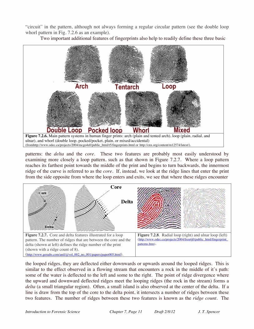
Fingerprint classification systems typically begin by identifying three basic patterns: the loop, the arch, and the whorl, shown in Figure 7.2.6. An arch pattern, found in about 5% of all fingerprints, has ridges beginning at one side of the fingerprint and running completely to the other side of the fingerprint without a backwards turn. In contrast, loop patterns, found in about 60% to 70% of fingerprints, contain ridge lines that enter on one side of the fingerprint, run towards the middle of the print, and then curve backwards to exit on the same side that they entered the pattern. Whorls, found in about 25-35% of fingerprints, contain ridges that complete at least one 360°
Introduction to Forensic Science Chapter 7, Page 11 Draft 2/8/12 J. T. Spencer
“circuit” in the pattern, although not always forming a regular circular pattern (see the double loop whorl pattern in Fig. 7.2.6 as an example).
Two important additional features of fingerprints also help to readily define these three basic
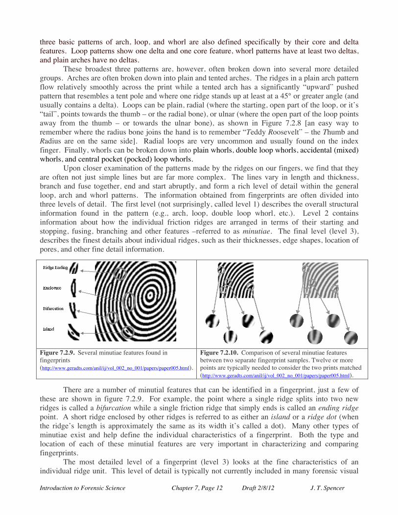
Figure 7.2.6. Main pattern systems in human finger prints: arch (plain and tented arch), loop (plain, radial, and ulnar), and whorl (double loop, pocked/pocket, plain, or mixed/accidental) (fromhttp://www.odec.ca/projects/2004/mcgo4s0/public_html/t5/fingerprints.html or http://cnx.org/content/m12574/latest/). patterns: the delta and the core. These two features are probably most easily understood by examining more closely a loop pattern, such as that shown in Figure 7.2.7. Where a loop pattern reaches its farthest point towards the middle of the print and begins to turn backwards, the innermost ridge of the curve is referred to as the core. If, instead, we look at the ridge lines that enter the print from the side opposite from where the loop enters and exits, we see that where these ridges encounter
Figure 7.2.7. Core and delta features illustrated for a loop pattern. The number of ridges that are between the core and the delta (shown at left) defines the ridge number of the print (shown with a ridge count of 8). (http://www.geradts.com/anil/ij/vol_002_no_001/papers/paper005.html).
Figure 7.2.8. Radial loop (right) and ulnar loop (left) (http://www.odec.ca/projects/2004/fren4j0/public_html/fingerprint_patterns.htm).
the looped ridges, they are deflected either downwards or upwards around the looped ridges. This is similar to the effect observed in a flowing stream that encounters a rock in the middle of it’s path: some of the water is deflected to the left and some to the right. The point of ridge divergence where the upward and downward deflected ridges meet the looping ridges (the rock in the stream) forms a delta (a small triangular region). Often, a small island is also observed at the center of the delta. If a line is draw from the top of the core to the delta point, it intersects a number of ridges between these two features. The number of ridges between these two features is known as the ridge count. The
Introduction to Forensic Science Chapter 7, Page 12 Draft 2/8/12 J. T. Spencer
three basic patterns of arch, loop, and whorl are also defined specifically by their core and delta features. Loop patterns show one delta and one core feature, whorl patterns have at least two deltas, and plain arches have no deltas.
These broadest three patterns are, however, often broken down into several more detailed groups. Arches are often broken down into plain and tented arches. The ridges in a plain arch pattern flow relatively smoothly across the print while a tented arch has a significantly “upward” pushed pattern that resembles a tent pole and where one ridge stands up at least at a 45° or greater angle (and usually contains a delta). Loops can be plain, radial (where the starting, open part of the loop, or it’s “tail”, points towards the thumb – or the radial bone), or ulnar (where the open part of the loop points away from the thumb – or towards the ulnar bone), as shown in Figure 7.2.8 [an easy way to remember where the radius bone joins the hand is to remember “Teddy Roosevelt” – the Thumb and Radius are on the same side]. Radial loops are very uncommon and usually found on the index finger. Finally, whorls can be broken down into plain whorls, double loop whorls, accidental (mixed) whorls, and central pocket (pocked) loop whorls.
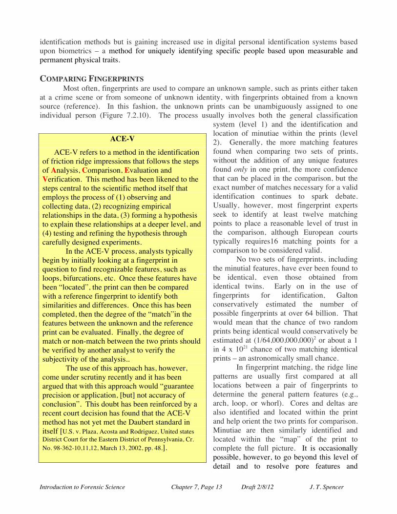
Upon closer examination of the patterns made by the ridges on our fingers, we find that they are often not just simple lines but are far more complex. The lines vary in length and thickness, branch and fuse together, end and start abruptly, and form a rich level of detail within the general loop, arch and whorl patterns. The information obtained from fingerprints are often divided into three levels of detail. The first level (not surprisingly, called level 1) describes the overall structural information found in the pattern (e.g., arch, loop, double loop whorl, etc.). Level 2 contains information about how the individual friction ridges are arranged in terms of their starting and stopping, fusing, branching and other features –referred to as minutiae. The final level (level 3), describes the finest details about individual ridges, such as their thicknesses, edge shapes, location of pores, and other fine detail information.
Figure 7.2.9. Several minutiae features found in fingerprints (http://www.geradts.com/anil/ij/vol_002_no_001/papers/paper005.html).
Figure 7.2.10. Comparison of several minutiae features between two separate fingerprint samples. Twelve or more points are typically needed to consider the two prints matched (http://www.geradts.com/anil/ij/vol_002_no_001/papers/paper005.html).
There are a number of minutial features that can be identified in a fingerprint, just a few of these are shown in figure 7.2.9. For example, the point where a single ridge splits into two new ridges is called a bifurcation while a single friction ridge that simply ends is called an ending ridge point. A short ridge enclosed by other ridges is referred to as either an island or a ridge dot (when the ridge’s length is approximately the same as its width it’s called a dot). Many other types of minutiae exist and help define the individual characteristics of a fingerprint. Both the type and location of each of these minutial features are very important in characterizing and comparing fingerprints. The most detailed level of a fingerprint (level 3) looks at the fine characteristics of an individual ridge unit. This level of detail is typically not currently included in many forensic visual
Introduction to Forensic Science Chapter 7, Page 13 Draft 2/8/12 J. T. Spencer
identification methods but is gaining increased use in digital personal identification systems based upon biometrics – a method for uniquely identifying specific people based upon measurable and permanent physical traits. COMPARING FINGERPRINTS
Most often, fingerprints are used to compare an unknown sample, such as prints either taken at a crime scene or from someone of unknown identity, with fingerprints obtained from a known source (reference). In this fashion, the unknown prints can be unambiguously assigned to one individual person (Figure 7.2.10). The process usually involves both the general classification
system (level 1) and the identification and location of minutiae within the prints (level 2). Generally, the more matching features found when comparing two sets of prints, without the addition of any unique features found only in one print, the more confidence that can be placed in the comparison, but the exact number of matches necessary for a valid identification continues to spark debate. Usually, however, most fingerprint experts seek to identify at least twelve matching points to place a reasonable level of trust in the comparison, although European courts typically requires16 matching points for a comparison to be considered valid.
No two sets of fingerprints, including the minutial features, have ever been found to be identical, even those obtained from identical twins. Early on in the use of fingerprints for identification, Galton conservatively estimated the number of possible fingerprints at over 64 billion. That would mean that the chance of two random prints being identical would conservatively be estimated at (1/64,000,000,000)2 or about a 1 in 4 x 10²¹ chance of two matching identical prints – an astronomically small chance.
In fingerprint matching, the ridge line patterns are usually first compared at all locations between a pair of fingerprints to determine the general pattern features (e.g., arch, loop, or whorl). Cores and deltas are also identified and located within the print and help orient the two prints for comparison. Minutiae are then similarly identified and located within the “map” of the print to complete the full picture. It is occasionally possible, however, to go beyond this level of detail and to resolve pore features and
ACE-V
ACE-V refers to a method in the identification of friction ridge impressions that follows the steps of Analysis, Comparison, Evaluation and Verification. This method has been likened to the steps central to the scientific method itself that employs the process of (1) observing and collecting data, (2) recognizing empirical relationships in the data, (3) forming a hypothesis to explain these relationships at a deeper level, and (4) testing and refining the hypothesis through carefully designed experiments. In the ACE-V process, analysts typically begin by initially looking at a fingerprint in question to find recognizable features, such as loops, bifurcations, etc. Once these features have been “located”, the print can then be compared with a reference fingerprint to identify both similarities and differences. Once this has been completed, then the degree of the “match”in the features between the unknown and the reference print can be evaluated. Finally, the degree of match or non-match between the two prints should be verified by another analyst to verify the subjectivity of the analysis.. The use of this approach has, however, come under scrutiny recently and it has been argued that with this approach would “guarantee precision or application, [but] not accuracy of conclusion”. This doubt has been reinforced by a recent court decision has found that the ACE-V method has not yet met the Daubert standard in itself [U.S. v. Plaza, Acosta and Rodriguez, United states District Court for the Eastern District of Pennsylvania, Cr. No. 98-362-10,11,12, March 13, 2002, pp. 48.].
Introduction to Forensic Science Chapter 7, Page 14 Draft 2/8/12 J. T. Spencer
locations in the ridges and also imperfections in edges of the ridges, usually from electronic scanning methods rather than other sampling methods (see below). Work is in progress to use this information in a way similar to how minutiae are employed in print comparison. Some features, such as scars and creases can be used but they are often changeable over time and are, therefore, of limited use. There also appears to be a relationship between fingerprint patterns and ethnicity with Europeans and Africans displaying relatively high incidences of loop patterns and Asians and Australian aborigines showing whorls.
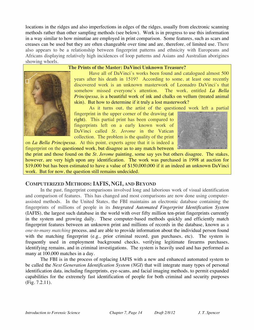
The Prints of the Master: DaVinci Unknown Treasure? Have all of DaVinci’s works been found and catalogued almost 500 years after his death in 1519? According to some, at least one recently discovered work is an unknown masterwork of Leonadro DaVinci’s that somehow missed everyone’s attention. The work, entitled La Bella Principessa, is a beautiful work of ink and chalks on vellum (treated animal skin). But how to determine if it truly a lost masterwork? As it turns out, the artist of the questioned work left a partial fingerprint in the upper corner of the drawing (at right). This partial print has been compared to fingerprints left on a early known work of DaVinci called St. Jerome in the Vatican collection. The problem is the quality of the print
on La Bella Principessa. At this point, experts agree that it is indeed a fingerprint on the questioned work, but disagree as to any match between the print and those found on the St. Jerome painting, some say yes but others disagree. The stakes, however, are very high upon any identification. The work was purchased in 1998 at auction for $19,000 but has been estimated to have a value of $150,000,000 if it an indeed an unknown DaVinci work. But for now, the question still remains undecided.
COMPUTERIZED METHODS: IAFIS, NGI, AND BEYOND
In the past, fingerprint comparisons involved long and laborious work of visual identification and comparison of features. This has changed and most comparisons are now done using computer-assisted methods. In the United States, the FBI maintains an electronic database containing the fingerprints of millions of people in its Integrated Automated Fingerprint Identification System (IAFIS), the largest such database in the world with over fifty million ten-print fingerprints currently in the system and growing daily. These computer-based methods quickly and efficiently match fingerprint features between an unknown print and millions of records in the database, known as a one-to-many matching process, and are able to provide information about the individual person found with the matching fingerprint (e.g., prior criminal record, gun purchases, etc). The system is frequently used in employment background checks, verifying legitimate firearms purchases, identifying remains, and in criminal investigations. The system is heavily used and has performed as many at 100,000 matches in a day.
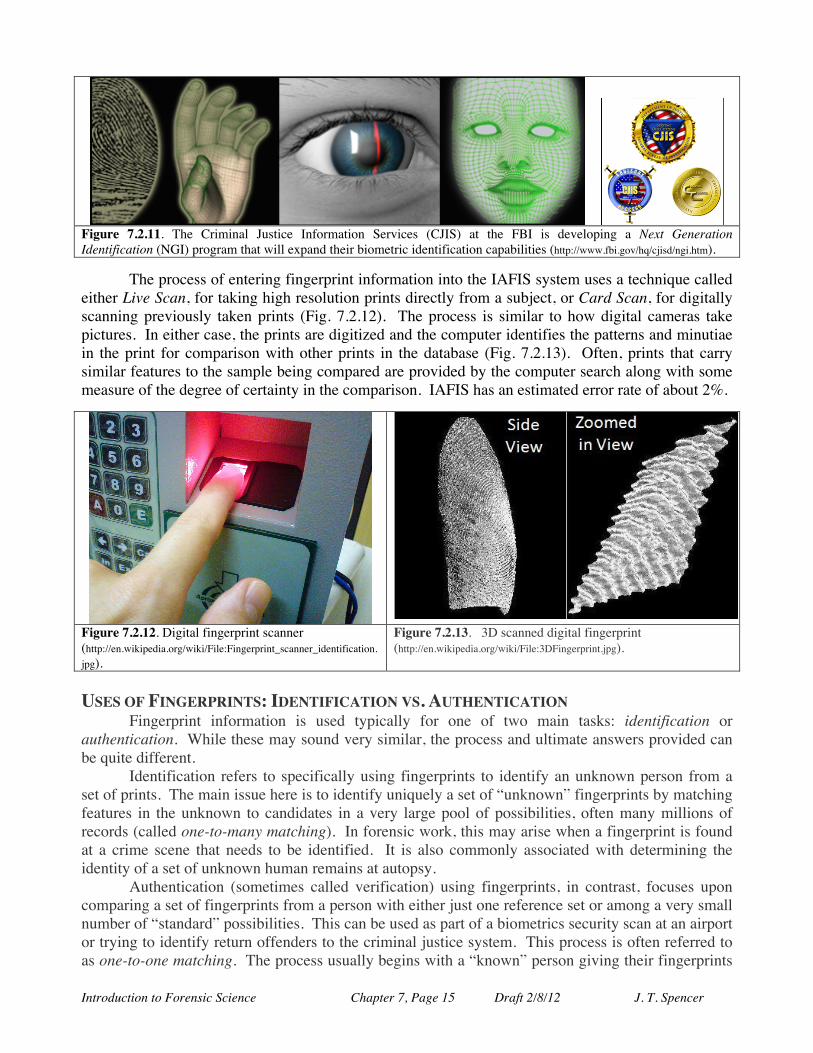
The FBI is in the process of replacing IAFIS with a new and enhanced automated system to be called the Next Generation Identification System (NGI) that will integrate many types of personal identification data, including fingerprints, eye-scans, and facial imaging methods, to permit expanded capabilities for the extremely fast identification of people for both criminal and security purposes (Fig. 7.2.11).
Introduction to Forensic Science Chapter 7, Page 15 Draft 2/8/12 J. T. Spencer
Figure 7.2.11. The Criminal Justice Information Services (CJIS) at the FBI is developing a Next Generation Identification (NGI) program that will expand their biometric identification capabilities (http://www.fbi.gov/hq/cjisd/ngi.htm).
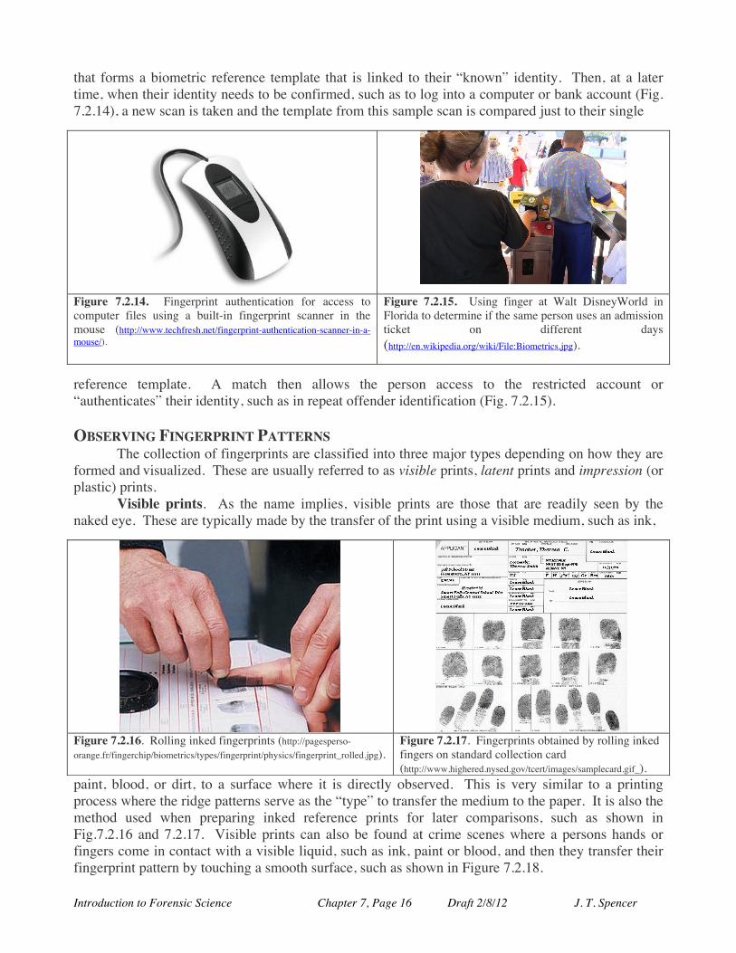
The process of entering fingerprint information into the IAFIS system uses a technique called
either Live Scan, for taking high resolution prints directly from a subject, or Card Scan, for digitally scanning previously taken prints (Fig. 7.2.12). The process is similar to how digital cameras take pictures. In either case, the prints are digitized and the computer identifies the patterns and minutiae in the print for comparison with other prints in the database (Fig. 7.2.13). Often, prints that carry similar features to the sample being compared are provided by the computer search along with some measure of the degree of certainty in the comparison. IAFIS has an estimated error rate of about 2%.
Figure 7.2.12. Digital fingerprint scanner (http://en.wikipedia.org/wiki/File:Fingerprint_scanner_identification.jpg).
Figure 7.2.13. 3D scanned digital fingerprint (http://en.wikipedia.org/wiki/File:3DFingerprint.jpg).
USES OF FINGERPRINTS: IDENTIFICATION VS. AUTHENTICATION Fingerprint information is used typically for one of two main tasks: identification or authentication. While these may sound very similar, the process and ultimate answers provided can be quite different.
Identification refers to specifically using fingerprints to identify an unknown person from a set of prints. The main issue here is to identify uniquely a set of “unknown” fingerprints by matching features in the unknown to candidates in a very large pool of possibilities, often many millions of records (called one-to-many matching). In forensic work, this may arise when a fingerprint is found at a crime scene that needs to be identified. It is also commonly associated with determining the identity of a set of unknown human remains at autopsy. Authentication (sometimes called verification) using fingerprints, in contrast, focuses upon comparing a set of fingerprints from a person with either just one reference set or among a very small number of “standard” possibilities. This can be used as part of a biometrics security scan at an airport or trying to identify return offenders to the criminal justice system. This process is often referred to as one-to-one matching. The process usually begins with a “known” person giving their fingerprints
Introduction to Forensic Science Chapter 7, Page 16 Draft 2/8/12 J. T. Spencer
that forms a biometric reference template that is linked to their “known” identity. Then, at a later time, when their identity needs to be confirmed, such as to log into a computer or bank account (Fig. 7.2.14), a new scan is taken and the template from this sample scan is compared just to their single
Figure 7.2.14. Fingerprint authentication for access to computer files using a built-in fingerprint scanner in the mouse (http://www.techfresh.net/fingerprint-authentication-scanner-in-a-mouse/).
Figure 7.2.15. Using finger at Walt DisneyWorld in Florida to determine if the same person uses an admission ticket on different days (http://en.wikipedia.org/wiki/File:Biometrics.jpg).
reference template. A match then allows the person access to the restricted account or “authenticates” their identity, such as in repeat offender identification (Fig. 7.2.15). OBSERVING FINGERPRINT PATTERNS The collection of fingerprints are classified into three major types depending on how they are formed and visualized. These are usually referred to as visible prints, latent prints and impression (or plastic) prints. Visible prints. As the name implies, visible prints are those that are readily seen by the naked eye. These are typically made by the transfer of the print using a visible medium, such as ink,
paint, blood, or dirt, to a surface where it is directly observed. This is very similar to a printing process where the ridge patterns serve as the “type” to transfer the medium to the paper. It is also the method used when preparing inked reference prints for later comparisons, such as shown in Fig.7.2.16 and 7.2.17. Visible prints can also be found at crime scenes where a persons hands or fingers come in contact with a visible liquid, such as ink, paint or blood, and then they transfer their fingerprint pattern by touching a smooth surface, such as shown in Figure 7.2.18.
Figure 7.2.16. Rolling inked fingerprints (http://pagesperso-orange.fr/fingerchip/biometrics/types/fingerprint/physics/fingerprint_rolled.jpg).
Figure 7.2.17. Fingerprints obtained by rolling inked fingers on standard collection card (http://www.highered.nysed.gov/tcert/images/samplecard.gif_).
Introduction to Forensic Science Chapter 7, Page 17 Draft 2/8/12 J. T. Spencer
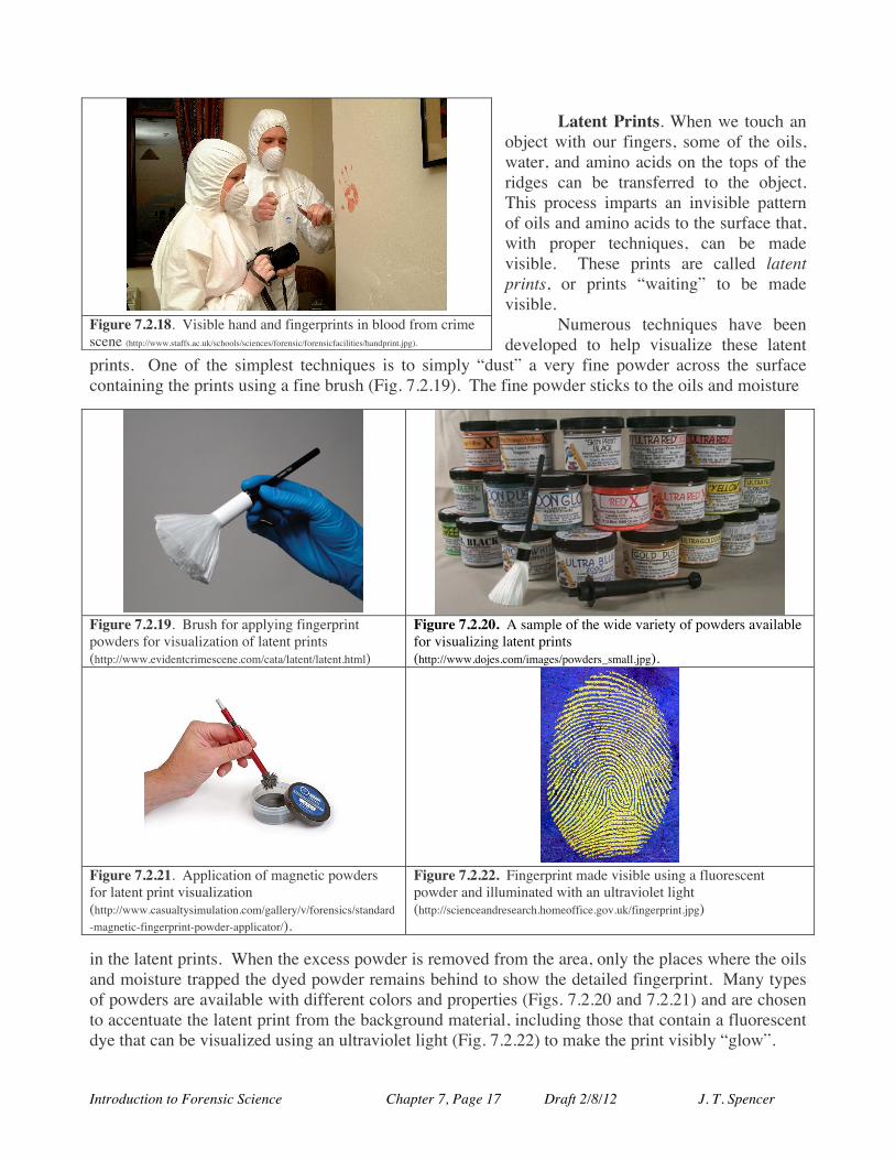
Latent Prints. When we touch an object with our fingers, some of the oils, water, and amino acids on the tops of the ridges can be transferred to the object. This process imparts an invisible pattern of oils and amino acids to the surface that, with proper techniques, can be made visible. These prints are called latent prints, or prints “waiting” to be made visible.
Numerous techniques have been developed to help visualize these latent
prints. One of the simplest techniques is to simply “dust” a very fine powder across the surface containing the prints using a fine brush (Fig. 7.2.19). The fine powder sticks to the oils and moisture
Figure 7.2.19. Brush for applying fingerprint powders for visualization of latent prints (http://www.evidentcrimescene.com/cata/latent/latent.html)
Figure 7.2.20. A sample of the wide variety of powders available for visualizing latent prints (http://www.dojes.com/images/powders_small.jpg).
Figure 7.2.21. Application of magnetic powders for latent print visualization (http://www.casualtysimulation.com/gallery/v/forensics/standard-magnetic-fingerprint-powder-applicator/).
Figure 7.2.22. Fingerprint made visible using a fluorescent powder and illuminated with an ultraviolet light (http://scienceandresearch.homeoffice.gov.uk/fingerprint.jpg)
in the latent prints. When the excess powder is removed from the area, only the places where the oils and moisture trapped the dyed powder remains behind to show the detailed fingerprint. Many types of powders are available with different colors and properties (Figs. 7.2.20 and 7.2.21) and are chosen to accentuate the latent print from the background material, including those that contain a fluorescent dye that can be visualized using an ultraviolet light (Fig. 7.2.22) to make the print visibly “glow”.
Figure 7.2.18. Visible hand and fingerprints in blood from crime scene (http://www.staffs.ac.uk/schools/sciences/forensic/forensicfacilities/handprint.jpg).
Introduction to Forensic Science Chapter 7, Page 18 Draft 2/8/12 J. T. Spencer
A very similar method employs a very fine magnetic powder that similarly adheres to the oils and moisture in the latent print. The excess powder, however, is cleanly removed by passing a magnet across the surface to extract any unadhered powder from the print, as shown in Fig. 7.2.21.
Another important way to visualize latent prints is to react the oils, amino acids or salts in the transferred fingerprint with some chemical reagent that allows us to see the print. The salts and amino acids deposited from our fingers and hands are essentially non-volatile and have been shown to remain in place for decades to render clear fingerprint patterns. Many such methods have been developed but four are particularly interesting and useful.
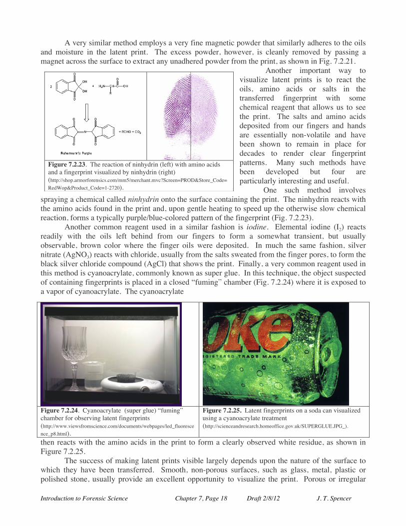
One such method involves spraying a chemical called ninhydrin onto the surface containing the print. The ninhydrin reacts with the amino acids found in the print and, upon gentle heating to speed up the otherwise slow chemical reaction, forms a typically purple/blue-colored pattern of the fingerprint (Fig. 7.2.23).
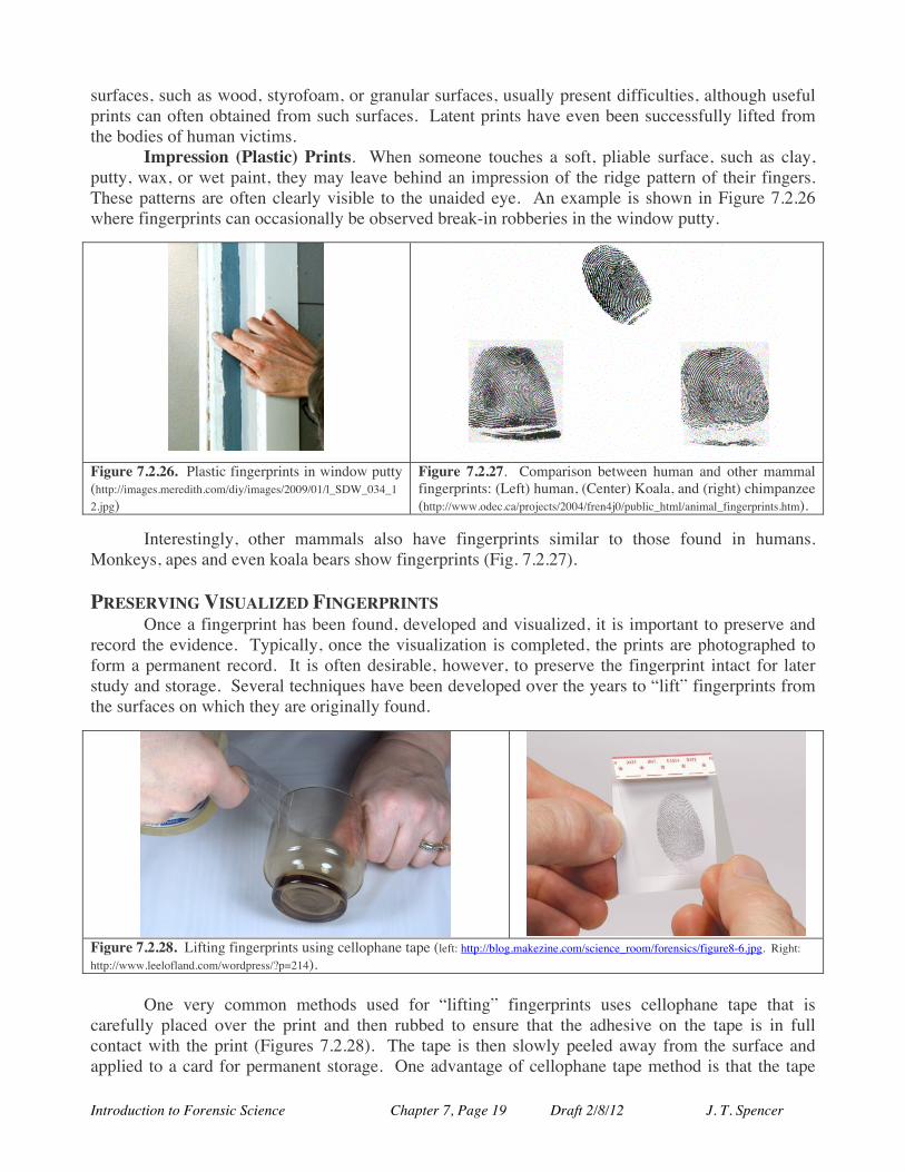
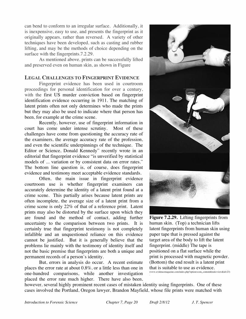
Another common reagent used in a similar fashion is iodine. Elemental iodine (I2) reacts readily with the oils left behind from our fingers to form a somewhat transient, but usually observable, brown color where the finger oils were deposited. In much the same fashion, silver nitrate (AgNO3) reacts with chloride, usually from the salts sweated from the finger pores, to form the black silver chloride compound (AgCl) that shows the print. Finally, a very common reagent used in this method is cyanoacrylate, commonly known as super glue. In this technique, the object suspected of containing fingerprints is placed in a closed “fuming” chamber (Fig. 7.2.24) where it is exposed to a vapor of cyanoacrylate. The cyanoacrylate
Figure 7.2.24. Cyanoacrylate (super glue) “fuming” chamber for observing latent fingerprints (http://www.viewsfromscience.com/documents/webpages/led_fluorescence_p8.html).
Figure 7.2.25. Latent fingerprints on a soda can visualized using a cyanoacrylate treatment (http://scienceandresearch.homeoffice.gov.uk/SUPERGLUE.JPG_).
then reacts with the amino acids in the print to form a clearly observed white residue, as shown in Figure 7.2.25.
The success of making latent prints visible largely depends upon the nature of the surface to which they have been transferred. Smooth, non-porous surfaces, such as glass, metal, plastic or polished stone, usually provide an excellent opportunity to visualize the print. Porous or irregular
Figure 7.2.23. The reaction of ninhydrin (left) with amino acids and a fingerprint visualized by ninhydrin (right) (http://shop.armorforensics.com/mm5/merchant.mvc?Screen=PROD&Store_Code=RedWop&Product_Code=1-2720).
Introduction to Forensic Science Chapter 7, Page 19 Draft 2/8/12 J. T. Spencer
surfaces, such as wood, styrofoam, or granular surfaces, usually present difficulties, although useful prints can often obtained from such surfaces. Latent prints have even been successfully lifted from the bodies of human victims.
Impression (Plastic) Prints. When someone touches a soft, pliable surface, such as clay, putty, wax, or wet paint, they may leave behind an impression of the ridge pattern of their fingers. These patterns are often clearly visible to the unaided eye. An example is shown in Figure 7.2.26 where fingerprints can occasionally be observed break-in robberies in the window putty.
Figure 7.2.26. Plastic fingerprints in window putty (http://images.meredith.com/diy/images/2009/01/l_SDW_034_12.jpg)
Figure 7.2.27. Comparison between human and other mammal fingerprints: (Left) human, (Center) Koala, and (right) chimpanzee (http://www.odec.ca/projects/2004/fren4j0/public_html/animal_fingerprints.htm).
Interestingly, other mammals also have fingerprints similar to those found in humans.
Monkeys, apes and even koala bears show fingerprints (Fig. 7.2.27).
PRESERVING VISUALIZED FINGERPRINTS Once a fingerprint has been found, developed and visualized, it is important to preserve and
record the evidence. Typically, once the visualization is completed, the prints are photographed to form a permanent record. It is often desirable, however, to preserve the fingerprint intact for later study and storage. Several techniques have been developed over the years to “lift” fingerprints from the surfaces on which they are originally found.
Figure 7.2.28. Lifting fingerprints using cellophane tape (left: http://blog.makezine.com/science_room/forensics/figure8-6.jpg. Right: http://www.leelofland.com/wordpress/?p=214).
One very common methods used for “lifting” fingerprints uses cellophane tape that is carefully placed over the print and then rubbed to ensure that the adhesive on the tape is in full contact with the print (Figures 7.2.28). The tape is then slowly peeled away from the surface and applied to a card for permanent storage. One advantage of cellophane tape method is that the tape
Introduction to Forensic Science Chapter 7, Page 20 Draft 2/8/12 J. T. Spencer
can bend to conform to an irregular surface. Additionally, it is inexpensive, easy to use, and presents the fingerprint as it originally appears, rather than reversed. A variety of other techniques have been developed, such as casting and rubber lifting, and may be the methods of choice depending on the surface with the fingerprints.7.2.29.
As mentioned above, prints can be successfully lifted and preserved even on human skin, as shown in Figure LEGAL CHALLENGES TO FINGERPRINT EVIDENCE
Fingerprint evidence has been used in courtroom proceedings for personal identification for over a century, with the first US murder conviction based on fingerprint identification evidence occurring in 1911. The matching of latent prints often not only determines who made the prints but they may also be used to indicate where that person has been, for example at the crime scene.
Recently, however, use of fingerprint information in court has come under intense scrutiny. Most of these challenges have come from questioning the accuracy rate of the examiners, the average accuracy rate of the profession, and even the scientific underpinnings of the technique. The Editor or Science, Donald Kennedy” recently wrote in an editorial that fingerprint evidence “is unverified by statistical models of ... variation or by consistent data on error rates.” The bottom line question is, of course, does fingerprint evidence and testimony meet acceptable evidence standards.
Often, the main issue in fingerprint evidence courtroom use is whether fingerprint examiners can accurately determine the identity of a latent print found at a crime scene. This partially arises because latent prints are often incomplete, the average size of a latent print from a crime scene is only 22% of that of a reference print. Latent prints may also be distorted by the surface upon which they are found and the method of contact, adding further uncertainty to the comparison between two prints. It is certainly true that fingerprint testimony is not completely infallible and an unquestioned reliance on this evidence cannot be justified. But it is generally believe that the problems lie mainly with the testimony of identity itself and not the basic premise that fingerprints are both a unique and permanent records of a person’s identity.
But, errors in analysis do occur. A recent estimate places the error rate at about 0.8%, or a little less than one in one-hundred comparisons, while another investigation placed the error rate much higher. There have also been, however, several highly prominent recent cases of mistaken identity using fingerprints. One of these cases involved the Portland, Oregon lawyer, Brandon Mayfield, whose file prints were matched with
Figure 7.2.29. Lifting fingerprints from human skin. (Top) a technician lifts latent fingerprints from human skin using paper tape that is pressed against the target area of the body to lift the latent fingerprint. (middle) The tape is positioned on a flat surface while the print is processed with magnetic powder. (Bottom) the end result is a latent print that is suitable to use as evidence. (www.evidencemagazine.com/index.php?option=com_content&task=view&id=23).
Introduction to Forensic Science Chapter 7, Page 21 Draft 2/8/12 J. T. Spencer
fingerprints obtained from the 2004 Madrid, Spain railcar bombing. Experts matched Mayfield’s fingerprints with those from Madrid, with the FBI calling the match "100 percent positive" and an "absolutely incontrovertible match". Mayfield was jailed for two weeks based upon this evidence before the Spanish National Police examiners showed the error in the analysis and he was released and exonerated.
Nonetheless, fingerprint evidence remains a powerful investigatory and courtroom technique that is generally relied upon as scientifically valid, reliable and trustworthy when applied in a rigorous manner. It has withstood significant Daubert challenges so far and remains an important part of forensic investigations and courtroom proceedings.
PALM AND FOOTPRINT EVIDENCE
Fingers are not the only portions of our bodies covered with epidermal friction ridges. Ridges are also found on the palms of our hands and on the soles of our feet that display many of the important pattern characteristics that are so important in fingerprint analysis. While palm and foot ridge pattern analysis is less well developed relative to fingerprint analysis, the information derived from palm and footprint pattern analysis can still be very useful.
The patterns observed on our palms and feet not only contain patterns of friction ridges but also show complex patterns of flexion creases - places where the skin flexes or folds to cause breaks in the observed ridge patterns. The major creases are formed prenatally and are places where the epidermis and dermis of the skin are very firmly anchored together, necessary because of their rugged use. Generally, our palms show three prominent creases and numerous smaller creases (Figure 7.2.30). One major crease runs in our palms “underneath” our fingers, called the distal transverse crease (“distal” meaning farther from the main part of our body and “transverse” meaning that it runs perpendicular to the axis of our hands). A second crease runs parallel this first crease but closer to the trunk of our body and arms and is called the proximal transverse crease (“proximal” meaning closer). The final main crease runs along the boundary of our thumbs in the palm and is called the radial transverse crease (“radial” since it close to the radius bone not that it radiates). These three creases break the palm into three separate regions for more detailed analysis. In addition to these major creases, our palms and feet show many, many very fine, thin creases that break up the ridge lines (Figure 7.2.31).
Figure 7.2.30. Palm print regions and generalized ridge patterns (Anil K. Jain, Fellow, IEEE, and Jianjiang Feng (IEEE Trans. on PAMI).
Figure 7.2.31. Palmprint with many thin creases (L) and the ridge characteristics completed computationally by VeriFinger (R) (Anil K. Jain, Fellow, IEEE, and Jianjiang Feng (IEEE Trans. on PAMI).
Introduction to Forensic Science Chapter 7, Page 22 Draft 2/8/12 J. T. Spencer
The ridges in our palms contain sweat pores like the fingers but our palms ridges do not contain any hair or oil glands. Like fingerprints, however, the ridges on our palms and feet show a variety of fine minutiae features that can be classified, identified and located for analysis and comparison. These patterns provide a unique set of pattern features that can be used to identify the prints in a very similar fashion to that described earlier for fingerprints (both one-to-one and one-to-many processes). A typical fingerprint pattern contains about 100 minutiae features while a palmprint contains about 800 minutiae features. Palmprint identification will be an important component of the FBI’s new NGI system, since it is estimated that about 30% of prints recovered from crime scenes are palm and not fingerprints.
The patterns on our feet and hands appear to be persistent throughout life and form entirely unique patterns that are individual to each person, as are fingerprints. The use of palm and foot prints does, however, suffer from the same problems encountered in fingerprint use; incomplete images, poorly resolved features, and small recovered areas of the prints. Palm and foot prints carry the added difficulty of the numerous complex patterns of fine creases and lines interrupting the ridge patterns, cause difficulties in automated identification of the prints.
Nonetheless, analysis of our palms and foot ridge and crease patterns is expected to become increasingly important as we understand more about how to analyze these features.
EAR AND LIP PATTERN EVIDENCE (pinnascopy and cheiloscopy)
The use of lip marks and ear shape patterns has been proposed as another way of linking a biological feature to a particular person. In order for any technique to be employed, it must first be demonstrated that it provides unique information and that the information is permanent (does not change over time or easily changed by design).
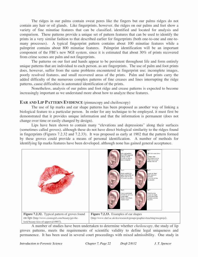
Lips have been shown to contain many “elevations and depressions” along their surfaces (sometimes called groves), although these do not have direct biological similarity to the ridges found in fingerprints (Figures 7.2.32 and 7.2.33). It was proposed as early at 1902 that the pattern formed by these groves could provide a means of personal identification. A number of methods for identifying lip marks features have been developed, although none has gained general acceptance.
A number of studies have been undertaken to determine whether cheiloscopy, the study of lip groves patterns, meets the requirements of scientific validity to define legal uniqueness and permanence. It has been used in several court proceedings with mixed admissibility. One study in
Figure 7.2.32. Typical pattern of groves found on lips (http://www.cosmogirl.com/beauty/get-the-look/beauty-kiss-of-approval-0907).
Figure 7.2.33. Examples of ear shapes (http://www.shef.ac.uk/dcs/research/groups/graphics/teaching/mscprojs).

Introduction to Forensic Science Chapter 7, Page 23 Draft 2/8/12 J. T. Spencer
Japan measured the lip marks of over 1300 subject, including a number of identical twins, and found them uniquely different and followed several others for several years at noted that the patterns did not appear to change. The scientific validity of the technique remains, however, untested and a great deal of work is needed before this technique can find acceptable use in forensic investigations and courtroom proceedings.
In a similar fashion, the shapes of a person’s ears has shown significant variation and have been used to tie a particular person with a “found” earprint (pinnascopy), such as on a window, door, or mirror.
Introduction to Forensic Science Chapter 7, Page 24 Draft 2/8/12 J. T. Spencer
7.3. Hair Analysis Learning Goals and Objectives Hair and fiber information can provide valuable forensic information on the origin and history of the sample. In order to understand how hair analysis can used, you will need to develop an understanding of:
Ø the chemical composition and structure of hair; Ø how hair is formed and how does it grow; Ø how we can tell human hair for that of other animals; Ø how we can tell where on the body a hair sample originated; Ø how ethnicity information can be gotten from a hair sample; Ø how information about the treatment of hair can be obtained; Ø how hair can be used in toxicological studies; Ø how can diseases and abnormalities be used to characterize hair samples; Ø how hair samples are collected and analyzed;
Introduction In the early biological development of a person, both the skin, with its friction ridges, and hair form from the ectoderm, or outermost, layer. As such, hair and skin not only share a strong biological connection, with hair considered as a derivative of our skin, but also more simply, both of these tissues are found primarily on the outside of an organism. Hair and skin are, therefore, the components of our bodies that directly face the environment and can very easily leave lasting and identifying forensic evidence. Because of the similarity of hair and fibers in their form and forensic function, we will consider them together in this section, beginning with a consideration of hair.
Hair and fiber samples are among the most durable of all biological materials and retain much of their forensic value for many, many years. While most biological tissues are usually quickly destroyed after death, hair samples have been know to persist virtually unchanged for thousands of years. These samples can provide both structural and chemical clues as to both their individual origin and the underlying biochemistry that formed them. Similarly, durable fibers, both natural and man-made, are found throughout our society in cloth and other items that can provide useful information about their origin, composition, form and use.
Hair and Fur. Hair is a complex appendage that grows from a follicle in the skin of only mammals and is a derivative of the epidermis of the skin. One of its main purposes is to help regulate the body temperature of an organism by either trapping or releasing warm air near the skin’s surface. The protective function of hair and it exposure to extreme conditions requires it to be strong and durable.
Hair has enormous diversity of form – both between two different species of organisms and from individual person to person. Traditionally, hair from non-human mammals is referred to as fur rather than hair, but the structures are typically very much the same.
Introduction to Forensic Science Chapter 7, Page 25 Draft 2/8/12 J. T. Spencer
Composition of Hair: Hair is composed of about 80 to 90% protein, mostly keratin and melanin, and between 8 to 15% water, with the remainder mostly as lipids. Keratin is a tough, durable, fibrous protein composed of long chains of amino acids typically found as a structural component of hair, nails, horns and claws. Melanin, however, is a pigment polymer derived mostly from the amino acid called tyrosine that imparts the color to a hair sample. Generally, the darker the hair, the more melanin it contains. There are, however, several types of melanin commonly found in hair. The dark pigment called eumelanin colors black and brown hair while the pigment called pheomelanin is the main coloration chemical found in red hair. Blonde hair simply has lower amounts of melanin overall while gray hair typically lacks melanin completely. All hair samples have very similar chemical compositions which limits the use of a chemical analysis in the individualization of hair sample as coming from a particular person.
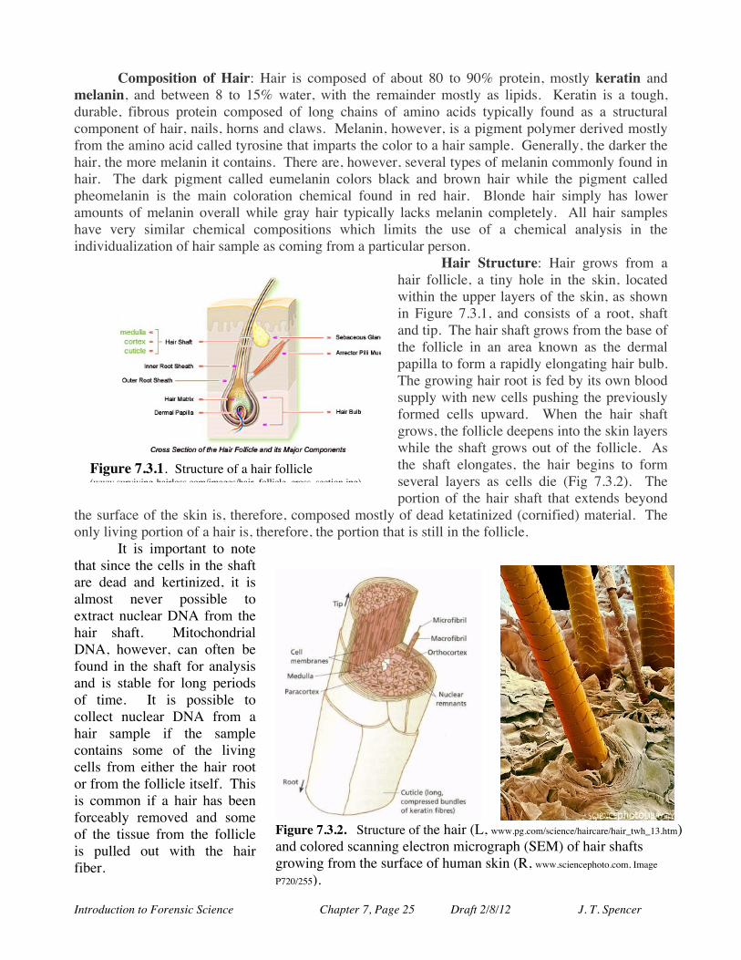
Hair Structure: Hair grows from a hair follicle, a tiny hole in the skin, located within the upper layers of the skin, as shown in Figure 7.3.1, and consists of a root, shaft and tip. The hair shaft grows from the base of the follicle in an area known as the dermal papilla to form a rapidly elongating hair bulb. The growing hair root is fed by its own blood supply with new cells pushing the previously formed cells upward. When the hair shaft grows, the follicle deepens into the skin layers while the shaft grows out of the follicle. As the shaft elongates, the hair begins to form several layers as cells die (Fig 7.3.2). The portion of the hair shaft that extends beyond
the surface of the skin is, therefore, composed mostly of dead ketatinized (cornified) material. The only living portion of a hair is, therefore, the portion that is still in the follicle.
It is important to note that since the cells in the shaft are dead and kertinized, it is almost never possible to extract nuclear DNA from the hair shaft. Mitochondrial DNA, however, can often be found in the shaft for analysis and is stable for long periods of time. It is possible to collect nuclear DNA from a hair sample if the sample contains some of the living cells from either the hair root or from the follicle itself. This is common if a hair has been forceably removed and some of the tissue from the follicle is pulled out with the hair fiber.
Figure 7.3.2. Structure of the hair (L, www.pg.com/science/haircare/hair_twh_13.htm) and colored scanning electron micrograph (SEM) of hair shafts growing from the surface of human skin (R, www.sciencephoto.com, Image
P720/255).
Figure 7.3.1. Structure of a hair follicle (www.surviving-hairloss.com/images/hair_follicle_cross_section.jpg).
Introduction to Forensic Science Chapter 7, Page 26 Draft 2/8/12 J. T. Spencer
Copernican Hair?
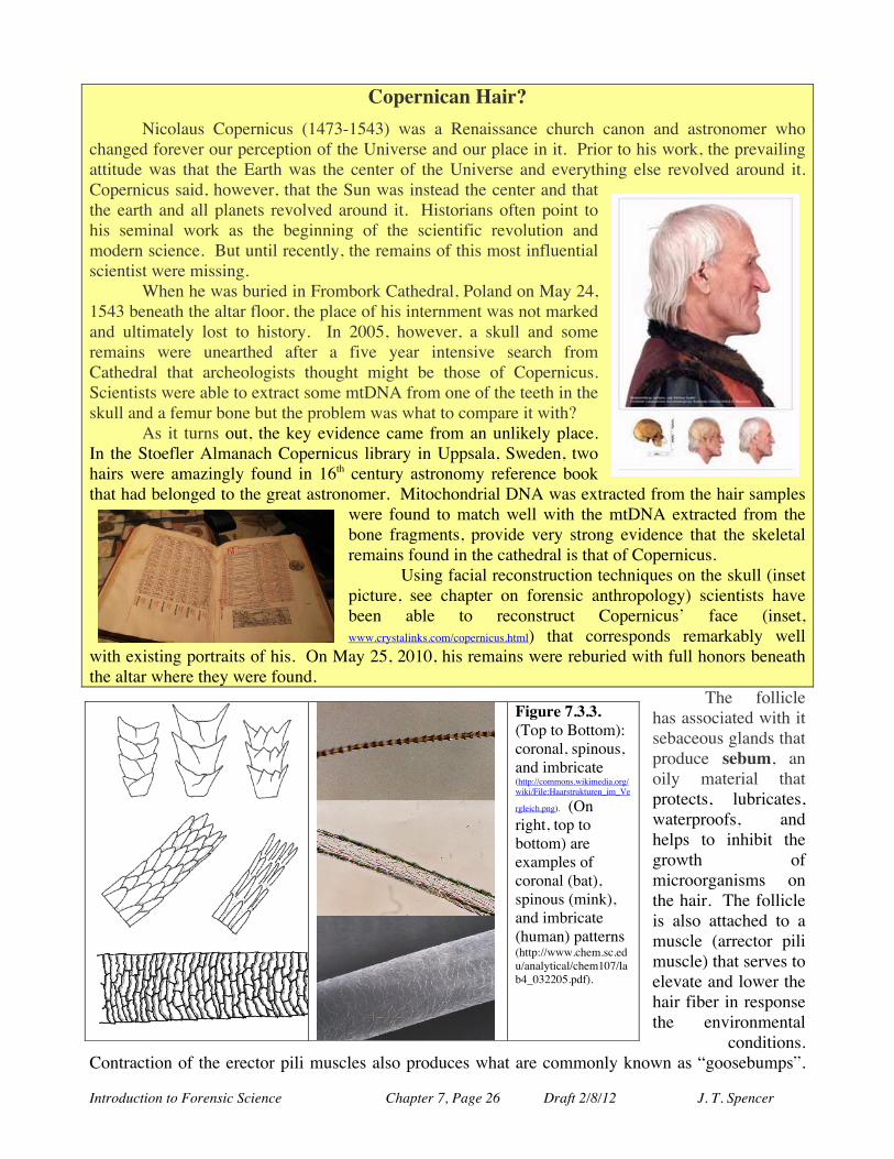
Nicolaus Copernicus (1473-1543) was a Renaissance church canon and astronomer who changed forever our perception of the Universe and our place in it. Prior to his work, the prevailing attitude was that the Earth was the center of the Universe and everything else revolved around it. Copernicus said, however, that the Sun was instead the center and that the earth and all planets revolved around it. Historians often point to his seminal work as the beginning of the scientific revolution and modern science. But until recently, the remains of this most influential scientist were missing.
When he was buried in Frombork Cathedral, Poland on May 24, 1543 beneath the altar floor, the place of his internment was not marked and ultimately lost to history. In 2005, however, a skull and some remains were unearthed after a five year intensive search from Cathedral that archeologists thought might be those of Copernicus. Scientists were able to extract some mtDNA from one of the teeth in the skull and a femur bone but the problem was what to compare it with?
As it turns out, the key evidence came from an unlikely place. In the Stoefler Almanach Copernicus library in Uppsala, Sweden, two hairs were amazingly found in 16th century astronomy reference book that had belonged to the great astronomer. Mitochondrial DNA was extracted from the hair samples
were found to match well with the mtDNA extracted from the bone fragments, provide very strong evidence that the skeletal remains found in the cathedral is that of Copernicus.
Using facial reconstruction techniques on the skull (inset picture, see chapter on forensic anthropology) scientists have been able to reconstruct Copernicus’ face (inset, www.crystalinks.com/copernicus.html) that corresponds remarkably well
with existing portraits of his. On May 25, 2010, his remains were reburied with full honors beneath the altar where they were found.
The follicle has associated with it sebaceous glands that produce sebum, an oily material that protects, lubricates, waterproofs, and helps to inhibit the growth of microorganisms on the hair. The follicle is also attached to a muscle (arrector pili muscle) that serves to elevate and lower the hair fiber in response the environmental
conditions. Contraction of the erector pili muscles also produces what are commonly known as “goosebumps”.
Figure 7.3.3. (Top to Bottom): coronal, spinous, and imbricate (http://commons.wikimedia.org/wiki/File:Haarstrukturen_im_Ve
rgleich.png). (On right, top to bottom) are examples of coronal (bat), spinous (mink), and imbricate (human) patterns (http://www.chem.sc.edu/analytical/chem107/lab4_032205.pdf).
Introduction to Forensic Science Chapter 7, Page 27 Draft 2/8/12 J. T. Spencer
Thus, when it’s cold outside, the erector muscles contract to raise the hair shaft to trap a layer of warm air next to the skin to help keep us warm and conserve body heat.
Each mature hair fiber is typically made up of three components: the cuticle, cortex, and the medulla. The outermost translucent layer of a hair shaft is called the cuticle which appears similar to the shingles on a roof or the scales on a snake skin, with the exposed portion of the “scale” aimed towards the tip of the shaft (Figures 7.3.3 and 7.3.4). You can sometimes feel this directionality of the cuticle scales by first
running your pinched fingers moving along a hair shaft from your head toward the end of the hair and comparing it with running your fingers in the opposite direction from the tip If often feels rougher when moving from the tip towards the scalp since this is moving against the “grain” of the cuticle scales towards your head. This relatively thin layer, usually just six to ten cells thick, protects the hair by forming a waterproof and rather impervious layer that coats and protects the entire shaft.
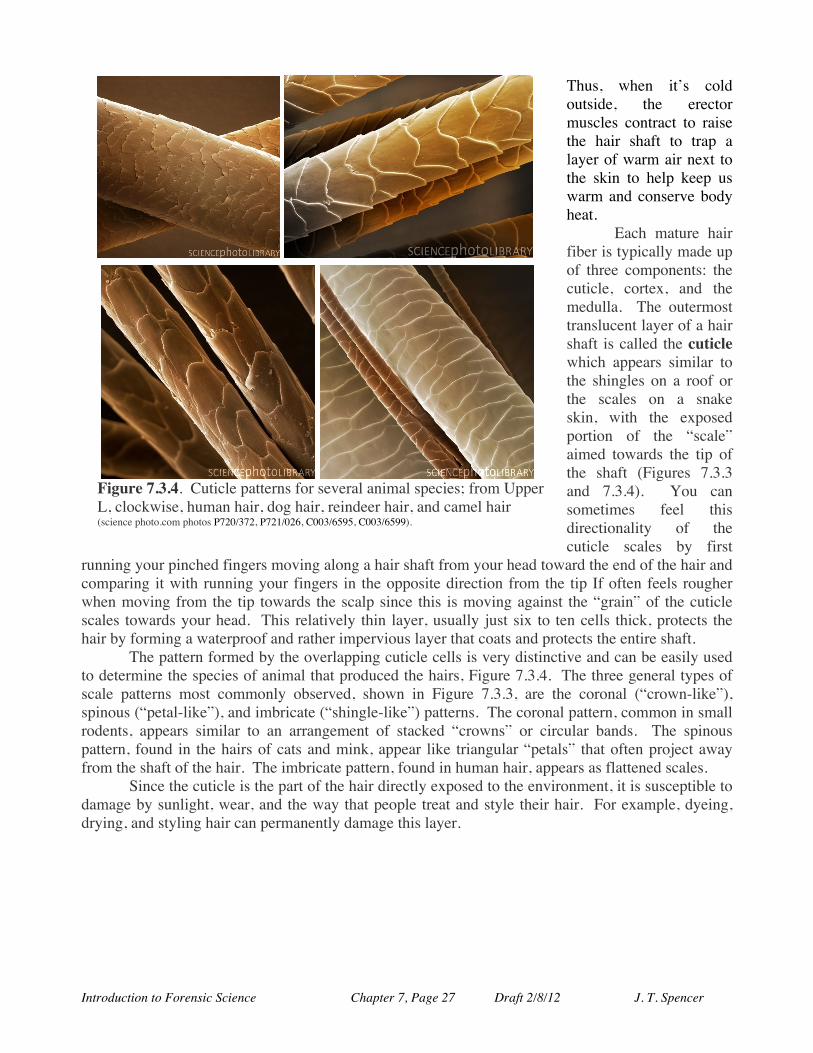
The pattern formed by the overlapping cuticle cells is very distinctive and can be easily used to determine the species of animal that produced the hairs, Figure 7.3.4. The three general types of scale patterns most commonly observed, shown in Figure 7.3.3, are the coronal (“crown-like”), spinous (“petal-like”), and imbricate (“shingle-like”) patterns. The coronal pattern, common in small rodents, appears similar to an arrangement of stacked “crowns” or circular bands. The spinous pattern, found in the hairs of cats and mink, appear like triangular “petals” that often project away from the shaft of the hair. The imbricate pattern, found in human hair, appears as flattened scales.
Since the cuticle is the part of the hair directly exposed to the environment, it is susceptible to damage by sunlight, wear, and the way that people treat and style their hair. For example, dyeing, drying, and styling hair can permanently damage this layer.
Figure 7.3.4. Cuticle patterns for several animal species; from Upper L, clockwise, human hair, dog hair, reindeer hair, and camel hair (science photo.com photos P720/372, P721/026, C003/6595, C003/6599).
Introduction to Forensic Science Chapter 7, Page 28 Draft 2/8/12 J. T. Spencer
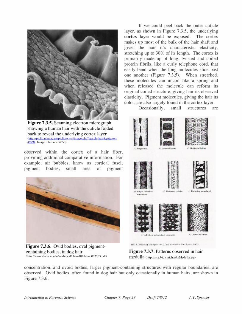
If we could peel back the outer cuticle layer, as shown in Figure 7.3.5, the underlying cortex layer would be exposed. The cortex makes up most of the bulk of the hair shaft and gives the hair it’s characteristic elasticity, stretching up to 30% of its length. The cortex is primarily made up of long, twisted and coiled protein fibrils, like a curly telephone cord, that easily bend when the long molecules slide past one another (Figure 7.3.5). When stretched, these molecules can uncoil like a spring and when released the molecule can reform its original coiled structure, giving hair its observed elasticity. Pigment molecules, giving the hair its color, are also largely found in the cortex layer.
Occasionally, small structures are
observed within the cortex of a hair fiber, providing additional comparative information. For example, air bubbles, know as cortical fusci, pigment bodies, small area of pigment
concentration, and ovoid bodies, larger pigment-containing structures with regular boundaries, are observed. Ovid bodies, often found in dog hair but only occasionally in human hairs, are shown in Figure 7.3.6.
Figure 7.3.5. Scanning electron micrograph showing a human hair with the cuticle folded back to reveal the underlying cortex layer (http://piclib.nhm.ac.uk/piclib/www/image.php?search=hair&getprev=49994, Image reference: 4650).
Figure 7.3.7. Patterns observed in hair medulla (http://atcg.bio.cmich.edu/Medulla.jpg)
Figure 7.3.6. Ovid bodies, oval pigment-containing bodies, in dog hair (http://www.chem.sc.edu/analytical/chem107/lab4_032205.pdf).
Introduction to Forensic Science Chapter 7, Page 29 Draft 2/8/12 J. T. Spencer
The third and innermost component of hair is the medulla. This part of the hair is characterized by either very spongy cells or no cells at all, forming a canal-like structure in the center of the shaft, often called the medullary canal. Melanin can also be found in this layer, contributing to the color of the hair. The medulla in human hair can form a continuous canal, be interrupted by areas without a medulla, or be missing a medulla altogether, Figure 7.4.7. The medulla pattern for some animals can be rather complex showing ladder-like or lattice-like patterns.
The ratio of the diameter of the shaft to the diameter of the medulla can be defined as the medullary index (MI) that can be used to help distinguish human hair from that of other animals. In many animals, the MI is greater than 0.5 while in humans it is typically found to be less than 0.3.
Hair varies greatly depending upon its location on the body. When we are fetuses, our entire bodies are covered with very fine colorless hair, called Lanugo. During early childhood, however, this lanugo hair is lost and the majority of our bodies are covered with fine short hairs, called Vellus hair or sometimes referred to as "peach fuzz". During puberty, humans develop longer, thicker, colored hair on various parts of the body, besides the scalp and eyebrows, called terminal hair, that forms part of our secondary sex characteristics. Terminal hair includes hair found on our scalps, in armpits, on legs, chest hair and elsewhere.
Usually, a single hair follicle produces only type of hair but sometimes a follicle can change to produce a different type of hair. For example, until puberty, the facial follicles on a male produce only fine vellus hair. During puberty, these follicles change to produce a characteristic male beard made up of thicker, longer terminal hair. Similarly, follicles on the scalp usually produce only terminal hair but in some instances (androgenetic alopecia) the follicle can change to produce short, thin, lightly colored hair.
Most animal hairs are divided into three basic types: guard hairs (from the outer coat for protection), fur (from the inner coat for insulation and temperature regulation), and tactile hairs (for sensing, such as whiskers). Human hair, however, is not so well differentiated, resembling animal fur most closely.
The overall shape and length of a human hair can give information about where on the body it originated, such as the scalp, face, public area or elsewhere on the body. For example, scalp hair is usually long with cut or split tips and a relatively narrow medulla, while pubic hairs are typically
short, with a tapered or rounded tip, and contain a relatively broad medulla. Because of the variation of hair structures, even on one individual person, it is often necessary to collect many hair samples in order to get a representative sample. This also makes it very difficult to determine if a particular hair fiber originated from a particular person.
How Hair Grows: Hair growth occurs in a cycle composed of three main stages: the anagen, catagen, and telogen phases (Figure 7.3.8). The lengths of these cycles are genetically programmed and can vary
Figure 7.3.8. Phases of hair growth (http://westchesterelectrolysis.com/services.html).
Introduction to Forensic Science Chapter 7, Page 30 Draft 2/8/12 J. T. Spencer
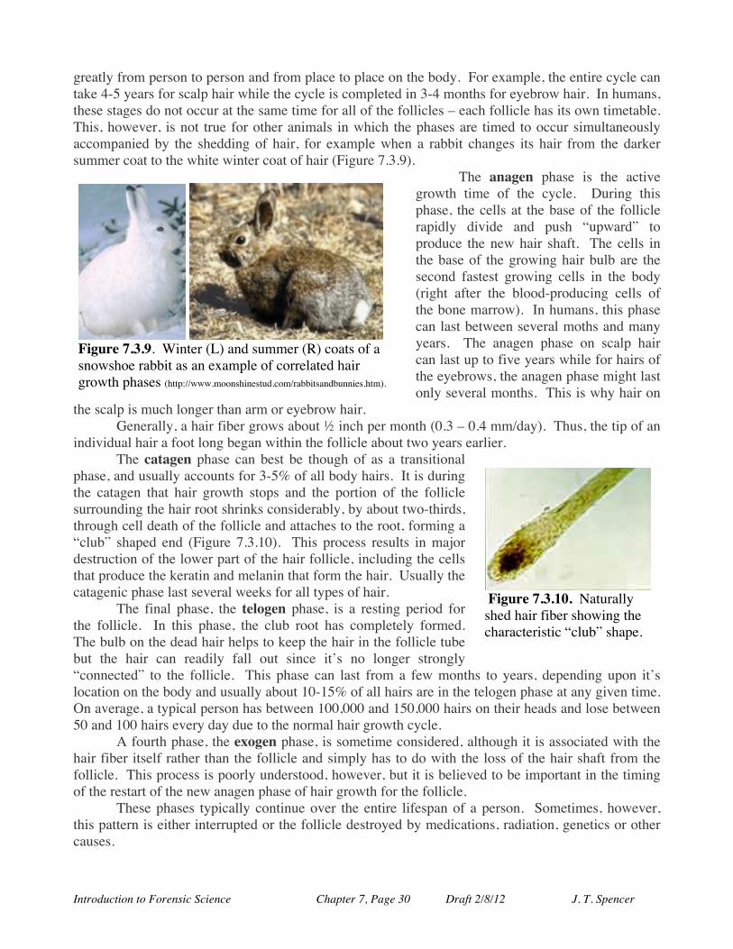
greatly from person to person and from place to place on the body. For example, the entire cycle can take 4-5 years for scalp hair while the cycle is completed in 3-4 months for eyebrow hair. In humans, these stages do not occur at the same time for all of the follicles – each follicle has its own timetable. This, however, is not true for other animals in which the phases are timed to occur simultaneously accompanied by the shedding of hair, for example when a rabbit changes its hair from the darker summer coat to the white winter coat of hair (Figure 7.3.9).
The anagen phase is the active growth time of the cycle. During this phase, the cells at the base of the follicle rapidly divide and push “upward” to produce the new hair shaft. The cells in the base of the growing hair bulb are the second fastest growing cells in the body (right after the blood-producing cells of the bone marrow). In humans, this phase can last between several moths and many years. The anagen phase on scalp hair can last up to five years while for hairs of the eyebrows, the anagen phase might last only several months. This is why hair on
the scalp is much longer than arm or eyebrow hair. Generally, a hair fiber grows about ½ inch per month (0.3 – 0.4 mm/day). Thus, the tip of an
individual hair a foot long began within the follicle about two years earlier. The catagen phase can best be though of as a transitional
phase, and usually accounts for 3-5% of all body hairs. It is during the catagen that hair growth stops and the portion of the follicle surrounding the hair root shrinks considerably, by about two-thirds, through cell death of the follicle and attaches to the root, forming a “club” shaped end (Figure 7.3.10). This process results in major destruction of the lower part of the hair follicle, including the cells that produce the keratin and melanin that form the hair. Usually the catagenic phase last several weeks for all types of hair.
The final phase, the telogen phase, is a resting period for the follicle. In this phase, the club root has completely formed. The bulb on the dead hair helps to keep the hair in the follicle tube but the hair can readily fall out since it’s no longer strongly “connected” to the follicle. This phase can last from a few months to years, depending upon it’s location on the body and usually about 10-15% of all hairs are in the telogen phase at any given time. On average, a typical person has between 100,000 and 150,000 hairs on their heads and lose between 50 and 100 hairs every day due to the normal hair growth cycle.
A fourth phase, the exogen phase, is sometime considered, although it is associated with the hair fiber itself rather than the follicle and simply has to do with the loss of the hair shaft from the follicle. This process is poorly understood, however, but it is believed to be important in the timing of the restart of the new anagen phase of hair growth for the follicle.
These phases typically continue over the entire lifespan of a person. Sometimes, however, this pattern is either interrupted or the follicle destroyed by medications, radiation, genetics or other causes.
Figure 7.3.10. Naturally shed hair fiber showing the characteristic “club” shape.
Figure 7.3.9. Winter (L) and summer (R) coats of a snowshoe rabbit as an example of correlated hair growth phases (http://www.moonshinestud.com/rabbitsandbunnies.htm).
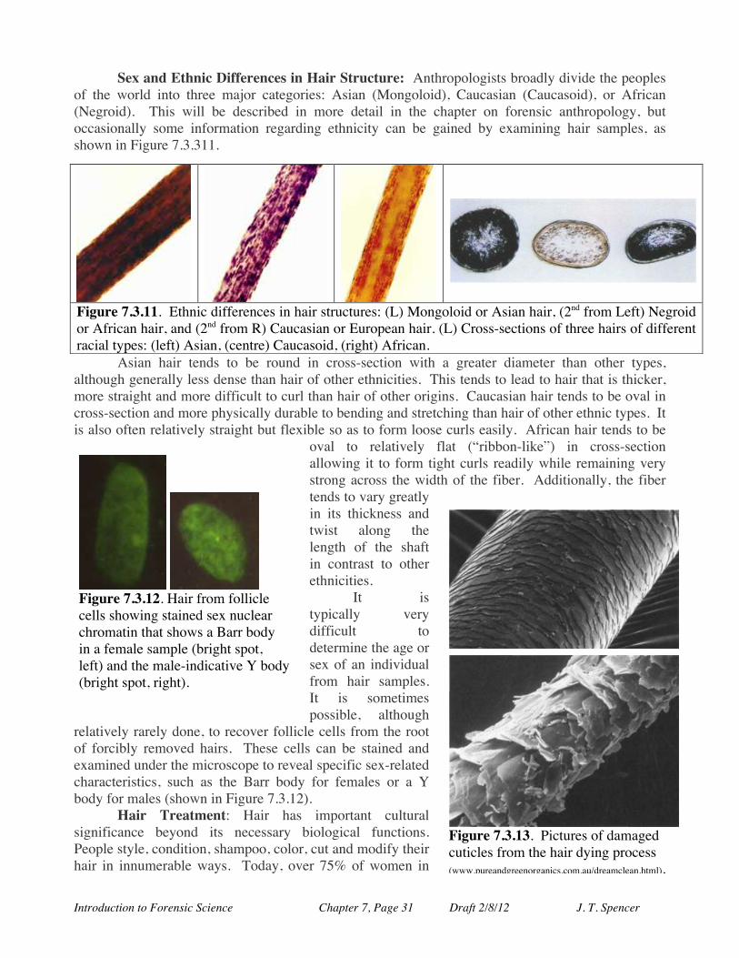
Introduction to Forensic Science Chapter 7, Page 31 Draft 2/8/12 J. T. Spencer
Sex and Ethnic Differences in Hair Structure: Anthropologists broadly divide the peoples of the world into three major categories: Asian (Mongoloid), Caucasian (Caucasoid), or African (Negroid). This will be described in more detail in the chapter on forensic anthropology, but occasionally some information regarding ethnicity can be gained by examining hair samples, as shown in Figure 7.3.311.
Asian hair tends to be round in cross-section with a greater diameter than other types, although generally less dense than hair of other ethnicities. This tends to lead to hair that is thicker, more straight and more difficult to curl than hair of other origins. Caucasian hair tends to be oval in cross-section and more physically durable to bending and stretching than hair of other ethnic types. It is also often relatively straight but flexible so as to form loose curls easily. African hair tends to be
oval to relatively flat (“ribbon-like”) in cross-section allowing it to form tight curls readily while remaining very strong across the width of the fiber. Additionally, the fiber tends to vary greatly in its thickness and twist along the length of the shaft in contrast to other ethnicities.
It is typically very difficult to determine the age or sex of an individual from hair samples. It is sometimes possible, although
relatively rarely done, to recover follicle cells from the root of forcibly removed hairs. These cells can be stained and examined under the microscope to reveal specific sex-related characteristics, such as the Barr body for females or a Y body for males (shown in Figure 7.3.12).
Hair Treatment: Hair has important cultural significance beyond its necessary biological functions. People style, condition, shampoo, color, cut and modify their hair in innumerable ways. Today, over 75% of women in
Figure 7.3.11. Ethnic differences in hair structures: (L) Mongoloid or Asian hair, (2nd from Left) Negroid or African hair, and (2nd from R) Caucasian or European hair. (L) Cross-sections of three hairs of different racial types: (left) Asian, (centre) Caucasoid, (right) African.
Figure 7.3.12. Hair from follicle cells showing stained sex nuclear chromatin that shows a Barr body in a female sample (bright spot, left) and the male-indicative Y body (bright spot, right).
Figure 7.3.13. Pictures of damaged cuticles from the hair dying process (www.pureandgreenorganics.com.au/dreamclean.html).
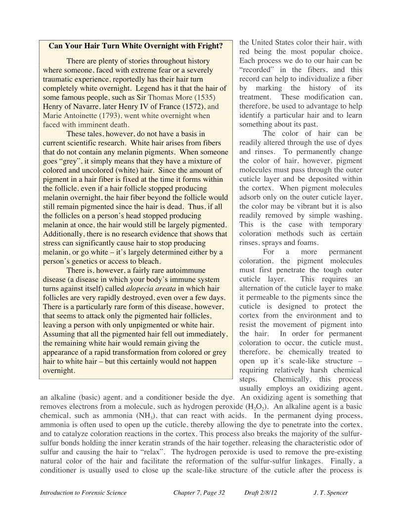
Introduction to Forensic Science Chapter 7, Page 32 Draft 2/8/12 J. T. Spencer
the United States color their hair, with red being the most popular choice. Each process we do to our hair can be “recorded” in the fibers, and this record can help to individualize a fiber by marking the history of its treatment. These modification can, therefore, be used to advantage to help identify a particular hair and to learn something about its past.
The color of hair can be readily altered through the use of dyes and rinses. To permanently change the color of hair, however, pigment molecules must pass through the outer cuticle layer and be deposited within the cortex. When pigment molecules adsorb only on the outer cuticle layer, the color may be vibrant but it is also readily removed by simple washing. This is the case with temporary coloration methods such as certain rinses, sprays and foams.
For a more permanent coloration, the pigment molecules must first penetrate the tough outer cuticle layer. This requires an alternation of the cuticle layer to make it permeable to the pigments since the cuticle is designed to protect the cortex from the environment and to resist the movement of pigment into the hair. In order for permanent coloration to occur, the cuticle must, therefore, be chemically treated to open up it’s scale-like structure – requiring relatively harsh chemical steps. Chemically, this process usually employs an oxidizing agent,
an alkaline (basic) agent, and a conditioner beside the dye. An oxidizing agent is something that removes electrons from a molecule, such as hydrogen peroxide (H2O2). An alkaline agent is a basic chemical, such as ammonia (NH3), that can react with acids. In the permanent dying process, ammonia is often used to open up the cuticle, thereby allowing the dye to penetrate into the cortex, and to catalyze coloration reactions in the cortex. This process also breaks the majority of the sulfur-sulfur bonds holding the inner keratin strands of the hair together, releasing the characteristic odor of sulfur and causing the hair to “relax”. The hydrogen peroxide is used to remove the pre-existing natural color of the hair and facilitate the reformation of the sulfur-sulfur linkages. Finally, a conditioner is usually used to close up the scale-like structure of the cuticle after the process is
Can Your Hair Turn White Overnight with Fright?
There are plenty of stories throughout history where someone, faced with extreme fear or a severely traumatic experience, reportedly has their hair turn completely white overnight. Legend has it that the hair of some famous people, such as Sir Thomas More (1535) Henry of Navarre, later Henry IV of France (1572), and Marie Antoinette (1793), went white overnight when faced with imminent death. These tales, however, do not have a basis in current scientific research. White hair arises from fibers that do not contain any melanin pigments. When someone goes “grey”, it simply means that they have a mixture of colored and uncolored (white) hair. Since the amount of pigment in a hair fiber is fixed at the time it forms within the follicle, even if a hair follicle stopped producing melanin overnight, the hair fiber beyond the follicle would still remain pigmented since the hair is dead. Thus, if all the follicles on a person’s head stopped producing melanin at once, the hair would still be largely pigmented. Additionally, there is no research evidence that shows that stress can significantly cause hair to stop producing melanin, or go white – it’s largely determined either by a person’s genetics or access to bleach. There is, however, a fairly rare autoimmune disease (a disease in which your body’s immune system turns against itself) called alopecia areata in which hair follicles are very rapidly destroyed, even over a few days. There is a particularly rare form of this disease, however, that seems to attack only the pigmented hair follicles, leaving a person with only unpigmented or white hair. Assuming that all the pigmented hair fell out immediately, the remaining white hair would remain giving the appearance of a rapid transformation from colored or grey hair to white hair – but this certainly would not happen overnight.
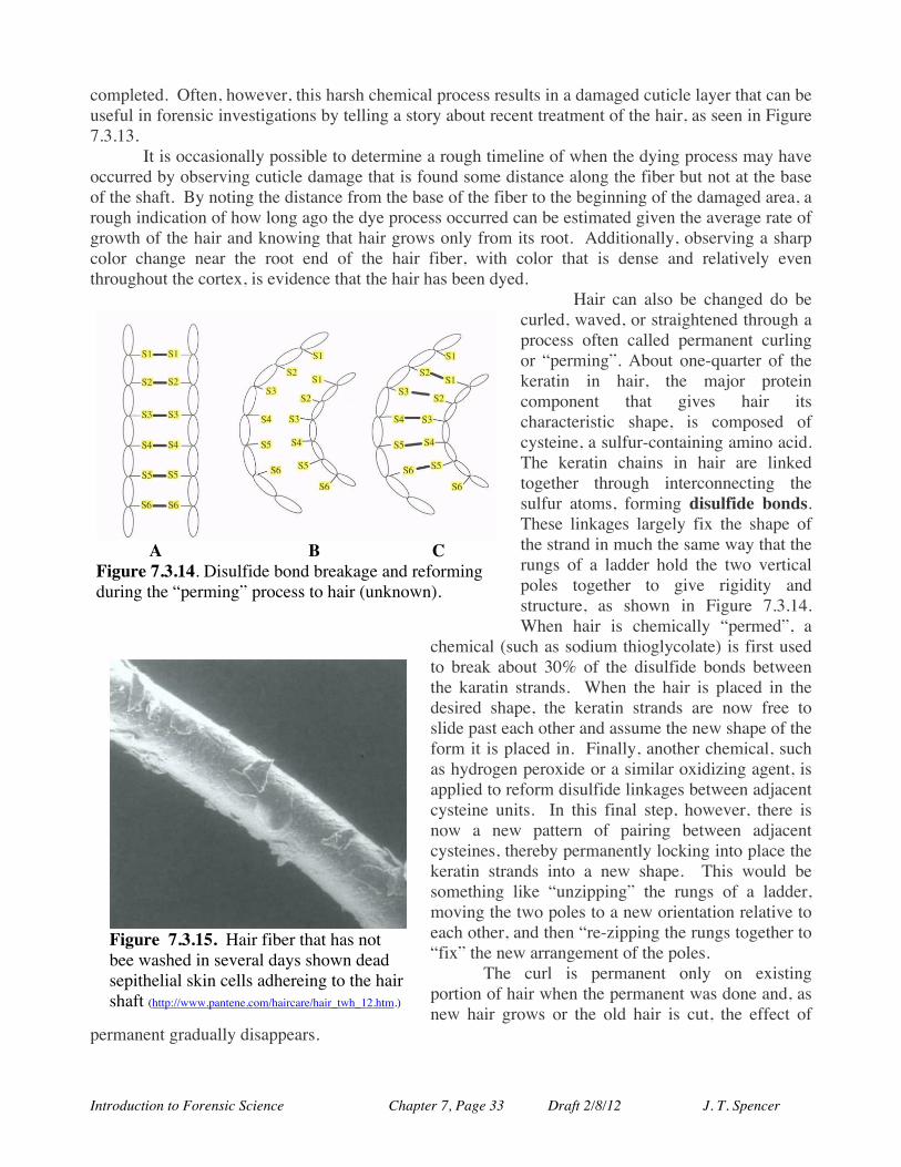
Introduction to Forensic Science Chapter 7, Page 33 Draft 2/8/12 J. T. Spencer
completed. Often, however, this harsh chemical process results in a damaged cuticle layer that can be useful in forensic investigations by telling a story about recent treatment of the hair, as seen in Figure 7.3.13.
It is occasionally possible to determine a rough timeline of when the dying process may have occurred by observing cuticle damage that is found some distance along the fiber but not at the base of the shaft. By noting the distance from the base of the fiber to the beginning of the damaged area, a rough indication of how long ago the dye process occurred can be estimated given the average rate of growth of the hair and knowing that hair grows only from its root. Additionally, observing a sharp color change near the root end of the hair fiber, with color that is dense and relatively even throughout the cortex, is evidence that the hair has been dyed.
Hair can also be changed do be curled, waved, or straightened through a process often called permanent curling or “perming”. About one-quarter of the keratin in hair, the major protein component that gives hair its characteristic shape, is composed of cysteine, a sulfur-containing amino acid. The keratin chains in hair are linked together through interconnecting the sulfur atoms, forming disulfide bonds. These linkages largely fix the shape of the strand in much the same way that the rungs of a ladder hold the two vertical poles together to give rigidity and structure, as shown in Figure 7.3.14. When hair is chemically “permed”, a
chemical (such as sodium thioglycolate) is first used to break about 30% of the disulfide bonds between the karatin strands. When the hair is placed in the desired shape, the keratin strands are now free to slide past each other and assume the new shape of the form it is placed in. Finally, another chemical, such as hydrogen peroxide or a similar oxidizing agent, is applied to reform disulfide linkages between adjacent cysteine units. In this final step, however, there is now a new pattern of pairing between adjacent cysteines, thereby permanently locking into place the keratin strands into a new shape. This would be something like “unzipping” the rungs of a ladder, moving the two poles to a new orientation relative to each other, and then “re-zipping the rungs together to “fix” the new arrangement of the poles.
The curl is permanent only on existing portion of hair when the permanent was done and, as new hair grows or the old hair is cut, the effect of
permanent gradually disappears.
A B C
Figure 7.3.14. Disulfide bond breakage and reforming during the “perming” process to hair (unknown).
Figure 7.3.15. Hair fiber that has not bee washed in several days shown dead sepithelial skin cells adhereing to the hair shaft (http://www.pantene.com/haircare/hair_twh_12.htm.)
Introduction to Forensic Science Chapter 7, Page 34 Draft 2/8/12 J. T. Spencer
People also very commonly clean and style their hair through regular cleaning and the application of hair products. Several forensically interesting pieces of information can be gained by considering the cleaning and styling of hair fibers. As shown in Figure 7.3.15, information about any recent washing can be can be revealed microscopically. Additionally, chemical analysis of the surface or rinsing of the fiber can also show residues of particular types of rinses and chemical treatments (see the section on chemical analysis).
Cutting hair, either to maintain a desired hairstyle, of through accident, injury, or assault can also be used to provide important on the history of a hair sample. Examples of a few of this type of
information from hair fibers can be seen in Figure 7.3.16. Diseases Involving Hair: Since everyone’s basic hair chemistry is about the same, it is
difficult to individualize a particular hair sample sufficiently to connect it with one individual person. Therefore, scientists try to find ways to help individualize a hair sample. One way is to identify the hair as associated with a hair-related disease, abnormality, or infestation. For example, a deficiency of certain vitamins or minerals, such as a zinc deficiency (Figure 7.3.17), can result in abnormal hair growth that can be chemically detected in the hair. If a suspect suffers from a similar deficiency, a better link can be made between the recovered sample and a hair sample of known origin. Abnormalities in metabolism, hormone levels, or other biochemical irregularities can also be detected
A
B
C
D
E
F
Figure 7.3.16. Hair patterns from treatment or injury: (a) hair root forcibly removed, (b) hair cut, (c) hair cut by razor, (d) split ends, (e) hair with dandruff, and (f) postmortem root band (Credits: (a), (c) and (e);http://www.fbi.gov/hq/lab/fsc/backissu/july2000/deedric1.htm; (b) and (d); sciencephoto.com images P720/154 and P720/344:(e) http://scienceray.com/biology/microbiology/see-through-the-eyes-of-the-bacteria/).
Figure 7.3.17. The effect of zinc deficiency on hair growth. The picture of left shows a child with a zinc deficiency wich thin and sparse hair. After treatment with a zinc supplement, his hair growth is more normal (www.pgbeautygroomingscience.com/the-hair-growth-cycle.html).
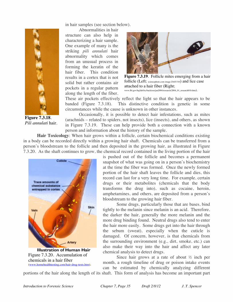
Introduction to Forensic Science Chapter 7, Page 35 Draft 2/8/12 J. T. Spencer
in hair samples (see section below). Abnormalities in hair
structure can also help in characterizing a hair sample. One example of many is the striking pili annulati hair abnormality which comes from an unusual process in forming the keratin of the hair fiber. This condition results in a cortex that is not solid but rather contains air pockets in a regular pattern along the length of the fiber. These air pockets effectively reflect the light so that the hair appears to be banded (Figure 7.3.18). This distinctive condition is genetic in some circumstances while the cause is unknown in other instances.
Occasionally, it is possible to detect hair infestations, such as mites (arachnids – related to spiders, not insects), lice (insects), and others, as shown in Figure 7.3.19. These can help provide both a connection with a known person and information about the history of the sample.
Hair Toxicology: When hair grows within a follicle, certain biochemical conditions existing in a body can be recorded directly within a growing hair shaft. Chemicals can be transferred from a person’s bloodstream to the follicle and then deposited in the growing hair, as illustrated in Figure 7.3.20. As the shaft continues to grow, the chemical record contained in the living portion of the hair
is pushed out of the follicle and becomes a permanent snapshot of what was going on in a person’s biochemistry at the time the fiber was formed. Once the newly formed portion of the hair shaft leaves the follicle and dies, this record can last for a very long time. For example, certain drugs or their metabolites (chemicals that the body transforms the drug into), such as cocaine, heroin, amphetamines, and others, are deposited from a person’s bloodstream to the growing hair fiber.
Some drugs, particularly those that are bases, bind tightly to the melanin since melanin is an acid. Therefore, the darker the hair, generally the more melanin and the more drug binding found. Neutral drugs also tend to enter the hair more easily. Some drugs get into the hair through the sebum (sweat), especially when the cuticle is damaged. Of concern, however, is that chemicals from the surrounding environment (e.g., dirt, smoke, etc.) can also make their way into the hair and affect any later chemical analysis to detect drugs.
Since hair grows at a rate of about ½ inch per month, a rough timeline of drug or poison intake events can be estimated by chemically analyzing different
portions of the hair along the length of its shaft. This form of analysis has become an important part
Figure 7.3.20. Accumulation of chemicals in a hair fiber (www.homehealthtesting.com/hair-drug-tests.htm).
Figure 7.3.18. Pili annulati hair.
Figure 7.3.19. Follicle mites emerging from a hair follicle (Left; sciencephoto.com image Z445/310) and lice case attached to a hair fiber (Right; www.fbi.gov/hq/lab/fsc/backissu/jan2004/research/2004_01_research01b.htm).

Introduction to Forensic Science Chapter 7, Page 36 Draft 2/8/12 J. T. Spencer
of drug surveillance for people on parole, checking to see if a patient has been complying with a therapeutic drug therapy, or checking the reliability of a person’s statement regarding drug use. Beside serving of a record of drug use, this toxicological information can also aid in determining if two fibers have a common source – for example, one found at a crime scene and one taken from a victim.
As mentioned before, analysis of hair fibers can provide record a person’s exposure to environmental toxins in the air, water, food or elsewhere. Hair can also serve as a strong piece of evidence showing a person’s long-term use of alcohol. When alcohol is consumed, the body produces fatty acid ethyl esters (FAEE), ethyl glucuronide (EtG), and ethylsulfate (EtS) (Figure 7.3.21). While alcohol is not deposited in hair, these three metabolites of alcohol (ethanol) are permanently deposited in growing hair fibers, making a lasting record of alcohol use – even years long.
There are a number of both important advantages and challenges in considering toxicology issues in hair samples. Hair is usually a readily available, non-invasive, inexpensive, and long-lasting medium for analysis. In addition, chemical analyses have been developed for trace levels of compounds found within hair fibers. But hair analysis also has some significant difficulties. For example, darker and coarser hair fibers retain drug information longer and better than lighter and finer hair. This may lead to a different drug use profile determined from hair for two people with identical usages. False positives may also present significant problems, such as in the determination of alcohol abuse through EtG analysis. For example, in a recent study, it was surprisingly shown that a false positive can come from a person using an alcohol-containing hand sanitizer before the analysis. There is also problems in establishing a relationship between the amount of a drug found in a hair sample with what was in a person’s blood system. Other problems currently being addressed include: (1) use of hair from different places on the body, (2) ethnic differences in drug absorption and retention in hair, and (3) the effect of cosmetics. Nonetheless, the use of hair analysis for drug and toxin exposure is increasing rapidly and forms an important forensic tool.
The End of an Emperor: Telltale Hair?
Napoleon Bonaparte (1769-1821) is one of the most studied, despised, and revered of all people in history, with tens of thousands of books published on his life and exploits, with thousands of new titles appearing every year. He is certainly a man of intrigue and mystery but one of the greatest mysteries regarding this enigmatic man is the cause and manner of his death. After Napoleon lost the Battle of Waterloo in 1815 and later surrendered to the British, he was exiled to the
remote tropical island of St. Helena in the South Atlantic, still one of the most isolated places on Earth. He and his retinue of about 20 friends and associates lived for six year on the island, under the close watch of the British commander, before his death in 1821. At the time of his death, an autopsy was performed by British surgeons and the cause of death was determined to be a perforated (“bleeding”) stomach ulcer that had become cancerous. But today, nearly two hundred years later, controversy still remains surrounding his death. During his exile, Napoleon often thought of escape and his relations with the British
Figure 7.3.21. Structure of ethyl Glucuronide and ethyl sulfate.
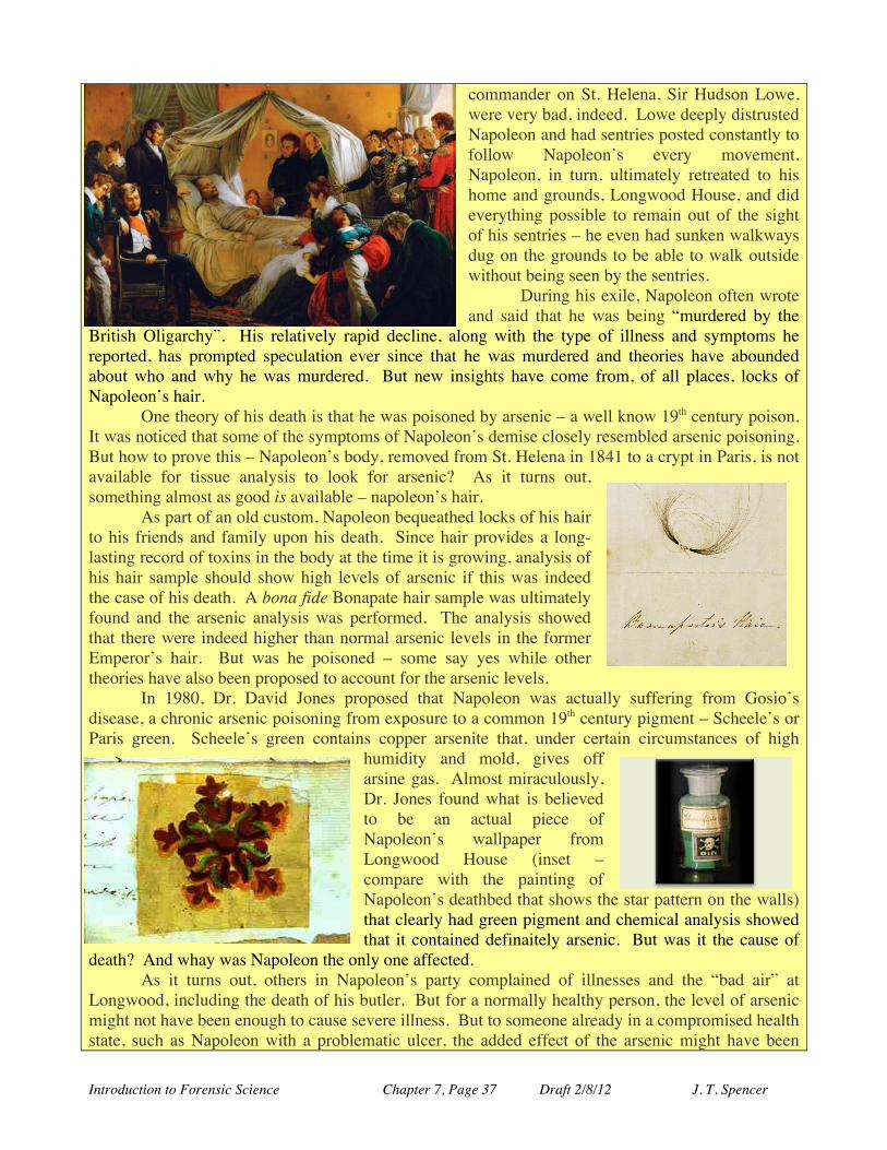
Introduction to Forensic Science Chapter 7, Page 37 Draft 2/8/12 J. T. Spencer
commander on St. Helena, Sir Hudson Lowe, were very bad, indeed. Lowe deeply distrusted Napoleon and had sentries posted constantly to follow Napoleon’s every movement. Napoleon, in turn, ultimately retreated to his home and grounds, Longwood House, and did everything possible to remain out of the sight of his sentries – he even had sunken walkways dug on the grounds to be able to walk outside without being seen by the sentries. During his exile, Napoleon often wrote and said that he was being “murdered by the
British Oligarchy”. His relatively rapid decline, along with the type of illness and symptoms he reported, has prompted speculation ever since that he was murdered and theories have abounded about who and why he was murdered. But new insights have come from, of all places, locks of Napoleon’s hair. One theory of his death is that he was poisoned by arsenic – a well know 19th century poison. It was noticed that some of the symptoms of Napoleon’s demise closely resembled arsenic poisoning. But how to prove this – Napoleon’s body, removed from St. Helena in 1841 to a crypt in Paris, is not available for tissue analysis to look for arsenic? As it turns out, something almost as good is available – napoleon’s hair. As part of an old custom, Napoleon bequeathed locks of his hair to his friends and family upon his death. Since hair provides a long-lasting record of toxins in the body at the time it is growing, analysis of his hair sample should show high levels of arsenic if this was indeed the case of his death. A bona fide Bonapate hair sample was ultimately found and the arsenic analysis was performed. The analysis showed that there were indeed higher than normal arsenic levels in the former Emperor’s hair. But was he poisoned – some say yes while other theories have also been proposed to account for the arsenic levels. In 1980, Dr. David Jones proposed that Napoleon was actually suffering from Gosio’s disease, a chronic arsenic poisoning from exposure to a common 19th century pigment – Scheele’s or Paris green. Scheele’s green contains copper arsenite that, under certain circumstances of high
humidity and mold, gives off arsine gas. Almost miraculously, Dr. Jones found what is believed to be an actual piece of Napoleon’s wallpaper from Longwood House (inset –compare with the painting of Napoleon’s deathbed that shows the star pattern on the walls) that clearly had green pigment and chemical analysis showed that it contained definaitely arsenic. But was it the cause of
death? And whay was Napoleon the only one affected. As it turns out, others in Napoleon’s party complained of illnesses and the “bad air” at Longwood, including the death of his butler. But for a normally healthy person, the level of arsenic might not have been enough to cause severe illness. But to someone already in a compromised health state, such as Napoleon with a problematic ulcer, the added effect of the arsenic might have been
Introduction to Forensic Science Chapter 7, Page 38 Draft 2/8/12 J. T. Spencer
enough to be a significant contributing cause of death. So, what did cause Napoleon’s death – at this point the evidence is not fully conclusive and we await more information. What do you think?
Hair Comparison and Identification: Probably the most important aspect of the examination of a hair sample is the observed microscopic structure of the medulla and cuticle of the sample. An individual hair fiber, however, cannot be individualized through its chemical composition or even usually from its structural features. The shaft of a mature human hair does not contain nuclear DNA so that only mitochondrial DNA analysis is possible for such samples. Often, however, tissue from the follicle may remain at the root of a hair sample, such as fibers forcibly removed from the scalp, so that a nuclear DNA analysis might be possible. Apart from DNA analyses, however, significant error rates are associated with the microscopic comparison of two hair samples. When comparing these samples, the color, length, and diameter of the hair fiber is particularly important. 7.3.2. Nails. Fingernails and toenails are, like hair, considered to be an appendage of the skin and are closely chemically related to the claws, hooves, and horns found in other animals. Like hair, nails are made up of the durable protein keratin. The importance of nails in forensic cases usually arises in
assault or other violent cases in which pieces of an attacker’s or victim’s fingernail become lodged in the others clothing or skin. Finding and analyzing the nail can be an important piece of evidence.
Nails primarily serve to protect the very sensitive ends of our fingers and toes and actually increase the sensitivity of the
fingers and toes. The tips of our fingers and toes are among the most sensitive portions of our bodies, through which we sense a great deal of information regarding the shape of the world. Fingernail Growth. Like hair, the majority of nails are dead keratinized material. The only living portion is the end of the nail is the nail root, or germinal matrix, that extends under the skin opposite to the end of the nail and is where the nerve, blood supply, and lymph vessels are found (Figure 7.2.22). The nail grows continuously from the root as long as it is healthy and is nourished, growing an average of 0.5 to 1.2 mm per week. Fingernail actually grow much faster than toenails – typically taking about 6 months to completely regrow a new fingernail while it make take up to two years to fully regrow a toe nail.
Figure 7.3.22. Fingernail structures (www.cures4beauty.com/nail.html).
Do Fingernails Continue to Grow After Death?
Many people believe that fingernails continue to grow after death and bodies exhumed after death appear to have much longer nails than expected. It turns out the this is actually an illusion based upon our normal “living” expectations.
The growth of the nails does indeed stop at death. After death, however, the tissue surround the nails shrinks and dehydrates, making it appear that the fingernails are longer. Since we are used to seeing fingernails grow and fingers remain the same size in life, we interpret what we see after death similarly – we think that the finger tissue remains the same size when it actually shrinks.
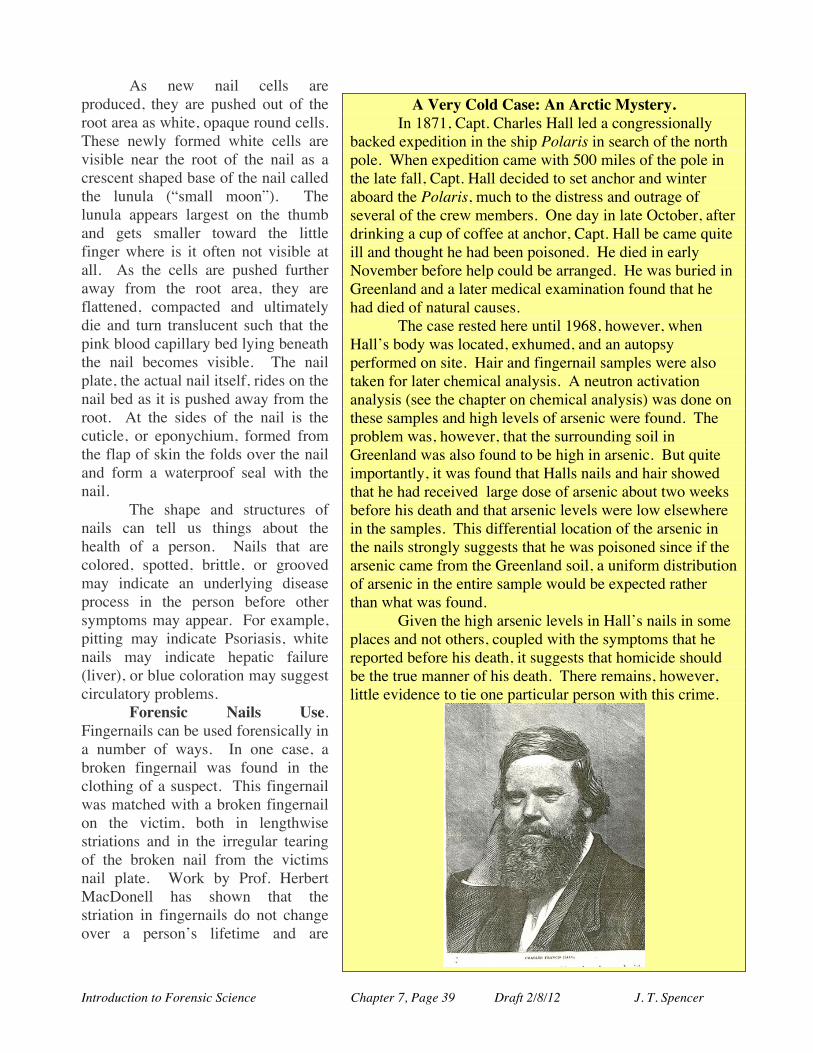
Introduction to Forensic Science Chapter 7, Page 39 Draft 2/8/12 J. T. Spencer
As new nail cells are produced, they are pushed out of the root area as white, opaque round cells. These newly formed white cells are visible near the root of the nail as a crescent shaped base of the nail called the lunula (“small moon”). The lunula appears largest on the thumb and gets smaller toward the little finger where is it often not visible at all. As the cells are pushed further away from the root area, they are flattened, compacted and ultimately die and turn translucent such that the pink blood capillary bed lying beneath the nail becomes visible. The nail plate, the actual nail itself, rides on the nail bed as it is pushed away from the root. At the sides of the nail is the cuticle, or eponychium, formed from the flap of skin the folds over the nail and form a waterproof seal with the nail.
The shape and structures of nails can tell us things about the health of a person. Nails that are colored, spotted, brittle, or grooved may indicate an underlying disease process in the person before other symptoms may appear. For example, pitting may indicate Psoriasis, white nails may indicate hepatic failure (liver), or blue coloration may suggest circulatory problems.
Forensic Nails Use. Fingernails can be used forensically in a number of ways. In one case, a broken fingernail was found in the clothing of a suspect. This fingernail was matched with a broken fingernail on the victim, both in lengthwise striations and in the irregular tearing of the broken nail from the victims nail plate. Work by Prof. Herbert MacDonell has shown that the striation in fingernails do not change over a person’s lifetime and are
A Very Cold Case: An Arctic Mystery. In 1871, Capt. Charles Hall led a congressionally backed expedition in the ship Polaris in search of the north pole. When expedition came with 500 miles of the pole in the late fall, Capt. Hall decided to set anchor and winter aboard the Polaris, much to the distress and outrage of several of the crew members. One day in late October, after drinking a cup of coffee at anchor, Capt. Hall be came quite ill and thought he had been poisoned. He died in early November before help could be arranged. He was buried in Greenland and a later medical examination found that he had died of natural causes. The case rested here until 1968, however, when Hall’s body was located, exhumed, and an autopsy performed on site. Hair and fingernail samples were also taken for later chemical analysis. A neutron activation analysis (see the chapter on chemical analysis) was done on these samples and high levels of arsenic were found. The problem was, however, that the surrounding soil in Greenland was also found to be high in arsenic. But quite importantly, it was found that Halls nails and hair showed that he had received large dose of arsenic about two weeks before his death and that arsenic levels were low elsewhere in the samples. This differential location of the arsenic in the nails strongly suggests that he was poisoned since if the arsenic came from the Greenland soil, a uniform distribution of arsenic in the entire sample would be expected rather than what was found. Given the high arsenic levels in Hall’s nails in some places and not others, coupled with the symptoms that he reported before his death, it suggests that homicide should be the true manner of his death. There remains, however, little evidence to tie one particular person with this crime.
!

Introduction to Forensic Science Chapter 7, Page 40 Draft 2/8/12 J. T. Spencer
similar to a unique “barcode” for a person’s nails. Fingernails often are found with tissue fragments attached to them when forceable removed
that yields viable DNA samples. This has been successfully used in many cases and this evidence has led to numerous convictions. Additionally, like hair, fingernails can “record” biochemical information in their composition. There are reported cases of using nails to show the presence of toxins, such as arsenic, that was non-uniform along the length of the nail – suggesting that arsenic was applied at specific times to the victim.
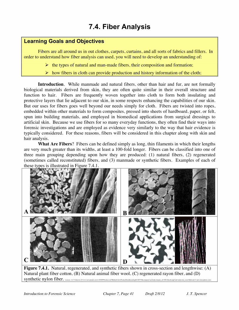
Introduction to Forensic Science Chapter 7, Page 41 Draft 2/8/12 J. T. Spencer
7.4. Fiber Analysis Learning Goals and Objectives Fibers are all around us in out clothes, carpets, curtains, and all sorts of fabrics and fillers. In order to understand how fiber analysis can used, you will need to develop an understanding of:
Ø the types of natural and man-made fibers, their composition and formation; Ø how fibers in cloth can provide production and history information of the cloth;
Introduction. While manmade and natural fibers, other than hair and fur, are not formally biological materials derived from skin, they are often quite similar in their overall structure and function to hair. Fibers are frequently woven together into cloth to form both insulating and protective layers that lie adjacent to our skin, in some respects enhancing the capabilities of our skin. But our uses for fibers goes well beyond our needs simply for cloth. Fibers are twisted into ropes, embedded within other materials to form composites, pressed into sheets of hardboard, paper, or felt, spun into building materials, and employed in biomedical applications from surgical dressings to artificial skin. Because we use fibers for so many everyday functions, they often find their ways into forensic investigations and are employed as evidence very similarly to the way that hair evidence is typically considered. For these reasons, fibers will be considered in this chapter along with skin and hair analysis.
What Are Fibers? Fibers can be defined simply as long, thin filaments in which their lengths are very much greater than its widths, at least a 100-fold longer. Fibers can be classified into one of three main grouping depending upon how they are produced: (1) natural fibers, (2) regenerated (sometimes called reconstituted) fibers, and (3) manmade or synthetic fibers. Examples of each of these types is illustrated in Figure 7.4.1.
A B
C D Figure 7.4.1. Natural, regenerated, and synthetic fibers shown in cross-section and lengthwise: (A) Natural plant fiber cotton, (B) Natural animal fiber wool, (C) regenerated rayon fiber, and (D) synthetic nylon fiber. (sources: A-C Unknown; D www.taiwantrade.com.tw:80/EP/Products.do?Method=showProductDetail&catalogId=260739&company=acelon&company_id=10812&setLangCode=en&come_soon=0&locale=2 and sciencephoto.com).
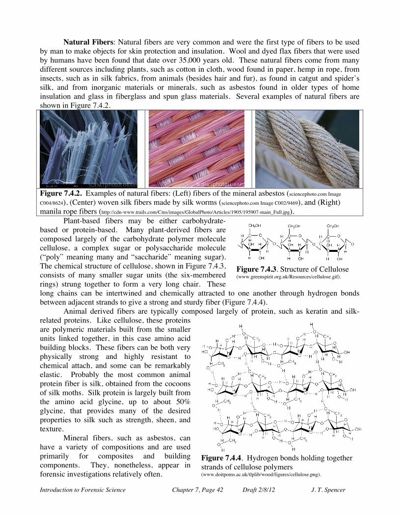
Introduction to Forensic Science Chapter 7, Page 42 Draft 2/8/12 J. T. Spencer
Natural Fibers: Natural fibers are very common and were the first type of fibers to be used by man to make objects for skin protection and insulation. Wool and dyed flax fibers that were used by humans have been found that date over 35,000 years old. These natural fibers come from many different sources including plants, such as cotton in cloth, wood found in paper, hemp in rope, from insects, such as in silk fabrics, from animals (besides hair and fur), as found in catgut and spider’s silk, and from inorganic materials or minerals, such as asbestos found in older types of home insulation and glass in fiberglass and spun glass materials. Several examples of natural fibers are shown in Figure 7.4.2.
Figure 7.4.2. Examples of natural fibers: (Left) fibers of the mineral asbestos (sciencephoto.com Image
C004/8624), (Center) woven silk fibers made by silk worms (sciencephoto.com Image C002/9469), and (Right) manila rope fibers (http://cdn-www.trails.com/Cms/images/GlobalPhoto/Articles/1905/195907-main_Full.jpg).
Plant-based fibers may be either carbohydrate-based or protein-based. Many plant-derived fibers are composed largely of the carbohydrate polymer molecule cellulose, a complex sugar or polysaccharide molecule (“poly” meaning many and “saccharide” meaning sugar). The chemical structure of cellulose, shown in Figure 7.4.3, consists of many smaller sugar units (the six-membered rings) strung together to form a very long chair. These long chains can be intertwined and chemically attracted to one another through hydrogen bonds between adjacent strands to give a strong and sturdy fiber (Figure 7.4.4).
Animal derived fibers are typically composed largely of protein, such as keratin and silk-related proteins. Like cellulose, these proteins are polymeric materials built from the smaller units linked together, in this case amino acid building blocks. These fibers can be both very physically strong and highly resistant to chemical attach, and some can be remarkably elastic. Probably the most common animal protein fiber is silk, obtained from the cocoons of silk moths. Silk protein is largely built from the amino acid glycine, up to about 50% glycine, that provides many of the desired properties to silk such as strength, sheen, and texture.
Mineral fibers, such as asbestos, can have a variety of compositions and are used primarily for composites and building components. They, nonetheless, appear in forensic investigations relatively often.
Figure 7.4.3. Structure of Cellulose (www.greenspirit.org.uk/Resources/cellulose.gif).
Figure 7.4.4. Hydrogen bonds holding together strands of cellulose polymers (www.doitpoms.ac.uk/tlplib/wood/figures/cellulose.png).
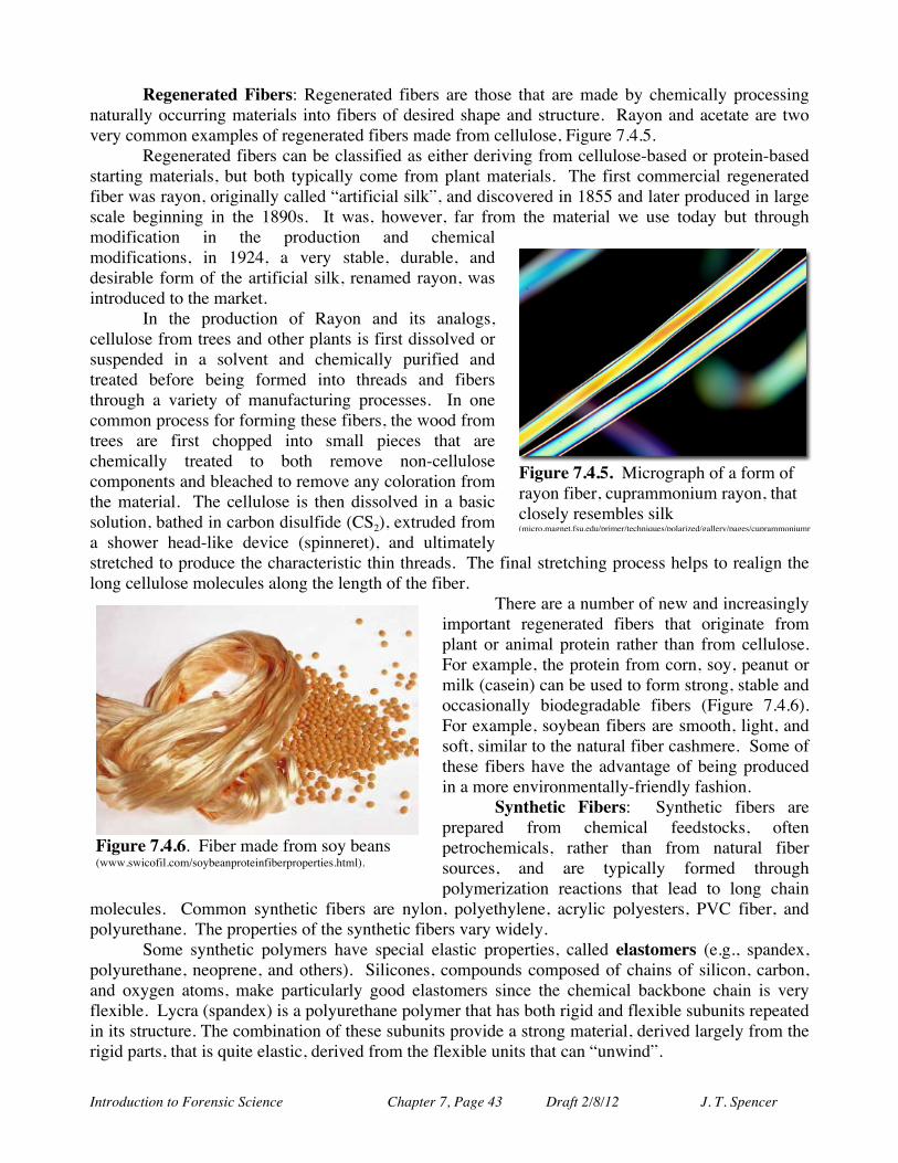
Introduction to Forensic Science Chapter 7, Page 43 Draft 2/8/12 J. T. Spencer
Regenerated Fibers: Regenerated fibers are those that are made by chemically processing naturally occurring materials into fibers of desired shape and structure. Rayon and acetate are two very common examples of regenerated fibers made from cellulose, Figure 7.4.5.
Regenerated fibers can be classified as either deriving from cellulose-based or protein-based starting materials, but both typically come from plant materials. The first commercial regenerated fiber was rayon, originally called “artificial silk”, and discovered in 1855 and later produced in large scale beginning in the 1890s. It was, however, far from the material we use today but through modification in the production and chemical modifications, in 1924, a very stable, durable, and desirable form of the artificial silk, renamed rayon, was introduced to the market.
In the production of Rayon and its analogs, cellulose from trees and other plants is first dissolved or suspended in a solvent and chemically purified and treated before being formed into threads and fibers through a variety of manufacturing processes. In one common process for forming these fibers, the wood from trees are first chopped into small pieces that are chemically treated to both remove non-cellulose components and bleached to remove any coloration from the material. The cellulose is then dissolved in a basic solution, bathed in carbon disulfide (CS2), extruded from a shower head-like device (spinneret), and ultimately stretched to produce the characteristic thin threads. The final stretching process helps to realign the long cellulose molecules along the length of the fiber.
There are a number of new and increasingly important regenerated fibers that originate from plant or animal protein rather than from cellulose. For example, the protein from corn, soy, peanut or milk (casein) can be used to form strong, stable and occasionally biodegradable fibers (Figure 7.4.6). For example, soybean fibers are smooth, light, and soft, similar to the natural fiber cashmere. Some of these fibers have the advantage of being produced in a more environmentally-friendly fashion.
Synthetic Fibers: Synthetic fibers are prepared from chemical feedstocks, often petrochemicals, rather than from natural fiber sources, and are typically formed through polymerization reactions that lead to long chain
molecules. Common synthetic fibers are nylon, polyethylene, acrylic polyesters, PVC fiber, and polyurethane. The properties of the synthetic fibers vary widely.
Some synthetic polymers have special elastic properties, called elastomers (e.g., spandex, polyurethane, neoprene, and others). Silicones, compounds composed of chains of silicon, carbon, and oxygen atoms, make particularly good elastomers since the chemical backbone chain is very flexible. Lycra (spandex) is a polyurethane polymer that has both rigid and flexible subunits repeated in its structure. The combination of these subunits provide a strong material, derived largely from the rigid parts, that is quite elastic, derived from the flexible units that can “unwind”.
Figure 7.4.5. Micrograph of a form of rayon fiber, cuprammonium rayon, that closely resembles silk (micro.magnet.fsu.edu/primer/techniques/polarized/gallery/pages/cuprammoniumrayonsmall.html).
Figure 7.4.6. Fiber made from soy beans (www.swicofil.com/soybeanproteinfiberproperties.html).
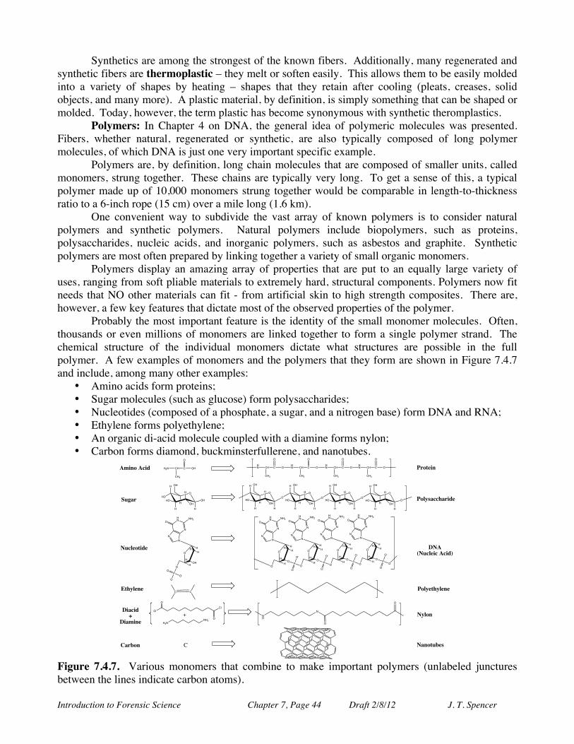
Introduction to Forensic Science Chapter 7, Page 44 Draft 2/8/12 J. T. Spencer
Synthetics are among the strongest of the known fibers. Additionally, many regenerated and synthetic fibers are thermoplastic – they melt or soften easily. This allows them to be easily molded into a variety of shapes by heating – shapes that they retain after cooling (pleats, creases, solid objects, and many more). A plastic material, by definition, is simply something that can be shaped or molded. Today, however, the term plastic has become synonymous with synthetic theromplastics.
Polymers: In Chapter 4 on DNA, the general idea of polymeric molecules was presented. Fibers, whether natural, regenerated or synthetic, are also typically composed of long polymer molecules, of which DNA is just one very important specific example.
Polymers are, by definition, long chain molecules that are composed of smaller units, called monomers, strung together. These chains are typically very long. To get a sense of this, a typical polymer made up of 10,000 monomers strung together would be comparable in length-to-thickness ratio to a 6-inch rope (15 cm) over a mile long (1.6 km).
One convenient way to subdivide the vast array of known polymers is to consider natural polymers and synthetic polymers. Natural polymers include biopolymers, such as proteins, polysaccharides, nucleic acids, and inorganic polymers, such as asbestos and graphite. Synthetic polymers are most often prepared by linking together a variety of small organic monomers.
Polymers display an amazing array of properties that are put to an equally large variety of uses, ranging from soft pliable materials to extremely hard, structural components. Polymers now fit needs that NO other materials can fit - from artificial skin to high strength composites. There are, however, a few key features that dictate most of the observed properties of the polymer.
Probably the most important feature is the identity of the small monomer molecules. Often, thousands or even millions of monomers are linked together to form a single polymer strand. The chemical structure of the individual monomers dictate what structures are possible in the full polymer. A few examples of monomers and the polymers that they form are shown in Figure 7.4.7 and include, among many other examples:
• Amino acids form proteins; • Sugar molecules (such as glucose) form polysaccharides; • Nucleotides (composed of a phosphate, a sugar, and a nitrogen base) form DNA and RNA; • Ethylene forms polyethylene; • An organic di-acid molecule coupled with a diamine forms nylon; • Carbon forms diamond, buckminsterfullerene, and nanotubes.
H2N CH C
CH3
OH
O
HN CH C
CH3
O
O
HN CH C
CH3
O
O
HN CH C
CH3
O
O
HN CH C
CH3
O
O
O
H
H
HO
H
HOHH
O
OH
O
H
H
HO
H
HOHH
O
OH
O
H
H
HO
H
HOHH
O
OH
O
H
H
HO
H
HOHH
O
OH
O
H
HO
H
HO
H
HOHH
OH
OH
HN
N
N
ONH2
N
O HH
H
H
HOH
O
P
-O
O
O-
HN
N
N
ONH2
N
O HH
H
H
HO
P
OOO
O-
HN
N
N
ONH2
N
O HH
H
H
HO
P
OO
O-
HN
N
N
ONH2
N
O HH
H
H
HO
P
OO
O-
HN
N
N
ONH2
N
O HH
H
H
HO
P
OO
O-
C C
ClCl
H2NNH2
NH
N
O
OO
O
Amino Acid
Sugar
Nucleotide
Ethylene
Diacid+
DiamineNylon
Polyethylene
DNA (Nucleic Acid)
Polysaccharide
Protein
CCarbon Nanotubes
+
Figure 7.4.7. Various monomers that combine to make important polymers (unlabeled junctures between the lines indicate carbon atoms).
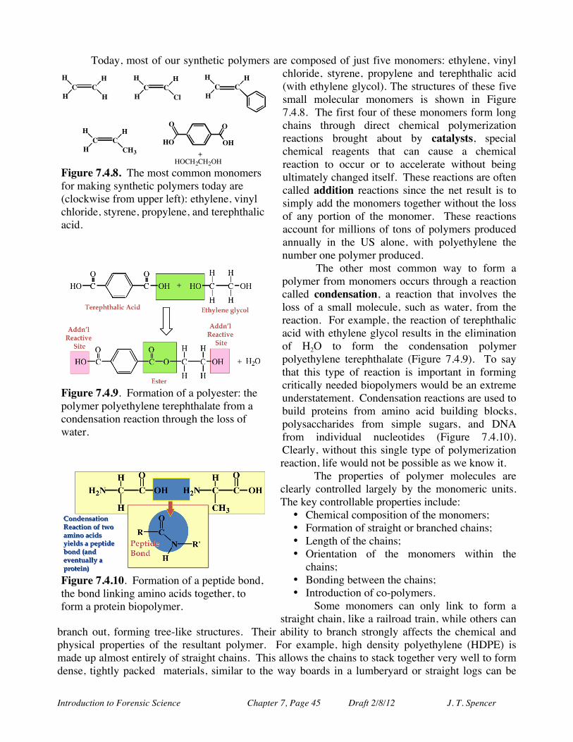
Introduction to Forensic Science Chapter 7, Page 45 Draft 2/8/12 J. T. Spencer
Today, most of our synthetic polymers are composed of just five monomers: ethylene, vinyl chloride, styrene, propylene and terephthalic acid (with ethylene glycol). The structures of these five small molecular monomers is shown in Figure 7.4.8. The first four of these monomers form long chains through direct chemical polymerization reactions brought about by catalysts, special chemical reagents that can cause a chemical reaction to occur or to accelerate without being ultimately changed itself. These reactions are often called addition reactions since the net result is to simply add the monomers together without the loss of any portion of the monomer. These reactions account for millions of tons of polymers produced annually in the US alone, with polyethylene the number one polymer produced.
The other most common way to form a polymer from monomers occurs through a reaction called condensation, a reaction that involves the loss of a small molecule, such as water, from the reaction. For example, the reaction of terephthalic acid with ethylene glycol results in the elimination of H2O to form the condensation polymer polyethylene terephthalate (Figure 7.4.9). To say that this type of reaction is important in forming critically needed biopolymers would be an extreme understatement. Condensation reactions are used to build proteins from amino acid building blocks, polysaccharides from simple sugars, and DNA from individual nucleotides (Figure 7.4.10). Clearly, without this single type of polymerization reaction, life would not be possible as we know it.
The properties of polymer molecules are clearly controlled largely by the monomeric units. The key controllable properties include: • Chemical composition of the monomers; • Formation of straight or branched chains; • Length of the chains; • Orientation of the monomers within the
chains; • Bonding between the chains; • Introduction of co-polymers.
Some monomers can only link to form a straight chain, like a railroad train, while others can
branch out, forming tree-like structures. Their ability to branch strongly affects the chemical and physical properties of the resultant polymer. For example, high density polyethylene (HDPE) is made up almost entirely of straight chains. This allows the chains to stack together very well to form dense, tightly packed materials, similar to the way boards in a lumberyard or straight logs can be
H
C C
H
H
H
H
C C
H
H
Cl
H
C C
H
H
H
C C
H
H
CH3
O
OH
O
HO
+
HOCH2CH2OH Figure 7.4.8. The most common monomers for making synthetic polymers today are (clockwise from upper left): ethylene, vinyl chloride, styrene, propylene, and terephthalic acid.
Figure 7.4.9. Formation of a polyester: the polymer polyethylene terephthalate from a condensation reaction through the loss of water.
Figure 7.4.10. Formation of a peptide bond, the bond linking amino acids together, to form a protein biopolymer.
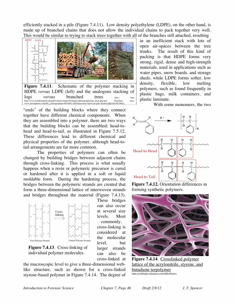
Introduction to Forensic Science Chapter 7, Page 46 Draft 2/8/12 J. T. Spencer
efficiently stacked in a pile (Figure 7.4.11). Low density polyethylene (LDPE), on the other hand, is made up of branched chains that does not allow the individual chains to pack together very well. This would be similar to trying to stack trees together with all of the branches still attached, resulting
in an inefficient stack with lots of open air-spaces between the tree trunks. The result of this kind of packing is that HDPE forms very strong, rigid, dense and high-strength materials, used in applications such as water pipes, snow boards, and storage sheds, while LDPE forms softer, low density, flexible, low melting polymers, such as found frequently in plastic bags, milk containers, and plastic laminate.
With some monomers, the two “ends” of the building blocks where they connect together have different chemical components. When they are assembled into a polymer, there are two ways that the building blocks can be assembled; head-to-head and head-to-tail, as illustrated in Figure 7.5.12. These differences lead to different chemical and physical properties of the polymer, although head-to-tail arrangements are far more common.
The properties of polymers can often be changed by building bridges between adjacent chains through cross-linking. This process is what usually happens when a resin or polymeric precursor is cured or hardened after it is applied in a soft or liquid moldable form. During the hardening process, the bridges between the polymeric strands are created that form a three-dimensional lattice of interwoven strands and bridges throughout the material (Figure 7.4.13).
These bridges can also occur at several size levels. Most
commonly, cross-linking is considered at the molecular level, but larger strands can also be cross-linked at
the macroscopic level to give a three-dimensional web-like structure, such as shown for a cross-linked styrene-based polymer in Figure 7.4.14. The degree of
Figure 7.4.11. Schematic of the polymer stacking in HDPE versus LDPE (left) and the analogous stacking of logs versus branched trees (pics; logs http://www.northerngrid.org/ngflwebsite/chopwell/images/photographs/log_stack.jpg;and branched trees www.istockphoto.com/file_closeup/nature/4814662-christmas-trees-fresh-cut-pile-stacked.php?id=4814662).
Figure 7.4.12. Orientation differences in forming synthetic polymers.
Figure 7.4.14. Crosslinked polymer lattice of the acrylonitrile, styrene, and butadiene terpolymer (http://retr0bright.wikispaces.com/ABS+Plastic).
Cross-linking
Free Polymer Strands Linked Polymer Strands
Figure 7.4.13. Cross-linking of individual polymer molecules.
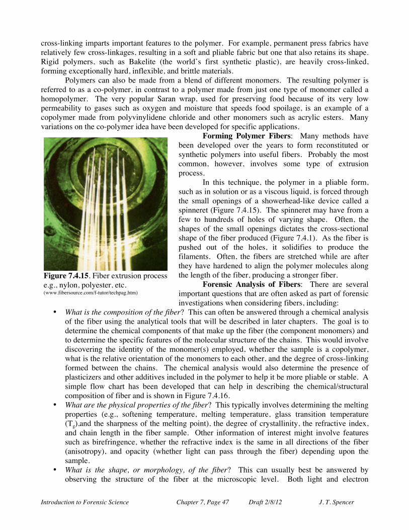
Introduction to Forensic Science Chapter 7, Page 47 Draft 2/8/12 J. T. Spencer
cross-linking imparts important features to the polymer. For example, permanent press fabrics have relatively few cross-linkages, resulting in a soft and pliable fabric but one that also retains its shape. Rigid polymers, such as Bakelite (the world’s first synthetic plastic), are heavily cross-linked, forming exceptionally hard, inflexible, and brittle materials.
Polymers can also be made from a blend of different monomers. The resulting polymer is referred to as a co-polymer, in contrast to a polymer made from just one type of monomer called a homopolymer. The very popular Saran wrap, used for preserving food because of its very low permeability to gases such as oxygen and moisture that speeds food spoilage, is an example of a copolymer made from polyvinylidene chloride and other monomers such as acrylic esters. Many variations on the co-polymer idea have been developed for specific applications.
Forming Polymer Fibers: Many methods have been developed over the years to form reconstituted or synthetic polymers into useful fibers. Probably the most common, however, involves some type of extrusion process.
In this technique, the polymer in a pliable form, such as in solution or as a viscous liquid, is forced through the small openings of a showerhead-like device called a spinneret (Figure 7.4.15). The spinneret may have from a few to hundreds of holes of varying shape. Often, the shapes of the small openings dictates the cross-sectional shape of the fiber produced (Figure 7.4.1). As the fiber is pushed out of the holes, it solidifies to produce the filaments. Often, the fibers are stretched while are after they have hardened to align the polymer molecules along the length of the fiber, producing a stronger fiber.
Forensic Analysis of Fibers: There are several important questions that are often asked as part of forensic investigations when considering fibers, including:
• What is the composition of the fiber? This can often be answered through a chemical analysis of the fiber using the analytical tools that will be described in later chapters. The goal is to determine the chemical components of that make up the fiber (the component monomers) and to determine the specific features of the molecular structure of the chains. This would involve discovering the identity of the monomer(s) employed, whether the sample is a copolymer, what is the relative orientation of the monomers to each other, and the degree of cross-linking formed between the chains. The chemical analysis would also determine the presence of plasticizers and other additives included in the polymer to help it be more pliable or stable. A simple flow chart has been developed that can help in describing the chemical/structural composition of fiber and is shown in Figure 7.4.16.
• What are the physical properties of the fiber? This typically involves determining the melting properties (e.g., softening temperature, melting temperature, glass transition temperature (Tg),and the sharpness of the melting point), the degree of crystallinity, the refractive index, and chain length in the fiber sample. Other information of interest might involve features such as birefringence, whether the refractive index is the same in all directions of the fiber (anisotropy), and opacity (whether light can pass through the fiber) depending upon the sample.
• What is the shape, or morphology, of the fiber? This can usually best be answered by observing the structure of the fiber at the microscopic level. Both light and electron
Figure 7.4.15. Fiber extrusion process e.g., nylon, polyester, etc. (www.fibersource.com/f-tutor/techpag.htm)
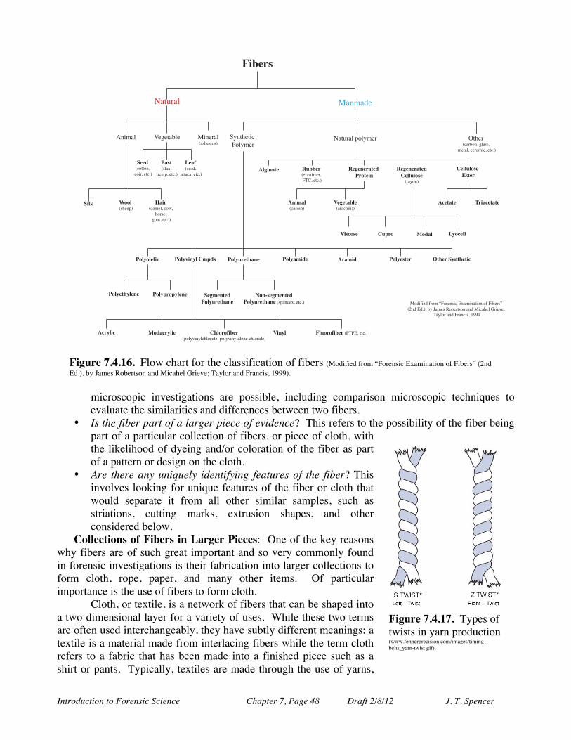
Introduction to Forensic Science Chapter 7, Page 48 Draft 2/8/12 J. T. Spencer
microscopic investigations are possible, including comparison microscopic techniques to evaluate the similarities and differences between two fibers.
• Is the fiber part of a larger piece of evidence? This refers to the possibility of the fiber being part of a particular collection of fibers, or piece of cloth, with the likelihood of dyeing and/or coloration of the fiber as part of a pattern or design on the cloth.
• Are there any uniquely identifying features of the fiber? This involves looking for unique features of the fiber or cloth that would separate it from all other similar samples, such as striations, cutting marks, extrusion shapes, and other considered below.
Collections of Fibers in Larger Pieces: One of the key reasons why fibers are of such great important and so very commonly found in forensic investigations is their fabrication into larger collections to form cloth, rope, paper, and many other items. Of particular importance is the use of fibers to form cloth. Cloth, or textile, is a network of fibers that can be shaped into a two-dimensional layer for a variety of uses. While these two terms are often used interchangeably, they have subtly different meanings; a textile is a material made from interlacing fibers while the term cloth refers to a fabric that has been made into a finished piece such as a shirt or pants. Typically, textiles are made through the use of yarns,
!""#$!"#$$#%&'"#()&'*$"+,
-%(./0 1*2*$/30* 4(%*)/0!/53*5$#5,
6/$7)/0 4/%./8*
%&'($!90/:&'
;*.<&'*$"+,
)"&*$!5(5/0&'
/3/"/&'*$"+,
!+,- .//,'!5;**<,
0&+1$!"/.*0&'"#=&';#)5*&'2#/$&'*$"+,
>$;*)'!"/)3#%&'20/55&'
.*$/0&'"*)/.("&'*$"+,
2+3"1'
6/$7)/0'<#0?.*)@?%$;*$("'A#0?.*)
4#8(9(*8'9)#.'BC#)*%5("'D:/.(%/$(#%'#9'C(3*)5E'!F%8'D8+,+'3?'G/.*5'H#3*)$5#%'/%8'4("/;*0'I)(*J*K'
L/?0#)'/%8'C)/%"(5&'MNNN
4,5+6&(" 7833"1$!*0/5$(.*)&'CLO&'*$"+,
7"5"6"1&("#$91/("+6
7"5"6"1&("#:",,8,/'"!)/?#%,
:",,8,/'"$;'("1
46+<&,!"/5*(%,
="5"(&3,"!/)/";(%,,
4>"(&(" ?1+&>"(&("
=+'>/'" :8@1/ A/#&, )B/>",,
9/,B/,"*+6 9/,BC+6B,$:<@#' 9/,B81"(D&6" 9/,B&<+#" 41&<+# 9/,B"'("1 E(D"1$!B6(D"(+>
9/,B"(DB,"6" 9/,B@1/@B,"6" !"5<"6("#9/,B81"(D&6"
F/6G'"5<"6("#9/,B81"(D&6"'!5</%8*:&'*$"+,
4>1B,+> A/#&>1B,+> :D,/1/*+3"1!<#0?J(%?0";0#)(8*&'<#0?J(%?0(8*%*'";0#)(8*,
=+6B, 2,8/1/*+3"1'!ALCD&'*$"+,
Figure 7.4.16. Flow chart for the classification of fibers (Modified from “Forensic Examination of Fibers” (2nd Ed.). by James Robertson and Micahel Grieve; Taylor and Francis, 1999).
Figure 7.4.17. Types of twists in yarn production (www.fennerprecision.com/images/timing-belts_yarn-twist.gif).
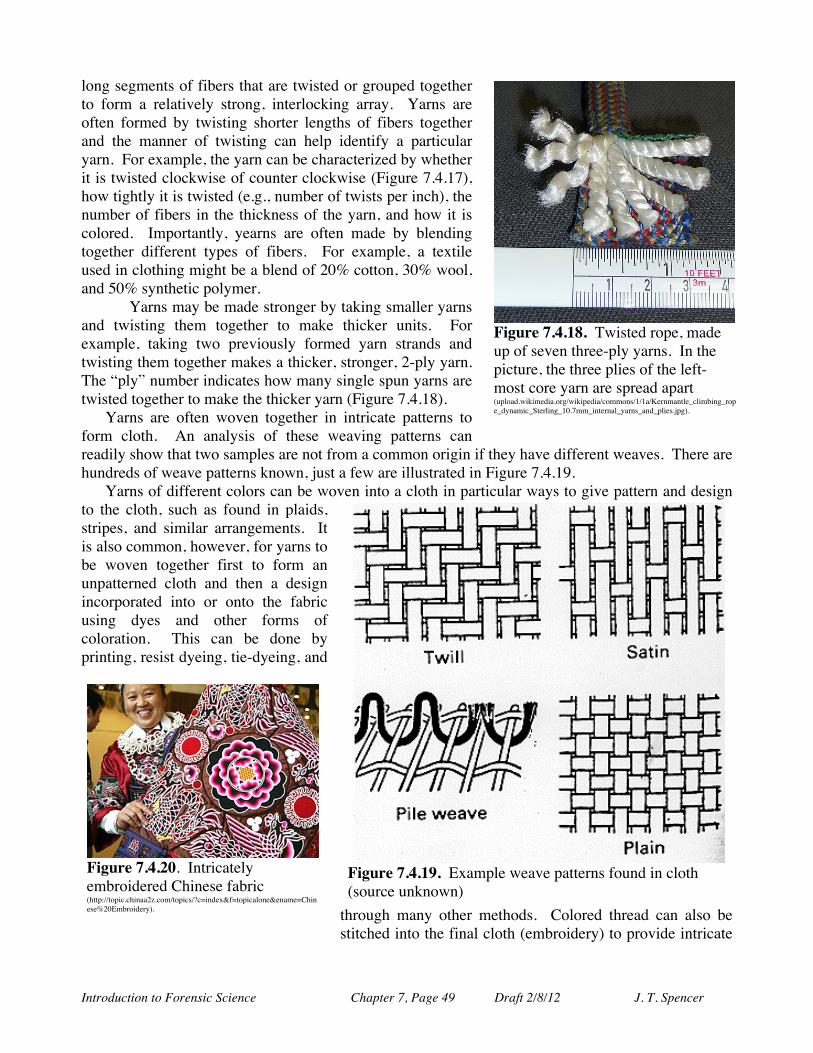
Introduction to Forensic Science Chapter 7, Page 49 Draft 2/8/12 J. T. Spencer
long segments of fibers that are twisted or grouped together to form a relatively strong, interlocking array. Yarns are often formed by twisting shorter lengths of fibers together and the manner of twisting can help identify a particular yarn. For example, the yarn can be characterized by whether it is twisted clockwise of counter clockwise (Figure 7.4.17), how tightly it is twisted (e.g., number of twists per inch), the number of fibers in the thickness of the yarn, and how it is colored. Importantly, yearns are often made by blending together different types of fibers. For example, a textile used in clothing might be a blend of 20% cotton, 30% wool, and 50% synthetic polymer. Yarns may be made stronger by taking smaller yarns and twisting them together to make thicker units. For example, taking two previously formed yarn strands and twisting them together makes a thicker, stronger, 2-ply yarn. The “ply” number indicates how many single spun yarns are twisted together to make the thicker yarn (Figure 7.4.18).
Yarns are often woven together in intricate patterns to form cloth. An analysis of these weaving patterns can readily show that two samples are not from a common origin if they have different weaves. There are hundreds of weave patterns known, just a few are illustrated in Figure 7.4.19.
Yarns of different colors can be woven into a cloth in particular ways to give pattern and design to the cloth, such as found in plaids, stripes, and similar arrangements. It is also common, however, for yarns to be woven together first to form an unpatterned cloth and then a design incorporated into or onto the fabric using dyes and other forms of coloration. This can be done by printing, resist dyeing, tie-dyeing, and
through many other methods. Colored thread can also be stitched into the final cloth (embroidery) to provide intricate
Figure 7.4.19. Example weave patterns found in cloth (source unknown)
Figure 7.4.18. Twisted rope, made up of seven three-ply yarns. In the picture, the three plies of the left-most core yarn are spread apart (upload.wikimedia.org/wikipedia/commons/1/1a/Kernmantle_climbing_rope_dynamic_Sterling_10.7mm_internal_yarns_and_plies.jpg).
Figure 7.4.20. Intricately embroidered Chinese fabric (http://topic.chinaa2z.com/topics/?c=index&f=topicalone&ename=Chinese%20Embroidery).
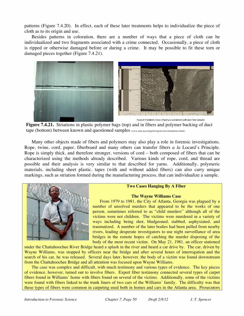
Introduction to Forensic Science Chapter 7, Page 50 Draft 2/8/12 J. T. Spencer
patterns (Figure 7.4.20). In effect, each of these later treatments helps to individualize the piece of cloth as to its origin and use.
Besides patterns in coloration, there are a number of ways that a piece of cloth can be individualized and two fragments associated with a crime connected. Occasionally, a piece of cloth is ripped or otherwise damaged before or during a crime. It may be possible to fit these torn or damaged pieces together (Figure 7.4.21).
Many other objects made of fibers and polymers may also play a role in forensic investigations. Rope, twine, cord, paper, fiberboard and many others can transfer fibers a la Locard’s Principle. Rope is simply thick, and therefore stronger, versions of cord – both composed of fibers that can be characterized using the methods already described. Various kinds of rope, cord, and thread are possible and their analysis is very similar to that described for yarns. Additionally, polymeric materials, including sheet plastic, tapes (with and without added fibers) can also carry unique markings, such as striation formed during the manufacturing process, that can individualize a sample.
Two Cases Hanging By A Fiber
The Wayne Williams Case
From 1979 to 1981, the City of Atlanta, Georgia was plagued by a number of unsolved murders that appeared to be the works of one person, sometimes referred to as “child murders” although all of the victims were not children. The victims were murdered in a variety of ways including being shot, bludgeoned, stabbed, asphyxiated, and traumatized. A number of the later bodies had been pulled from nearby rivers, leading desperate investigators to use night surveillance of area bridges in the remote hopes of catching the murder disposing of the body of the most recent victim. On May 21, 1981, an officer stationed
under the Chattahoochee River Bridge heard a splash in the river and heard a car drive by. The car, driven by Wayne Williams, was stopped by officers near the bridge and after several hours of interrogation and the search of his car, he was released. Several days later, however, the body of a victim was found downstream from the Chattahoochee Bridge and all attention was focused upon Wayne Williams.
The case was complex and difficult, with much testimony and various types of evidence. The key pieces of evidence, however, turned out to involve fibers. Expert fiber testimony connected several types of carpet fibers found in Williams’ home with fibers found on several of the victims. Additionally, some of the victims were found with fibers linked to the trunk liners of two cars of the Williams’ family. The difficulty was that these types of fibers were common in carpeting used both in homes and cars in the Atlanta area. Prosecutors
Figure 7.4.21. Striations in plastic polymer bags (top) and in fibers and polymer backing of duct tape (bottom) between known and questioned samples (www.state.nj.us/njsp/divorg/invest/criminalistics.html).

Introduction to Forensic Science Chapter 7, Page 51 Draft 2/8/12 J. T. Spencer
had to show that the fibers found on the victims had a high probability of coming from Williams’ home and car. Investigators examined the structure and colors of the fibers and determined how common these fibers were in the Atlanta area and determined the odds of a random match of these fibers at about 1 in 30,000,000. In the end, they were able to convince the jury that there was enough of a link between the two sets of fibers to convict Wayne Williams of two of the homicides.
There remains, however, some unanswered questions in this case to many people. It appears that the car from which the fibers were deemed to match was not available to Williams at the time of the crimes. The fibers were relatively commonly found in hotels, homes and other residential buildings. And Williams himself has maintained his innocence throughout his imprisonment.
Jeffrey MacDonald Case
Early on the morning of Feb. 17, 1970, military police on Fort Bragg were called to a reported violent attack. At the home of Capt. Jeffrey MacDonald, a doctor on the base, they found a violent scene with MacDonald’s wife and two small children dead. They had been violently attacked and repeatedly stabbed and beaten. MacDonald was found wounded but none of his wounds were life threatening.
The case quickly centered upon Dr. MacDonald as the prime suspect since investigators felt that Dr. MacDonald’s account did not seem to match the physical evidence. MacDonald’s account was that intruders had broken into his home and had done the crime.
A great deal of evidence was collected but, once again, fiber analysis proved important to the case. Investigators were able to find fiber fibers from MacDonald’s pajama tops in several places at the crime scene and number of hole in the cloth. He had said that he used the top to wrap around his hands to help ward off the blows from the ice-pick wielding attacker. The tears in the cloth, both in location and in the smooth nature of the holes, were, however, thought not to be consistent with a defensive posture but more consistent with MacDonald attacking his wife with the pajama top laid over her.
Capt. MacDonald was convicted on Aug. 29, 1979 and sentenced to life imprisonment. Capt. MacDonald continues to declare his innocence and has lodged a number of unsuccessful appeals. Subsequent DNA analysis has failed to turn up any connection with someone other that a member of the MacDonald household.
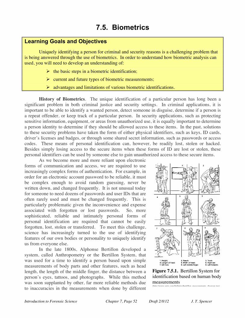
Introduction to Forensic Science Chapter 7, Page 52 Draft 2/8/12 J. T. Spencer
7.5. Biometrics Learning Goals and Objectives Uniquely identifying a person for criminal and security reasons is a challenging problem that is being answered through the use of biometrics. In order to understand how biometric analysis can used, you will need to develop an understanding of:
Ø the basic steps in a biometric identification; Ø current and future types of biometric measurements; Ø advantages and limitations of various biometric identifications.
History of Biometrics. The unique identification of a particular person has long been a significant problem in both criminal justice and security settings. In criminal applications, it is important to be able to identify a wanted person, detect someone in disguise, determine if a person is a repeat offender, or keep track of a particular person. In security applications, such as protecting sensitive information, equipment, or areas from unauthorized use, it is equally important to determine a person identity to determine if they should be allowed access to these items. In the past, solutions to these security problems have taken the form of either physical identifiers, such as keys, ID cards, driver’s licenses and badges, or through some shared secret information, such as passwords or access codes. These means of personal identification can, however, be readily lost, stolen or hacked. Besides simply losing access to the secure items when these forms of ID are lost or stolen, these personal identifiers can be used by someone else to gain unauthorized access to these secure items.
As we become more and more reliant upon electronic forms of communication and access, we are required to use increasingly complex forms of authentication. For example, in order for an electronic account password to be reliable, it must be complex enough to avoid random guessing, never be written down, and changed frequently. It is not unusual today for someone to need dozens of passwords and user IDs that are often rarely used and must be changed frequently. This is particularly problematic given the inconvenience and expense associated with forgotten or lost passwords. So, more sophisticated, reliable and intimately personal forms of personal identification are required that cannot be easily forgotten, lost, stolen or transferred. To meet this challenge, science has increasingly turned to the use of identifying features of our own bodies or personality to uniquely identify us from everyone else.
In the late 1800s, Alphonse Bertillon developed a system, called Anthropometry or the Bertillon System, that was used for a time to identify a person based upon simple measurements of body parts and other features, such as head length, the length of the middle finger, the distance between a person’s eyes, tattoos, and photographs. While this method was soon supplanted by other, far more reliable methods due to inaccuracies in the measurements when done by different
Figure 7.5.1. Bertillon System for identification based on human body measurements (http://www.onin.com/fp/fmiru/bertillon_measurements_diagram.jpg).
Introduction to Forensic Science Chapter 7, Page 53 Draft 2/8/12 J. T. Spencer
people, it did form the first attempt to base personal identification on body traits. As presented earlier in this chapter, fingerprints have formed a powerful tool for uniquely
identifying people for well over one hundred years. Increasingly, however, other forms of recognition based upon various aspects of the human body are being used for identification.
Biometrics Basics: Biometrics, the application of statistical methods to biological data, is based upon finding some individualized trait that a person has that can distinguish them from all others. These traits can be biological, such as fingerprint or iris pattern, or behavioral in nature, such as how you act or even type. But all forms of biometric identification, in order to be useful, must meet a specific set of criteria that includes: Universality, Uniqueness, Permanence, Measurability, and Ease of Use.
Universality. In order to be useful, the trait must be something that all people to be authenticated have in common. The analysis must be based upon common biological structures or developed behavioral patterns. For example, a biometric marker based upon how someone types on a computer keyboard makes sense only to populations where keyboard use is common and of limited use in computer-less populations. In contrast, an analysis based upon iris patterns in the eye would be generally useful for the entire human population.
Uniqueness. A biometric feature must be able to differentiate between all people needing authentication. An analysis based upon number of fingers on a person’s right hand is based upon a common biological trait but is not very useful in distinguished between most people. An analysis based upon fingerprints, however, is far more able to discern between two people. It is important to realize, however, that any such analysis must be based in a good understanding of the statistics involved – in other words, how probable is it that two people would randomly have the same measured value such that the test could not determine the difference between them. In the chapter on DNA, we said that the chance of two people having the same DNA profile of 14 markers is often 1 in several trillion, making it a very reliable probe of personal ID. A biometric analysis based upon ABO blood typing, however, might have a random match rate in the percent range, making it far less useful when compared to DNA as a biometric tool.
Permanence. Any biometric tool must persist unchanged for a very long time in a person, ideally for an entire lifetime. Most biological biometrics typically don’t change much at all over a lifetime, such as DNA, eye iris patterns, and fingerprints. Some biological and behavioral markers do change, however, with age, illness, accident, growth, or learned patterns, such as voice pattern and writing patterns. These changes do not render the method useless, however, but care must be taken to understand the possibility for change and account for it in the analysis.
Measurability. Any biometric tool must employ a trait that can be measured reliably and with straightforward measuring devices. If a trait cannot be measured, it cannot be used in automated ID systems.
Ease of Use. There are many types of probes that could be used in a biometric method that are impractical, expensive or difficult to use. Preferred methods employ measurements that are easy and quick and give highly reliable data for comparison.
Circumvention. The refers to how easily the biometric trait can be defeated through the use of a substitute, such as a fingerprint casting or a contact lens with a false iris pattern.
Biometric Methods: Biometric methods typically try establish someone’s identity by answering one of three specific questions; (1) who are you?, (2) what information do you possess?, or
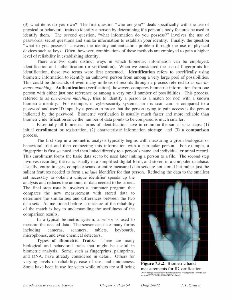
Introduction to Forensic Science Chapter 7, Page 54 Draft 2/8/12 J. T. Spencer
(3) what items do you own? The first question “who are you?” deals specifically with the use of physical or behavioral traits to identify a person by determining if a person’s body features be used to identify them. The second question, “what information do you possess?” involves the use of passwords, secret questions and similar information to establish your identity. Finally, the question “what to you possess?” answers the identity authentication problem through the use of physical devices such as keys. Often, however, combinations of these methods are employed to gain a higher level of reliability in establishing identity.
There are two quite distinct ways in which biometric information can be employed: identification and authentication (or verification). When we considered the use of fingerprints for identification, these two terms were first presented. Identification refers to specifically using biometric information to identify an unknown person from among a very large pool of possibilities. This could be thousands of even many millions of records through a process referred to as one-to-many matching. Authentication (verification), however, compares biometric information from one person with either just one reference or among a very small number of possibilities. This process, referred to as one-to-one matching, tries to identify a person as a match (or not) with a known biometric identity. For example, in cybersecurity systems, an iris scan can be compared to a password and user ID input by a person to prove that the person trying to gain access is the person indicated by the password Biometric verification is usually much faster and more reliable than biometric identification since the number of data points to be compared is much smaller.
Essentially all biometric forms of identification have in common the same basic steps: (1) initial enrollment or registration, (2) characteristic information storage, and (3) a comparison process.
The first step in a biometric analysis typically begins with measuring a given biological or behavioral trait and then connecting this information with a particular person. For example, a fingerprint is first scanned and then linked directly to a person’s name and individual criminal record. This enrollment forms the basic data set to be used later linking a person to a file. The second step involves recording the data, usually in a simplified digital form, and stored in a computer database. Usually, entire images, complete scans or entire measured data sets are not stored but rather just the salient features needed to form a unique identifier for that person. Reducing the data to the smallest set necessary to obtain a unique identifier speeds up the analysis and reduces the amount of data needed to be stored. The final step usually involves a computer program that compares the new measurement with stored data to determine the similarities and differences between the two data sets. As mentioned before, a measure of the reliability of the match is key to understanding the usefulness of the comparison results.
In a typical biometric system, a sensor is used to measure the needed data. The sensor can take many forms including cameras, scanners, tablets, keyboards, microphones, and even chemical detectors.
Types of Biometric Traits. There are many biological and behavioral traits that might be useful in biometric analysis. Some, such as fingerprints, palmprints, and DNA, have already considered in detail. Others for varying levels of reliability, ease of use, and uniqueness. Some have been in use for years while others are still being
Figure 7.5.2. Biometric hand measurements for ID verification (www.theage.com.au/news/national/schools-to-fingerprint-students-for-security/2007/09/01/1188067438565.html).
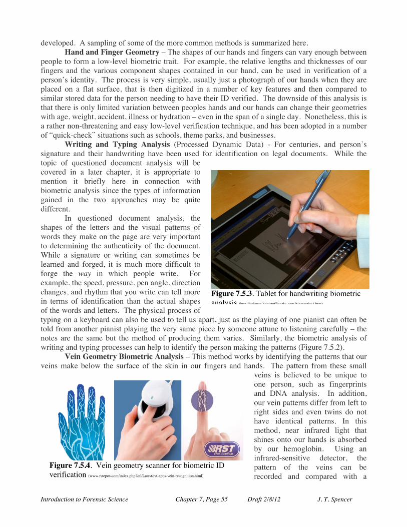
Introduction to Forensic Science Chapter 7, Page 55 Draft 2/8/12 J. T. Spencer
developed. A sampling of some of the more common methods is summarized here. Hand and Finger Geometry – The shapes of our hands and fingers can vary enough between
people to form a low-level biometric trait. For example, the relative lengths and thicknesses of our fingers and the various component shapes contained in our hand, can be used in verification of a person’s identity. The process is very simple, usually just a photograph of our hands when they are placed on a flat surface, that is then digitized in a number of key features and then compared to similar stored data for the person needing to have their ID verified. The downside of this analysis is that there is only limited variation between peoples hands and our hands can change their geometries with age, weight, accident, illness or hydration – even in the span of a single day. Nonetheless, this is a rather non-threatening and easy low-level verification technique, and has been adopted in a number of “quick-check” situations such as schools, theme parks, and businesses.
Writing and Typing Analysis (Processed Dynamic Data) - For centuries, and person’s signature and their handwriting have been used for identification on legal documents. While the topic of questioned document analysis will be covered in a later chapter, it is appropriate to mention it briefly here in connection with biometric analysis since the types of information gained in the two approaches may be quite different.
In questioned document analysis, the shapes of the letters and the visual patterns of words they make on the page are very important to determining the authenticity of the document. While a signature or writing can sometimes be learned and forged, it is much more difficult to forge the way in which people write. For example, the speed, pressure, pen angle, direction changes, and rhythm that you write can tell more in terms of identification than the actual shapes of the words and letters. The physical process of typing on a keyboard can also be used to tell us apart, just as the playing of one pianist can often be told from another pianist playing the very same piece by someone attune to listening carefully – the notes are the same but the method of producing them varies. Similarly, the biometric analysis of writing and typing processes can help to identify the person making the patterns (Figure 7.5.2).
Vein Geometry Biometric Analysis – This method works by identifying the patterns that our veins make below the surface of the skin in our fingers and hands. The pattern from these small
veins is believed to be unique to one person, such as fingerprints and DNA analysis. In addition, our vein patterns differ from left to right sides and even twins do not have identical patterns. In this method, near infrared light that shines onto our hands is absorbed by our hemoglobin. Using an infrared-sensitive detector, the pattern of the veins can be recorded and compared with a
Figure 7.5.3. Tablet for handwriting biometric analysis (http://science.howstuffworks.com/biometrics1.htm).
Figure 7.5.4. Vein geometry scanner for biometric ID verification (www.rstepos.com/index.php?/nl/Latest/rst-epos-vein-recognition.html).
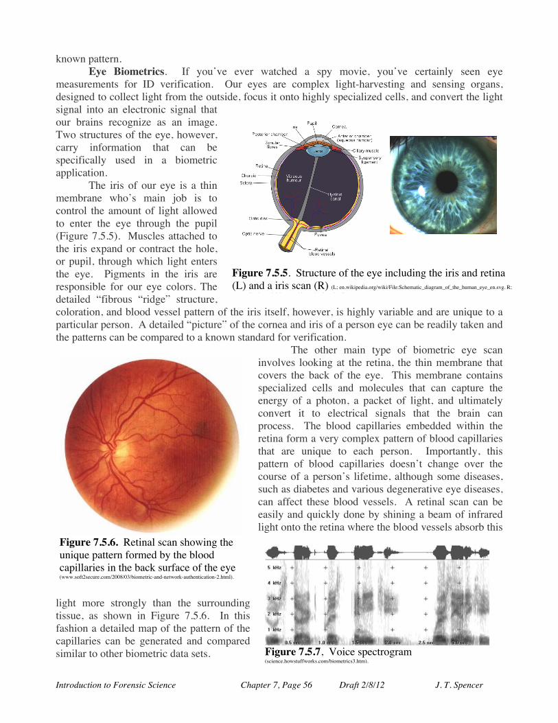
Introduction to Forensic Science Chapter 7, Page 56 Draft 2/8/12 J. T. Spencer
known pattern. Eye Biometrics. If you’ve ever watched a spy movie, you’ve certainly seen eye
measurements for ID verification. Our eyes are complex light-harvesting and sensing organs, designed to collect light from the outside, focus it onto highly specialized cells, and convert the light signal into an electronic signal that our brains recognize as an image. Two structures of the eye, however, carry information that can be specifically used in a biometric application.
The iris of our eye is a thin membrane who’s main job is to control the amount of light allowed to enter the eye through the pupil (Figure 7.5.5). Muscles attached to the iris expand or contract the hole, or pupil, through which light enters the eye. Pigments in the iris are responsible for our eye colors. The detailed “fibrous “ridge” structure, coloration, and blood vessel pattern of the iris itself, however, is highly variable and are unique to a particular person. A detailed “picture” of the cornea and iris of a person eye can be readily taken and the patterns can be compared to a known standard for verification.
The other main type of biometric eye scan involves looking at the retina, the thin membrane that covers the back of the eye. This membrane contains specialized cells and molecules that can capture the energy of a photon, a packet of light, and ultimately convert it to electrical signals that the brain can process. The blood capillaries embedded within the retina form a very complex pattern of blood capillaries that are unique to each person. Importantly, this pattern of blood capillaries doesn’t change over the course of a person’s lifetime, although some diseases, such as diabetes and various degenerative eye diseases, can affect these blood vessels. A retinal scan can be easily and quickly done by shining a beam of infrared light onto the retina where the blood vessels absorb this
light more strongly than the surrounding tissue, as shown in Figure 7.5.6. In this fashion a detailed map of the pattern of the capillaries can be generated and compared similar to other biometric data sets.
Figure 7.5.5. Structure of the eye including the iris and retina (L) and a iris scan (R) (L; en.wikipedia.org/wiki/File:Schematic_diagram_of_the_human_eye_en.svg. R; www.aditech.co.uk/irisrecognitiontechnology.html).
!
Figure 7.5.6. Retinal scan showing the unique pattern formed by the blood capillaries in the back surface of the eye (www.soft2secure.com/2008/03/biometric-and-network-authentication-2.html).
Figure 7.5.7, Voice spectrogram (science.howstuffworks.com/biometrics3.htm).
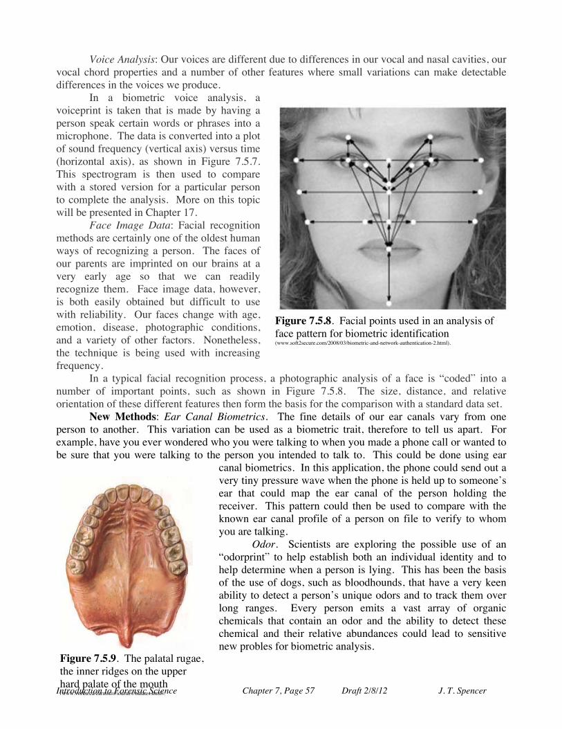
Introduction to Forensic Science Chapter 7, Page 57 Draft 2/8/12 J. T. Spencer
Voice Analysis: Our voices are different due to differences in our vocal and nasal cavities, our vocal chord properties and a number of other features where small variations can make detectable differences in the voices we produce.
In a biometric voice analysis, a voiceprint is taken that is made by having a person speak certain words or phrases into a microphone. The data is converted into a plot of sound frequency (vertical axis) versus time (horizontal axis), as shown in Figure 7.5.7. This spectrogram is then used to compare with a stored version for a particular person to complete the analysis. More on this topic will be presented in Chapter 17.
Face Image Data: Facial recognition methods are certainly one of the oldest human ways of recognizing a person. The faces of our parents are imprinted on our brains at a very early age so that we can readily recognize them. Face image data, however, is both easily obtained but difficult to use with reliability. Our faces change with age, emotion, disease, photographic conditions, and a variety of other factors. Nonetheless, the technique is being used with increasing frequency.
In a typical facial recognition process, a photographic analysis of a face is “coded” into a number of important points, such as shown in Figure 7.5.8. The size, distance, and relative orientation of these different features then form the basis for the comparison with a standard data set.
New Methods: Ear Canal Biometrics. The fine details of our ear canals vary from one person to another. This variation can be used as a biometric trait, therefore to tell us apart. For example, have you ever wondered who you were talking to when you made a phone call or wanted to be sure that you were talking to the person you intended to talk to. This could be done using ear
canal biometrics. In this application, the phone could send out a very tiny pressure wave when the phone is held up to someone’s ear that could map the ear canal of the person holding the receiver. This pattern could then be used to compare with the known ear canal profile of a person on file to verify to whom you are talking.
Odor. Scientists are exploring the possible use of an “odorprint” to help establish both an individual identity and to help determine when a person is lying. This has been the basis of the use of dogs, such as bloodhounds, that have a very keen ability to detect a person’s unique odors and to track them over long ranges. Every person emits a vast array of organic chemicals that contain an odor and the ability to detect these chemical and their relative abundances could lead to sensitive new probles for biometric analysis.
Figure 7.5.8. Facial points used in an analysis of face pattern for biometric identification (www.soft2secure.com/2008/03/biometric-and-network-authentication-2.html).
Figure 7.5.9. The palatal rugae, the inner ridges on the upper hard palate of the mouth (www.yorku.ca/earmstro/journey/palates.html).
Introduction to Forensic Science Chapter 7, Page 58 Draft 2/8/12 J. T. Spencer
Palatal Rugae (palatoscopy) – The palatal rugae are the ridges on the inner parts of the roof of our mouths, closest to the upper teeth. These for patterns that differ from person - and can persist after death in remains identification. There is still considerable debate, however, about the reliability of these structures for biometric identification.
Automated Biometric Identification System (IDENT). The US Department of Homeland Security, in cooperation with several other agencies is working to develop an comprehensive system for using biometric data to link a particular person rapidly with biographic information, such as criminal arrests, personal identification, and travel restrictions, for security and law enforcement work. The IDENT system provides verification to a wide range of government programs that collect biometric data and compile personal biographical information. Recently the IAFIS fingerprint system has been incorporated into the broader IDENT system.
Countries all over the world are faced with increasing needs for identification and authentication of people for both criminal and security threats. Thus, the use of biometric data is expected to grow rapidly in the near future as one of the best ways to rapidly connect a person with biographic information about them.

Introduction to Forensic Science Chapter 7, Page 59 Draft 2/8/12 J. T. Spencer
Chapter 7 References and Bibliography
FINGERPRINT REFERENCES
Henry C. Lee and R. E. Gaensslen (Eds.), Advances in Fingerprint Technology (2nd Ed.), Second Edition. CRC Press, 2001.
Christophe Champod, Chris J. Lennard Pierre Margot, and Milutin Stoilovic Fingerprints and Other Ridge Skin Impressions (International Forensic Science and Investigation), CRC Press, 2004.
Davide Maltoni, Dario Maio, Anil K. Jain, Salil Prabhakar, Handbook of Fingerprint Recognition, Springer, 2009.
Mikael Jägerbrand (Ed.), Journal of Ancient Fingerprints. 1, 2007. Brian Innes, Fingerprints and Impressions, M.E. Sharpe Publ., 2007.
HAIR AND FIBER ANALYSIS REFERENCES
James Robertson , Forensic Examination of Human Hair, Taylor & Francis, Inc. (Series: Taylor and Francis Forensic Science Series), 1999.
International Symposium on Forensic Hair Comparisons: Proceedings, Federal Bureau of Investigation, Laboratory Division, 1985.
James Robertson and Michael Grieve (Eds), Forensic Investigation of Fibres, Taylor & Francis, Inc., 1999.
R.E. Bisbing, The Forensic Identification and Association of Human Hair, Forensic Science Handbook, R. Saferstein, R. (Ed.), Prentice Hall, 1982.
D.R. Cousins, The Use of Microspectrophotometry in the Examination of Paints, Forensic Science Review, 1(2),141-162, 1989.
B.D. Gaudette, The Forensic Aspects of Textile Fiber Examinations, Forensic Science Handbook, Vol. II, R. Saferstein (Ed.), Prentice Hall, 1988.
B.D. Gaudette, Probabilities in Human Pubic Hair Comparisons, J. For. Sci. Vol. 21(3), 514-517, 1976.
B.D. Gaudette, and E.S. Keeping, An Attempt at Determining Probabilities in Human Scalp hair Comparison, J For. Sci. 19, 599- 606, 1974.
P.L. Kirk, Microscopic Evidence - Its Use in the Investigation of Crime, J. Criminal Law, Criminology and Police Science, 40 362-369, 1949-1950.
John D. Wright, Hair and Fibers (Forensic Evidence), Sharpe Focus, 2007. BIOMETRICS REFERENCES
Anil Jain, Ruud Bolle, and Sharath Pankanti, Biometrics: Personal Identification in Networked Society, Kluwer Academic Publ., 1998.
Anil K. Jain, Patrick Flynn, and Arun A. Ross (Eds.), Handbook of Biometrics, Springer Scientific, 2008.
Anil K. Jain, Arun A. Ross, and Karthik Nandakumar, Introduction to Biometrics, Springer Science, 2011.
Ronald Hall, Biometrics 100 Most Asked Questions of Physicological and Behavioral Biometric Technologies, Verification Systems, Design, Implementation and Performance Evaluation, Emereo Publ., 2008.
Introduction to Forensic Science Chapter 7, Page 60 Draft 2/8/12 J. T. Spencer
GLOSSARY OF TERMS Addition Reactions: A chemical reaction where the net result is to simply add monomers together
without the loss of any portion of the monomer AFIS: A fingerprint database abbreviation for Automated Fingerprint Identification System. Anagen Phase: The active growth time for hair formation. Apocrine Gland: The scent glands. Arch Pattern: The fingerprint pattern that has ridges beginning at one side of the fingerprint and
running completely to the other side of the fingerprint without a backwards turn. Authentication: The process of using fingerprints to compare a set of fingerprints from a person with
either just one reference set or among a very small number of “standard” possibilities. Automated Biometric Identification System (IDENT): The US Department of Homeland Security
comprehensive system for using biometric data to link a particular person rapidly with biographic information, such as criminal arrests, personal identification, and travel restrictions, for security and law enforcement work.
Bifurcation: The point in a fingerprint pattern where a single ridge splits into two new ridges. Biometrics: A branch of biology that analyzes statistically biological dat. Forensic applications of
biometrics are based upon finding individualized traits that can statistically distinguish one organism from all others.
Catagen Phase: The transitional phase in hair growth when the hair stops growing and the portion of the follicle surrounding the hair root shrinks considerably.
Catalysts: Chemical reagents that can cause a chemical reaction to occur or to accelerate without being ultimately changed itself.
Cheiloscopy: The furrows of the human lips used for individual identification. Cellulose: A carbohydrate polymer molecule found in many plant-based fibers. Condensation Reactions: A chemical reaction that involves the loss of a small molecule, such as
water, from the reaction and couples together the two starting molecules. Core: The feature formed where a loop pattern reaches its farthest point towards the middle of the
print and begins to turn backwards and constitutes the innermost ridge of the curve. Cortex: The inner potion of a hair fiber that makes up most of the bulk of the hair shaft and gives the
hair it’s characteristic elasticity. Cuticle: The outermost translucent protective layer of a hair shaft which appears similar to the
shingles on a roof. Delta: The point of ridge divergence in a fingerprint pattern where the upward and downward
deflected ridges meet the looping ridges. Dermis: The middle skin layer that is composed mostly collagen (protein) fibers, elastic tissue, and
reticular fibers. Disulfide Bonds: The chemical linkages between sulfur atoms (in cysteine) in difference keratin
chains that hold the keratin molecules together and give rigidity and structure to the hair fiber. Eccrine Glands: The sweat glands. Ectoderm: The outmost of developmental germ layers of the human body that gives rise to our
epidermis (skin), hair, eyes and nervous system. Elastomers: Synthetic polymers with special elastic properties. Ending Ridge Point: The point in a fingerprint pattern where a single friction ridge that ends. Epidermis: The outermost layer of our skin that ranges in thickness from very thin on our eyelids
(about 0.05 mm) to rather thick on the palms of our hands and the soles of our feet. Eumelanin: The dark pigment that colors black and brown hair.
Introduction to Forensic Science Chapter 7, Page 61 Draft 2/8/12 J. T. Spencer
Fibers: Small structures that are defined simply as long, thin filaments in which their lengths are very much greater than its widths, at least a 100-fold longer.
Fingernails: Fingernails, composed primarily of keratin, are considered to be an appendage of the skin and are closely chemically related to the claws, hooves, and horns found in other animals.
Fingerprint: The impression made on a surface by the unique set of friction ridges found on a person’s fingers and used for the individual identification of the individual.
Fingerprint Lifting: The process of preserving fingerprints by using cellophane tape (or similar) that has been carefully placed over the print and then rubbed to ensure that the adhesive on the tape is in full contact with the print. The tape then carries the pattern of the fingerprint when it is lifted from the surface.
Friction Ridges: Raised surfaces formed from furrows in the skin that form the observed pattern of our fingerprints and serve to greatly increase the skin’s surface area and increase its griping ability.
Fuming Fingerprint Visualization (super glue): A technique where a object suspected of containing fingerprints is placed in a closed “fuming” chamber where it is exposed to a vapor of cyanoacrylate where the fingerprints show up as a white residue.
Furrows: The adjacent lower valleys next to the ridges of fingerprints. Hair: The complex appendage, composed largely of keratin, that grows from a follicle in the skin of
only mammals and is a derivative of the epidermis of the skin and used to help regulate the body temperature of an organism by either trapping or releasing warm air near the skin’s surface.
Henry System: An early system of classifying fingerprint patterns. Hypodermis: See Subcutaneous Layer. IAFIS: A revised automated fingerprint database abbreviation for Integrated Automated Fingerprint
Identification System. Identification: The process of using fingerprints to identify an unknown person from a set of prints. Impression Prints (Plastic): Fingerprints left in a soft, pliable surface, such as clay, putty, or soil. Integumentary System: The biological system that forms the outer “boundary” of our bodies and
includes our skin, hair and nails. Iodine (I2): An element that reacts readily with the oils left behind from fingers to form a somewhat
transient, observable, brown color where the finger oils were deposited. Keratin: A tough, durable, fibrous protein composed of long chains of amino acids typically found
as a structural component of hair, nails, horns and claws. Latent Fingerprints: Fingerprints are not observable to the naked eye but are present in oils and
amino acids that have been left behind on a surface when touched by a finger. These may later be developed to become visible.
Loop Pattern: The fingerprint pattern that contains ridge lines that enter on one side of the fingerprint, run towards the middle of the print, and then curve backwards to exit on the same side that they entered the pattern.
Medulla: The part of the hair at the center of the fiber that is characterized by either very spongy cells or no cells at all, forming a canal-like structure in the center of the shaft (medullary canal).
Medullary Canal: See medulla. Medullary Index (MI): The ratio of the diameter of the shaft to the diameter of the medulla. Melanin: A pigment polymer derived mostly from the amino acid called tyrosine that imparts the
color to a hair sample. Minutiae: The fine details of fingerprint patterns.

Introduction to Forensic Science Chapter 7, Page 62 Draft 2/8/12 J. T. Spencer
Monomers: the small building blocks that make up polymers. Natural Fibers: Fibers come from many different naturally-occurring sources including plants,
animals and inorganic sources. Ninhydrin: A chemical that reacts with the amino acids found in a fingerprint and, upon gentle
heating, forms a typically purple/blue-colored pattern of the fingerprint. Palmprint: Patterns left by the complex ridge patterns of flexion creases found in the palm of the
hand or sole of the foot. Pheomelanin: The pigment that is the main coloration chemical found in red hair. Pinnascopy: The patterns of the ears used for individual identification. Polymers: Long chain molecules that are composed of smaller units, called monomers, strung
together. Principle of Persistency: The principle states that once our fingerprints, once formed during
prenatal development, remain unchanged throughout our lives and often last even well beyond death to the latter stages of decay.
Regenerated Fibers: Fibers that are made by chemically processing naturally occurring materials into fibers of desired shape and structure.
Ridge: The top of the fingerprint ridge pattern on our fingers. Ridge Count: The number of ridges between these two features in a fingerprint pattern. Sebaceous Glands: Glands in the skin that produce sebum, an oily material that protects, lubricates,
waterproofs, and helps to inhibit the growth of microorganisms on the hair. Stratum Basale: The lowest layer of the skin’s epidermis. Stratum Corneum: The topmost layer of the skin’s epidermis. Subcutaneous Layer (hypodermis): The lowest layer of skin that is composed largely of fat and
connective tissue that contains larger blood vessels and nerves. Synthetic Fibers: Fibers that are prepared from chemical feedstocks and are typically formed
through polymerization reactions that lead to long chain molecules. Telogen Phase: The resting period for the follicle in the hair growth cycle. Thermoplastic: Sunthetic fibers that melt or soften easily. Trace Evidence: Evidence that includes fingerprints, hair, fiber, glass, soil, and explosives, among
others. Trace analysis often involves the comparison of small pieces of evidence with a standard in an attempt to see if the origin or use of the evidence can be identified.
Vellus Hair: The fine short hairs that covers the majority of the body. Visible Fingerprints: Fingerprints are readily observable by the naked eye. Whorl Pattern: The fingerprint pattern that contains ridges that complete at least one 360° “circuit”
in the pattern, although not always forming a regular circular pattern.

Introduction to Forensic Science Chapter 7, Page 63 Draft 2/8/12 J. T. Spencer
QUESTIONS FOR FURTHER PRACTICE AND MASTERY 7.1. The fingerprint pattern class-type shown at right displays a(n):
(a) arch (b) whorl (c) loop (d) bifurcation (e) double loop
7.2. Which of the following types of fingerprints require dusting powder or a chemical like ninhydrin or iodine in order to see them?
(a) visible (b) plastic (c) latent (d) hidden (e) None of the above.
7.3. The middle, often hollow, portion of a human hair is called the (a) cuticle. (b) cortex. (c) root . (d) follicle. (e) medulla.
7.4. The minutae detail shown at right (black lines) is a:
(a) bifurcation (b) trifurcation (c) delta (d) island (e) crossover
7.5. The outermost portion of the hair that resembles scales is the a) cortex b) cuticle c) medulla d) root e) folicle
7.6. The fingerprint pattern class type shown at right displays a(n)
a) arch b) whorl c) loop d) bifurcation e) double loop
7.7. Which of the following types of fingerprints will most likely be found impressed in soft wax?
a) visible b) plastic c) latent
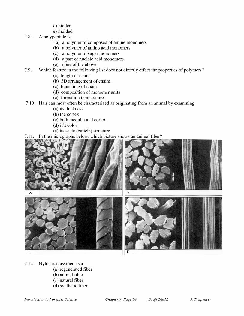
Introduction to Forensic Science Chapter 7, Page 64 Draft 2/8/12 J. T. Spencer
d) hidden e) molded
7.8. A polypeptide is (a) a polymer of composed of amine monomers (b) a polymer of amino acid monomers (c) a polymer of sugar monomers (d) a part of nucleic acid monomers (e) none of the above
7.9. Which feature in the following list does not directly effect the properties of polymers? (a) length of chain (b) 3D arrangement of chains (c) branching of chain (d) composition of monomer units (e) formation temperature
7.10. Hair can most often be characterized as originating from an animal by examining (a) its thickness (b) the cortex (c) both medulla and cortex (d) it’s color (e) its scale (cuticle) structure
7.11. In the micrographs below, which picture shows an animal fiber?
7.12. Nylon is classified as a
(a) regenerated fiber (b) animal fiber (c) natural fiber (d) synthetic fiber

Introduction to Forensic Science Chapter 7, Page 65 Draft 2/8/12 J. T. Spencer
(e) plant fiber 7.13. In what stage of a hair’s growth cycle is the hair actively growing?
(a) anagenic (b) telogenic (c) catagenic (d) analgesic (e) none of the above
7.14. Which part of the hair shaft is most resistant to chemical decomposition? (a) medulla (b) cortex (c) follicle (d) cuticle (e) shaft
7.15. The central canal running through many, but not all, human hairs is known as the: (a) medulla (b) shaft (c) cortex (d) cuticle (e) shaft
7.16. Hair, fibers and fingerprints are examples of what classification of evidence? 7.17. Who is considered the “father of fingerprinting?” 7.18. What is IAFIS? 7.19. Define the following terms used in fingerprint identification: core, delta, bifurcation, ridge
line and island. 7.20. What is the current minimum accepted number of matching points needed to place a
reasonable level of trust in deciding two fingerprints are a match? a) 6 b) 8 c) 10 d) 12 e) 14
7.21. What is the difference between identification and authentication methods? 7.22. What are some of the legal questions that have arisen in the use of latent prints in court cases? 7.23. What are the three general scale patterns on a hair shaft? 7.24. What are the three layers in a hair? 7.25. In order for a hair dye process to be effective(last longer than a rinse), what must the hair
stylist do to the cuticle layer of the hair shaft? 7.26. How does a “perm” work? 7.27. What forensic information may be gathered when examining a dyed or ‘permed’ hair shaft? 7.28. What are the three main groupings of fibers? 7.29. Plant fibers are typically _____________based while animal fibers are typically
______________ based. 7.30. What is a regenerated fiber? What is a synthetic fiber? 7.31. What property do thermoplastics have that make them desirable polymers? 7.32. What are the two main polymer synthesis reactions? 7.33. What are the criteria that any biometric identification must satisfy? 7.34. Which of the following could not be used as a means of biometric analysis? A) fingerprints B)
vein geometry C) iris scan D) face imaging E) right hand digit count EXTENSIVE QUESTIONS
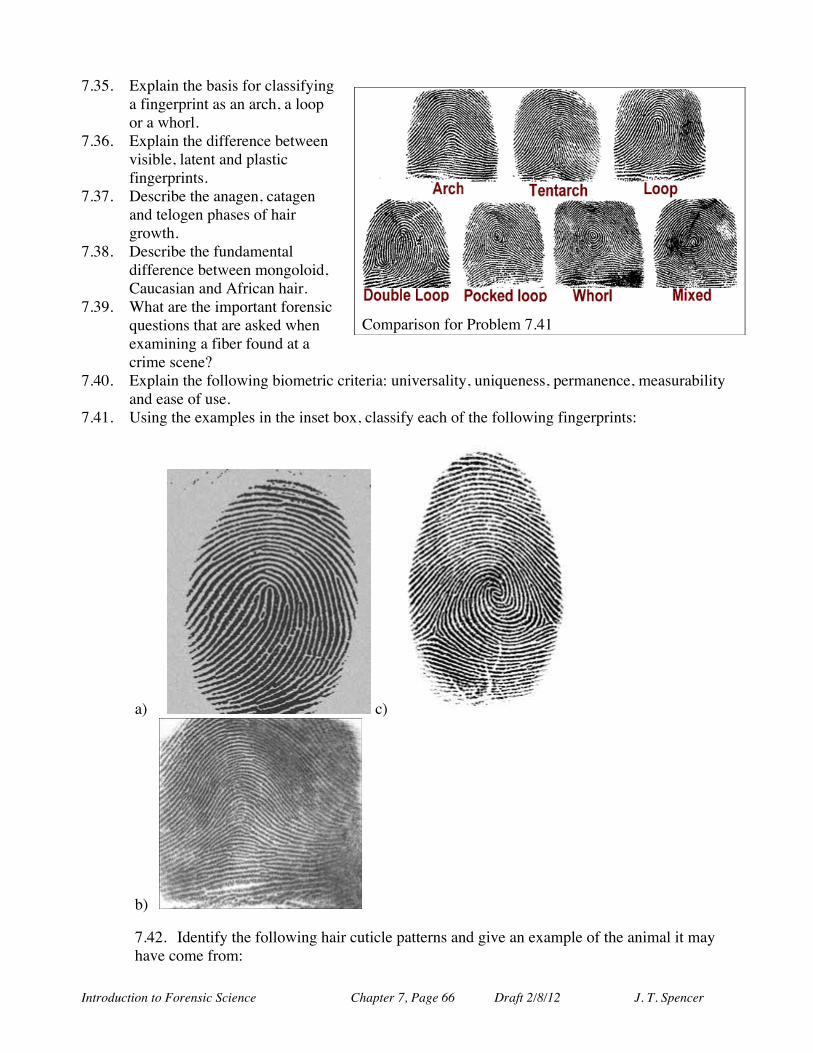
Introduction to Forensic Science Chapter 7, Page 66 Draft 2/8/12 J. T. Spencer
7.35. Explain the basis for classifying a fingerprint as an arch, a loop or a whorl.
7.36. Explain the difference between visible, latent and plastic fingerprints.
7.37. Describe the anagen, catagen and telogen phases of hair growth.
7.38. Describe the fundamental difference between mongoloid, Caucasian and African hair.
7.39. What are the important forensic questions that are asked when examining a fiber found at a crime scene?
7.40. Explain the following biometric criteria: universality, uniqueness, permanence, measurability and ease of use.
7.41. Using the examples in the inset box, classify each of the following fingerprints:
a) c)
b) 7.42. Identify the following hair cuticle patterns and give an example of the animal it may have come from:
Comparison for Problem 7.41
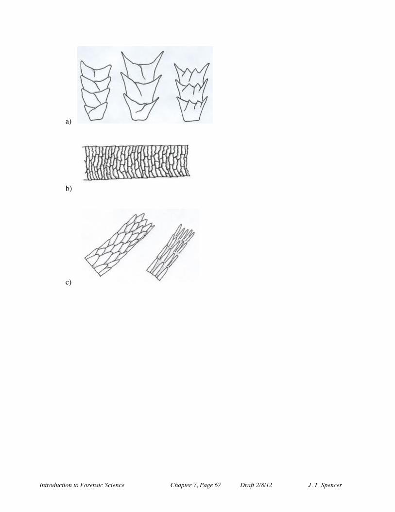
Introduction to Forensic Science Chapter 7, Page 67 Draft 2/8/12 J. T. Spencer
a)
b)
c)
Related Documents