CLINICAL MICROBIOLOGY REVIEWS, Apr. 2002, p. 223–246 Vol. 15, No. 2 0893-8512/02/$04.000 DOI: 10.1128/CMR.15.2.223–246.2002 Copyright © 2002, American Society for Microbiology. All Rights Reserved. Dracunculiasis (Guinea Worm Disease) and the Eradication Initiative Sandy Cairncross, 1 * Ralph Muller, 1 and Nevio Zagaria 2 Department of Infectious and Tropical Diseases, London School of Hygiene and Tropical Medicine, London WC1E 7HT, United Kingdom, 1 Department of Control, Prevention and Eradication of Communicable Diseases, World Health Organization, Geneva, Switzerland 2 INTRODUCTION .......................................................................................................................................................223 BIOLOGY OF THE DISEASE .................................................................................................................................224 Morphology ..............................................................................................................................................................224 Life Cycle .................................................................................................................................................................224 Zoonotic Aspects .....................................................................................................................................................224 CLINICAL ASPECTS.................................................................................................................................................226 Clinical Manifestations and Pathogenesis ..........................................................................................................226 Diagnosis ..................................................................................................................................................................227 Treatment .................................................................................................................................................................227 SOCIOECONOMIC IMPACT ..................................................................................................................................227 Disability ..................................................................................................................................................................227 Economic Impact ....................................................................................................................................................228 Nutrition, Education, and Perpetual Benefits ....................................................................................................229 EPIDEMIOLOGY .......................................................................................................................................................229 Water Sources .........................................................................................................................................................229 Villages of Endemicity ............................................................................................................................................230 Seasonality ...............................................................................................................................................................230 Individual Risk Factors .........................................................................................................................................231 THE ERADICATION INITIATIVE ..........................................................................................................................231 INTERVENTIONS ......................................................................................................................................................233 Safe Water Supply ..................................................................................................................................................233 Filtration of Drinking Water.................................................................................................................................235 Case Management...................................................................................................................................................235 Preventing Patients’ Contact with Ponds ............................................................................................................236 Killing or Removing Cyclops.................................................................................................................................237 Case Containment...................................................................................................................................................237 Intervention Conclusions .......................................................................................................................................239 CURRENT ISSUES ....................................................................................................................................................239 The Integration Debate ..........................................................................................................................................239 Geographic Information Systems (GIS) ..............................................................................................................240 The Certification of Dracunculiasis Eradication................................................................................................241 Concluding Remarks ..............................................................................................................................................242 ACKNOWLEDGMENTS ...........................................................................................................................................243 REFERENCES ............................................................................................................................................................243 INTRODUCTION Guinea worm disease, also known as dracunculiasis (or dra- cunculosis), is a long-established human infection which was clearly referred to by various authors from India, Greece, and the Middle East in antiquity; female worms have been seen in Egyptian mummies (2, 150). A curious monograph by Vels- chius (159) discussed real and imagined references in ancient writings and sculptures, including a supposed relationship with the caduceus motif. James Africanus Horton, the first West African to be trained in Europe as a medical doctor, wrote a book about the disease (87), mistakenly supposing that it was transmitted through the soles of the feet. Connection of infection with water sources was recognized early, and it is probable that, if the prepatent period were not so long, the mode of infection would have been obvious many cen- turies earlier. As it was, this was determined in 1870 by a Russian naturalist, Alexei Fedchenko (67), who found that larvae expelled from emerging female worms in the limbs of sufferers developed in freshwater microcrustaceans (cyclops) living in ponds, which were then ingested in drinking water. In historical times infection occurred in Algeria, Egypt (162), Gambia, Guinea Conakry, Iraq, Brazil, and the West Indies (163) but died out spontaneously in those countries and was eliminated from Uzbekistan in 1932 and from southern Iran in 1972. * Corresponding author. Mailing address: Department of Infectious and Tropical Diseases, London School of Hygiene and Tropical Med- icine, Keppel Street, London WC1E 7HT, United Kingdom. Phone: 44 20 7927 2211. Fax: 44 20 7636 7843. E-mail: sandy.cairncross@lshtm .ac.uk. 223 on May 9, 2016 by PENN STATE UNIV http://cmr.asm.org/ Downloaded from

Welcome message from author
This document is posted to help you gain knowledge. Please leave a comment to let me know what you think about it! Share it to your friends and learn new things together.
Transcript

CLINICAL MICROBIOLOGY REVIEWS, Apr. 2002, p. 223–246 Vol. 15, No. 20893-8512/02/$04.00�0 DOI: 10.1128/CMR.15.2.223–246.2002Copyright © 2002, American Society for Microbiology. All Rights Reserved.
Dracunculiasis (Guinea Worm Disease) and theEradication Initiative
Sandy Cairncross,1* Ralph Muller,1 and Nevio Zagaria2
Department of Infectious and Tropical Diseases, London School of Hygiene and Tropical Medicine, London WC1E 7HT,United Kingdom,1 Department of Control, Prevention and Eradication of Communicable Diseases,
World Health Organization, Geneva, Switzerland2
INTRODUCTION .......................................................................................................................................................223BIOLOGY OF THE DISEASE .................................................................................................................................224
Morphology ..............................................................................................................................................................224Life Cycle .................................................................................................................................................................224Zoonotic Aspects .....................................................................................................................................................224
CLINICAL ASPECTS.................................................................................................................................................226Clinical Manifestations and Pathogenesis ..........................................................................................................226Diagnosis..................................................................................................................................................................227Treatment.................................................................................................................................................................227
SOCIOECONOMIC IMPACT ..................................................................................................................................227Disability ..................................................................................................................................................................227Economic Impact ....................................................................................................................................................228Nutrition, Education, and Perpetual Benefits ....................................................................................................229
EPIDEMIOLOGY.......................................................................................................................................................229Water Sources .........................................................................................................................................................229Villages of Endemicity............................................................................................................................................230Seasonality ...............................................................................................................................................................230Individual Risk Factors .........................................................................................................................................231
THE ERADICATION INITIATIVE..........................................................................................................................231INTERVENTIONS......................................................................................................................................................233
Safe Water Supply ..................................................................................................................................................233Filtration of Drinking Water.................................................................................................................................235Case Management...................................................................................................................................................235Preventing Patients’ Contact with Ponds ............................................................................................................236Killing or Removing Cyclops.................................................................................................................................237Case Containment...................................................................................................................................................237Intervention Conclusions .......................................................................................................................................239
CURRENT ISSUES....................................................................................................................................................239The Integration Debate ..........................................................................................................................................239Geographic Information Systems (GIS) ..............................................................................................................240The Certification of Dracunculiasis Eradication................................................................................................241Concluding Remarks ..............................................................................................................................................242
ACKNOWLEDGMENTS ...........................................................................................................................................243REFERENCES ............................................................................................................................................................243
INTRODUCTION
Guinea worm disease, also known as dracunculiasis (or dra-cunculosis), is a long-established human infection which wasclearly referred to by various authors from India, Greece, andthe Middle East in antiquity; female worms have been seen inEgyptian mummies (2, 150). A curious monograph by Vels-chius (159) discussed real and imagined references in ancientwritings and sculptures, including a supposed relationship withthe caduceus motif. James Africanus Horton, the first West
African to be trained in Europe as a medical doctor, wrote abook about the disease (87), mistakenly supposing that it wastransmitted through the soles of the feet.
Connection of infection with water sources was recognizedearly, and it is probable that, if the prepatent period were not solong, the mode of infection would have been obvious many cen-turies earlier. As it was, this was determined in 1870 by a Russiannaturalist, Alexei Fedchenko (67), who found that larvae expelledfrom emerging female worms in the limbs of sufferers developedin freshwater microcrustaceans (cyclops) living in ponds, whichwere then ingested in drinking water.
In historical times infection occurred in Algeria, Egypt (162),Gambia, Guinea Conakry, Iraq, Brazil, and the West Indies (163)but died out spontaneously in those countries and was eliminatedfrom Uzbekistan in 1932 and from southern Iran in 1972.
* Corresponding author. Mailing address: Department of Infectiousand Tropical Diseases, London School of Hygiene and Tropical Med-icine, Keppel Street, London WC1E 7HT, United Kingdom. Phone: 4420 7927 2211. Fax: 44 20 7636 7843. E-mail: [email protected].
223
on May 9, 2016 by P
EN
N S
TA
TE
UN
IVhttp://cm
r.asm.org/
Dow
nloaded from

BIOLOGY OF THE DISEASE
Morphology
The single species causing the disease in humans, Dracun-culus medinensis (Linnaeus, 1758; Gallandant, 1773), belongsto the nematode superfamily Dracunculoidea of the orderSpirurida. Most spirurids are tissue parasites and produce eggscontaining larvae or free larvae which require arthropod inter-mediate hosts. The best-known examples of this order are thefilariae (superfamily Filarioidea), including the important hu-man parasites Wuchereria bancrofti and Brugia malayi (againstboth of which a global campaign is just beginning), Onchocercavolvulus, and Loa loa. For many years Dracunculus was in-cluded among the filariae, but it differs from them, mainly inthe disparate sizes of the sexes and life history. The validity ofthe relationships among the various groups of spirurids hasbeen supported by recent analyses of small-subunit rRNA se-quences (16).
The same or similar species of Dracunculus have been re-ported sporadically for mammals and reptiles (snakes and tur-tles) in many parts of the world (116); species from birds havebeen separated into a different genus, Avioserpens.
A mature female D. medinensis is one of the longest nema-todes, measuring up to 100 cm, but is only 1 to 2 mm wide (Fig.1). The vulva is halfway down the body. In filarial females themicrofilarial prelarvae emerge from this opening throughout
the life of the worm. In Dracunculus, however, it is closed witha plug, and the whole body cavity is filled with the uterus, whichextends anteriorly and posteriorly and contains from 1 to 3million first-stage larvae. The gut is also completely flattenedand is nonfunctional. Bright red worms have been reportedfrom Pakistan (56), but their significance is not known. Maleshave been recovered only doubtfully from humans, but thosefrom experimental animals measure 15 to 40 by 0.4 mm; thetail has three to six preanal and four to six pairs of postanalpapillae, with subequal spicules (490 to 750 �m long) and anaccessory organ, the gubernaculum (about 117 �m).
Life Cycle
The mature female worm lives in the subcutaneous connec-tive tissues, moves to the surface of the skin, and provokes theformation of a blister, which bursts, causing the anterior end ofthe worm to be exposed. If the affected portion of the body(usually the foot or leg) is cooled by immersion in water,first-stage larvae are expelled in large numbers from the rup-tured uterus. These measure 643 (490 to 737) by 23 (18 to 24)�m with a pointed tail and a fully formed gut, although they donot feed. They move actively in the water, resembling a free-living nematode, and can live for a few days in water.
For further development, the larvae need to be ingested bysuitable predatory species of copepods, measuring 1 to 2 mm.Until a few years ago, these were all included in the singlegenus Cyclops, but this has now been subdivided, and the mostimportant intermediate hosts belong to the genera Mesocyclops(M. aequatorialis and M. kieferi), Metacyclops (M. margaretae),and Thermocyclops (T. crassus, T. incisus, T. inopinus, and T.oblongatus) (119). Larvae develop to the infective third stage inthe body cavity in 14 days at 26°C. For further development ofthe larva, the host copepod needs to be ingested in drinkingwater obtained from ponds or open wells. Once in the humanbody, larvae are released in the stomach and migrate throughthe intestinal wall, across the peritoneal cavity, and into thewall of the abdomen and thorax by 15 days (Fig. 2). After twomolts the sexually mature males and females meet and mate inabout 100 days, while they are both of comparable size. Themales remain in the tissues and in a few months become en-capsulated and die. In experimental infections by Dracunculusinsignis in ferrets, males can live for at least 330 days (19) andtherefore are potentially able to fertilize females for anotheryear. The females move down the muscle planes and by 10months have grown greatly, with the uterus being filled withlarvae. They emerge about 1 year after infection, usually fromthe feet and lower legs (Fig. 3) but occasionally from any otherpart of the body.
The process from ingestion of a larva to emergence of anadult worm typically takes about a year, enabling transmissionto occur annually at the most favorable season. However, inindividual cases it can last from 10 to 14 months.
Zoonotic Aspects
The eradication of any human disease becomes a vastlymore difficult enterprise if there are animal reservoirs of in-fection. There is no evidence that animals act or have everacted as reservoir hosts of human guinea worm infection, but
FIG. 1. Male and female D. medinensis worms. The female worm isthe larger of the two. The ruler at the left is in centimeters.
224 CAIRNCROSS ET AL. CLIN. MICROBIOL. REV.
on May 9, 2016 by P
EN
N S
TA
TE
UN
IVhttp://cm
r.asm.org/
Dow
nloaded from

the theoretical possibility that they could do so has not beenconclusively disproved. Studies of animal infections in areaswhere dracunculiasis has been recently eliminated but withoutmuch improvement in drinking water sources might provideuseful information regarding this question.
Table 1 lists the species of Dracunculus which have beenreported. Emerging female worms are recovered sporadicallyfrom a wide range of mammals both from parts of the worldwhere dracunculiasis is endemic and from parts where it is not
(12, 49, 69, 71, 89, 90, 103, 106, 110, 135, 140; review in refer-ence 116). Unfortunately, in almost all cases only a portion ofa female worm is recovered, usually in a flaccid state after theenclosed larvae have been ejected, and identification with re-gard to species is impossible. In general, worms in mammalsare regarded as D. medinensis in the Old World and SouthAmerica and as D. insignis in North America. Dracunculus hasa very short patent period, at the most a few weeks, and anemerging worm is not easy to see in fur-bearing carnivores(Fig. 4), so it is likely that infection is much more common inmany countries than is currently recognized. This is well illus-trated by the situation in North America, where there are oneor two reports of infection in wild carnivores and domesticdogs each year, mostly from states in the southeastern UnitedStates and from southern Ontario, Canada (e.g., see reference
FIG. 2. Development of Dracunculus in the mammalian host. S.C.CONN., subcutaneous connective tissues. Sizes at left are given in centimeters.
FIG. 3. A human case of guinea worm disease, with an emergingworm. The worm is often wound on a stick, a practice which is believed tohave given rise to the caduceus symbol of medicine. (Copyright A. Tayeh.)
TABLE 1. Reported species of Dracunculusa
Species Host Geographical distribution
D. alii Snakes IndiaD. coluberensis Snakes IndiaD. dahomensis Snakes West and Central AfricaD. doi Snakes IndiaD. houdemeri Snakes VietnamD. ophidensis Snakes Italy, USAb
D. oesophageus Snakes Italy, MadagascarDracunculus species Snakes England, Brazil, Trinidad,
USAD. globocephalus Turtles USAD. fuellebornius Opossums BrazilD. insignis Dogs, wild
carnivoresCanada, USA
D. lutrae Otters CanadaD. medinensis Many mammals Africa, Americas, Asia
a Note that some authorities have recognized only two species: D. medinensisin mammals and D. oesophageus in reptiles (116).
b USA, United States of America.
VOL. 15, 2002 DRACUNCULIASIS AND THE ERADICATION INITIATIVE 225
on May 9, 2016 by P
EN
N S
TA
TE
UN
IVhttp://cm
r.asm.org/
Dow
nloaded from

14). However, in studies where large numbers of raccoons weredissected and immature worms were found, prevalence rates of13.8% (144) to about 50% (47) were recorded.
The taxonomic status of this North American parasite is alsoworthy of investigation. It was described by Leidy in a shortaccount in 1858 and confirmed as a different species (D. insig-nis) by Chitwood (43) in 1950, because of differences in thelength of the gubernaculum and number of preanal papillaefrom males of D. medinensis. However, when more males ofthe latter species were described (116), these differences werenot found to be valid, and the species was never accepted by allauthorities: e.g., Faust et al. in a standard textbook (66) stated,“In reservoir hosts D. medinensis is found in fur-bearing mam-mals in North America.” Species of copepods which transmitD. medinensis are also susceptible to larvae of D. insignis (149).If these parasites are all of the human-infecting species, thenthis is encouraging for world elimination of human infections,since almost no autochthonous infections have been reportedfrom North America (see reference 146 for one such case;Spearman et al. [145] reported on imported cases), althoughcalcified worms have been seen on roentgenograms (E. H.Michelson, personal communication).
An isolated autochthonous human case of dracunculiasis hasbeen reported from Japan (105), and similar older cases oc-curred in Korea (76) and Indonesia (157). In all of thesepresumably zoonotic cases, there was the possibility that infec-tion was contracted from ingesting raw freshwater fishes asparatenic hosts (in which the immature parasites ingested incyclops can survive but do not develop) rather than from drink-ing water: certainly Dracunculus in North America can betransmitted experimentally by amphibians (47, 54). Experi-mentally, rodents can be infected with larvae of the humanparasite, which can live for months in the peritoneal cavitywithout development, and these could act as paratenic hostsfor infections in carnivores (R. Muller, unpublished data). In-fections in reptiles appear to be due to separate species (seereference 116).
The infections in domestic animals reported from areas ofendemicity are possibly of human origin. This supposition is sup-ported by the higher prevalence found by surveys of the humanpopulation than by the dissection of dogs culled during the 1920sin Bukhara, Uzbekistan (Fig. 5). However, worms are still foundin dogs in the former areas of endemicity of Uzbekistan (99) andTamil Nadu (106) and also in dogs and cats in the areas ofAzerbaijan, Kazakhstan, and possibly Turkmenia where the dis-ease is not endemic (44, 45, 71, 110, 158). It is very likely that inmany parts of the world there are widespread but underreportedanimal cycles completely independent of human infection: forexample, there was a recent report from China of infection withD. medinensis in a cat (69). It is significant that the disease has notreturned to Uzbekistan in the 70 years since it was eliminatedthere, in spite of the fact that about 40% of the rural populationstill does not have piped water and only 33% in the area sur-rounding Bukhara, the historic focus (171), have piped water. Norhas it returned to Egypt, Iran, Gambia, or Guinea, where itdisappeared decades ago.
The question of whether animal reservoirs of D. medinensisexist should be clarified by projected DNA sequence studies,but in any event animal infections are unlikely to prove ahuman public health problem once complete eradication of thehuman disease has been achieved, particularly if safe watersources are provided.
CLINICAL ASPECTS
Clinical Manifestations and Pathogenesis
Preemergent female worms can move easily through theconnective tissues, but when they are about to emerge to the
FIG. 4. Guinea worm in a laboratory cat infected with cyclops froma pond in Nigeria.
FIG. 5. Prevalence of dracunculiasis in people and dogs in OldBukhara, Uzbekistan, 1926 to 1931. Data are from reference 171.
226 CAIRNCROSS ET AL. CLIN. MICROBIOL. REV.
on May 9, 2016 by P
EN
N S
TA
TE
UN
IVhttp://cm
r.asm.org/
Dow
nloaded from

surface, a few larvae are released into the subdermis through arupture at the anterior end. The host reaction results in theformation of a burning, painful blister, which bursts in a fewdays to give a shallow ulcer, and there is then a marked in-flammatory response against the cuticle of the entire worm,preventing its removal. The bacteriologically sterile blisterfluid contains larvae surrounded principally by polymorphonu-clear neutrophils with macrophages, lymphocytes, and eosino-phils (117). After expulsion of thousands of larvae, the end ofthe worm dries up, and this process is repeated a few times,with the complete worm being extruded in a few weeks. Thelesion then resolves quickly. Unfortunately, the track of theworm becomes secondarily infected in about half of all cases,and patients become severely incapacitated. In one study in anarea of Nigeria, 58% of patients, mostly in the 15- to 49-year-old working-age group or of school age, were disabled for anaverage of 12.7 weeks during the yam and rice harvest time(143). In a study in Ghana, 28% of patients had continuingpain 12 to 18 months after emergence of worms and 0.5% (asin many studies) had permanent physical impairment, in theform of “locked” knees or other joints (88). In another study inBenin there was 0.3% mortality from tetanus and septicemia(42). Female worms sometimes burst in the tissues, resulting ina very large pus-filled abscess and severe cellulitis. Infertilefemales or males elicit a slight inflammatory reaction andsometimes calcify, showing up on a roentgenogram.
Dracunculiasis is unusual among parasitic infections in thatthere is little evidence of acquired immunity and the sameindividual can be reinfected many times. The response to theextrusion of larvae is indicative of an Arthus reaction followedby a delayed hypersensitivity response (117).
Diagnosis
Patients in an area of endemicity have no doubt about thediagnosis when, or just before, the blister forms from the localitching and then sharp pain and often general allergic symp-toms including urticaria follow. Once the blister has burst, coldwater will encourage the release of larvae, which can be seenmicroscopically under low power. Immunodiagnostic methodsare not useful in practice because it has not been proved thatthey can detect prepatent infections, mainly because of the lackof prepatent serum samples. However, antibodies can be de-tected in patent infections by enzyme-linked immunosorbentassay or dot-enzyme-linked immunosorbent assay, usingwhole-worm antigens. The most specific reaction appears to befor detection of immunoglobulin G4 (17). This test might beable to detect prepatent infections up to 6 months beforeemergence (11), in which case it could have practical impor-tance. No evidence was found for the presence of circulatingantigen (18).
Treatment
Treatment by winding worms out on a stick, a few centime-ters a day, has been practiced since antiquity and is still useful,particularly when combined with a clean dressing and antibi-otic ointment to prevent secondary bacterial infection (115).There is no evidence that any chemotherapeutic agent has adirect action against guinea worms, although various benzimi-
dazoles may have an anti-inflammatory action, aiding elimina-tion (123), and aspirin was found elsewhere to be equallyeffective (Muller, unpublished). Ivermectin is effective againstmany other nematodes but had no action in one trial, nor inexperimental infections (55, 96). Chippaux (37) found thattreatment with mebendazole was associated with aberrant mi-gration of the worms, which were more likely than usual toemerge at places other than the lower limbs.
SOCIOECONOMIC IMPACT
In recent years, the understanding has grown that biologicaland technical feasibility is not the only criterion to considerbefore launching an eradication program. Costs and benefitsare no less important (52). The benefits of dracunculiasis erad-ication, in contrast to those of smallpox and polio, will accruealmost exclusively to the population in which the disease isendemic (10).
In the past, most cases of dracunculiasis went unreported fora number of reasons: most health centers had little to offer thepatient besides palliative treatment; most patients live in poor,remote rural areas and are hindered by their disease fromwalking to a health facility; and most recover spontaneouslyafter expulsion of the worm. For example, a large teachinghospital in Nigeria never saw dracunculiasis cases in the Ca-sualty Department, although the disease was endemic in vil-lages a few kilometers away. Because few cases were reported,the disease was often considered an exotic curiosity rather thana major public health problem. However, in areas of endemic-ity its social, economic, nutritional, and educational conse-quences, and the costs incurred by the individuals, households,and communities which suffer from it, can be substantial.
Disability
Dracunculiasis is rarely fatal; studies in India based on med-ical records suggest a case fatality rate of 0.1% or less, and thisis probably a generous estimate, because only persons withsevere complications usually seek treatment from health facil-ities (4). The proportion of patients permanently disabled bythe disease is also small; a number of studies have found it tobe less than 1% (94, 132, 142).
The social impact of guinea worm disease is mainly attrib-utable to the temporary disability suffered by the patient. Twolongitudinal studies in Nigeria (5, 143) found that 58 to 76% ofpatients were unable to leave their beds for approximately amonth during and after emergence of the worm. The moresevere and protracted disability is associated with secondaryinfection of the lesion; this occurs in roughly half the cases(124, 184).
The impact of this temporary disability is reinforced by theseasonal pattern of worm emergence, often peaking at stagesof the agricultural year when labor is in maximum demand.This is illustrated in Fig. 6, where the topmost section showsthe seasonal variation in agricultural activity in northernBurkina Faso, peaking with the sowing season in June. Themiddle section shows the corresponding variation in cases ofdracunculiasis, also peaking in June. The pattern typical of theforest zone to the south is shown in the bottom section of Fig.6; here cases peak in the dry season, but this is the time for yam
VOL. 15, 2002 DRACUNCULIASIS AND THE ERADICATION INITIATIVE 227
on May 9, 2016 by P
EN
N S
TA
TE
UN
IVhttp://cm
r.asm.org/
Dow
nloaded from
and rice harvesting (143). This seasonality means that a wholecommunity can be laid prostrate simultaneously and householdmembers can be prevented from substituting for one anotherin agricultural and other tasks (13). Indeed, it has been claimedthat the effect of the disease on agricultural productivity can bedetected in satellite photographs (6). The Dogon people ofMali refer to the infection as “the disease of the empty gra-nary” (172).
The impact of guinea worm disease does not end when theworm is out and the sufferer returns to work. A study in Ghana(88) found that, between 12 and 18 months after emergence of
a worm, 34% of patients still had some difficulty performingeveryday activities, usually due to pain attributable by its loca-tion and the date of onset to the episode of dracunculiasis.While this disability is not necessarily permanent, it extendsbeyond the incapacity occurring during worm emergence.
Economic Impact
Some attempts to estimate the economic impact of dracun-culiasis have simply multiplied the number of days of labor lostby the mean value of production per day or by the wage rate.
FIG. 6. Seasonality of dracunculiasis in the Sahel (A) and the forest zone of West Africa (B), compared with the rainfall and the Sahel’sseasonal pattern of agricultural labor demand. Reproduced from reference 147 with permission of Taylor & Francis Ltd. Months are January,February, March, April, May, June, July, August, September, October, November, and December, from left to right, respectively.
228 CAIRNCROSS ET AL. CLIN. MICROBIOL. REV.
on May 9, 2016 by P
EN
N S
TA
TE
UN
IVhttp://cm
r.asm.org/
Dow
nloaded from
From such a simplification, it is a small step to multiply the lossper household to derive an estimated cost for a whole region.One such study (50), based on a survey of 87 households,estimated that the rice-growing areas in three states of south-ern Nigeria sustained an annual loss of $20 million due toguinea worm disease. In spite of its simplistic argument, thisstudy was extremely effective in mobilizing the support of se-nior politicians in Nigeria for the eradication of the disease(64).
It has been argued previously that this method of calculationuses an oversimplified approach and is likely to overestimatethe cost (75, 128), as it does not allow for the various copingstrategies by which households respond to illness (such asabandoning other tasks and using additional labor), whichqualitative studies have found to be common in peasant farm-ing (23, 38). A more sophisticated approach is to examine theimpact on actual production (24) or even to include the inci-dence and duration of dracunculiasis-induced disability as pre-dictive variables in an agricultural production function. Audi-bert (9) used this approach, in a setting in northeastern Maliwhere the incidence of guinea worm disease was “relativelylow” (between 3 and 33% in the villages studied), to show thattemporary disability accounted for a reduction of 5% in theoverall production of two important subsistence crops: sor-ghum, mainly grown by men, and peanuts, cultivated bywomen.
However, there is also a cost to the coping strategies, whichcannot be measured using this approach. Mutual assistance(164) simply transfers the cost of the disease to other house-holds and is of little help to wage laborers (36). The simplisticapproach used for the Nigerian study mentioned above may bemore accurate than it seems.
Nutrition, Education, and Perpetual Benefits
Such economic calculations do not take into account theunequal distribution of costs within the family and the way inwhich disease, by impacting more on the production of somecrops than others (24), can have a disproportionate effect onnutritional status. A survey in South Kordofan, Sudan (151),found that, in households where more than half the adultmembers had suffered from dracunculiasis in the previous year,the children under 6 years old were nearly three times as likelyto be malnourished, as indicated by wasting.
Children also suffer in other ways from guinea worm disease
in their families. Children miss school when they have guineaworm (in rural Africa, school is usually a long walk away) andalso when they have to substitute for their ill parents in doingagricultural work and other household tasks. As a result,school attendance suffers during the peak season (22, 60, 62,92, 124), and schools in areas of endemicity often have to closefor 1 month in each year as a result.
Guinea worm disease is of special interest to economists notonly because of its direct and measurable impact on produc-tion but also because of the singular ability of disease eradi-cation to produce a perpetual stream of benefits at no ongoingcost. It is, therefore, surprising that a recent cost-benefit anal-ysis of the eradication effort by the World Bank (104) considersonly the benefits from incidence reduction during the cam-paign; their estimate of a 29% economic rate of return for theglobal campaign, which they estimate to have cost some $90million to date, is certainly pessimistic.
EPIDEMIOLOGY
Water Sources
Because Dracunculus larvae need a period of 12 to 14 daysto develop in the cyclops and become infective, dracunculiasisis not normally caught from flowing water sources such asrivers and streams. Deep wells are rarely implicated in trans-mission (31, 118); few cyclops are found in them, probablybecause the lack of light at the bottom constrains the popula-tion of zooplankton, which are the cyclops’ natural diet. Thus,ponds and sometimes shallow or step wells are the mainsources of the disease, and the epidemiology of dracunculiasisis chiefly determined by the use of such sources for drinkingwater.
Numerous studies have illustrated the predominant role ofponds in dracunculiasis transmission in various parts of Nigeria(57, 58, 59, 60, 61, 100, 127), Ghana (112, 138), Burkina Faso(70), Togo (129), Uganda (77), Pakistan (86), India (98), andwhat is now Uzbekistan (171).
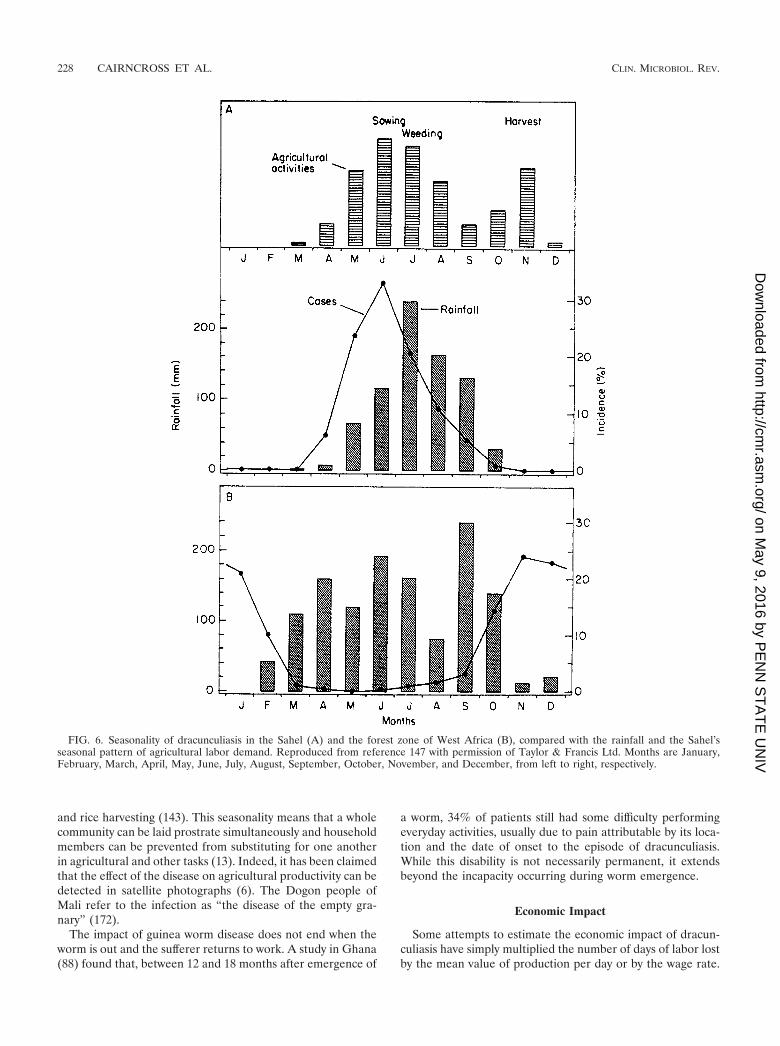
Many (probably most) of the ponds involved in transmissionare human-made. Table 2 shows the findings of Steib andMayer (147) for a village in Northeast Burkina Faso, whichillustrate the large number of ponds which can be found in asingle village, even in a semiarid, Sahelian setting, and thedegree to which relatively few human-made ponds, with spe-cific characteristics, play a significant role in transmission.
TABLE 2. Types of water sources and their relative importance in guinea worm transmission in Dara village, Burkina Fasoa
Characteristic Draw wells Periodic streams Large naturalponds Cattle waterings Small natural
pondsSmall human-made
ponds
Diam (m) 2 10 50–200 17–45 7–15 4–9Maximum depth (cm) Up to 500 250 100–130 45–80 25–30 30–55Potential focus? No No Yes Yes Yes YesNo. found and
studied4 4 4 8
No. of guinea wormpatients regularlyusing them
9 2 5 90
% 8.5 1.9 4.7 84.9
a Adapted from reference 147 with permission of Taylor & Francis Ltd.
VOL. 15, 2002 DRACUNCULIASIS AND THE ERADICATION INITIATIVE 229
on May 9, 2016 by P
EN
N S
TA
TE
UN
IVhttp://cm
r.asm.org/
Dow
nloaded from

Other types of human-made ponds, sometimes larger thanthose identified by Steib and Mayer, have been implicated else-where. These have included “boullies,” which are large dew pondsexcavated for community water storage on the Mossi plateau ofcentral Burkina Faso (101); small dams in northern Ghana (152);“ataparas” (also known as valley tanks), which are similar reser-voirs in northern Uganda (77); hundreds of drinking-water pondsrecently built in Anambra State, Nigeria; “hafirs,” built to storewater from ephemeral streams in Sudan (31); and municipalponds in Old Bukhara, Uzbekistan.
Outbreaks of guinea worm disease have occasionally beenblamed on the construction of large dams (3, 59). However,their role in transmission has usually resulted from the use ofponds left by the receding water during drawdown of the waterlevel.
Various types of wells and storage tanks can become sourcesof the disease. Rectangular masonry-lined step wells were theprincipal sources of infection in Rajasthan, India (142), andshallow wells have been implicated in Mali (130). Scoop wellsdug in sandy riverbeds can also be a source, although if thesewater holes are exhausted by the drawing of water each day, nocyclops population can be sustained in them, and so transmis-sion cannot occur (31). In Iran, the “berkeh” (traditional cov-ered water storage cisterns with a diameter of over 10 m) wereimplicated (137).
Transmission has also occurred from rainwater storage res-ervoirs used by individual households, such as the “karkour” ofthe Nuba Mountains of Sudan (31) and in an isolated incidentin 1993 in El Rohaibat, Libya, from a buried reservoir filledfrom a farm tank which an infected migrant worker had con-taminated (102). Relatively few cases are caused in this way,because there must be an index case in the household tocontaminate the reservoir, but when they do occur, transmis-sion is more intense than usual because the infected cyclopsare contained in a smaller volume of water, and this is reflectedin a higher average number of worms per patient (32).
Villages of Endemicity
The fact that dracunculiasis transmission depends on thecharacteristics of the water source has an important conse-
quence for the design of eradication programs. It occurs only ina limited number of so-called “villages of endemicity,” onwhich eradication activities can focus. For example, in 1990 thenational case search conducted in all 8,068 villages of BurkinaFaso found cases in only 2,621 (101). Local health staff in BamProvince found that villages known to have ponds were twice aslikely as others to be among these villages of endemicity. Therehas been a tendency in some countries for the list of villages ofendemicity to “drift” by some 30% per year and sometimeseven twice this, but the appearance of a large proportion ofnew villages seems to be largely the consequence of casesreappearing in villages removed prematurely from the “ende-micity” list (111), rather than a change in the set of villagessusceptible to the disease. A period of 1 year without cases isinsufficient evidence that transmission has been interrupted, asthe full cycle from emergence of a worm in the index case todetection of a secondary case can take up to 16 months.
Seasonality
Dracunculus takes roughly a year from ingestion of an in-fected cyclops by the human host to emergence of an adultworm. This makes it well suited for environments in whichtransmission can occur only at a particular time of year. As aresult, there is a strong seasonal peak in incidence rates in mostcommunities of endemicity.
Two broad patterns of seasonality are found in the Africanareas of endemicity, depending on climatic factors. In somecountries, both patterns occur, each in a different climatic zone(Table 3). To the north in the Sahelian zone, transmission ofdracunculiasis is generally limited to the rainy season fromMay to August with a peak in June and July (73). Steib andMayer (147) attributed this pattern to the presence of T. in-opinus in the surface and shallow water used for drinking;however, others have found it more difficult to correlate oc-currence of cyclopoids in the local water sources with theprevalence of infection among the people using them (185).More fundamentally, many water sources involved dry up andhand pumps are repaired in the dry season, so that the popu-lation turns to safer groundwater sources (48).
Further south in the humid savanna and forest zone, the
TABLE 3. Peak transmission seasons for dracunculiasis in 14 African countries
CountryOccurrence in montha:
Jan Feb Mar Apr May June July Aug Sept Oct Nov Dec
Mauritania X X X XChad X X X X XMali X X X X XNiger X X X X XBurkina Faso X X X X X XCameroon X X X X X XSudan X X X X X XUganda X X X X XEthiopia X X X X XIvory Coast X X X X X X XGhana X X X X X X XBenin X X X X X XTogo X X X X XNigeria X X X X X X X X
a Abbreviations: Jan, January; Feb, February; Mar, March; Apr, April; Aug, August; Sept, September; Oct, October; Nov, November; Dec, December.
230 CAIRNCROSS ET AL. CLIN. MICROBIOL. REV.
on May 9, 2016 by P
EN
N S
TA
TE
UN
IVhttp://cm
r.asm.org/
Dow
nloaded from

opposite pattern is found, with a peak in the dry season. Thismay be the early dry season (September to January), as insome parts of Oyo State, Nigeria (61, 100); Danfa in Ghana(13); and southern Togo (129), particularly in villages withshallow ponds which usually dry up by January. The diseaseoften occurs or continues towards the end of the dry season(January to May) in Ghana (13, 25, 112, 138), southern Benin(41), Kwara and Anambra states in Nigeria (1, 58, 60, 124), andUganda (77). This dry season transmission is often associated
with the consumption of water from ponds or water holesformed (or dug) in the beds of seasonal rivers when flow hasceased (38, 114). It has also been suggested (41) that transmis-sion does not occur when there is less than one susceptiblecyclopoid per liter in the pond and that this accounts forseasonal variations in incidence.
There are local variations in these patterns. The durationand intensity of transmission in particular villages often de-pend on whether and when the local dams or ponds dried upthe previous year (152), and some villages have very differentseasonal peaks from those of the surrounding area because oflocal circumstances. For example, in some villages along thebanks of the Niger and Volta rivers the incidence peaks whenthe river level falls and water is taken from holes dug in theriverbed.
Individual Risk Factors
In the same way as dracunculiasis exhibits a variety of sea-sonal patterns, which can be explained in terms of seasonalvariations in water source use, the incidence of the disease hasbeen found to vary with age and sex in different ways, but thesecan generally be understood from the way that people of dif-ferent ages and genders behave with regard to their sources ofdrinking water.
For example, a significantly higher prevalence has beenfound in women in Ethiopia (97) and in men in India (98) andsometimes in West Africa (3, 39, 123); however, when behav-ioral risk factors (such as work in the fields or collection ofwater) are taken into account, the difference between the sexesis not significant (153).
Two possible age prevalence profiles are illustrated in Fig. 7.The graph for four high-prevalence villages, each with an in-fected water source, shows similar prevalences in children andin adults. The remaining 23 villages in the study area show alower prevalence in all ages but significantly less in childrenthan in adults. The former is characteristic of communitieswhere the water carried home is infected, while the latter isindicative of an association with mobility, where infection isacquired from water sources outside the community.
Other individual risk factors are important, particularlythose associated with mobility (160); however, the strongest ofall is infection in the previous year. A minority of people sufferrecurrent infection in spite of drinking from the same watersources as the rest of the population, reflecting the variabilityin individuals’ susceptibility to the disease (39, 112, 153).
THE ERADICATION INITIATIVE
As one of us first pointed out (118), guinea worm disease isa promising candidate for successful eradication. The cyclops isnot a mobile vector like a mosquito, and the carrier state inboth the cyclops and human hosts is of limited duration. Di-agnosis is easy and unambiguous; cheap and effective measuresare available to prevent transmission. The disease has a limitedgeographical distribution, and even within this area it is foundonly in certain communities of endemicity. Its markedly sea-sonal distribution in time also permits a more intensive focuson its prevention in seasonal campaigns. Lastly, as discussed
FIG. 7. Age prevalence curves for 4 high-endemicity and 23 lower-endemicity villages in South Kordofan, Sudan. The curves were plottedfrom data in reference 31.
TABLE 4. Characteristics of past and present eradicationcandidatesa
Characteristic
Presence of characteristic for diseaseb:
Malaria Small-pox Polio Dracun-
culiasis
AgentNo animal reservoir � � � �No mobile vector � � � �
HostLimited carrier state � � � �Easy diagnosis � � � �
DistributionLimited area of endemicity � � � ��Seasonal transmission � � �/� �Threat to Western countries
and visitors� � � �
InterventionExistence of effective drugs � � � �Cheapness and completeness
of prevention� � � �
No behavioral changeneeded for prevention
� � � �
a Adapted from reference 79 with permission of the publisher.b ��, highly present and favorable for eradication; �, present;�/�,somewhat
present; �, absent.
VOL. 15, 2002 DRACUNCULIASIS AND THE ERADICATION INITIATIVE 231
on May 9, 2016 by P
EN
N S
TA
TE
UN
IVhttp://cm
r.asm.org/
Dow
nloaded from

above, transmission from animals to people is practically un-known.
The suggestion that dracunculiasis might be eradicablefell on fertile ground. Smallpox had been eradicated in 1977,and some of those associated with that achievement werelooking for a suitable candidate to adopt for the sequel (7,78, 141), while taking care to avoid a mistake like the tar-geting of malaria in 1955 (109). Another possible candidatewas poliomyelitis, and indeed some of those involved inguinea worm eradication have seen it as a rehearsal for theeradication of polio, particularly in the difficult African ter-rain where the last cases of both diseases will probablyoccur. Table 4 compares the prospects of all four diseases inthe eradication stakes.
Choosing a target is easy, however, compared with the taskof mobilizing the resources for the battle. Much of the creditfor that achievement goes to members and former members ofthe staff of the Centers for Disease Control and Prevention,
who through an advocacy campaign beginning in 1980 andsustained over more than a decade (79, 80, 82, 83) succeededin convincing former U.S. president Jimmy Carter, the UnitedNations Children’s Fund (UNICEF) Executive Board, the1989 African Regional Committee of the World Health Orga-nization (WHO), and the 1990 World Summit for Children totake up the challenge. In 1991 the World Health Assemblydeclared “its commitment to the goal of eradicating dracuncu-liasis by the end of 1995, this date being technically feasiblegiven appropriate political, social, and economic support.” Thetarget date was set in order to enhance the advocacy effort atthe international level and in the countries of endemicity aswell.
This advocacy effort needed to be replicated in each countryto get a national program established (64). In 1982, India wasthe first to initiate a national eradication campaign. By 1990,four other countries had followed: Pakistan, Ghana, Nigeria,and Cameroon. In the following 5 years, all the other known
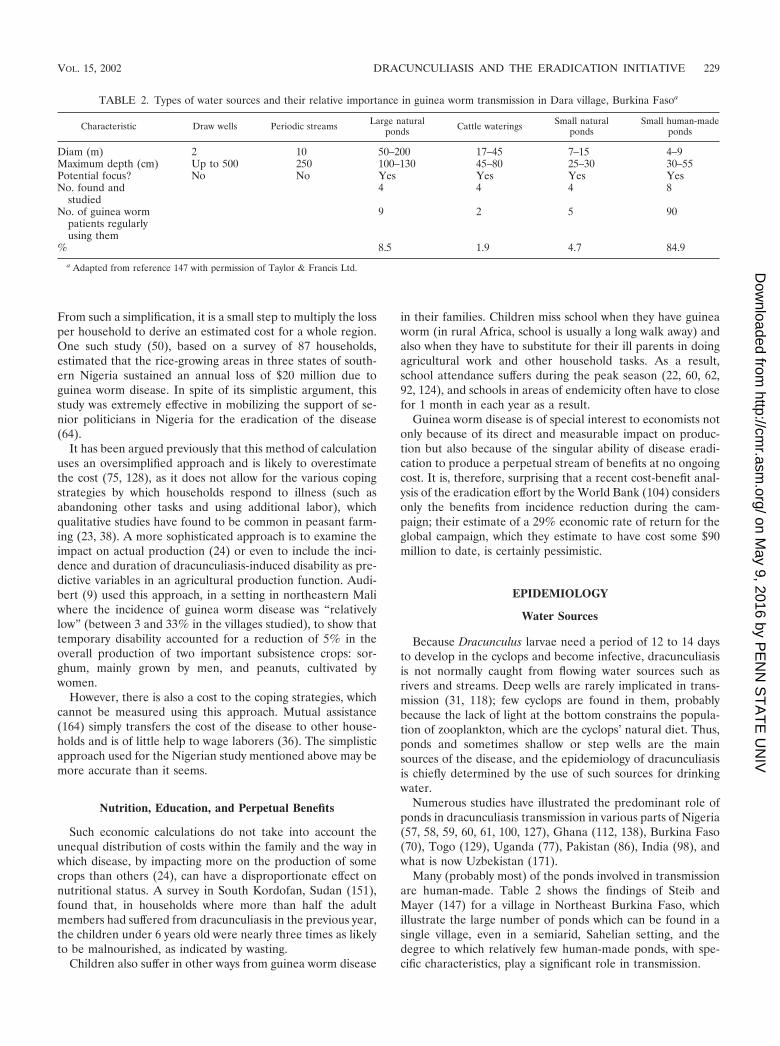
FIG. 8. The decline in cases of guinea worm disease by country and by year, 1989 to 2000. Note the logarithmic vertical scale. (a) Selectedcountries of endemicity. The poor performances of Sudan, due to the civil war, and of Ghana and Nigeria in the late 1990s can be seen. (b) Theremaining countries of endemicity. The rise in cases in the initial years of some national programs (e.g., Togo and Ethiopia) is attributable togradual improvements in case detection. C.A.R., Central African Republic. (c) The countries which have interrupted transmission; the last pointshown for each country is for the last year in which autochthonous cases were reported. Data are courtesy of WHO.
232 CAIRNCROSS ET AL. CLIN. MICROBIOL. REV.
on May 9, 2016 by P
EN
N S
TA
TE
UN
IVhttp://cm
r.asm.org/
Dow
nloaded from
countries of endemicity also established national eradicationprograms, and substantial and progressive reductions in dis-ease incidence were recorded each year, particularly at thebeginning of the campaign (Fig. 8a and b).
The advocacy effort needed to be maintained as the initiativeadvanced. On one hand, this was to kindle and sustain theinterest of donor agencies which supported water supply pro-grams, technical assistance, vehicles, filter cloth, temephos,training of staff and volunteers, and field allowances. On theother, it aimed to keep up the commitment of governments ofcountries of endemicity and the enthusiasm of program staff,an essential function since the bulk of the staff engaged in theinitiative were health workers whose salaries were supportedfrom the budgets of national ministries of health.
An important means to this end has been the series ofregional conferences and meetings of national program coor-dinators, together with program review meetings at which eachnational coordinator would give a presentation on her or hisprogram and then answer a barrage of penetrating questionsfrom her or his skeptical colleagues from neighboring coun-tries. Donor involvement and coordination were also sup-
ported by regular interagency meetings, usually held in theUnited States or Africa and attended by representatives of theCarter Center, UNICEF, the U.S. Agency for InternationalDevelopment, the World Bank, WHO, and other agencies.High-level advocacy was also supported by the continuing in-volvement of Jimmy Carter and two African former heads ofstate whom he persuaded to play a similar role, A. T. Touré ofMali and Yakubu Gowon of Nigeria.
The principal international stakeholders in the programhave been relatively few in number: the Carter Center,UNICEF, the World Bank, and WHO. Other major support-ers, such as the Bill and Melinda Gates Foundation and theBritish and Japanese bilateral aid programs, have usually chan-neled their funding through these agencies. The major playersliaise periodically to identify major gaps in the funding ofnational programs, and in most countries of endemicity, one oranother of them has, by consensus, taken the leading role.From 1992 to 1996, when national eradication programs werebeing established, UNICEF and WHO maintained a jointtechnical team based in Ouagadougou, Burkina Faso, to pro-vide technical support to national program coordinators in theregion and to external support agencies; the Carter Center alsoseconded one of its staff to this team in 1994.
The outcome of all this effort has been a remarkable 98%reduction in the number of cases, from an estimated 3.3 millionworldwide in 1986 (161) to only 75,223 cases reported in 2000.Only 14 countries, all in Africa, reported indigenous cases in2000 (180).
INTERVENTIONS
The development and selection of the most effective inter-ventions for dracunculiasis prevention illustrate some generalpoints about the design of programs to combat infectious dis-eases: field experience is more reliable than theory or basicbiology, but successful programs are often based on remark-ably little systematic evidence from the field. As a result, mis-takes are made along the way, and there is a need to learn fromexperience (108).
Given the transmission cycle of the parasite and the absenceof an effective vaccine, a number of interventions would seema priori to be worth considering: (i) provision of a safe watersupply, (ii) filtration of one’s drinking water to remove cyclops,(iii) searching for patients with active cases and proper man-agement of cases, (iv) ensuring that patients avoid contact withponds, and (v) killing or removing cyclops in ponds. These areconsidered in turn.
Safe Water Supply
In the early discussion of strategies for eradication of dra-cunculiasis, provision of a safe water supply was generally seenas the intervention of choice (121), although water supplies arebuilt and maintained for many other reasons besides the pre-vention of guinea worm disease. The eradication goal wasoriginally proposed as a target for the International WaterDecade (79, 80). Early eradication efforts in Nigeria (61, 62,91) and Benin (139) invested the major part of their budgets inwater supply construction.
There is plenty of evidence for the impact of water supplies
FIG. 8—Continued.
VOL. 15, 2002 DRACUNCULIASIS AND THE ERADICATION INITIATIVE 233
on May 9, 2016 by P
EN
N S
TA
TE
UN
IVhttp://cm
r.asm.org/
Dow
nloaded from

on dracunculiasis (15, 31, 62, 77, 98, 112, 133, 156). Table 5provides a summary; all the impacts reported there were as-sessed after at least a year and in one case (62) after 3 years.India’s rural water supply program gave priority to villages ofendemicity and, by the time that the national eradication pro-gram was concluded, had provided a supply to every village ofendemicity in the country. This was an important contributionto that country’s successful elimination of the disease in 1997.However, there are also some important limitations to theeffectiveness of water supply as a preventive intervention.
First, water supplies cannot be expected to function withoutmaintenance. Many water systems in Africa have fallen intodisuse for this reason within a few years of construction, and insome cases the resulting reversion to unprotected watersources has allowed the disease to persist (15, 147) or, inperiurban settings, to develop in epidemic form (21, 57). Theexperience of the Ivory Coast is a salutary reminder of this.
In the 1970s, the Ivory Coast was in the vanguard of Africancountries providing rural water supplies. Between 1973 and1985, a total of 12,500 new boreholes were installed in ruralareas, at a cost of many millions of dollars. As a result, theannual number of cases of guinea worm disease fell from67,123 in 1966 to only 1,889 in 1985 (165). In 1991, however thenational case search found an estimated 12,690 cases (134).The case enumeration methods may not have been exactlycomparable, but the main reason behind this recrudescence ofcases became clear in 1992 when UNICEF carried out a surveyof hand pumps in three of the subprefectures of highest ende-micity of the country and found that more than half of themwere out of order, in spite of a recently concluded hand pumprehabilitation project in the area. Since then, hand pump main-tenance has much improved, and the incidence of dracuncu-liasis has fallen sharply, with only 467 cases reported during1999.
The second limitation is that provision of water supply toevery village and hamlet is not always feasible. In the Beninproject mentioned above, the prevalence of guinea worm dis-
ease in villages of endemicity with fewer than 150 inhabitantswas four times that in the largest villages (154), but they werespecifically excluded from the borehole program as they wereconsidered too small to justify the cost of drilling and to main-tain a hand pump sustainably (186).
Third, a functioning water supply will still be ineffective if itis not used. The most common cause of nonuse is that thesupply is not close enough to people’s homes. In the countriesof endemicity of the Sahel, a hand pump may be the onlysource of water for miles around in the dry season; however,guinea worm transmission peaks during the rains, when peopleare often infected from the many ephemeral ponds which arewithin a few hundred yards of their houses. In southern San-matenga Province, one of the major foci of guinea worm dis-ease in Burkina Faso, there is a working hand pump for every600 people, but for more than 1 household in 10 that pump ismore than 1 km away.
Fourth, much of the population in the rural Sahel migrates.In addition to the movement of the nomadic pastoral popula-tion, it is common practice in countries such as Burkina Fasoand Niger for a village to disperse during the growing season(which is also the peak dracunculiasis transmission season) toa number of small and seasonally occupied hamlets, some ofwhich may be in other districts, or even to sow their crops inseveral different areas and tend those where the region’s un-predictable rainfall turns out to be most plentiful. When aborehole can cost as much as $10,000, it is not a cost-effectiveoption to provide one for every such hamlet.
Fifth, water supplies alone cannot eliminate dracunculiasis ifthey are not used exclusively. Guinea worm infection is oftenacquired through casual use of unprotected sources when peo-ple are away from home, especially when they are working inthe fields. This is confirmed by the common finding that adults,particularly farmers, are more commonly infected than arechildren (13, 31) and that people traveling away from theirvillage are at greater risk (153).
Finally, water supplies are expensive. Typical rural water
TABLE 5. Studies of the impact of water supply on dracunculiasisa
Country and reference % Reduction in annual incidenceb Comment
Ghana (112) 37% reduction for boreholes, deep wells, and dams,compared to ponds, rivers, and shallow wells.
Less risky sources were generally not used exclusively; oftenthey were used only when ponds and pools dried up.
India (133) 78% reduction for village with decreased use ofunprotected step wells.
Prevalence in 4 villages increased with proportion of thepopulation using step wells.
India (15) 84% of affected families used pond water, 65%used well water, and 38% used tube wells.
Majority of families using wells and tube wells switched topond water in summer when improved supplies becamemore salty and inadequate
India (98) 98% reduction associated with draw well use asopposed to pond use.
93% of those surveyed used pond water sources.
Nigeria (62) 81% reduction in 20 villages provided withboreholes and pumps, along with healtheducation.
Less reduction in villages where boreholes were notconvenient or had unpalatable water; no change incontrol villages.
Nigeria (156) 72% reduction 18 mo after provision of watersupply.
Not clear how postprevalence was determined; prevalencehigher in those �20 yr old; reductions greater in those�20 yr old.
Uganda (77) �90% reduction for borehole use in rainy season;�60% reduction in dry season.
Boreholes, on average, were closer than other sources;exact figures for percent reduction not given.
Sudan (31) 63% reduction for well or borehole use comparedto surface sources
Many hand pump users sometimes used other sourcesbecause of breakdown, queuing, and inadequate yield.
a Adapted from reference 65.b All reductions were assessed at least 1 year after intervention.
234 CAIRNCROSS ET AL. CLIN. MICROBIOL. REV.
on May 9, 2016 by P
EN
N S
TA
TE
UN
IVhttp://cm
r.asm.org/
Dow
nloaded from
supplies in sub-Saharan Africa have a median capital cost ofover $40 per person served (167), with an additional recurrentmaintenance cost; while the maintenance costs are probablycomparable with the recurrent costs of delivering other inter-ventions, the capital cost is an order of magnitude greater.
This does not mean that water supply has no role to play; insome cases, particularly in the few urban foci of dracunculiasis,it has been decisive. However, its impact must be assessedrealistically. It can be seen as transforming a high-prevalencecommunity where all water is contaminated (like the four vil-lages in Fig. 7) to a low-prevalence community where onlythose who use unprotected sources are at risk (like the 23villages in Fig. 7).
Filtration of Drinking Water
Since an adult cyclopoid is over 1 mm long, it can easily beremoved by filtering the water through an ordinary cloth. Thefiltration may be easy, but that does not mean people will do it.For millions of poor and mostly illiterate villagers, living inthousands of remote and frequently inaccessible communitiesand speaking hundreds of different languages, to change theirbehavior in this way is by any standard a major challenge tohealth education planning. It is remarkable that this has beenachieved at all in practice, bearing in mind the low level ofinvolvement of health education professionals in most nationalprograms; with a few laudable exceptions, the approachadopted has been what Brieger (20) describes as “the behav-ioristic mode utilizing simplistic, professionally determinedmessages,” and in many countries of endemicity the involve-ment of health educators has been largely confined to theproduction of visual aids and a walk-on part in the trainingprogram.
As the national eradication programs took shape during theearly 1990s, it became clear that they would rely largely onhealth education to promote the use of cloth filters. The initialevidence base for this emphasis was little more than the expe-rience of three villages in Burkina Faso (70). However, thestrategies of a number of successful public health initiatives inthe past have been based on debatable evidence or worked outonly during implementation; for example, there were cogentarguments at the outset of the Onchocerciasis Control Pro-gram that it would fail (H. Disney, personal communication),and the “search and contain” strategy which successfully erad-icated smallpox was discovered only when the campaign wasalready under way (68). Perhaps the decisive argument in favorof health education was the fact that this was something whichministries of health could do without having to mobilize thesupport of other ministries (such as water) or hefty financialresources.
Fortunately, the filtering of water builds on existing practicesin the region of endemicity, as cloth or sieves are widely usedin Africa to filter various liquids. Early eradication programsdistributed cotton cloth, but this was sometimes used as cloth-ing or for decoration, and homemakers also complained that itsoon became clogged with the sediment in the water so that toomuch time was needed to do the family’s filtering.
An important additional step to successfully inducing behav-ior change was, therefore, the introduction of the right type offilter cloth. In the early 1990s, the cotton cloth was replaced by
a monofilament nylon cloth, which was donated in huge quan-tities by Precision Fabrics Group through the Carter Centerand which is less susceptible to clogging (53). More recently, asomewhat cheaper polyester fabric has been found to beequally effective and acceptable (125). These filters haveproved so popular that many households have bought themand filtered their water with them whether or not they haveheard that it can prevent guinea worm disease.
Several hundred thousand square meters of this cloth weredonated during the 1990s; at the sale price of the fabric, thisrepresents a donation of over $14 million (33). A study inPakistan (93) found the filters in satisfactory condition after 12to 15 months of use. Unfortunately, it gradually became clearthat in Africa the cloth could not be expected to last for morethan a year, particularly when people washed it regularly, withthe vigor that they customarily used on other items of domesticlaundry. This meant that filters had to be replaced regularly.Since the ending of the fabric donation program in 1998, thenational programs have had to distribute cotton cloth or to findthe funding to purchase monofilament fabric at some $4 persquare meter. The cost of the cloth has encouraged nationalprograms to find more economical ways of using it, such asstitching a patch of it into a hole in a larger piece of ordinarycotton baft (35).
One particularly cost-effective use of the material is to fix itover the end of a piece of 10- to 20-mm-diameter plastic pipe,100 to 200 mm long. This “straw” filter can then be taken onjourneys or to the fields and used to drink from ponds. A holecan be drilled through the pipe so that it can hang around theneck from a string. First introduced with success by the Mau-ritanian program, it has also been used in Niger and in south-ern Sudan.
When the nylon cloth was being donated, some nationalprograms, such as those of Burkina Faso and Togo, sold thefilters for a nominal sum. This was to help pay for the cost ofmaking up the cloth into a handy form and also seen as en-suring that those who acquired the filters would value them.During Togo’s general strike in the mid-1990s, the revenuefrom sale of filters kept health workers motivated and active inthe eradication program when no other government programswere functioning. However, not every household chose to buya filter. An evaluation of Burkina Faso’s program in 1994found that only 59% of households in villages of endemicityhad acquired filters; the constraint was not in the distributionsystem, as some filters had reached all but 3 of the 31 villagessurveyed (S. Cairncross, unpublished data). Ironically, nowthat the national programs have to purchase the filter cloth, allof them have decided to distribute filters free of charge incommunities of endemicity, with a view to ensuring completecoverage.
Case Management
Surgical extraction of the worm was first recommended byAvicenna (980 to 1037) and was practiced by traditional heal-ers in Iran (168) and in what is now Uzbekistan (171). Greatskill is required to avoid breaking the worm, which is some-times caught around joints or tendons. Extraction before emer-gence avoids the pain and suffering caused as the wormemerges and also contains the case by preventing contamina-
VOL. 15, 2002 DRACUNCULIASIS AND THE ERADICATION INITIATIVE 235
on May 9, 2016 by P
EN
N S
TA
TE
UN
IVhttp://cm
r.asm.org/
Dow
nloaded from
tion of water sources. When the technique was refined andapplied in India by B. L. Sharma, an ayurvedic practitionersupported by UNICEF, a further advantage was apparent:people would come from far and wide to have their wormsextracted, greatly improving the effectiveness of case detectionand hence of case containment (136).
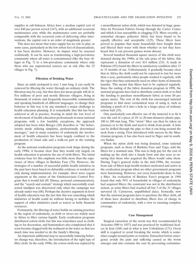
Evidence of the effectiveness of this treatment, particularlyin the final stages of a local campaign to eliminate dracuncu-liasis, is provided in Fig. 9, showing the decline in cases in thethree districts where Sharma practiced, by comparison with theother districts of endemicity of Rajasthan, India.
However, there has also been some unease at the ethics ofusing an invasive treatment in an age of human immunodefi-ciency virus and about the difficulty of maintaining qualitystandards in the rough conditions of the field. Even in Rajas-than, where the technique was an important component of theeradication program, the extractions were performed by onlytwo people (mainly by Sharma himself), and the measure wasnever accepted by the Indian medical profession or adopted bythe national eradication program. Ghana, the only Africancountry to include this intervention in its dracunculiasis erad-ication program, abandoned it in early 2000, considering that itdistracted the attention of health workers (who received areward for each worm extracted) from the implementation and
supervision of the basic community-based interventions de-scribed above.
A more widely used form of case management is to apply anocclusive bandage to the lesion where the worm is emerging.This does not prevent the release of larvae into the environ-ment, but it does discourage the patient from immersing theaffected part in a pond; immersion transforms a neat bandageinto a soggy mass of wet cotton wool, which is likely to fall off.Coupled with other palliative treatments by the village healthworker, such as “controlled immersion” of the lesion in abucket of water, disinfection of the lesion, topical applicationof emollient creams, and even antibiotic ointment (166), ban-daging by a village health worker has been used to promoteearly self-reporting of cases.
Preventing Patients’ Contact with Ponds
Preventing patient contact with ponds was an importantcomponent of the world’s first endeavor at dracunculiasis elim-ination, conducted in the Old City of Bukhara, Uzbekistan, inthe 1920s (171). Efforts were made to identify patients earlyand keep them under observation during the 2 weeks followinginitial emergence of their worms, and Muslim worshippers
FIG. 9. Decline in guinea worm cases from 1985 to 1993 in three districts of Rajasthan State, India, where surgical extraction was practiced from1991 onward. The total of cases for all other districts in the state is also shown. Information provided by UNICEF.
236 CAIRNCROSS ET AL. CLIN. MICROBIOL. REV.
on May 9, 2016 by P
EN
N S
TA
TE
UN
IVhttp://cm
r.asm.org/
Dow
nloaded from

were prevented from approaching the main ponds in the townto perform their ablutions.
In the early stages of the worldwide eradication campaign,however, such measures were ruled out as impractical in arural context where case detection within even a month ofemergence could be considered an impressive achievement.Instead, the message that patients should not contaminateponds was usually a secondary message in the general healtheducation materials. As a result, people were far more awareof how one catches the disease than of how one passes it on.For example, an evaluation survey of 26 villages in Niger in1994 showed that, while 54% of householders knew that dra-cunculiasis is transmitted in drinking water, only 13% could sayhow ponds become infected with it (26).
More recently, it has become clear that people sometimesrespond so well to this message that it can have a significanteffect on transmission in the complete absence of filter cloth.For example, in an area of some 160 villages in Adior Districtin Bahr el Ghazal Province, Sudan, in 1999, not enough clothfilters were available, so that only 44% of households had one.Nevertheless, a reduction of 88% in the number of cases wasachieved, with only 515 cases reported in the first 10 months of2000 compared with 4,177 in the same period in 1999. Theprevention of pond contamination by patients was a majorfeature of the 4,304 health education sessions performed byvillage volunteers during 1999, following a training course de-veloped by WHO and the Carter Center in collaboration withthe nongovernmental organizations (NGOs) operating locallyand the Sudan Ministry of Health.
Killing or Removing Cyclops
The intervention of killing or removing cyclops has also beentried since the earliest efforts to control transmission of thedisease: Leiper (107) used steam to kill cyclops in Indian stepwells, and Turkhud (155) used potassium permanganate. Tenyears later, when Isaev tried to apply their methods to theponds of Old Bukhara, he found them impractical; an averagepond required 300 kg of disinfectant, and the water becameundrinkable (95).
In more recent times, temephos, an organophosphate insec-ticide safe for use in drinking water sources, has been used inmany countries of endemicity, and $2 million worth has beendonated to the campaign by its manufacturers (33). Cyclopi-cide played a prominent role in the eradication programslaunched in the 1980s in India, Pakistan, and Cameroon. Allthese programs have successfully achieved elimination, but thistook many years (Fig. 8c). Did vector control help?
In Africa, vector control has not proved as easy as initiallyanticipated, in spite of there being fewer ponds per personthan there were step wells in India. Chemical treatment ofAfrican ponds, even by highly qualified research teams, hasbeen found on a number of occasions to be of questionableeffectiveness 74, 148; O. Doumbo, personal communication).When the treatment is not fully effective, there is an increasedrisk of cyclops developing resistance to the cyclopicide. Eventreatment which successfully removes the cyclops from thepond does not always eliminate guinea worm disease, as othercontaminated water sources are often in use (40, 113).
Transmission is most intense in ponds which are in the final
stages of drying up (13, 152) and which therefore may bemissed by the treatment team. This is because the infectedcyclops, which tend to sink to the bottom (122, 126), are in-creasingly likely to be scooped up as the pond becomes ashallow puddle.
Treatment of ponds can also consume substantial resources,particularly in terms of trained staff. It is harder to calculatethe volume of an irregular pond than of a rectangular Indianstep well, although this is essential in order to estimate thedose of insecticide required (34). Given the difficulty, it isimpressive that local health technicians have demonstrated ontraining courses that they can measure pond volumes to within�10% (A. Maı̈ga, personal communication). However, it usu-ally takes them the best part of half a day to do so.
Moreover, the pond volume varies with time; if rainfall afterthe treatment does not dilute the insecticide to harmless levels,the pond may still need to be remeasured the following month,although a solution used in India to avoid continual remea-surement of the volume is to insert a calibrated scale in thepond. Treatment has to be applied at least monthly to beeffective, and there is evidence to suggest that an even shortertreatment interval is required (40). With as many as 10 pondsto treat per village (Table 2), it becomes a very labor-intensiveactivity. There is some evidence that it has diverted staff fromother, more important activities (26).
As always, there are exceptions. In 1997, the eradicationprogram staff in northern Ghana found that most of their casescame from only four district towns, each of which used waterfrom a dam. The dams were nearly 10 times the maximum sizerecommended for treatment (34). Nevertheless, after someexperimentation, they found that cyclopicide could be appliedvery effectively to these (28)
Case Containment
By the middle 1990s, as case numbers began to come down,there was increased enthusiasm to step up the level of inter-vention and move towards “case containment.” Essentially,this involves a shift of emphasis from helping individuals toprotect their own health by avoidance or filtration of infectedwater towards the protection of ponds and the community atlarge from contamination by infected people.
If this is to be effective, it requires detection of each casebefore or immediately after the emergence of the worm andmeasures to ensure that it could give rise to no subsequentcase. Controlled immersion and bandaging, as describedabove, help to encourage self-reporting of cases, essential if thecases are to be detected in time, and also discourage patientsfrom immersing their lesions in water. Patients and their fam-ilies are urged to keep out of water sources until the wormshave completely emerged and are also interviewed to ascertainwhether they have already contaminated any ponds. If so,remedial measures are taken, such as alerting the communityto this possibility, treatment of the affected ponds with cyclo-picide, and checking that every household in the village has acloth filter in good condition and knows how to use it. Toensure the effective implementation of case containment, eachcase should be reported to the supervisor of the village healthworker within 7 days, and the supervisor should visit to verify
VOL. 15, 2002 DRACUNCULIASIS AND THE ERADICATION INITIATIVE 237
on May 9, 2016 by P
EN
N S
TA
TE
UN
IVhttp://cm
r.asm.org/
Dow
nloaded from
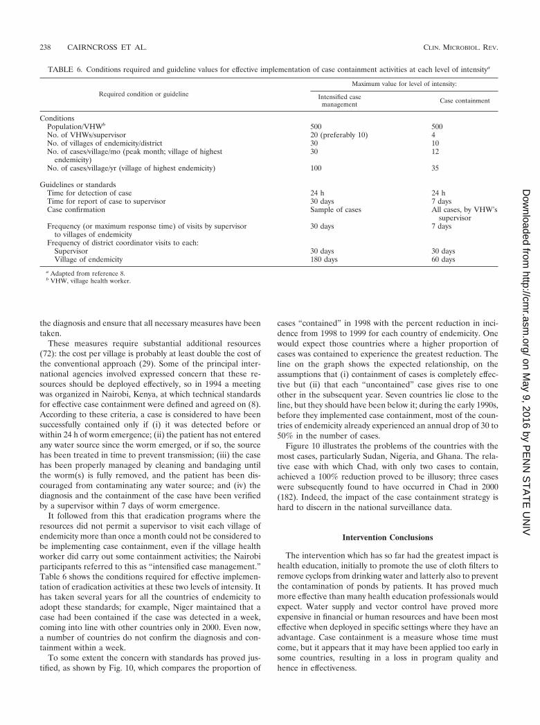
the diagnosis and ensure that all necessary measures have beentaken.
These measures require substantial additional resources(72): the cost per village is probably at least double the cost ofthe conventional approach (29). Some of the principal inter-national agencies involved expressed concern that these re-sources should be deployed effectively, so in 1994 a meetingwas organized in Nairobi, Kenya, at which technical standardsfor effective case containment were defined and agreed on (8).According to these criteria, a case is considered to have beensuccessfully contained only if (i) it was detected before orwithin 24 h of worm emergence; (ii) the patient has not enteredany water source since the worm emerged, or if so, the sourcehas been treated in time to prevent transmission; (iii) the casehas been properly managed by cleaning and bandaging untilthe worm(s) is fully removed, and the patient has been dis-couraged from contaminating any water source; and (iv) thediagnosis and the containment of the case have been verifiedby a supervisor within 7 days of worm emergence.
It followed from this that eradication programs where theresources did not permit a supervisor to visit each village ofendemicity more than once a month could not be considered tobe implementing case containment, even if the village healthworker did carry out some containment activities; the Nairobiparticipants referred to this as “intensified case management.”Table 6 shows the conditions required for effective implemen-tation of eradication activities at these two levels of intensity. Ithas taken several years for all the countries of endemicity toadopt these standards; for example, Niger maintained that acase had been contained if the case was detected in a week,coming into line with other countries only in 2000. Even now,a number of countries do not confirm the diagnosis and con-tainment within a week.
To some extent the concern with standards has proved jus-tified, as shown by Fig. 10, which compares the proportion of
cases “contained” in 1998 with the percent reduction in inci-dence from 1998 to 1999 for each country of endemicity. Onewould expect those countries where a higher proportion ofcases was contained to experience the greatest reduction. Theline on the graph shows the expected relationship, on theassumptions that (i) containment of cases is completely effec-tive but (ii) that each “uncontained” case gives rise to oneother in the subsequent year. Seven countries lie close to theline, but they should have been below it; during the early 1990s,before they implemented case containment, most of the coun-tries of endemicity already experienced an annual drop of 30 to50% in the number of cases.
Figure 10 illustrates the problems of the countries with themost cases, particularly Sudan, Nigeria, and Ghana. The rela-tive ease with which Chad, with only two cases to contain,achieved a 100% reduction proved to be illusory; three caseswere subsequently found to have occurred in Chad in 2000(182). Indeed, the impact of the case containment strategy ishard to discern in the national surveillance data.
Intervention Conclusions
The intervention which has so far had the greatest impact ishealth education, initially to promote the use of cloth filters toremove cyclops from drinking water and latterly also to preventthe contamination of ponds by patients. It has proved muchmore effective than many health education professionals wouldexpect. Water supply and vector control have proved moreexpensive in financial or human resources and have been mosteffective when deployed in specific settings where they have anadvantage. Case containment is a measure whose time mustcome, but it appears that it may have been applied too early insome countries, resulting in a loss in program quality andhence in effectiveness.
TABLE 6. Conditions required and guideline values for effective implementation of case containment activities at each level of intensitya
Required condition or guideline
Maximum value for level of intensity:
Intensified casemanagement Case containment
ConditionsPopulation/VHWb 500 500No. of VHWs/supervisor 20 (preferably 10) 4No. of villages of endemicity/district 30 10No. of cases/village/mo (peak month; village of highest
endemicity)30 12
No. of cases/village/yr (village of highest endemicity) 100 35
Guidelines or standardsTime for detection of case 24 h 24 hTime for report of case to supervisor 30 days 7 daysCase confirmation Sample of cases All cases, by VHW’s
supervisorFrequency (or maximum response time) of visits by supervisor
to villages of endemicity30 days 7 days
Frequency of district coordinator visits to each:Supervisor 30 days 30 daysVillage of endemicity 180 days 60 days
a Adapted from reference 8.b VHW, village health worker.
238 CAIRNCROSS ET AL. CLIN. MICROBIOL. REV.
on May 9, 2016 by P
EN
N S
TA
TE
UN
IVhttp://cm
r.asm.org/
Dow
nloaded from

CURRENT ISSUES
The Integration Debate
The strategy followed by most countries of endemicity asthey set up eradication programs during the early 1990s was setout in a seminal paper which built on the experiences of Pa-kistan, Ghana, and Nigeria (85). Volunteer village healthworkers, providing health education, distributing cloth filters,and carrying out surveillance, were central to the strategy. Itwas foreseen that they would cover more villages in less timethan the provision of safe water supplies required. While theinitial search for cases should be a vertically organized opera-tion, the authors proposed that “where they exist already, pri-mary health care (PHC) workers in endemic villages should beused; where they do not yet exist, an appropriate villagershould be designated (who may later be incorporated into thecountry’s PHC programme).“
In the ensuing years, the networks of village volunteerswhich were being established proved remarkably effective incarrying out surveillance and health education (29). However,the degree of their integration into the primary health careprogram was a subject of intense debate. Their success ledsome stakeholders in the global campaign to advocate that theyshould be used to monitor conditions other than dracunculia-sis, such as immunization status and acute flaccid paralysis as amarker for possible polio epidemics. As they saw it, the cost ofestablishing the network was an investment which should serveother health objectives (131). The cost of setting up such anetwork is little more than the annual cost of its maintenance,but the main item is the salaries of those who supervise thevolunteers, and in most countries these are met from the gen-eral health budget and not from funds earmarked for guineaworm eradication. The other major items are the cost of an-nual retraining for the volunteers and occasional gifts in kind
to motivate them, such as T-shirts or the salt and soap given tothe volunteers in South Sudan; other health programs, partic-ularly those with external support, have been ready to contrib-ute toward these.
Others doubted the wisdom of integration, wary of puttingthe eradication effort at the mercy of an often weak primaryhealth care system and dissipating eradication resources onother activities (84). Prior experience of the inappropriate “in-tegration” of smallpox eradication with measles control, and ofyaws control in Ghana (81), influenced their thinking. Therewere other concerns, too. One was the danger of overburden-ing the volunteers, though a recent field assessment of inte-grated surveillance in northern Ghana found no objective ev-idence that the surveillance activities were interfering withhealth workers’ ability to carry out the eradication work effec-tively (187). Another source of unease, with some justification,was that the collection of surveillance data is useless if, as hasoccurred in a number of countries of endemicity, it is not usedfor any action. Without a system to make use of it, the datawould be collected for their own sake. Moreover, most of thenational programs operate in only a limited number of villagesof endemicity, whereas some of the wider objectives wouldrequire complete coverage.
The integration debate led to a joint statement in favor ofintegrated surveillance by the four principal agencies involvedin the eradication effort (183), but of course a question such as“to integrate, or not to integrate?” does not have a universalanswer; the answer can only be worked out on the ground ineach specific context. Moreover, surveillance is only one com-ponent of a public health system, and others may benefit froma degree of integration. Certainly, there have been substantialsynergies from combining dracunculiasis eradication activitieswith others in a number of cases. Both dracunculiasis eradica-tion and the wider health agenda can benefit, as long as thecombination is opportune and has been made judiciously. Ex-amples include (i) the integrated surveillance for guinea worm,immunization, yellow fever, and cerebrospinal meningitis inthe 3,743 villages of Northern Region, Ghana (187), whereinitial difficulties in harmonizing case definitions were ironedout during the first year of operation; (ii) the mobilization ofunderused government field staff to supervise the village healthworkers in Mopti Region, Mali (30), while also supportingtheir work to promote latrine construction, better agriculture,etc.; (iii) use of the guinea worm surveillance system for im-plementation and monitoring of trachoma elimination activi-ties in Zinder Region, Niger; (iv) the use of the 6-month“guinea worm cease-fire” in southern Sudan in 1995 to treatpeople at risk for onchocerciasis; treat children with vitamin A;and vaccinate cattle against rinderpest and children againstmeasles, polio, and tuberculosis (84); (v) also in southern Su-dan, the use of the guinea worm program’s training and super-vision system for a pilot program in which village health vol-unteers in remote villages were trained to identify and treatfever and malaria, cough and difficult breathing, and dehydra-tion due to diarrhea; and (vi) the use of the polio nationalimmunization days to detect 400 new villages of endemicity insouthern Sudan during the first campaign in this area in 1998(174), where the same process in the following 2 years led tothe detection of a further 350 villages of endemicity.
FIG. 10. Case containment: proportion of cases contained in 1998and year-on-year national case reductions from 1998 to 1999 for coun-tries of endemicity in Africa. Each point on the graph represents onecountry’s performance.
VOL. 15, 2002 DRACUNCULIASIS AND THE ERADICATION INITIATIVE 239
on May 9, 2016 by P
EN
N S
TA
TE
UN
IVhttp://cm
r.asm.org/
Dow
nloaded from
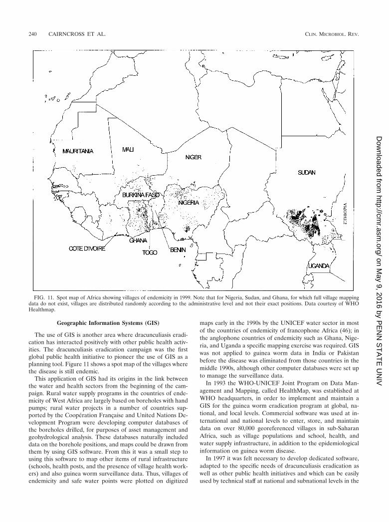
Geographic Information Systems (GIS)
The use of GIS is another area where dracunculiasis eradi-cation has interacted positively with other public health activ-ities. The dracunculiasis eradication campaign was the firstglobal public health initiative to pioneer the use of GIS as aplanning tool. Figure 11 shows a spot map of the villages wherethe disease is still endemic.
This application of GIS had its origins in the link betweenthe water and health sectors from the beginning of the cam-paign. Rural water supply programs in the countries of ende-micity of West Africa are largely based on boreholes with handpumps; rural water projects in a number of countries sup-ported by the Coopération Française and United Nations De-velopment Program were developing computer databases ofthe boreholes drilled, for purposes of asset management andgeohydrological analysis. These databases naturally includeddata on the borehole positions, and maps could be drawn fromthem by using GIS software. From this it was a small step tousing this software to map other items of rural infrastructure(schools, health posts, and the presence of village health work-ers) and also guinea worm surveillance data. Thus, villages ofendemicity and safe water points were plotted on digitized
maps early in the 1990s by the UNICEF water sector in mostof the countries of endemicity of francophone Africa (46); inthe anglophone countries of endemicity such as Ghana, Nige-ria, and Uganda a specific mapping exercise was required. GISwas not applied to guinea worm data in India or Pakistanbefore the disease was eliminated from those countries in themiddle 1990s, although other computer databases were set upto manage the surveillance data.
In 1993 the WHO-UNICEF Joint Program on Data Man-agement and Mapping, called HealthMap, was established atWHO headquarters, in order to implement and maintain aGIS for the guinea worm eradication program at global, na-tional, and local levels. Commercial software was used at in-ternational and national levels to enter, store, and maintaindata on over 80,000 georeferenced villages in sub-SaharanAfrica, such as village populations and school, health, andwater supply infrastructure, in addition to the epidemiologicalinformation on guinea worm disease.
In 1997 it was felt necessary to develop dedicated software,adapted to the specific needs of dracunculiasis eradication aswell as other public health initiatives and which can be easilyused by technical staff at national and subnational levels in the
FIG. 11. Spot map of Africa showing villages of endemicity in 1999. Note that for Nigeria, Sudan, and Ghana, for which full village mappingdata do not exist, villages are distributed randomly according to the administrative level and not their exact positions. Data courtesy of WHOHealthmap.
240 CAIRNCROSS ET AL. CLIN. MICROBIOL. REV.
on May 9, 2016 by P
EN
N S
TA
TE
UN
IVhttp://cm
r.asm.org/
Dow
nloaded from
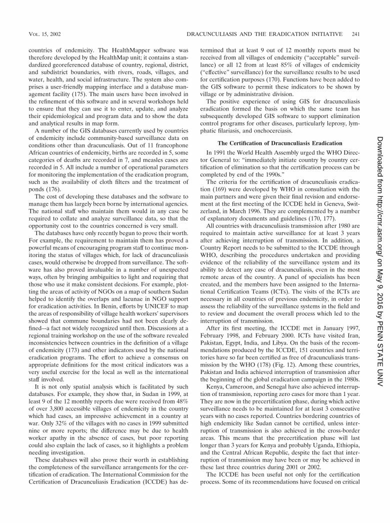
countries of endemicity. The HealthMapper software wastherefore developed by the HealthMap unit; it contains a stan-dardized georeferenced database of country, regional, district,and subdistrict boundaries, with rivers, roads, villages, andwater, health, and social infrastructure. The system also com-prises a user-friendly mapping interface and a database man-agement facility (175). The main users have been involved inthe refinement of this software and in several workshops heldto ensure that they can use it to enter, update, and analyzetheir epidemiological and program data and to show the dataand analytical results in map form.
A number of the GIS databases currently used by countriesof endemicity include community-based surveillance data onconditions other than dracunculiasis. Out of 11 francophoneAfrican countries of endemicity, births are recorded in 5, somecategories of deaths are recorded in 7, and measles cases arerecorded in 5. All include a number of operational parametersfor monitoring the implementation of the eradication program,such as the availability of cloth filters and the treatment ofponds (176).
The cost of developing these databases and the software tomanage them has largely been borne by international agencies.The national staff who maintain them would in any case berequired to collate and analyze surveillance data, so that theopportunity cost to the countries concerned is very small.
The databases have only recently begun to prove their worth.For example, the requirement to maintain them has proved apowerful means of encouraging program staff to continue mon-itoring the status of villages which, for lack of dracunculiasiscases, would otherwise be dropped from surveillance. The soft-ware has also proved invaluable in a number of unexpectedways, often by bringing ambiguities to light and requiring thatthose who use it make consistent decisions. For example, plot-ting the areas of activity of NGOs on a map of southern Sudanhelped to identify the overlaps and lacunae in NGO supportfor eradication activities. In Benin, efforts by UNICEF to mapthe areas of responsibility of village health workers’ supervisorsshowed that commune boundaries had not been clearly de-fined–-a fact not widely recognized until then. Discussions at aregional training workshop on the use of the software revealedinconsistencies between countries in the definition of a villageof endemicity (173) and other indicators used by the nationaleradication programs. The effort to achieve a consensus onappropriate definitions for the most critical indicators was avery useful exercise for the local as well as the internationalstaff involved.
It is not only spatial analysis which is facilitated by suchdatabases. For example, they show that, in Sudan in 1999, atleast 9 of the 12 monthly reports due were received from 48%of over 3,800 accessible villages of endemicity in the countrywhich had cases, an impressive achievement in a country atwar. Only 32% of the villages with no cases in 1999 submittednine or more reports; the difference may be due to healthworker apathy in the absence of cases, but poor reportingcould also explain the lack of cases, so it highlights a problemneeding investigation.
These databases will also prove their worth in establishingthe completeness of the surveillance arrangements for the cer-tification of eradication. The International Commission for theCertification of Dracunculiasis Eradication (ICCDE) has de-
termined that at least 9 out of 12 monthly reports must bereceived from all villages of endemicity (“acceptable” surveil-lance) or all 12 from at least 85% of villages of endemicity(“effective” surveillance) for the surveillance results to be usedfor certification purposes (170). Functions have been added tothe GIS software to permit these indicators to be shown byvillage or by administrative division.
The positive experience of using GIS for dracunculiasiseradication formed the basis on which the same team hassubsequently developed GIS software to support eliminationcontrol programs for other diseases, particularly leprosy, lym-phatic filariasis, and onchocerciasis.
The Certification of Dracunculiasis Eradication
In 1991 the World Health Assembly urged the WHO Direc-tor General to: “immediately initiate country by country cer-tification of elimination so that the certification process can becompleted by end of the 1990s.”
The criteria for the certification of dracunculiasis eradica-tion (169) were developed by WHO in consultation with themain partners and were given their final revision and endorse-ment at the first meeting of the ICCDE held in Geneva, Swit-zerland, in March 1996. They are complemented by a numberof explanatory documents and guidelines (170, 177).
All countries with dracunculiasis transmission after 1980 arerequired to maintain active surveillance for at least 3 yearsafter achieving interruption of transmission. In addition, aCountry Report needs to be submitted to the ICCDE throughWHO, describing the procedures undertaken and providingevidence of the reliability of the surveillance system and itsability to detect any case of dracunculiasis, even in the mostremote areas of the country. A panel of specialists has beencreated, and the members have been assigned to the Interna-tional Certification Teams (ICTs). The visits of the ICTs arenecessary in all countries of previous endemicity, in order toassess the reliability of the surveillance systems in the field andto review and document the overall process which led to theinterruption of transmission.
After its first meeting, the ICCDE met in January 1997,February 1998, and February 2000. ICTs have visited Iran,Pakistan, Egypt, India, and Libya. On the basis of the recom-mendations produced by the ICCDE, 151 countries and terri-tories have so far been certified as free of dracunculiasis trans-mission by the WHO (178) (Fig. 12). Among these countries,Pakistan and India achieved interruption of transmission afterthe beginning of the global eradication campaign in the 1980s.
Kenya, Cameroon, and Senegal have also achieved interrup-tion of transmission, reporting zero cases for more than 1 year.They are now in the precertification phase, during which activesurveillance needs to be maintained for at least 3 consecutiveyears with no cases reported. Countries bordering countries ofhigh endemicity like Sudan cannot be certified, unless inter-ruption of transmission is also achieved in the cross-borderareas. This means that the precertification phase will lastlonger than 3 years for Kenya and probably Uganda, Ethiopia,and the Central African Republic, despite the fact that inter-ruption of transmission may have been or may be achieved inthese last three countries during 2001 or 2002.
The ICCDE has been useful not only for the certificationprocess. Some of its recommendations have focused on critical
VOL. 15, 2002 DRACUNCULIASIS AND THE ERADICATION INITIATIVE 241
on May 9, 2016 by P
EN
N S
TA
TE
UN
IVhttp://cm
r.asm.org/
Dow
nloaded from
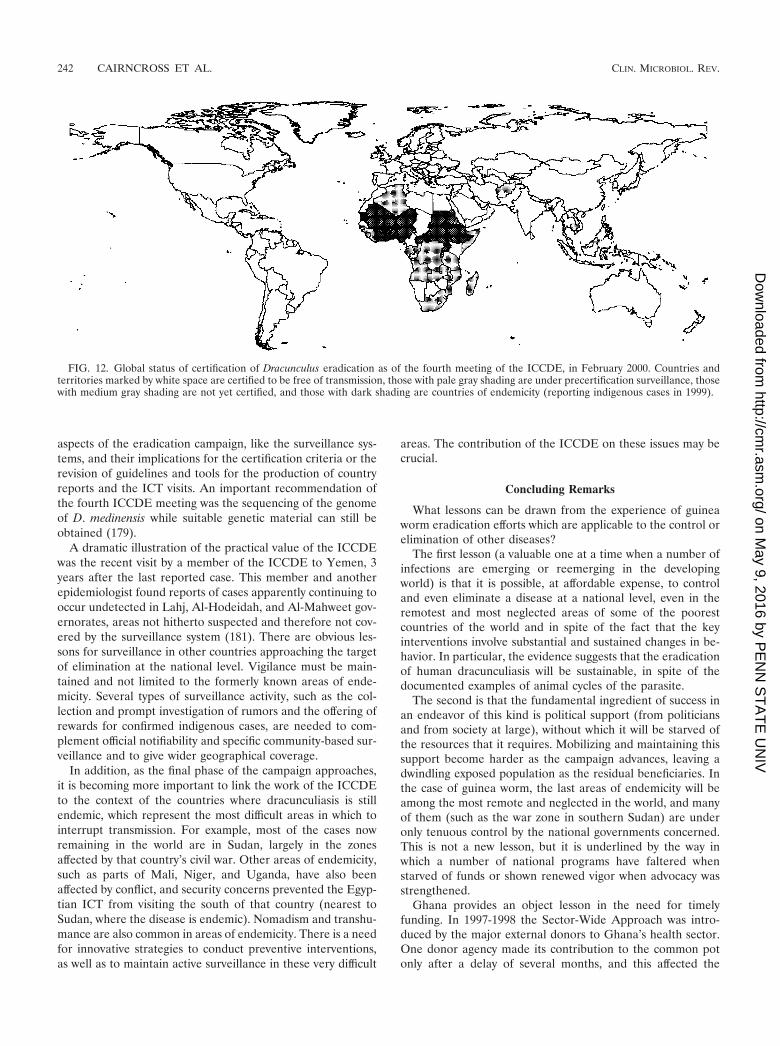
aspects of the eradication campaign, like the surveillance sys-tems, and their implications for the certification criteria or therevision of guidelines and tools for the production of countryreports and the ICT visits. An important recommendation ofthe fourth ICCDE meeting was the sequencing of the genomeof D. medinensis while suitable genetic material can still beobtained (179).
A dramatic illustration of the practical value of the ICCDEwas the recent visit by a member of the ICCDE to Yemen, 3years after the last reported case. This member and anotherepidemiologist found reports of cases apparently continuing tooccur undetected in Lahj, Al-Hodeidah, and Al-Mahweet gov-ernorates, areas not hitherto suspected and therefore not cov-ered by the surveillance system (181). There are obvious les-sons for surveillance in other countries approaching the targetof elimination at the national level. Vigilance must be main-tained and not limited to the formerly known areas of ende-micity. Several types of surveillance activity, such as the col-lection and prompt investigation of rumors and the offering ofrewards for confirmed indigenous cases, are needed to com-plement official notifiability and specific community-based sur-veillance and to give wider geographical coverage.
In addition, as the final phase of the campaign approaches,it is becoming more important to link the work of the ICCDEto the context of the countries where dracunculiasis is stillendemic, which represent the most difficult areas in which tointerrupt transmission. For example, most of the cases nowremaining in the world are in Sudan, largely in the zonesaffected by that country’s civil war. Other areas of endemicity,such as parts of Mali, Niger, and Uganda, have also beenaffected by conflict, and security concerns prevented the Egyp-tian ICT from visiting the south of that country (nearest toSudan, where the disease is endemic). Nomadism and transhu-mance are also common in areas of endemicity. There is a needfor innovative strategies to conduct preventive interventions,as well as to maintain active surveillance in these very difficult
areas. The contribution of the ICCDE on these issues may becrucial.
Concluding Remarks
What lessons can be drawn from the experience of guineaworm eradication efforts which are applicable to the control orelimination of other diseases?
The first lesson (a valuable one at a time when a number ofinfections are emerging or reemerging in the developingworld) is that it is possible, at affordable expense, to controland even eliminate a disease at a national level, even in theremotest and most neglected areas of some of the poorestcountries of the world and in spite of the fact that the keyinterventions involve substantial and sustained changes in be-havior. In particular, the evidence suggests that the eradicationof human dracunculiasis will be sustainable, in spite of thedocumented examples of animal cycles of the parasite.
The second is that the fundamental ingredient of success inan endeavor of this kind is political support (from politiciansand from society at large), without which it will be starved ofthe resources that it requires. Mobilizing and maintaining thissupport become harder as the campaign advances, leaving adwindling exposed population as the residual beneficiaries. Inthe case of guinea worm, the last areas of endemicity will beamong the most remote and neglected in the world, and manyof them (such as the war zone in southern Sudan) are underonly tenuous control by the national governments concerned.This is not a new lesson, but it is underlined by the way inwhich a number of national programs have faltered whenstarved of funds or shown renewed vigor when advocacy wasstrengthened.
Ghana provides an object lesson in the need for timelyfunding. In 1997-1998 the Sector-Wide Approach was intro-duced by the major external donors to Ghana’s health sector.One donor agency made its contribution to the common potonly after a delay of several months, and this affected the
FIG. 12. Global status of certification of Dracunculus eradication as of the fourth meeting of the ICCDE, in February 2000. Countries andterritories marked by white space are certified to be free of transmission, those with pale gray shading are under precertification surveillance, thosewith medium gray shading are not yet certified, and those with dark shading are countries of endemicity (reporting indigenous cases in 1999).
242 CAIRNCROSS ET AL. CLIN. MICROBIOL. REV.
on May 9, 2016 by P
EN
N S
TA
TE
UN
IVhttp://cm
r.asm.org/
Dow
nloaded from

funding of health activities across the board. The Ministry ofHealth saw little option but to maintain curative health servicesby cutting the funding of preventive activities, and the guineaworm eradication program, previously supported by ear-marked funds, lost out. By the time the money came through itwas too late, as the peak transmission season had passed. As aresult, the number of cases nearly doubled in 1999. This set-back was only partially reversed in 2000.
The example of Nigeria illustrates the potential of high-leveladvocacy. In the late 1980s, an intensive advocacy effort led tothe initial launch of Nigeria’s eradication program (63, 64), butby 1996 the number of cases was stable. In 1998, YakubuGowon, former head of state, became “guinea worm eradica-tion ambassador,” and during 1999 he worked hard at this task,traveling across the country, visiting political leaders in thestates of highest endemicity, and returning a few months laterto monitor their progress. After 4 years of stability, the numberof cases reported fell by nearly half in 2000.
It is important, however, not to confuse advocacy goals withplanning targets. Some countries pushed forward to implementcase containment, or began to offer cash rewards for reportedcases, when there were still such large numbers of cases thatthe quality of the case containment system could not be guar-anteed or when the reliability, transparency, and even thefunding of the reward system were not secure. The resourcesinvested in these premature additions might have been betterspent on the basic strategy.
There have been times when the international agencies in-volved in the campaign have failed to maintain the collegialdialogue required to build and maintain consensus about strat-egy as the situation evolves. This was most evident in thedebate about integration but has also affected some more de-tailed questions. It is not clear to what extent, if any, suchpolemics have harmed the eradication effort; they have at leastwasted a substantial amount of energy. What is clearer is thatinteragency collaboration and consensus will become moreimportant, not less, if innovative strategies are to be identifiedand adopted to respond to the new challenges of the finalphases of the eradication campaign. When such consensus hasbeen achieved, the eradication effort has always benefited. Ithas been noted that “a sense of common purpose, dedicationand commitment, and a collaborative mode of behaviour” is ahallmark of successful disease control programs (108); theguinea worm eradication effort needs these more than ever.
The fear has sometimes been expressed that the resourcesallocated for guinea worm eradication will divert the energy ofhealth workers from other essential tasks, and on the otherhand some have hoped that the eradication campaign willblaze a trail where other health programs can follow. In prac-tice, there is little evidence that the functioning of governmenthealth systems has been greatly affected, for good or ill, bydracunculiasis eradication. However, in the many communitieswhere the disease has disappeared in recent years, thanks tothe eradication initiative, this has surely helped to build publicconfidence in health workers and the health system and in-creased people’s readiness for change. Future health interven-tions, especially those requiring behavioral change, will fall onmore fertile ground as a result.
Whatever the degree of support afforded to guinea wormeradication by politicians, international agencies, and health
staff, much of the success achieved so far is attributable to theremarkable support by simple villagers, who have demon-strated commitment, skill, and resourcefulness beyond whatmany would have imagined. Not least among the unsung he-roes of the campaign are the thousands of volunteer villagehealth workers who have spread the word about prevention,conscientiously detected and reported cases of disease, andincreasingly taken steps to ensure that those cases were con-tained. They and the population have often surprised the pro-gram managers with their willingness and ability to respond tochallenges. For example, early skepticism that health educa-tion would have much impact on the contamination of pondsby infected cases has been shown in recent years to be un-founded.
Above all, the guinea worm eradication campaign has shownthat even in poor, small, and remote villages in developingcountries, health interventions based in the community andimplemented by the villagers are feasible and can have a sig-nificant impact on the burden of disease. The challenge is tomobilize the proven capacity of the population and of localvolunteers to improve the health of their communities and tofight not only guinea worm disease but also the other diseasesof poverty.
ACKNOWLEDGMENTS
The eradication of guinea worm disease is a collaborative endeavortowards which many people have contributed their efforts and ideas, inaddition to those cited above. The help of Ahmed Tayeh and thecomments of Philippe Ranque on a draft of the manuscript wereparticularly valuable.
REFERENCES
1. Abolarin, M. O. 1981. Guinea worm infection in a Nigerian village. Trop.Geogr. Med. 33:83–88.
2. Adamson, P. 1988. Dracontiasis in antiquity. Med. Hist. 32:204–209.3. Adekolu-John, E. A. 1983. The impact of lake creation on guinea-worm
transmission in Nigeria on the Eastern side of Kainji Lake. Int. J. Parasitol.13:427–432.
4. Adeyeba, O. A. 1985. Secondary infection in dracunculiasis: bacteria andmorbidity. Int. J. Zoonoses 12:147–149.
5. Adeyeba, O. A., and O. O. Kale. 1991. Epidemiology of dracunculiasis andits socio-economic impact in a village in south-west Nigeria. West Afr.J. Med. 10:208–215.
6. Ahearn, S. C., and C. de Rooy. 1996. Monitoring the effects of dracuncu-liasis remediation on agricultural productivity using satellite data. RemoteSens. 17:917–929.
7. Anonymous. 1983. After smallpox, guinea worm? Lancet i:161–162.8. Anonymous. 1994. Case containment strategy for eradication of dracuncu-
liasis in Africa. Centers for Disease Control with Global 2000, UnitedNations Children’s Fund, and World Health Organization, Atlanta, Ga.
9. Audibert, M. 1993. Invalidité temporaire et production agricole: les effetsde la dracunculose dans une agriculture de subsistance. Rev. Écon. Dév.1:23–36.
10. Aylward, B., K. A. Hennessey, N. Zagaria, J.-M. Olivé, and S. Cochi. 2000.When is a disease eradicable? 100 years of lessons learned. Am. J. PublicHealth 90(10):1–10.
11. Bapna, S., and D. M. Renapurkar. 1996. Immunodiagnosis of early dra-cunculiasis. J. Commun. Dis. 28:33–37.
12. Batliwala, J. C. 1983. Guinea-worm in the horse. Vet. J. 36:412–413.13. Belcher, D. W., F. K. Wurapa, W. B. Ward, and I. M. Lourie. 1975. Guinea
worm in southern Ghana; its epidemiology and impact on agriculturalproductivity. Am. J. Trop. Med. Hyg. 24:243–249.
14. Beyer, T. A., J. P. Tao, Z. X. Wang, et al. 1999. Massive Dracunculus insignisin a dog. Am. Vet. Med. Assoc. 214:366–368.
15. Bhatt, A. N., and K. H. Palan. 1978. Guinea-worm infection in BanaskanthaDistrict of Gujarat—some important epidemiological aspects. IndianJ. Med. Sci. 32:1–4.
16. Blaxter, M., M. Aslett, D. Guitiano, J. Daub, et al. 1999. Parasitic helminthgenomes. Parasitology 118(Suppl.):S39–S61.
17. Bloch, P., P. E. Simonsen, and B. J. Vennervald. 1993. The antibody re-sponse to Dracunculus medinensis in an endemic human population ofnorthern Ghana. J. Helminthol. 67:37–48.
VOL. 15, 2002 DRACUNCULIASIS AND THE ERADICATION INITIATIVE 243
on May 9, 2016 by P
EN
N S
TA
TE
UN
IVhttp://cm
r.asm.org/
Dow
nloaded from

18. Bloch, P., B. J. Vennervald, and P. E. Simonsen. 1998. Studies on immu-nodiagnosis of dracunculiasis. II. Search for circulating antigens. Acta Trop.70:303–315.
19. Brandt, F. H., and M. L. Eberhard. 1990. Distribution, behavior, and courseof patency of Dracunculus insignis in experimentally infected ferrets. J.Parasitol. 76:515–518.
20. Brieger, W. R. 1996. Health education to promote community involvementin the control of tropical diseases. Acta Trop. 61:93–106.
21. Brieger, W. R., J. D. Adeniyi, and S. U. Akpovi. 1982. Complexities ofguinea worm disease. World Health Forum 3:216–217.
22. Brieger, W. R., E. A. Olumide, and O. O. Kale. 1983. Effect of guinea wormon schoolchildren. World Health Forum 4:324–325.
23. Brieger, W. R., S. Watts, and M. Yacoob. 1989. Guinea worm, maternalmorbidity and child health. J. Trop. Paediatr. 35:285–288.
24. Brieger, W. R., and J. Guyer. 1990. Farmers’ loss due to guinea wormdisease: a pilot study. J. Trop. Med. Hyg. 93:106–111.
25. Bugri, S. Z. 1981. Guinea worm: an indicator of the quality and quantity ofrural water supply in Northern Ghana. M.S. dissertation. London School ofHygiene and Tropical Medicine, London, United Kingdom.
26. Cairncross, S. 1995. Evaluation du PNEVG du Niger, 6–15 décembre de1994. UNICEF Mission Report. United Nations Children’s Fund, NewYork, N.Y.
27. Reference deleted.28. Cairncross, S., S. D. Anemana, and A. Olsen. 1999. Towards the eradication
of guinea worm; a Danish-Ghanaian collaboration. Parasitol. Today 15:127–129.
29. Cairncross, S., E. I. Braide, and S. Z. Bugri. 1996. Community participationin the eradication of guinea worm disease. Acta Trop. 61:121–136.
30. Cairncross, S., F. T. Cutts, and H. Periès. 1997. Vertical programmes; whatare they good for? Lancet 349:SIII20–SIII22.
31. Cairncross, S., and A. Tayeh. 1988. Guinea worm and water supply inKordofan, Sudan. J. Inst. Water Environ. Management 2:268–274.
32. Cairncross, S., and A. Tayeh. 1989. Aggregation of Dracunculus medinensisin communities using different types of water source in Sudan. Trans. R.Soc. Trop. Med. Hyg. 83:431.
33. Carter, J. 1999. The power of partnership; the eradication of guinea wormdisease, p. 141–147. In Cooperation South no. 2. United Nations Develop-ment Program, New York, N.Y.
34. Centers for Disease Control. 1989. Guidelines for chemical control ofcopepod populations in dracunculiasis eradication programs. WHO Col-laborating Center for Research, Training and Eradication of Dracunculiasisat Centers for Disease Control, Atlanta, Ga.
35. Centers for Disease Control and Prevention. 1999. Guinea worm wrap-upno. 92. WHO Collaborating Center for Research, Training and Eradicationof Dracunculiasis, Atlanta, Ga.
36. Chippaux, J. P. 1992. The impact of dracunculiasis in a sugar-cane planta-tion. Trans. R. Soc. Trop. Med. Hyg. 86:72.
37. Chippaux, J. P. 1991. Mebendazole treatment of dracunculiasis. Trans. R.Soc. Trop. Med. Hyg. 85:280.
38. Chippaux, J. P., A. Banzou, and K. Agbede. 1992. Social and economicimpact of dracunculosis; a longitudinal study carried out in 2 villages inBenin. Bull. W. H. O. 70:73–78.
39. Chippaux, J. P., L. de Souza, and A. Massougbodji. 1991. Aspects epidé-miologiques de la dracunculose au Bénin. 1. Incidences, localisation desemergences et fréquence de réinfestations. Bull. Soc. Pathol. Exot. 84:345–350.
40. Chippaux, J. P., I. Laniyan, and M. Akogbeto. 1991. Evaluation del’efficacité du Témephos dans la lutte contre la dracunculose. Ann. Soc.Belg. Méd. Trop. 71:279–285.
41. Chippaux, J. P., and A. Massougbodji. 1991. Aspects épidémiologiques dela dracunculose au Bénin 2. Relations entre la périodicité des émergenceset l’origine de l’eau de boisson. Bull. Soc. Pathol. Exot. 84:351–357.
42. Chippaux, J. P., and A. Massougbodji. 1991. Évaluation clinique et épidé-miologique de la dracunculose au Bénin. Méd. Trop. 51:269–274.
43. Chitwood, B. G. 1950. The male of Dracunculus insignis (Leidy, 1858)Chandler, 1942. Proc. Helminthol. Soc. Wash. 17:14–15.
44. Chun Sun, F. 1958. Dracunculus infection in dogs in Kazakhstan. Med.Parazitol. 27:219. (In Russian.)
45. Chun Sun, F. 1966. A case of Dracunculus infection in a domestic cat in theKazakh SSR. Med. Parazitol. 35:374–375. (In Russian.)
46. Clarke, K. C., J. P. Osleeb, J. M. Sherry, J.-P. Meert, and R. W. Larsson.1991. The use of remote sensing and geographic information systems inUNICEF’s dracunculiasis (Guinea worm) eradication effort. Prev. Vet.Med. 11:229–235.
47. Crichton, V. F. J., and M. Beverley-Burton. 1977. Observations on theseasonal prevalence, pathology and transmission of Dracunculus insignis(Nematodoidea: Dracunculoidea) in the raccoon (Procyon lotor (L.)) inOntario. J. Wildl. Dis. 13:273–280.
48. Curtis, V., I. Diallo, J. Konaté, and T. Skitt. 1993. The hand-pump main-tenance project in Seno and Oudalan, Burkina Faso; evaluation report.Save the Children Fund, London, United Kingdom.
49. Davidson, W. R., V. F. Nettles, L. F. Hayes, E. W. Howerth, and C. E.
Coavillion. 1992. Disease diagnosed in gray foxes (Urocyon cinereoargeneus)from the southeast United States. J. Wildl. Dis. 28:28–33.
50. de Rooy, C., and L. D. Edungbola. 1988. Guinea worm control as a majorcontributor to self-sufficiency in rice production in Nigeria. UNICEF work-ing document. United Nations Children’s Fund, New York, N.Y.
51. Reference deleted.52. Dowdle, W. R., and D. R. Hopkins. ed. 1998. Report of the Dahlem Work-
shop on the Eradication of Infectious Diseases. John Wiley & Sons, Chich-ester, England.
53. Duke, B. L. 1984. Filtering out the guinea worm. World Health March1984:29.
54. Eberhard, M. L., and F. H. Brandt. 1995. The role of tadpoles and frogs asparatenic hosts in the life cycle of Dracunculus insignis (Nematoidea: Dra-cunculoidea). J. Parasitol. 81:792–793.
55. Eberhard, M. L., F. H. Brandt, and A. Hightower. 1990. Chemoprophylacticdrug trials for treatment of dracunculiasis using the Dracunculus insignisferret model. J. Helminthol. 64:79–86.
56. Eberhard, M. L., M. A. Rab, and M. N. Dilshad. 1989. Red Dracunculusmedinensis. Am. J. Trop. Med. Hyg. 41:479–481.
57. Edungbola, L. D. 1980. Water utilization and its health implications inIlorin, Kwara State, Nigeria. Acta Trop. 37:73.
58. Edungbola, L. D. 1983. Babana Parasitic Disease Project. II. Prevalenceand impact of dracontiasis in Babana District. Trans. R. Soc. Trop. Med.Hyg. 77:310–315.
59. Edungbola, L. D., and S. J. Watts. 1984. An outbreak of dracunculiasis ina peri-urban community of Ilorin, Kwara State, Nigeria. Acta Trop. 41:155–163.
60. Edungbola, L. D., and S. J. Watts. 1985. Epidemiological assessment of thedistribution and endemicity of guinea worm infection in Asa, Kwara Sate,Nigeria. Trop. Geogr. Med. 37:22–28.
61. Edungbola, L. D., and S. J. Watts. 1990. The elimination of dracunculiasisin Igbon, Oyo State, Nigeria: the success of self-help activities. J. Trop.Med. Hyg. 93:1–6.
62. Edungbola, L. D., S. J. Watts, T. O. Alabi, and A. B. Bello. 1988. The impactof a UNICEF-assisted rural water project on the prevalence of guineaworm disease in Asa, Kwara State, Nigeria. Am. J. Trop. Med. Hyg. 39:79–85.
63. Edungbola, L. D., S. J. Watts, O. O. Kale, G. S. Smith, and D. R. Hopkins.1986. A method of rapid assessment of the distribution and endemicity ofdracunculiasis in Nigeria. Soc. Sci. Med. 23:550–558.
64. Edungbola, L. D., P. C. Withers, Jr., E. I. Braide, O. O. Kale, L. O. Sadiq,B. C. Nwobi, T. Alakija, P. McConnon, and D. R. Hopkins. 1992. Mobili-zation strategy for guinea worm eradication in Nigeria. Am. J. Trop. Med.Hyg. 47:529–538.
65. Esrey, S. A., J. B. Potash, L. Roberts, and C. Shiff. 1990. Health benefitsfrom improvements in water supply and sanitation; survey and analysis ofthe literature on selected diseases. WASH technical report no. 66. WASHfor U.S. Agency for International Development, Arlington, Va.
66. Faust, E. C., P. C. Beaver, and R. C. Jung. 1968. Animal agents and vectorsof human disease, 3rd ed., p. 297. Lea & Febiger, Philadelphia, Pa.
67. Fedchenko, A. P. 1971. Concerning the structure and reproduction of theguinea-worm (Filaria medinensis L.). Am. J. Trop. Med. Hyg. 20:511–523.(Translation of 1870 original.)
68. Foege, W. H. 1998. Smallpox eradication in West and Central Africa revis-ited. Bull. W. H. O. 76:233–235.
69. Fu, A. Q., J. P. Tao, Z. X. Wang, B. L. Jiang, Y. X. Liu, and H. H. Qui. 1999.Observations on the morphology of Dracunculus medinensis from a cat inChina. Chin. J. Zoonoses 15:35–38. (In Chinese with English summary.)
70. Gbary, A. R., T. R. Guiguémdé, and J.-B. Ouédraogo. 1987. La dracuncu-lose, un fléau éradiqué dans trois villages du Burkina Faso par l’éducationsanitaire. Bull. Soc. Pathol. Exot. 80:390–395.
71. Ghenis, D. E. 1972. New cases of Dracunculus medinensis L., 1758 detectedin domestic cats and dogs in Kazakhstan. Med. Parazitol. 41:365.
72. Greer, G., M. Dama, S. Graham, R. Migliani, M. Alami, and A. Sam-Abbenyi. 1994. Cameroon: an African model for final stages of guinea wormeradication. Am. J. Trop. Med. Hyg. 50:393–400.
73. Guiguémdé, T. R. 1985. Climatic characteristics of endemic zones andepidemiologic modalities of drancunculosis in Africa. Bull. Soc. Pathol.Exot. 79:89–95.
74. Guiguémdé, T. R., A. R. Gbary, and J.-B. Ouédraogo. 1990. Lutte contre ladracunculose: problematique du traitement chimique des points d’eau auTemephos (Abate) en Afrique. Pub. Méd. Afr. 110:19–22.
75. Guiguemdé, T. R., F. Orivel, B. Millot, A. R. Gbary, and J.-B. Ouédraogo.1986. Le poids socio-économique de la dracunculose; méthode de calcul ducoût économique chiffré de la maladie. Étud. Méd 3:113–124.
76. Hashikura, T. 1927. A case of Filaria medinensis in Chosen (Korea). Jpn.Med. World 7:145–146. (In Japanese.)
77. Henderson, P. L., R. E. Fontaine, and G. Kyeyune. 1988. Guinea wormdisease in northern Uganda: a major public health problem controllablethrough an effective water programme. Int. J. Epidemiol. 17:434–440.
78. Hopkins, D. R. 1976. After smallpox eradication: yaws? Am. J. Trop. Med.Hyg. 25:860–865.
244 CAIRNCROSS ET AL. CLIN. MICROBIOL. REV.
on May 9, 2016 by P
EN
N S
TA
TE
UN
IVhttp://cm
r.asm.org/
Dow
nloaded from
79. Hopkins, D. R. 1983. Dracunculiasis: an eradicable scourge. Epidemiol.Rev. 5:208–219.
80. Hopkins, D. R. 1984. Guinea worm and the decade. World Health Novem-ber 1984:22–23.
81. Hopkins, D. R. 1985. Control of yaws and other endemic treponematoses:implementation of vertical and/or integrated programs. Rev. Infect. Dis.7(Suppl. 2):S338–S342.
82. Hopkins, D. R. 1987. Dracunculiasis eradication: a mid-decade status re-port. Am. J. Trop. Med. Hyg. 37:115–118.
83. Hopkins, D. R. 1990. We can get rid of guinea worm. World Health Jan-uary-February 1990:26–27.
84. Hopkins, D. R. 1998. Perspectives from the dracunculiasis eradication pro-gramme. Bull. W. H. O. 76(Suppl. 2):38–41.
85. Hopkins, D. R., and E. Ruiz-Tiben. 1991. Strategies for dracunculiasiseradication. Bull. W. H. O. 69:533–540.
86. Hopkins, D. R., M. Azam, E. Ruiz-Tiben, and K. D. Kappus. 1995. Eradi-cation of dracunculiasis from Pakistan. Lancet 346:621–624.
87. Horton, J. A. 1868. Guinea worm, or dracunculus, its symptoms andprogress, causes, pathological anatomy, results and radical cure. W. J.Johnson, London, United Kingdom.
88. Hours, M., and S. Cairncross. 1994. Long-term disability due to guineaworm disease. Trans. R. Soc. Trop. Med. Hyg. 88:559–560.
89. Hoyos, C. B., G. A. Jara, and C. M. Monzon. 1995. Reporte de un caso dedracunculosis en un cano en la Provincia de Formosa, Argentina. Rev. Inst.Med. Trop. Sao Paulo 37:273–275.
90. Hsu, P. K., and D. N. Li. 1981. Dracunculus medinensis (Linnaeus, 1788)from a cat in Guangdong. Ann. Bull. Soc. Parasitol. Guangdong Province3:92.
91. Huttly, S. R. A., D. Blum, B. R. Kirkwood, R. N. Emeh, N. Okeke, M. Ajala,G. S. Smith, D. C. Carson, O. Dosunmu-Ogunbi, and R. G. Feachem. 1990.The Imo State (Nigeria) Drinking Water Supply and Sanitation Project 2.Impact on dracunculiasis, diarrhoea and nutritional status. Trans. R. Soc.Trop. Med. Hyg. 84:316–321.
92. Ilegbodu, V. A., O. O. Kale, R. A. Wise, B. L. Christensen, J. H. Steele, Jr.,and L. A. Chambers. 1986. Impact of guinea worm disease on children inNigeria. Am. J. Trop. Med. Hyg. 35:962–964.
93. Imtiaz, R., J. D. Anderson, V. Long, J. J. Sullivan, and B. L. Cline. 1990.Monofilament nylon filters for preventing dracunculiasis: durability andcopepod retention after long term field use in Pakistan. Trop. Med. Para-sitol. 41:251–253.
94. Imtiaz, R., D. R. Hopkins, and E. Ruiz-Tiben. 1990. Permanent disabilityfrom dracunculiasis. Lancet 336:630.
95. Isaev, L. M. 1956. Guinea worm and its eradication from Uzbekistan. Proc.Uzbekistan Inst. Malaria Med. Parasitol. (Samarkand) II:3–14.
96. Issaka-Tinorgah, A., P. Magnussen, P. Bloch, and A. Yakubu. 1994. Lack ofeffect of ivermectin on prepatent guinea-worm: a single-blind, placebo-controlled trial. Trans. R. Soc. Trop. Med. Hyg. 88:346–348.
97. Jemaneh, L., and S. Taticheff. 1993. Dracunculiasis (guinea worm disease)in the Bume (Nyangaton) people of South Omo, Ethiopia. Ethiop. Med. J.31:209–222.
98. Johnson, S., and V. Joshi. 1982. Dracontiasis in Rajasthan. VI. Epidemi-ology of dracontiasis in Barmer District, Western Rajasthan, India. Int. J.Epidemiol. 11:26–30.
99. Kairov, I. K. 1973. Helminthiases with a natural focal occurrence in theKarakalpak area. Akad. Nauk. Kazakhskoi SSR 6:136–138.
100. Kale, O. O. 1977. The clinico-epidemiological profile of guinea worm in theIbadan District of Nigeria. Am. J. Trop. Med. Hyg. 26:208–214.
101. Kambiré, S. R., L. T. Kangoye, R. Hien, G. Yameogo, Y. Hutin, J.-B.Ouédraogo, J.-P. Meert, and T. R. Guiguémdé. 1993. Dracunculiasis inBurkina Faso: results of a national survey. J. Trop. Med. Hyg. 96:357–362.
102. Karam, M., and A. Tayeh. 1999. Eradication of Dracunculiasis in theLibyan Arab Jamahiriya; report of the International Certification Team.WHO/CDS/CEE/DRA.99.7. World Health Organization, Geneva, Switzer-land.
103. Keeling, N. G., M. E. Roelke, and D. J. Forrester. 1993. Subcutaneoushelminths of the raccoon (Procyon lotor) in southern Florida. J. Hel-minthol. Soc. Wash. 60:115–117.
104. Kim, A., A. Tandon, and E. Ruiz-Tiben. 1997. Cost-benefit analysis of theglobal dracunculiasis eradication campaign. Policy working paper no. 1835.Africa Human Development Department, The World Bank, Washington,D.C.
105. Kobayashi, A., A. Katatura, A. Hamada, T. Suzuki, et al. 1986. Human caseof dracunculiasis in Japan. Am. J. Trop. Med. Hyg. 35:159–161.
106. Lalitha, C. M., and R. Anandan. 1980. Guinea worm infection in dogs.Cheiron 9:198–199.
107. Leiper, R. T. 1911. A method for dealing with town wells infected withguinea worm. J. Lond. School Trop. Med. 1:28–30.
108. Liese, B. H., P. S. Sachdeva, and D. G. Cochrane. 1991. Organizing andmanaging tropical disease control programs: lessons of success. World Banktechnical paper no. 159. The World Bank, Washington, D.C.
109. Litsios, S. 1996. The tomorrow of malaria. Pacific Press, Wellington, NewZealand.
110. Litvinov, V. F., and V. P. Vitvinov. 1981. Helminths of predatory mammalsfrom eastern Azerbaijan SSR, USSR. Parazitologiya 15:219–223. (In Rus-sian.)
111. Lucas, P., J.-P. Chippaux, N. Zagaria, J.-P. Meert, A. Maiga, and D.Yameogo. 1999. Les nouveaux villages d’endémie de la dracunculose: re-emergence de la maladie ou dysfonctionnement du système de surveillance.Méd. Trop. 59:141–145.
112. Lyons, G. R. L. 1972. Guinea-worm infection in the Wa District of north-western Ghana. Bull. W. H. O. 47:601–610.
113. Lyons, G. R. L. 1973. The control of guineaworm with Abate: a trial in avillage of North-West Ghana. Bull. W. H. O. 49:215–216.
114. Macpherson, C. N. 1981. The existence of Dracunculus medinensis (Lin-naeus, 1758) in Turkana, Kenya. Trans R. Soc. Trop. Med Hyg. 75:680–681.
115. Magnussen, P., A. Yakubu, and P. Bloch. 1994. The effect of antibiotic- andhydrocortisone-containing ointments in preventing secondary infection inguinea worm disease. Am. J. Trop. Med. Hyg. 51:797–799.
116. Muller, R. 1971. Dracunculus and dracunculiasis. Adv. Parasitol. 9:73–151.117. Muller, R. 1976. The pathology of experimental Dracunculus infection and
its relevance to chemotherapy, p. 133–148. In E. J. L. Soulsby (ed.), Patho-physiology of parasitic infection. Academic Press, New York, N.Y.
118. Muller, R. 1979. Guinea worm disease; epidemiology, control and treat-ment. Bull. W. H. O. 57:683–689.
119. Muller, R. 1991. Dracunculus in Africa, p. 204–223. In C. N. L. Macphersonand P. S. Craig (ed.), Parasitic helminths and zoonoses in Africa. UnwinHyman, London, United Kingdom.
120. Reference deleted.121. National Research Council. 1983. Opportunities for control of dracuncu-
liasis; report of a workshop. National Academy Press, Washington, D.C.122. Nugent, D. A. W., D. Scott, and B. B. Waddy. 1955. Effect of water-point
treatment with DDT on the incidence of guinea worm infection. Trans. R.Soc. Trop. Med. Hyg. 49:476–477.
123. Nwoke, B. E. 1992. Behavioural aspects and their possible uses in thecontrol of dracontiasis (guinea-worm) in Igwun river basin area of ImoState, Nigeria. Angew. Parasitol. 33:205–210.
124. Nwosu, A. B. C., E. O. Ifezulike, and A. O. Anya. 1982. Endemic dracontiasisin Anambra State of Nigeria; geographic distribution, clinical features,epidemiology and socio-economic impact of the disease. Ann. Trop. Med.Parasitol. 76:187–200.
125. Olsen, A., P. Magnussen, and S. Anemana. 1997. The acceptability andeffectiveness of a polyester drinking-water filter in a dracunculiasis-endemicvillage in Northern Region, Ghana. Bull. W. H. O. 75:449–452.
126. Onabamiro, S. D. 1954. The diurnal migration of cyclops infected with thelarvae of Dracunculus medinensis (Linnaeus) with some observations on thedevelopment of the larval worms. W. Afr. Med. J. 3:189–194.
127. Osisanya, J. O. S., E. I. Elueze, and F. I. Okoro. 1986. Dracontiasis: patternof morbidity in a north-western village in Sokoto State, Nigeria. Trans. R.Soc. Trop. Med. Hyg. 80:293–294.
128. Paul, J. E. 1988. A field test report of implementation planning and acost-benefit model for Guinea worm eradication in Pakistan. WASH fieldreport no. 231, March 1988. WASH Project for U.S. Agency for Interna-tional Development, Arlington, Va.
129. Petit, M. M., M. Denia’u, C. Tourte-Schaefer, and K. Amegbo. 1989. Étudeépidémiologique longitudinale de la dracunculose dans le Sud du Togo.Bull. Soc. Pathol. Exot. 82:520–530.
130. Ranque, P., I. S. Degoga, A. Tounkara, H. Balique, and M. Quilici. 1979.Répartition de la dracunculose au Mali; étude des biotopes à Cyclops. Méd.Trop. 39:545–548.
131. Ranque, P., H. Periès, J.-P. Meert, and K. O’Neill. 1996. Situation actuellede la campagne d’éradication de la dracunculose. Méd. Trop. 56:289–296.
132. Rao, C. K., and G. V. M. Reddy. 1965. Dracontiasis in West Godavari andKurnool Districts, Andhra Pradesh. Bull. Indian Soc. Malaria Commun.Dis. 2:275–295.
133. Reddy, C. R. R. M., I. L. Narasaiah, and G. Parvathi. 1969. Epidemiologicalstudies on guinea-worm infection. Bull. W. H. O. 40:521–529.
134. République du Côte d’Ivoire. 1992. Situation Epidémiologique du Ver deGuinée en Côte d’Ivoire. Ministère de la Santé et de la Protection Sociale,Abidjan, Ivory Coast.
135. Richardson, O. J., W. B. Owen, and D. E. Snyder. 1992. Helminth parasitesof the raccoon (Procyon lotor) from north-central Arkansas. J. Parasitol.78:163–166.
136. Rohde, J. E., B. L. Sharma, H. Patton, C. Deegan, and J. M. Sherry. 1993.Surgical extraction of guinea worm: disability reduction and contribution todisease control. Am. J. Trop. Med. Hyg. 48:71–76.
137. Sahba, G. H., F. Arfaa, A. Fardin, and A. Ardalan. 1973. Studies on dra-cunculiasis in Iran. Am. J. Trop. Med. Hyg. 22:343–347.
138. Scott, D. 1960. An epidemiological note on guinea-worm infection in north-west Ashanti, Ghana. Ann. Trop. Med. Parasitol. 54:32–43.
139. Sebastian, H., J. Collbran, and M. Saint-Lot. 1991. Assessment of inter-agency collaboration in the Bénin Rural Water and Sanitation Project andits guinea worm reduction component. VBC report no. 81152. VectorBiology and Control Project for U.S. Agency for International Develop-ment, Arlington, Va.
VOL. 15, 2002 DRACUNCULIASIS AND THE ERADICATION INITIATIVE 245
on May 9, 2016 by P
EN
N S
TA
TE
UN
IVhttp://cm
r.asm.org/
Dow
nloaded from

140. Seville, R. S., and E. M. Addison. 1995. Nongastrointestinal helminths inmarten (Martes americana) from Ontario, Canada. J. Wildl. Dis. 31:529–533.
141. Sharma, M. I. D. 1980. Lessons learnt from the intensified campaignagainst smallpox in India and their possible applicability to other healthprogrammes, with particular reference to eradication of dracunculiasis.J. Commun. Dis. 12:59–64.
142. Singh, J., and N. G. S. Raghavan. 1957. Dracontiasis in India, its publichealth importance and its control. Bull. Natl. Soc. India Malaria Mosquito-Borne Dis. 5:141–158.
143. Smith, G. S., D. Blum, S. R. A. Huttly, N. Okeke, B. R. Kirkwood, and R. G.Feachem. 1989. Disability from dracunculiasis: effect on mobility. Ann.Trop. Med. Parasitol. 83:151–158.
144. Smith, R. A., M. L. Kennedy, and W. E. Wilhelm. 1985. Helminth parasitesof the raccoon (Procyon lotor) from Tennessee and Kentucky. J. Parasitol.71:599–603.
145. Spearman, P., M. Spring, W. Moore, Jr., M. Barry, T. Minichiello, and J.Hadler. 1998. Imported dracunculiasis—United States 1995 and 1997.Morb. Mortal. Wkly. Rep. 47:209–211.
146. Spiers, R. E., and A. J. Baum. 1953. Dracunculosis. J. Kans. Med. Soc.54:553–554.
147. Steib, K., and P. Mayer. 1988. Epidemiology and vectors of Dracunculusmedinensis in northwest Burkina Faso, West Africa. Ann. Trop. Med. Para-sitol. 82:189–199.
148. Sullivan, J. J. 1991. Field evaluation of slow-release temephos formulationsfor control of copepods in dracunculiasis elimination programs: updateSeptember 1991. Centers for Disease Control, Atlanta, Ga.
149. Sullivan, J. J., H. S. Bishop, and A. W. Hightower. 1991. Susceptibility offour species of copepods from areas of endemic Dracunculus medinensis, tothe North American Dracunculus insignis. Ann. Trop. Med. Parasitol. 85:637–643.
150. Tayeh, A. 1996. Dracunculiasis, p. 286–293. In F. E. G. Cox (ed.), TheWellcome illustrated history of tropical diseases. The Wellcome Trust,London, United Kingdom.
151. Tayeh, A., and S. Cairncross. 1996. The impact of dracunculiasis on thenutritional status of children in South Kordofan, Sudan. Ann. Trop. Pae-diatr. 16:221–226.
152. Tayeh, A., and S. Cairncross. 1998. The effect of size of surface drinkingwater sources on dracunculiasis prevalence in the Northern Region ofGhana. Int. J. Environ. Health Res. 8:285–292.
153. Tayeh, A., S. Cairncross, and G. H. Maude. 1993. Water sources and otherdeterminants of dracunculiasis in the Northern Region of Ghana. J. Hel-minthol. 67:213–225.
154. Tempalski, B. 1991. The spatial distribution of dracunculiasis in NorthernZou Province, Bénin. M.A. thesis. City University of New York, New York,N.Y.
155. Turkhud, D. A. 1914. Report of the Bombay bacteriological laboratory forthe year 1913, p. 14–16. Government Central Press, Bombay, India.
156. Udonsi, J. K. 1987. Control of endemic dracontiasis by provision of watersupply in rural communities of Imo State, Nigeria. Public Health 101:63–70.
157. van Heutsz. 1926. One indigenous and two imported cases of Dracunculusin Java. Geneeskd. Tijdschr. Ned.-Indie 66:25. (In Dutch.)
158. Velikanov, B. P. 1984. A case of Dracunculus medinensis infection in a dogin Turkmenia. Izv. Akad. Nauk Turkm. SSR Biol. Nauk 1:64–65. (In Rus-sian.)
159. Velschius, G. H. 1674. Exercitationes de Vena medinensis et de vermiculiscapillaribus infantium. Th. Goeblius, Augsburg, Germany.
160. Watts, S. J. 1984. Population mobility, development and dracunculiasis inKwara State, Nigeria. Soc. Sci. Med. 19:471–473.
161. Watts, S. J. 1987. Dracunculiasis in Africa in 1986: its geographic extent,incidence, and at-risk population. Am. J. Trop. Med. Hyg. 37:119–125.
162. Watts, S. J. 1998. An ancient scourge: the end of dracunculiasis in Egypt.Soc. Sci. Med. 46:811–819.
163. Watts, S. J. 2000. Dracunculiasis in the Caribbean and South America: acontribution to the history of dracunculiasis eradication. Med. Hist. 45:227–250.
164. Watts, S. J., W. R. Brieger, and M. Yacoob. 1989. Guinea worm: an in-depthstudy of what happens to mothers, families and communities. Soc. Sci. Med.29:1043–1049.
165. World Health Organization. 1987. Dracunculiasis: Ivory Coast. Wkly. Epi-demiol. Rec. 62:169–170.
166. World Health Organization. 1990. Press release WHO/45, 31 August 1990.167. World Health Organization. 1992. The International Drinking Water Sup-
ply and Sanitation Decade; end of decade review (as of December 1990).WHO/CWS/92.12. World Health Organization, Geneva, Switzerland.
168. World Health Organization. 1992. Study mission to the Islamic Republic ofIran, 7 to 20 October 1992, by P. Ranque, Division of Tropical Diseases.World Health Organization, Geneva, Switzerland.
169. World Health Organization. 1996. Certification of dracunculiasis eradica-tion; criteria, strategies, procedures—a practical guide. WHO/FIL/96.188REV.1. World Health Organization, Geneva, Switzerland.
170. World Health Organization. 1996. Criteria for the certification of dracun-culiasis eradication. WHO/FIL/96.187 Rev. 1. World Health Organization,Geneva, Switzerland.
171. World Health Organization. 1998. Dracunculiasis eradication inUzbekistan: country report. WHO/CDS/CEE/DRA/99.9. World Health Or-ganization, Geneva, Switzerland.
172. World Health Organization. 1998. Yoro, the empty granary. Film (35 mm)and videotape. Coproduction by WHO/ORSTOM/ENMP, Geneva, Swit-zerland.
173. World Health Organization. 1998. Dracunculiasis surveillance. Wkly. Epi-demiol. Rec. 73(45):345–346.
174. World Health Organization. 1999. Dracunculiasis surveillance, 1998. Wkly.Epidemiol. Rec. 74(19):146–152.
175. World Health Organization. 1999. Geographical information systems(GIS); mapping for epidemiological surveillance. Wkly. Epidemiol. Rec.74(34):281–284.
176. World Health Organization. 1999. Gestion des données épidémiologiqueset systèmes d’information géographique appliquées au programmed’éradication de la dracunculose. Rapport de l’atelier. WHO/CDS/CEE/DRA/99.4. World Health Organization, Geneva, Switzerland.
177. World Health Organization. 2000. Eradication of dracunculiasis; guidelinesfor international certification teams. WHO/CDS/CEE/DRA/2000.12. WorldHealth Organization, Geneva, Switzerland.
178. World Health Organization. 2000. Dracunculiasis; certification of absenceof transmission. Wkly. Epidemiol. Rec. 75(10):77–79.
179. World Health Organization. 2000. International Commission for the Cer-tification of Dracunculiasis Eradication; 4th Meeting, report and recom-mendations. WHO/CDS/CPE/2000.6. World Health Organization, Geneva,Switzerland.
180. World Health Organization. 2001. Dracunculiasis—global surveillancesummary, 2000. Wkly. Epidemiol. Rec. 76(18):133–139.
181. World Health Organization. 2001. Dracunculiasis, Yemen. Wkly. Epide-miol. Rec. 76(3):22–23.
182. World Health Organization. 2001. Dracunculiasis surveillance, Chad. Wkly.Epidemiol. Rec. 76(17):131–132.
183. World Health Organization-United Nations Children’s Fund. 1993. Inte-grated surveillance in dracunculiasis eradication programmes; joint state-ment by WHO, UNICEF, Global 2000 and the WHO Collaborating Centerat CDC. WHO-UNICEF Joint Committee on Health Policy, JCHPSS/94/2.9, Annex 5.
184. Wurapa, F. K., D. W. Belcher, and W. B. Ward. 1975. A clinical picture ofguinea worm disease in southern Ghana. Ghana Med. J. 14:10–15.
185. Yelifari, L., E. Frempong, and A. Olsen. 1997. The intermediate hosts ofDracunculus medinensis in Northern Region, Ghana. Ann. Trop. Med.Parasitol. 91:403–409.
186. Yellott, H. L. 1990. Enquête sur l’incidence de la dracunculose dans sixdistricts du Zou Nord de la Républiue du Bénin. The Pragma Corporation,Cotonou, Benin.
187. Zucker, J. 1998. Assessment of community-based surveillance: NorthernRegion, Ghana. UNICEF mission report. United Nations Children’s Fund,New York, N.Y.
246 CAIRNCROSS ET AL. CLIN. MICROBIOL. REV.
on May 9, 2016 by P
EN
N S
TA
TE
UN
IVhttp://cm
r.asm.org/
Dow
nloaded from
Related Documents