Lecture No.7 Dr. Mustafa Zuhair Mahmoud Mr Ali B Alhailiy م ي ح ر ل ا ن م ح ر ل ها ل ل ما س ب1 Display of Radiographs

Dr. Mustafa Zuhair Mahmoud Mr Ali B Alhailiy بسم الله الرحمن الرحيم 1 Display of Radiographs.
Dec 15, 2015
Welcome message from author
This document is posted to help you gain knowledge. Please leave a comment to let me know what you think about it! Share it to your friends and learn new things together.
Transcript
1
Lecture No.7 Dr. Mustafa Zuhair Mahmoud
Mr Ali B Alhailiy
الرحيم الرحمن الله بسم
Display of Radiographs

Display of Radiographs
1. Introduction:
• Radiographs are usually placed on the illuminator (viewing box) and oriented the body part placed at anatomical position.
2Assistant Prof. Dr. Mustafa Zuhair Mahmoud
Display of Radiographs
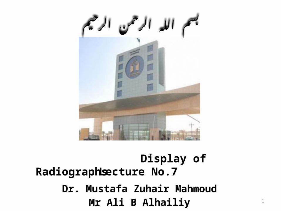
1. Introduction:
• When the radiograph is displayed at this manner, the patient left side is on the viewer’s right side and vice versa.
3Assistant Prof. Dr. Mustafa Zuhair Mahmoud

Display of Radiographs
2. What is required in a radiograph in addition to adequate demonstration of a part examined?
• Any radiograph what so ever should included on it, preferably in delegable form the following information:1. The subject (patient) name.
4Assistant Prof. Dr. Mustafa Zuhair Mahmoud

Display of Radiographs
1. The subject (patient) name.
5Assistant Prof. Dr. Mustafa Zuhair Mahmoud

Display of Radiographs
2. Correct sign of right or left side of the patient.
6Assistant Prof. Dr. Mustafa Zuhair Mahmoud
Display of Radiographs
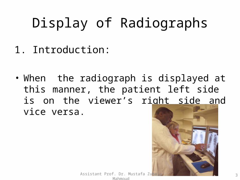
3. The date and time on which the radiograph was taken.
7Assistant Prof. Dr. Mustafa Zuhair Mahmoud
Display of Radiographs
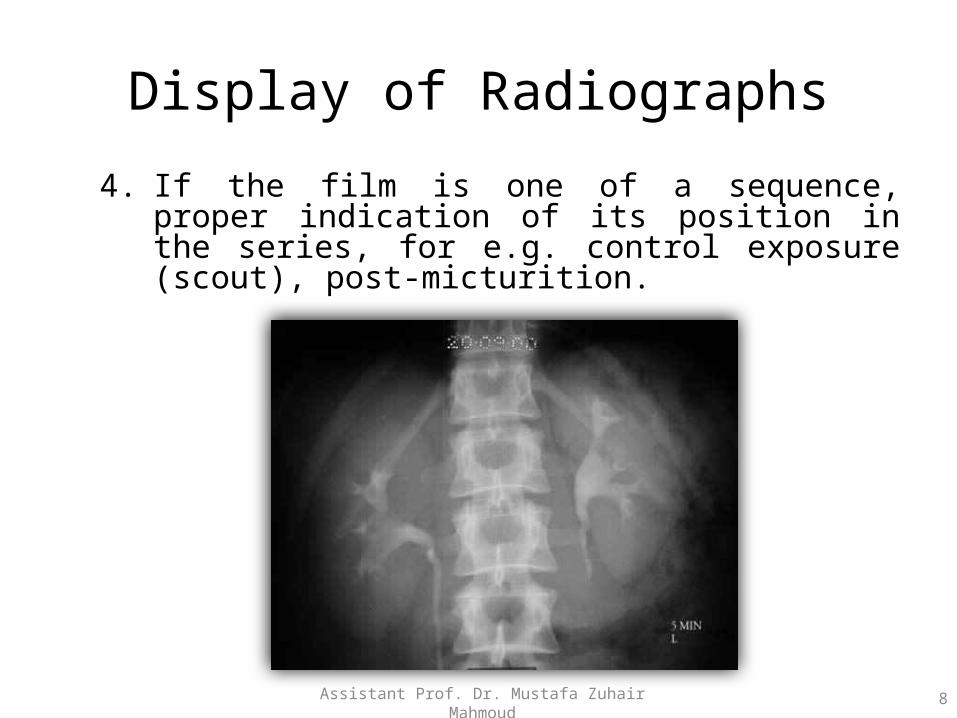
4. If the film is one of a sequence, proper indication of its position in the series, for e.g. control exposure (scout), post-micturition.
8Assistant Prof. Dr. Mustafa Zuhair Mahmoud

Display of Radiographs
3. Radiographic Image Identification Systems:
• Radiograph image can be identify through a system of identification by a different method of identification such as:1. Opaque letters.
9Assistant Prof. Dr. Mustafa Zuhair Mahmoud
Display of Radiographs
3. Radiographic Image Identification Systems:
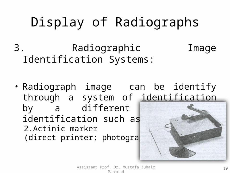
• Radiograph image can be identify through a system of identification by a different method of identification such as:2.Actinic marker(direct printer; photographic marker).
10Assistant Prof. Dr. Mustafa Zuhair Mahmoud
Display of Radiographs
3. Radiographic Image Identification Systems:
• The advantages of radiograph image identification systems are as follow:1. Permanent identification.2. Is economical in time.3. Shows information neatly and uniformly.4. Reduces the likelihood of error(s).
11Assistant Prof. Dr. Mustafa Zuhair Mahmoud

Display of Radiographs
4. Precaution When Using Lead Letters and Legends:
The character should not be placed where to obscure a feature of diagnostic importance.
12Assistant Prof. Dr. Mustafa Zuhair Mahmoud
Display of Radiographs
4. Precaution When Using Lead Letters and Legends:
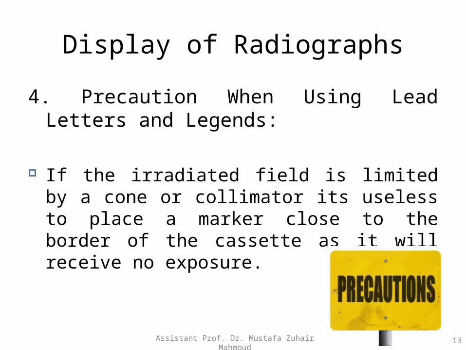
If the irradiated field is limited by a cone or collimator its useless to place a marker close to the border of the cassette as it will receive no exposure.
13Assistant Prof. Dr. Mustafa Zuhair Mahmoud
Display of Radiographs
5. Hanging Radiograph:
When hanging a radiographic image in the illuminator (viewing box) there are several tips can be use for hanging which include:
1. Dorsal, vertebral, cranial, shoulder, hip radiograph: as if the patient is standing in an upright position.
14Assistant Prof. Dr. Mustafa Zuhair Mahmoud

Display of Radiographs
1. Dorsal, vertebral, cranial, shoulder, hip radiograph: as if the patient is standing in an upright position.
15Assistant Prof. Dr. Mustafa Zuhair Mahmoud

Display of Radiographs
1. Dorsal, vertebral, cranial, shoulder, hip radiograph: as if the patient is standing in an upright position.
16Assistant Prof. Dr. Mustafa Zuhair Mahmoud

Display of Radiographs
2. Decubitus chest and abdominal radiographs: so that the side of the patient which was positioned upward when radiograph was taken is upward on the hung radiograph.
17Assistant Prof. Dr. Mustafa Zuhair Mahmoud

Display of Radiographs
2. Decubitus chest and abdominal radiographs: so that the side of the patient which was positioned upward when radiograph was taken is upward on the hung radiograph.
18Assistant Prof. Dr. Mustafa Zuhair Mahmoud

Display of Radiographs
5. Hanging Radiograph:
When hanging a radiographic image in the illuminator (viewing box) there are several tips can be use for hanging which include:
3. Toe and AP and oblique foot radiographs: as if the patient is hanging from toes.
19Assistant Prof. Dr. Mustafa Zuhair Mahmoud

Display of Radiographs
3. Toe and AP and oblique foot radiographs: as if the patient is hanging from toes.
20Assistant Prof. Dr. Mustafa Zuhair Mahmoud
Display of Radiographs
5. Hanging Radiograph:
When hanging a radiographic image in the illuminator (viewing box) there are several tips can be use for hanging which include:
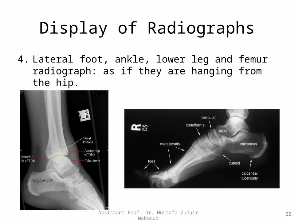
4. Lateral foot, ankle, lower leg and femur radiograph: as if they are hanging from the hip.
21Assistant Prof. Dr. Mustafa Zuhair Mahmoud
Display of Radiographs
4. Lateral foot, ankle, lower leg and femur radiograph: as if they are hanging from the hip.
22Assistant Prof. Dr. Mustafa Zuhair Mahmoud

Display of Radiographs
4. Lateral foot, ankle, lower leg and femur radiograph: as if they are hanging from the hip.
23Assistant Prof. Dr. Mustafa Zuhair Mahmoud
Display of Radiographs
5. Hanging Radiograph:
When hanging a radiographic image in the illuminator (viewing box) there are several tips can be use for hanging which include:
5. Fingers, wrist, and forearm radiographs: as if the patient is hanging from fingertips.
24Assistant Prof. Dr. Mustafa Zuhair Mahmoud

Display of Radiographs
5. Fingers, wrist, and forearm radiographs: as if the patient is hanging from fingertips.
25Assistant Prof. Dr. Mustafa Zuhair Mahmoud

Display of Radiographs
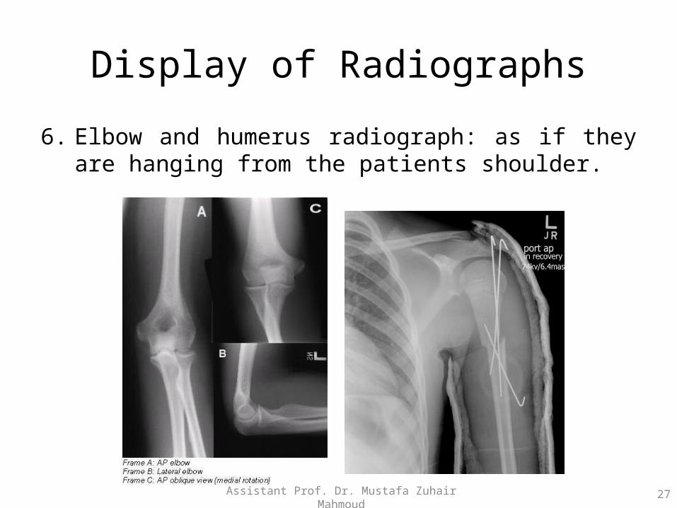
5. Hanging Radiograph:
When hanging a radiographic image in the illuminator (viewing box) there are several tips can be use for hanging which include:
6. Elbow and humerus radiograph: as if they are hanging from the patients shoulder.
26Assistant Prof. Dr. Mustafa Zuhair Mahmoud
Display of Radiographs
6. Elbow and humerus radiograph: as if they are hanging from the patients shoulder.
27Assistant Prof. Dr. Mustafa Zuhair Mahmoud

28Lecture No.8
Related Documents