1 This is the author manuscript accepted for publication and has undergone full peer review but has not been through the copyediting, typesetting, pagination and proofreading process, which may lead to differences between this version and the Version of Record. Please cite this article as doi: 10.1111/EPI.16480 This article is protected by copyright. All rights reserved DR. FELIPPE BORLOT (Orcid ID : 0000-0001-7897-4700) DR. MAJID ALFADHEL (Orcid ID : 0000-0002-9427-7240) DR. RAVINDRA ARYA (Orcid ID : 0000-0003-0873-9718) DR. KENNETH ALEXIS MYERS (Orcid ID : 0000-0001-7831-4593) DR. JITENDRA KUMAR SAHU (Orcid ID : 0000-0001-5194-9951) DR. SUVASINI SHARMA (Orcid ID : 0000-0002-3135-3306) Article type : Full length original research paper KCNT1-related epilepsy: An International Multicenter Cohort of 27 Pediatric Cases Authors: Felippe Borlot MD MSc 1 , Ahmed Abushama MD 1 , Nadine Morrison-Levy MBBS MSc 1,2 , Puneet Jain MD DM 1,3 , Kollencheri Puthenveettil Vinayan MD DM 4 , Musaad Abukhalid MD 5 , Hesham M. Aldhalaan MD FRCPC 5 , Hanin S. Almuzaini MBBS 5 , Sheffali Gulati MD FAMS 6 , Tova Hershkovitz MD 7 , Ramesh Konanki DM 8 , Lokesh Lingappa DM 8 , Aimee F. Luat MD 9 , Shatha Shafi MD 10 , Brahim Tabarki MD 10 , Maya Thomas MD DCH DM 11 , Sangeetha Yoganathan MD DNB DM 11 , Majid Alfadhel MD MHSC FCCMG 12,13 , Ravindra Arya MD DM 14,15 , Elizabeth J. Donner MD MSc FRCPC 1 , Salleh N. Ehaideb MS PhD 12 , Vykuntaraju K. Gowda DM 16 , Vivek Jain MBBS, FRCPCH 17 , Priyanka Madaan DM 18 , Kenneth A. Myers MD PhD FRCPC 19,20,21 , Hiroshi Otsubo MD 1 , Prateek Panda DM 6 , Jitendra K. Sahu DM 18 , Letícia P. B. Sampaio MD Author Manuscript

Welcome message from author
This document is posted to help you gain knowledge. Please leave a comment to let me know what you think about it! Share it to your friends and learn new things together.
Transcript
1
This is the author manuscript accepted for publication and has undergone full peer review but has
not been through the copyediting, typesetting, pagination and proofreading process, which may
lead to differences between this version and the Version of Record. Please cite this article as doi:
10.1111/EPI.16480
This article is protected by copyright. All rights reserved
DR. FELIPPE BORLOT (Orcid ID : 0000-0001-7897-4700)
DR. MAJID ALFADHEL (Orcid ID : 0000-0002-9427-7240)
DR. RAVINDRA ARYA (Orcid ID : 0000-0003-0873-9718)
DR. KENNETH ALEXIS MYERS (Orcid ID : 0000-0001-7831-4593)
DR. JITENDRA KUMAR SAHU (Orcid ID : 0000-0001-5194-9951)
DR. SUVASINI SHARMA (Orcid ID : 0000-0002-3135-3306)
Article type : Full length original research paper
KCNT1-related epilepsy: An International Multicenter Cohort of 27 Pediatric Cases
Authors: Felippe Borlot MD MSc1, Ahmed Abushama MD1, Nadine Morrison-Levy
MBBS MSc1,2, Puneet Jain MD DM1,3, Kollencheri Puthenveettil Vinayan MD DM4,
Musaad Abukhalid MD5, Hesham M. Aldhalaan MD FRCPC5, Hanin S. Almuzaini
MBBS5, Sheffali Gulati MD FAMS6, Tova Hershkovitz MD7, Ramesh Konanki DM8,
Lokesh Lingappa DM8, Aimee F. Luat MD9, Shatha Shafi MD10, Brahim Tabarki MD10,
Maya Thomas MD DCH DM11, Sangeetha Yoganathan MD DNB DM11, Majid Alfadhel
MD MHSC FCCMG12,13 , Ravindra Arya MD DM14,15, Elizabeth J. Donner MD MSc
FRCPC1, Salleh N. Ehaideb MS PhD12, Vykuntaraju K. Gowda DM16 , Vivek Jain MBBS,
FRCPCH17, Priyanka Madaan DM18, Kenneth A. Myers MD PhD FRCPC19,20,21 , Hiroshi
Otsubo MD1, Prateek Panda DM6, Jitendra K. Sahu DM18, Letícia P. B. Sampaio MD
Auth
or
Manuscript

Borlot et al.
This article is protected by copyright. All rights reserved
PhD22, Suvasini Sharma MD DM23, Elisabeth Simard-Tremblay MD FRCPC 19,20, Maria
Zak NP MN1, Robyn Whitney MD FRCPC1.
Affiliations:
1- Division of Neurology, Department of Paediatrics, The Hospital for Sick Children,
Toronto, Ontario, Canada.
2- Children’s Hospital of Eastern Ottawa, Ontario, Canada.
3- Division of Pediatric Neurology, Department of Pediatrics, Danat Al Emarat
Hospital for Women and Children, Abu Dhabi, United Arab Emirates.
4- Department of Paediatric Neurology, Amrita Institute of Medical Sciences,
Cochin, Kerala, India.
5- Department of Neurosciences, King Faisal Specialist Hospital and Research
Center, Riyadh, Saudi Arabia
6- Child Neurology Division, Center of Excellence & Advanced Research on
Childhood Neurodevelopmental Disorders, Department of Pediatrics, All India
Institute of Medical Sciences, New Delhi, India.
7- The Genetic Institute, Rambam Medical Center, Haifa, Israel.
8- Department of Neurology, Rainbow Children's Hospital, Hyderabad, India.
9- Children’s Hospital of Michigan, Detroit Medical Center, Wayne State University
School of Medicine, Detroit, MI, USA.
10-Division of Neurology, Department of Pediatrics, Prince Sultan Military Medical
City, Riyadh, Saudi Arabia.
11-Paediatric Neurology, Department of Neurological Sciences, Christian Medical
College, Vellore, Tamil Nadu, India.
12-King Abdullah International Medical Research Centre (KAIMRC), King Saud bin
Abdulaziz University for Health Sciences, King Abdulaziz Medical City, Ministry of
National Guard-Health Affairs (MNGHA), Riyadh, Saudi Arabia.
13-Division of Genetics, Department of Pediatrics, King Abdulaziz Medical City,
Ministry of National Guard-Health Affairs (MNGHA), Riyadh, Saudi Arabia.
Auth
or
Manuscript

Borlot et al.
This article is protected by copyright. All rights reserved
14-Comprehensive Epilepsy Center, Division of Neurology, Cincinnati Children's
Hospital Medical Center, Cincinnati, OH, USA.
15- Department of Pediatrics, University of Cincinnati College of Medicine,
Cincinnati, Ohio, USA.
16-Indira Gandhi Institute of Child Health, Bangalore, Karnataka, India.
17-Santokba Durlabhji Hospital, Jaipur, Rajasthan, India.
18-Pediatric Neurology Unit, Department of Pediatrics, Postgraduate Institute of
Medical Education and Research, Chandigarh, India.
19-Division of Neurology, Department of Pediatrics, Montreal Children’s Hospital,
McGill University Health Centre, Montreal, Québec, Canada
20-Department of Neurology & Neurosurgery, McGill University Health Centre,
Montreal, Québec, Canada
21-Research Institute of the McGill University Health Centre, Montreal, Québec,
Canada.
22-Department of Neurology, Faculdade de Medicina da Universidade de Sao Paulo
(USP), Sao Paulo, Brazil.
23-Neurology Division, Department of Pediatrics, Lady Harding Medical College and
associated Kalawati Saran Children Hospital, New Delhi, India.
Corresponding author:
Dr. Robyn Whitney
Division of Neurology, The Hospital for Sick Children
555 University Ave, Toronto ON
M5G 1X8
Auth
or
Manuscript

Borlot et al.
This article is protected by copyright. All rights reserved
Email: [email protected]
Phone: 416-813-6660
Word count: 3780
Tables: 02
References: 31
ABSTRACT
Objective: Through international collaboration, we evaluated the phenotypic aspects of
a multiethnic cohort of KCNT1-related epilepsy and explored genotype-phenotype
correlations associated with frequently encountered variants.
Methods: A cross-sectional analysis of children harboring pathogenic or likely
pathogenic KCNT1 variants was completed. Children with one of the two more common
Auth
or
Manuscript

Borlot et al.
This article is protected by copyright. All rights reserved
recurrent KCNT1 variants were compared to the rest of the cohort for the presence of
particular characteristics.
Results: Twenty-seven children (15 males, mean age 40.8 months) were included.
Seizure onset ranged from one day to six months, and half (48.1%) exhibited
developmental plateauing upon onset. Two-thirds had epilepsy of infancy with migrating
focal seizures (EIMFS) and focal tonic seizures were common (48.1%). The most
frequent recurrent KCNT1 variants were c.2800G>A; p.Ala934Thr (n=5) and c.862G>A;
p.Gly288Ser (n=4). De novo variants were found in 96% of tested parents (23/24). Sixty
percent had abnormal MRI findings. Delayed myelination, thin corpus callosum, and
brain atrophy were most common. One child had grey-white matter interface
indistinctness, suggesting a malformation of cortical development. Several anti-epileptic
drugs (mean 7.4/patient) were tried with no consistent response to any one agent.
Eleven tried quinidine, 45% had marked (>50% seizure reduction); or some
improvement (25-50% seizure reduction). Seven used cannabidiol, 71% experienced
marked or some improvement. Fourteen tried diet therapies, 57% had marked or some
improvement. When comparing the recurrent variants to the rest of the cohort, with
respect to developmental trajectory, presence of EIMFS, >500 seizures/month,
abnormal MRI and treatment response, there were no statistically significant
differences. Four patients died (15%); none of SUDEP.
Significance: Our cohort reinforces common aspects of this highly pleiotropic entity.
EIMFS manifesting with refractory tonic seizures was most common. Cannabidiol, diet
therapy, and quinidine seem to offer better chances of seizure reduction, although
evidence-based practice is still unavailable.
Key words: KCNT1, Epilepsy of infancy with migrating focal seizures, microcephaly,
CBD, ketogenic diet, quinidine. Auth
or
Manuscript

Borlot et al.
This article is protected by copyright. All rights reserved
INTRODUCTION
KCNT1 encodes a ligand-gated potassium channel, which is activated by intracellular
sodium binding (also called SLACK, SLO2.2, KC4.1). KCNT1 has several functions,
which include regulating neuronal firing rate, contributing to the slow hyperpolarization
after repetitive firing, and it also has an important role in neuronal response to hypoxia.1-
3 Gain-of-function effects produce higher action potential firing frequency due to faster
action potential repolarization and increased fast after-hyperpolarization.4 KCNT1
channels are widely expressed in the central nervous system and are found in the
olfactory bulb, brainstem, hippocampal and cortical embryonic neurons.1,2,5
Gain of function KCNT1 pathogenic variants are known to cause pleiotropic effects and
a number of epilepsy phenotypes have been described: (I) epilepsy of infancy with
migrating focal seizures (EIMFS);1,6 (II) a severe form of autosomal dominant sleep-
related hypermotor epilepsy (ADSHE);7 (III) Early onset epileptic encephalopathy
(EOEE) (i.e. Ohtahara syndrome, West syndrome, and unclassified EOEE);8 (IV)
temporal lobe epilepsy with intellectual disability;9 and (V) myoclonic-atonic epilepsy.10
Although most patients harbouring KCNT1 pathogenic variants have no causative
underlying structural brain abnormalities, patients with ADSHE may rarely exhibit
malformations of cortical development.11
Auth
or
Manuscript

Borlot et al.
This article is protected by copyright. All rights reserved
There has been inconsistent data with respect to clinical efficacy of quinidine (broad-
spectrum potassium channel blocker) in patients with EIMFS. Unblinded assessment of
seizure reduction may range from complete to no response.12-17 Overall, half of EIMFS
or EOEE patients will have no response to quinidine, and only 20% may benefit with at
least a 50% seizure reduction.18 The main adverse effect attributed to quinidine therapy
is cardiotoxicity with prolonged QTc interval and arrhythmias, but sedation, elevated
liver function tests, rash, and skin discoloration have also been described.18,19
We sought to evaluate the clinical aspects including phenotypic presentation, EEG,
neuroimaging, response to pharmacological and non-pharmacological therapies of a
multiethnic cohort of children with pathogenic or likely pathogenic KCNT1 variants.
Despite the high pleiotropy and heterogeneity associated with KCNT1-related
epilepsies, we also aimed to explore any possible phenotype-genotype correlations
associated with the most common identified variants.
METHODS
Patients and Institutional Review Board Approval
Ethics approval for the study was granted by the Research Ethics Board at The Hospital
for Sick Children, Toronto, Ontario, Canada (REB #1000061319) and the other
participant centres, as per the respective hospital policies. Children were enrolled in the
study after consent was obtained from legal guardians, unless the local board institution
waived the need to obtain an informed consent from a given center. Recruitment started
within the Division of Neurology at the Hospital for Sick Children, which was the
coordinating research site. Henceforth, an international collaborative network joined the
study allowing us to recruit patients from Brazil, Canada, India, Israel, Saudi Arabia, and
the United States. All mutations were identified by commercial gene panels or whole
exome sequencing.
Auth
or
Manuscript

Borlot et al.
This article is protected by copyright. All rights reserved
Study Design, Inclusion and Exclusion Criteria
Inclusion criteria for this retrospective case series study were: (I) age 18 years or
younger, (II) diagnosed with KCNT1-related epilepsy at any point until September 2018,
(III) patients with pathogenic or likely pathogenic KCNT1 variants (Class 5 or 4),
according to The American College of Medical Genetics and Genomics (ACMG)20,21,
regardless of the phenotypic presentation. The exclusion criteria were: (I) patients
harboring KCNT1 variants of uncertain significance (VUS) or benign variants, and (II)
failure to gather enough data for phenotypic characterization.
Data collection, analysis and interpretation
Study data were collected, stored and managed using REDCap (Research Electronic
Data Capture) secure electronic data capture tool at The Hospital for Sick Children.22,23
Data abstraction protocol included: demographics, age of presentation, first symptom
developed by the patient, age of seizure onset, seizure type(s), presence of known
epilepsy syndrome, developmental history, neurological/psychiatric/systemic
comorbidities, genotype and inheritance (if available), EEG reports, neuroimaging
abnormalities and pharmacological and non-pharmacological treatment and treatment
response. For each treatment modality, the following options were gathered from
caregivers and clinicians: (I) seizure freedom; (ii) marked improvement (i.e. greater than
50% reduction in seizures); (III) some improvement (i.e. 25-50% seizure reduction); (IV)
minimal improvement (i.e. less than 25% seizure reduction); (V) no improvement; and
(VI) worsening of seizures. In addition, tolerability, adverse effects, reason for
discontinuation of therapy, and duration of therapies were gathered. Data were
summarized using descriptive statistics, including mean, median, range and standard
deviation (SD) for continuous variables and percentages for categorical variables.
Clinical genetic testing (i.e. through gene panels or whole exome sequencing) was
performed through different companies internationally, using their specific protocols to
obtain genomic DNA from blood. Testing of family members was performed according
Auth
or
Manuscript
Borlot et al.
This article is protected by copyright. All rights reserved
to clinical indication and/or availability using Sanger sequencing in nearly all cases.
Variants of unknown significance, benign and likely benign variants in other genes, as
well as heterozygous pathogenic variants detected in genes associated with autosomal
recessive conditions were considered out of the scope of the study and will not be
reported.
To address the second aim of the study (i.e. to determine phenotype-genotype
correlations associated with more frequently encountered variants), we performed the
Chi-square test (IBM SPSS statistics version 20.0 Armonk, NY: IBM Corp.) to compare
the presence of specific findings (such as developmental plateauing, presence of
EIMFS, reported seizure frequency greater than 500/month, abnormal MRI) in the most
frequently encountered KCNT1 variants found in our cohort (i.e. 2800G>A; p.Ala934Thr
and c.862G>A; p.Gly288Ser). In addition, the Chi-square test was used to compare
children with and without these variants, with respect to treatment responses to
quinidine, diet therapy, and CBD. Moreover, given the previous literature data
suggesting that an earlier age of onset of quinidine therapy could result in a better
response, we compared the age of initiation of quinidine therapy to the seizure
reduction response using Spearman’s Rho calculator. Statistically significant
correlations were considered positive if P < .05.
All data were reviewed by two authors from the main research site for completeness;
whenever incomplete or unclear information was noted, further clarification was sought
from the contributing centre. Individual genetic results (KCNT1 variants) obtained from
each center were checked and re-classified (if required) in accordance with the ACMG
criteria.20,21 If, on re-classification, patients were found to have VUS or benign variants,
they were then subsequently excluded from the analysis.
RESULTS
Thirty children were initially recruited from all centers. However, three patients were
excluded upon review, as their KCNT1 variants were classified as VUS. Our cohort was
Auth
or
Manuscript
Borlot et al.
This article is protected by copyright. All rights reserved
composed of 27 children, 15 males and 12 females (ratio 1.25), current mean age 40.8
months (SD 28.3, median 38 months). Three patients were previously published (case#
15, case#16, and case #25)24, 25 and 24 children are newly reported. For all children,
seizures were the first concern that brought them to attention of a neurologist, with age
of onset of seizures ranging from one day to six months of life (mean 1.7 months, SD
1.7, median 1 month). Nevertheless, the mean age for a definitive diagnosis of KCNT1-
related epilepsy was later and only at mean of 18.8 months, SD 20.1 (median 10
months). Developmental trajectory was characterized by plateauing in nearly half of
children (48.1%, 13/27) upon seizure onset, whereas 22.2% (6/27) had slow
developmental gains, and 22.2% (6/27) had developmental regression, followed by
either slow gains or plateauing.
Recurrent KCNT1 variants were reported in 18 patients. The most common KCNT1
variants were (i) c.2800G>A; p.Ala934Thr; (ii) c.862G>A; p.Gly288Ser; and (iii)
c.1421G>A; p.Arg474His. These variants were respectively seen in five (cases #4, #8,
#16, #24, and #27), four (cases #7, #9, #10, and #11), and three (cases #6, #17, and
#20) children. In addition, (iv) c.1283 G>A; p.Arg428Gln (cases #1 and #23); (v)
c.1420C>T; p.Arg474Cys (cases #13 and #22); and (vi) c.2849G>A; p.Arg950Gln
(cases #3 and #15) were found in two children each. From the nine non-recurrent
KCNT1 variants found in this cohort, four have not been previously reported in the
literature (cases #2, 12, #14, and #18). Case 12’s nucleotide variant (c.1130G>C) is
novel, however a nucleotide variant affecting the same amino acid (c.1129T>A;
p.Cys377Ser) has been reported in a patient presenting with EIMFS.26
Parental testing was completed in 24 out of the 27 families, and 23 children (95.8%,
23/24) were found to have de novo variants. Despite having a de novo variant, six out of
the 23 children (26%, 6/23) with a de novo variant had a positive family history of
seizures. We had only limited information to the specific epilepsy syndromes in the
affected relatives: (i) case 3’s maternal great uncle had unclassified epilepsy; (ii) three
third cousins of case 9 had recurrent seizures in adulthood; (iii) case 14’s grandmother
had unprovoked seizures in adulthood and this maternal grandmother’s sister had
febrile seizures in childhood; (iv) case 15’s paternal grandfather's brother had seizures
Auth
or
Manuscript

Borlot et al.
This article is protected by copyright. All rights reserved
in childhood only; (v) a paternal first cousin of case 16 had seizures, and there is also a
history of febrile seizures in this patient's second cousins; finally (vi) case 26’s father
had recurrent seizures attributed to a previous history of traumatic brain injury. Only one
child (case #10) presented with an inherited variant from an unaffected parent. Case
10’s mother has no neurological manifestations, but is heterozygous for the variant
c.862G>A; p.Gly288Ser. Consanguinity was reported in four families from Saudi Arabia,
cases #5, #6, #7 and #8, and three out of these four variants (c.1885A>G, p.Lys629Glu;
c.862G>A, p Gly288Ser; and c.2800 G>A; p.Ala 934Thr) were found to be de novo.
Parents of case #6 were not available for testing (unknown inheritance of the variant
c.1421G>A; p.Arg474His)
EIMFS was the most common electroclinical syndrome, present in 18 patients (66.7%,
18/27). Four children had an unclassified EOEE, three children were diagnosed with
West syndrome, and two children were diagnosed with ADSHE at some point. Three
children have had more than one electroclinical phenotype throughout their trajectories:
cases #3 and #4 initially presented with EIMFS evolving to West syndrome, whereas
case #22’s phenotype was initially consistent with unclassified EOEE, but later on
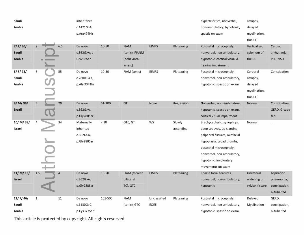
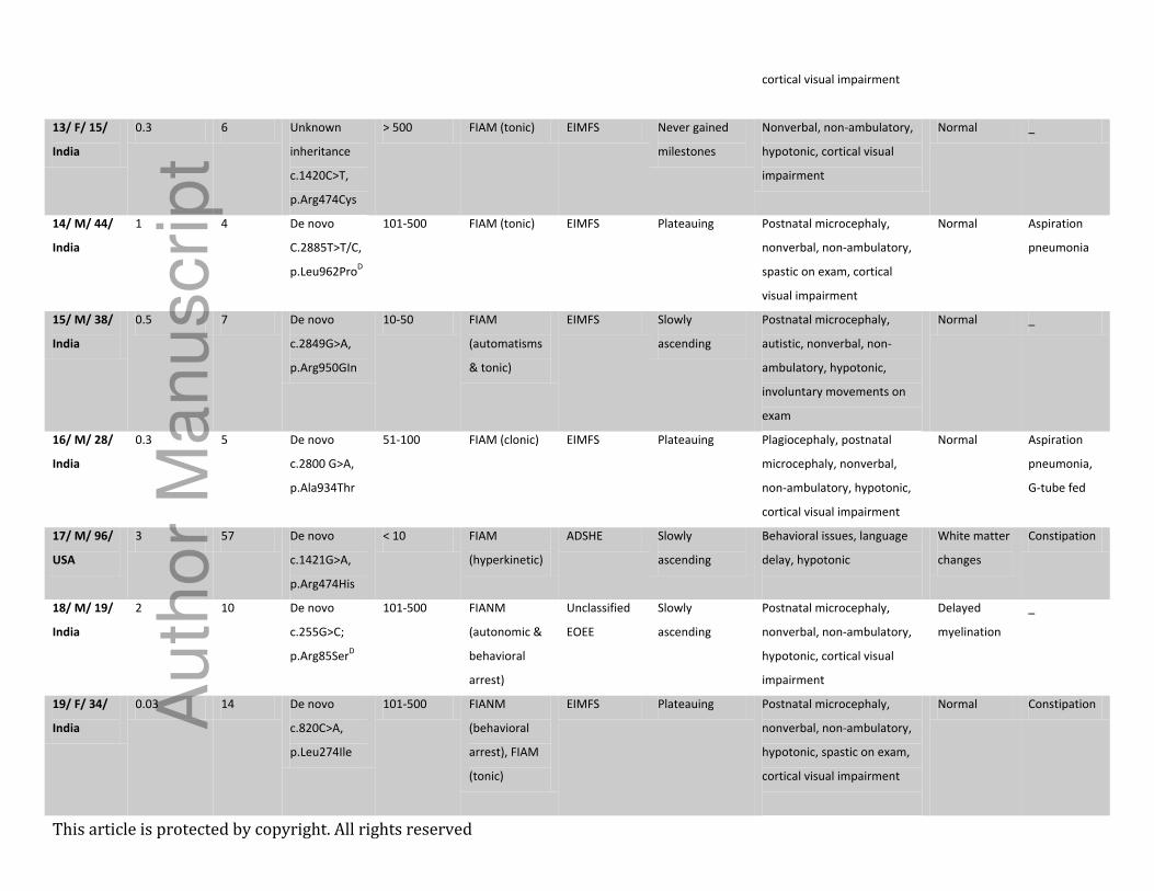
developed ADSHE. Table 1 summarizes the demographics, relevant clinical
information, KCNT1 variants details, and neuroimaging findings.
Focal impaired awareness motor (tonic, or with tonic component) seizures were the
most common seizure type (48.1%, 13/27) observed in our cohort. In addition, a variety
of other seizures were also observed such as: focal impaired awareness non-motor
(behavioural arrest, autonomic, emotional), focal impaired awareness motor
(hyperkinetic, clonic), generalized tonic, generalized tonic-clonic, generalized
myoclonic-tonic-clonic, generalized myoclonic, and generalized absence seizures
(atypical). Estimated current seizure frequency reported by caregivers was markedly
variable at the time of data collection, with three patients (11.1%, 3/27) having more
than 500 seizures/month, nine patients (33.4%, 9/27) having more than 100 and up to
500 seizures/month, three patients (11.1%, 3/27) having more than 50 up to 100
seizures/month, seven patients (26%, 7/27) having 10 to 50 seizures/month, and five
Auth
or
Manuscript
Borlot et al.
This article is protected by copyright. All rights reserved
patients (18.5%, 5/27) having less than 10 seizures/month. None of the caregivers
reported seizure freedom.
The presence of developmental plateauing, EIMFS, and seizure burden were not
associated with a specific genetic variant. Five children harboring c.2800G>A;
p.Ala934Thr were compared to the rest of the cohort with respect to the presence of
developmental plateauing (P=.13) phenotype consistent with EIMFS (P=.48), and
estimated seizure frequency greater than 500 per month (P=.22). In addition, four
children harboring c.862G>A; p.Gly288Ser were compared to the rest of the cohort with
respect to the presence of developmental plateauing (P=.93) phenotype consistent with
EIMFS (P=.44), and estimated seizure frequency (P=.38). There were no significant
differences between the genotypes analyzed compared the rest of the cohort with
respect to the aforementioned characteristics. The most common phenotype seen in our
cohort consisted of non-ambulatory (92.3%, 24/26; unavailable data in one patient),
nonverbal (88%, 22/25; unavailable data in two patients), hypotonic (74%, 20/27) and
spastic (48.1%, 13/27) patients, with acquired microcephaly (65.3%, 17/26; unavailable
data in one patient ) and cortical visual impairment (60%, 15/25; unavailable data in two
patients). Only five children (18.5%, 5/27) were diagnosed with involuntary movements
(see Table 1).
Brain MRI was abnormal in the majority of children (59.3%, 16/27). The most common
findings were delayed myelination (68.7%, 11/16; or 40.7%, 11/27 of all patients), thin
corpus callosum (43.7%, 7/16; or 26%, 7/27 of all patients), usually but not always
accompanied by brain atrophy (37.5% 6/16; or 22%, 6/27 of all patients). The presence
of an abnormality in the brain MRI was further evaluated in the two most common
genetic variants in comparison to the rest of the cohort. There was no correlation
between the presence of an abnormality in patients with specific genotypes in
comparison to the rest of the cohort (i.e. c.2800G>A; p.Ala934Thr versus other patients,
P= .97; c.862G>A; p.Gly288Ser versus other patients, P=.68).
Systemically, these children often had constipation (40.7%, 11/27), gastroesophageal
reflux disease (33.4%, 9/27), and aspiration pneumonia (33.4%, 9/27). Only one child
Auth
or
Manuscript
Borlot et al.
This article is protected by copyright. All rights reserved
(case #7) had a cardiac malformation (ventricular septal defect) and supraventricular
ectopic activity.
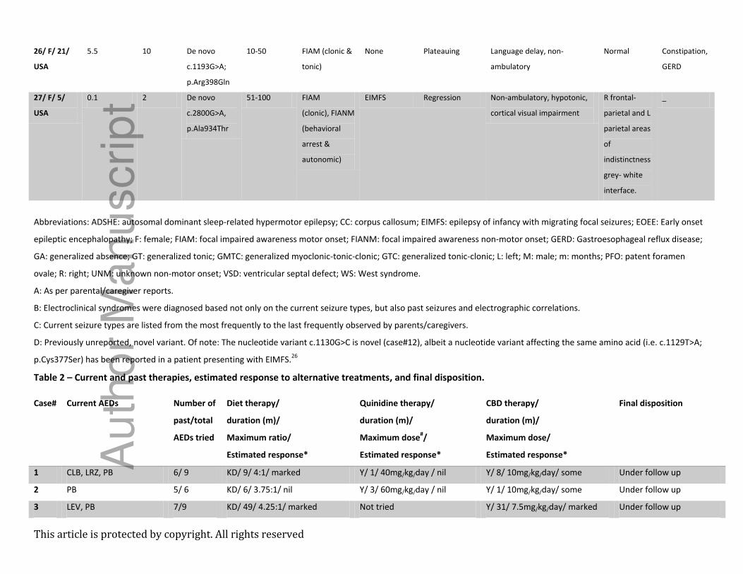
Table 2 shows the current AEDs, number of total AEDs tried for each patient, alternative
therapies and estimated response for each, and final disposition for each patient.
Twenty-three children are still under follow up (85.2%, 23/27) and four passed away
(14.8%, 4/27). Cause of death was due to complications of systemic diseases (n=1),
progression of illness (n=1), and redirection of the goal of care (n=2). There were no
reports of sudden unexpected death in epilepsy (SUDEP). Out of the 23 children being
followed, the number of current anti-epileptic drugs (AEDs) in use ranged from one to
five (mean 2.7, SD 1.2, median 2). Including the patients who died, the total number of
AEDs tried ranged from three to 16 (mean 7.4, SD 3.1, median 7). Given the high
number of medications tried in a relatively short period of time, subjective nature of
responses, along with a variety of different combinations, we were unable to identify one
or two AEDs that were particularly effective in controlling seizures. Eleven patients
were tried on quinidine, with doses ranging from 20 to 60mg/kg/day (mean 35.9
mg/kg/day, SD 11.1, median 40 mg/kg/day) and duration of therapy from one to 28
months (mean 9.1 months, SD 9.5, median 4 months). As per caregivers, estimated
response was marked or some improvement in five (45.4%, 5/11), but no response
whatsoever in six children (54.6%, 6/11). Five children (45.4%, 5/11) developed QTc
prolongation while on quinidine, but none discontinued therapy for this reason. Irritability
and vomiting were reported in one child each. The age at administration of quinidine
ranged from two to 37 months (mean 11.4 months, SD 9.8, median 8 months). There
was no statistically significant correlation between responses to quinidine and the earlier
age of onset of therapy (rs = .22917, P (2-tailed) = .49).
Cannabidiol (CBD) was used in seven patients (26%, 7/27), with highly variable doses
ranging from 0.25 to 10 mg/kg/day (mean 6 mg/kg/day, SD 4.4, median 7.5 mg/kg/day).
Estimated response was reported as marked or some improvement in five children
(71.4%, 5/7), and minimal in two (28.6%, 2/7). CBD was well tolerated in all patients,
with no side effects reported by the caregivers. Diet therapies were tried in fourteen
patients (51.8%, 14/27), which included the ketogenic diet in twelve and low glycemic
Auth
or
Manuscript

Borlot et al.
This article is protected by copyright. All rights reserved
index diet in two. Eight children (57.2%, 8/14) had marked or some improvement, and
six (42.8%, 6/14) had minimal or no improvement. There was no statistically significant
correlation when children harboring c.2800G>A; p.Ala934Thr or c.862G>A; p.Gly288Ser
were compared to the rest of the cohort, with respect to treatment response to
quinidine, CBD, or diet therapy.
DISCUSSION
This observational study is one of the largest international cohorts examining pediatric
patients diagnosed with KCNT1-related epilepsy. There was no selection bias with
respect to the phenotypic characterization of this patients’ sample, given that the
essential criterion was related to the presence of pathogenic or likely pathogenic
KCNT1 variants. Some may argue that the presence of ADSHE could be
underestimated due the cut-off age of the inclusion criteria, but we believe our data
reflects the current circumstances of clinicians dealing with KCNT1-related epilepsy in
infancy and childhood. The 17-month gap between first seizures (i.e. mean age at onset
of seizures 1.7 months) and diagnosis of KCNT1-related epilepsy (mean age at
diagnosis 18.8 months) in our cohort reflects the true odyssey for families and
physicians alike.
As previously reported by different authors, KCNT1 pathogenic variants are highly
pleiotropic and associated with a variety of phenotypes.8,18,27 In our study, two-thirds of
patients presented with EIMFS, however, unclassified EOEE, West syndrome and
ADSHE were also present, similar to previous reports. Further analysis of recurrent
variants and seizure burden related to specific genotypes was not significant, reinforcing
the variability in gene expression and high pleiotropy of KCNT1 pathogenic variants.
However, our data obtained from 27 affected children enabled us to refine the
phenotypic characterization, which may help in earlier recognition of these patients:
early onset of tonic seizures which are medically refractory, with plateauing of
milestones, hypotonia, cortical visual impairment, and acquired microcephaly should
promptly raise a heightened index of suspicion for KCNT1-related epilepsy, likely
Auth
or
Manuscript

Borlot et al.
This article is protected by copyright. All rights reserved
manifesting as EIMFS or unclassified EOEE. Obviously, other seizure types and
developmental trajectories can be seen. Similar to our results, the high prevalence of
acquired microcephaly was also noted by Kuchenbuch et al. as high as 90% after three
years of follow up.28 Given the cross-sectional nature of our study, we were not able to
delineate different phases experienced by EIMFS patients over time, recently described
as stormy phase, stabilization period, and chronic phase.28
Somatic mosaicism (i.e. DNA alteration occurring at the post-zygotic stage) may also
contribute to the phenotypic heterogeneity seen in some epilepsy genes. As previously
demonstrated for “de novo” epileptic encephalopathies, 8.3% of parents have
mosaicism of their child’s pathogenic variant, particularly when there is parental history
of seizures.29 Interestingly, the high prevalence of positive family history of seizures (~
30%) in our patients and in another study28 might indicate the need for future research
focused on relatives of patients with KCNT1-related epilepsy.
Although we often consider that neuroimaging is normal in the genetic epileptic
encephalopathies, our data strengthen that in KCNT1-related epilepsies brain MRI can
be abnormal (59% of our children). From those with abnormal imaging, we found that
delayed myelination, thin corpus callosum, and brain atrophy were the most common
findings, albeit some of these abnormalities (i.e. delayed myelination and brain atrophy)
are likely depending whether the neuroimaging was obtained at earlier or later stages of
the disease. One of our patients (case #27) was found to have areas of indistinctness
in the grey-white matter interface, which could suggest an underlying malformation of
cortical development, as recently described in patients with KCNT1 pathogenic
mutations and ADSHE.11
Given the expression of KCNT1 in muscle tissue, gonads, and the pituitary gland, it has
been proposed that KCNT1 mutations can be involved in cardiac anomalies, SUDEP,
and precocious puberty.5,27,28 Indeed, one of our patients (case#7) was diagnosed with
a ventricular septal defect, and supraventricular ectopic activity. However, unlike in the
other reported cases, neither SUDEP nor precocious puberty were reported in our
series, possibly due to limited long term follow up of this cohort. A cross-sectional study
including older patients as well as long term follow up assessments could help
Auth
or
Manuscript

Borlot et al.
This article is protected by copyright. All rights reserved
identifying precocious puberty and, perhaps, SUDEP if these conditions are related to
KCNT1-related epilepsy.
Poor response to AEDs is a common characteristic among KCNT1-related epilepsy
patients regardless of the phenotype. Not different from other studies,18,28 our patients
were exposed to several AEDs over time (mean 7.4, SD 3.1), with no seizure freedom
achieved. Moreover, more than half (55%, 15/27) reported more than 50 seizures
monthly. These data promptly led to clinicians and families to look for alternative
treatments. After some case reports successfully reported treating KCNT1-related
epilepsy with quinidine,12,13 several authors unfortunately have not been able to
reproduce the same outcomes.14-17 In addition, a small blinded, placebo-controlled,
crossover trial that included six ADSHE patients did not show significant difference in
seizure frequency during the quinidine phase compared to placebo, and paroxysmal
arousals were similarly unchanged. There were no patients achieving the 50%-
reduction mark.30
In our retrospective analysis, nearly 50% of (5/11) patients had marked or some seizure
reduction with quinidine. Twenty-seven percent (3/11) reported greater than 50%
seizure reduction and 18.1% (2/11) reported from 25 to 50% seizure reduction. Our
findings were similar to those from the largest cohort of KCNT1-related epilepsy that
evaluated 20 patients taking quinidine and found nearly 50% (9/20) of patients with
some response, including 20% of patients having at least 50% seizure reduction.18
Neither seizure freedom nor worsening of seizures was reported in our patients. In
addition, we were not able to establish statistical correlation suggesting an age-
dependent response to quinidine, as previously reported in the literature.15
Other than quinidine, we were also able to record reasonable efficacy of diet therapy
and CBD. When taking into consideration patients exposed to diet therapies, 43% (6/14)
had greater than 50% seizure reduction. Analyzing the response to the ketogenic diet
(not including the low glycemic index diet), half of the patients (6/12) had greater than
50% seizure reduction. Reasonable response to the ketogenic diet has also been
reported, with a response rate of 31% (9/29) as per caregivers and physicians.18 For
Auth
or
Manuscript

Borlot et al.
This article is protected by copyright. All rights reserved
those who responded well to CBD, two out of three were previously on the ketogenic
diet making any conclusions of the CBD efficacy merely speculative. In addition, the
small number of patients on CBD and the lack of consistency of the dosage prescribed
(from 0.25 to 10 mg/kg/day) limit our capacity to draw meaningful conclusions.
In addition to the limitations related to our study method, which include missing data and
lack of longitudinal follow up, our study is hampered by the limited number of patients
for subset analysis. This is difficult to overcome when gathering data in a rare condition.
Parental reports of seizure frequency (sometimes hard to recognize due to subtle
manifestations, or sometimes overcalled due to the presence of abnormal movements
and behavioral issues) as well as the lack of standardized use of some therapies (i.e.
CBD and quinidine doses) are further limitations of our study. Moreover, with our current
data it is still unclear whether somatic mutations are particularly relevant in KCNT1-
related epilepsy or not. Low level of parental mosaicism could be underestimated given
that the great majority of parental testing in our study was through Sanger sequencing,
and not through next generation sequencing analysis.31
In summary, through international collaboration and in comparison with previous
literature data, we were able to delineate the common aspects within this highly
pleiotropic entity, KCNT1-related epilepsy: early-onset refractory tonic seizures, likely
(but not exclusively) manifesting as EIMFS or unclassified EOEE, along with milestones
plateauing, hypotonia, cortical visual impairment, and acquired microcephaly.
Supportive but not mandatory neuroimaging findings included delayed myelination, thin
corpus callosum, brain atrophy, and rarely malformations of cortical development.
Despite the lack of satisfactory evidence, alternative treatments such as the ketogenic
diet and quinidine seem to be well tolerated and may help achieving seizure reduction
greater than 50%.
ACKNOWLEDGEMENT
Auth
or
Manuscript

Borlot et al.
This article is protected by copyright. All rights reserved
Dr. Camila C. Henriques de Aquino for her valuable assistance with the statistical
analysis and interpretation.
DISCLOSURE OF CONFLICTS OF INTEREST
Dr. Vinayan was supported by an institutional seed grant for infantile epilepsy registry
from the Amrita University. He was also supported by academic grants from the Kerala
Association of Neurologists and the Indian Epilepsy Association, Kochi. The other
authors have no conflict of interest to disclose.
ETHICAL PUBLICATION STATEMENT
We confirm that we have read the Journal’s position on issues involved in ethical
publication and affirm that this report is consistent with those guidelines.
Key Point Box
KCNT1-related epilepsy in children usually manifests as early-onset refractory
focal tonic seizures and EIMFS.
Most children will become non-ambulatory, nonverbal, hypotonic, spastic, with
acquired microcephaly and cortical visual impairment.
Supportive MRI findings include: delayed myelination, thin corpus callosum, brain
Auth
or
Manuscript
Borlot et al.
This article is protected by copyright. All rights reserved
atrophy, and malformations of cortical developmental.
There have been no well-stablished genotype-phenotype specific correlations so
far.
Despite the lack of evidence-based practice, ketogenic diet and quinidine are
well tolerated and may help with seizure reduction.
REFERENCES
1- Barcia G, Fleming MR, Deligniere A, et al. De novo gain-of-function KCNT1
channel mutations cause malignant migrating partial seizures of infancy. Nat
Genet. 2012;44:1255-1259.
2- Bhattacharjee A, Kaczmarek LK. For K+ channels, Na+ is the new Ca2+. Trends
Neurosci. 2005;28:422-428.
3- Ruffin VA, Gu XQ, Zhou D, et al. The sodium-activated potassium channel Slack
is modulated by hypercapnia and acidosis. Neuroscience. 2008;151:410-418.
4- Niday Z, Tzingounis AV. Potassium Channel Gain of Function in Epilepsy: An
Unresolved Paradox. Neuroscientist. 2018;24:368-380.
5- The Human Protein Atlas. Tissue expression of KCNT1 [Internet]. Available from:
https://www.proteinatlas.org/ENSG00000107147-KCNT1/tissue (October 3 2019,
date last accessed).
Auth
or
Manuscript

Borlot et al.
This article is protected by copyright. All rights reserved
6- Ishii A, Shioda M, Okumura A, et al. A recurrent KCNT1 mutation in two sporadic
cases with malignant migrating partial seizures in infancy. Gene 2013; 531: 467–
71
7- Heron SE, Smith KR, Bahlo M, et al. Missense mutations in the sodium-gated
potassium channel gene KCNT1 cause severe autosomal dominant nocturnal
frontal lobe epilepsy. Nat Genet 2012; 44: 1188–90.
8- Ohba C, Kato M, Takahashi N, et al. De novo KCNT1 mutations in early-onset
epileptic encephalopathy. Epilepsia 2015; 56: e121–128.
9- Hansen N, Widman G, Hattingen E, Elger CE, Kunz WS. Mesial temporal lobe
epilepsy associated with KCNT1 mutation. Seizure 2017; 45:181-183.
10-Routier L, Verny F, Barcia G, et al. Exome sequencing findings in 27 patients
with myoclonic-atonic epilepsy: Is there a major genetic factor? Clin Genet.
2019;96:254-260.
11-Rubboli G, Plazzi G, Picard F, Nobili L, et al. Mild malformations of cortical
development in sleep-related hypermotor epilepsy due to KCNT1 mutations.
Annals of Clinical and Translational Neurology 2019; 6: 386–391.
12-Bearden D, Strong A, Ehnot J, DiGiovine M, Dlugos D, Goldberg EM. Targeted
treatment of migrating partial seizures of infancy with quinidine. Ann Neurol 2014;
76: 457–456
13-Mikati MA, Jiang YH, Carboni M, et al. Quinidine in the treatment of KCNT1-
positive epilepsies. Ann Neurol 2015; 78: 995–999.
14-Chong PF, Nakamura R, Saitsu H, Matsumoto N, Kira R. Ineffective quinidine
therapy in early onset epileptic encephalopathy with KCNT1 mutation. Ann
Neurol 2016; 79: 502–503.
15-Abdelnour E, Gallentine W, McDonald M, Sachdev M, Jiang Y-H, Mikati MA.
Does age affect response to quinidine in patients with KCNT1 mutations? Report
of three new cases and review of the literature. Seizure 2018; 55:1-3.
16-Dilena R, DiFrancesco JC, Soldovieri MV, et al. Early Treatment with quinidine in
2 patients with epilepsy of infancy with migrating focal seizures (EIMFS) due to
gain-of-function KCNT1 mutations: functional studies, clinical responses, and
critical issues for personalized therapy. Neurotherapeutics 2018; 15: 1112–1126.
Auth
or
Manuscript

Borlot et al.
This article is protected by copyright. All rights reserved
17-Numis AL, Nair U, Datta AN, et al. Lack of response to quinidine in KCNT1-
related neonatal epilepsy. Epilepsia 2018; 59: 1889–1898.
18-Fitzgerald MP, Fiannacca M, Smith DM, et al. Treatment responsiveness in
KCNT1-related epilepsy. Neurotherapeutics 2019: 1–10.
19-Baumer FM, Sheehan M. Quinidine-associated skin discoloration in KCNT1 -
associated pediatric epilepsy. Neurology 2017; 89: 2212.
20-Richards S, Aziz N, Bale S, et al. Standards and guidelines for the interpretation
of sequence variants: a joint consensus recommendation of the American
College of Medical Genetics and Genomics and the Association for Molecular
Pathology. Genet Med. 2015;17: 405-424.
21-Nykamp K, Anderson M, Powers M. et al. Sherloc: a comprehensive refinement
of the ACMG-AMP variant classification criteria. Genet Med. 2017;19:1105-1117.
22-Harris PA, Taylor R, Thielke R, Payne J, Gonzalez N, Conde JG. Research
electronic data capture (REDCap)--a metadata-driven methodology and workflow
process for providing translational research informatics support. J Biomed Inform.
2009;42:377-381.
23-Harris PA, Taylor R, Minor BL, Elliott V, et al. The REDCap consortium: Building
an international community of software platform partners. J Biomed Inform.
2019;95:103208.
24-Patil AA, Vinayan KP, Roy AG. Two south Indian children with KCNT1-related
malignant migrating focal seizures of infancy - clinical characteristics and
outcome of targeted treatment with quinidine. Ann Indian Acad Neurol.
2019;22:311-315.
25-Mandaan P, Jauhari P, Gupta A, Chakrabarty B, Gulati S. A quinidine non
responsive novel KCNT1 mutation in an Indian infant with epilepsy of infancy with
migrating focal seizures. Brain Dev 2018; 40:229-232.
26-Kawasaki Y, Kuki I, Ehara E, et al. Three Cases of KCNT1 Mutations: Malignant
Migrating Partial Seizures in Infancy with Massive Systemic to Pulmonary
Collateral Arteries. J Pediatr 2017;191:270–274.
27-Møller RS, Heron SE, Larsen LHG, et al. Mutations in KCNT1 cause a spectrum
of focal epilepsies. Epilepsia 2015; 56: e114–120.
Auth
or
Manuscript

Borlot et al.
This article is protected by copyright. All rights reserved
28-Kuchenbuch M, Barcia G, Chemaly N, Carme E, et al. KCNT1 epilepsy with
migrating focal seizures shows a temporal sequence with poor outcome, high
mortality and SUDEP. Brain 2019; 142:2996-3008.
29-Myers CT, Hollingsworth G, Muir AM, et al. Parental mosaicism in “de novo”
epileptic encephalopathies. N Engl J Med 2018;378:1646-1648.
30-Mullen SA, Carney PW, Roten A, et al. Neurology 2018; 2018;90:e67-72.
31-Qin L, Wang J, Tian X, et al. Detection and Quantification of Mosaic Mutations in
Disease Genes by Next-Generation Sequencing. J Mol Diagn 2016;18:446–453.
Auth
or
Manuscript

This article is protected by copyright. All rights reserved
Table 1 – Demographics, phenotype, genotype, and neuroimaging findings
Case#/ Sex/
Age at
inclusion
(m)/Origin
Seizure
onset (m)
Age at
genetic
diagnosis
(m)
KCNT1
variant and
inheritance
Estimated
seizure
frequency
/monthA
Seizure
type(s)B
Electroclinical
syndrome(s)C
Developmental
trajectory
Dysmorphism, neurological
and psychiatric
manifestations
Brain MRI Additional
features
1/ F/ 16 /
Canada
1.5 5 De novo
c.1283 G>A,
p.Arg428Gln
> 500 FIAM (tonic),
GT, GTC
EIMFS Slowly
ascending
Postnatal microcephaly,
nonverbal, non-ambulatory,
hypotonic, spastic on exam,
cortical visual impairment
Delayed
myelination
Aspiration
pneumonia,
GERD, G-tube
fed
2/ M/ 21/
Canada
0.1 9 De novo
c.1438G>A,
p.Asp480AsnD
> 500 FIAM (tonic &
clonic), GT
EIMFS Plateauing Postnatal microcephaly,
nonverbal, non-ambulatory,
involuntary movements,
hypotonic, spastic on exam,
cortical visual impairment
Delayed
myelination,
thin CC
GERD, G-tube
fed
3/ M/ 67/
Canada
1.5 28 De novo
c.2849 G>A;
p.Arg950Gln
< 10 GTC, GT,
FIANM
(behavioral
arrest)
EIMFS, WS Regression >
plateauing
Postnatal microcephaly,
nonverbal, non-ambulatory,
involuntary movements,
hypotonic, spastic on exam
Cerebral
atrophy,
delayed
myelination,
thin CC
Aspiration
pneumonia,
constipation,
GERD, G-tube
fed
4/ F/ 59/
Canada
0.7 32 De novo
c.2800 G>A,
p.Ala934Thr
< 10 GT EIMFS, WS Plateauing Postnatal microcephaly,
nonverbal, non-ambulatory,
hypotonic, cortical visual
impairment
Cerebral
atrophy,
delayed
myelination,
thin CC
Aspiration
pneumonia,
constipation,
GERD, G-tube
fed
5/ F/ 72/
Saudi
Arabia
1 24 De novo
c.1885A>G,
p.Lys629Glu
101-500 GMTC EIMFS Regression Postnatal microcephaly,
nonverbal, non-ambulatory,
hypotonic
Cerebral
atrophy,
delayed
myelination,
thin CC
_
6/ M/ 48/ 3 24 Unknown 10-50 GTC EIMFS Regression Prominent forehead, Cerebral _
Auth
or
Manuscript
This article is protected by copyright. All rights reserved
Saudi
Arabia
inheritance
c.1421G>A,
p.Arg474His
hypertelorism, nonverbal,
non-ambulatory, hypotonic,
spastic on exam
atrophy,
delayed
myelination,
thin CC
7/ F/ 30/
Saudi
Arabia
2 6.5 De novo
c.862G>A, p
Gly288Ser
10-50 FIAM
(tonic), FIANM
(behavioral
arrest)
EIMFS Plateauing Postnatal microcephaly,
nonverbal, non-ambulatory,
hypotonic, cortical visual &
hearing impairment
Verticalized
splenium of
the CC
Cardiac
arrhythmia,
PFO, VSD
8/ F/ 75/
Saudi
Arabia
5 55 De novo
c.2800 G>A,
p.Ala 934Thr
10-50 FIAM (tonic) EIMFS Plateauing Postnatal microcephaly,
nonverbal, non-ambulatory,
hypotonic, spastic on exam
Cerebral
atrophy,
delayed
myelination,
thin CC
Constipation
9/ M/ 39/
Brazil
6 20 De novo
c.862G>A,
p.Gly288Ser
51-100 GT None Regression Nonverbal, non-ambulatory,
hypotonic, spastic on exam,
cortical visual impairment
Normal Constipation,
GERD, G-tube
fed
10/ M/ 38/
Israel
4 34 Maternally
inherited
c.862G>A,
p.Gly288Ser
< 10 GTC, GT WS Slowly
ascending
Brachycephalic, synophrys,
deep set eyes, up-slanting
palpebral fissures, midfacial
hypoplasia, broad thumbs,
postnatal microcephaly,
nonverbal, non-ambulatory,
hypotonic, involuntary
movements on exam
Normal _
11/ M/ 13/
Israel
1.5 4 De novo
c.862G>A,
p.Gly288Ser
10-50 FIAM (focal to
bilateral
TC), GTC
EIMFS Plateauing Coarse facial features,
nonverbal, non-ambulatory,
hypotonic
Unilateral
widening of
sylvian fissure
Aspiration
pneumonia,
constipation,
G-tube fed
12/ F/ 46/
Saudi
Arabia
1 11 De novo
c.1130G>C,
p.Cys377SerD
101-500 FIAM
(tonic), GTC
Unclassified
EOEE
Plateauing Postnatal microcephaly,
nonverbal, non-ambulatory,
hypotonic, spastic on exam,
Delayed
Myelination
GERD,
constipation,
G-tube fed
Auth
or
Manuscript
This article is protected by copyright. All rights reserved
cortical visual impairment
13/ F/ 15/
India
0.3 6 Unknown
inheritance
c.1420C>T,
p.Arg474Cys
> 500 FIAM (tonic) EIMFS Never gained
milestones
Nonverbal, non-ambulatory,
hypotonic, cortical visual
impairment
Normal _
14/ M/ 44/
India
1 4 De novo
C.2885T>T/C,
p.Leu962ProD
101-500 FIAM (tonic) EIMFS Plateauing Postnatal microcephaly,
nonverbal, non-ambulatory,
spastic on exam, cortical
visual impairment
Normal Aspiration
pneumonia
15/ M/ 38/
India
0.5 7 De novo
c.2849G>A,
p.Arg950GIn
10-50 FIAM
(automatisms
& tonic)
EIMFS Slowly
ascending
Postnatal microcephaly,
autistic, nonverbal, non-
ambulatory, hypotonic,
involuntary movements on
exam
Normal _
16/ M/ 28/
India
0.3 5 De novo
c.2800 G>A,
p.Ala934Thr
51-100 FIAM (clonic) EIMFS Plateauing Plagiocephaly, postnatal
microcephaly, nonverbal,
non-ambulatory, hypotonic,
cortical visual impairment
Normal Aspiration
pneumonia,
G-tube fed
17/ M/ 96/
USA
3 57 De novo
c.1421G>A,
p.Arg474His
< 10 FIAM
(hyperkinetic)
ADSHE Slowly
ascending
Behavioral issues, language
delay, hypotonic
White matter
changes
Constipation
18/ M/ 19/
India
2 10 De novo
c.255G>C;
p.Arg85SerD
101-500 FIANM
(autonomic &
behavioral
arrest)
Unclassified
EOEE
Slowly
ascending
Postnatal microcephaly,
nonverbal, non-ambulatory,
hypotonic, cortical visual
impairment
Delayed
myelination
_
19/ F/ 34/
India
0.03 14 De novo
c.820C>A,
p.Leu274Ile
101-500 FIANM
(behavioral
arrest), FIAM
(tonic)
EIMFS Plateauing Postnatal microcephaly,
nonverbal, non-ambulatory,
hypotonic, spastic on exam,
cortical visual impairment
Normal Constipation Auth
or
Manuscript

This article is protected by copyright. All rights reserved
20/ F/ 13/
Canada
0.5 6 De novo
c.1421G>A,
p.Arg474His
101-500 FIAM (tonic &
clonic)
EIMFS Plateauing Nonverbal, non-ambulatory,
hypotonic, cortical visual
impairment
Delayed
Myelination
Aspiration
pneumonia,
constipation,
GERD
21/ F/ 66/
India
3 37 Unknown
inheritance
c.2882G>G/A,
p.Arg961His
101-500 FIAM
(tonic), GM,
GA (atypical)
Unclassified
EOEE
Regression >
slowly
ascending
Postnatal microcephaly,
autistic, behavioral issues,
nonverbal, non-ambulatory,
involuntary movements,
spastic on exam, cortical
visual impairment
Cerebral
atrophy,
delayed
myelination,
thin CC
_
22/ M/
125/ India
1 84 De novo
c.1420C>T,
p.Arg474Cys
< 10 FIAM
(clonic), UNM
(behavioral
arrest)
Unclassified
EOEE, ADSHE
Slowly
ascending
Elongated facies, inverted V-
shaped upper lip, smooth
philtrum, high arched palate,
small ears, autistic,
behavioral issues, language
delay
Increased
perivascular
spaces
_
23/ M/ 29/
India
1 4 De novo
c.1283 G>A,
p.Arg428Gln
10-50 FIANM
(emotional)
EIMFS Plateauing Palpebral upslant, long
philtrum, postnatal
microcephaly, behavioral
issues, nonverbal, non-
ambulatory, spastic on exam,
cortical visual impairment
Normal Constipation,
GERD
24/ M/ 38/
India
1 6 De novo
c.2800 G>A,
p.Ala934Thr
101-500 FIAM
(tonic), GT
Unclassified
EOEE
Plateauing Postnatal microcephaly,
behavioral issues, nonverbal,
non-ambulatory, spastic on
exam
Normal Aspiration
pneumonia
25/ M/ 9/
India
0.1 4 De novo c.808
C>G.
p.Gln270Glu
101-500 FIANM
(autonomic)
EIMFS Not available Sloping forehead, long
philtrum, thin upper lip, long
slender fingers, spastic on
exam
Normal Aspiration
pneumonia
Auth
or
Manuscript
This article is protected by copyright. All rights reserved
26/ F/ 21/
USA
5.5 10 De novo
c.1193G>A;
p.Arg398Gln
10-50 FIAM (clonic &
tonic)
None Plateauing Language delay, non-
ambulatory
Normal Constipation,
GERD
27/ F/ 5/
USA
0.1 2 De novo
c.2800G>A,
p.Ala934Thr
51-100 FIAM
(clonic), FIANM
(behavioral
arrest &
autonomic)
EIMFS Regression Non-ambulatory, hypotonic,
cortical visual impairment
R frontal-
parietal and L
parietal areas
of
indistinctness
grey- white
interface.
_
Abbreviations: ADSHE: autosomal dominant sleep-related hypermotor epilepsy; CC: corpus callosum; EIMFS: epilepsy of infancy with migrating focal seizures; EOEE: Early onset
epileptic encephalopathy; F: female; FIAM: focal impaired awareness motor onset; FIANM: focal impaired awareness non-motor onset; GERD: Gastroesophageal reflux disease;
GA: generalized absence; GT: generalized tonic; GMTC: generalized myoclonic-tonic-clonic; GTC: generalized tonic-clonic; L: left; M: male; m: months; PFO: patent foramen
ovale; R: right; UNM: unknown non-motor onset; VSD: ventricular septal defect; WS: West syndrome.
A: As per parental/caregiver reports.
B: Electroclinical syndromes were diagnosed based not only on the current seizure types, but also past seizures and electrographic correlations.
C: Current seizure types are listed from the most frequently to the last frequently observed by parents/caregivers.
D: Previously unreported, novel variant. Of note: The nucleotide variant c.1130G>C is novel (case#12), albeit a nucleotide variant affecting the same amino acid (i.e. c.1129T>A;
p.Cys377Ser) has been reported in a patient presenting with EIMFS.26
Table 2 – Current and past therapies, estimated response to alternative treatments, and final disposition.
Case# Current AEDs Number of
past/total
AEDs tried
Diet therapy/
duration (m)/
Maximum ratio/
Estimated response*
Quinidine therapy/
duration (m)/
Maximum dose#/
Estimated response*
CBD therapy/
duration (m)/
Maximum dose/
Estimated response*
Final disposition
1 CLB, LRZ, PB 6/ 9 KD/ 9/ 4:1/ marked Y/ 1/ 40mg/kg/day / nil Y/ 8/ 10mg/kg/day/ some Under follow up
2 PB 5/ 6 KD/ 6/ 3.75:1/ nil Y/ 3/ 60mg/kg/day / nil Y/ 1/ 10mg/kg/day/ some Under follow up
3 LEV, PB 7/9 KD/ 49/ 4.25:1/ marked Not tried Y/ 31/ 7.5mg/kg/day/ marked Under follow up
Auth
or
Manuscript

This article is protected by copyright. All rights reserved
4 LEV 2/ 3 KD/ 46/ 4:1/ marked Not tried Y/ 8/ 0.5mg/kg/day / marked Under follow up
5 N/A 9/ 9 Not tried Not tried Not tried Died (systemic illness)
6 LEV, VPA 1/ 3 Not tried Not tried Not tried Under follow up
7 LEV, TPM 2/ 4 Not tried Not tried Not tried Under follow up
8 PB, TPM 2/ 4 Not tried Not tried Not tried Under follow up
9 CLB, OXC, TPM 4/7 KD/ 21/ 4:1/ marked Y/ 19/ 20mg/kg/day / some Not tried Under follow up
10 CLB, LEV, PB 4/ 7 KD/ 31/ 3:1/ marked Y/ 3/ 40mg/kg/day / marked Not tried Under follow up
11 CBZ, CLN 10/ 12 KD/ 1/ 3:1/ nil Y/ 9/ 40mg/kg/day / some Y/ 9/ 10mg/kg/day/ marked Under follow up
12 GBP, LEV, TPM, VGB 2/ 6 Not tried Not tried Not tried Under follow up
13 CLB, LEV, PB, VPA 3/ 7 Not tried Not tried Not tried Under follow up
14 LEV, PER, TPM, VPA 6/ 10 KD/ 0.5/ 3:1/ some Not tried Not tried Under follow up
15 CLB, LEV, PB 3/ 6 LGID/ 30/ unknown/ some Y/ 28/ 40mg/kg/day / marked Not tried Under follow up
16 CLB, PB, DPH, TPM 7/ 11 LGID/ 3/ N/A/ minimal Y/ 22/ 20mg/kg/day / marked Not tried Under follow up
17 DZP 5/ 6 KD/ unknown/ 3.75:1/ marked Not tried Not tried Under follow up
18 CLB, LEV 4/ 6 Not tried Not tried Not tried Under follow up
19 ACTH, CLB, LEV, PB, TPM 6/ 11 KD/ 9/3:1/ minimal Not tried Y/unknown/ 4mg/kg/day/ nil Under follow up
20 N/A 6/ 6 Not tried Y/ 4/ 30mg/kg/day / nil Not tried Died (disease progression
and redirection of care)
21 PIR, VPA 8/ 10 Not tried Not tried Not tried Under follow up
22 CBZ, CLB, TPM 3/ 7 Not tried Not tried Not tried Under follow up
23 CBZ, CLN, LEV, TPM, VPA 11/ 16 Not tried Not tried Not tried Under follow up
24 LEV 9/ 10 Not tried Not tried Not tried Under follow up
25 N/A 11/ 11 KD/ 1/ 3.5:1/ nil Y/ 2/ 35mg/kg/day / nil Not tried Died (systemic illness)
26 CLN, LEV 2/ 4 Not tried Y/ 9/ 30mg/kg/day / nil Not tried Under follow up
27 N/A 5/ 5 KD/ 2/ 4:1/ nil Y/ 1/ 40mg/kg/day / nil Y/ unknown/ 0.25mg/kg/day/ nil Died (disease progression
and redirection of care)
Auth
or
Manuscript

This article is protected by copyright. All rights reserved
Abbreviations: ACTH: adrenocorticotropic hormone; CBZ: carbamazepine; CLB: clobazam; CLN: clonazepam; DPH: Phenytoin; DZP: Diazepam; GBP: gabapentin; KD: ketogenic
diet (classical); LEV: levetiracetam; LIGD: low glycemic index diet; LRZ: lorazepam; N/A: not applicable; OXC: oxcarbazepine; PB: phenobarbital; PER: perampanel, PIR: piracetam;
TPM: topiramate; VGB: vigabatrin; VPA: valproate; Y: yes. *Estimated response scale: marked improvement: > 50% reduction in seizures; some improvement: 25-50% reduction
in seizures; minimal: < 25% reduction in seizures; nil: no changes in seizure frequency.
Auth
or
Manuscript
Related Documents


