Descriptive statistical analysis of a real life cohort of 2419 patients with brain metastases of solid cancers Anna S Berghoff, 1,2 Sophie Schur, 1,2 Lisa M Füreder, 1,2 Brigitte Gatterbauer, 2,3 Karin Dieckmann, 2,4 Georg Widhalm, 2,3 Johannes Hainfellner, 2,5 Christoph C Zielinski, 1,2 Peter Birner, 2,6 Rupert Bartsch, 1,2 Matthias Preusser 1,2 To cite: Berghoff AS, Schur S, Füreder LM, et al. Descriptive statistical analysis of a real life cohort of 2419 patients with brain metastases of solid cancers. ESMO Open 2016;1:e000024. doi:10.1136/esmoopen-2015- 000024 ▸ Prepublication history and additional material for this paper is available online. To view these files please visit the journal online (http://dx.doi.org/10.1136/ esmoopen-2015-000024). Received 17 December 2015 Revised 4 February 2016 Accepted 12 February 2016 Find the definition of the Karnofsky and ECOG performance scale here: http://oncologypro.esmo. org/Guidelines-Practice/ Practice-Tools/ Performance-Scales For numbered affiliations see end of article. Correspondence to Professor Matthias Preusser; Matthias.preusser@ meduniwien.ac.at ABSTRACT Aim: We provide a descriptive statistical analysis of baseline characteristics and the clinical course of a large real-life cohort of brain metastases (BM) patients. Methods: We performed a retrospective chart review for patients treated for BM of solid cancers at the Medical University of Vienna between 1990 and 2011. Results: We identified a total of 2419 BM patients (50.5% male, 49.5% female, median age 59 years). The primary tumour was lung cancer in 43.2%, breast cancer in 15.7%, melanoma in 16.4%, renal cell carcinoma in 9.1%, colorectal cancer in 9.3% and unknown in 1.4% of cases. Rare tumour types associated with BM included genitourinary cancers (4.1%), sarcomas (0.7%). gastro-oesophageal cancer (0.6%) and head and neck cancers (0.2%). 48.7% of patients presented with a singular BM, 27.7% with 2–3 and 23.5% with >3 BM. Time from primary tumour to BM diagnosis was shortest in lung cancer (median 11 months; range 1–162) and longest in breast cancer (median 44 months; 1–443; p<0.001). Multiple BM were most frequent in breast cancer (30.6%) and least frequent in colorectal cancer (8.5%; p<0.001). Patients with breast cancer had the longest median overall survival times (8 months), followed by patients with lung cancer (7 months), renal cell carcinoma (7 months), melanoma (5 months) and colorectal cancer (4 months; p<0.001; log rank test). Recursive partitioning analysis and graded prognostic assessment scores showed significant correlation with overall survival (both p<0.001, log rank test). Evaluation of the disease status in the past 2 months prior to patient death showed intracranial progression in 35.9%, extracranial progression in 27.5% and combined extracranial and intracranial progression in 36.6% of patients. Conclusions: Our data highlight the heterogeneity in presentation and clinical course of BM patients in the everyday clinical setting and may be useful for rational planning of clinical studies. INTRODUCTION Brain metastases (BM) are a common chal- lenge in oncology and have a devastating impact on the quality of life and the survival prognosis of patients. 1 2 Incidence of BM differs between tumour entities with lung cancer being the most frequent primary tumour causing BM, followed by breast cancer, melanoma, renal cell carcinoma and colorectal cancer. 3 Treatment strategies include neurosurgical resection, stereotactic radiosurgery and whole brain radiotherapy, depending on the number, size and localisa- tion of BM. Systemic therapies may achieve intracranial responses, however, high-level evidence is lacking, as patients with BM have routinely been excluded from phase III trials of new systemic treatment strategies. 4–6 BM specific trials are urgently needed in order to improve treatment strategies and BM are an area of high medical need. However, the design of BM specific trials is challenging due to the heterogeneity of clin- ical presentations and the multitude of Key questions What is already known about this subject? ▸ Brain metastases (BM) are an increasing chal- lenge in modern oncology. ▸ Conduction of BM specific trials is challenging. What does this study add? ▸ We describe the clinical characteristics of a large real-life cohort of 2419 BM patients and high- light the heterogeneity in presentation and clin- ical course. ▸ Disease status in the past 2 months prior to patient death showed intracranial progression in 35.9%, extracranial progression in 27.5% and combined extracranial and intracranial progres- sion in 36.6% of patients. How might this impact on clinical practice? ▸ Our data provide important information for clin- ical trial planning in BM specific trials, which in the future will translate to the improvement of treatment in BM patients. Berghoff AS, et al. ESMO Open 2016;1:e000024. doi:10.1136/esmoopen-2015-000024 1 Open Access Original article on 2 July 2018 by guest. Protected by copyright. http://esmoopen.bmj.com/ ESMO Open: first published as 10.1136/esmoopen-2015-000024 on 16 March 2016. Downloaded from

Welcome message from author
This document is posted to help you gain knowledge. Please leave a comment to let me know what you think about it! Share it to your friends and learn new things together.
Transcript
Descriptive statistical analysis of a reallife cohort of 2419 patients with brainmetastases of solid cancers
Anna S Berghoff,1,2 Sophie Schur,1,2 Lisa M Füreder,1,2 Brigitte Gatterbauer,2,3
Karin Dieckmann,2,4 Georg Widhalm,2,3 Johannes Hainfellner,2,5
Christoph C Zielinski,1,2 Peter Birner,2,6 Rupert Bartsch,1,2 Matthias Preusser1,2
To cite: Berghoff AS,Schur S, Füreder LM, et al.Descriptive statistical analysisof a real life cohort of 2419patients with brainmetastases of solid cancers.ESMO Open 2016;1:e000024.doi:10.1136/esmoopen-2015-000024
▸ Prepublication history andadditional material for thispaper is available online. Toview these files please visitthe journal online(http://dx.doi.org/10.1136/esmoopen-2015-000024).
Received 17 December 2015Revised 4 February 2016Accepted 12 February 2016
Find the definition of theKarnofsky and ECOGperformance scale here:http://oncologypro.esmo.org/Guidelines-Practice/Practice-Tools/Performance-Scales
For numbered affiliations seeend of article.
Correspondence toProfessor Matthias Preusser;[email protected]
ABSTRACTAim: We provide a descriptive statistical analysis ofbaseline characteristics and the clinical course of alarge real-life cohort of brain metastases (BM) patients.Methods: We performed a retrospective chart reviewfor patients treated for BM of solid cancers at theMedical University of Vienna between 1990 and 2011.Results: We identified a total of 2419 BM patients(50.5% male, 49.5% female, median age 59 years).The primary tumour was lung cancer in 43.2%, breastcancer in 15.7%, melanoma in 16.4%, renal cellcarcinoma in 9.1%, colorectal cancer in 9.3% andunknown in 1.4% of cases. Rare tumour typesassociated with BM included genitourinary cancers(4.1%), sarcomas (0.7%). gastro-oesophageal cancer(0.6%) and head and neck cancers (0.2%). 48.7% ofpatients presented with a singular BM, 27.7% with 2–3and 23.5% with >3 BM. Time from primary tumour toBM diagnosis was shortest in lung cancer (median11 months; range 1–162) and longest in breast cancer(median 44 months; 1–443; p<0.001). Multiple BMwere most frequent in breast cancer (30.6%) and leastfrequent in colorectal cancer (8.5%; p<0.001). Patientswith breast cancer had the longest median overallsurvival times (8 months), followed by patients withlung cancer (7 months), renal cell carcinoma(7 months), melanoma (5 months) and colorectalcancer (4 months; p<0.001; log rank test). Recursivepartitioning analysis and graded prognosticassessment scores showed significant correlation withoverall survival (both p<0.001, log rank test).Evaluation of the disease status in the past 2 monthsprior to patient death showed intracranial progressionin 35.9%, extracranial progression in 27.5% andcombined extracranial and intracranial progression in36.6% of patients.Conclusions: Our data highlight the heterogeneity inpresentation and clinical course of BM patients in theeveryday clinical setting and may be useful for rationalplanning of clinical studies.
INTRODUCTIONBrain metastases (BM) are a common chal-lenge in oncology and have a devastatingimpact on the quality of life and the survival
prognosis of patients.1 2 Incidence of BMdiffers between tumour entities with lungcancer being the most frequent primarytumour causing BM, followed by breastcancer, melanoma, renal cell carcinoma andcolorectal cancer.3 Treatment strategiesinclude neurosurgical resection, stereotacticradiosurgery and whole brain radiotherapy,depending on the number, size and localisa-tion of BM. Systemic therapies may achieveintracranial responses, however, high-levelevidence is lacking, as patients with BM haveroutinely been excluded from phase III trialsof new systemic treatment strategies.4–6
BM specific trials are urgently needed inorder to improve treatment strategies andBM are an area of high medical need.However, the design of BM specific trials ischallenging due to the heterogeneity of clin-ical presentations and the multitude of
Key questions
What is already known about this subject?▸ Brain metastases (BM) are an increasing chal-
lenge in modern oncology.▸ Conduction of BM specific trials is challenging.
What does this study add?▸ We describe the clinical characteristics of a large
real-life cohort of 2419 BM patients and high-light the heterogeneity in presentation and clin-ical course.
▸ Disease status in the past 2 months prior topatient death showed intracranial progression in35.9%, extracranial progression in 27.5% andcombined extracranial and intracranial progres-sion in 36.6% of patients.
How might this impact on clinical practice?▸ Our data provide important information for clin-
ical trial planning in BM specific trials, which inthe future will translate to the improvement oftreatment in BM patients.
Berghoff AS, et al. ESMO Open 2016;1:e000024. doi:10.1136/esmoopen-2015-000024 1
Open Access Original article
on 2 July 2018 by guest. Protected by copyright.
http://esmoopen.bm
j.com/
ES
MO
Open: first published as 10.1136/esm
oopen-2015-000024 on 16 March 2016. D
ownloaded from
factors influencing the clinical course and patient out-comes.7 8 Unfortunately, there is a lack of systematicallycollected data on baseline characteristics and clinicalcourse of real-life BM patients. The available data aremainly based on small retrospective patient cohorts andfew clinical trial populations that were selected based onspecific inclusion criteria and thus do thus not representeveryday clinical reality.9–13 However, a profound knowl-edge on the natural clinical course of BM patients isnecessary for rational planning of clinical trials, particu-larly for patient selection and definition of appropriatestudy end points.7 Therefore, we collected data on alarge unselected cohort of BM patients managed at ourinstitution over a time period of more than 20 years andperformed descriptive statistical analyses to provide his-torical benchmarks as a basis for rational design of clin-ical studies on BM.
METHODSPatientsPatients aged over 18 years and treated for BM from solidcancer at the Medical University of Vienna between 1990and 2011 were identified from the clinical files of ourinstitution. Clinical Data on the course of disease wereobtained by chart review. Prognostic scores includingrecursive portioning analysis (retrisive partioning analysis(RPA)) and graded prognostic assessment (GradedPrognostic assesment (GPA)) were calculated as pub-lished previously based on clinical characteristics.8 11 13–15
Disease status in the end of life period was evaluated bylast available restaging including extracranial and intra-cranial disease within the past 60 days of life. If applic-able, radiological screening procedure used MRI andsecondary CT scan. Patient data were collected in a pass-word secured database and handled anonymously.
StatisticsDifferences according to histology of primary tumourwere analysed for patients diagnosed with lung cancer,breast cancer, melanoma, renal cell carcinoma and colo-rectal cancer. Synchronous diagnosis was defined asdiagnosis of primary tumour and/or new extracranialmetastases and BM within 30 days. Analysis of patternover time were conducted in patients diagnosedbetween 1994 and 2010, as during for this time periodall patients treated either with neurosurgical resection,stereotactic radiosurgery or whole brain radiotherapywere identified. Overall survival time from primarytumour diagnosis was defined as time from radiologicaldiagnosis of primary tumour to death or last follow-up.Overall survival from BM was defined as time from radio-logical diagnosis of BM to death or last follow-up.Descriptive analysis of clinical characteristics was per-
formed. For correlation of two parameters the χ2 test,the Kruskal Wallis test and the spearman correlationwere used as appropriate. For estimation survival analysisthe Kaplan-Meier product limit method was used.
Survival times were analysed in months. To test differ-ences between groups respective to survival, the log-ranktest was used. p Values ≤0.05 were considered to indicatestatistical significance. Due to the hypothesis generatingand exploratory approach of the present study theBonferroini method as wells as no other adjustment formultiple testing was applied.16
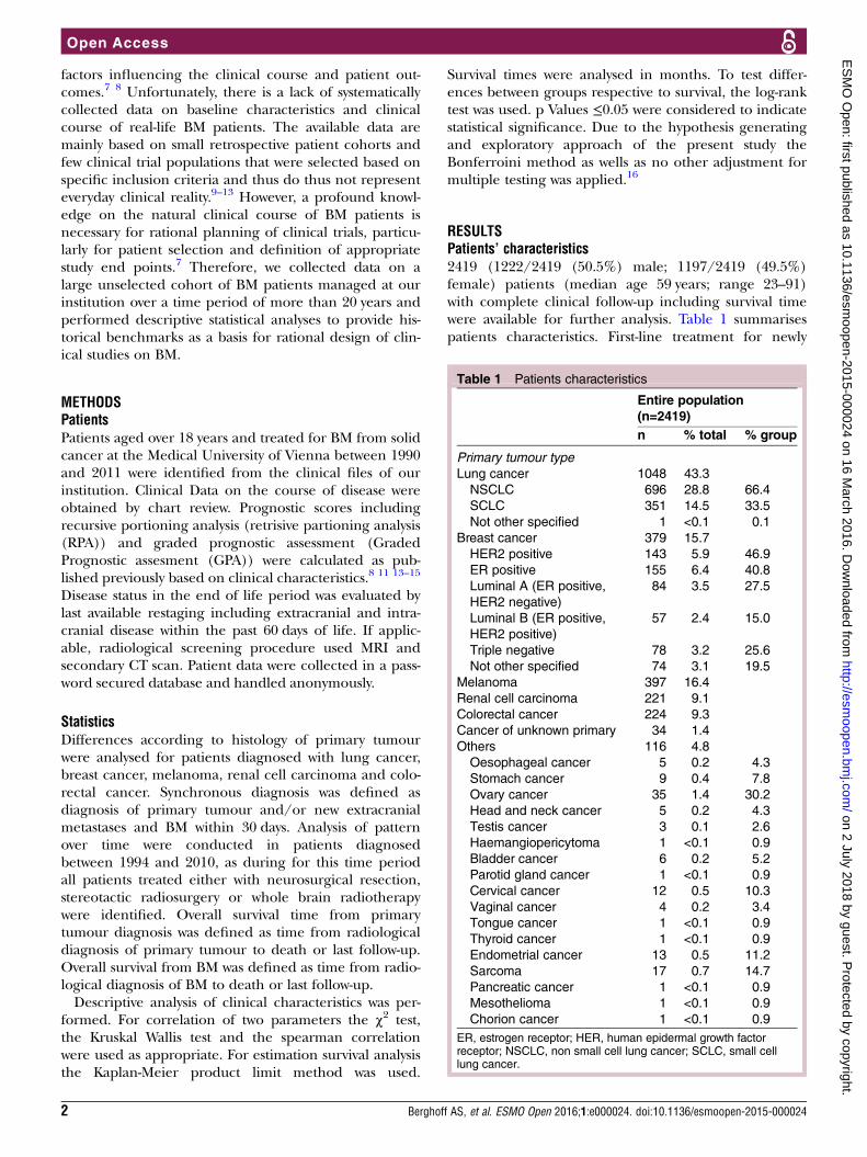
RESULTSPatients’ characteristics2419 (1222/2419 (50.5%) male; 1197/2419 (49.5%)female) patients (median age 59 years; range 23–91)with complete clinical follow-up including survival timewere available for further analysis. Table 1 summarisespatients characteristics. First-line treatment for newly
Table 1 Patients characteristics
Entire population(n=2419)n % total % group
Primary tumour typeLung cancer 1048 43.3
NSCLC 696 28.8 66.4
SCLC 351 14.5 33.5
Not other specified 1 <0.1 0.1
Breast cancer 379 15.7
HER2 positive 143 5.9 46.9
ER positive 155 6.4 40.8
Luminal A (ER positive,
HER2 negative)
84 3.5 27.5
Luminal B (ER positive,
HER2 positive)
57 2.4 15.0
Triple negative 78 3.2 25.6
Not other specified 74 3.1 19.5
Melanoma 397 16.4
Renal cell carcinoma 221 9.1
Colorectal cancer 224 9.3
Cancer of unknown primary 34 1.4
Others 116 4.8
Oesophageal cancer 5 0.2 4.3
Stomach cancer 9 0.4 7.8
Ovary cancer 35 1.4 30.2
Head and neck cancer 5 0.2 4.3
Testis cancer 3 0.1 2.6
Haemangiopericytoma 1 <0.1 0.9
Bladder cancer 6 0.2 5.2
Parotid gland cancer 1 <0.1 0.9
Cervical cancer 12 0.5 10.3
Vaginal cancer 4 0.2 3.4
Tongue cancer 1 <0.1 0.9
Thyroid cancer 1 <0.1 0.9
Endometrial cancer 13 0.5 11.2
Sarcoma 17 0.7 14.7
Pancreatic cancer 1 <0.1 0.9
Mesothelioma 1 <0.1 0.9
Chorion cancer 1 <0.1 0.9
ER, estrogen receptor; HER, human epidermal growth factorreceptor; NSCLC, non small cell lung cancer; SCLC, small celllung cancer.
2 Berghoff AS, et al. ESMO Open 2016;1:e000024. doi:10.1136/esmoopen-2015-000024
Open Access
on 2 July 2018 by guest. Protected by copyright.
http://esmoopen.bm
j.com/
ES
MO
Open: first published as 10.1136/esm
oopen-2015-000024 on 16 March 2016. D
ownloaded from

diagnosed BM was neurosurgical resection in 852/2418(35.2%), SRS in 1017/2418 (42.0%), whole brain radio-therapy in 495/2418 (20.5%), chemotherapy in 20/2418(0.8%) and best supportive care in 35/2418 (1.4%)patients (see online supplementary table S1).
Time from diagnosis of primary tumour to diagnosis of BM649/2419 (26.8%) patients presented with synchronousdiagnosis of BM and primary tumour. Patients with lungcancer (501/1048 (47.8%)) presented most frequently,while patients with breast cancer (13/366 (3.4%) pre-sented least frequently with synchronous diagnosis ofBM and primary tumour (p<0.001; χ2 test; see onlinesupplementary table S2; figure 1A). Importantly, BMwere detected through routinely performed radiologicalstaging procedures in 130/649 (20.0%) patients withsynchronous diagnosis of primary tumour and BM, indi-cating that these patients presented with asymptomaticBM at diagnosis of the primary tumour (figure 1B).Median time to diagnosis of BM in patients with subse-quent diagnosis of BM after diagnosis of primarytumour was 24 months (range 1–502). Patients withbreast cancer (median 44 months; 1–443) presentedwith the longest time to diagnosis of BM, while patientswith lung cancer (median 11 months; range 1–162) pre-sented with the shortest time between diagnosis ofprimary tumour and development of BM (p<0.001;logrank test; figure 1C).
Clinical characteristics at diagnosis of BMExtracranial involvement1564/2419 (64.7%) patients presented with extracranialmetastases at diagnosis of BM. BM as the only site ofmetastatic disease was most frequently observed amongpatients with lung cancer (535/1048 (51.0%)) and leastfrequently among patients with melanoma (72/397(18.1%); p<0.001; χ2 test; see online supplementarytable S2; figure 2A).1231/2419 (50.9%) patients presented with visceral
metastases, while 60/2419 (2.5%) presented with onlywith osseous metastases and 188/2419 (7.8%) onlylymph node metastases. Lung metastases were present in1545/2419 (36.1%) and liver metastases in 490/2419(20.3%) patients.727/2419 (30.1%) patients presented with progressive
extracranial disease at diagnosis of BM. Further,671/2419 (27.7%) patients presented with synchronousnew metastatic sites simultaneously with the diagnosis ofBM. Patients with melanoma presented most frequently204/397 (51.4%)), while patients with lung cancer(180/1048 (17.2%) presented least frequently with pro-gressive extracranial disease at diagnosis of BM (p<0.001;χ2 test; see online supplementary table S2; figure 2B).421/2419 (17.4%) patients had no evidence of extra-
cranial disease at diagnosis of BM. 136/421 (32.3%) ofthese patients develop systemic progression after diagno-sis of BM. 627/2419 (25.9%) patients presented with
brain only metastatic behaviour without any extracranialmetastases during their course of disease. The brainonly metastatic behaviour was most frequently observedamong patients with lung cancer (349/1048 (33.3%)and least frequently among patients with melanoma(37/397 (9.3%); p<0.001; χ2 test; see onlinesupplementary table S2; figure 2C).
Figure 1 (A) Frequency of synchronous diagnosis of
primary tumour and BM according to primary tumour type
(B) Frequency of patients with asymptomatic BM at first
diagnosis of primary tumour (C) Time from diagnosis to BM
according to primary tumour type.
Berghoff AS, et al. ESMO Open 2016;1:e000024. doi:10.1136/esmoopen-2015-000024 3
Open Access
on 2 July 2018 by guest. Protected by copyright.
http://esmoopen.bm
j.com/
ES
MO
Open: first published as 10.1136/esm
oopen-2015-000024 on 16 March 2016. D
ownloaded from

Figure 2 Clinical characteristics at diagnosis of brain metastases. (A) Extracranial involvement at diagnosis of BM according
to primary tumour type (B) Frequency of progressive extracranial disease at diagnosis of BM according to primary tumour type
(C) Frequency of brain only metastatic behaviour according to primary tumour type (D) Number of BM at first diagnosis of BM
(E) Number of BM according to primary tumour type (F) Localisation of BM at diagnosis (G) Frequency of epileptic seizures at
diagnosis of BM according to primary tumour type (H) Occurrence of additional leptomeningeal carcinomatosis after diagnosis
of BM according to primary tumour type.
4 Berghoff AS, et al. ESMO Open 2016;1:e000024. doi:10.1136/esmoopen-2015-000024
Open Access
on 2 July 2018 by guest. Protected by copyright.
http://esmoopen.bm
j.com/
ES
MO
Open: first published as 10.1136/esm
oopen-2015-000024 on 16 March 2016. D
ownloaded from
Number of BM1177/2419 (48.7%) patients presented with a singularBM, 670/2419 (27.7%) with 2–3 and 569/2419 (23.5%)with >3 BM (see online supplementary table S2 andfigure 2C). Patients with colorectal cancer presentedmost frequently (145/224 (64.7%)), while patients withmelanoma presented least frequently with singular BM(136/397 (34.3%)). Multiple BM were most frequentlyobserved among patients with breast cancer (115/376(30.6%)) and least frequently among patients with colo-rectal cancer (19/224 (8.5%); p<0.001; χ2 test; seeonline supplementary table S2; figure 2E).
Symptoms445/2419 (18.6%) patients presented with asymptomaticBM, that were detected trough staging. 340/2419(14.1%) patients presented with epileptic seizures, 768/2419 (31.7%) with signs of increased intracranial pres-sure and 1698/2419 (70.2%) with neurologicalsymptoms.
Leptomeningeal carcinomatosis54/2419 (2.2%) patients presented with leptomeningealinvolvement at diagnosis of BM and 120/2419 (5.0%)
patients developed leptomeningeal involvement withinthe course of disease. Patient with breast cancer pre-sented most frequently with additional leptomeningealcarcinomatosis (41/379 (10.8%) and patients with renalcell carcinoma least frequently (4/221 (1.8%; p<0.001;χ2 test; see online supplementary table S2; figure 2H).
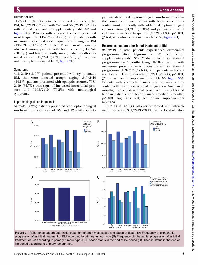
Recurrence pattern after initial treatment of BM980/2419 (40.5%) patients experienced extracranialprogression after diagnosis of BM (see onlinesupplementary table S3). Median time to extracranialprogression was 3 months (range 0–207). Patients withmelanoma presented most frequently with extracranialprogression (189/397 (47.6%)) and patients with colo-rectal cancer least frequently (66/224 (29.5%); p<0.001;χ2 test; see online supplementary table S3; figure 3A).Patients with colorectal cancer and melanoma pre-sented with fastest extracranial progression (median 2months), while extracranial progression was observedlater in patients with breast cancer (median 5 months;p<0.001; log rank test; see online supplementarytable S3).1057/2419 (43.7%) patients presented with intracra-
nial progression, 381/2419 (20.4%) at the local site after
Figure 3 Recurrence pattern after initial treatment of brain metastases and cause of death. (A) Frequency of extracranial
progression after initial treatment of BM according to primary tumour type (B) Frequency of intracranial progression after initial
treatment of BM according to primary tumour type (C) Disease status in the end of life period (D) Disease status in the end of
life period according to primary tumour type.
Berghoff AS, et al. ESMO Open 2016;1:e000024. doi:10.1136/esmoopen-2015-000024 5
Open Access
on 2 July 2018 by guest. Protected by copyright.
http://esmoopen.bm
j.com/
ES
MO
Open: first published as 10.1136/esm
oopen-2015-000024 on 16 March 2016. D
ownloaded from

surgery or stereotactic radiosurgery (see onlinesupplementary table S3). Patients with melanoma pre-sented with the highest risk to experience intracranialprogression (207/397 (52.1%), while patients with colo-rectal cancer presented less frequently with intracranialprogression (85/224 (37.9%); p=0.002; χ2 test; seeonline supplementary table S3; figure 3B). No differ-ence in likelihood of intracranial progression wasobserved in patients with lung cancer according to histo-logical subtype. 311/696 (44.7%) non small cell lungcancer BM patients experienced intracranial progressioncompared to 155/351 (44.2%) small cell lung cancerBM patients (p=0.872; χ2 test). Median time to intracra-nial progression was 5 months (range 0–100). Patientswith melanoma presented with shortest time to intracra-nial progression (median 3 months; p<0.001; log ranktest; see online supplementary table S3).
Disease status in the end of life periodEvaluation of the disease status in the past 2 monthsprior to patient death showed intracranial progressionin 35.9%, extracranial progression in 27.5% andcombined extracranial and intracranial progression in36.6% of patients (see online supplementary table S3;figure 3C). In the end of life period, combined extra-cranial and intracranial progression was most frequentin patients with lung cancer (226/599 (37.7%),intracranial progression was most common in patientswith breast cancer (87/184 (47.3%)), combinedextracranial and intracranial progression was mostcommon in patients with melanoma (103/272(37.9%)), extracranial and intracranial progression wasmost frequent in patients with renal cell carcinoma(41/96 (42.7%)) and intracranial progression wasmost frequent in patients with colorectal cancer(53/119 (44.5%); see online supplementary table S3;figure 3D).Intracranial progression in the end of life period was
most frequently observed among patients with breastcancer (87/184 (47.3%)) and least frequently amongpatients with lung cancer (180/599 (30.1%); p<0.001; χ2
test; see online supplementary table S3; figure 3D).Extracranial progression in the end of life period wasmost frequently evident in patients with lung cancer(193/599 (32.2%)) and least frequently in patients withrenal cell carcinoma (18/96 (18.8%); p<0.001; χ2 test;see online supplementary table S3; figure 3D). Onlinesupplementary table S4 lists disease status in the end oflife period in specific BM subgroups.
SURVIVAL ANALYSISPrognostic scoresRPAPatients in RPA class I presented with a median overallsurvival of 12 months, compared to 7 months in RPAclass II and 2 months in RPA class III (p<0.001; log ranktest; see online supplementary figure S2B).
GPAPatients in GPA class I presented with a median overallsurvival of 15 months, compared to 11 months in classII, 7 months in class III and 3 months in class IV(p<0.001; log rank test; see online supplementaryfigure S2C).
Treatment modalitySurvival time from diagnosis of BM differed significantlyaccording to applied first-line treatment approach.Median overall survival was longest in patients treatedwith chemotherapy (median 10 months), followed byneurosurgical resection (median OS 8 months), SRS(median 6 months), whole brain radiotherapy (median5 months) and best supportive care (median 0 months;p<0.001; log rank test). Patients treated with chemother-apy after diagnosis of BM presented with an improvedoverall survival of median 11 months, compared topatients not treated with chemotherapy after diagnosisof BM (median 4 months; p<0.001; log rank test).
Frequency patterns of BM over timeThe frequency of patients treated for BM at our institu-tion increased numerically in the past two decades(Spearman correlation coefficient 0.598; p=0.011;figure 4A). Further, a trend towards an increasedfrequency in lung cancer BM could be observed(Spearman correlation coefficient 0.621; p<0.001;figure 4B). Increase in lung cancer BM differed accord-ing to gender, as female patients presented with strongerincrease (Spearman correlation coefficient 0.798;p<0.001) as compared to male patients (Spearman cor-relation coefficient 0.558; p=0.020; figure 4C). Noincreased frequency was observed for breast cancer BM(Spearman correlation coefficient 0.479; p=0.052), mel-anoma BM (Spearman correlation coefficient 0.371;p=0.142), renal cell carcinoma BM (Spearman correl-ation coefficient 0.730; p=0.780) or colorectal cancerBM (Spearman correlation coefficient −0.145; p=0.578;figure 4B).The survival prognosis increased over the past two
decades as patients diagnosed before 2000 presentedwith a median overall survival of 5 months compared to7 months in patients diagnosed and treated after 2000(p=0.005; log rank test; figure 4D).
DISCUSSIONBM patients are a heterogeneous patient population.Prognosis as well as clinical course varies significantlyaccording to clinical factors like histology of the primarytumour, number of BM and status of the extracranialdisease.8 15 14 Definition of appropriate patient cohortsfor clinical trials is challenging, as investigated cohortsshould resemble prognostically comparable patientpopulations.7 In the current study, we investigated aunique, large real-life cohort of BM patients and provide
6 Berghoff AS, et al. ESMO Open 2016;1:e000024. doi:10.1136/esmoopen-2015-000024
Open Access
on 2 July 2018 by guest. Protected by copyright.
http://esmoopen.bm
j.com/
ES
MO
Open: first published as 10.1136/esm
oopen-2015-000024 on 16 March 2016. D
ownloaded from

information on baseline characteristics and the clinicalcourse of BM patients.Expectedly and in line with prior epidemiological
studies, the majority of BM cases in our cohort were fromlung cancer, breast cancer, melanoma, renal cell cancerand colorectal cancers.9 However, our cohort alsoincluded some cases of tumour types that rarely lead toBM such gastro-oesophageal cancers, genitourinarycancers, head and neck cancers and sarcomas. Our dataclearly show considerable differences in BM frequency,clinical presentation and clinical course between patientswith different tumour types. For example, we detectedsignificant differences in symptoms at BM diagnosis, timefrom primary tumour to BM diagnosis, rate of synchron-ous BM and primary tumour diagnoses, number of BM atpresentation, and median overall survival times betweentumour types. The reasons for these differences need tobe elucidated in further studies, but are likely related tomolecular factors and the interaction of metastatictumour cells with the specific microenvironment of thecentral nervous system.17–19 In any case, these findingshighlight the profound heterogeneity of BM patients andthe need for informed patient selection and stratification
for conduction of meaningful clinical trials in this patientpopulation. Importantly, pooling of BM patients of differ-ent primary tumour types in clinical studies seems unad-visable and focused enrolment of BM patients withspecific tumour types is likely to produce more robustclinical trial data.7
Several prognostic scores based on clinical parametershave been developed for BM patients. The RPA andGPA scores, which take into account age, status of theextracranial disease, number of BM and the Karnofskyperformance score are most commonly used and weconfirm the strong statistical correlation of these scoreswith median overall survival times in our cohort.11 13
The use of these scores as inclusion criterion or as strati-fication factor in BM-specific trials can help to designclinical studies based on rational and robust statisticalassumptions. It must be noted, however, that predictionof individual patient survival time in the clinical settingbased on these clinical appears not to be accurately pos-sible.20 Refinement of prognostic scores by inclusion ofadditional parameters, for example, of laboratory, tissue-based, imaging-based factors, may help to increase theirprognostic accuracy.21–24
Figure 4 Frequency patterns of brain metastases over time. (A) Frequency of BM diagnosed between 1994 and 2010 (B)
Frequency of BM according to primary tumour between 1994 and 2010 (C) Frequency of lung cancer BM in female and male
patients between 1994 and 2010 (D) Overall survival from diagnosis of BM according to year of diagnosis.
Berghoff AS, et al. ESMO Open 2016;1:e000024. doi:10.1136/esmoopen-2015-000024 7
Open Access
on 2 July 2018 by guest. Protected by copyright.
http://esmoopen.bm
j.com/
ES
MO
Open: first published as 10.1136/esm
oopen-2015-000024 on 16 March 2016. D
ownloaded from

Response assessment is particularly challenging in BMpatients, as intracranial and extracranial disease statushas to be considered. Recently, the Response Assessmentin Neuro-Oncology (RANO) group has proposed analgorithm that integrates response assessment in theintracranial and extracranial compartment for use andstandardised reporting in clinical trials.25 26 Our datasupport this concept, as 43.7% of patients showed intra-cranial and 40.5% of cases showed extracranial diseaseprogression during the clinical course after initialBM-directed treatment. Moreover, radiological evalu-ation of the disease status in the end-of life period (ie,the past 2 months prior to patient death) showed intra-cranial progression in 35.9%, extracranial progression in27.5% and combined extracranial and intracranial pro-gression in 36.6% of patients in our series. Our datathus are in line with some smaller and older studies thatindicated that at least half of the patients die with pro-gression of their extracranial disease.27–29 Cumulatively,the available data show that in a considerable fraction ofpatients systemic disease contributes significantly to thepoor outcome of patients with BM and therefore thereis a strong need and rationale to perform studies withnovel systemic agents in BM patients.7 Unfortunately, sofar BM patients have been systematically excluded frommost clinical trials evaluating systemic drugs in patientswith cancer. Recently however, some studies have shownclinically meaningful activity of systemic agents such asimmunotherapies and tyrosine-kinase inhibitors in BMpatient populations of melanoma, breast cancer andlung cancer, thus providing proof of concept for the effi-cacy of such approaches.30–33
Analysing changes over time in our cohort of BMpatients treated at our institution in a period spanningmore than 20 years, we noted some interesting findings.First, we found a modest increase of BM treated for BMat our institution over time. This finding may beexplained by improvements and increased accessibilityof diagnostic methods such as cranial CT and MRI, butcould also be explained by a higher number of long-term surviving patients with cancer with novel thera-peutic regimens. Also, changes in referral patterns withincreased patient concentration due to the specificexpertise in our tertiary care centre may have played arole. Second, a disproportionate and gender-specificincrease in lung cancer BM cases was evident withwomen showing a significantly steeper increase thanmales. This finding is probably related to the increasingpercentage of smokers among women and reflects thegeneral increase of lung cancer cases in females.34
Third, we found a statistically significant, albeit verymodest, increase in median overall survival times of BMpatients over time. Again, this finding may relate torecent advances in oncological care. However, overallthe poor prognosis of most BM patients documentedalso in our study shows that this patient populationrepresents a significant unmet clinical need that shouldbe met with adequate research efforts. However, the
poor prognosis of most BM patients as documented alsoin our study shows that this patient population has a sig-nificant unmet clinical need that should be met withadequate research efforts.In summary, we present here a detailed clinical char-
acterisation of the largest unselected real-life cohort ofBM patients published to date. Our data highlight theheterogeneity in presentation and clinical course of BMpatients in the everyday clinical setting and may beuseful for rational planning of clinical studies.
Author affiliations1Department of Medicine I, Medical University of Vienna, Vienna, Austria2Comprehensive Cancer Center, Medical University of Vienna, Vienna, Austria3Department of Neurosurgery, Medical University of Vienna, Vienna, Austria4Department of Radiotherapy, Medical University of Vienna, Vienna, Austria5Institute of Neurology, Medical University of Vienna, Vienna, Austria6Department of Pathology, Medical University of Vienna, Vienna, Austria
Acknowledgements The authors thank Patricia Heilcappel, FranziskaSchlieter, Romina Koller, Marion Anderl, Hanna Plazer, Pedro Ferreira,Benjamin Prascher, Robert Reumann, Mela Medjedovic and AleksandraAngelovski, Thomas Melchardt and Josef Pichler for assistance with dataacquisition. The authors thank Harald Heinzl (Center for Medical Statistics,Medical University of Vienna) for statistical advice and supervision.
Funding The costs for this project were covered by the research budget ofthe Medical University of Vienna.
Competing interests None declared.
Provenance and peer review Commissioned; externally peer reviewed.
Data sharing statement No additional data are available.
Open Access This is an Open Access article distributed in accordance withthe Creative Commons Attribution Non Commercial (CC BY-NC 4.0) license,which permits others to distribute, remix, adapt, build upon this work non-commercially, and license their derivative works on different terms, providedthe original work is properly cited and the use is non-commercial. See: http://creativecommons.org/licenses/by-nc/4.0/
REFERENCES1. Caissie A, Nguyen J, Chen E, et al. Quality of life in patients with
brain metastases using the EORTC QLQ-BN20+2 andQLQ-C15-PAL. Int J Radiat Oncol Biol Phys 2012;83:1238–45.
2. Gavrilovic IT, Posner JB. Brain metastases: epidemiology andpathophysiology. J Neurooncol 2005;75:5–14.
3. Barnholtz-Sloan JS, Sloan AE, Davis FG, et al. Incidenceproportions of brain metastases in patients diagnosed (1973 to2001) in the Metropolitan Detroit Cancer Surveillance System. J ClinOncol 2004;22:2865–72.
4. Bartsch R, Berghoff AS, Preusser M. Optimal management of brainmetastases from breast cancer. Issues and considerations.CNS Drugs 2013;27:121–34.
5. Eichler AF, Chung E, Kodack DP, et al. The biology of brainmetastases-translation to new therapies. Nat Rev Clin Oncol2011;8:344–56.
6. Jenkinson MD, Haylock B, Shenoy A, et al. Management of cerebralmetastasis: evidence-based approach for surgery, stereotacticradiosurgery and radiotherapy. Eur J Cancer 2011;47:649–55.
7. Preusser M, Winkler F, Collette L, et al. Trial design on prophylaxis andtreatment of brain metastases: lessons learned from the EORTC BrainMetastases Strategic Meeting 2012. Eur J Cancer 2012;48:3439–47.
8. Sperduto PW, Kased N, Roberge D, et al. Summary report on thegraded prognostic assessment: an accurate and facilediagnosis-specific tool to estimate survival for patients with brainmetastases. J Clin Oncol 2012;30:419–25.
9. Johnson JD, Young B. Demographics of brain metastasis.Neurosurg Clin N Am 1996;7:337–44.
10. Andrews DW, Scott CB, Sperduto PW, et al. Whole brain radiationtherapy with or without stereotactic radiosurgery boost for patientswith one to three brain metastases: phase III results of the RTOG9508 randomised trial. Lancet 2004;363:1665–72.
8 Berghoff AS, et al. ESMO Open 2016;1:e000024. doi:10.1136/esmoopen-2015-000024
Open Access
on 2 July 2018 by guest. Protected by copyright.
http://esmoopen.bm
j.com/
ES
MO
Open: first published as 10.1136/esm
oopen-2015-000024 on 16 March 2016. D
ownloaded from
11. Gaspar L, Scott C, Rotman M, et al. Recursive partitioning analysis(RPA) of prognostic factors in three Radiation Therapy OncologyGroup (RTOG) brain metastases trials. Int J Radiat Oncol Biol Phys1997;37:745–51.
12. Gaspar LE, Scott C, Murray K, et al. Validation of the RTOGrecursive partitioning analysis (RPA) classification for brainmetastases. Int J Radiat Oncol Biol Phys 2000;47:1001–6.
13. Sperduto CM, Watanabe Y, Mullan J, et al. A validation study of anew prognostic index for patients with brain metastases: the GradedPrognostic Assessment. J Neurosurg 2008;109:87–9.
14. Sperduto PW, Berkey B, Gaspar LE, et al. A new prognostic indexand comparison to three other indices for patients with brainmetastases: an analysis of 1,960 patients in the RTOG database.Int J Radiat Oncol Biol Phys 2008;70:510–14.
15. Sperduto PW, Chao ST, Sneed PK, et al. Diagnosis-specificprognostic factors, indexes, and treatment outcomes for patients withnewly diagnosed brain metastases: a multi-institutional analysis of4,259 patients. Int J Radiat Oncol Biol Phys 2010;77:655–61.
16. Bender R, Lange S. Adjusting for multiple testing—when and how?J Clin Epidemiol 2001;54:343–9.
17. Bos PD, Zhang XH, Nadal C, et al. Genes that mediate breastcancer metastasis to the brain. Nature 2009;459:1005–9.
18. Fidler IJ. The role of the organ microenvironment in brainmetastasis. Semin Cancer Biol 2011;21:107–12.
19. Langley RR, Fidler IJ. The seed and soil hypothesis revisited—therole of tumor-stroma interactions in metastasis to different organs.Int J Cancer 2011;128:2527–35.
20. Kondziolka D, Parry PV, Lunsford LD, et al. The accuracy ofpredicting survival in individual patients with cancer. J Neurosurg2014;120:24–30.
21. Berghoff AS, Ilhan-Mutlu A, Dinhof C, et al. Differential role ofangiogenesis and tumour cell proliferation in brain metastasesaccording to primary tumour type: analysis of 639 cases.Neuropathol Appl Neurobiol 2015;41:e41–55.
22. Berghoff AS, Spanberger T, Ilhan-Mutlu A, et al. Preoperativediffusion-weighted imaging of single brain metastases correlates withpatient survival times. PLoS ONE 2013;8:e55464.
23. Spanberger T, Berghoff AS, Dinhof C, et al. Extent of peritumoralbrain edema correlates with prognosis, tumoral growth pattern,HIF1a expression and angiogenic activity in patients with singlebrain metastases. Clin Exp Metastasis 2013;30:357–68.
24. Berghoff A, Koller R, Widhalm G, et al. Laboratory parameters haveindependent prognostic impact in patients with newly diagnosedbrain metastases: analysis of 1201 cases. ECC. 2015:Abstractnumber: 2928.
25. Lin NU, Lee EQ, Aoyama H, et al. Challenges relating to solidtumour brain metastases in clinical trials, part 1: patient population,response, and progression. A report from the RANO group. LancetOncol 2013;14:e396–406.
26. Lin NU, Lee EQ, Aoyama H, et al. Response assessment criteria forbrain metastases: proposal from the RANO group. Lancet Oncol2015;16:e270–8.
27. Patchell RA, Tibbs PA, Regine WF, et al. Postoperative radiotherapyin the treatment of single metastases to the brain: a randomized trial.JAMA 1998;280:1485–9.
28. Patchell RA, Tibbs PA, Walsh JW, et al. A randomized trial ofsurgery in the treatment of single metastases to the brain. N Engl JMed 1990;322:494–500.
29. Lucas JT Jr, Colmer HGt, White L, et al. Competing Risk Analysis ofNeurologic versus Nonneurologic Death in Patients UndergoingRadiosurgical Salvage After Whole-Brain Radiation Therapy Failure:Who Actually Dies of Their Brain Metastases? Int J Radiat OncolBiol Phys 2015;92:1008–15.
30. Margolin K, Ernstoff MS, Hamid O, et al. Ipilimumab in patients withmelanoma and brain metastases: an open-label, phase 2 trial.Lancet Oncol 2012;13:459–65.
31. Long GV, Trefzer U, Davies MA, et al. Dabrafenib in patients withVal600Glu or Val600Lys BRAF-mutant melanoma metastatic to thebrain (BREAK-MB): a multicentre, open-label, phase 2 trial. LancetOncol 2012;13:1087–95.
32. Bachelot T, Romieu G, Campone M, et al. Lapatinib pluscapecitabine in patients with previously untreated brainmetastases from HER2-positive metastatic breast cancer(LANDSCAPE): a single-group phase 2 study. Lancet Oncol2013;14:64–71.
33. Fekrazad MH, Ravindranathan M, Jones DV Jr. Response ofintracranial metastases to erlotinib therapy. J Clin Oncol2007;25:5024–6.
34. Wang A, Kubo J, Luo J, et al. Active and passive smoking in relationto lung cancer incidence in the Women’s Health InitiativeObservational Study prospective cohort. Ann Oncol2015;26:221–30.
Berghoff AS, et al. ESMO Open 2016;1:e000024. doi:10.1136/esmoopen-2015-000024 9
Open Access
on 2 July 2018 by guest. Protected by copyright.
http://esmoopen.bm
j.com/
ES
MO
Open: first published as 10.1136/esm
oopen-2015-000024 on 16 March 2016. D
ownloaded from
Related Documents