© AGO e.V. in der DGGG e.V. sowie in der DKG e.V. Guidelines Breast Version 2016.1 Diagnosis and Treatment of Patients with Primary and Metastatic Breast Cancer Lesions of Uncertain Malignant Potential (B3) (ADH, LIN, FEA, Papilloma, Radial Scar)

Welcome message from author
This document is posted to help you gain knowledge. Please leave a comment to let me know what you think about it! Share it to your friends and learn new things together.
Transcript

© AGO e.V. in der DGGG e.V.
sowie
in der DKG e.V.
Guidelines Breast
Version 2016.1
Diagnosis and Treatment of Patients with
Primary and Metastatic Breast Cancer
Lesions of Uncertain Malignant
Potential (B3)
(ADH, LIN, FEA, Papilloma, Radial Scar)

© AGO e.V. in der DGGG e.V.
sowie
in der DKG e.V.
Guidelines Breast
Version 2016.1
www.ago-online.de
Lesions of Uncertain Malignant Potential (B3)
(including “Precursor Lesions”)
Versions 2005–2015:
Albert / Audretsch / Brunnert / Fersis /
Friedrich / Gerber / Kreipe / Nitz / Rody /
Schreer / Sinn / Thomssen
Version 2016:
Friederichs / Sinn

© AGO e.V. in der DGGG e.V.
sowie
in der DKG e.V.
Guidelines Breast
Version 2016.1
www.ago-online.de
Pathology Reporting for Minimal
Invasive Biopsies
B-Classification*
B1 = unsatisfactory / normal tissue only
B2 = benign lesion
B3 = lesion of uncertain malignant potential
B4 = suspicion of malignancy
B5 = malignant B5a = non-invasive
B5b = invasive
B5c = in-situ/invasion not assessable
B5d = non epithelial, metastatic
* National Coordinating Group for Breast Screening Pathology (NHSBSP), E.C.
Working Group on Breast Screening Pathology, S3-Leitlinien

© AGO e.V. in der DGGG e.V.
sowie
in der DKG e.V.
Guidelines Breast
Version 2016.1
www.ago-online.de
B3-Lesions
Lesions with risk of associated DCIS or invasive Ca:
• Atypical ductal hyperplasia (ADH)
• Lobular neoplasia (ALH, LCIS)
• Flat epithelial atypia (FEA)
Inhomogenous lesions with sampling risk:
• Phyllodes tumor, cellular fibroadenoma
• Atypical papilloma, if incompletely removed
• Radial scar, complex sclerosing lesion

© AGO e.V. in der DGGG e.V.
sowie
in der DKG e.V.
Guidelines Breast
Version 2016.1
www.ago-online.de
Major B3-Lesions and Prospektive Prediktive
Value (PPV) for Malignancy in Resection
B3-Lesions: ~PPV
Atypical ductal hyperplasia (ADH) 20-30%
Lobular intraepithelial neoplasia (LN/LIN) 0-10%
Flat epithelial atypia (FEA) 0-10%
Radial scar / Complex sclerosing lesion 0-10%
Papilloma without atypia 0-10%
Cellular fibroepithelial tumors / phyllodes tumors 0%

© AGO e.V. in der DGGG e.V.
sowie
in der DKG e.V.
Guidelines Breast
Version 2016.1
www.ago-online.de
Management after Minimally
Invasive Biopsy
Interdisciplinary conference: Concordant findings in pathology and imaging?
yes: proceed according to histologic type 3a C ++
no: open biopsy 3a C ++
Oxford / AGO
LoE / GR

© AGO e.V. in der DGGG e.V.
sowie
in der DKG e.V.
Guidelines Breast
Version 2016.1
www.ago-online.de
Synonyms: Atypical intraductal epithelial proliferation (AIDEP),
atypical epithelial proliferation of ductal type
Definition: Atypical intraductal proliferations with cytologic and
structural features of well differentiated DCIS, such as rigid
bridging or micropapillae, well demarcated cell borders and
occupy less than two separate duct spaces. The extension of all
involved lumina within one ductulo-lobular unit is less than 2 mm.
Atypical ductal proliferations larger than 2 mm or in at least two
ductules are classified as DCIS (low-grade).
Indicator/Precursor lesion: Ipsi- and contralateral breast cancer
risk: RR 3 - 5 x after 3 - 5 years.
Classification in ductal intraepithelial neoplasia grade 1 - 3 is not
sufficiently validated.
Atypical Ductal Hyperplasia (ADH)

© AGO e.V. in der DGGG e.V.
sowie
in der DKG e.V.
Guidelines Breast
Version 2016.1
www.ago-online.de
Strategy after Diagnosis of ADH
in Core Biopsy
ADH in core- / vacuum-assisted biopsy:
Open excisional biopsy 3a C ++
Open excisional biopsy may be omitted, with:
a) no mass lesion radiologically and
b) a small lesion (≤ 2 TDLU* in vacuum biopsy) and
c) complete removal of imaging abnormality 5a C +/-
ADH at margins in resection specimen: No further surgery, if incidental finding
accompanying invasive or intraductal carcinoma 3a C ++
Oxford / AGO
LoE / GR
* Terminal ductal-lobular unit

© AGO e.V. in der DGGG e.V.
sowie
in der DKG e.V.
Guidelines Breast
Version 2016.1
www.ago-online.de
Risk of Breast Cancer after Atypical
Hyperplasie (ADH, ALH)
Stratification of breast cancer risk*
Number of Foci: 1 RR = 2,33
2 RR = 5,26
> 3 RR = 7,97
Microcalcifications: present RR = 3,21
not present RR = 4,21
Type ductal RR = 3,83
lobular RR = 3,67
both RR = 7,10
Age < 45 RR = 6,76
45 – 55 RR = 5,10
> 55 RR = 2,67
*AC Degnim et al. J Clin Oncol 2007; 25: 2671-2677

© AGO e.V. in der DGGG e.V.
sowie
in der DKG e.V.
Guidelines Breast
Version 2016.1
www.ago-online.de
Includes: Atypical lobular hyperplasia, lobular
carcinoma in situ, LCIS/CLIS
LIN1 - 3 classification is not sufficiently validated
prognostically
Pleomorphic LIN and LIN with comedotype necrosis
are classified as → B5a
Indicator/Precursor lesion:
Ipsi- and contralateral enhanced breast cancer risk:
7 x at 10 years
Lobular Intraepithelial Neoplasia (LIN)
© AGO e.V. in der DGGG e.V.
sowie
in der DKG e.V.
Guidelines Breast
Version 2016.1
www.ago-online.de
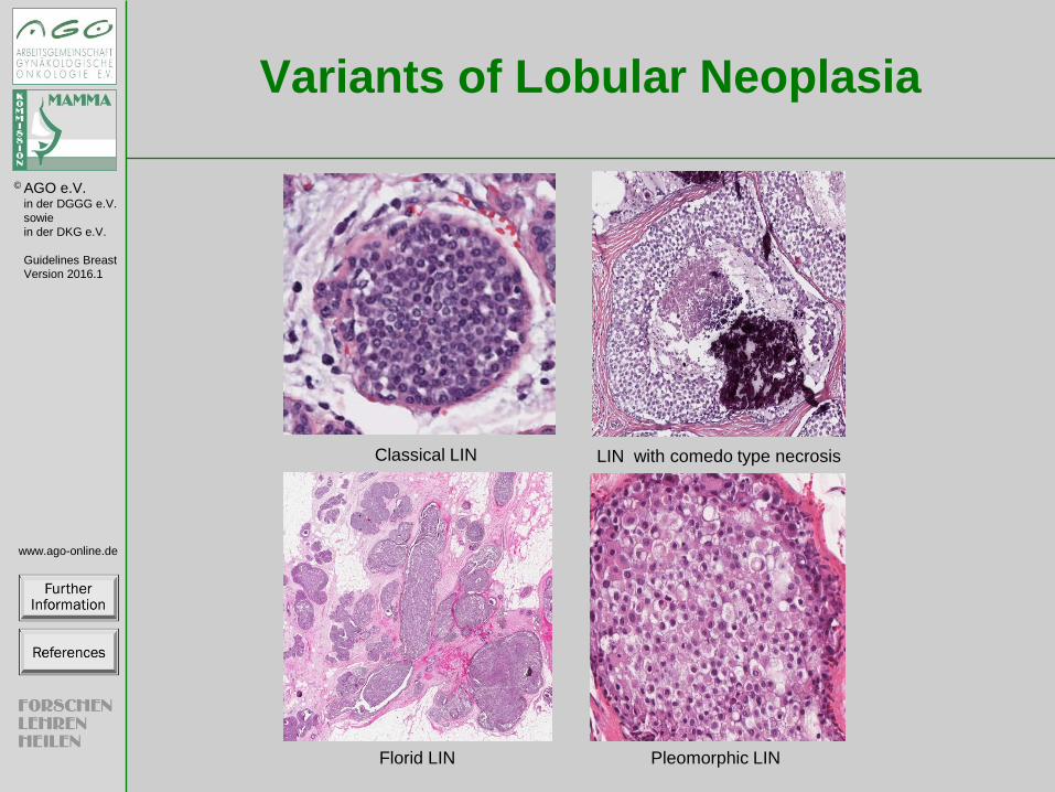
Variants of Lobular Neoplasia
Classical LIN LIN with comedo type necrosis
Florid LIN Pleomorphic LIN
© AGO e.V. in der DGGG e.V.
sowie
in der DKG e.V.
Guidelines Breast
Version 2016.1
www.ago-online.de
Pleomorphic LCIS: high grade cellular atypia,
frequent involvement of ductules, comedo-type
necroses, microcalcifications
Florid LCIS: Involvement of numerous lobuli with
distension and near confluence, extension to
ductules and neighbouring TDLU
Type of LCIS with 21 cases of LCIS with
microinvasion*:
- classical LCIS: n=11
- florid LCIS: n=4
- pleomorphic LCIS: n=1
LIN with High Risk
* Ross DS. Am J Surg Pathol 2011 35: 750–6

© AGO e.V. in der DGGG e.V.
sowie
in der DKG e.V.
Guidelines Breast
Version 2016.1
www.ago-online.de
Strategy after Diagnosis of LIN
LIN in core- / vacuum-assisted biopsy:
Open excisional biopsy, with pleomorphic LIN, florid LIN, or LIN with comedo type necrosis
or when not concordant with imaging findings 2b C ++
LIN at margins of resection specimen (BCT):
No further surgery 2a C ++
Exceptions:
a) Pleomorphic LIN, florid LIN, or LIN with necrosis b) Imaging abnormality is not removed
Complete resection 5 D ++
Oxford / AGO
LoE / GR

© AGO e.V. in der DGGG e.V.
sowie
in der DKG e.V.
Guidelines Breast
Version 2016.1
www.ago-online.de
Flat Epithelial Atypia (FEA)
Synonyms: Columnar cell hyperplasia with atypia,
columnar cell metaplasia with atypia, ductal
intraepithelial neoplasia grade 1A (DIN 1A)
Differential diagnosis:
ADH is discriminated by architectural features
(micropapillary, cribriform) → B3
Clinging carcinoma is discriminated by high grade
nuclear atypia (G2/G3) and classified as → B5a
Marker lesion:
FEA is frequently associated with calcifications and
may be associated with intraductal carcinoma.
Therefore, histologic step sectioning and correlation
with imaging are mandatory.

© AGO e.V. in der DGGG e.V.
sowie
in der DKG e.V.
Guidelines Breast
Version 2016.1
www.ago-online.de
Strategy after Diagnosis of FEA
FEA in core biopsy/vacuum-assisted biopsy:
Open excisional biopsy 3b C + Open excisional biopsy may be
omitted, with:
a small lesion (≤ 2 TDLU* in vacuum biopsy) and
complete removal of imaging abnormality 5 C +
FEA at margins in resection specimen: 3b C ++ No further surgery, unless calcifications
have not been completely removed
Oxford / AGO
LoE / GR
* Terminal ductal-lobular unit

© AGO e.V. in der DGGG e.V.
sowie
in der DKG e.V.
Guidelines Breast
Version 2016.1
www.ago-online.de
Papilloma
Includes: Central and peripheral papilloma > 2 mm,
atypical intraductal papilloma (B3)
To be discriminated from peripheral micropapilloma
arising in the TDLU, size ≤ 2 mm, may be multiple
To be discriminated from papilloma with DCIS, from
intraductal papillary carcinoma, and from encapsulated
papillary carcinoma
Indicator lesion:
May be associated with in-situ or invasive cancer (10%,
in case of atypical papilloma up to 20%), increased
ipsilateral risk for cancer (4.6% to 13% in case of
atypical papilloma)
© AGO e.V. in der DGGG e.V.
sowie
in der DKG e.V.
Guidelines Breast
Version 2016.1
www.ago-online.de
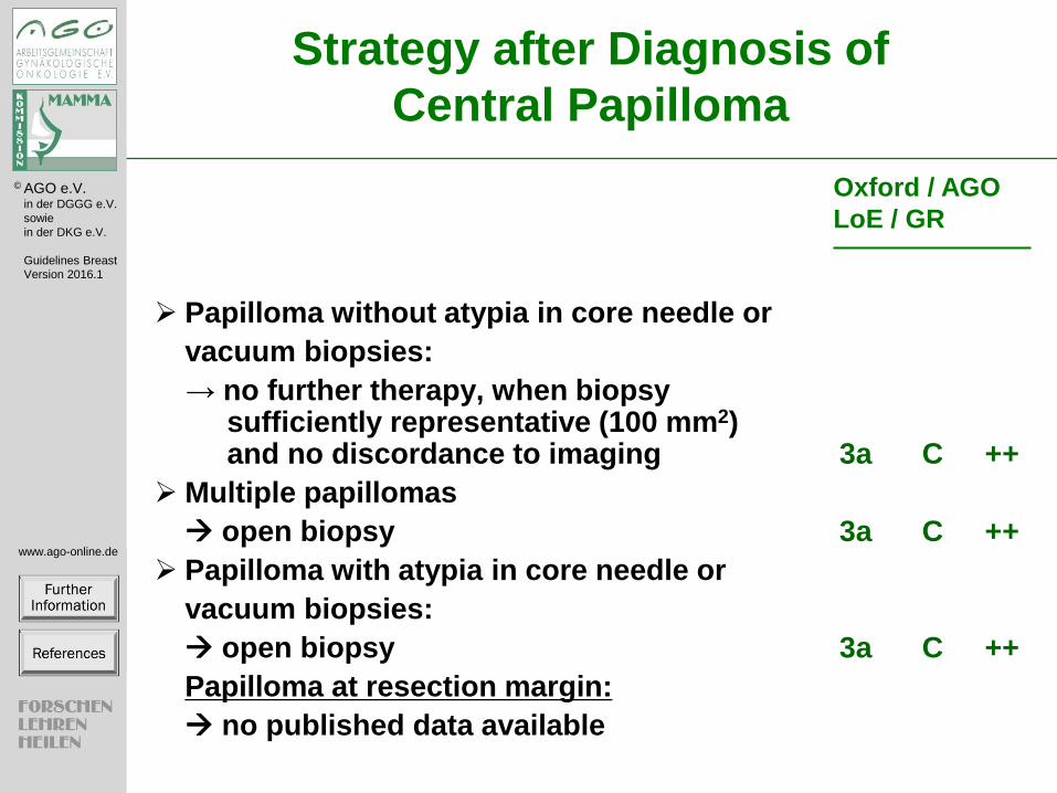
Strategy after Diagnosis of
Central Papilloma
Papilloma without atypia in core needle or
vacuum biopsies:
→ no further therapy, when biopsy sufficiently representative (100 mm2) and no discordance to imaging 3a C ++
Multiple papillomas
open biopsy 3a C ++
Papilloma with atypia in core needle or
vacuum biopsies:
open biopsy 3a C ++
Papilloma at resection margin:
no published data available
Oxford / AGO
LoE / GR

© AGO e.V. in der DGGG e.V.
sowie
in der DKG e.V.
Guidelines Breast
Version 2016.1
www.ago-online.de
Radially Sclerosing Lesion
Benign pseudoinfiltrative lesion with central fibroelastic
core and radical configuration.
Includes:
- radial scar
- complex sclerosing lesion (> 1 cm)
Additional risk factor in patients with benign epithelial
hyperplasia (proliferating breast disease)
Risk for upgrade in open biopsy after diagnosis of
radial-sclerosing lesion in core biopsy: 8.3% (79/948)*
* Bianchi S et al. Breast. (2012) 21: 159–64.

© AGO e.V. in der DGGG e.V.
sowie
in der DKG e.V.
Guidelines Breast
Version 2016.1
www.ago-online.de
Strategy after Diagnosis of Radial Scar,
Complex Sclerosing Lesion (CSL)
Radial scar / CSL in core biopsy/ vacuum-assisted biopsy:
Open excisional biopsy 3b C + Open excisional biopsy may be omitted, with a small lesion and complete
removal of imaging abnormality 5a C +
Radial scar / CSL at margins in resection specimen:
No further surgery 3b C ++
Oxford / AGO
LoE / GR

© AGO e.V. in der DGGG e.V.
sowie
in der DKG e.V.
Guidelines Breast
Version 2016.1
www.ago-online.de
Follow-up Imaging for Women Age
50-69 Years with B3-Lesions
Oxford / AGO
LoE / GR
FEA, non-atypical papilloma
Screening mammography 5 C ++
LIN
Mammography (12 months) 3a C ++
ADH
Mammography (12 months) 3a C ++
Women with LIN and ADH should
be informed about their elevated
risk of breast cancer 3a C ++

© AGO e.V. in der DGGG e.V.
sowie
in der DKG e.V.
Guidelines Breast
Version 2016.1
www.ago-online.de
Medical Prevention for Women at
Increased Risk (including Women with LIN and ADH)
Tamoxifen for women >35 years –
Risk reduction of invasive BrCa and DCIS 1a A +
Raloxifen for postmenopausal women -
Risk reduction of invasive BrCa only 1b A +/-*
Aromatase inhibitors (Exemestan, Anastrozole)
for postmenopausal women 1b A +/-
*Risk situation as defined in NSABP P1-trial (1,66% in 5 years)
Oxford / AGO
LoE / GR
Medical prevention should only be offered after
individual and comprehensive counseling; the
net benefit strongly depends on risk status, age
and pre-existing risk factors for side effects.
© AGO e.V. in der DGGG e.V.
sowie
in der DKG e.V.
Guidelines Breast
Version 2016.1
www.ago-online.de
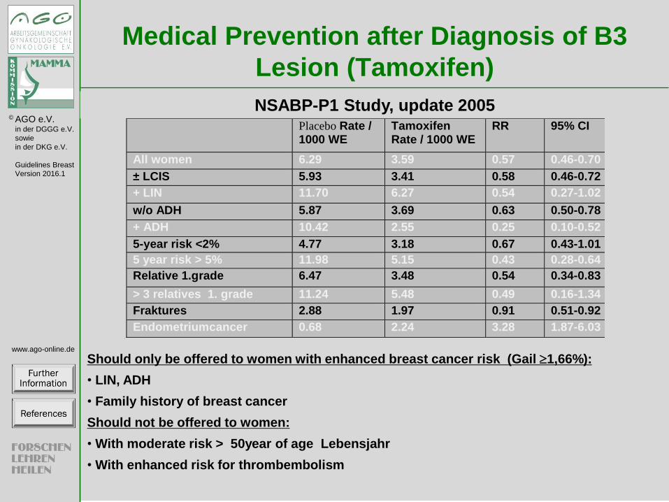
Medical Prevention after Diagnosis of B3
Lesion (Tamoxifen)
Should only be offered to women with enhanced breast cancer risk (Gail 1,66%):
• LIN, ADH
• Family history of breast cancer
Should not be offered to women:
• With moderate risk > 50year of age Lebensjahr
• With enhanced risk for thrombembolism
Placebo Rate / 1000 WE
Tamoxifen Rate / 1000 WE
RR 95% CI
All women 6.29 3.59 0.57 0.46-0.70
± LCIS 5.93 3.41 0.58 0.46-0.72
+ LIN 11.70 6.27 0.54 0.27-1.02
w/o ADH 5.87 3.69 0.63 0.50-0.78
+ ADH 10.42 2.55 0.25 0.10-0.52
5-year risk <2% 4.77 3.18 0.67 0.43-1.01
5 year risk > 5% 11.98 5.15 0.43 0.28-0.64
Relative 1.grade 6.47 3.48 0.54 0.34-0.83
> 3 relatives 1. grade 11.24 5.48 0.49 0.16-1.34
Fraktures 2.88 1.97 0.91 0.51-0.92
Endometriumcancer 0.68 2.24 3.28 1.87-6.03 5
NSABP-P1 Study, update 2005
© AGO e.V. in der DGGG e.V.
sowie
in der DKG e.V.
Guidelines Breast
Version 2016.1
www.ago-online.de
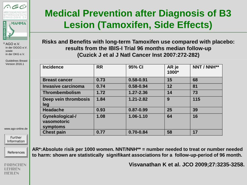
Medical Prevention after Diagnosis of B3
Lesion (Tamoxifen, Side Effects)
AR*:Absolute risik per 1000 women. NNT/NNH** = number needed to treat or number needed
to harm: shown are statistically signifikant associations for a follow-up-period of 96 month.
Visvanathan K et al. JCO 2009;27:3235-3258.
Incidence RR 95% CI AR je 1000*
NNT / NNH**
Breast cancer 0.73 0.58-0.91 15 68
Invasive carcinoma 0.74 0.58-0.94 12 81
Thrombembolism 1.72 1.27-2.36 14 73
Deep vein thrombosis leg
1.84 1.21-2.82 9 115
Headache 0.93 0.87-0.99 25 39
Gynekological-/ vasomotoric symptoms
1.08 1.06-1.10 64 16
Chest pain 0.77 0.70-0.84 58 17 5
Risks and Benefits with long-term Tamoxifen use compared with placebo:
results from the IBIS-I Trial 96 months median follow-up
(Cuzick J et al J Natl Cancer Inst 2007:272-282)

© AGO e.V. in der DGGG e.V.
sowie
in der DKG e.V.
Guidelines Breast
Version 2016.1
www.ago-online.de
Medical Prevention after Diagnosis of B3
Lesion (Raloxifen)
NSABP-P2 Study, STAR trial 2006
Should only be offered to women with enhanced breast cancer risk :
(Gail 1,66%) or postmeopausal
Should not be offered to women:
• With moderate risk > 50year of age
• With enhanced risk for thrombembolism
Tamoxifen
: Rate / 1000 WE
Raloxifen Rate /
1000 WE RR 95% CI
All women 4.30 4.41 1.02 0.82-1.28
± LIN 3.76 3.89 1.03 0.81-1.33
+ LIN 9.83 9.61 0.98 0.58-1.63
± ADH 4.06 4.03 0.99 0.76-1.28
+ ADH 5.21 5.81 1.12 0.72-1.74

© AGO e.V. in der DGGG e.V.
sowie
in der DKG e.V.
Guidelines Breast
Version 2016.1
www.ago-online.de
Prevention for Lesions with Uncertain
Biological Behaviour (Aromatase Inhibitors)
Inclusion criteria:
IBIS.2:
Prior ADH, ALH, or LCIS
Anastrozole:154 (8.0%);
Placebo: 190 (9.7%)
MAP.3:
Prior ADH, ALH, or LCIS:
Exemestane: 185 (8.1%);
Placebo: 188 (8.3%)
Results for prior ALH, ADH,
LCIS (HR AI vs Plac):
Yes (7y-BC-risk 12.1%):
HR 0.31 (0.12–0.84)
No (7y-BC-risk 4.9%):
HR 0.52 (0.31–0.78)
Yes: HR=0.61 (0.20–1.82)
No HR=0.26 (0.11–0.64)
Cuzick J et al. Lancet 2014; 383: 1041–48
Goss PE et al. N Engl J Med. 2011 Jun 23;364(25):2381-91

Lesions of Uncertain Malignant Potential (B3) (2/25)
Pubmed 2010-2015 (plus earlier publications if relevant): Lobular neoplasia (162 Results): (Breast Diseases/CL[mh] OR Breast Diseases/DI[mh] OR Breast Diseases/EP[mh] OR
Breast Diseases/GE[mh] OR Breast Diseases/MO[mh] OR Breast Diseases/PA[mh] OR Breast Diseases/RA[mh] OR
Breast Diseases/RT[mh] OR Breast Diseases/SU[mh] OR Breast Diseases/TH[mh] OR Breast Diseases/US[mh]) AND
("2005/01/01"[dp] : "2016/01/01"[dp]) AND ("lobular neoplasia"[ti] OR "lobular intraepithelial neoplasia"[ti] OR
"atypical lobular hyperplasia"[ti] OR "lobular carcinoma in situ"[ti] OR "LIN"[ti] OR "ALH"[ti] OR "LCIS"[ti]) AND
("english"[la] OR "german"[la])
Atypical ductal hyperplasia (78 Results): (Breast Diseases/CL[mh] OR Breast Diseases/DI[mh] OR Breast
Diseases/EP[mh] OR Breast Diseases/GE[mh] OR Breast Diseases/MO[mh] OR Breast Diseases/PA[mh] OR Breast
Diseases/RA[mh] OR Breast Diseases/RT[mh] OR Breast Diseases/SU[mh] OR Breast Diseases/TH[mh] OR Breast
Diseases/US[mh]) AND ("2005/01/01"[dp] : "2016/01/01"[dp]) AND ("atypical ductal hyperplasia"[ti] OR "atypical
hyperplasia"[ti] OR "ADH"[ti]) AND ("english"[la] OR "german"[la])
Flat epithelial atypia (88 Results): (Breast Diseases/CL[mh] OR Breast Diseases/DI[mh] OR Breast Diseases/EP[mh]
OR Breast Diseases/GE[mh] OR Breast Diseases/MO[mh] OR Breast Diseases/PA[mh] OR Breast Diseases/RA[mh] OR
Breast Diseases/RT[mh] OR Breast Diseases/SU[mh] OR Breast Diseases/TH[mh] OR Breast Diseases/US[mh]) AND
("2005/01/01"[dp] : "2016/01/01"[dp]) AND ("flat epithelial atypia"[ti] OR "columnar cell"[ti] OR "FEA"[ti]) AND
("english"[la] OR "german"[la])
Papilloma (274 Results): (Breast Diseases/CL[mh] OR Breast Diseases/DI[mh] OR Breast Diseases/EP[mh] OR Breast
Diseases/GE[mh] OR Breast Diseases/MO[mh] OR Breast Diseases/PA[mh] OR Breast Diseases/RA[mh] OR Breast
Diseases/RT[mh] OR Breast Diseases/SU[mh] OR Breast Diseases/TH[mh] OR Breast Diseases/US[mh]) AND
("2005/01/01"[dp] : "2016/01/01"[dp]) AND ("papilloma"[ti] OR "papillary"[ti]) AND ("english"[la] OR "german"[la])
NOT virus[Title]
Radial scar (22 Results): (Breast Diseases/CL[mh] OR Breast Diseases/DI[mh] OR Breast Diseases/EP[mh] OR Breast
Diseases/GE[mh] OR Breast Diseases/MO[mh] OR Breast Diseases/PA[mh] OR Breast Diseases/RA[mh] OR Breast
Diseases/RT[mh] OR Breast Diseases/SU[mh] OR Breast Diseases/TH[mh] OR Breast Diseases/US[mh]) AND

("2005/01/01"[dp] : "2016/01/01"[dp]) AND ("radial scar"[ti] OR "complex sclerosing lesion"[ti] OR "radial sclerosing
lesion"[ti]) AND ("english"[la] OR "german"[la])
Screened Guidlines: • Interdisziplinäre S3-Leitlinie für die Diagnostik, Therapie und Nachsorge des Mammakarzinoms. Aktualisierung
2012
• NCCN Breast cancer V.I.2014
• NCCN Breast Cancer Risk Reduction I 2013
• NCCN Breast Cancer Screening and Diagnosis 2.2013
• NZ: HTA risk assesment 2007
• CMJA: no update
• NICE: no update
• SIGN: no update
• Cochrane: Decision aids for risk communication update 2009
• DARE: no relevant references. 2010
• ASCO 2012: done
• National Institute of health (NIH): done
• San Antonio Breast Cancer Conference (SABCC 2013): done
References
National and international guidelines Albert US, Altland H, Duda V et al. 2008 update of the guideline early detection of breast cancer in Germany. J Cancer
Res Clin Oncol 2009; 135:339-354
Albert US, (Hrsg). Stufe-3-Leitlinie Brustkrebs-Früherkennung in Deutschland 1.Aktualisierung 2008. 1 ed. Muenchen:
Zuckschwerdt Verlag, 2008.
Leitlinienprogramm Onkologie der AWMF, Deutschen Krebsgesellschaft e.V. und Deutschen Krebshilfe e.V. (Hrsg.).
Interdisziplinäre S3-Leitlinie für die Diagnostik, Therapie und Nachsorge des Mammakarzinoms. Langversion 3.0,
Aktualisierung 2012 AWMF-Register-Nummer: 032 – 045OL
Lebeau A, Kreipe H, Dietel M, Schlake W, Kreienberg R. Mammakarzinom: aktuelle Empfehlungen fur Pathologen auf
Basis der S3-Leitlinie. Pathologe. 2013;34(4):293-302

Visvanathan K, Chlebowski R, Hurley P et al. American Society of Clinical Oncology Clincal Practice Guideline: Update
on the use of pharmacologic interventions including tamoxifen, raloxifen and aromatase inhibition for breast cancer risk
reduction. JCO 2009; 27:3235-3258
Weir R, Day P, Ali W. Risk factors for breast cancer in women:a systematic review of the literature. Christchurch: New
Zealand Health Technology Assessment (NZHTA), 2007.
NCCN, National Comprehensive Cancer Network. Breast cancer V.I.2014. 2014 ed. USA: NCCN, 2014.
NCCN, National Comprehensive Cancer Network. Breast cancer risk reduction V.I.2013. 2013 ed. USA: NCCN, 2013.
NCCN, National Comprehensive Cancer Network. Breast cancer screening and diagnosis V.2.2013. 2013 ed. USA:
NCCN, 2013.
O'Connor A, Bennett C, Stacey D et al. Decision aids for people facing health treatment or screening decisions (Review).
The Cochrane Library 2009;(4):1-35.

National and international guidelines
Albert US, Altland H, Duda V et al. 2008 update of the guideline early detection of breast cancer in Germany. J Cancer
Res Clin Oncol 2009; 135:339-354
Albert US, (Hrsg). Stufe-3-Leitlinie Brustkrebs-Früherkennung in Deutschland 1.Aktualisierung 2008. 1 ed. Muenchen:
Zuckschwerdt Verlag, 2008.
Leitlinienprogramm Onkologie der AWMF, Deutschen Krebsgesellschaft e.V. und Deutschen Krebshilfe e.V. (Hrsg.).
Interdisziplinäre S3-Leitlinie für die Diagnostik, Therapie und Nachsorge des Mammakarzinoms. Langversion 3.0,
Aktualisierung 2012 AWMF-Register-Nummer: 032 – 045OL
Lebeau A, Kreipe H, Dietel M, Schlake W, Kreienberg R. Mammakarzinom: aktuelle Empfehlungen fur Pathologen auf
Basis der S3-Leitlinie. Pathologe. 2013;34(4):293-302
Visvanathan K, Chlebowski R, Hurley P et al. American Society of Clinical Oncology Clincal Practice Guideline: Update
on the use of pharmacologic interventions including tamoxifen, raloxifen and aromatase inhibition for breast cancer risk
reduction. JCO 2009; 27:3235-3258
Weir R, Day P, Ali W. Risk factors for breast cancer in women:a systematic review of the literature. Christchurch: New
Zealand Health Technology Assessment (NZHTA), 2007.
NCCN, National Comprehensive Cancer Network. Breast cancer V.I.2014. 2014 ed. USA: NCCN, 2014.
NCCN, National Comprehensive Cancer Network. Breast cancer risk reduction V.I.2013. 2013 ed. USA: NCCN, 2013.
NCCN, National Comprehensive Cancer Network. Breast cancer screening and diagnosis V.2.2013. 2013 ed. USA:
NCCN, 2013.
O'Connor A, Bennett C, Stacey D et al. Decision aids for people facing health treatment or screening decisions (Review).
The Cochrane Library 2009;(4):1-35.

Pathology Reporting for Minimal Invasive Biopsies (3/25)
Further information:
The histologic B-classification of breast core biopsies as based on recommendations of the National Coordinating Group
for Breast Screening Pathology (NHSBSP), and E. C. Working Group on breast screening pathology encompasses the
heterogeneous B3 category.
References:
1. World Health Organization: WHO Classification of Tumours of the Breast. Lakhani SR, Ellis IO, Schnitt SJ, Tan
PH, editors. World Health Organization; 2012.
2. Wells C: Quality assurance guidelines for pathology: Cytological and histological non-operative procedures. In:
European guidelines for quality assurance in breast cancer screening and diagnosis. Perry N, Broeders M, de Wolf C,
Törnberg S, Holland R, Koch von F, editors. Luxembourg: Office for Official Publications of the European
Communities, 2006: 221-256
3. NHSBSP. Guidelines Working Group of the National Coordinating Committee for Breast Pathology. Pathology
reporting of breast disease. Sheffield: NHS Screening Programmes and The Royal College of Pathologists, 2005.
4. Kluttig A, Trocchi P, Heinig A, Holzhausen HJ, Taege C, Hauptmann S, Boecker W, Decker T, Loening T, Schmidt-
Pokrzywniak A, Thomssen C, Lantzsch T, Buchmann J, Stang A. Reliability and validity of needle biopsy evaluation
of breast-abnormalities using the B-categorization--design and objectives of the Diagnosis Optimisation Study
(DIOS). BMC Cancer. 2007 Jun 14;7:100.

B3-Lesions (4/25)
Further information:
Lesions of uncertain malignant potential include atypical ductal hyperplasia (ADH), lobular neoplasia (LN), flat epithelial
atypia (FEA), atypical papillary proliferations, and lesions with sampling risk because of inhomogeneity, such as phyllodes
tumor, cellular fibroadenoma, and radial scars. The lesions with atypical proliferations (ADH, ALH, LCIS, FEA) are
regarded both as an indicator of increased risk, but also as precursor lesions, and are part of the low-grade pathway of
breast cancers [1-4]. The accurate pathological identification and classification of lesions with atypical proliferations is
important to assess the individual risk of the patient, and to decide if the lesion should be excised. The recognition of
atypical epithelial proliferation is based on the distinction of hyperplastic from neoplastic lesions, that is on the
identification of a clonal process. As a general rule, usual type epithelial hyperplasia is morphologically and
phenotypically heterogeneous, while ADH, FEA, and LN are characterized by a homogeneity of cell type and marker
expression. With all types of precursor lesions, careful attention must be paid to the pathologic-radiologic correlation for
the guidance of the clinical management. B3 lesions are associated with a high rate of 6-16% discordance among first and
second pathology compared to 0.5-1,3% discordance for B5 lesions [5].
References:
1. Andreu FJ, Sáez A, Sentís M, Rey M, Fernández S, Dinarès C, et al. Breast core biopsy reporting categories. An
internal validation in a series of 3054 consecutive lesions. Breast. 2007 Jan 31;16(1):94–101.
2. Bombonati A, Sgroi DC. The molecular pathology of breast cancer progression. Ladanyi M, Hogendoorn PC,
editors. J Pathol. 2010 Nov 16;223(2):308–18.
3. Hayes BD, Quinn CM. Pathology of B3 lesions ofthe breast. Diagnostic Histopathology. Elsevier Ltd; 2009 Oct
1;15(10):459–69.

4. Houssami N, Ciatto S, Bilous M, Vezzosi V, Bianchi S. Borderline breast core needle histology: predictive values
for malignancy in lesions of uncertain malignant potential (B3). Br J Cancer. 2007 Apr 22;96(8):1253–7.
5. Kreipe H-H, Höfler H, Lebeau A, Pickartz H, Schmidt D. Ergebnisse der Referenzpathologie im Mammographie-
Screening. Pathologe. 2008 Oct 9;29(S2):178–80.
6. Kluttig A, Trocchi P, Heinig A, Holzhausen HJ, Taege C, Hauptmann S, Boecker W, Decker T, Loening T, Schmidt-
Pokrzywniak A, Thomssen C, Lantzsch T, Buchmann J, Stang A. Reliability and validity of needle biopsy evaluation
of breast-abnormalities using the B-categorization--design and objectives of the Diagnosis Optimisation Study
(DIOS). BMC Cancer. 2007 Jun 14;7:100.

Major B3-Lesions and Prospektive Prediktive Value (PPV) for Malignancy in Resection (5/25)
Further information:
In this category atypical intraductal hyperplasia (ADH), flat epithelial atypia (FEA), and lobular intraepithelial neoplasia
(LN/LIN) are grouped together as lesions of uncertain biological behaviour. Besides these diagnoses papillomas, radial
scar and phyllodes-tumour belong to the B3 group. In older studies approximately one-third of CNB results classified as
B3 were malignant on excision, but the likelihood of malignancy varied substantially between specific lesion groups.
Whereas cases may be selectively managed without surgery, the majority warrant excision biopsy (Rakha 2010, Houssami
2010). No clinical and radiologic findings and/or comprehensive evaluation of multiple histologic parameters on CNB
specimen are distinctive enough to predict final classification of equivocal cellular fibroepithelial lesions.
In recent years publications demonstrated a decline in PPV except for ADH. This is partiularly obvious for LIN, which
only rarely shows upgrade to higher lesions in resection when carful correlation between imaging and histology of CNB
has been performed. Also papilloma without atypia usually shows no upgrade in resection. With regard to FEA different
frequencies of upgrade to higher lesions are published.
B3 lesions are diagnosed with less than 10% in mammography screening (6000 core biopsies, with central pathology). But
B3 lesions are associated with a high rate of 6-16% disconcordance among first and second pathology compared to 0.5-
1,3% disconcordance for B5 lesions (Kreipe HH et al 2008).
Current systematic review:
Calhoun, B. C., & Collins, L. C. (2016). Recommendations for excision following core needle biopsy of the breast: a
contemporary evaluation of the literature. Histopathology, 68(1), 138–151. http://doi.org/10.1111/his.12852
Other References:
1. Atkins KA, Cohen MA, Nicholson B, Rao S. Atypical lobular hyperplasia and lobular carcinoma in situ at core
breast biopsy: use of careful radiologic-pathologic correlation to recommend excision or observation. Radiology.
2013 Nov;269(2):340-7.
2. Becker AK, Gordon PB, Harrison DA, Hassell PR, Hayes MM, van Niekerk D, Wilson CM. Flat ductal
intraepithelial neoplasia 1A diagnosed at stereotactic core needle biopsy: is excisional biopsy indicated? AJR Am J
Roentgenol. 2013 Mar;200(3):682-8.
3. Bianchi S, Bendinelli B, Castellano I, Piubello Q, Renne G, Cattani MG, Stefano DD, Carrillo G, Laurino L, Bersiga
A, Giardina C, Dante S, Loreto CD, Quero C, Antonacci CM, Palli D; VANCB Study Group. Morphological
parameters of lobular in situ neoplasia in stereotactic 11-gauge vacuum-assisted needle core biopsy do not predict the
presence of malignancy on subsequent surgical excision. Histopathology. 2013 Jul;63(1):83-95.
4. Buckley ES, Webster F, Hiller JE, Roder DM, Farshid G. A systematic review of surgical biopsy for LCIS found at
core needle biopsy - do we have the answer yet? Eur J Surg Oncol. 2014 Feb;40(2):168-75.
5. Calhoun BC, Sobel A, White RL, Gromet M, Flippo T, Sarantou T, Livasy CA. Management of flat epithelial atypia
on breast core biopsy may be individualized based on correlation with imaging studies. Mod Pathol. 2014 Nov 21.
doi: 10.1038/modpathol.2014.159. [Epub ahead of print]
6. Ceugnart L, Doualliez V, Chauvet MP, Robin YM, Bachelle F, Chaveron C, Rocourt N, Pouliquen G, Jarraya H,
Taieb S. Pure flat epithelial atypia: is there a place for routine surgery? Diagn Interv Imaging. 2013 Sep;94(9):861-9.
7. D'Alfonso TM, Wang K, Chiu YL, Shin SJ. Pathologic upgrade rates on subsequent excision when lobular
carcinoma in situ is the primary diagnosis in the needle core biopsy with special attention to the radiographic target.
Arch Pathol Lab Med. 2013 Jul;137(7):927-35.
8. Dialani V, Venkataraman S, Frieling G, Schnitt SJ, Mehta TS. Does isolated flat epithelial atypia on vacuum-assisted
breast core biopsy require surgical excision? Breast J. 2014 Nov-Dec;20(6):606-14.
9. Dillon MF: Predictive Value of Breast Lesions of "Uncertain Malignant Potential" and "Suspicious for Malignancy"
Determined by Needle Core Biopsy. Annals of Surgical Oncology 2007; 14(2):704-711
10. Hartmann LC, Degnim AC, Santen RJ, Dupont WD, Ghosh K. Atypical hyperplasia of the breast--risk assessment
and management options. N Engl J Med. 2015 Jan 1;372(1):78-89.
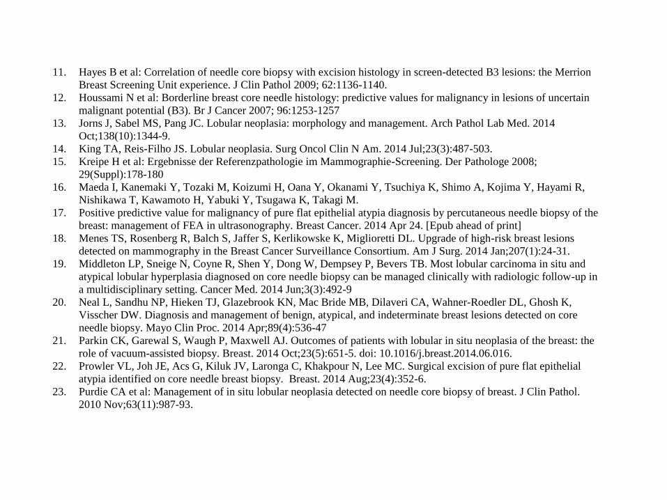
11. Hayes B et al: Correlation of needle core biopsy with excision histology in screen-detected B3 lesions: the Merrion
Breast Screening Unit experience. J Clin Pathol 2009; 62:1136-1140.
12. Houssami N et al: Borderline breast core needle histology: predictive values for malignancy in lesions of uncertain
malignant potential (B3). Br J Cancer 2007; 96:1253-1257
13. Jorns J, Sabel MS, Pang JC. Lobular neoplasia: morphology and management. Arch Pathol Lab Med. 2014
Oct;138(10):1344-9.
14. King TA, Reis-Filho JS. Lobular neoplasia. Surg Oncol Clin N Am. 2014 Jul;23(3):487-503.
15. Kreipe H et al: Ergebnisse der Referenzpathologie im Mammographie-Screening. Der Pathologe 2008;
29(Suppl):178-180
16. Maeda I, Kanemaki Y, Tozaki M, Koizumi H, Oana Y, Okanami Y, Tsuchiya K, Shimo A, Kojima Y, Hayami R,
Nishikawa T, Kawamoto H, Yabuki Y, Tsugawa K, Takagi M.
17. Positive predictive value for malignancy of pure flat epithelial atypia diagnosis by percutaneous needle biopsy of the
breast: management of FEA in ultrasonography. Breast Cancer. 2014 Apr 24. [Epub ahead of print]
18. Menes TS, Rosenberg R, Balch S, Jaffer S, Kerlikowske K, Miglioretti DL. Upgrade of high-risk breast lesions
detected on mammography in the Breast Cancer Surveillance Consortium. Am J Surg. 2014 Jan;207(1):24-31.
19. Middleton LP, Sneige N, Coyne R, Shen Y, Dong W, Dempsey P, Bevers TB. Most lobular carcinoma in situ and
atypical lobular hyperplasia diagnosed on core needle biopsy can be managed clinically with radiologic follow-up in
a multidisciplinary setting. Cancer Med. 2014 Jun;3(3):492-9
20. Neal L, Sandhu NP, Hieken TJ, Glazebrook KN, Mac Bride MB, Dilaveri CA, Wahner-Roedler DL, Ghosh K,
Visscher DW. Diagnosis and management of benign, atypical, and indeterminate breast lesions detected on core
needle biopsy. Mayo Clin Proc. 2014 Apr;89(4):536-47
21. Parkin CK, Garewal S, Waugh P, Maxwell AJ. Outcomes of patients with lobular in situ neoplasia of the breast: the
role of vacuum-assisted biopsy. Breast. 2014 Oct;23(5):651-5. doi: 10.1016/j.breast.2014.06.016.
22. Prowler VL, Joh JE, Acs G, Kiluk JV, Laronga C, Khakpour N, Lee MC. Surgical excision of pure flat epithelial
atypia identified on core needle breast biopsy. Breast. 2014 Aug;23(4):352-6.
23. Purdie CA et al: Management of in situ lobular neoplasia detected on needle core biopsy of breast. J Clin Pathol.
2010 Nov;63(11):987-93.

24. Rakha EA et al: Characterisation and outcome of breast needle core biopsy diagnoses of lesions of uncertain
malignant potential (B3) in abnormalities detected by mammographic screening. Int J Cancer. 2010 Dec 2. [Epub
ahead of print]
25. Resetkova E et al: Clinical and radiologic data and core needle biopsy findings should dictate management of
cellular fibroepithelial tumors of the breast. Breast J. 2010 Nov-Dec;16(6):573-80
26. Shamonki J, Chung A, Huynh KT, Sim MS, Kinnaird M, Giuliano A. Management of papillary lesions of the breast:
can larger core needle biopsy samples identify patients who may avoid surgical excision? Ann Surg Oncol. 2013
Dec;20(13):4137-44
27. Weir R et al: Risk factors for breast cancer in women:a systematic review of the literature. Christchurch: New
Zealand Health Technology Assessment (NZHTA), 2007.
28. Whiffen A: Predictors of Breast Cancer Development in Women with Atypical Ductal Hyperplasia and Atypical
Lobular Hyperplasia. Ann Surg Oncol. 2010 Sep 28.
29. Wyss P, Varga Z, Rössle M, Rageth CJ. Papillary lesions of the breast: outcomes of 156 patients managed without
excisional biopsy. Breast J. 2014 Jul-Aug;20(4):394-401.

Management after Minimally Invasive Biopsy (6/25)
Further information:
What kind of treatment has to follow when a B3 diagnosis has been rendered should be individually determined in an
interdisciplinary discussion of the imaging findings and the patholgy results. Algorithm for quality assurance of minimal
invasive guided biopsies.
After a review and quality assessment of 21 studies, diagnostic accuracy of VAB were evaluated. The summary estimates
for VAB in diagnosis of breast carcinoma were as follows: sensitivity, 0.981 (95% confidence interval [CI], 0.972-0.987);
specificity, 0.999 (95% CI, 0.997-0.999); positive likelihood ratio (PLR), 93.84 (95% CI, 41.55-211.95); negative
likelihood ratio, 0.05 (95% CI, 0.03-0.09); diagnostic odds ratio, 1891.7 (95% CI, 683.8-5233.4); underestimate rate of
ADH and DCIS were 20.9% (95% CI, 0.177-0.245) and 11.2% (95% CI, 0.098-0.128), respectively. VAB is a highly
sensitive and specific biopsy method for evaluating mammographically detected breast in women.
References:
1. Brem RF, Behrndt VS, Sanow L, Gatewood OM: Atypical ductal hyperplasia: histologic underestimation of
carcinoma in tissue harvested from impalpable breast lesions using 11-gauge stereotactically guided directional
vacuum-assisted biopsy. AJR 1999, 172:1405-1407
2. Ciatto S, Houssami N, Ambrogetti D, Bianchi S, Bonari R, Brancato B, Catarzi S, Risso G: Accuracy and
underestimation of malignancy of breast core needle biopsy: the florence experience of over 4000 consecutive
biopsies. Breast Cancer res Treat 2007, 101:291-307
3. Bedei L, Falcini F, Sanna P: Atypical ductal hyperplasia of the breast: the controversional management of a
borderline lesion. Experience of 47 cases diagnosed at vaccumassisted biopsy. Breast 2006, 15:196-202.
4. Liberman L, Holland A, Marjan D, Murray M, Bertelle L, Morris E, Dershew D, Wynn R: Underestimation of
atypical ductal hyperplasia at MRI-guided 9-Gauge vacuum-assisted breast biopsy. AJR 2007, 188:684-690.

5. Zagrafos G, Zagouri F, Sergentanis T, Nonni A, Koulocheri D, Folou M, Panopoulou E, Paranas N, Foliadis C,
Bramis J: Minimizing underestimation rate of microcalcifications excised via vacuum-assisted breast biopsy: ablind
study. Breast Cancer Res and Treat 2008, 109:397-402.
6. Arora S, Menes T, Moung C, Nagi C, Bleiweiss I, Jaffer S. Atypical ductal hyperplasia at margin of breast biopsy -
Is Re-excision indicated? Ann Surg Oncol 2007; 15(3):843-847
7. Neal L, Sandhu NP, Hieken TJ, Glazebrook KN, Mac Bride MB, Dilaveri CA, Wahner-Roedler DL, Ghosh K,
Visscher DW. Diagnosis and management of benign, atypical, and indeterminate breast lesions detected on core
needle biopsy. Mayo Clin Proc. 2014 Apr;89(4):536-47
8. Hartmann LC, Degnim AC, Santen RJ, Dupont WD, Ghosh K. Atypical hyperplasia of the breast--risk assessment
and management options. N Engl J Med. 2015 Jan 1;372(1):78-89.
9. Atkins KA, Cohen MA, Nicholson B, Rao S. Atypical lobular hyperplasia and lobular carcinoma in situ at core
breast biopsy: use of careful radiologic-pathologic correlation to recommend excision or observation. Radiology.
2013 Nov;269(2):340-7.
10. Middleton LP, Sneige N, Coyne R, Shen Y, Dong W, Dempsey P, Bevers TB. Most lobular carcinoma in situ and
atypical lobular hyperplasia diagnosed on core needle biopsy can be managed clinically with radiologic follow-up in
a multidisciplinary setting. Cancer Med. 2014 Jun;3(3):492-9

Atypical Ductal Hyperplasia (ADH) (7/25)
Further information:
ADH and breast cancer are asoziated with postmenopausal hormone treatment. According to the data of the Breast Cancer
Surveillance Consortium (USA) rates of ADH decreased from 5.5/10000 mammograms 1999 to 2.4/10000 mammograms
in 2005
Statement: indicator-/ precursor-lesion:
Women have an enhanced breast cancer risk after ADH: one lesion RR 3.88 (95%CI 3.00-4.94), three lesions RR10.35
(95%CI 6.13-16.4). Less than 45 years at diagnosis of ADH RR 6.78 (95%CI 3.24-12.4).
References:
1. Menes T: Rates of atypical ductal hyperplasia have declined with less use of postmenopausal hormone treatment:
Findings from the Breast Cancer Surveillance Consortium. Cancer Epidemiol Biomarkers Prev 2009;18:2822-2828
2. Degnim A:. Stratification of breast cancer risk in women with atypia: A Mayo Cohort Study. JCO 2007;
25(19):2671-2677
3. Ellis IO: Impact of a national external quality assessment scheme for breast pathology in the UK.J Clin Pathol.
2006;59:138-45.
4. Böcker W, Hungermann D, Weigel S, Roterberg K, Decker T. Atypical ductal hyperplasia and atypical epithelial
proliferation of ductal type]. Pathologe. 2009 Feb;30(1):42-8. doi: 10.1007/s00292-008-1101-4.

Strategy after Diagnosis of ADH (8/25)
Further information:
Significant histologic predictors of upgrade from ADH to carcinoma included number of terminal duct-lobular units
(TDLU; >2) involved (P = .0306), presence of significant cytologic atypia suspicious for intermediate or high-grade
carcinoma (P < .0001), and necrosis (P = .0006). Therefore, ADH lesions with significant cytologic atypia and/or necrosis
are most likely to be associated with carcinoma and should be excised. ADH without these features, regardless of extent of
involvement, and with complete removal of the targeted calcifications, is associated with a minimal risk (<3%) of
carcinoma and may undergo mammographic follow-up only (Nguyen CV 2010, Allison KH 2010). Radiological
calcification with supicious or malignant characteristics and histological B3 with evidence of epithelial atypia has the
highest positive predictive value (50%) (Rhaka et al. 2010). Even in the case of complete removal of microcalcifications
there is a risk of 5 % of underestimation of malignancy (Penco 2010). An open excisional is recommended with exception
of very small lesions (≤ 2 TDLU) and minimal atypia and complete removed imaging abnormality.
ADH in core- / vacuum-assisted biopsy (LoE 3a)
ADH at margins in resection specimen (LoE 3a)
References:
1. Atkins KA, Cohen MA, Nicholson B, Rao S. Atypical lobular hyperplasia and lobular carcinoma in situ at core
breast biopsy: use of careful radiologic-pathologic correlation to recommend excision or observation. Radiology.
2013 Nov;269(2):340-7.
2. Dillon MF: Predictive Value of Breast Lesions of "Uncertain Malignant Potential" and "Suspicious for Malignancy"
Determined by Needle Core Biopsy. Annals of Surgical Oncology 2007; 14(2):704-711
3. Hartmann LC, Degnim AC, Santen RJ, Dupont WD, Ghosh K. Atypical hyperplasia of the breast--risk assessment
and management options. N Engl J Med. 2015 Jan 1;372(1):78-89.

4. Hayes B et al: Correlation of needle core biopsy with excision histology in screen-detected B3 lesions: the Merrion
Breast Screening Unit experience. J Clin Pathol 2009; 62:1136-1140.
5. Houssami N et al: Borderline breast core needle histology: predictive values for malignancy in lesions of uncertain
malignant potential (B3). Br J Cancer 2007; 96:1253-1257
6. Menes TS, Rosenberg R, Balch S, Jaffer S, Kerlikowske K, Miglioretti DL. Upgrade of high-risk breast lesions
detected on mammography in the Breast Cancer Surveillance Consortium. Am J Surg. 2014 Jan;207(1):24-31.
7. Neal L, Sandhu NP, Hieken TJ, Glazebrook KN, Mac Bride MB, Dilaveri CA, Wahner-Roedler DL, Ghosh K,
Visscher DW. Diagnosis and management of benign, atypical, and indeterminate breast lesions detected on core
needle biopsy. Mayo Clin Proc. 2014 Apr;89(4):536-47
8. Rakha EA et al: Characterisation and outcome of breast needle core biopsy diagnoses of lesions of uncertain
malignant potential (B3) in abnormalities detected by mammographic screening. Int J Cancer. 2010 Dec 2. [Epub
ahead of print]
9. Whiffen A: Predictors of Breast Cancer Development in Women with Atypical Ductal Hyperplasia and Atypical
Lobular Hyperplasia. Ann Surg Oncol. 2010 Sep 28.

Risk of Breast Cancer after Atypical Hyperplasie (ADH, ALH) (9/25)
No further information
References:
1. Degnim A, Visscher W, Berman H et al. Stratification of breast cancer risk in women with atypia: A Mayo Cohort
Study. JCO 2007; 25(19):2671-2677
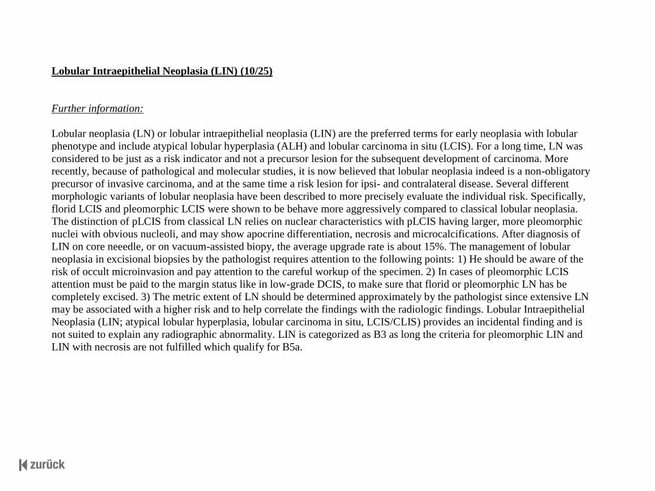
Lobular Intraepithelial Neoplasia (LIN) (10/25)
Further information:
Lobular neoplasia (LN) or lobular intraepithelial neoplasia (LIN) are the preferred terms for early neoplasia with lobular
phenotype and include atypical lobular hyperplasia (ALH) and lobular carcinoma in situ (LCIS). For a long time, LN was
considered to be just as a risk indicator and not a precursor lesion for the subsequent development of carcinoma. More
recently, because of pathological and molecular studies, it is now believed that lobular neoplasia indeed is a non-obligatory
precursor of invasive carcinoma, and at the same time a risk lesion for ipsi- and contralateral disease. Several different
morphologic variants of lobular neoplasia have been described to more precisely evaluate the individual risk. Specifically,
florid LCIS and pleomorphic LCIS were shown to be behave more aggressively compared to classical lobular neoplasia.
The distinction of pLCIS from classical LN relies on nuclear characteristics with pLCIS having larger, more pleomorphic
nuclei with obvious nucleoli, and may show apocrine differentiation, necrosis and microcalcifications. After diagnosis of
LIN on core neeedle, or on vacuum-assisted biopy, the average upgrade rate is about 15%. The management of lobular
neoplasia in excisional biopsies by the pathologist requires attention to the following points: 1) He should be aware of the
risk of occult microinvasion and pay attention to the careful workup of the specimen. 2) In cases of pleomorphic LCIS
attention must be paid to the margin status like in low-grade DCIS, to make sure that florid or pleomorphic LN has be
completely excised. 3) The metric extent of LN should be determined approximately by the pathologist since extensive LN
may be associated with a higher risk and to help correlate the findings with the radiologic findings. Lobular Intraepithelial
Neoplasia (LIN; atypical lobular hyperplasia, lobular carcinoma in situ, LCIS/CLIS) provides an incidental finding and is
not suited to explain any radiographic abnormality. LIN is categorized as B3 as long the criteria for pleomorphic LIN and
LIN with necrosis are not fulfilled which qualify for B5a.
References:
1. Contreras A: Lobular Neoplasia of the breast: An update. Arch Pathol Lab Med 2009; 133(7):1116-1120
2. Pinder S: Lobular in situ neoplasia and columnar cell lesions:diagnosis in breast core biopsies and implications for
management. Pathology 2007, 39:208-216.
3. Lakhani I: The management of lobular carcinoma in situ (LCIS). Is LCIS the same as ductal carcinoma in situ
(DCIS)? Eur J Cancer 2006; 42:2205-2211
4. Arpino G:: Lobular neoplasia on core-needle biopsy: clinical significance. Cancer 2004, 101:242-250
Statement: Indicator-/ precusor lesion
1. Chuba PJ: Bilateral Risk for Subsequent Breast Cancer After Lobular Carcinoma-In-Situ: Analysis of Surveillance,
Epidemiology, and End Results Data. Journal of Clinical Oncology 2005, 23:5534-5541
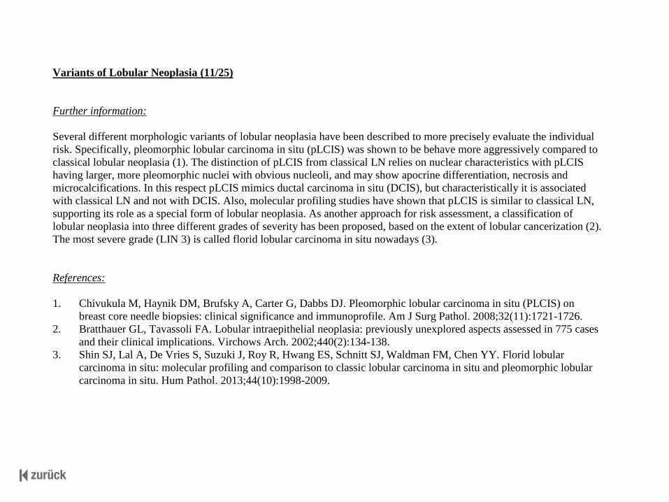
Variants of Lobular Neoplasia (11/25)
Further information:
Several different morphologic variants of lobular neoplasia have been described to more precisely evaluate the individual
risk. Specifically, pleomorphic lobular carcinoma in situ (pLCIS) was shown to be behave more aggressively compared to
classical lobular neoplasia (1). The distinction of pLCIS from classical LN relies on nuclear characteristics with pLCIS
having larger, more pleomorphic nuclei with obvious nucleoli, and may show apocrine differentiation, necrosis and
microcalcifications. In this respect pLCIS mimics ductal carcinoma in situ (DCIS), but characteristically it is associated
with classical LN and not with DCIS. Also, molecular profiling studies have shown that pLCIS is similar to classical LN,
supporting its role as a special form of lobular neoplasia. As another approach for risk assessment, a classification of
lobular neoplasia into three different grades of severity has been proposed, based on the extent of lobular cancerization (2).
The most severe grade (LIN 3) is called florid lobular carcinoma in situ nowadays (3).
References:
1. Chivukula M, Haynik DM, Brufsky A, Carter G, Dabbs DJ. Pleomorphic lobular carcinoma in situ (PLCIS) on
breast core needle biopsies: clinical significance and immunoprofile. Am J Surg Pathol. 2008;32(11):1721-1726.
2. Bratthauer GL, Tavassoli FA. Lobular intraepithelial neoplasia: previously unexplored aspects assessed in 775 cases
and their clinical implications. Virchows Arch. 2002;440(2):134-138.
3. Shin SJ, Lal A, De Vries S, Suzuki J, Roy R, Hwang ES, Schnitt SJ, Waldman FM, Chen YY. Florid lobular
carcinoma in situ: molecular profiling and comparison to classic lobular carcinoma in situ and pleomorphic lobular
carcinoma in situ. Hum Pathol. 2013;44(10):1998-2009.
LIN with High Risk (12/25)
Further information:
Several different morphologic variants of lobular neoplasia have been described to more precisely evaluate the individual
risk. Specifically, pleomorphic lobular carcinoma in situ (pLCIS) was shown to be behave more aggressively compared to
classical lobular neoplasia [1]. The distinction of pLCIS from classical LN relies on nuclear characteristics with pLCIS
having larger, more pleomorphic nuclei with obvious nucleoli, and may show apocrine differentiation, necrosis and
microcalcifications. In this respect pLCIS mimics ductal carcinoma in situ (DCIS), but characteristically it is associated
with classical LN and not with DCIS. Also, molecular profiling studies have shown that pLCIS is similar to classical LN,
supporting its role as a special form of lobular neoplasia. As another approach for risk assessment, a classification of
lobular neoplasia into three different grades of severity has been proposed, based on the extent of lobular cancerization [2].
The most severe grade (LIN 3) is called florid lobular carcinoma in situ nowadays [3]. It may be associated with
microinvasion [4].
References:
1. Chivukula M, Haynik DM, Brufsky A, Carter G, Dabbs DJ. Pleomorphic lobular carcinoma in situ (PLCIS) on
breast core needle biopsies: clinical significance and immunoprofile. Am J Surg Pathol. 2008;32(11):1721-1726.
2. Bratthauer GL, Tavassoli FA. Lobular intraepithelial neoplasia: previously unexplored aspects assessed in 775 cases
and their clinical implications. Virchows Arch. 2002;440(2):134-138.
3. Shin SJ, Lal A, De Vries S, Suzuki J, Roy R, Hwang ES, Schnitt SJ, Waldman FM, Chen YY. Florid lobular
carcinoma in situ: molecular profiling and comparison to classic lobular carcinoma in situ and pleomorphic lobular
carcinoma in situ. Hum Pathol. 2013;44(10):1998-2009.
4. Ross DS, Hoda SA. Microinvasive (T1mic) lobular carcinoma of the breast: clinicopathologic profile of 16 cases.
The American journal of surgical pathology. 2011 May;35(5):750–6.
Strategy after Diagnosis of LIN (13/25)
Further information:
In contrast to atypical ductal hyperplasia, it is less clear if a follow-up excisional biopsy is beneficial to the outcome of a
patient with the finding of lobular neoplasia in the core biopsy, and therefore there is some disagreement if excision should
be recommended as a rule or not. This is mainly due to the relative infrequency of lobular neoplasia as the most severe
finding in core biopsies and the even lower number of excisional biopsies in this situation. Not surprisingly these small
studies have led to widely discrepant results and conflicting interpretations of published data. An excisional biopsy was
recommended in fully developed LCIS because of an upgrade rate of greater than of 25% [1] or 16% [2], but results were
inconclusive with lesions of lesser extent, namely atypical lobular hyperplasia. The argument against a routine follow-up
biopsy is that LN as the most significant pathology usually is an incidental finding in an otherwise benign core biopsy and
if there is no other clinical or radiological detectable lesion, it is unlikely that an excisional biopsy could yield anything
more significant [3]. This argument has to be taken seriously, and at least all cases with LCIS and a mass lesion should be
followed up by a surgical biopsy. However, because of the reported upgrade rates in fully developed LCIS, the nature of
these lesions as non-obligate precursors, and risk of missing a radiologically occult invasive cancer, an open biopsy in
classical LCIS should be considered as an option also [2], especially if multiple lobules are involved.
References:
LIN in core- / vacuum-assisted biopsy (LoE 2b)
1. Atkins KA, Cohen MA, Nicholson B, Rao S. Atypical lobular hyperplasia and lobular carcinoma in situ at core
breast biopsy: use of careful radiologic-pathologic correlation to recommend excision or observation. Radiology.
2013 Nov;269(2):340-7.

2. Bianchi S, Bendinelli B, Castellano I, Piubello Q, Renne G, Cattani MG, Stefano DD, Carrillo G, Laurino L, Bersiga
A, Giardina C, Dante S, Loreto CD, Quero C, Antonacci CM, Palli D; VANCB Study Group. Morphological
parameters of lobular in situ neoplasia in stereotactic 11-gauge vacuum-assisted needle core biopsy do not predict the
presence of malignancy on subsequent surgical excision. Histopathology. 2013 Jul;63(1):83-95.
3. Buckley ES, Webster F, Hiller JE, Roder DM, Farshid G. A systematic review of surgical biopsy for LCIS found at
core needle biopsy - do we have the answer yet? Eur J Surg Oncol. 2014 Feb;40(2):168-75.
4. D'Alfonso TM, Wang K, Chiu YL, Shin SJ. Pathologic upgrade rates on subsequent excision when lobular
carcinoma in situ is the primary diagnosis in the needle core biopsy with special attention to the radiographic target.
Arch Pathol Lab Med. 2013 Jul;137(7):927-35.
5. Jorns J, Sabel MS, Pang JC. Lobular neoplasia: morphology and management. Arch Pathol Lab Med. 2014
Oct;138(10):1344-9.
6. King TA, Reis-Filho JS. Lobular neoplasia. Surg Oncol Clin N Am. 2014 Jul;23(3):487-503.
7. Menes TS, Rosenberg R, Balch S, Jaffer S, Kerlikowske K, Miglioretti DL. Upgrade of high-risk breast lesions
detected on mammography in the Breast Cancer Surveillance Consortium. Am J Surg. 2014 Jan;207(1):24-31.
8. Middleton LP, Sneige N, Coyne R, Shen Y, Dong W, Dempsey P, Bevers TB. Most lobular carcinoma in situ and
atypical lobular hyperplasia diagnosed on core needle biopsy can be managed clinically with radiologic follow-up in
a multidisciplinary setting. Cancer Med. 2014 Jun;3(3):492-9
9. Neal L, Sandhu NP, Hieken TJ, Glazebrook KN, Mac Bride MB, Dilaveri CA, Wahner-Roedler DL, Ghosh K,
Visscher DW. Diagnosis and management of benign, atypical, and indeterminate breast lesions detected on core
needle biopsy. Mayo Clin Proc. 2014 Apr;89(4):536-47
10. Parkin CK, Garewal S, Waugh P, Maxwell AJ. Outcomes of patients with lobular in situ neoplasia of the breast: the
role of vacuum-assisted biopsy. Breast. 2014 Oct;23(5):651-5. doi: 10.1016/j.breast.2014.06.016.
11. Purdie CA et al: Management of in situ lobular neoplasia detected on needle core biopsy of breast. J Clin Pathol.
2010 Nov;63(11):987-93.
12. Rakha EA et al: Characterisation and outcome of breast needle core biopsy diagnoses of lesions of uncertain
malignant potential (B3) in abnormalities detected by mammographic screening. Int J Cancer. 2010 Dec 2. [Epub
ahead of print]

13. Whiffen A: Predictors of Breast Cancer Development in Women with Atypical Ductal Hyperplasia and Atypical
Lobular Hyperplasia. Ann Surg Oncol. 2010 Sep 28.
LIN accompanying intraductal or invasive carcinoma in patients with BCT (LoE 2a)
1. Ciocca R: Presence of lobular carcinoma in situ does not increase recurrence in patients treated with breast-
conserving therapy. Ann Surg Oncol 2008; 15:2263-2271

Flat Epithelial Atypia (FEA) (14/25)
Further information:
FEA represents one of the earliest morphologically recognizable neoplastic alterations of the breast. It is characterized by
mildly to severely atypical cells simply replacing the single layer of native epithelial cells in a flat fashion without
appreciable proliferation.
Marker Lesion
FEA is highly associated with microcalcification (77%). The mammographic features are amorphous and pleomorphic
microcalcification.
In about one-third to one-quarter of cases of FEA seen at core biopsy, a more advanced lesion is found at excision: ADH,
DCIS and tubulär carcinoma. A 2- to 3-fold increase in the occurrence of ADH in the presence of FEA versus in their
absence (P < .005) was observed. A finding of FEA on benign breast biopsy may indicate the presence of ADH, a more
worrisome lesion (Boulos FI). FEA might be associated with noninvasive cancer but not with invasive cancer.
References:
1. Purdie CA et al: Management of in situ lobular neoplasia detected on needle core biopsy of breast. J Clin Pathol.
2010 Nov;63(11):987-93.
2. Moinfar F. Flat ductal intraepithelial neoplasia of the breast: A review of diagnostic criteria, differential diagnoses,
molecular-genetic findings, and clinical relevance - It is time to appreciate the Azzopardi concept! Arch Pathol Lab
Med 2009; 133(6):879-892.
3. Böcker W: Flache epitheliale Atypie. Pathologe 2009; 30:36-41.

Statement: Marker Lesion (LoE 3b)
1. Kunju L: Significance of flat epithelial atypia on mammotome core needle biopsy:should it be excised? Hum Pathol
2006; 38:35-41
2. Noske A: Flat eoithelial atypia is a common subtyp of B3 breast lesions and associated with noninvasive cancer but
not with invasive cancer in final excision histology. Hum Pathol 2009; Epub ahead of print.
3. Pandey S: Columnar Cell Lesions of the Breast: Mammographic Findings with Histopathologic Correlation.
Radiographics 2007; 27(suppl_1):S79-S89
4. Collins L: Clinical and pathological features of ductal carcinoma in situ associated with the presence of flat epithelial
atypia: an analysis of 543 patients. Modern Pathology 2007; 20:1149-1155
5. Boulos F: Histologic Associations and long-term cancer risk in columnar cell lesions of the breast. Cancer 2008;
113:2415-2421

Strategy after Diagnosis of FEA (15/25)
Further information:
If a FEA is detected in core biopsy further no further (open) biopsy is indicated if the underlaying lesion / calcification is
completely removed (Lee TJ, 2010). In cases of FEA combined with an ADH further surgery depends on the ADH lesion
(Ingegnoli A, 2010).
Statement: FEA in core (LoE 3a)
Statement: FEA at margins in resection specimens (LoE 3b)
References:
1. Becker AK, Gordon PB, Harrison DA, Hassell PR, Hayes MM, van Niekerk D, Wilson CM. Flat ductal
intraepithelial neoplasia 1A diagnosed at stereotactic core needle biopsy: is excisional biopsy indicated? AJR Am J
Roentgenol. 2013 Mar;200(3):682-8.
2. Calhoun BC, Sobel A, White RL, Gromet M, Flippo T, Sarantou T, Livasy CA. Management of flat epithelial atypia
on breast core biopsy may be individualized based on correlation with imaging studies. Mod Pathol. 2014 Nov 21.
doi: 10.1038/modpathol.2014.159. [Epub ahead of print]
3. Ceugnart L, Doualliez V, Chauvet MP, Robin YM, Bachelle F, Chaveron C, Rocourt N, Pouliquen G, Jarraya H,
Taieb S. Pure flat epithelial atypia: is there a place for routine surgery? Diagn Interv Imaging. 2013 Sep;94(9):861-9.
4. Dialani V, Venkataraman S, Frieling G, Schnitt SJ, Mehta TS. Does isolated flat epithelial atypia on vacuum-assisted
breast core biopsy require surgical excision? Breast J. 2014 Nov-Dec;20(6):606-14.
5. Dillon MF: Predictive Value of Breast Lesions of "Uncertain Malignant Potential" and "Suspicious for Malignancy"
Determined by Needle Core Biopsy. Annals of Surgical Oncology 2007; 14(2):704-711
6. Hayes B et al: Correlation of needle core biopsy with excision histology in screen-detected B3 lesions: the Merrion
Breast Screening Unit experience. J Clin Pathol 2009; 62:1136-1140.

7. Houssami N et al: Borderline breast core needle histology: predictive values for malignancy in lesions of uncertain
malignant potential (B3). Br J Cancer 2007; 96:1253-1257
8. Maeda I, Kanemaki Y, Tozaki M, Koizumi H, Oana Y, Okanami Y, Tsuchiya K, Shimo A, Kojima Y, Hayami R,
Nishikawa T, Kawamoto H, Yabuki Y, Tsugawa K, Takagi M. Positive predictive value for malignancy of pure flat
epithelial atypia diagnosis by percutaneous needle biopsy of the breast: management of FEA in ultrasonography.
Breast Cancer. 2014 Apr 24. [Epub ahead of print]
9. Neal L, Sandhu NP, Hieken TJ, Glazebrook KN, Mac Bride MB, Dilaveri CA, Wahner-Roedler DL, Ghosh K,
Visscher DW. Diagnosis and management of benign, atypical, and indeterminate breast lesions detected on core
needle biopsy. Mayo Clin Proc. 2014 Apr;89(4):536-47
10. Prowler VL, Joh JE, Acs G, Kiluk JV, Laronga C, Khakpour N, Lee MC. Surgical excision of pure flat epithelial
atypia identified on core needle breast biopsy. Breast. 2014 Aug;23(4):352-6.
11. Rakha EA et al: Characterisation and outcome of breast needle core biopsy diagnoses of lesions of uncertain
malignant potential (B3) in abnormalities detected by mammographic screening. Int J Cancer. 2010 Dec 2. [Epub
ahead of print]

Papilloma (16/25)
Further information:
Benign intraductal papillomas occur either as a central papilloma originating from the ducts in the subareolar region, or
peripherally, and both locations can be either solitary or multiple. Both central and peripheral papillomas are characterized
by fibrovascular cores with epithelial and myoepithelial cell layers. Central intraductal papillomas with a predominant or
exclusive glandular differentiation are called ductal adenoma [1]. Intraductal papillomas and ductal adenomas may show
regressive changes, such as sclerosis or infarction, also also epithelial or myoepithelial hyperplasia or squamous or
apocrine metaplasia. These changes may cause diagnostic difficulties in core needle biopsy [2]. The term papillomatosis is
not used in the WHO classification of the breast, because was historically used both for usual type ductal hyperplasia and
for papillomas.
Atypical epithelial proliferations (ADH and DCIS) may occur in papillomas, and are usually of low grade. As with
atypical intraductal proliferative lesions, the distinction of ADH and DCIS within a papilloma rests with quantitative
criteria [1]. An intraductal papilloma with ADH is diagnosed when the atypical epithelial proliferation is < 3 mm, while
larger atypical epithelial proliferations within a papilloma fulfill the criteria of an intraductal papilloma with low grade [3].
This definition replaces alternative terminologies that were focused on the proportion of atypical cells (30% or 90%)
within a papilloma. An intermediate or high grade DCIS within a papilloma can be diagnosed regardless of the extent of
atypia.
References:
1. World Health Organization. Who Classification of Tumours of the Breast. Lakhani SR, Ellis IO, Schnitt SJ, Tan PH,
editors. World Health Organization; 2012.
2. Bilous M. Breast core needle biopsy: issues and controversies. Mod Pathol. 2010 May 1;23 Suppl 2:S36–45.

3. Page DL, Salhany KE, Jensen RA, Dupont WD. Subsequent breast carcinoma risk after biopsy with atypia in a
breast papilloma. Cancer. 1996 Jul 14;78(2):258–66.

Strategy after Diagnosis of Central Papilloma (17/25)
Further information:
A policy of open excisional biopsy after the diagnosis of a central papilloma has been recommended by the European
guidelines for quality assurance in breast cancer screening. However, this recommendation has been questioned by newer
studies. The risk of up-grade is to be considered very low in central papilloma without atypia and not sufficient to justify
routine surgical resection.
References:
1. Collins L, Schnitt S. Papillary lesions of the breast: selected diagnostic and management issues. Histopathology.
2008;52(1):20–9.
2. European guidelines for quality assurance in breast cancer screening and diagnosis. Perry N, Broeders M, de Wolf C,
Törnberg S, Holland R, Karsa von L, editors. 2008
3. Lewis, J., Hartmann, L., Vierkant, R., Maloney, S., Shane Pankratz, V., Allers, T., et al. (2006). An analysis of breast
cancer risk in women with single, multiple, and atypical papilloma. Am J Surg Pathol, 30(6), 665–672.
4. Neal L, Sandhu NP, Hieken TJ, Glazebrook KN, Mac Bride MB, Dilaveri CA, Wahner-Roedler DL, Ghosh K,
Visscher DW. Diagnosis and management of benign, atypical, and indeterminate breast lesions detected on core needle
biopsy. Mayo Clin Proc. 2014 Apr;89(4):536-47
5. Rakha EA et al: Characterisation and outcome of breast needle core biopsy diagnoses of lesions of uncertain malignant
potential (B3) in abnormalities detected by mammographic screening. Int J Cancer. 2010 Dec 2. [Epub ahead of print]
6. Shamonki J, Chung A, Huynh KT, Sim MS, Kinnaird M, Giuliano A. Management of papillary lesions of the breast:
can larger core needle biopsy samples identify patients who may avoid surgical excision? Ann Surg Oncol. 2013
Dec;20(13):4137-44

7. Swapp RE, Glazebrook KN, Jones KN, Brandts HM, Reynolds C, Visscher DW, et al. Management of benign
intraductal solitary papilloma diagnosed on core needle biopsy. Ann Surg Oncol. 2013 Jun;20(6):1900–5.
8. Wen X, Cheng W. Nonmalignant Breast Papillary Lesions at Core-Needle Biopsy: A Meta-analysis of
Underestimation and Influencing Factors. Ann Surg Oncol. 2012 Aug 10;20(1):94–101.
9. Wyss P, Varga Z, Rössle M, Rageth CJ. Papillary lesions of the breast: outcomes of 156 patients managed without
excisional biopsy. Breast J. 2014 Jul-Aug;20(4):394-401.

Radially Sclerosing Lesion (18/25)
References
1. Bunting, D. M., Steel, J. R., Holgate, C. S., & Watkins, R. M. (2011). Long term follow-up and risk of breast cancer
after a radial scar or complex sclerosing lesion has been identified in a benign open breast biopsy. European Journal of
Surgical Oncology, 37(8), 709–713. http://doi.org/10.1016/j.ejso.2011.04.011
2. Doyle, E., Banville, N., Quinn, C., Flanagan, F., O'Doherty, A., Hill, A., et al. (2007). Radial scars/complex sclerosing
lesions and malignancy in a screening programme: incidence and histological features revisited. Histopathology, 50(5),
607–614.
3. Eusebi, V., & Millis, R. R. (2010). Epitheliosis, infiltrating epitheliosis, and radial scar. Semin Diagn Pathol, 27(1), 5–
12. http://doi.org/10.1053/j.semdp.2009.12.008
4. Li, Z., Ranade, A., & Zhao, C. (2016). Pathologic findings of follow-up surgical excision for radial scar on breast core
needle biopsy. Human Pathology, 48, 76–80. http://doi.org/10.1016/j.humpath.2015.06.028
5. Matrai, C., D'Alfonso, T. M., Pharmer, L., Drotman, M. B., Simmons, R. M., & Shin, S. J. (2015). Advocating
Nonsurgical Management of Patients With Small, Incidental Radial Scars at the Time of Needle Core Biopsy: A Study
of 77 Cases. Archives of Pathology & Laboratory Medicine, 139(9), 1137–1142. http://doi.org/10.5858/arpa.2014-
0550-OA
6. Manfrin, E., Remo, A., Falsirollo, F., Reghellin, D., & Bonetti, F. (2007). Risk of neoplastic transformation in
asymptomatic radial scar. Analysis of 117 cases. Breast Cancer Research and Treatment, 107(3), 371–377.
http://doi.org/10.1007/s10549-007-9569-9
7. Patterson, J., Scott, M., Anderson, N., & Kirk, S. (2004). Radial scar, complex sclerosing lesion and risk of breast
cancer. Analysis of 175 cases in Northern Ireland. European Journal of Surgical Oncology : the Journal of the
European Society of Surgical Oncology and the British Association of Surgical Oncology, 30(10), 1065–1068.

Strategy after Diagnosis of Radial Scar, Complex Sclerosing Lesion (CSL) (19/25)
References
8. Bunting, D. M., Steel, J. R., Holgate, C. S., & Watkins, R. M. (2011). Long term follow-up and risk of breast cancer
after a radial scar or complex sclerosing lesion has been identified in a benign open breast biopsy. European Journal of
Surgical Oncology, 37(8), 709–713. http://doi.org/10.1016/j.ejso.2011.04.011
9. Doyle, E., Banville, N., Quinn, C., Flanagan, F., O'Doherty, A., Hill, A., et al. (2007). Radial scars/complex sclerosing
lesions and malignancy in a screening programme: incidence and histological features revisited. Histopathology, 50(5),
607–614.
10. Eusebi, V., & Millis, R. R. (2010). Epitheliosis, infiltrating epitheliosis, and radial scar. Semin Diagn Pathol, 27(1), 5–
12. http://doi.org/10.1053/j.semdp.2009.12.008
11. Li, Z., Ranade, A., & Zhao, C. (2016). Pathologic findings of follow-up surgical excision for radial scar on breast core
needle biopsy. Human Pathology, 48, 76–80. http://doi.org/10.1016/j.humpath.2015.06.028
12. Matrai, C., D'Alfonso, T. M., Pharmer, L., Drotman, M. B., Simmons, R. M., & Shin, S. J. (2015). Advocating
Nonsurgical Management of Patients With Small, Incidental Radial Scars at the Time of Needle Core Biopsy: A Study
of 77 Cases. Archives of Pathology & Laboratory Medicine, 139(9), 1137–1142. http://doi.org/10.5858/arpa.2014-
0550-OA
13. Manfrin, E., Remo, A., Falsirollo, F., Reghellin, D., & Bonetti, F. (2007). Risk of neoplastic transformation in
asymptomatic radial scar. Analysis of 117 cases. Breast Cancer Research and Treatment, 107(3), 371–377.
http://doi.org/10.1007/s10549-007-9569-9
14. Patterson, J., Scott, M., Anderson, N., & Kirk, S. (2004). Radial scar, complex sclerosing lesion and risk of breast
cancer. Analysis of 175 cases in Northern Ireland. European Journal of Surgical Oncology : the Journal of the
European Society of Surgical Oncology and the British Association of Surgical Oncology, 30(10), 1065–1068.

Follow-up Imaging for Women Age 50-69 Years with B3-Lesions (20/25)
Further information:
Women with ADH and LIN need to be informed about their elevated risk for breast cancer. Risk communication should
provide women with information of risk reduction strategies (e.g. follow-up and medical intervention) providing
comprehensive disclosure of risks and benefits in absolute terms, helping women to make an informed decision to her
personal needs and values. Atypia patients who drank alcohol and had a first-degree relative with breast cancer have an
increased risk of breast cancer compared to those without atypia [1].
References:
1. Whiffen A: Predictors of Breast Cancer Development in Women with Atypical Ductal Hyperplasia and Atypical
Lobular Hyperplasia. Ann Surg Oncol. 2010 Sep 28. [Epub ahead of print]
2. Weir R: Risk factors for breast cancer in women:a systematic review of the literature. Christchurch: New Zealand
Health Technology Assessment (NZHTA); 2007.
3. Chuba PJ: Bilateral Risk for Subsequent Breast Cancer After Lobular Carcinoma-In-Situ: Analysis of Surveillance,
Epidemiology, and End Results Data. Journal of Clinical Oncology 2005; 23(24):5534-5541
4. Degnim A: Stratification of breast cancer risk in women with atypia: A Mayo Cohort Study. JCO 2007;
25(19):2671-2677.
5. Youk J: Sonographically guided 14-gauge core needle biopsy of breast mass: A review of 2.420 cases with long-
term follow-up. AJR 2007; 190:202-207
6. Albert U, (Hrsg). Stufe-3-Leitlinie Brustkrebs-Früherkennung in Deutschland 1.Aktualisierung 2008. 1 ed.
Muenchen: Zuckschwerdt Verlag, 2008.
7. NCCN, National Comprehensive Cancer Network: Breast cancer screening and diagnosis V.1.2010, ed 2010. USA,
NCCN, 2010
8. O'Connor A: Decision aids for people facing health treatment or screening decisions (Review). The Cochrane
Library 2009;(4):1-35

Medical Prevention for Women at Increased Risk (including Women with LIN and ADH) (21/25)
Further information:
Risk communication should provide women with information of risk reduction strategies (e.g. follow-up or medical
intervention) providing comprehensive disclosure of risks and benefits in absolut terms (numbers needed to treat and
numbers needed to harm), helping women to make an informed decision to her personal needs and values.
References:
1. Visvanathan K:. American Society of Clinical Oncology Clincal Practice Guideline: Update on the use of
pharmacologic interventions including tamoxifen, raloxifen and aromatase inhibition for breast cancer risk reduction.
JCO 2009; 27:3235-3258
2. Cuzick J: Long-term results of tamoxifen prophylaxis for breast cancer - 96 months follow-up of the randomized
IBIS-I trial. J Natl Cancer Inst 2007; 99:272-282
3. O'Connor A: Decision aids for people facing health treatment or screening decisions (Review). The Cochrane
Library 2009;(4):1-354
4. Bozovic-Spasojevic I1, Azambuja E, McCaskill-Stevens W, Dinh P, Cardoso F. Chemoprevention for breast
cancer. Cancer Treat Rev. 2012 Aug;38(5):329-39.
Studies on medical prevention for women at increased risk that included women with LIN and ADH are in bold.
Tamoxifen für Frauen > 35 Jahre –Reduktion von DCIS und invasivem Karzinom (LoE 1a A AGO +)
NSABP.P1:
1. Fischer B: Tamoxifen for the prevention of breast cancer: current status of the national surgical adjuvant breast and
bowel project P-1 study. J Natl Cancer Inst 2005, 97:1652-1662
IBIS.1
1. Cuzick J: Long-term results of tamoxifen prophylaxis for breast cancer - 96 months follow-up of the randomized
IBIS-I trial. J Natl Cancer Inst 2007; 99:272-282.
Royal Marsden
Italian Trial
Aromataseinhibitor (Exemestan, Anastrozol) für postmenopausale Frauen (LoE 1b A AGO +/-)
MAP.3
1. Goss PE, Ingle JN, Alés-Martínez JE, Cheung AM, Chlebowski RT, Wactawski-Wende J, McTiernan A, Robbins J,
Johnson KC, Martin LW, Winquist E, Sarto GE, Garber JE, Fabian CJ, Pujol P, Maunsell E, Farmer P, Gelmon KA,
Tu D, Richardson H; NCIC CTG MAP.3 Study Investigators. Exemestane for breast-cancer prevention in
postmenopausal women. N Engl J Med. 2011 Jun 23;364(25):2381-91.
2. Maunsell E, Goss PE, Chlebowski RT, Ingle JN, Alés-Martínez JE, Sarto GE, Fabian CJ, Pujol P, Ruiz A, Cooke
AL, Hendrix S, Thayer DW, Rowland KM, Dubé P, Spadafora S, Pruthi S, Lickley L, Ellard SL, Cheung AM,
Wactawski-Wende J, Gelmon KA, Johnston D, Hiltz A, Brundage M, Pater JL, Tu D, Richardson H. Quality of life
in MAP.3 (Mammary Prevention 3): a randomized, placebo-controlled trial evaluating exemestane for prevention of
breast cancer. J Clin Oncol. 2014 May 10;32(14):1427-36.
IBIS.2
1. Cuzick J, Sestak I, Forbes JF, Dowsett M, Knox J, Cawthorn S, Saunders C, Roche N, Mansel RE, von Minckwitz
G, Bonanni B, Palva T, Howell A, on behalf of the IBIS-II investigators. Anastrozole for prevention of breast cancer
in high-risk postmenopausal women (IBIS-II): an international, double-blind, randomised placebo-controlled trial
Lancet 2014; 383: 1041–48

Medical Prevention after Diagnosis of B3 Lesion (Tamoxifen) (22/25)
No further information
References:
1. Visvanathan K: American Society of Clinical Oncology Clincal Practice Guideline: Update on the use of
pharmacologic interventions including tamoxifen, raloxifen and aromatase inhibition for breast cancer risk reduction.
JCO 2009; 27:3235-3258
2. Cuzick J: Long-term results of tamoxifen prophylaxis for breast cancer - 96 months follow-up of the randomized
INIS-I trial. J Natl Cancer Inst 2007; 99:272-282
3. Vogel V: Effects of tamoxifen versus raloxifene on the risk of developing invasive breast cancer and other disease
outcomes: the NSABP Study of Tamoxifen and Raloxifene (STAR) P2 trial. JAMA 2006; 295(23):2727-2741.
4. Fischer B: Tamoxifen for the prevention of breast cancer: current status of the national surgical adjuvant breast and
bowel project P-1 study. J Natl Cancer Inst 2005, 97:1652-1662

Medical Prevention after Diagnosis of B3 Lesion (Tamoxifen, Side Effects) (23/25)
Further information:
Risk communication should provide women with information of risk reduction strategies (e.g. follow-up and medical
intervention) providing comprehensive disclosure of risks and benefits in absolut terms (numbers needed to treat and
numbers needed to harm), helping women to make an informed decision to her personal needs and values.
References:
1. Visvanathan K:. American Society of Clinical Oncology Clincal Practice Guideline: Update on the use of
pharmacologic interventions including tamoxifen, raloxifen and aromatase inhibition for breast cancer risk reduction.
JCO 2009; 27:3235-3258
2. Cuzick J: Long-term results of tamoxifen prophylaxis for breast cancer - 96 months follow-up of the randomized
INIS-I trial. J Natl Cancer Inst 2007; 99:272-282
3. O'Connor A: Decision aids for people facing health treatment or screening decisions (Review). The Cochrane
Library 2009;(4):1-354

Medical Prevention after Diagnosis of B3 Lesion (Raloxifen) (24/25)
No further information
References:
1. Visvanathan K: American Society of Clinical Oncology Clincal Practice Guideline: Update on the use of
pharmacologic interventions including tamoxifen, raloxifen and aromatase inhibition for breast cancer risk reduction.
JCO 2009; 27:3235-3258
2. Cuzick J: Long-term results of tamoxifen prophylaxis for breast cancer - 96 months follow-up of the randomized
INIS-I trial. J Natl Cancer Inst 2007; 99:272-282
3. Vogel V: Effects of tamoxifen versus raloxifene on the risk of developing invasive breast cancer and other disease
outcomes: the NSABP Study of Tamoxifen and Raloxifene (STAR) P2 trial. JAMA 2006; 295(23):2727-2741.
4. Fischer B: Tamoxifen for the prevention of breast cancer: current status of the national surgical adjuvant breast and
bowel project P-1 study. J Natl Cancer Inst 2005, 97:1652-1662

Prevention for Lesions with Uncertain Biological Behaviour (Aromatase Inhibitors) (25/25)
No further information
References:
Exemestane for breast-cancer prevention in postmenopausal women.
1. Goss PE, Ingle JN, Alés-Martínez JE, Cheung AM, Chlebowski RT, Wactawski-Wende J, McTiernan A, Robbins J,
Johnson KC, Martin LW, Winquist E, Sarto GE, Garber JE, Fabian CJ, Pujol P, Maunsell E, Farmer P, Gelmon KA,
Tu D, Richardson H; NCIC CTG MAP.3 Study Investigators. N Engl J Med. 2011 Jun 23;364(25):2381-91.
2. Cancer Treat Rev. 2012 Aug;38(5):329-39.
Chemoprevention for breast cancer.
1. Bozovic-Spasojevic I1, Azambuja E, McCaskill-Stevens W, Dinh P, Cardoso F.
Related Documents
![Download [3.98 MB]](https://static.cupdf.com/doc/110x72/5863eeb91a28ab0e30920ab3/download-398-mb.jpg)

![Download [1.89 MB]](https://static.cupdf.com/doc/110x72/58667b051a28ab59408b4ee4/download-189-mb.jpg)


![Download [5.80 MB]](https://static.cupdf.com/doc/110x72/58779df21a28abaf098b9695/download-580-mb.jpg)


![Download [1.58 MB]](https://static.cupdf.com/doc/110x72/5849c3c01a28aba93a938c49/download-158-mb.jpg)