SYNTHESIS, CHARACTERIZATION, AND RISK ASSESSMENT PLANNING FOR NOVEL DEGRADABLE AND IMAGEABLE EMBOLIC AGENTS by Jensen S Doucet Submitted in partial fulfillment of the requirements for the degree of Master of Applied Science at Dalhousie University Halifax, Nova Scotia June 2018 © Copyright by Jensen Doucet, 2018

Welcome message from author
This document is posted to help you gain knowledge. Please leave a comment to let me know what you think about it! Share it to your friends and learn new things together.
Transcript
SYNTHESIS, CHARACTERIZATION, AND RISK ASSESSMENT PLANNING FOR NOVEL DEGRADABLE AND IMAGEABLE
EMBOLIC AGENTS
by
Jensen S Doucet
Submitted in partial fulfillment of the requirements for the degree of Master of Applied Science
at
Dalhousie University Halifax, Nova Scotia
June 2018
© Copyright by Jensen Doucet, 2018
ii
Table of Contents
LIST OF TABLES .................................................................................................. vi
LIST OF FIGURES ................................................................................................ vii
ABSTRACT ............................................................................................................ ix
LIST OF ABBREVIATIONS AND SYMBOLS USED ....................................... x
ACKNOWLEDGEMENTS .................................................................................... xiii
CHAPTER 1: INTRODUCTION .......................................................................... 1
1.1. Uterine Leiomyoma .......................................................................................... 1
1.1.1. Pathophysiology of Uterine Leiomyomas ................................................ 1
1.1.2. Current Treatments .................................................................................. 3
1.1.2.1. Surgical Options ............................................................................... 3
1.1.2.2. Commercially Available UAE Microspheres .................................. 6
1.2. Advances in Degradable Embolic Microspheres: A State of the Art Review ........................................................................................................ 8
1.2.1. Abstract .................................................................................................... 9
1.2.2. Introduction .............................................................................................. 9
1.2.3. Methodology ............................................................................................ 12
1.2.4. Current State of the Art ............................................................................ 14
1.2.4.1. PLGA ................................................................................................ 14
1.2.4.1.1. PLGA: Basic Chemistry and Mechanisms of Degradation .... 14
1.2.4.1.2. PLGA: Safety, Efficacy, and Performance ............................ 15
1.2.4.1.3. Key Advantages of PLGA Microspheres ............................... 19
1.2.4.1.4. Key Limitations of PLGA Microspheres ............................... 19
1.2.4.2. PLGA-PEG-PLGA ........................................................................... 20
1.2.4.2.1. PLGA-PEG-PLGA: Basic Chemistry and Mechanisms of Degradation ................................................... 20
1.2.4.2.2. PLGA-PEG-PLGA: Safety, Efficacy, and Performance ........ 20
iii
1.2.4.2.3. Key Advantages of PLGA-PEG-PLGA Microspheres .......... 24
1.2.4.2.4. Key Limitations of PLGA-PEG-PLGA Microspheres ........... 24
1.2.4.3. CMC-CNN ....................................................................................... 25
1.2.4.3.1. CMC-CNN: Basic Chemistry and Mechanisms of Degradation ............................................................................. 25
1.2.4.3.2. CMC-CNN: Safety, Efficacy, and Performance .................... 26
1.2.4.3.3. Key Advantages of CMC-CNN Microspheres ....................... 29
1.2.4.3.4. Key Limitations of CMC-CNN Microspheres ....................... 29
1.2.4.4. Chitosan ............................................................................................ 29
1.2.4.4.1. Chitosan: Basic Chemistry and Mechanisms of Degradation ............................................................................. 29
1.2.4.4.2. Chitosan: Safety, Efficacy, and Performance ......................... 30
1.2.4.4.3. Key Advantages of Chitosan Microspheres ........................... 34
1.2.4.4.4. Key Limitations of Chitosan Microspheres ........................... 34
1.2.4.5 HEA .................................................................................................. 34
1.2.4.5.1. HEA: Basic Chemistry and Mechanisms of Degradation ...... 34
1.2.4.5.2. HEA: Safety, Efficacy, and Performance ............................... 35
1.2.4.5.3. Key Advantages of HEA Microspheres ................................. 38
1.2.4.5.4. Key Limitations of HEA Microspheres ................................. 38
1.2.5. Preclinical Models .................................................................................... 38
1.2.6. Conclusion ............................................................................................... 40
1.3. Consolidated Design Inputs ............................................................................. 41
1.4. Glasses as Candidate Embolic Agents ............................................................ 42
1.4.1. Borate Glasses .......................................................................................... 43
1.4.2. Modifier Ions ............................................................................................ 45
1.4.2.1. Rubidium .......................................................................................... 46
1.4.2.2. Strontium .......................................................................................... 47
1.4.2.3. Gallium ............................................................................................. 48
1.5. The Problem Statement ................................................................................... 50
CHAPTER 2: OVERARCHING THESIS OBJECTIVES ................................. 51
iv
CHAPTER 3: EXPERIMENT 1&2: MULTI-MODAL IMAGEABILITY & DEGRADATION CHARACTERISTICS OF BORATE NETWORKS ........... 53
3.1. Abstract ............................................................................................................. 56
3.2. Introduction ...................................................................................................... 56
3.3. Methods ............................................................................................................. 59
3.3.1. Glass Synthesis ........................................................................................ 59
3.3.2. X-Ray Diffraction Analysis ..................................................................... 60
3.3.3. Solid State NMR Spectroscopy ................................................................ 60
3.3.4. Glass Density ........................................................................................... 61
3.3.5. Thermal Analysis ..................................................................................... 61
3.3.6. Computed Tomography Imaging ............................................................. 61
3.3.7. Magnetic Resonance Imaging .................................................................. 62
3.3.8. Glass Cylinder Synthesis for Mass Loss Evaluation ............................... 62
3.3.9. Mass Loss Evaluation .............................................................................. 63
3.3.10. Statistical Analysis ................................................................................. 63
3.4. Results ............................................................................................................... 64
3.5. Discussion .......................................................................................................... 73
3.6. Limitations ........................................................................................................ 77
3.7. Conclusion ......................................................................................................... 78
CHAPTER 4: EXPERIMENT 3: PILOT BENCHTOP MIGRATION AND PRE-CLINCAL PREPARATION ............................................................... 80
4.1. Part 1: Benchtop Migration Method Development ....................................... 80
4.1.1. Objective .................................................................................................. 80
4.1.2. Rationale .................................................................................................. 80
4.1.3. Materials & Methods ............................................................................... 82
4.1.4.1. Model Set-Up ................................................................................... 82
4.1.4.2. Experimental Design ........................................................................ 82
4.1.4. Results and Discussion ............................................................................. 83
4.1.5. Limitations and Future Considerations .................................................... 86
4.2. Part 2: Microsphere Synthesis and Embolization Effectiveness................... 87
4.2.1. Objective .................................................................................................. 87
v
4.2.2. Hypothesis ................................................................................................ 87
4.2.3. Rationale .................................................................................................. 87
4.2.4. Materials & Methods ............................................................................... 91
4.2.4.1. Glass Synthesis and Recharacterization ........................................... 91
4.2.4.2. Spherical Processing and Recharacterization ................................... 91
4.2.4.3. Packaging and Sterilization .............................................................. 92
4.2.4.4. Statistical Analysis of Glass Properties ............................................ 93
4.2.4.5. Pre-Clinical Protocol ........................................................................ 93
4.2.5. Results ...................................................................................................... 96
4.2.6. Discussion ................................................................................................ 100
4.2.6. Conclusion ............................................................................................... 104
CHAPTER 5: CONCLUSIONS ............................................................................. 106
REFERENCES ........................................................................................................ 116
APPENDIX A .......................................................................................................... 132
APPENDIX B .......................................................................................................... 135
APPENDIX C: PRECLINICAL RESEARCH PROTOCOL ............................. 138
vi
List of Tables Table 1.1: Comparison of quality of life after UAE and hysterectomy ................... 5
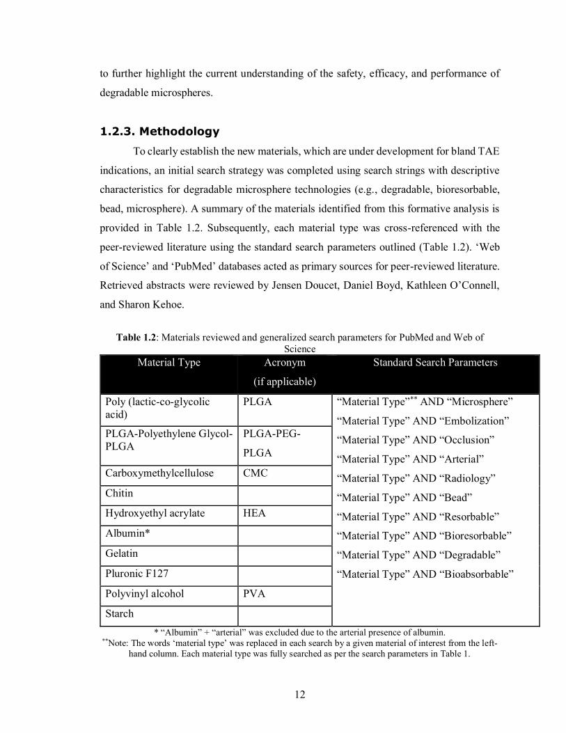
Table 1.2: Materials reviewed and generalized search parameters for PubMed and Web of Science ................................................................. 12
Table 1.3: Initial Returned Searches based on Table 1, with Articles Meeting Inclusion Criteria .................................................................................... 13
Table 1.4: Pre-clinical safety summary for hydrolysis mediated degradable PLGA microspheres ............................................................................... 16
Table 1.5: Pre-clinical safety summary for hydrolysis mediated degradable PEG-PLGA-PEG microspheres.............................................................. 21
Table 1.6: Pre-clinical safety summary for hydrolysis mediated degradable CMC microspheres ................................................................................. 27
Table 1.7: Pre-clinical safety summary for hydrolysis mediated degradable Chitin microspheres................................................................................. 31
Table 1.8: Pre-clinical safety summary for hydrolysis mediated degradable HEA microspheres ................................................................................. 36
Table 1.9: Mass attenuation and toxicity information for B, Rb, Sr and Ga ............ 46
Table 3.1: Glass compositions by molar fraction (pre-fired material) ..................... 60
Table 3.2: 100% volume fraction values of R1, R2, and R2* extrapolated
from given data ....................................................................................... 71
Table 4.1: Test and Control Article Allocation ........................................................ 93
Table 4.2: Characterization data of BRS2 glass frit ................................................. 97
Table 4.3: Thermal Analysis Data for Frit versus Spheres ...................................... 99
Table 4.4: Spherical Data and Particle Sizes of BRS2 Microspheres ...................... 100
vii
List of Figures Figure 1.1: Vascular anatomy of uterine leiomyoma ............................................... 2
Figure 1.2: Distribution, xii, of the borate basic structural unit species, B(n), in monovalent substituted glasses. The solid lines correspond to the model distribution of (super)structural units .................................... 44
Figure 3.1: XRD analysis of A) 5 gallium series glass compositions and B) 5 strontium series glass compositions showing two amorphous peaks, at 2Θ values of approximately 25 and 45, corresponding to 3 and 4 coordinated boron centers in the glass................................... 64
Figure 3.2: Particle size analysis of A) BRG series and B) BRS series where D90, D50 and D10 stand for particle diameters at 90%, 50% and 10% cumulative size, respectively. Error bars are plotted for all points, but are contained within the size of the symbol.......................... 64
Figure 3.3: 11B MAS NMR line spectra of A) the BRS series, and B) the BRG series. The X value indicated the percentage of Sr or Ga substituted ............................................................................................... 65
Figure 3.4: Fraction of four coordinated boron content by mol% of substituted ion ........................................................................................ 66
Figure 3.5: Plot of B4 concentrations versus oxygen to boron ratios of the BRS series (overlapping blue squares) and the BRG series (red). The blue line marks the theoretical values predicted in alkali modified glasses (see text), and the orange points mark the estimated values when the B4 percentage is compared to both Ga and B as network formers (circle) and when all tetrahedral (B and presumably Ga) are compared to both Ga and B as network formers (dash) ........................................................................................ 66
Figure 3.6: A) Density and B) molar density analysis of all 11 glass compositions displayed by percentage of ion substitution. Error bars are plotted for all points, but are contained within the size of the symbol ............................................................................. 67
Figure 3.7: Glass transition temperatures, both onset and inflection, of the BRS glass compositions ......................................................................... 68
Figure 3.8: Glass stability (∆T= Tp1– Tg) of the BRS glass compositions, calculated with both Tg onset and Tg inflection ..................................... 68
viii
Figure 3.9: CT radiopacity values of all 11 glass compositions at (A) 120 kVp and 80 kVp, and (B) 120kVp with comparisons. The dashed line at 7733 HU represents 40-150 µm ORP5 radiopacity, and the dashed line at 2455 HU represents the radiopacity of half strength contrast media ........................................................................................ 69
Figure 3.10: MRI results for A) R1 values of i) BRG and ii) BRS, B) R2 values of i) BRG and ii) BRS, and C) R2* values of i) BRG and ii) BRS ............................................................................................. 70
Figure 3.11: MRI delta chi (∆) values for A) BRG compositions, and B) BRS compositions ............................................................................. 70
Figure 3.12: Percentage dissolved by mass of A) the BRG series and B) the BRS series over 6, 12, 24, 36, and 48 hours ................................ 72
Figure 3.13: Slope of dissolution data vs composition for the BRS series .............. 72
Figure 4.1: Benchtop renal artery model. (A) represents the fluid reservoir and the pump which approximates the heart. (B) indicates the area that has been modified to incorporate the capillary bed (C) denotes the percutaneous introducer used to insert the catheter (<20Fr) into the model for embolization .................................................................... 82
Figure 4.2: A) Complete setup of the benchtop model, B) stepwise reduction from the ‘renal artery’ to the ‘arteriole’ to the capillary bed .................. 83
Figure 4.3: Reduction adaptor and micro-tubing filled with EmboSphere® suspended in DMEM following completion of one trial ........................ 85
Figure 4.4: Division System Method: D1 refers to the immediate and largest branches of the caudal artery, subsequent branches of this artery are labelled D2, further branches labelled D3, etc ................ 96
Figure 4.5: A) XRD data for the BRS2 glass frit and the microspheres. Two amorphous peaks can be seen at 2Ø values of approximately 25 and 45, corresponding to 3 and 4 coordinated boron centers in the glass. B) Density and molar density data for the original BRS2 glass frit and the microspheres. C) Glass transition temperatures and glass stabilities (both onset and inflection) for the BRS2 glass frit and
the microspheres. D) CT radiopacity for the BRS2 glass frit and the microspheres at 80 kVp and 120 kVp .................................................... 98
Figure 4.6: SEM images of microspherical particles (100x) ................................... 99
Figure 4.7: The volume-temperature diagram for a glass-forming liquid, superimposed with the accompanying atomic structure. “L” signifies liquid phase, “G” signifies glass, and “X” signifies ................ 101
ix
Abstract Transarterial embolization (TAE) is a minimally invasive procedure proven to reduce
health care costs and recovery times while maximizing quality of life for patients. Next
generation microspheres for TAE are required to be degradable and exhibit multi-modal
imageability. Accordingly, two series of borate networks were investigated as candidate
degradable radiopaque embolic agents for use in uterine artery embolization (UAE). The
effect of substitutions of SrO or Ga2O3 for Rb2O on the structure and properties of borate
networks was evaluated using density, differential scanning calorimetry, 11B MAS-NMR,
mass loss, CT and MRI experiments. The glasses exhibited high radiopacities (over 3200
HU) and various degradation timeframes (from under 6 hours to over 48 hours). To assess
the safety and efficacy of novel degradable, imageable microsphere for use in UAE an in
vivo animal study protocol was developed. Additionally, an in vitro methodology was
developed to objectively assess the risk of migration.
x
List of Abbreviations and Symbols Used % Percent ° Degree °C Degree Celsius ∆T Melt/Glass Stability ∆ Delta Chi λ Wavelength Å Angstrom A Atomic Mass ANOVA Standard Analysis of Variance ATP Adenosine Tri-Phosphate ATSDR Agency for Toxic Substances and Disease Registry B Boron B3 Three-fold Coordinate Boron B4 Four-fold Coordinate Boron BIC Best In Class BRMS Bio-Resorbable Microspheres C6NCL N,N’-(dimethacryloyloxy)adipamide Crosslinker CBCT Cone Beam Computed Tomography CCAC Canadian Council on Animal Care CHUM Centre Hospitalier de l’Université de Montréal cm Centimeter CMC Carboxymethylcellulose CMC-CNN Carboxymethylcellulose-chitosan CT Computed Tomography DEB TACE Drug Eluting Bead Transarterial Chemoembolization dFMEA Design Failure Mode and Effect Analysis DMEM Dulbecco's Modified Eagle's Medium DSC Differential Scanning Calorimetry FBS Fetal Bovine Serum FDA Food and Drug Administration g gram Ga Gallium Gy Gray HCl Hydrochloric Acid HEA Hydroxyethyl acrylate HU Hounsfield Units ICP-OES Inductively Coupled Plasma Optical Emission Spectroscopy ID Inner Diameter IEL Internal Elastic Lamina K Potassium kGy Kilogray kHz Kilohertz kVp Peak kilo Voltage La Lanthanum
xi
LOAEL Lowest Observed Adverse Effect Level LRA Left Renal Artery mg Milligram min minute mL Milliliter mm Millimeter mol Mole mol% Mole Percent MAS-NMR Magic Angle Spinning Nuclear Magnetic Resonance MRI Magnetic Resonance Imaging Na Sodium NaBH4 Sodium Borohydride NBO Non-Bridging Oxygen NHS National Health Service NIST National Institute of Standards and Technology NMR Nuclear Magnetic Resonance NOAEL No Observable Adverse Effects Level NSAID Nonsteroidal Anti-Inflammatory Drugs NTE Non-Target Embolization O Oxygen PBS Phosphate Buffered Saline PEG Polyethylene Glycol PES Post-Embolization Syndrome PET Positron Emission Tomography PLA Polylactic Acid PLG Polyglycolic Acid PLGA Polylactic-co-glycolic Acid PSA Particle Size Analysis PVA Polyvinyl Alcohol Rb Rubidium RbF Rubidium Fluoride ROI Region of Interest rpm Revolutions per Minute RRA Right Renal Artery s second SD Standard Deviation SEM Scanning Electron Microscopy Sr Strontium TA Test Article TAE Transarterial Embolization TAG Tris-Acryl Gelatin TAGM Tris-Acryl Gelatin Microspheres TGMS Tris-Acryl Gelatin Microspheres Tg Glass Transition Temperature Tm Melting Temperature Tp1 First Crystalline Peak
xii
UAE Uterine Artery Embolization UFE Uterine Fibroid Embolization Vf Volume Fraction V-T Diagram Volume-Temperature Diagram wt% Weight Percent XRD X-Ray Diffraction μm Micron μs microsecond Z Atomic Number
xiii
Acknowledgments First and foremost, I would like to thank my supervisor, Dr. Daniel Boyd for all the work you put into turning me into the student I am now (and I know that wasn’t easy). You made it clear right from the beginning that you were going to do everything in your power to help get me to where I want to be, and I wouldn’t have been able to do it without your enthusiasm and expertise. Even though you are officially old now, and ditched me for most of the year to go on sabbatical, I still think you’re pretty cool. I would also like to thank my committee members, Dr. Robert Abraham, Dr. Steven Beyea, and Dr. Mark Filiaggi for their time and energy. Your insightful comments and questions helped me to develop my research further, and answer questions more completely. Thank you very much for putting up with the early morning meetings, and for being so helpful and kind. I would like to thank Dr. Kimberly Brewer, Dr. Elena Tonkopi, and Dr. Ulriki Werner-Zwanzinger for their expertise – both technical and intellectual – in their respective fields. Their willingness to share their immense knowledge with me helped to bring my project to new heights. It has been a privilege working with you all. I would not have been able to complete this project without my labmates and group members, Dr. Kathleen MacDonald-Parsons Dr. Kathleen O’Connell, Dr. Alicia Oickle, and soon to be (different kind of) Dr. Lauren Kiri. You guys have put up with my stupid questions and annoying tendency to be around when everything breaks, and for that I want to thank you. Even though most of you ditched me at the end for bigger and better things, I’m so glad to have met you. I also want to thank my office pals, Brendan, Camryn, Hayden, Kat (again), Taylor, and Tyler for always being available for a chat and a laugh. I would like to acknowledge the financial assistance of NSERC that made this project possible. Last but certainly not least, I would like to thank my family for their continuing love and support. To my parents, Cyndy and Gerry: Thank you for helping me get through the hard parts and taking time to celebrate the good parts. You are the strongest people I know and I love you to MB and back. To my siblings/pseudo-children, Breton, Camryn, and Aidan: I would like to say that even though you are a pain in my neck and take up energy & time I should be spending on my research, I wouldn’t want it any other way. I love you all so much and am grateful for you each and every day. Camryn, I want to thank you specially for putting up with my crazy school schedule and helping me complete this project; you’re the best room/labmate a girl could ask for.
Halifax, June 2018 Jensen Doucet
1
CHAPTER 1
Introduction
The fundamental objective of this thesis is to examine the composition-structure-
property relationship of ternary, high borate glass networks, and to assess their potential as
novel degradable, imageable embolic agents. The specific clinical context for this work
involves the management of hypervascular tumors, specifically uterine leiomyomas. To
fully understand the results of this study, this chapter provides an overview on uterine
leiomyomas, existing treatment approaches, and limitations with current embolic materials.
1.1. Uterine Leiomyoma
Uterine leiomyomas are the most common benign tumors of the female
reproductive tract for premenopausal women [1]. The exact prevalence of the disease is
difficult to ascertain due to their low symptomatic nature in many patients; however, it is
accepted that uterine leiomyomas are present in up to 70% of women of reproductive age
[2,3]. Although technically benign, leiomyomas may cause a variety of debilitating
symptoms such as heavy bleeding, pain, subfertility, pelvic pressure, dyspareunia, urinary
frequency and urgency, and other pelvic symptoms. Treatment of leiomyomas is provided
when patients experience severely uncomfortable symptoms, particularly those that inhibit
day-to-day tasks and compromise quality of life [1,2]. Generally, their overall incidence is
reported to be 29.7 per 1000 patient/year, with peak incidence occurring among women
who are in their early to mid-40s [1].
1.1.1. Pathophysiology of Uterine Leiomyomas
Uterine leiomyomas are benign monoclonal tumors of the uterus composed of
smooth muscle cells and an extracellular matrix of collagen, fibronectin, and proteoglycans
[1]. Their etiology remains unclear; however it is believed that the growth of leiomyomas
is affected by the presence of growth factors such as estrogen and progesterone, as
leiomyomas are not seen in children and regress after menopause [1]. As they develop,
leiomyomas cause enlargement of the uterus. Leiomyomas located in a submucosal
2
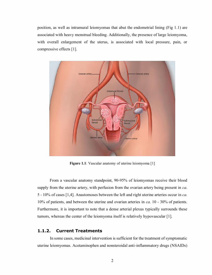
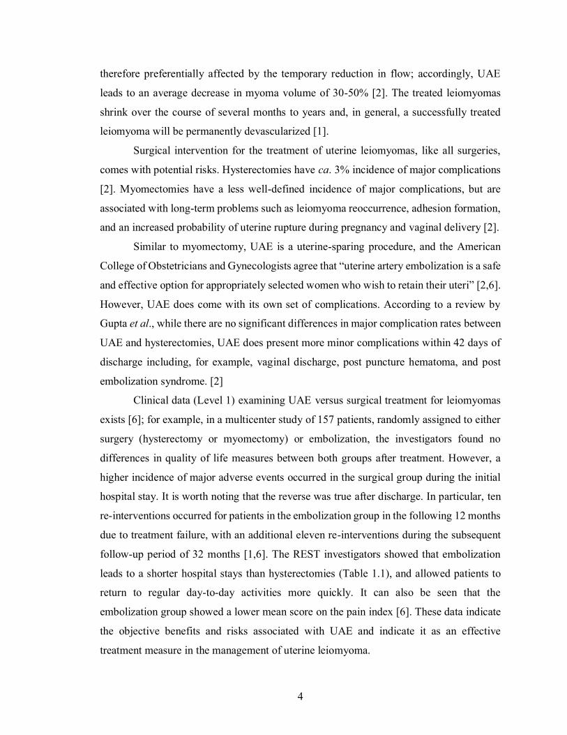
position, as well as intramural leiomyomas that abut the endometrial lining (Fig 1.1) are
associated with heavy menstrual bleeding. Additionally, the presence of large leiomyoma,
with overall enlargement of the uterus, is associated with local pressure, pain, or
compressive effects [1].
Figure 1.1: Vascular anatomy of uterine leiomyoma [1]
From a vascular anatomy standpoint, 90-95% of leiomyomas receive their blood
supply from the uterine artery, with perfusion from the ovarian artery being present in ca.
5 - 10% of cases [1,4]. Anastomoses between the left and right uterine arteries occur in ca.
10% of patients, and between the uterine and ovarian arteries in ca. 10 - 30% of patients.
Furthermore, it is important to note that a dense arterial plexus typically surrounds these
tumors, whereas the center of the leiomyoma itself is relatively hypovascular [1].
1.1.2. Current Treatments
In some cases, medicinal intervention is sufficient for the treatment of symptomatic
uterine leiomyomas. Acetaminophen and nonsteroidal anti-inflammatory drugs (NSAIDs)
3
are often effective for the relief of pain, although these drugs do not reduce bleeding [1].
Gonadotrophin releasing hormone-analogues can effectively reduce bleeding and decrease
leiomyoma size; however, they can only be used for a limited time because of their adverse
effect on bone mass [2]. Other hormone therapies are considered in the literature, including:
(i) a combination of oral contraceptive pills, which are effective at decreasing heavy
menstrual bleeding, but are generally not recommended as they have no effect on
decreasing leiomyoma size, or (ii) Danazol, an antiestrogenic therapy, which decreases
leiomyoma size and increases hemoglobin concentrations but commonly causes unwanted
side effects (e.g. painful muscle cramps, edema, depression, etc.) [3]. In addition to these
limitations it is important to understand that many patients wish to avoid hormonal therapy
and/or do not tolerate it well [1].
1.1.2.1. Surgical Options
Many women with symptomatic leiomyomas seek surgical options for treatment.
To clarify, it is estimated that 30-70% of the ~ 600,000 hysterectomies performed each
year in the USA are uterine leiomyoma related [2]. In fact, leiomyomas are the most
common indication for hysterectomy in the USA. The total direct cost for treating
leiomyomas was estimated to range from US$4.1 to $9.4 billion in 2010 [5], and more than
70% of those costs were directly related to hysterectomies [1]. In addition to hysterectomy,
patients may also be eligible for other surgical treatments of leiomyomas including, for
example, hysteroscopic endometrial ablation, trans-cervical resection of the submucous
myoma, laparoscopic myomectomy, laparoscopic bipolar coagulation and/or dissection of
uterine vessels, or myomectomy [2].
Uterine-artery embolization (UAE), frequently referred to as uterine fibroid
embolization (UFE), was introduced in 1995 as an alternative technique for the treatment
of leiomyomas. Since then, UAE has become increasingly accepted as a minimally
invasive, uterine-sparing procedure [6]. UAE involves occlusion of the uterine arteries with
particulate emboli. The intention is to cause ischaemic necrosis of the uterine leiomyomas,
without permanent adverse effects on otherwise healthy tissues (supported by a healthy
myometrium which can rapidly establish new blood supply through collateral vessels from
the ovarian and vaginal circulations) [2]. Myomas appear to be fed by end arteries, and are
4
therefore preferentially affected by the temporary reduction in flow; accordingly, UAE
leads to an average decrease in myoma volume of 30-50% [2]. The treated leiomyomas
shrink over the course of several months to years and, in general, a successfully treated
leiomyoma will be permanently devascularized [1].
Surgical intervention for the treatment of uterine leiomyomas, like all surgeries,
comes with potential risks. Hysterectomies have ca. 3% incidence of major complications
[2]. Myomectomies have a less well-defined incidence of major complications, but are
associated with long-term problems such as leiomyoma reoccurrence, adhesion formation,
and an increased probability of uterine rupture during pregnancy and vaginal delivery [2].
Similar to myomectomy, UAE is a uterine-sparing procedure, and the American
College of Obstetricians and Gynecologists agree that “uterine artery embolization is a safe
and effective option for appropriately selected women who wish to retain their uteri” [2,6].
However, UAE does come with its own set of complications. According to a review by
Gupta et al., while there are no significant differences in major complication rates between
UAE and hysterectomies, UAE does present more minor complications within 42 days of
discharge including, for example, vaginal discharge, post puncture hematoma, and post
embolization syndrome. [2]
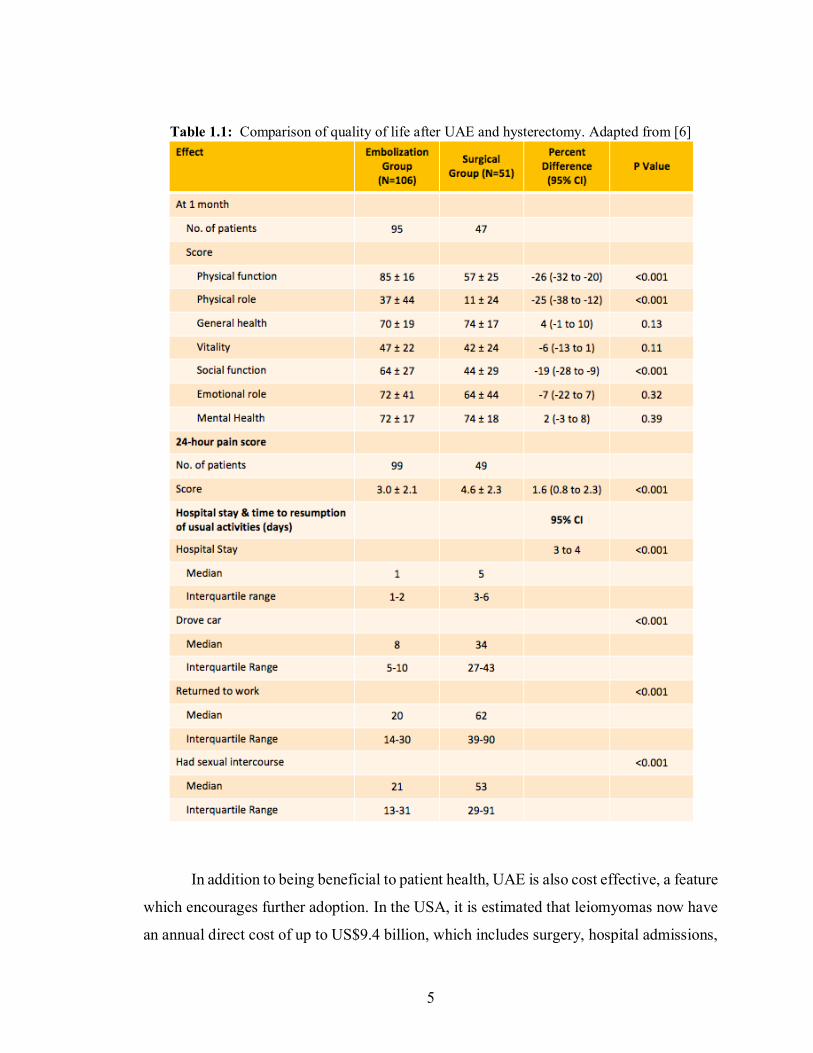
Clinical data (Level 1) examining UAE versus surgical treatment for leiomyomas
exists [6]; for example, in a multicenter study of 157 patients, randomly assigned to either
surgery (hysterectomy or myomectomy) or embolization, the investigators found no
differences in quality of life measures between both groups after treatment. However, a
higher incidence of major adverse events occurred in the surgical group during the initial
hospital stay. It is worth noting that the reverse was true after discharge. In particular, ten
re-interventions occurred for patients in the embolization group in the following 12 months
due to treatment failure, with an additional eleven re-interventions during the subsequent
follow-up period of 32 months [1,6]. The REST investigators showed that embolization
leads to a shorter hospital stays than hysterectomies (Table 1.1), and allowed patients to
return to regular day-to-day activities more quickly. It can also be seen that the
embolization group showed a lower mean score on the pain index [6]. These data indicate
the objective benefits and risks associated with UAE and indicate it as an effective
treatment measure in the management of uterine leiomyoma.
5
Table 1.1: Comparison of quality of life after UAE and hysterectomy. Adapted from [6]
In addition to being beneficial to patient health, UAE is also cost effective, a feature
which encourages further adoption. In the USA, it is estimated that leiomyomas now have
an annual direct cost of up to US$9.4 billion, which includes surgery, hospital admissions,
6
outpatient visits, and medications [5]. Accordingly, any progress towards cost reduction,
without additional risk to patients, is beneficial from a cost containment perspective. The
National Health Service (NHS) of Great Britain and Northern Ireland has established that
embolization is more cost-effective than surgery for patients with symptomatic uterine
leiomyomas. In particular it has been determined that UAE had a mean decrease in cost of
£951 ($1,712 at an exchange rate of £1 = $1.80) over hysterectomies, and remained a cost-
effective option even when the assumed cost of follow up imaging was increased [6]. A
similar study conducted in the USA supports the UK data, showing the average total cost
of UAE (including follow up and morbidity costs) until menopause to be $6,915, vs. $7,847
for hysterectomies [7].
1.1.2.2. Commercially Available UAE Microspheres
While there are many types of products available for uterine artery embolization –
ranging from coils, to balloons, to liquids [8] – embolic agents consisting of micro-particles
are the focus of this thesis. Tris-acryl gelatine (TAG) in the form of EmboSphere® and
PVA microspheres are amongst the most commonly used for UAE [8]. Both particles are
spherical in shape, minimizing the risk of particle aggregation and catheter occlusion. PVA
and TAG microspheres operate via similar mechanisms of occlusion: permanent occlusion
leading to an inflammatory reaction and focal angionecrosis, with vessel fibrosis
developing over time [8]. Multiple studies have shown no significant differences in the
outcomes of procedures done with either of the particles [9,10], and they are cheap, easy
to use and effective [11]. These particles afford consistent controllable embolization at
defined levels in the vascular bed and may also be used as drug delivery systems should
this be so desired [12].
A prominent characteristic of these materials is their compressibility (more so in
TAG particles), allowing for essentially effortless delivery through a catheter [12,13]. It
has been shown, however, that many failure mechanisms of embolization therapy such as
particle deformation and lack of durable occlusion are directly related to compressible
beads [13]. These particles are also radiolucent and permanent, inhibiting visualization of
the particles themselves and recanalization, respectively. Recently, patients have expressed
concerns about foreign materials remaining in their body indefinitely, and therefore are
7
more partial to degradable embolic agents. To assess the range of products being developed
to address the need for degradable embolic agents, a state of the art review of current
preclinical degradable technologies available was conducted and published in the Journal
of Functional Biomaterials in January of 2018.
8
1.2. Advances in Degradable Embolic Microspheres: A State of
the Art Review
Jensen Doucet1, Lauren Kiri2, Kathleen O’Connell2, Sharon Kehoe3, Robert J.
Lewandowski4, David M. Liu5, Robert J. Abraham6, Daniel Boyd3
1 School of Biomedical Engineering, Dalhousie University, Halifax, NS Canada 2 Department of Applied Oral Sciences, Dalhousie University, Halifax, NS, Canada 3 ABK Biomedical Inc., Halifax, NS Canada 4 Department of Radiology, Division of Vascular and Interventional Radiology, Fienberg
School of Medicine, Northwestern University, Chicago, IL, USA 5 Department of Radiology, University of British Columbia, Vancouver, BC, Canada 6 Interventional Radiology and Diagnostic Imaging Department, QEII Health Sciences
Center, Halifax, NS, Canada
This manuscript was written by the candidate (Jensen Doucet) under the supervision of Dr.
Boyd, who provided guidance and assistance throughout all aspects of the review. Ms.
Doucet searched the databases for the determined search terms, reviewed each abstract,
filtered abstracts based on the pre-determined inclusion/exclusion criteria, and wrote the
preliminary draft of the manuscript based on FDA guidance documentation. Dr. O’Connell
and Ms. Kiri aided in the review of the selected articles, as well as with the editing of the
manuscript. Dr. Kehoe, Dr. Lewandowski, Dr. Liu, and Dr. Abraham provided clinical and
technical oversight, reviewed the final manuscript and proposed suggestions with respect
to the clinical significance of the review. Published in the Journal of Functional
Biomaterials on Jan 26, 2018.
9
1.2.1. Abstract
Considerable efforts have been placed on the development of degradable microspheres for
use in transarterial embolization indications. Using the guidance of the U.S. Food and
Drug Administration (FDA) special controls document for the preclinical evaluation of
vascular embolization devices, this review consolidates all relevant data pertaining to novel
degradable microsphere technologies for bland embolization into a single reference. This
review emphasizes intended use, chemical composition, degradative mechanisms, and pre-
clinical safety, efficacy, and performance, while summarizing the key advantages and
disadvantages for each degradable technology which is currently under development for
transarterial embolization. This review is intended to provide an inclusive reference for
clinicians that may facilitate an understanding of clinical and technical concepts related to
this field of interventional radiology. For materials scientists, this review highlights
innovative devices and current evaluation methodologies (i.e. preclinical models), and is
designed to be instructive in the development of innovative/new technologies and
evaluation methodologies.
1.2.2. Introduction
Over the past decade, there has been growing interest in the development of
degradable microspheres for transarterial embolization (TAE) procedures, especially for
applications in trauma, gastrointestinal bleeding, and for the treatment of uterine
leiomyoma. Degradable microspheres are intended to provide effective embolization on a
transient basis. Ideally, after achieving their clinical outcome, they are removed from the
body without interfering with the functionality of other organs. Unlike conventional
permanent agents, degradable microspheres should be designed to optimize the window of
therapeutic intent (e.g., embolization). In so doing, these agents may then balance
therapeutic requirements, while minimizing the potential of long-term sequelae because of
permanent alterations in histological architecture, vascular capacitance and/or injury to
both ‘on target’ and ‘off target’ deposition of therapy. A significant driver for the
development and utilization of degradable microspheres is that “patients commonly
express worries about foreign materials remaining in the body”, and while this may not be
a physiological problem, it is certainly an important consideration for patients and may
10
provide competitive marketing advantages for next generation technologies [14].
Although the safety, efficacy, and performance of permanent embolic agents are
well established in the clinical literature, degradable microspheres may present new safety
concerns. Fortunately, when developing new biomaterials for clinical applications,
researchers benefit from the existence of international standards and guidance documents
to help address potential risks. With respect to vascular embolization devices, specific
guidance documents have been published by regulatory agencies. For example, in 2004
FDA published a document entitled: “Class II Special Controls Guidance Document:
Vascular and Neurovascular Embolization Devices”, which lays out special controls for
establishing the preclinical safety and efficacy of bland embolic microspheres. This
document emphasizes (i) ease of deliverability (from a friction and tortuosity standpoint),
(ii) acute complications, (iii) local and systemic foreign body reactions, (iv) recanalization,
(v) embolization effectiveness, and (vi) device migration. Given the potential new safety
risks that may arise from the use of degradable microspheres, these considerations are
critical in the design and evaluation of new microsphere technologies.
Further to such guidance documents, it is also instructive to consider the ideal
characteristics of degradable microspheres. These innovative technologies must provide
predictable and effective occlusion while also providing:
1. Tailored degradation timeframes—to provide adequate infarction to the target
tissues in a variety of indications, subsequently allowing return of flow (e.g., 5–7 h for
uterine artery embolization—based on Doppler-guided transvaginal clamping) [15]
2. A variety of tightly calibrated particle size distributions—to optimize particle
delivery according to target artery anatomy [16]
3. Ease of delivery through conventional microcatheters—to facilitate adoption of the
novel technology into established embolization techniques
4. Full biological compatibility as per the relevant sections of ISO-10993—to
minimize safety concerns [17]
5. Multi-modal imageability (e.g., fluoroscopy, CT)—to allow for efficiency and
standardization of embolization endpoints [18].
While most of the above points are reasonably self-evident, the last point of multi-modal
imageability raises an important and additional design consideration. Specifically, an
11
understanding of the temporal and spatial distribution of embolic microspheres is clinically
beneficial [18], with the assurance that degradation byproducts should not, for instance,
generate artifacts arising from degradation.
Prior to developing this article further, readers new to TAE are encouraged to
review technical information on techniques and therapies, for example “Transcatheter
Embolization and Therapy; Techniques in Interventional Radiology” [16]. It is also
important to clarify the definitions and terms utilized in the literature related to degradable
microspheres. Terms such as ‘resorbable’ and ‘absorbable’ (with or without the prefix
“bio”) are commonly utilized to describe these technologies. However, it must be
acknowledged that these terms, which are often used as synonyms for one another, are
poorly defined and that despite significant efforts to find consensus about such terms, no
agreed consensus in the interventional radiology or broader biomaterials literature exists
[19]. Conversely, terms such as ‘degradation’ or ‘degradable’ are scientifically defined
throughout the literature. Broadly, degradation refers to “a deleterious change in the
chemical structure, physical properties and appearance of materials” [20]. More
specifically, and within the context of TAE, degradation may be defined as the cleavage of
bonds arising from oxidation, hydrolysis, or enzymatic activity, ultimately culminating in
the complete removal of the agent from the human body. Preferably, the degradation
mechanism(s) and concomitant byproduct(s) provoke minimal adverse local and systemic
responses. For clarity, the remainder of this review will utilize the term ‘degradable’ as per
the aforementioned definition.
Based on the special controls described by FDA, as they relate to degradable
microspheres for TAE applications, this paper intends to consolidate the highest levels of
preclinical evidence relating to the safety, efficacy, and performance of new technologies
which are under development as degradable microspheres – specifically, those that are in
development for bland embolization procedures. This paper is structured to cross-reference
microsphere compositions with the special controls provided by the FDA. This format was
deliberately chosen to provide a robust framework for discussing the current state of the
art technologies with respect to potential risks that may need to be considered as part of a
design control process for the development of new degradable microsphere technologies.
Finally, a review of the preclinical models utilized by the identified papers will be provided
12
to further highlight the current understanding of the safety, efficacy, and performance of
degradable microspheres.
1.2.3. Methodology
To clearly establish the new materials, which are under development for bland TAE
indications, an initial search strategy was completed using search strings with descriptive
characteristics for degradable microsphere technologies (e.g., degradable, bioresorbable,
bead, microsphere). A summary of the materials identified from this formative analysis is
provided in Table 1.2. Subsequently, each material type was cross-referenced with the
peer-reviewed literature using the standard search parameters outlined (Table 1.2). ‘Web
of Science’ and ‘PubMed’ databases acted as primary sources for peer-reviewed literature.
Retrieved abstracts were reviewed by Jensen Doucet, Daniel Boyd, Kathleen O’Connell,
and Sharon Kehoe.
Table 1.2: Materials reviewed and generalized search parameters for PubMed and Web of Science
Material Type Acronym
(if applicable)
Standard Search Parameters
Poly (lactic-co-glycolic acid)
PLGA “Material Type”** AND “Microsphere”
“Material Type” AND “Embolization”
“Material Type” AND “Occlusion”
“Material Type” AND “Arterial”
“Material Type” AND “Radiology”
“Material Type” AND “Bead”
“Material Type” AND “Resorbable”
“Material Type” AND “Bioresorbable”
“Material Type” AND “Degradable”
“Material Type” AND “Bioabsorbable”
PLGA-Polyethylene Glycol-PLGA
PLGA-PEG-
PLGA
Carboxymethylcellulose CMC
Chitin
Hydroxyethyl acrylate HEA
Albumin*
Gelatin
Pluronic F127
Polyvinyl alcohol PVA
Starch
* “Albumin” + “arterial” was excluded due to the arterial presence of albumin. **Note: The words ‘material type’ was replaced in each search by a given material of interest from the left-
hand column. Each material type was fully searched as per the search parameters in Table 1.
13
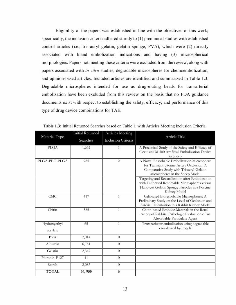
Eligibility of the papers was established in line with the objectives of this work;
specifically, the inclusion criteria adhered strictly to (1) preclinical studies with established
control articles (i.e., tris-acryl gelatin, gelatin sponge, PVA), which were (2) directly
associated with bland embolization indications and having (3) microspherical
morphologies. Papers not meeting these criteria were excluded from the review, along with
papers associated with in vitro studies, degradable microspheres for chemoembolization,
and opinion-based articles. Included articles are identified and summarized in Table 1.3.
Degradable microspheres intended for use as drug-eluting beads for transarterial
embolization have been excluded from this review on the basis that no FDA guidance
documents exist with respect to establishing the safety, efficacy, and performance of this
type of drug device combinations for TAE.
Table 1.3: Initial Returned Searches based on Table 1, with Articles Meeting Inclusion Criteria.
Material Type Initial Returned
Searches
Articles Meeting
Inclusion Criteria Article Title
PLGA 1,662 1 A Preclinical Study of the Safety and Efficacy of OcclusinTM 500 Artificial Embolization Device
in Sheep
PLGA-PEG-PLGA 985 2 A Novel Resorbable Embolization Microsphere for Transient Uterine Artery Occlusion: A Comparative Study with Trisacryl-Gelatin
Microspheres in the Sheep Model
Targeting and Recanalization after Embolization with Calibrated Resorbable Microspheres versus Hand-cut Gelatin Sponge Particles in a Porcine
Kidney Model
CMC 417 1 Calibrated Bioresorbable Microspheres: A Preliminary Study on the Level of Occlusion and Arterial Distribution in a Rabbit Kidney Model
Chitin 585 1 Chitin-based Embolic Materials in the Renal Artery of Rabbits: Pathologic Evaluation of an
Absorbable Particulate Agent
Hydroxyethyl
acrylate
65 1 Transcatheter embolization using degradable crosslinked hydrogels
PVA 2,014 0
Albumin 6,751 0
Gelatin 2,347 0
Pluronic F127 41 0
Starch 2,083 0
TOTAL 16, 950 6
14
1.2.4. Current State of the Art
Based on the search methods, five materials were identified as candidates for
review in this paper. The materials are summarized in Table 1.3, and comprise: Polylactic-
co-glycolic acid (PLGA), PLGA-Polyethylene glycol (PEG)-PLGA,
Carboxymethylcellulose-chitosan (CMC-CCN), Chitosan, and Hydroxyethyl acrylate
(HEA). Although Chitin was the search term originally entered, its derivative in
microsphere form (Chitosan microspheres) warranted inclusion within the assessment, as
the chitin agents were all irregular particles. This paper is structured to deal with each of
these materials individually based on (1) their basic chemistry as it pertains to their
mechanism(s) of degradation, and (2) their respective safety, efficacy, and performance
data tabulated against the specific risk mitigation requirements identified by FDA [17].
Further information on the details of the individual parameters regarding safety, efficacy,
and performance, can be found in the Class II Special Controls Guidance Document
provided by FDA [17]. Finally, the paper provides a brief commentary on preclinical
investigation methodologies utilized by those articles included for review.
1.2.4.1. PLGA
1.2.4.1.1. PLGA: Basic Chemistry and Mechanisms of Degradation
PLGA is a hydrophobic, degradable polymer commonly used in drug delivery and
medical sutures [21]. It is a linear co-polymer that can be synthesized with different ratios
of lactic and glycolic acids [22]. The monomers are linked with an ester bond and,
depending on the ratio of lactic acid to glycolic acid used in polymerization, different forms
of PLGA can be obtained with variable degradation rates [23]. These forms are usually
identified based on the ratio of monomers used; for example, PLGA 75:25 identifies a
copolymer consisting of 75% lactic acid and 25% glycolic acid [24]. In general, low
molecular weight PLGA has been found to degrade more quickly than high molecular
weight PLGA, most likely due to its decreased entanglements, allowing water to penetrate
the structure more readily and hydrolyze the ester bonds [21]. The degradation of PLGA is
well understood and described in detail elsewhere [25-27]. Succinctly, PLGA degrades in
vivo by hydrolysis of the ester bonds between polylactic acid (PLA) and polyglycolic acid
15
(PGA), yielding PLA and PGA as degradation byproducts [22]. PLA undergoes further
hydrolysis to produce monomers, which are metabolized to form lactic acid and then easily
excreted through normal cellular activity or converted to glucose to produce adenosine
triphosphate [28-30]. The degradation of PGA in vivo follows this same process, however
the monomer produced is glycolic acid, which is excreted via the kidney or converted to
pyruvate for use in the tricarboxylic acid cycle.
1.2.4.1.2. PLGA: Safety, Efficacy and Performance
This review identified 1662 articles relating to PLGA based on the search
parameters identified in Table 1.2. Only one of these papers met the inclusion criteria; the
remainder of the articles were substantially focused on in vitro studies and materials for
chemoembolization. The article that met the inclusion criteria was published by Owen et
al. in 2012 [31] and provides a comprehensive and detailed analysis of the safety and
efficacy of PLGA-based microspheres for TAE. This paper utilized a uterine artery sheep
model over a period of 12 months, with animals divided into four cohorts (1, 3, 6, and 12
months). The control article was EmboSphere® (300–500 µm, Merit Medical Systems Inc.,
South Jordan, UT, USA). A summary of the article’s findings versus the specific animal
testing requirements to establish safety and efficacy as per FDA are provided in Table 1.4.
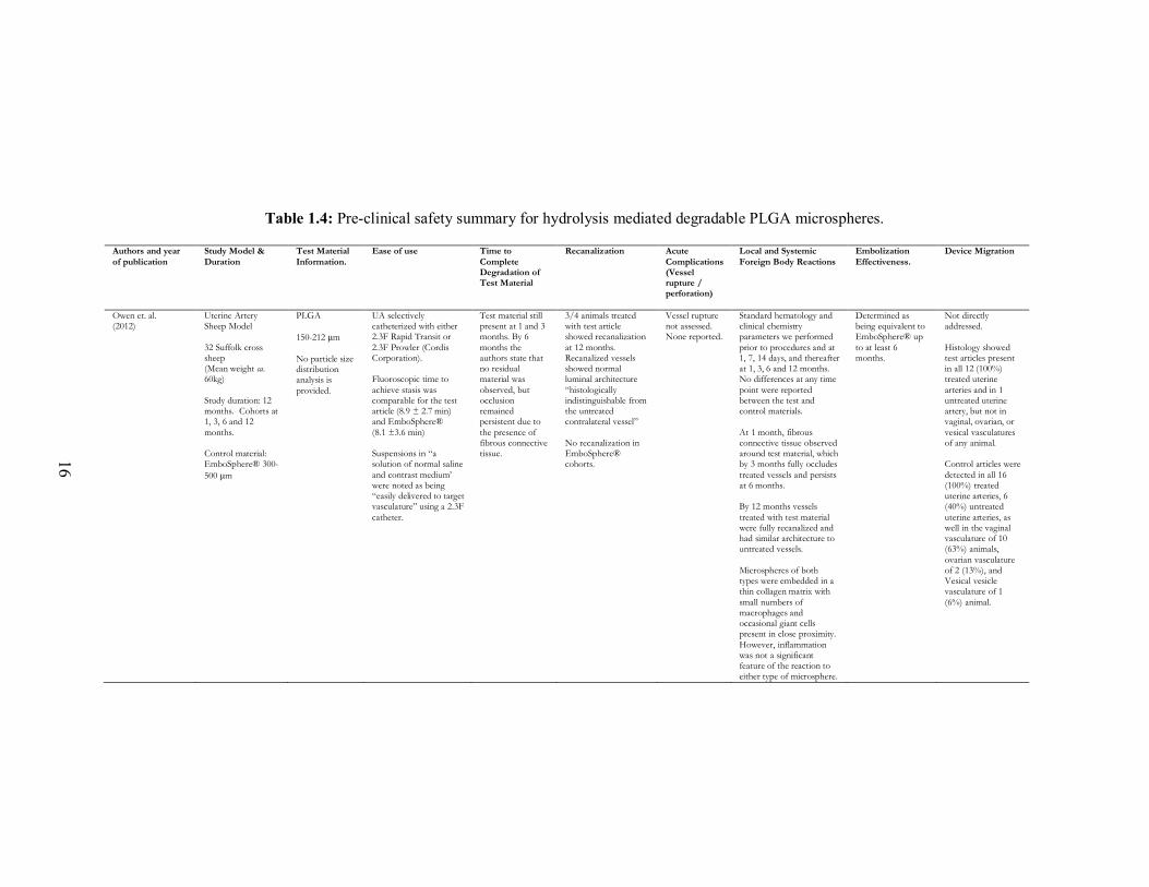
16
Table 1.4: Pre-clinical safety summary for hydrolysis mediated degradable PLGA microspheres.
Authors and year
of publication
Study Model &
Duration
Test Material
Information.
Ease of use Time to
Complete Degradation of Test Material
Recanalization Acute
Complications (Vessel rupture / perforation)
Local and Systemic
Foreign Body Reactions
Embolization
Effectiveness.
Device Migration
Owen et. al. (2012)
Uterine Artery Sheep Model
32 Suffolk cross sheep (Mean weight ca. 60kg)
Study duration: 12 months. Cohorts at 1, 3, 6 and 12
months. Control material: EmboSphere® 300-
500 μm
PLGA
150-212 μm
No particle size distribution analysis is
provided.
UA selectively catheterized with either 2.3F Rapid Transit or
2.3F Prowler (Cordis Corporation). Fluoroscopic time to
achieve stasis was comparable for the test article (8.9 ± 2.7 min) and EmboSphere®
(8.1 ±3.6 min) Suspensions in “a solution of normal saline
and contrast medium’ were noted as being “easily delivered to target vasculature” using a 2.3F
catheter.
Test material still present at 1 and 3 months. By 6
months the authors state that no residual material was
observed, but occlusion remained persistent due to
the presence of fibrous connective tissue.
3/4 animals treated with test article showed recanalization
at 12 months. Recanalized vessels showed normal luminal architecture
“histologically indistinguishable from the untreated contralateral vessel”
No recanalization in EmboSphere® cohorts.
Vessel rupture not assessed. None reported.
Standard hematology and clinical chemistry parameters we performed
prior to procedures and at 1, 7, 14 days, and thereafter at 1, 3, 6 and 12 months. No differences at any time
point were reported between the test and control materials.
At 1 month, fibrous connective tissue observed around test material, which by 3 months fully occludes
treated vessels and persists at 6 months. By 12 months vessels
treated with test material were fully recanalized and had similar architecture to untreated vessels.
Microspheres of both types were embedded in a thin collagen matrix with
small numbers of macrophages and occasional giant cells present in close proximity.
However, inflammation was not a significant feature of the reaction to either type of microsphere.
Determined as being equivalent to EmboSphere® up
to at least 6 months.
Not directly addressed.
Histology showed test articles present in all 12 (100%) treated uterine
arteries and in 1 untreated uterine artery, but not in vaginal, ovarian, or
vesical vasculatures of any animal. Control articles were
detected in all 16 (100%) treated uterine arteries, 6 (40%) untreated
uterine arteries, as well in the vaginal vasculature of 10 (63%) animals,
ovarian vasculature of 2 (13%), and Vesical vesicle vasculature of 1
(6%) animal.
16
17
Degradable PLGA microspheres have already been approved by FDA and are
available on the market under the brand name Occlusin® 500 Artificial Embolization
Device from IMBiotechnologies Ltd. (Edmonton, AB, Canada) [31,32]; however human
clinical studies have not been published up to the period leading to literature review. To
assess the safety, efficacy, and performance of these microspheres, Owen et al. used a
particle size distribution of 150–212 µm, comparing this to a conventional product,
EmboSphere®, with a size range of 300–500 µm in a sheep model [31]. This PLGA particle
size range selected by Owen et al. is substantially smaller than that used in most clinical
indications, such as hepatic and renal tumor embolization (e.g., 300–500 µm) and uterine
fibroid embolization (e.g., 500–700 µm), as well as the control article included in this paper
[16,31]. However, it is reasonable to assume this small particle size represents a higher risk
with respect to biological response (i.e., higher surface area) and migration, and therefore
safety risks in this study were evaluated using approximated worst-case conditions. With
regards to delivery, Owen et al. reported that it was possible to suspend and visualize the
PLGA microspheres in conventional contrast media, and that the microspheres were
“easily delivered to target vasculature” using a standard 2.3-French microcatheter without
clogging the syringe [31]. The authors note, similar to other published literature, that no
significant difference in (i) the volume of test and control materials delivered or (ii) the
fluoroscopic times required to achieve effective stasis for either product [33].
The PLGA microspheres were shown to degrade in ca. six months; however
occlusion persisted up to nine months due to the presence of fibrous ingrowth [31]. Initially,
occlusion was mechanical in nature due to aggregation of microspheres within the target
vessel. This shifted over time to include biological occlusion at one month and three
months, as fibrous ingrowth formed “a matrix that held the microspheres in place as they
degraded”, maintaining complete occlusion of the treated artery at six months despite the
complete degradation of the PLGA microspheres [31]. This is an anticipated biological
response given the acidic nature of the degradation byproducts arising from PLGA [34].
By 12 months, normal vessel luminal architecture was observed to be “histologically
indistinguishable from the untreated contralateral vessel”, suggesting vessel recanalization
(The term ‘recanalization’ is used in a variety of ways in the literature, ranging from re-
opening of the occluded vessel to the formation of new vasculature [31]. In this study, the

18
term appears to refer to the reopening of the vessel that has been embolized) [31]. Although
PLGA is considered to be a degradable embolic agent in the literature and does technically
degrade in vivo, this extended occlusion time may not meet the intended purpose of
degradable microspheres or be suitable for the indications proposed for such products (e.g.,
<24 h for Uterine Arterty Embolization (UAE) [35]). Furthermore, based on its contact
type and duration (>30 days), PLGA is technically categorized as a permanent agent
according to ISO 10993-1, the international standards used to assess the biological
performance of medical devices [36].
Acute complications, such as vessel rupture and perforation, were not assessed in
this paper. However, it is reasonable to assume that these complications are not likely a
risk associated with PLGA given that it has been cleared by FDA. Further to the generation
of fibrous tissue as discussed, both the PLGA and control microspheres were associated
with a small number of macrophages and occasional giant cells. Nevertheless, the authors
state that “inflammation was not a significant feature of the reaction to either type of
microsphere” [31] and no significant systemic foreign body reactions were reported on
hematological and clinical chemistry analyses.
Although the risk of migration did not appear to be directly assessed by Owen et
al., the authors noted that the control microspheres were detected in non-target vasculature
including vaginal, ovarian, and vesicle arteries (63%, 13%, and 6% of the animals,
respectively). Conversely, PLGA microspheres were not observed in these structures. This
difference may be explained by the compressibility of the control microspheres [37], which
likely facilitated passage of the material through small diameter anastomoses joining the
uterine artery with the vaginal and ovarian arteries [38]. Furthermore, and perhaps more
concerning, was the presence of particles in the vesicle artery, as this was likely a result of
reflux out of the uterine artery back into the umbilical artery, resulting in possible non-
target embolization [38]. The authors attributed the lack of retrograde flow (reflux to
vesicle artery) observed with PLGA to its increased density over the control; it is important
to point out that biological occlusion (in the form of fibrotic encapsulation) likely secures
the PLGA microspheres at the target level [31]. These observations are of import with
respect to designing degradable microspheres. Firstly, inherent to the design, the
degradation must be predictable and proceed in a manner that avoids complications
19
associated with non-targeted embolization due passage of smaller particles through the
target vascular bed. This may raise safety concerns with respect to the clinical utility of
materials designed to degrade in a timeframe shorter than that associated with the
development of a sufficient foreign body response (encapsulation of material at the target
area), which may mitigate the risk of migration. For example, it is considered in the
literature that degradation timeframes of ca. 24 h are sufficient for UAE [15]; however, the
host response at this time point likely represents transient edema and migration of
inflammatory cells without fibrosis. Dichotomously, engineering microspheres that
degrade over time periods sufficient to cause biological responses, suitable to mitigating
migration risk (i.e., fibrous ingrowth), may contradict the design requirement underpinning
the development of degradable microspheres—balancing therapeutic requirements while
minimizing collateral damage to adjacent tissue.
1.2.4.1.3. Key Advantages of PLGA Microspheres (Occlusin® 500
Artificial Embolization Device)
• Approved by FDA for the treatment of unresectable/inoperable hypervascularized
tumors (k093813) [32]
• Available in multiple particle size ranges for a variety of applications
• Easily suspended in conventional contrast media and delivered using standard
embolization equipment
• Demonstrated full biological compatibility (via testing performed to obtain device
clearance)
• Mitigate the risk of migration through biological occlusion (fibrous ingrowth
anchoring the particles in place as they degrade).
1.2.4.1.4. Key Limitations of PLGA Microspheres (Occlusin® 500
Artificial Embolization Device)
• Lack of tailorable degradation timeframes—6 to 12 months occlusion timeframe
only
• Lacks multi-modal imageability.
20
1.2.4.2. PLGA-PEG-PLGA
1.2.4.2.1. PLGA-PEG-PLGA: Basic Chemistry and Mechanisms of
Degradation
The chemical composition of PLGA and the mechanisms by which it degrades are
described in the previous Section 1.2.4.1.1. Polyethylene glycols (PEG) are polymers of
ethylene oxide with a chemical formula of HO–(CH2–CH2–O)n–H, where n can range from
4 to >400 [39]. From a mechanistic standpoint, the degradation of PLGA-PEG-PLGA
begins with hydrolysis of the PLGA crosslinks, yielding PLGA and PEG as the initial
degradation byproducts [40]. It is typically regarded that PEG does not degrade, but rather
is excreted unchanged in urine, leading to a limited risk of toxicity [39]. However, should
PEG degrade, it is metabolized in the kidney and can be evaluated by the presence of
ethylene glycol metabolites, such as calcium oxalate and carbon dioxide, which may pose
risk of toxicity [39].
1.2.4.2.2. PLGA-PEG-PLGA: Safety, Efficacy and Performance
This review identified 985 articles relating to PLGA-PEG-PLGA based on the
keyword search identified in Table 1.2. Only two of these papers met the inclusion criteria;
the remainder of articles were substantially focused on in vitro studies and drug eluting
materials. Papers meeting the inclusion criteria were published by Verret et al. in 2014 [15]
and Maeda et al. in 2013 [41]. With respect to the former, the study utilized a uterine artery
sheep model for a duration of seven days, with tris-acryl gelatin microspheres (500–700
µm) as a control. The latter study used a porcine kidney model for a period of up to seven
days with gelatin sponge particles as a control. Summaries of the articles’ findings versus
the specific requirements to establish safety and efficacy as per FDA are provided in Table
1.5.
21
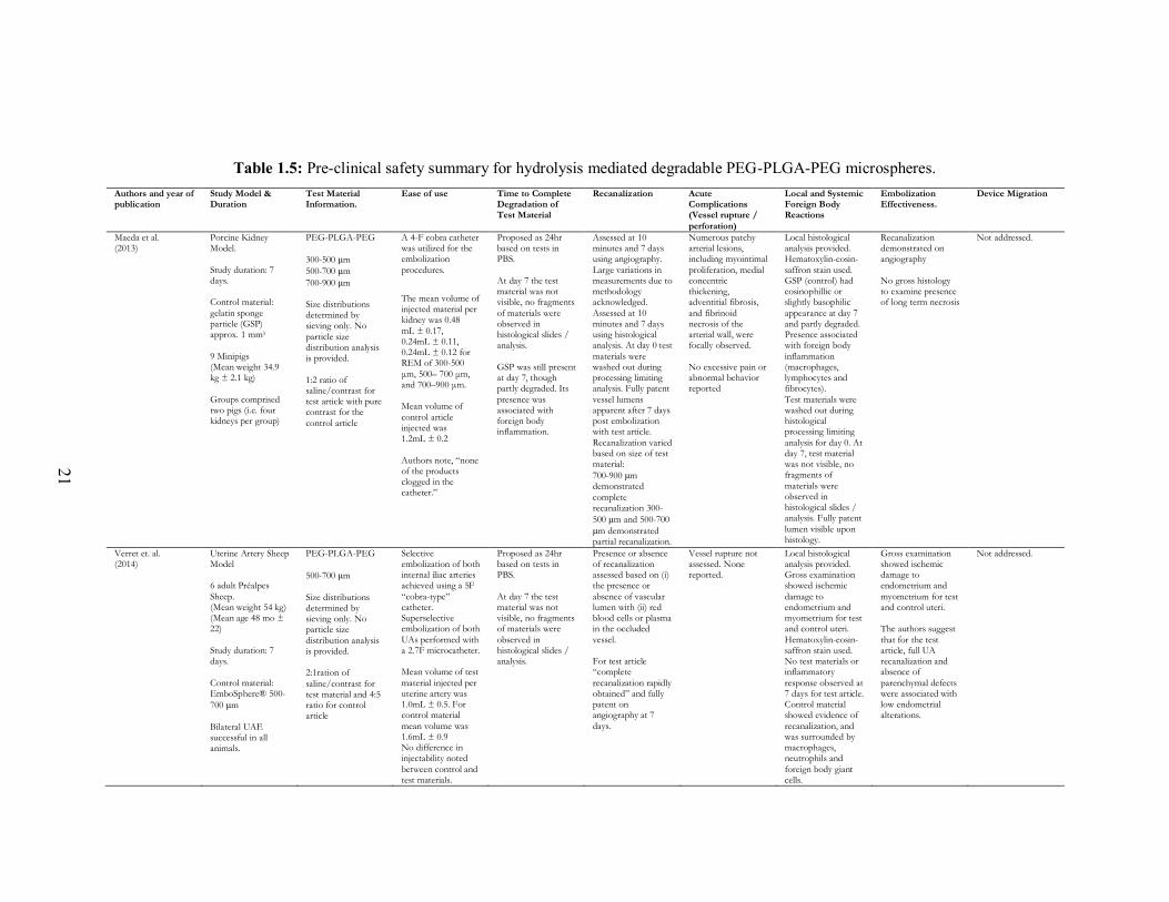
Table 1.5: Pre-clinical safety summary for hydrolysis mediated degradable PEG-PLGA-PEG microspheres.
Authors and year of publication
Study Model & Duration
Test Material Information.
Ease of use Time to Complete Degradation of Test Material
Recanalization Acute Complications (Vessel rupture /
perforation)
Local and Systemic Foreign Body Reactions
Embolization Effectiveness.
Device Migration
Maeda et al. (2013)
Porcine Kidney Model.
Study duration: 7 days. Control material:
gelatin sponge particle (GSP) approx. 1 mm3
9 Minipigs (Mean weight 34.9 kg ± 2.1 kg)
Groups comprised two pigs (i.e. four kidneys per group)
PEG-PLGA-PEG
300-500 μm
500-700 μm
700-900 μm Size distributions determined by sieving only. No
particle size distribution analysis is provided.
1:2 ratio of saline/contrast for test article with pure contrast for the
control article
A 4-F cobra catheter was utilized for the embolization
procedures.
The mean volume of injected material per kidney was 0.48
mL ± 0.17, 0.24mL ± 0.11, 0.24mL ± 0.12 for REM of 300-500
μm, 500– 700 μm, and 700–900 μm. Mean volume of
control article injected was 1.2mL ± 0.2
Authors note, “none of the products clogged in the catheter.”
Proposed as 24hr based on tests in PBS.
At day 7 the test material was not visible, no fragments
of materials were observed in histological slides / analysis.
GSP was still present at day 7, though partly degraded. Its
presence was associated with foreign body inflammation.
Assessed at 10 minutes and 7 days using angiography.
Large variations in measurements due to methodology acknowledged.
Assessed at 10 minutes and 7 days using histological analysis. At day 0 test
materials were washed out during processing limiting analysis. Fully patent
vessel lumens apparent after 7 days post embolization with test article.
Recanalization varied based on size of test material:
700-900 μm demonstrated
complete recanalization 300-
500 μm and 500-700 μm demonstrated partial recanalization.
Numerous patchy arterial lesions, including myointimal
proliferation, medial concentric thickening, adventitial fibrosis,
and fibrinoid necrosis of the arterial wall, were focally observed.
No excessive pain or abnormal behavior reported
Local histological analysis provided. Hematoxylin-eosin-
saffron stain used. GSP (control) had eosinophillic or slightly basophilic
appearance at day 7 and partly degraded. Presence associated with foreign body
inflammation (macrophages, lymphocytes and fibrocytes).
Test materials were washed out during histological processing limiting
analysis for day 0. At day 7, test material was not visible, no fragments of
materials were observed in histological slides / analysis. Fully patent
lumen visible upon histology.
Recanalization demonstrated on angiography
No gross histology to examine presence of long term necrosis
Not addressed.
Verret et. al. (2014)
Uterine Artery Sheep Model 6 adult Préalpes
Sheep. (Mean weight 54 kg) (Mean age 48 mo ± 22)
Study duration: 7 days.
Control material: EmboSphere® 500-
700 μm
Bilateral UAE successful in all animals.
PEG-PLGA-PEG
500-700 μm
Size distributions determined by sieving only. No particle size
distribution analysis is provided. 2:1ration of
saline/contrast for test material and 4:5 ratio for control article
Selective embolization of both internal iliac arteries achieved using a 5F
“cobra-type” catheter. Superselective embolization of both
UAs performed with a 2.7F microcatheter. Mean volume of test
material injected per uterine artery was 1.0mL ± 0.5. For control material
mean volume was 1.6mL ± 0.9 No difference in injectability noted
between control and test materials.
Proposed as 24hr based on tests in PBS.
At day 7 the test material was not visible, no fragments of materials were
observed in histological slides / analysis.
Presence or absence of recanalization assessed based on (i) the presence or
absence of vascular lumen with (ii) red blood cells or plasma in the occluded
vessel. For test article “complete
recanalization rapidly obtained” and fully patent on angiography at 7
days.
Vessel rupture not assessed. None reported.
Local histological analysis provided. Gross examination showed ischemic
damage to endometrium and myometrium for test and control uteri.
Hematoxylin-eosin-saffron stain used. No test materials or inflammatory
response observed at 7 days for test article. Control material showed evidence of
recanalization, and was surrounded by macrophages, neutrophils and
foreign body giant cells.
Gross examination showed ischemic damage to endometrium and
myometrium for test and control uteri. The authors suggest
that for the test article, full UA recanalization and absence of
parenchymal defects were associated with low endometrial alterations.
Not addressed.
21
22
The performance, safety, and efficacy of the PLGA-PEG-PLGA microspheres were
assessed by Verret et al. using a particle size distribution of 500–700 µm, as it represented
the “most common diameter used for uterine fibroid embolization in clinical practice” [15].
Maeda et al. studied this particle size range as well, also incorporating 300–500 µm and
700–900 µm for comparison [41]. No data confirming actual particle size distribution was
listed in either study, thus it may be assumed that the size classifications were based on
sieve aperture utilized to produce the microspheres. It is worth noting sieve aperture
tolerances allow for a degree of error and the actual particle size distributions may be as
low as 286 µm and as high as 585 µm for the 300–500 µm range, 480 µm to 815 µm for
the 500–700 µm range, and 670 µm to 970 µm for the 700–900 µm range [42]. With respect
to injectability and ease of use, no substantial differences between the test and control
articles were reported in either study [15,41]. The reported mean volume of particles
delivered by both groups showed variability, suggesting different volumes may have been
utilized from one animal to next. For example, Verret et al. reported delivering 1.0 ± 0.5mL
of the test article and 1.6 ± 0.9mL of the control [15]. The volumes of test article delivered
by Maeda et al. were notably smaller (0.48 ± 0.17 mL, 0.24 ± 0.11 mL, and 0.24 ± 0.12
mL for the so called ‘REM’ (resorbable embolic microspheres) of 300–500 µm, 500–700
µm, and 700–900 µm respectively). However, the volume of control article delivered was
comparable to the volume of test article delivered by Verret et al. These discrepancies may
be due to variability in animal vasculature—both between and within species but are
worthy of note since they may confound the observations.
PLGA-PEG-PLGA microspheres were reported by both Verret et al. and Maeda et
al. to degrade in vitro in PBS in <24 h, and in vivo in less than seven days. Both papers
angiographically monitored the animals at three time points, as follows: before delivery of
microspheres, 10 min after embolization was achieved, and after seven days. Verret et al.
characterized degradation and recanalization at day seven using a three-tier graded system:
“normal flow, reduced flow (defined as contrast material visible during five heartbeats
before disappearing), and stasis (defined as the blockade of the contrast column in the
[uterine artery])” [15]. All animals treated with the PLGA-PEG-PLGA regained ‘normal
flow’ by day seven and histological analysis showed no remaining fragments and no
arterial wall modifications [15]. Conversely, Maeda et al. reported recanalization as
23
‘patency rates’, showing it correlated with particle size, as well as level of occlusion
(particle distribution), extent of necrosis, and the total percentage of the embolized vessels
that recanalized (‘recanalization rate’). It was observed that decreased particle size
distributions resulted in more distal occlusion, greater necrosis, and lower recanalization
rates [41].These investigations were conducted with angiography and the authors made
strong efforts to correlate them histologically; however, unfortunately, the test
microspheres ‘washed out’ into solvent baths during processing, and so the distribution of
PLGA-PEG-PLGA could not be directly observed [15]. Accordingly, with respect to
Maeda et al., it is difficult to fully evaluate the safety, efficacy, and performance of PLGA-
PEG-PLGA microspheres. However, given the encouraging results, future work will likely
buttress and substantiate this early data; it would be of benefit to develop methodologies
that make it possible to definitively determine the in vivo degradation timeframes of
degradable microspheres. Such methodologies would be of immense benefit since it is
widely accepted that initial host-material responses, including (but not limited to) protein
deposition and cellular interactions, may accelerate or impede the degradation rates of
biomaterials [43].
While it may be possible to argue that the degradation byproducts of PLGA-PEG-
PLGA (e.g., oxalic acid and its calcium salt) may be a concern with respect to systemic
toxicity, evidence for such claims is limited in the literature. Although it is commonly
accepted that PEG is excreted unchanged in urine, PEG byproducts can be as large as
20,000 Da, which is significantly larger than the size exclusion of the glomerulus (ca. 7265
Da) [39,44,45]. However, no animals treated with PEG-PLGA-PEG suffered any obvious
systemic toxicities, pain, abnormal behavior, or atypical blood counts/biochemistry. With
regards to necrosis, Verret et al. found the PLGA-PEG-PLGA microspheres produced
significantly less ischemic damage relative to the control (tris-acryl gelatin), attributing
this, in part, to the short degradation timeframe of PLGA-PEG-PLGA [15]. Maeda et al.
found their control (gelatin sponge) yielded a similar level of necrosis to the smallest
PLGA-PEG-PLGA size investigated (300–500 µm), which was significantly higher than
the two larger PLGA-PEG-PLGA particle sizes explored. This group did not comment on
degradation time as a factor, rather stated the level of necrosis correlated with the
distribution of the microspheres, with those positioned distal to the arcuate artery yielding
24
significantly more necrosis. Given the variability in the size distribution of gelatin sponge,
it is likely the control agent was present both proximal and distal to this anatomical
location, resulting in a higher level of necrosis [41].
Neither Verret et al. or Maeda et al. directly investigated the risk of migration of
degradable PLGA-PEG-PLGA microspheres (as pointed out by the FDA special control
document), and it would be unreasonable to assume that these studies could fully evaluate
the multiplicity of risks identified by FDA. Nevertheless, in consideration of the risk,
Verret et al. did comment on the absence of observable ovarian damage, concluding
migration could not be ruled out solely based on these findings [15]. Given that the
predicted degradation timeframe of PLGA-PEG-PLGA is <24 h, embolization would likely
be limited to mechanical occlusion (i.e., the material itself) and thrombus formation,
without the presence of fibrotic encapsulation (i.e., biological occlusion) to ‘anchor’ the
microspheres in places while they degrade. As discussed previously, the absence of this
biological occlusion may increase the risk of migration and non-target embolization, as
small microsphere fragments may break off and travel forward through small anastomoses,
or reflux retrograde into neighboring vasculature [15,41].
1.2.4.2.3. Key Advantages of PLGA-PEG-PLGA Microspheres
• Limit necrotic damage due to rapid degradation timeframe (ca. <24 h)
• Available in multiple particle size ranges for a variety of applications
• Comparable ease of delivery to control article.
1.2.4.2.4. Key Limitations of PLGA-PEG-PLGA Microspheres
• Lack of tailorable degradation timeframes—ca. seven-day timeframe only
• Toxicity concerns related to PEG degradation byproducts not directly addressed,
may not offer full biological compatibility
• Lacks multi-modal imageability.
25
1.2.4.3. CMC-CNN
1.2.4.3.1. CMC-CNN: Basic Chemistry and Mechanisms of Degradation
Carboxymethylcellulose-chitosan (CMC-CCN) has been proposed as a material for
use in TAE based on its ability to rapidly degrade. CMC-CCN polymers can be created
with varying degradation times by altering the degree of oxidation of the
carboxymethylcellulose (CMC); however, only two time points have been validated in the
literature (14 days for 10% oxidated CMC and 30 days for 25% oxidated CMC) [46,47].
In manufacturing, the two components (CMC and chitosan) are combined in a water-in-oil
emulsion to form crosslinked polymers via a Schiff base reaction between the aldehyde
groups on oxidized-CMC and the amino groups on chitosan [47]. This two-part system
avoids small molecular cross-linking agents, which are usually considered to have higher
cytotoxic potential [47]. The macrostructure of the resulting polymer is susceptible to
degradation by lysozyme, an enzyme that is abundantly present in most parts of the human
body; lysozyme hydrolytically cleaves the Schiff base, separating the material into two
components (CMC and chitosan) [47].
The first component, CMC, is a non-toxic, biodegradable polymer that is widely
used in the pharmaceutical industry [48]. CMC is not degradable by mammalian enzymes
but has demonstrated limited in vivo degradation via hydrolysis of its 1,4-glucosidic
linkages, producing small amounts of glucose [49,50]. The exact extent of CMC
degradation is likely to be determined in future work as the research teams continue to
consider the degradation kinetics and compatibility of degradation by-products in future
work.
Chitosan is a naturally occurring polysaccharide derived from the exoskeleton of
crustaceans that is commonly used in medicine and pharmaceuticals [51]. It is widely
considered non-toxic, having been cleared by FDA for use in wound dressing. Chitosan is
not one chemical entity but varies in composition depending on manufacturing; during
alkaline hydrolysis of chitin to form chitosan, N-deacetylation and depolymerization occur
to varying extents. Structurally, chitosan is considered a polymer of glucosamine and N-
acetylglucosamine, linked by 1,4-glucosidic bonds [52]. Like CMC-CCN, chitosan is also
degraded in vivo by lysozyme, which breaks glucosamine-glucosamine, glucosamine-N-
acetyl-glucosamine, and N-acetyl-glucosamine-N-acetyl-glucosamine linkages, leaving
26
only glucosamine. Glucosamine then goes on to produce glycosaminoglycans,
proteoglycans, and glycolipids in the body. The rate of degradation is historically believed
to depend on the acetylation of chitosan, with more acetylated and thus more crystalline
chitosans (like chitin) showing faster rates of degradation [51-54].
1.2.4.3.2. CMC-CNN: Safety, Efficacy and Performance
This review identified 417 articles relating to CMC-CCN based on the keyword
search identified in Table 1.2. Only one of these papers met the inclusion criteria, the
remainder of articles were substantially focused on drug-carrying materials and
neuroprotective effects in ischemic brain injury. The paper meeting the inclusion criteria
was published by Weng et al. in 2013 [55] and provides a detailed analysis of the safety
and efficacy of CMC-CCN-based microspheres for TAE, which they call ‘bioresorbable
microspheres’ (‘BRMS’). This paper used a renal artery rabbit model over a period of 15
min. The animals were divided into three groups: partial occlusion with BRMS-I (CMC-
CCN with a theoretical oxidation degree of 10%), total occlusion with BRMS-I, and total
occlusion with BRMS-II (CMC-CCN with a theoretical oxidation degree of 25%). Tris-
acryl gelatin microspheres (TGMS) with a particle size range of 100–300 m were included
as the control. Angiography was performed before, immediately after, and 15 min after the
embolization procedure. A summary of the article’s findings versus the specific
requirements to establish safety and efficacy as per FDA are provided in Table 1.6.
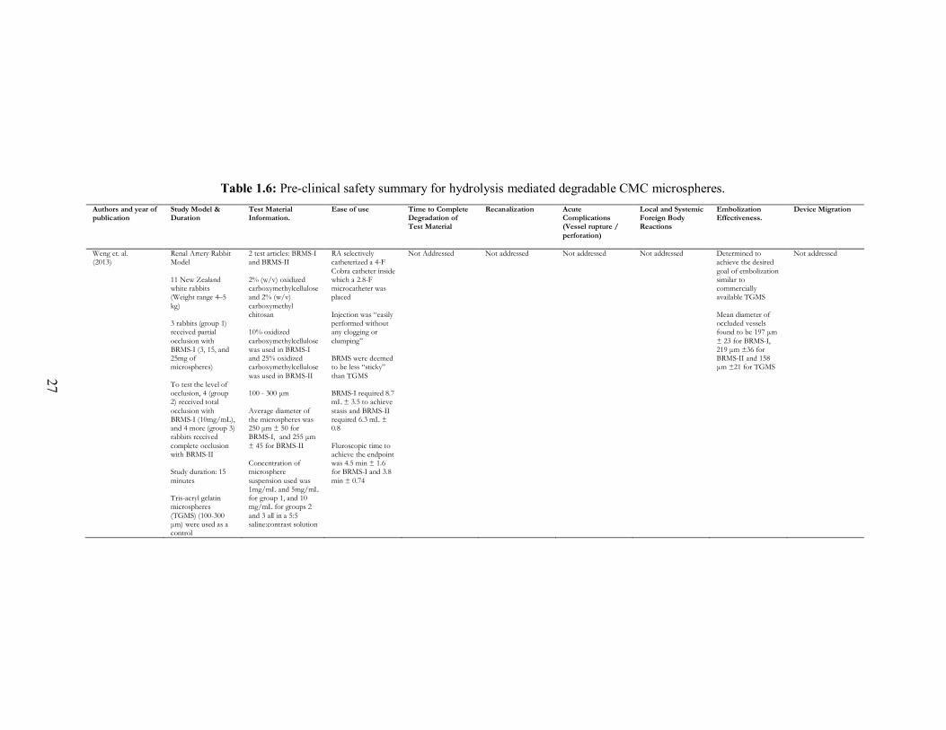
27
Table 1.6: Pre-clinical safety summary for hydrolysis mediated degradable CMC microspheres.
Authors and year of publication
Study Model & Duration
Test Material Information.
Ease of use Time to Complete Degradation of Test Material
Recanalization Acute Complications (Vessel rupture /
perforation)
Local and Systemic Foreign Body Reactions
Embolization Effectiveness.
Device Migration
Weng et. al. (2013)
Renal Artery Rabbit Model
11 New Zealand white rabbits (Weight range 4–5
kg) 3 rabbits (group 1) received partial
occlusion with BRMS-I (3, 15, and 25mg of microspheres)
To test the level of occlusion, 4 (group 2) received total
occlusion with BRMS-I (10mg/mL), and 4 more (group 3) rabbits received
complete occlusion with BRMS-II Study duration: 15
minutes Tris-acryl gelatin microspheres
(TGMS) (100-300 μm) were used as a control
2 test articles: BRMS-I and BRMS-II
2% (w/v) oxidized carboxymethylcellulose and 2% (w/v)
carboxymethyl chitosan 10% oxidized
carboxymethylcellulose was used in BRMS-I and 25% oxidized carboxymethylcellulose
was used in BRMS-II 100 - 300 μm
Average diameter of the microspheres was 250 μm ± 50 for BRMS-I, and 255 μm
± 45 for BRMS-II Concentration of microsphere
suspension used was 1mg/mL and 5mg/mL for group 1, and 10 mg/mL for groups 2
and 3 all in a 5:5 saline:contrast solution
RA selectively catheterized a 4-F
Cobra catheter inside which a 2.8-F microcatheter was placed
Injection was “easily performed without any clogging or
clumping” BRMS were deemed to be less “sticky”
than TGMS BRMS-I required 8.7 mL ± 3.5 to achieve
stasis and BRMS-II required 6.3 mL ± 0.8
Fluroscopic time to achieve the endpoint was 4.5 min ± 1.6 for BRMS-I and 3.8
min ± 0.74
Not Addressed Not addressed Not addressed Not addressed
Determined to achieve the desired
goal of embolization similar to commercially available TGMS
Mean diameter of occluded vessels found to be 197 μm
± 23 for BRMS-I, 219 μm ±36 for BRMS-II and 158 μm ±21 for TGMS
Not addressed
27
28
Beneficially, CMC-CCN microspheres may be produced in a wide variety of
particle size distributions ranging from 100 to 1550 µm [47]. For assessment of this
technology, Weng et al. utilized a size range of 100–300 µm, providing comprehensive
details specific to particle size distribution [55]. This selection of particle size may be
justified, similarly to Owen et al. [31], as representing a worst-case scenario in terms of
biocompatibility (i.e., surface area) and risk of migration (Section 1.2.4.1.2). Importantly,
and with regards to ease of use, Weng et al. stated the injection of CMC-CCN microspheres
was “easily performed without any clogging or clumping” using a 2.8-F microcatheter [55].
The control article was deemed significantly more difficult to use as it tended to stick to
both the syringe and the microcatheter, substantially reducing the percentage of particles
delivered per syringe. The authors attributed the decreased ‘stickiness’ observed for the
CMC-CCN microspheres to the low coefficient of surface friction inherent to hydrogels
[55].
Degradation rates of CMC-CCN microspheres have been previously demonstrated
in vitro and depend on the level of oxidation from processing [56]. In the present study, the
timeframe of 15 min did not allow for assessment of in vivo degradation or recanalization.
Nonetheless from a performance standpoint, the authors stated that more cross-linking can
provide for a slower rate of degradation [55,56]. The key focus of this paper was related to
the acute phase of embolization, focusing on initial particle distribution and level of
occlusion. Comparisons regarding material performance were made when total occlusion
(effective stasis) was used as an end point (versus a pre-determined dose). Interestingly,
microsphere distribution was reported as being dependent on the level of CMC-CCN
crosslinking. Weng et al. found BRMS-II microspheres (oxidation degree of 25%)
occluded slightly more proximal than BRMS-I (oxidation degree of 10%) and the control
(which was equal to BRMS-I), likely due to their lower compressibility. Despite these
observations, the authors reported no statistical differences in the diameter of occluded
vessels or the magnitude of particle deformation between any of the materials [55]. The
CMC-CCN microspheres remained intact in all histological specimens; however, 20% of
the control particles showed pitting or ‘peeling’. The authors stated this may have been a
result of the histochemical processing and agree that it may have obscured their analysis of
microsphere deformation [55].
29
Given the focused and acute scope of this study, it was not possible to comment on
acute complications and foreign body reactions. On histological evaluation, the authors
discussed the presence of ‘white spaces’ surrounding the microspheres, attributing them to
either “dehydration of the microspheres and shrinking during the staining process, or
slicing that was not through the cross-section of the vessel diameter” [55]. It may also be
possible these spaces resulted from thrombus formation, as the authors noted “visible tissue
and blood cells that surrounded the microspheres were erased” prior to analysis [55].
Although risk assessment was limited in this paper due to its short timeframe, the authors
have published a subsequent study, which is considerably more focused on the special
controls published by FDA; this follow-up study was not included in the present review as
it did not meet the inclusion criteria (lack of commercial control), but is cited so as to direct
the readers toward additional data for these materials [57].
1.2.4.3.3. Key Advantages of CMC-CNN Microspheres
• Potentially offers a range of tailorable degradation timeframes based on in vitro
evaluations
• Available in a wide variety of particle sizes from 100 to 1550 µm
• Superior ease of delivery as compared to the control (less adhesive) using
conventional embolization equipment.
1.2.4.3.4. Key Limitations of CMC-CNN Microspheres
• No information on in vivo degradation timeframes or recanalization
• Toxicity concerns related to degradation byproducts and their size(s) not addressed,
may not offer full biological compatibility
• Lacks multi-modal imageability.
1.2.4.4. Chitosan
1.2.4.4.1. Chitosan: Basic Chemistry and Mechanisms of Degradation
Chitin is a biopolymer found in the shells and exoskeletons of animals, such as
insects, squids, and crustaceans, and serves a similar structural role to that of cellulose in
plants or collagen in higher species [58,59]. Structurally, chitin has a highly-extended
30
hydrogen bonded semi-crystalline arrangement, which limits its solubility in water [58].
The chemical composition of chitin is poly(ß-(1-4)-N-acetyl-D-glucosamine). Chitosan is
the N-deacetlyated derivative of chitin and contains multiple free amino groups, typically
with a degree of deacetylation > 0.65 (deacetylation values of 75% and 99% were found in
the specific body of literature explored for this paper) [60]. The degradation mechanism
and products of chitosan have already been addressed in the section on CMC-CCN (Section
1.2.4.3.1).
1.2.4.4.2. Chitosan: Safety, Efficacy and Performance
This review identified 585 articles relating to chitin and chitosan based on the
keyword search identified in Table 1.2. Only 1 of these papers met the inclusion criteria,
the remainder of the articles were substantially focused on in vitro studies and drug-loaded
materials. The paper meeting the inclusion criteria was published by Kwak et al. in 2005
[60] and provides a comprehensive and detailed analysis of the safety and efficacy of
chitin- and chitosan-based particles for TAE. While this paper examined many
morphologies (e.g., thin, scale-shaped chitin and chitosan plates), the current review
focuses solely on data relating to the chitosan microspheres (75% deacetylated). This paper
used a renal artery rabbit model over a period of 32 weeks. The animals were divided into
nine cohorts: 1 and 3 days, and 1, 2, 4, 8, 16, 24, and 32 weeks, and PVA particles (150–
250 m) were used as the control. A summary of the article’s findings versus the specific
requirements to establish safety and efficacy as per FDA are provided in Table 1.7.
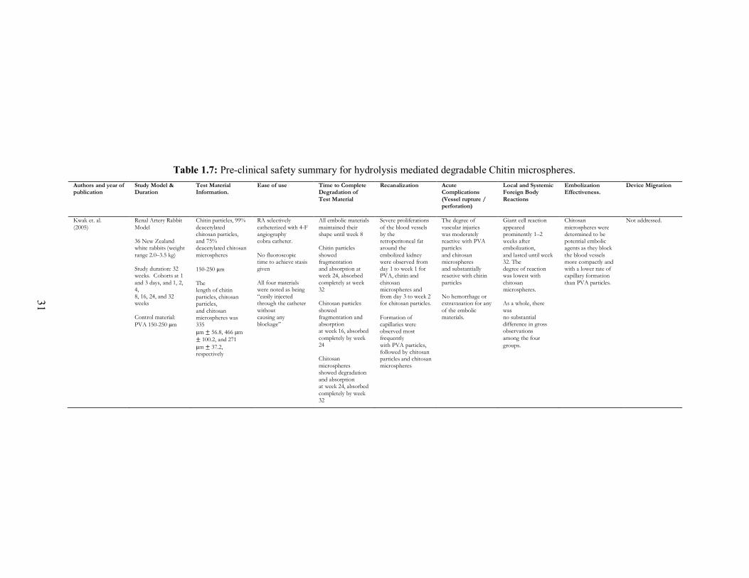
31
Table 1.7: Pre-clinical safety summary for hydrolysis mediated degradable Chitin microspheres. Authors and year of publication
Study Model & Duration
Test Material Information.
Ease of use Time to Complete Degradation of
Test Material
Recanalization Acute Complications
(Vessel rupture / perforation)
Local and Systemic Foreign Body
Reactions
Embolization Effectiveness.
Device Migration
Kwak et. al.
(2005)
Renal Artery Rabbit
Model 36 New Zealand white rabbits (weight
range 2.0–3.5 kg) Study duration: 32 weeks. Cohorts at 1
and 3 days, and 1, 2, 4, 8, 16, 24, and 32 weeks
Control material:
PVA 150-250 μm
Chitin particles, 99%
deacetylated chitosan particles, and 75% deacetylated chitosan
microspheres
150-250 μm
The length of chitin particles, chitosan particles,
and chitosan microspheres was 335
μm ± 56.8, 466 μm
± 100.2, and 271
μm ± 37.2, respectively
RA selectively
catheterized with 4-F angiography cobra catheter.
No fluoroscopic time to achieve stasis given
All four materials were noted as being “easily injected through the catheter
without causing any blockage”
All embolic materials
maintained their shape until week 8 Chitin particles
showed fragmentation and absorption at week 24, absorbed
completely at week 32 Chitosan particles
showed fragmentation and absorption at week 16, absorbed
completely by week 24 Chitosan
microspheres showed degradation and absorption at week 24, absorbed
completely by week 32
Severe proliferations
of the blood vessels by the retroperitoneal fat around the
embolized kidney were observed from day 1 to week 1 for PVA, chitin and
chitosan microspheres and from day 3 to week 2 for chitosan particles.
Formation of capillaries were observed most frequently
with PVA particles, followed by chitosan particles and chitosan microspheres
The degree of
vascular injuries was moderately reactive with PVA particles
and chitosan microspheres and substantially reactive with chitin
particles No hemorrhage or extravasation for any
of the embolic materials.
Giant cell reaction
appeared prominently 1–2 weeks after embolization,
and lasted until week 32. The degree of reaction was lowest with
chitosan microspheres. As a whole, there
was no substantial difference in gross observations
among the four
groups.
Chitosan
microspheres were determined to be potential embolic agents as they block
the blood vessels more compactly and with a lower rate of capillary formation
than PVA particles.
Not addressed.
31
32
To assess the safety, efficacy, and performance of chitosan microspheres, a uniform
size distribution of 150–250 µm was used for all test and control agents. The authors
provided detailed particle size distributions, reporting the mean ± standard deviation of
chitosan microspheres as 271 ± 37.2 µm and non-spherical PVA particles as 326 ± 89.1
µm [60]. The notable increase in mean PVA particle size as compared to the labelled size
range can likely be attributed to the rod-shaped morphology of this material, which enabled
passage through 150–250 µm sieves. Spherical agents were not included for control
purposes, as the authors reported they were “not available at the time [their] study was
performed”. With regards to ease of use, the authors reported both the chitosan
microspheres and control were “easily injected through the catheter without causing any
blockage” [60]. The distinct morphologies of the two materials lead to varied degrees of
occlusion; the chitosan microspheres filled the vessel lumen more compactly than the
irregular PVA particles. The authors do not provide the volume of material delivered per
injection.
Chitosan microspheres were found to have a degradation timeframe of ca. 32
weeks. The first signs of degradation appeared at 24 weeks, with complete absence of the
microspheres at 32 weeks [60]. Although PVA is known to be non-degradable and is
therefore regarded as a permanent agent in the literature, this group found the size and
number of PVA particles decreased over time. This began after eight weeks and persisted
for the duration of the study. Unlike the chitosan microspheres, PVA particles were still
present in situ after 32 weeks. The authors suggested this observation, along with the
presence of PVA fragmentation and foreign body giant cell infiltration, may indicate PVA
underwent some sort of degradation process(es) [60]. Recanalization (reopening of
occluded blood vessels) was not explicitly studied in this paper, although the authors did
comment on the formation of new capillary growth that correlated with the degree of
fibrosis [60]. This was present for both chitosan microspheres and PVA particles, though
it occurred earlier in the PVA cohort (week two vs. week four) and to a more substantial
degree. The presence of fibrosis, in combination with the extended amount of time both
embolic agents were present in the vessels, suggest there was a very good chance that
particle degradation did not lead to vessel reperfusion in either case [60]. This extended
occlusion time lends itself to the same argument as noted above for PLGA (Section
33
1.2.4.1.2); although both materials may eventually degrade in situ, both chitosan and PVA
can essentially be categorized as permanent embolic materials due to their protracted
biological occlusion times [36].
Acute complications were discussed effectively in this paper, owing to the use of
two additional stains, Victoria blue and Masson trichrome, allowing for thorough
examination of elastic lamina damage and fibrosis, respectively. Victoria blue staining
demonstrated that vascular injuries became more severe over time in both groups,
progressing from damage of only internal elastic lamina to damage of the middle
membrane. There was, however, no hemorrhage or extravasation of either material. The
authors proposed that the destruction of the elastic lamina occurred because of either
“ischemia, direct toxic effects of embolized materials, or focal angionecrosis by embolized
materials to the vessel wall” [60]. Inflammation was seen in relation to both particle types
to varying degrees, beginning immediately after embolization, and persisting up to roughly
one week. The inflammation observed with chitosan microspheres, although more severe
than that of the control, was not severe enough to destroy the vessel wall. After
approximately one-week, inflammatory infiltrates aggregated into foreign body giant cells,
which persisted up to week 32 for both materials; these foreign body giant cells presented
before degradation of the material and persisted continuously after the chitosan
microspheres were completely degraded [60]. With regards to systemic reactivity, neither
material altered blood chemistry. Specifically, the authors emphasized the importance of
the low eosinophil count, which suggests chitosan microspheres are unlikely to evoke
allergic reactions [60].
Although not formally assessed, the issue of migration arose due to complications
seen in this study. One rabbit died following reflux of chitin particles (irregular particles
not discussed in detail in this review) from the right renal artery to the left, resulting in
occlusion of both renal arteries. The authors discussed this complication in relation to the
volume of material, stating this occurred when an excessive number of particles were
injected. Unfortunately, the volumes of each material delivered were not provided [60] and
the issue of reflux was not discussed any further or specifically in relation to material
properties, such as embolic morphology or density. As previously mentioned, the extended
timeframe of chitosan microsphere degradation likely mitigates, to a degree, the risk of
34
embolic migration, as fibrous ingrowth anchors the microspheres in place as they degrade.
Nonetheless, this theory merits proper exploration prior to use in human subjects.
1.2.4.4.3. Key Advantages of Chitosan Microspheres
• Easily delivered through conventional microcatheters and provides for more
compact vessel occlusion (relative to irregular PVA particles)
• Low risk of local and systemic toxicity (unlikely a high-risk allergen)—potential to
fulfill full biological compatibility
• Extended degradation timeframe potentially mitigates risk of migration through
stimulation of fibrous ingrowth.
1.2.4.4.4. Key Limitations of Chitosan Microspheres
• No information provided on ability to manufacture different particle size ranges
• Lack of tailorable degradation timeframes—24 to 34 week occlusion timeframe
only
• Lacks multi-modal imageability.
1.2.4.5. HEA
1.2.4.5.1. HEA: Basic Chemistry and Mechanisms of Degradation
Hydroxyethyl acrylate (HEA) is a hydrolytically degradable material that has been
proposed for embolization procedures due to its resiliency and lack of dependence on
enzymatic degradation [61]. HEA is synthesized to contain two degradation sites using a
N,N’-(dimethacryloyloxy)adipamide crosslinker (C6NCL) to modulate the rate of
degradation while maintaining desired mechanical properties. HEA undergoes degradation
in basic conditions, which is an important consideration when designing HEA
microspheres, since embolization procedures are intended to produce ischemic events
(resulting in acidic conditions) and cessation of flow (preventing elimination of carbon
dioxide) [62]. The degradation of hydroxylamines substituted into the material, such as
C6NCL, is known to lead to production of primary amines (e.g., putrescine) and linear
carboxylic acids with the loss of carbon dioxide [61]. Although these degradation
byproducts (e.g., putrescine) are often naturally present in the body, they may be toxic in
35
large quantities [63].
1.2.4.5.2. HEA: Safety, Efficacy and Performance
This review identified 65 articles relating to HEA based on the keyword search
identified in Table 1.2. Only one of these papers met the inclusion criteria; the remainder
of articles were substantially focused on in vitro testing and studies related to high
environmental ammonia. The paper meeting the inclusion criteria was published by
Schwarz et al. in 2003 [61]. This paper used both renal artery canine and auricular artery
rabbit models over a period of three weeks, with EmboGold microspheres (300–500 µm,
Merit Medical Systems Inc., South Jordan, UT, USA) as the control. A summary of the
article’s findings versus the specific requirements to establish safety and efficacy as per
FDA are provided in Table 1.8.
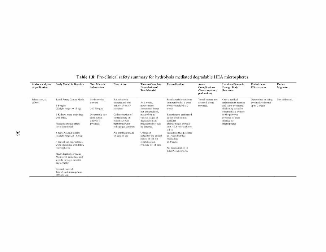
36
Table 1.8: Pre-clinical safety summary for hydrolysis mediated degradable HEA microspheres.
Authors and year of publication
Study Model & Duration Test Material Information.
Ease of use Time to Complete Degradation of Test Material
Recanalization Acute Complications (Vessel rupture /
perforation)
Local and Systemic Foreign Body Reactions
Embolization Effectiveness.
Device Migration
Schwarz et. al. (2003)
Renal Artery Canine Model
5 Beagles (Weight range 10-15 kg) 3 Kidneys were embolized
with HEA Median auricular artery occlusion model
5 New Zealand rabbits (Weight range 2.5–3.5 kg)
4 central auricular arteries were embolized with HEA microspheres
Study duration: 3 weeks. Monitored immediate and weekly through catheter angiography
Control material: EmboGold microspheres 300-500 μm
Hydroxyethyl acrylate
300-500 μm No particle size
distribution analysis is provided.
RA selectively catheterized with
either 4-F or 5-F catheters. Catheterization of
central artery of rabbit ears was performed with radiopaque catheters
No comment made on ease of use
At 3 weeks,
microspheres (sometimes intact but encapsulated, most often in
various stages of degradation and phagocytosis) could be detected
Occlusion lasted for the critical period at risk for
recanalization, typically 10–14 days
Renal arterial occlusions that persisted at 1 week
were recanalized at 3 weeks Experiments performed
in the rabbit central auricular arterial model showed that HEA microspheres
led to occlusions that persisted at 1 week but that recanalized
at 2 weeks No recanalization in EmboGold cohorts.
Vessel rupture not assessed. None
reported.
Only a residual inflammatory reaction
and some neointimal thickening could be observed as a witness to the previous
presence of these degradable microspheres
Determined as being potentially effective
up to 2 weeks
Not addressed.
36
37
The safety, efficacy, and performance of HEA microspheres were evaluated by
Schwarz et al. using a particle size range of 300–500 µm, which was obtained via sieving
[61]. The authors did not provide any commentary regarding the ease of use of either
product, and no values for average volume delivered per injection were provided. It was,
however, mentioned that the transparency of HEA microspheres rendered them practically
invisible in a syringe. To mitigate this issue and facilitate easier handling of this product,
HEA microspheres were colored orange using small amounts of fluorescein acrylate [61].
Schwarz et al. began their study by investigating the degradation rate of synthesized HEA
microspheres in vitro (pH 7.4, 37°C), demonstrating complete degradation of the material
at 22 days. This correlated, to a degree, with what was observed in their in vivo
investigation; on angiography, embolized vessels were recanalized at three weeks.
However, histopathology showed HEA microspheres were often present in situ at three
weeks in “various stages of degradation and phagocytosis”, indicating (i) that degradation
was incomplete at this time point (ii) highlighting potential discrepancies between in vivo
and in vitro degradation findings [61]. In areas where the HEA microspheres had
completely degraded, residual inflammation and neointimal thickening were observed. As
discussed by Shwarz et al., this discrepancy between in vitro and in vivo degradation
timeframes may be a result of the acidic conditions produced during ischemic events and
cessation of blood flow [61]. Nevertheless, the authors effectively showed using both renal
and auricular models that temporary arterial occlusion could be achieved using HEA
microspheres for up to two weeks [61].
Risks associated with acute complications and systemic toxicity were not reported
for HEA microspheres in this work. Little information was provided regarding foreign
body reactions other than noting that both HEA microspheres and the control lead to
inflammation and tissue infarction, with the permanent agent demonstrating notably higher
levels of both. In line with this, the authors stated, “the theoretical advantages of temporary
embolization were not convincingly demonstrated in [their] model since temporary renal
branch occlusions for the time periods investigated still led to tissue infarction” [61]. It
should be noted that a renal artery model is an end vessel model and may not be ideal for
correlating infarction and degradation. The ideal degradation timeframe for embolic
microspheres remains unclear and likely varies across different clinical indications.
38
Although HEA yielded ischemic insult to the healthy tissues examined by Schwarz et al.,
this may not have occurred, to the same degree, in the presence of a tumor/fibroid. Due to
the hypervascular nature of tumors/fibroids, these diseased tissues have been shown to
preferentially attract blood flow and therefore microspheres, sparing neighboring healthy
tissues. Consequently, the ischemia observed by Schwarz et al. may not be representative
of a true clinical scenario. HEA microspheres reliably degraded, succeeding in providing
patients with a dependable material that will be eliminated from their body—a key driver
in the development of degradable microspheres for TAE.
The risk of microsphere reflux was briefly mentioned in the methods of this paper
as a potential complication to avoid; however, it was not discussed in detail, as unlike Kwak
et al. (Chitosan, Section 1.2.4.4), no animals died from microsphere migration. The authors
alluded to microsphere encapsulation in some tissue samples, but it appears this was not
always the case. As such, it is unclear, and perhaps unlikely, a biological mechanism would
be reliably in place to anchor HEA microspheres during degradation, suggesting migration
would be possible.
1.2.4.5.3. Key Advantages of HEA Microspheres
• Produced notably lower levels of ischemia relative to control agent
1.2.4.5.4. Key Limitations of HEA Microspheres
• No information provided on ability to manufacture different particle size ranges
• No information provided on ease of use of microspheres
• Lack of tailorable degradation timeframes—2 to 3 week occlusion timeframe only
• Toxicity of degradation byproducts (e.g., putrescine) not assessed
• Lacks multi-modal imageability.
1.2.5. Preclinical Models
Selecting an appropriate model for the preclinical evaluation of embolization
products is critical for accurately assessing safety, efficacy, and performance prior to
human use. While some parameters can be assessed in vitro (e.g., particle size and ease of
use), animal models are required to best evaluate the material in an environment that
39
approximates the intended clinical scenario [64]. Specifically, with regards to the list of
special controls provided by FDA, in vivo models are required to assess degradation and
recanalization, host response (acute complications and local and systemic reactions), and
migration [64]. As no animal model can truly represent the intended clinical conditions in
their entirety, it is necessary to match the specific objectives under consideration to the best
available model. Unfortunately, this proves to be a complicated process, as several
variables are at play that can significantly alter outcomes and distort results – anatomy,
vascular distribution, blood flow, and immune and coagulation responses are examples of
such [64]. The selected animal, organ, and tissue condition (e.g., tumor vs. healthy) are
three key factors that affect these variables, which must be carefully selected to meet a
study’s objectives [65]. The following section is intended to outline the advantages and
disadvantages of the models incorporated in the papers considered in this review.
The kidney and uterus were the two main organs used to carry out the preclinical
analyses reviewed in this paper. Embolization procedures are performed clinically in both
of these organ structures (e.g., for renal cell carcinoma, uterine fibroids), and thus these
models are clinically relevant. Both of these organs act as a filter, in which microspheres
may be delivered, and entrapped, as the vascular diameters decrease from the renal/uterine
artery down to end arterioles [66]. The uterus is perhaps less straightforward, in that the
uterine artery often anastomoses with the contralateral uterine artery, ovarian artery, and/or
vaginal artery [66]. In theory, these structures are suitable for evaluating the migration risk
associated with degradation, as they allow the material to advance deeper into the vascular
bed as it decreases in size, but remain entrapped within the organ (i.e., not shunt elsewhere).
In the papers under review, the kidney and uterine models enabled, for the most part,
effective evaluation of degradation, recanalization, and host response; however,
commentary regarding migration was limited.
Microsphere migration is perhaps the most difficult parameter to investigate, as
vascular patterns and blood flow are so variable in different species, organs, and disease
states [65]. None of the papers discussed in this review considered a diseased model, and
thus the vascular distribution and blood flow dynamics limited the applicability of these
results to clinical use for several reasons. Firstly, vascular tumors (including uterine
fibroids) tend to show significant increases in blood flow relative to their non-diseased
40
counterparts, and preferentially draw microspheres from the circulation to the tumor [67].
Secondly, the vasculature of tumors is generally more disorganized, potentiating migration
of the material through poorly developed vessels beyond the tumor itself [67]. Finally, a
significant complication of uterine fibroid embolization is post-embolization syndrome
(PES), which is often seen when small particles (e.g., <500 µm) are used and thought to
result from microspheres reaching arterioles in close proximity to the fibroid [68,69]. As
degradable microspheres shrink and likely advance further downstream within the tumor
vascular bed, the risk of this complication may be increased. The complicated nature of
diseased tissue combined with the novelty of degradable microspheres questions the
applicability of the healthy models used in the papers under review as preclinical
evaluations prior to clinical use.
Embolization is performed clinically for several different indications not covered
by the models explored in this review, such as arteriovenous malformation and
hepatocellular carcinoma [19]. It should be noted these indications have a unique set of
inherent risks, perhaps the most concerning of which is bypass of the target tissue with
shunting to vital organs. The vascular networks of these pathologies, and hypervascular
tumors in general, is highly variable and poorly organized, potentially enabling passage of
microspheres to the systemic circulation. This may be of extreme clinical importance,
especially with degradable materials, which are designed to decrease in size over time; the
pressure of the pulsatile flow, coupled with the decreased size and altered compressibility
of partially degraded microspheres may push the embolics further downstream. Several
groups report pulmonary emboli when particles <10 µm are utilized [70], and radioactive
Y-90 microspheres are designed to limit pulmonary radiation below a given threshold,
indicating migration beyond target tissue is an acceptable event associated with the use of
these products. Although the kidney and uterine models discussed above are clinically
relevant, they do not address this important issue and therefore the commentary made in
relation to migration does not translate to all indications.
1.2.6. Conclusions
This review identified five state of the art materials that have been investigated for
use as degradable microspheres for TAE. In considering the FDA’s list of special controls
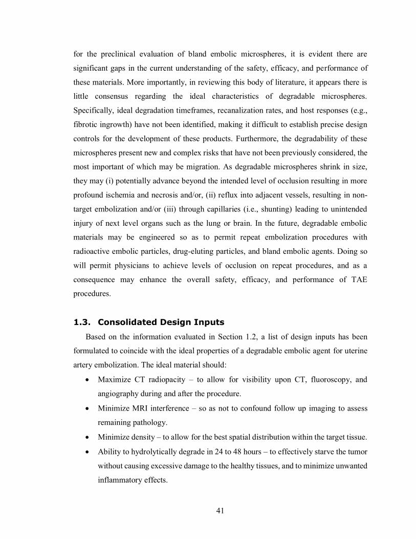
41
for the preclinical evaluation of bland embolic microspheres, it is evident there are
significant gaps in the current understanding of the safety, efficacy, and performance of
these materials. More importantly, in reviewing this body of literature, it appears there is
little consensus regarding the ideal characteristics of degradable microspheres.
Specifically, ideal degradation timeframes, recanalization rates, and host responses (e.g.,
fibrotic ingrowth) have not been identified, making it difficult to establish precise design
controls for the development of these products. Furthermore, the degradability of these
microspheres present new and complex risks that have not been previously considered, the
most important of which may be migration. As degradable microspheres shrink in size,
they may (i) potentially advance beyond the intended level of occlusion resulting in more
profound ischemia and necrosis and/or, (ii) reflux into adjacent vessels, resulting in non-
target embolization and/or (iii) through capillaries (i.e., shunting) leading to unintended
injury of next level organs such as the lung or brain. In the future, degradable embolic
materials may be engineered so as to permit repeat embolization procedures with
radioactive embolic particles, drug-eluting particles, and bland embolic agents. Doing so
will permit physicians to achieve levels of occlusion on repeat procedures, and as a
consequence may enhance the overall safety, efficacy, and performance of TAE
procedures.
1.3. Consolidated Design Inputs
Based on the information evaluated in Section 1.2, a list of design inputs has been
formulated to coincide with the ideal properties of a degradable embolic agent for uterine
artery embolization. The ideal material should:
• Maximize CT radiopacity – to allow for visibility upon CT, fluoroscopy, and
angiography during and after the procedure.
• Minimize MRI interference – so as not to confound follow up imaging to assess
remaining pathology.
• Minimize density – to allow for the best spatial distribution within the target tissue.
• Ability to hydrolytically degrade in 24 to 48 hours – to effectively starve the tumor
without causing excessive damage to the healthy tissues, and to minimize unwanted
inflammatory effects.
42
• Be easily manufactured – to allow for sufficient material to be synthesized and
processed into spheres.
• Be fully biocompatible - to minimize unwanted adverse effects.
1.4. Glasses as Candidate Embolic Agents
Based on these design inputs and previous work in the field of bioactive glasses, it
is possible that glasses may be strong candidate materials for the development of next
generation embolic agents [18]. Their use in dentistry and orthopaedics has illustrated their
ability to add radiopacity to medical materials (e.g. restorative dental composites & bone
cements) without compromising biocompatibility, and while the indications are
substantially different, there is reason to consider their applicability in the embolization of
hypervascular tumours including uterine leiomyoma [13]. In particular, glasses allow for
radiopacity through the incorporation of heavy elements into the glass network [18], the
engineering of variable degradation timeframes by varying the stability of the glass
network, and the ability to manufacture different sized particles based on the methods used
to grind/sieve the manufactured glass frits.
To understand the remainder of the work enclosed in this thesis, it is first important
to understand some vocabulary related to glass science. A glass, in the broadest sense, is
an amorphous solid which exhibits “glass transformation behaviour”; essentially glasses
are considered solid liquids, as they display comparable properties to solids, but their
structure is more similar to that of a traditional liquid [71]. Sun explained that elements
with high single bond strength are less able to rearrange during cooling from a liquid to a
solid, thereby creating a glass through melt quenching [72]. He then divided glass
components into three classes: network formers, network modifiers, and network
intermediates. Network formers are the elements which form the network structure of the
glass, and typically bond very strongly to oxygen. Network modifiers have a weaker single
bond strength, and can therefore rearrange easily and modify the glass network. Network
intermediates are elements that can behave either as a network former or a network
modifier, depending on the environment in which they are found. The addition of network
modifiers or network intermediates can greatly alter the glass structure (or network
connectivity) and thereby alter the properties that said glass displays.
43
1.4.1. Borate Glasses
Borate glasses are significantly understudied in the literature due to their hydrolytic
instability; ca. 2% of glass science literature consists of borate glass compositions, and the
majority of this literature relates to the borosilicates (e.g. Pyrex) [71]. Over 20% of the
literature focuses on silicates because of their high durability and use in industry [71].
Traditionally, the degradable properties of borate glasses were unfavorable for industrial
purposes; however, when considering medical devices, this shortcoming becomes an
advantage for potential degradable embolic materials. It is thus important to investigate
simple high boron glass compositions to develop better understanding of the structure-
property relationship of these networks.
Relative to silicates and phosphates, there is little understood about borate glasses
(in particular those where B is the only network forming element). Nevertheless, there are
some areas of the literature that are well studied. The basic structure of a borate glass
networks has been extensively studied and has identified an interesting phenomenon called
the borate anomaly [72]. Pure B2O3 consists of trigonal boron, found largely in the form of
boroxyl rings; however, boron has the potential to form more than one stable configuration
with oxygen, allowing for the formation of super-structural units. Upon the addition of a
monovalent network modifier, trigonal BØ3 (where Ø signifies an oxygen atom bridging
two boron atoms) converts to tetrahedral BØ4- up to about 33 mol% as seen in Figure 1.2
[72]. The mole percent at which this conversion is maximized depends on a variety of
factors such as the charge density and size of the modifier ion. This conversion stabilizes
the glass network and leads to longer dissolution times (i.e. enhanced hydrolytic stability).
Thereafter, (> 33 mol% addition of modifier) the network becomes more disrupted leading
to non-bridging oxygen and more reactive glass species [72,73]. This feature provides a
compositional and structural basis upon which the degradation of borate glasses may be
modulated by the inclusion of specific elements, which can themselves modulate critical
network properties (e.g. radiopacity).
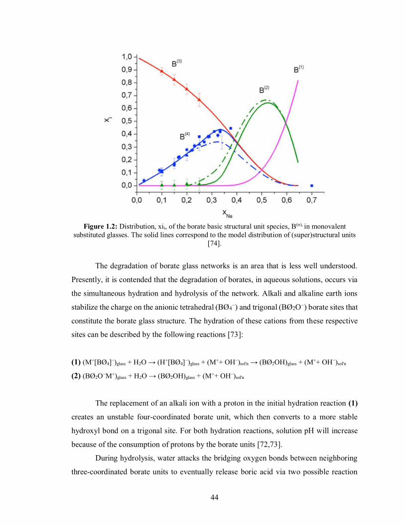
44
Figure 1.2: Distribution, xii, of the borate basic structural unit species, B(n), in monovalent
substituted glasses. The solid lines correspond to the model distribution of (super)structural units [74].
The degradation of borate glass networks is an area that is less well understood.
Presently, it is contended that the degradation of borates, in aqueous solutions, occurs via
the simultaneous hydration and hydrolysis of the network. Alkali and alkaline earth ions
stabilize the charge on the anionic tetrahedral (BØ4−) and trigonal (BØ2O−) borate sites that
constitute the borate glass structure. The hydration of these cations from these respective
sites can be described by the following reactions [73]:
(1) (M+[BØ4]−)glass + H2O → (H+[BØ4]−)glass + (M++ OH−)sol'n → (BØ2OH)glass + (M++ OH−)sol'n
(2) (BØ2O−M+)glass + H2O → (BØ2OH)glass + (M++ OH−)sol'n
The replacement of an alkali ion with a proton in the initial hydration reaction (1)
creates an unstable four-coordinated borate unit, which then converts to a more stable
hydroxyl bond on a trigonal site. For both hydration reactions, solution pH will increase
because of the consumption of protons by the borate units [72,73].
During hydrolysis, water attacks the bridging oxygen bonds between neighboring
three-coordinated borate units to eventually release boric acid via two possible reaction
45
pathways. Reaction 3 shows the hydrolysis of the hydrated borate sites formed by the
hydration reactions described above in Reactions 1 and 2, and Reaction 4 shows the
hydrolysis of two trigonal sites that constitute the original glass structure [73]. The release
of boric acid to solution by the hydrolysis of the borate sites will decrease the pH of the
solution. As there is a much larger number of boron centers than modifier ions, the overall
pH of the local environment will decrease with the degradation of the glass system.
(3) (BØ2OH)glass + H2O → B(OH)3 (sol'n)
(4) (Ø2B-O-BØ2)glass + 3H2O → 2B(OH)3 (sol'n)
.
The dissolution of borate glasses progresses by the simultaneous hydration of metal
ions and the hydrolysis of the borate network [73]. The identity and concentration of the
network modifier plays a large role in the dissolution kinetics; however, these dissolution
pathways are only fully understood for alkali, and partially understood for alkali earth
metals; there is little information on how higher valency ions may modulate structural and
degradation characteristics. An understanding of how the modifier ions modulate the
borate glass network is crucial to understanding the mechanism by which the glass
degrades.
1.4.2. Modifier Ions
The role of monovalent (alkali) ions on borate structure has been studied in the
literature, with less information available about how divalent ions or trivalent ions modify
borate glass networks [76]. When divalent and trivalent ions are studied, the compositions
chosen to assess their effects are typically very complex, which inhibits the understanding
of the role of individual elements. Assessing the effects of substituting higher valency ions
into a simple binary or ternary system will develop our knowledge by providing a clear
platform on which to compare the effects of modifier ions, with respect to glass structure
and properties.
Glasses intended for human use require additional considerations when choosing a
modifier ion. For example, for use as uterine embolic agents, glasses should not cause
cytotoxic effects, lead to acute or systemic toxicity, or generally be harmful to the patients.
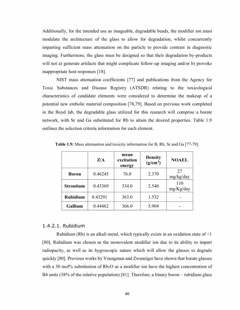
46
Additionally, for the intended use as imageable, degradable beads, the modifier ion must
modulate the architecture of the glass to allow for degradation, whilst concurrently
imparting sufficient mass attenuation on the particle to provide contrast in diagnostic
imaging. Furthermore, the glass must be designed so that their degradation by-products
will not a) generate artifacts that might complicate follow-up imaging and/or b) provoke
inappropriate host responses [18].
NIST mass attenuation coefficients [77] and publications from the Agency for
Toxic Substances and Disease Registry (ATSDR) relating to the toxicological
characteristics of candidate elements were considered to determine the makeup of a
potential new embolic material composition [78,79]. Based on previous work completed
in the Boyd lab, the degradable glass utilized for this research will comprise a borate
network, with Sr and Ga substituted for Rb to attain the desired properties. Table 1.9
outlines the selection criteria information for each element.
Table 1.9: Mass attenuation and toxicity information for B, Rb, Sr and Ga [77-79].
Z/A mean
excitation energy
Density (g/cm3) NOAEL
Boron 0.46245 76.0 2.370 27 mg/kg/day
Strontium 0.43369 334.0 2.540 110 mg/Kg/day
Rubidium 0.43291
363.0 1.532 -
Gallium 0.44462 366.0 5.904 -
1.4.2.1. Rubidium
Rubidium (Rb) is an alkali metal, which typically exists in an oxidation state of +1
[80]. Rubidium was chosen as the monovalent modifier ion due to its ability to impart
radiopacity, as well as its hygroscopic nature which will allow the glasses to degrade
quickly [80]. Previous works by Youngman and Zwanziger have shown that borate glasses
with a 30 mol% substitution of Rb2O as a modifier ion have the highest concentration of
B4 units (38% of the relative population) [81]. Therefore, a binary boron – rubidium glass
47
with 30 mol% rubidium was chosen as the basis for this study, so as to allow for the highest
number of B4 units, theoretically imparting the highest hydrolytic stability of all binary B-
Rb glasses. Additionally, rubidium has a high atomic number to mass ratio and excitation
energy compared to boron, and as a result will impart radiopacity into the glass [77,82].
Although the exact toxicological specifications on rubidium have not been reported
by ATSDR, studies have been done on the potential harmful effects (or lack thereof) of
rubidium [83-85]. A Provisional Peer-Reviewed Toxicity evaluation conducted by the U.S.
Environmental Protection Agency gives rubidium chloride a LOAEL of between 5.1 and
7.7 mg/kg/day. Rubidium has been shown not to have an effect on glomerular filtration
rates in rats, and only demonstrates mild hepatotoxicity in its fluoride salt form (RbF) [86].
It was concluded that the counter ion used in rubidium salts, such as fluoride and carbonate,
are primarily responsible for the toxic effects reported, despite having blood serum
rubidium concentrations of ca. 11 mg/L. Similar results were reported for humans with a
blood serum rubidium concentration of 13.7 mg/L [87]. Rubidium is therefore only slightly
toxic on an acute toxicological basis, and would need to be ingested in large quantities to
pose an acute health hazard [88]. As such, rubidium compounds have been used as both an
antidepressant (as rubidium chloride) and as an iodine source for the treatment of goiters
(as rubidium iodide) [84]. Additionally, rubidium was found to be readily taken up by
tumor cells, as is behaves similarly to potassium [85] in cells whose Na+-K+-ATPase
activities are elevated, which may impart radiopacity into the tumors themselves.
1.4.2.2. Strontium
Strontium (Sr) is an alkali earth metal, typically found in the 2+ oxidation state.
The selection of strontium as the divalent modifier ion for this research was done with the
intent to impart the highest level of radiopacity, without adding any toxicological concerns.
Strontium has a very high NOAEL threshold, allowing for the administration of a large
amount of ion in a short amount of time without adverse toxicological effects [78].
However, care should be taken to avoid strontium treatments in children, as Sr is taken up
in lieu of calcium, which may lead to bone growth problems. As can also be seen in Table
1.9, strontium has a high atomic number to mass ratio, excitation energy, and density
48
compared to boron, and as a result will impart radiopacity into the glass [77] whilst
providing a means to control degradation.
Strontium is similar to calcium both physiologically in terms of absorption and
secretion in the body, but also in effectively controlling the degradation rates of degradable
glasses [88]. From a glass structure standpoint, an increasing SrO fraction in a borate glass
composition favors the conversion of BO3 to BO4 units [89] creating a more durable glass
by (i) acting as the ionic crosslink between non-bridging oxygens (NBOs) and/or
negatively charged tetrahedral boron centers, and (ii) increasing the bond strength of this
ionic crosslink. It has also been shown that binary strontium-borate glass exhibit a higher
glass transition temperature than binary rubidium-boron glasses, demonstrating an increase
in network stability; theoretically this is most likely due to an increase in BO4 units [76].
This increase in tetrahedral coordinated boron can stabilize the glass network, thereby
modulating the time required to achieve complete hydrolytic degradation.
1.4.2.3. Gallium
Initially, lanthanum was chosen as the trivalent modifier ion to be used in this
research due to its nature as a network intermediate; it can act as a network modifier and,
due to its high field strength, may also resemble a network former [90]. Substituting an
alkali oxide for lanthanum has been shown to increase the number of oxygen atoms within
the network, forcing an increase in network volume [91]. This indicates that increasing
substitution of La causes the glass network to stabilize and densify, possibly via the
formation of BO4 [90,91]. The stabilization of the borate network would have been
effective in modulating the degradation time frame of the glass, while imparting significant
radiopacity on the network. However, upon attempting this substitution into a rubidium-
borate glass, it was observed that lanthanum caused rapid crystallization of the glass.
Accordingly, alternative trivalent ions were considered; due to the design criteria, limited
options are available when searching for trivalent ions. Gallium (Ga) was chosen for its
ability to modify the network similarly to lanthanum, and its potential to impart radiopacity.
Gallium is a post-transition metal, commonly found in the 3+ oxidation state
(however 1+ oxidation state is also possible). Gallium has been shown to transition from a
network modifier to a network former (at ca. >6 mol% substitution) and may exist in both
49
tetrahedral and octahedral co-ordination states [92]. This transition may decrease the BO4
concentration by sequestering bridging oxygens away from the boron network, causing the
formation of hydrolysable BO3 units [92]. Regardless of this decrease in BO4, gallium as a
network former appears to stabilize borate networks, and should therefore enable the
production of a more stable glass, with increased resistance to hydrolytic degradation (i.e.
provide an opportunity to modulate longer degradation timeframes as necessary). The
observation of gallium as a substitution for a monovalent ion in a simple ternary system
will give better insight to its exact role in the borate glass architecture. As seen in Table
1.9, gallium has a high atomic number to mass ratio, excitation energy, and density
compared to both boron and strontium. It can be expected to impart significant radiopacity
into the glass, while also offering an ability to modulate network architecture and properties
to achieve the desired technological characteristics [77].
Unlike strontium, gallium does not have an accompanying ATSDR document, and
therefore its exact toxicological specifications become more difficult to predict. It has no
known physiologic function in humans, however it may be able to interact with certain
cellular processes and proteins, especially those related to iron metabolism [93]. This
introduces a potential risk in the design, which will need considerable further research
should proof of principle be delivered with the current work. However, gallium has been
increasingly used in the literature as a therapeutic ion for multiple disorders such as
pathologic bone resorption, autoimmune disease, allograft rejection, infectious disease,
Non-Hodgkin’s lymphoma, bladder cancer, and hypercalcemia [93,94], and as such may
carry limited risk.
50
1.5. The Problem Statement
Based on a comprehensive review of the literature, it is clear that the ideal
requirements for imageable degradable embolic agents may be satisfied by examining
modified borate glasses in a way which permits (i) the balance of sufficient radiopacity (to
provide angiographic visualization), whilst (ii) altering the structure of the borate network
so as to tailor appropriate degradation timeframes. However, the understanding of the
composition-structure-property relationship for borate glasses is not well understood. This
thesis presents the first investigative analysis into borate based degradable radiopaque
embolic agents, while also expanding the compositional palette of borate glasses under
consideration in the literature in a fundamental way. In this study, two series of borate
glasses will be investigated: 1) the BRS series with the composition 70B/30-xRb/xSr
(where x=2,4,6,8,10), and 2) the BRG series with the composition 70B/30-xRb/xGa (where
x=2,4,6,8,10). Each formulation will be fully characterized, and their imaging and
degradation properties examined. Arising from this analysis, a best in class composition
will be chosen and designed to evaluate embolization safety and efficacy in a swine renal
model. This model will be used to evaluate the risks laid out by FDA in the “Class II Special
Controls Guidance Document: Vascular and Neurovascular Embolization Devices” [17].
Additionally, the feasibility of using a benchtop model to examine the risk of migration
will be investigated.
51
CHAPTER 2
Overarching Thesis Objectives
The overall objective of this thesis was to investigate glass compositions suitable for
use as degradable, radiopaque embolic agents, potentially for use in uterine artery
embolization procedures. To do so, two glass series were synthesized and characterized to
determine their potential use as embolic agents, and consequently the potential risks
associated degradable embolic agents were assessed. The following are the specific
objectives of this thesis:
• The first experiment of this work consists of synthesizing and characterizing eleven
glasses of composition 70 B2O3 – 30-X Rb2O – X Y, where Y = Ga or Sr, and
0≤X≤10 (in 2 mol% increments). The aim of this experiment was to contribute to
the expansion of the compositional palate for borate glasses by bettering our
understanding of the composition-structure-property relationship found in simple
borate systems. The objective was to assess the effect of divalent (Sr) or trivalent
(Ga) substitutions of monovalent (Rb) network modifiers on the basic properties of
the glass systems.
• The second experiment of this work consists of assessing each glass for their the
critical, indication specific, performance attributes that would make them effective
embolic agents (i.e. degradability and radiopacity). This portion of the work aims
to advance potential uses of bioactive glasses outside of the skeletal system, by
assessing their potential as TAE materials. These indication specific properties
were then compared to target degradation timeframes, CT radiopacities, and MRI
susceptibilities collected from the literature and from interventional radiologist
opinions, to determine their potential utility as embolic agents.
52
• The issue of migration remains a concern for regulatory agencies and the
development of bench top models may provide a simple mechanism to examine this
risk in an objective manner. Accordingly, the first objective of experiment three
was to develop an in vitro benchtop method to assess the risk of migration. The aim
of this section was to determine the feasibility of assessing migration in vitro, prior
to implantation in an animal model.
• The second objective of experiment three was to choose a best in class (BIC)
composition for the intended indication (based on the information collected in
experiments one and two). This composition was then used to determine the
usefulness of conducting in vitro testing on glass frit over microspheres, when
microspheres will be used in the final application. The comparison of the properties
of both the frit and the microspheres was used to determine if the thermal
augmentation of the glass led to changes in the structure and/or properties of the
selected glass.
• The final objective of experiment three was to develop a pilot in vivo protocol to
provide a preliminary assessment of the safety, efficacy, and performance of the
chosen composition. This pre-clinical protocol was designed to assess the following
criteria (as per FDA guidance): (i) ease of deliverability (from a friction and
tortuosity standpoint), (ii) acute complications, (iii) local and systemic foreign body
reactions, (iv) recanalization, (v) embolization effectiveness, and (vi) device
migration. The aim of this study was to design a pilot model which will demonstrate
how safety, efficacy and performance could be established for degradable agents.
53
CHAPTER 3
Multi-Modal Imageability and Degradation Characteristics of Borate Networks
J. Doucet1, E. Tonkopi2, A. Nuschke3, M.L. Tremblay3, K. Brewer1,3, S. Beyea1,3, M.
Filiaggi1,2,4, R. Abraham2, U. Werner-Zwanzinger5, D. Boyd1,2,4*
1 School of Biomedical Engineering, Dalhousie University, Halifax, NS, Canada
2 Department of Diagnostic Imaging and Interventional Radiology, QE II Health Sciences
Centre, Halifax, NS, Canada
3 BIOTIC, IWK Health Centre, Halifax, NS, Canada
4 Department of Applied Oral Science, Faculty of Dentistry, Dalhousie University,
Halifax, NS, Canada
5 Department of Chemistry, Dalhousie University, Halifax, NS, Canada
* Corresponding Author:
Daniel Boyd, PhD
Department of Applied Oral Science
Faculty of Dentistry
Dalhousie University
Halifax, NS, Canada
54
This manuscript was written by the candidate (Jensen Doucet) under the
supervision of Dr. Daniel Boyd, who provided guidance and assistance to all aspects of the
work. Ms. Doucet proposed and conducted all the experiments described, with the
exception of the solid-state NMR evaluations, which was designed and performed in
collaboration with Dr. Ulrike Werner-Zwanziger from the Chemistry Department. Dr.
Werner-Zwanzinger provided critical support and manuscript review concerning the solid-
state NMR evaluations. Dr. Elena Tonkopi conducted the data collection for the CT
radiopacity measurements and provided assistance with the manuscript preparation
regarding CT radiopacity experiment. Dr. Kimberly Brewer and her team at BIOTIC
conducted the data collection for the MRI radiopacity measurements and provided
assistance with the manuscript preparation regarding MR radiopacity experiments. As first
author of the manuscript presented in Chapter Two, Ms. Doucet prepared the manuscript
and incorporated the suggested edits from the co-authors. All co-authors reviewed the
respective manuscripts and provided critical feedback.
The first two experiments of this thesis are enclosed in the following chapter.
Experiment one encompasses basic characterization of all eleven glass compositions; the
experiments consisted of x-ray diffraction, particle size analysis, solid state 11B NMR
spectroscopy, density, and thermal analysis. Experiment two consisted of specific
characterization of the desired properties investigated for uterine artery embolization; the
experiments consisted of CT imaging, MRI, and mass loss analysis. The results of these
two experiments have been combined into the following manuscript included in Chapter 3.
The following statements were hypothesized for experiments one and two:
• 11B NMR will show a higher ratio of four to three coordinated boron structural units
upon higher concentration of substituted Sr, however the ratio of four to three
coordinated boron will reach a maximum during the Ga series at 6 mol%, and begin
to descend, due to the transition of gallium from a network modifier to a network
former.
• Due to the large charge densities of Sr2+ and Ga3+, addition of these modifier ions
will modulate the super-structural units in the borate network in a way that requires
the formation of space to accompany these large ions, thereby decreasing the

55
density of the borate glass network, and thus of the glass, as the concentration of
substituted ions increases.
• Given that the atomic number to mass ratio, excitation energy, and density of
gallium exceed that of strontium, it will be possible to extend the CT radiopacity of
the gallium containing borate networks to a level above that which is possible for
the strontium containing glass of the same range
• Due to gallium’s ability to transition from a network modifier to a network former,
the gallium substituted glasses will show a change in the rate of mass loss per unit
surface area per time from a decrease to an increase upon increasing substitution.
The strontium substituted glasses will decrease in degradation rate as the amount
of strontium is increased, since the network structure and architecture modulation
will lead to increased four-coordinate boron, hindering hydrolytic degradation.
• The stabilizing nature of the modifier ions will balance with the radiopacity they
impart to yield a particle that has both a degradable time frame of 48 hours ± 10
and a radiopacity of at least 2,500 HU (which is suitable for uterine artery
embolization) in the middle of one of the series.
56
3.1. Abstract
Two glass series were developed based on the substitution of a monovalent glass
modifier for a di- or trivalent ion in a high borate glass system. The BRS series consists of
compositions 0.70 B2O3 – 0.30-X Rb2O – X SrO, and the BRG series consists of
compositions 0.70 B2O3 – 0.30-X Rb2O – X Ga2O3, where 0.00≤X≤0.10 in increments of
0.2. All glasses were characterized in order to examine their composition-structure-
property relationships, and to assess their potential for use as degradable, radiopaque
embolic agents. Glasses were melt quenched and evaluated in terms of structural changes
(11B MAS-NMR, density, and glass transition temperature), changes in radiopacity (both
CT and MRI), and changes in degradation timeframes under simulated physiological
conditions. Structural analysis revealed no change in the tetrahedral boron fraction (B4%)
of the BRS series, despite a linear increase in density and Tg. The strontium acts as a
crosslinker, creating a more hydrolytically stable network, which leads to longer
dissolution times. Conversely, the BRG series displayed a linear decrease in the B4%, a
decrease in density (Tg data not available), yet a slight increase in hydrolytic stability is
also observed. Gallium most likely acts more as a glass former than a glass modifier,
thereby sequestering oxygens from the tetrahedral boron centers, creating trigonal B3
centers and tetrahedral gallium. All glasses were found to be imageable on CT (intensity
>3200 HU at 120 kVp) and invisible on clinical MR imaging modalities.
3.2. Introduction
Despite their unique characteristics, including congruent hydrolytic degradation, low
processing temperatures, high melt stability, and their potential for biomedical
applications, borate glasses are under-represented in the glass science literature.
Specifically, less than two percent of the published literature considers borate glass
compositions, the majority of which relates to borosilicates (e.g. Pyrex) [73]. Increasing
emphasis on the development of degradable biomedical materials, coupled with the
philosophy that development of new biomaterials should be directed towards unlocking
“the body's innate powers of organization and self-repair” [95], is increasing the popularity
of borate glass systems [96]. Contrary to conventional silicate-based bioactive glasses,
borates degrade in water at a rate that may be precisely modulated via compositional or
57
structural modifications [97]. Accordingly, borate glasses have been considered in
application such as wound healing, where it is contended that they stimulate angiogenesis,
support neovascularization, and direct soft tissue repair [98]. With respect to hard tissue
regeneration, objective evidence exists to show that borates support natural healing
processes concurrent to providing an osteoconductive and osseointegrative platform
suitable for skeletal applications [99].
While the ability of borate networks to degrade hydrolytically offers significant
advantages in biomedical engineering, the mechanisms underpinning degradation are
poorly understood. Our limited understanding of factors that may modulate degradation,
such as ionic radius, field strength, and valency, further complicates this situation. It is
understood that pure B2O3 glass consists solely of trigonal boron in mostly boroxyl rings
[100]; however, boron has more than one stable configuration with oxygen [74]. For
example, upon the addition of a monovalent network modifier, trigonal BO3 (B3) converts
to tetrahedral BO4 (B4) up to ca. 33 mol% [74]. The mole percent at which this conversion
is maximized depends on a variety of factors such as the charge density and size of the
modifier ion(s). This conversion stabilizes the glass network and leads to longer dissolution
times (i.e. enhanced hydrolytic stability). Thereafter, (> 33 mol% addition of modifier) the
network becomes more disrupted, leading to higher concentrations of non-bridging
oxygens, resulting in more reactive glasses [74,75]. This feature, termed the ‘borate
anomaly’ [74], provides a unique compositional and structural basis upon which the
degradation of borate glasses may be modulated by the inclusion of specific elements
which can themselves modulate critical network properties. To date, this anomaly has been
highly characterized with alkaline modifier ions, and to a lesser extent alkali-earth ions;
the effect of higher valency ions on the properties of simple borate systems is less
understood, particularly with respect to degradation [75,76].
From a mechanistic standpoint, it is contended that the degradation of borates in
aqueous solutions occurs via the simultaneous hydration and hydrolysis of the network
[75]; however, existing models are limited and based largely on alkali-borate glasses. With
respect to modulating degradation, it is believed that modifier ions “do not passively
occupy sites (cages) formed by the surrounding vitreous network” [74]. Rather, the
structure of the local environment surrounding the modifier is altered to suit their
58
requirements, and thus the size and density of the network is based largely on the modifier
ions present [101]. Consequently, for a given compositional palette, it is possible to
modulate both the material and host responses of borate glasses for a variety of medical
indications.
Recently, glass materials have been increasingly considered for therapeutic
applications in oncology [102] and, in particular, as imageable microspheres for the
transarterial embolization (TAE) of hypervascular tumors. Briefly, TAE is a minimally
invasive, fluoroscopically guided transcatheter procedure that uses microspherical particles
to block blood flow to targeted tissues [16]. Glass microspheres for TAE provide the unique
clinical advantage of CT imageability under clinical conditions without confounding
follow up MRI scans. Specifically, this allows for both intra- and post-procedural feedback
regarding the temporal and spatial distribution of microspheres within target tissues [18]
and consequently may support personalized treatments, standardization of procedures, and
optimized clinical outcomes. In addition to being imageable via CT and X-ray, there is now
an increasing emphasis on the development of degradable microspheres for TAE.
Degradable microspheres may be preferred over permanent embolics as degradable
microspheres could 1) optimize ischemic time to target arteries while 2) minimizing
ischemic effects on adjacent normal tissue, thereby balancing therapeutic requirements
with excessive ischemia of normal tissue; the latter of which can lead to complications
including non-target tissue infarction and post-embolization syndrome [103]. Secondly,
patients have expressed “worries about foreign materials remaining in the body”, and the
use of degradable microspheres may mitigate patient’s anxiety [14]. However, due to the
relative novelty of degradable microspheres, imageability is necessitary to accurately and
objectively assess their efficacy and risks.
We hypothesize that borate glasses will be ideal candidates for use as degradable
embolic agents, as they have tailorable degradation rates as well as the ability to incorporate
CT radiopacifying elements without compromising biocompatibility [18,103]. These
microspheres will thus be visible using CT, but invisible on MRI to allow for follow up
pathological imaging. This study will investigate the multi-modal imageability and
degradation of borate networks modified with gallium and strontium. This objective
satisfies two functions: firstly, it is intended that the work will provide information on the
59
influence of monovalent for divalent, and monovalent for trivalent substitutions in borate
networks. Secondly, this work is intended to allow for the assessment of critical
performance attributes of borate glasses with respect to TAE (i.e. modulation of
degradation, and imageability characteristics).
3.3. Methods
3.3.1. Glass Synthesis
Eleven glasses, separated into two series – one with a monovalent for divalent
substitution and the other monovalent for trivalent – were synthesized through melt
quenching according to the compositions listed in Table 3.1. The glasses were divided into
two series: The BRG series consisting of glasses substituting Rb2O for Ga2O3, and the BRS
consisting of glasses substituting Rb2O for SrO. Glass precursor compositions were
weighed into high density polypropylene (HDPP) jars and mixed mechanically in a blender
for one hour to ensure a homogeneous mixture. Glasses were then melted in 10%Rh /90%
Pt crucibles using a high temperature box furnace (Carbolite RHF 1600, UK), programmed
to heat at 25 °C/min to an initial dwelling temperature of 600 °C and held for 60 min (to
allow calcination). Thereafter the furnace was ramped at 20 °C/min to a final dwelling
temperature of 1100 °C and held for 60 min. Each glass melt was quenched between two
stainless steel plates and the resulting glasses ground with a planetary micromill
(Pulverisette 7), then sieved to retrieve particles of <100 μm, 100-300 μm, and >300 μm
(ASTM standard sieves, Cole Parmer, USA). Glasses were stored in labelled glass vials in
vacuum desiccators for subsequent analysis. Particle size distribution was verified through
laser diffraction of a wet suspension using a Mastersizer 3000 model laser diffraction
particle size analyzer using distilled water as the dispersant.
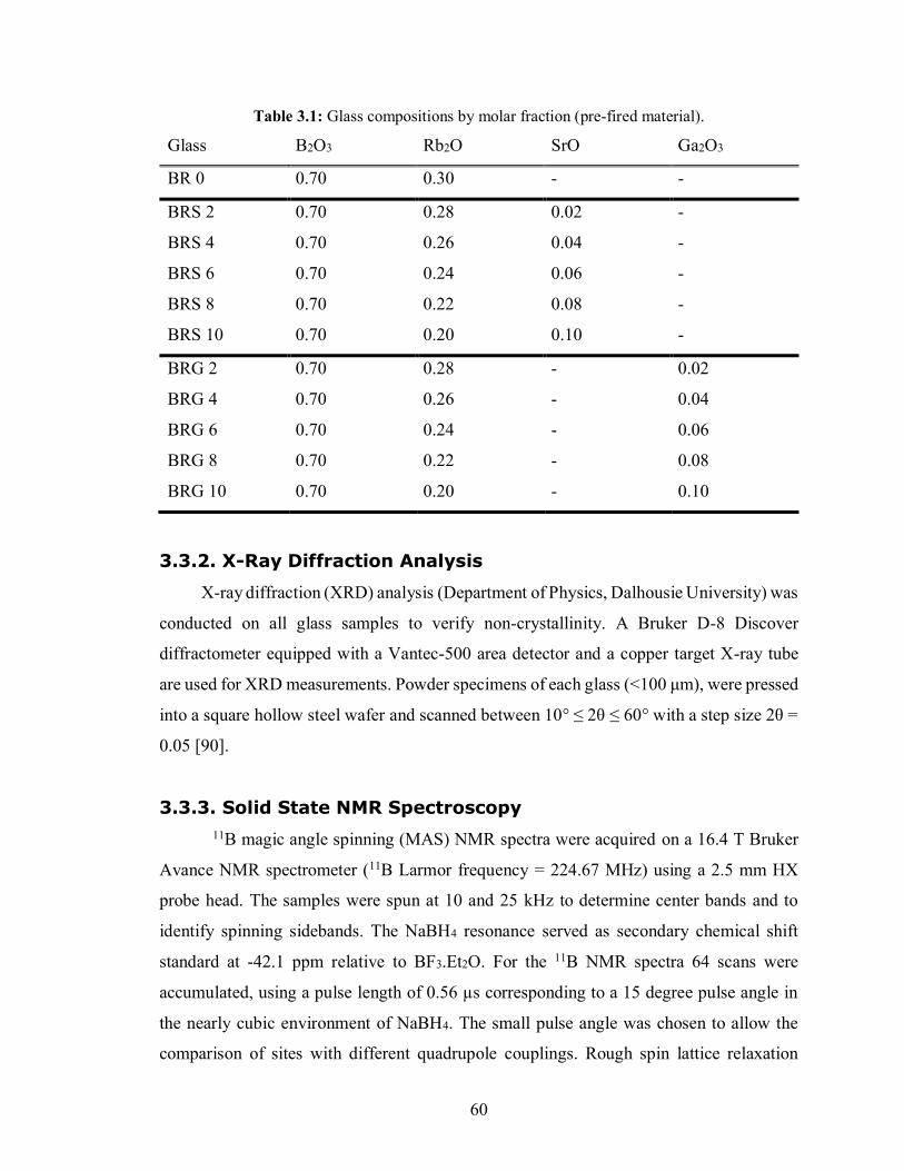
60
Table 3.1: Glass compositions by molar fraction (pre-fired material).
Glass B2O3 Rb2O SrO Ga2O3
BR 0 0.70 0.30 - -
BRS 2 0.70 0.28 0.02 -
BRS 4 0.70 0.26 0.04 -
BRS 6 0.70 0.24 0.06 -
BRS 8 0.70 0.22 0.08 -
BRS 10 0.70 0.20 0.10 -
BRG 2 0.70 0.28 - 0.02
BRG 4 0.70 0.26 - 0.04
BRG 6 0.70 0.24 - 0.06
BRG 8 0.70 0.22 - 0.08
BRG 10 0.70 0.20 - 0.10
3.3.2. X-Ray Diffraction Analysis
X-ray diffraction (XRD) analysis (Department of Physics, Dalhousie University) was
conducted on all glass samples to verify non-crystallinity. A Bruker D-8 Discover
diffractometer equipped with a Vantec-500 area detector and a copper target X-ray tube
are used for XRD measurements. Powder specimens of each glass (<100 μm), were pressed
into a square hollow steel wafer and scanned between 10° ≤ 2θ ≤ 60° with a step size 2θ =
0.05 [90].
3.3.3. Solid State NMR Spectroscopy
11B magic angle spinning (MAS) NMR spectra were acquired on a 16.4 T Bruker
Avance NMR spectrometer (11B Larmor frequency = 224.67 MHz) using a 2.5 mm HX
probe head. The samples were spun at 10 and 25 kHz to determine center bands and to
identify spinning sidebands. The NaBH4 resonance served as secondary chemical shift
standard at -42.1 ppm relative to BF3.Et2O. For the 11B NMR spectra 64 scans were
accumulated, using a pulse length of 0.56 µs corresponding to a 15 degree pulse angle in
the nearly cubic environment of NaBH4. The small pulse angle was chosen to allow the
comparison of sites with different quadrupole couplings. Rough spin lattice relaxation

61
times were determined using a saturation recovery sequence and were on the order of 4-5
seconds. The pulse repetition times were chosen to be approximately three times the longest
relaxation time. In addition to sample spectra, a spectrum of an empty rotor was acquired
under identical conditions. The substantial boron background was removed by subtracting
that spectrum of the empty rotor. The intensities of the different sites were determined by
integration with shift limits of 23.3 to 6.0 ppm for the B3 range and 6.0 to -6.3 ppm for the
B4 resonances [90].
3.3.4. Glass Density
Density measurements were conducted using an AccuPyc 1340 helium pycnometer
(Micromeritics, USA) equipped with a 1 cm3 insert, and packed with 0.9–1.0 g of powdered
glass specimens of each glass (n=10 per glass, 100–300 μm). The results are reported as
the average ± standard deviation (SD) [90]. Molar density was calculated for each glass
composition by pm = p/M, where p is the density of each glass and M is the molecular
weight.
3.3.5. Thermal Analysis
Glass transition temperature (Tg) and first crystallization temperature (Tp1) were
analysed by DSC using a simultaneous thermal analysis — STA 409 PC Luxx® (Netzsch-
Geratebau-GMBH, USA). Approximately 35mg of powdered glass (n=3 per glass, <100
μm) was packed into a platinum/rhodium crucible and heated at 10 °C/min from 50 to 1000
°C. Prior to heating, the BRG series was baked in an oven at 120°C to ensure no surface
water was present to interfere with measurements. Tg onset, Tg inflection and Tp1 were
determined using Proteus Analysis software (VERSION 5.1.1) and are reported as the
average ± SD. The glass stability is reported as ∆T= Tp1 – Tg [105].
3.3.6. Computed Tomography Imaging
Quantitative CT radiopacity measurements of each glass composition (n=5) were
determined using clinical 128-slice CT scanner Somatom Definition AS+ (Siemens
Healthcare, Erlangen, Germany). Glass vials containing particles were imaged at 80 kVp
and 120 kVp, 400 mAs, with 1 mm acquisition slice thickness and pitch=0.5. The
62
radiodensity, or radiopacity, was assessed by placing region of interest (ROI) on axial
images at different locations inside the vial. All measurements were performed on 100–
300 μm particles. A clinically used contrast agent, Isovue® 370 (Iopamidol; 370 mg of
non-ionic iodine per mL, Bracco Diagnostics Inc., Monroe Township, NJ) at half strength
(i.e. mixed 50:50 with saline), and a permanent radiopaque embolic agent, ORP5 [105]
were scanned under the same CT settings in order to benchmark the radiopacity of the glass
compositions. Radiopacity is reported as an average of 5 measurements in Hounsfield Units
(HU) ± standard deviation (SD) [105].
3.3.7. Magnetic Resonance Imaging
Each particles glass composition (100-300 μm) was dispersed at varying volume
fractions (2.5, 5, 7.5 10 and 12.5% w/w) in an 8% aqueous porcine gelatin for the BRS
series. Due to the rapid degradation of the BRG series, these particles were dispersed in a
non-aqueous gel made with 1% Evonik Intelimer IPA 13-1 NG polymer (Evonik
Industries, Essen, Germany) in peanut oil (Our Compliments, NS, Canada). Prior to
addition of the particles, the gels were subjected to magnetic stirring and heating for proper
mixture of all components (40° C for gelatin and 50 °C for the oil gel). Melted gels then
were added to 5 mm Nuclear Magnetic Resonance (NMR) tubes pre-filled with appropriate
amounts of particles. The NMR tubes were then subjected to horizontal rotation to properly
distribute particles throughout the tube and to prevent gravitational settling of the
microspheres prior to rapid cooling on ice to solidify the gel. Measurements of MRI
susceptometry, bulk R2* (full-width at half maximum of the spectral linewidth), bulk R2
(CPMG) and bulk R1 (arrayed inversion recovery) [106] were conducted for all samples at
room temperature using an Agilent 3T preclinical MRI. Values for a purely glass
composition are obtained from linear regression analysis of each imaging parameter and
extrapolated to 100% volume fraction (Vf).
3.3.8. Glass Cylinder Synthesis for Mass Loss Evaluation
The precursor blends for each composition were prepared and melted as per section
2.1, Glass Synthesis. On removal, each glass melt was quenched into stainless steel moulds
(6 mm in length and 4 mm in diameter), which were set between two stainless steel plates.
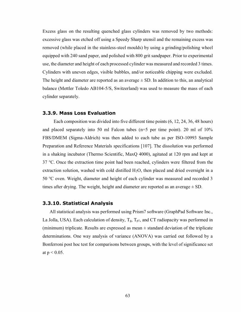
63
Excess glass on the resulting quenched glass cylinders was removed by two methods:
excessive glass was etched off using a Speedy Sharp utensil and the remaining excess was
removed (while placed in the stainless-steel moulds) by using a grinding/polishing wheel
equipped with 240 sand paper, and polished with 800 grit sandpaper. Prior to experimental
use, the diameter and height of each processed cylinder was measured and recorded 3 times.
Cylinders with uneven edges, visible bubbles, and/or noticeable chipping were excluded.
The height and diameter are reported as an average ± SD. In addition to this, an analytical
balance (Mettler Toledo AB104-5/S, Switzerland) was used to measure the mass of each
cylinder separately.
3.3.9. Mass Loss Evaluation
Each composition was divided into five different time points (6, 12, 24, 36, 48 hours)
and placed separately into 50 ml Falcon tubes (n=5 per time point). 20 ml of 10%
FBS/DMEM (Sigma-Aldrich) was then added to each tube as per ISO-10993 Sample
Preparation and Reference Materials specifications [107]. The dissolution was performed
in a shaking incubator (Thermo Scientific, MaxQ 4000), agitated at 120 rpm and kept at
37 °C. Once the extraction time point had been reached, cylinders were filtered from the
extraction solution, washed with cold distilled H2O, then placed and dried overnight in a
50 °C oven. Weight, diameter and height of each cylinder was measured and recorded 3
times after drying. The weight, height and diameter are reported as an average ± SD.
3.3.10. Statistical Analysis
All statistical analysis was performed using Prism7 software (GraphPad Software Inc.,
La Jolla, USA). Each calculation of density, Tg, Tp1, and CT radiopacity was performed in
(minimum) triplicate. Results are expressed as mean ± standard deviation of the triplicate
determinations. One way analysis of variance (ANOVA) was carried out followed by a
Bonferroni post hoc test for comparisons between groups, with the level of significance set
at p < 0.05.
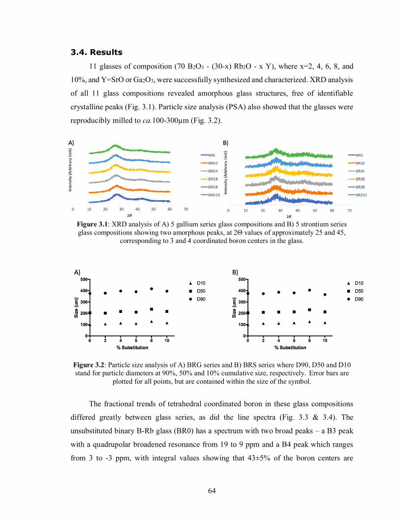
64
3.4. Results
11 glasses of composition (70 B2O3 - (30-x) Rb2O - x Y), where x=2, 4, 6, 8, and
10%, and Y=SrO or Ga2O3, were successfully synthesized and characterized. XRD analysis
of all 11 glass compositions revealed amorphous glass structures, free of identifiable
crystalline peaks (Fig. 3.1). Particle size analysis (PSA) also showed that the glasses were
reproducibly milled to ca.100-300µm (Fig. 3.2).
Figure 3.1: XRD analysis of A) 5 gallium series glass compositions and B) 5 strontium series glass compositions showing two amorphous peaks, at 2Θ values of approximately 25 and 45,
corresponding to 3 and 4 coordinated boron centers in the glass.
Figure 3.2: Particle size analysis of A) BRG series and B) BRS series where D90, D50 and D10 stand for particle diameters at 90%, 50% and 10% cumulative size, respectively. Error bars are
plotted for all points, but are contained within the size of the symbol.
The fractional trends of tetrahedral coordinated boron in these glass compositions
differed greatly between glass series, as did the line spectra (Fig. 3.3 & 3.4). The
unsubstituted binary B-Rb glass (BR0) has a spectrum with two broad peaks – a B3 peak
with a quadrupolar broadened resonance from 19 to 9 ppm and a B4 peak which ranges
from 3 to -3 ppm, with integral values showing that 43±5% of the boron centers are
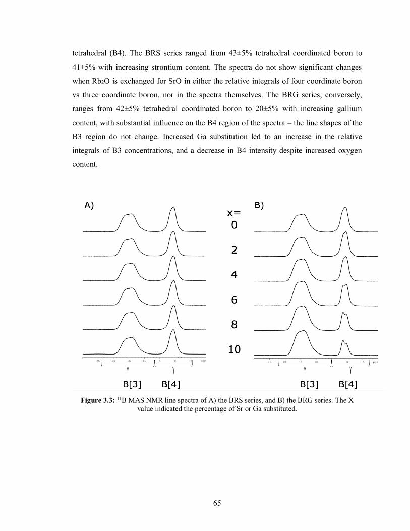
65
tetrahedral (B4). The BRS series ranged from 43±5% tetrahedral coordinated boron to
41±5% with increasing strontium content. The spectra do not show significant changes
when Rb2O is exchanged for SrO in either the relative integrals of four coordinate boron
vs three coordinate boron, nor in the spectra themselves. The BRG series, conversely,
ranges from 42±5% tetrahedral coordinated boron to 20±5% with increasing gallium
content, with substantial influence on the B4 region of the spectra – the line shapes of the
B3 region do not change. Increased Ga substitution led to an increase in the relative
integrals of B3 concentrations, and a decrease in B4 intensity despite increased oxygen
content.
Figure 3.3: 11B MAS NMR line spectra of A) the BRS series, and B) the BRG series. The X
value indicated the percentage of Sr or Ga substituted.
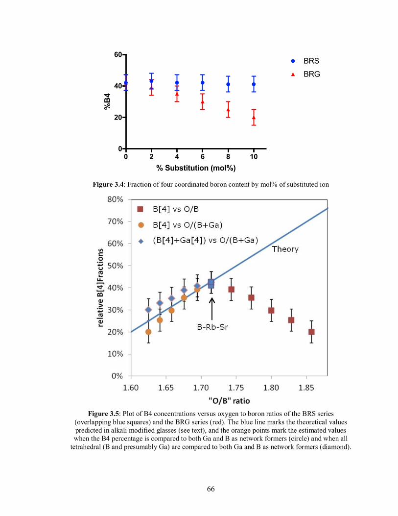
66
Figure 3.4: Fraction of four coordinated boron content by mol% of substituted ion
Figure 3.5: Plot of B4 concentrations versus oxygen to boron ratios of the BRS series (overlapping blue squares) and the BRG series (red). The blue line marks the theoretical values predicted in alkali modified glasses (see text), and the orange points mark the estimated values when the B4 percentage is compared to both Ga and B as network formers (circle) and when all
tetrahedral (B and presumably Ga) are compared to both Ga and B as network formers (diamond).
0 2 4 6 8 100
20
40
60
% Substitution (mol%)
%B
4
BRG
BRS
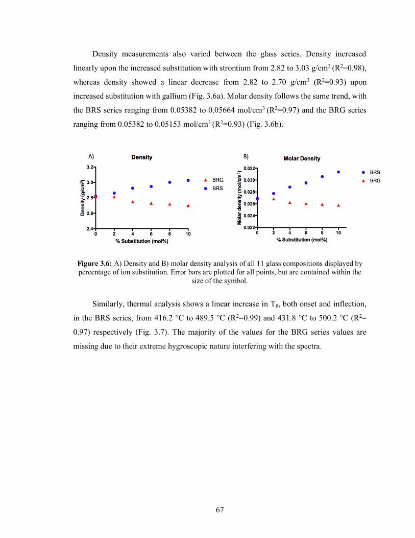
67
Density measurements also varied between the glass series. Density increased
linearly upon the increased substitution with strontium from 2.82 to 3.03 g/cm3 (R2=0.98),
whereas density showed a linear decrease from 2.82 to 2.70 g/cm3 (R2=0.93) upon
increased substitution with gallium (Fig. 3.6a). Molar density follows the same trend, with
the BRS series ranging from 0.05382 to 0.05664 mol/cm3 (R2=0.97) and the BRG series
ranging from 0.05382 to 0.05153 mol/cm3 (R2=0.93) (Fig. 3.6b).
Figure 3.6: A) Density and B) molar density analysis of all 11 glass compositions displayed by percentage of ion substitution. Error bars are plotted for all points, but are contained within the
size of the symbol.
Similarly, thermal analysis shows a linear increase in Tg, both onset and inflection,
in the BRS series, from 416.2 °C to 489.5 °C (R2=0.99) and 431.8 °C to 500.2 °C (R2=
0.97) respectively (Fig. 3.7). The majority of the values for the BRG series values are
missing due to their extreme hygroscopic nature interfering with the spectra.
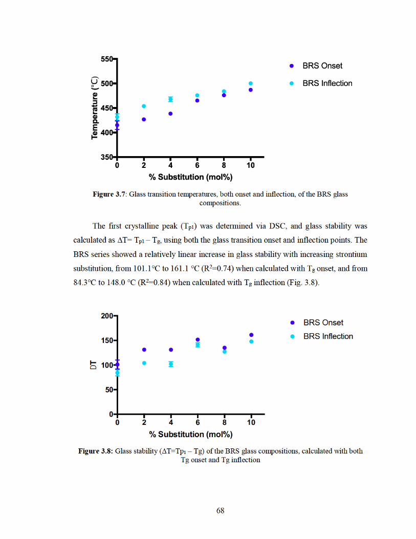
6 8
Fi g u r e 3 . 7: Gl a ss tr a nsiti o n t e m p er at ur es, b ot h o ns et a n d i nfl e cti o n, of t h e B R S gl a ss c o m p ositi o ns.
T h e first cr yst alli n e p e a k ( T p 1 ) w as d et er mi n e d vi a D S C, a n d g l as s st a bilit y w as
c al c ul at e d as ∆ T = T p 1 – T g , usi n g b ot h t h e gl as s tr a nsiti o n o ns et a n d i nfl e cti o n p oi nts. T h e
B R S s eri es s h o w e d a r el ati v el y li n e ar i n cr e as e i n gl a s s st a bilit y wit h i n cr e asi n g str o nti u m
s u bstit uti o n, fr o m 1 0 1. 1 ° C t o 1 6 1. 1 ° C ( R 2 = 0. 7 4) w h e n c al c ul at e d wit h T g o ns et, a n d fr o m
8 4. 3 ° C t o 1 4 8. 0 ° C ( R 2 = 0. 8 4) w h e n c al c ul at e d wit h T g i nfl e cti o n ( Fi g. 3 .8).
Fi g u r e 3 . 8: Gl a ss st a bilit y ( ∆ T = T p 1 – T g) of t h e B R S gl a ss c o m p ositi o ns, c al c ul at e d wit h b ot h T g o ns et a n d T g i nfl e cti o n
0 2 4 6 8 1 00
5 0
1 0 0
1 5 0
2 0 0
% S u b stit uti o n ( m ol %)
DT
Gl a s s St a bilit y
B R S O n s et
B R S I nfl e cti o n
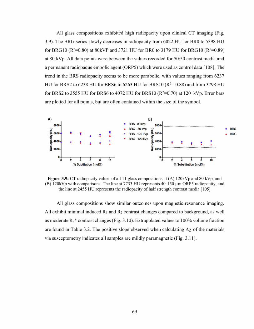
69
All glass compositions exhibited high radiopacity upon clinical CT imaging (Fig.
3.9). The BRG series slowly decreases in radiopacity from 6022 HU for BR0 to 5398 HU
for BRG10 (R2=0.80) at 80kVP and 3721 HU for BR0 to 3179 HU for BRG10 (R2=0.89)
at 80 kVp. All data points were between the values recorded for 50:50 contrast media and
a permanent radiopaque embolic agent (ORP5) which were used as control data [108]. The
trend in the BRS radiopacity seems to be more parabolic, with values ranging from 6237
HU for BRS2 to 6238 HU for BRS6 to 6263 HU for BRS10 (R2= 0.88) and from 3798 HU
for BRS2 to 3555 HU for BRS6 to 4072 HU for BRS10 (R2=0.70) at 120 kVp. Error bars
are plotted for all points, but are often contained within the size of the symbol.
Figure 3.9: CT radiopacity values of all 11 glass compositions at (A) 120kVp and 80 kVp, and (B) 120kVp with comparisons. The line at 7733 HU represents 40-150 µm ORP5 radiopacity, and
the line at 2455 HU represents the radiopacity of half strength contrast media [105]
All glass compositions show similar outcomes upon magnetic resonance imaging.
All exhibit minimal induced R1 and R2 contrast changes compared to background, as well
as moderate R2* contrast changes (Fig. 3.10). Extrapolated values to 100% volume fraction
are found in Table 3.2. The positive slope observed when calculating ∆ of the materials
via susceptometry indicates all samples are mildly paramagnetic (Fig. 3.11).
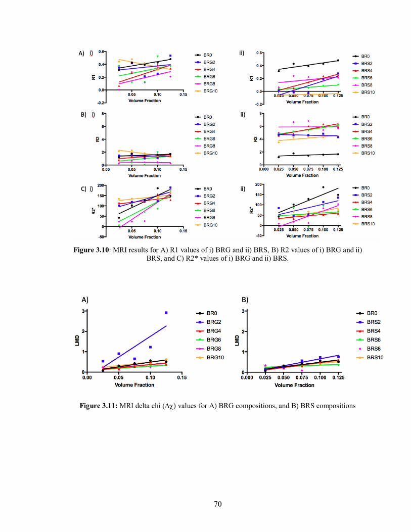
70
Figure 3.10: MRI results for A) R1 values of i) BRG and ii) BRS, B) R2 values of i) BRG and ii)
BRS, and C) R2* values of i) BRG and ii) BRS.
Figure 3.11: MRI delta chi () values for A) BRG compositions, and B) BRS compositions
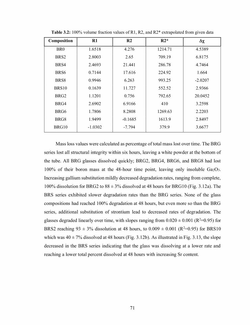
71
Table 3.2: 100% volume fraction values of R1, R2, and R2* extrapolated from given data
Composition R1 R2 R2*
BR0 1.6518 4.276 1214.71 4.5389
BRS2 2.8003 2.65 709.19 6.8175
BRS4 2.4693 21.441 286.78 4.7464
BRS6 0.7144 17.616 224.92 1.664
BRS8 0.9946 6.263 993.25 -2.0207
BRS10 0.1639 11.727 552.52 2.9366
BRG2 1.1201 0.756 792.65 20.0452
BRG4 2.6902 6.9166 410 3.2598
BRG6 1.7806 8.2808 1269.63 2.2203
BRG8 1.9499 -0.1685 1613.9 2.8497
BRG10 -1.0302 -7.794 379.9 3.6677
Mass loss values were calculated as percentage of total mass lost over time. The BRG
series lost all structural integrity within six hours, leaving a white powder at the bottom of
the tube. All BRG glasses dissolved quickly; BRG2, BRG4, BRG6, and BRG8 had lost
100% of their boron mass at the 48-hour time point, leaving only insoluble Ga2O3.
Increasing gallium substitution mildly decreased degradation rates, ranging from complete,
100% dissolution for BRG2 to 88 ± 3% dissolved at 48 hours for BRG10 (Fig. 3.12a). The
BRS series exhibited slower degradation rates than the BRG series. None of the glass
compositions had reached 100% degradation at 48 hours, but even more so than the BRG
series, additional substitution of strontium lead to decreased rates of degradation. The
glasses degraded linearly over time, with slopes ranging from 0.020 ± 0.001 (R2=0.95) for
BRS2 reaching 93 ± 3% dissolution at 48 hours, to 0.009 ± 0.001 (R2=0.95) for BRS10
which was 40 ± 7% dissolved at 48 hours (Fig. 3.12b). As illustrated in Fig. 3.13, the slope
decreased in the BRS series indicating that the glass was dissolving at a lower rate and
reaching a lower total percent dissolved at 48 hours with increasing Sr content.
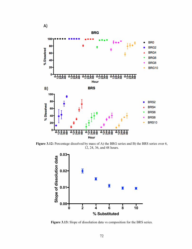
72
Figure 3.12: Percentage dissolved by mass of A) the BRG series and B) the BRS series over 6,
12, 24, 36, and 48 hours.
Figure 3.13: Slope of dissolution data vs composition for the BRS series.
0 2 4 6 8 100.00
0.01
0.02
0.03
% Substituted
Slo
pe o
f d
isso
luti
on
data
Data 13
73
3.5. Discussion
All 11 glass compositions were found to form a glass through plate quenching with
no evidence of crystallization. Interestingly, many properties of both series behaved
differently from one another and from what one would expect based on existing literature.
It is commonly assumed that an increase in four-fold coordination in borate networks leads
to a decrease in dissolution rates under aqueous conditions due to an increase in glass
connectivity [74,101]. However, the structural characterization and degradation
experiments in this work reinforce the need to consider other components and factors which
may influence the properties of borate glasses. The BRS system studied here displayed a
fraction of the four-fold coordinated boron which remained constant at the value predicted
from theory (Fig 3.5), changing only within the uncertainty limits from 43±5% to 41±5%
over the series; yet despite this observation, increased Rb substitution by Sr decreased the
dissolution rates (Fig. 3.12 and 3.13). This can be understood from several points of view:
(i) the alkali (Rb+) and alkaline-earth (Sr2+) cations both act as glass modifiers, and the
replacement ratios were chosen such that the oxygen concentration, and therefore the O/B
ratio remains constant, (ii) replacing two singly charged Rb+ cations with one doubly
charged Sr2+ cation increases network connectivity and (iii) the substitution range covered
is small [74].
In contrast, the substitution of Rb2O with Ga2O3 in the BRG series created substantial
structural changes within the borate network, which affected the glass properties and
demonstrated a decrease in four coordinate boron species. While the line shapes of the B3
regions do not change (Fig. 3.3), the B4 region of the 11B NMR spectra varies, indicating
that Ga3+ preferentially bonds to B4 tetrahedra. Most importantly, with increased Ga2O3
content, the relative integrals of B3 species increase, lowering the percentage of B4's (Fig.
3.4). This trend contrasts to what would be expected for alkali modified borated glasses
[101,109] represented by the blue line in Figure 3.5; for this compositional range, the
theoretical B4 fraction is calculated as B4=x/(1-x) at each O/B ratio, for alkali (R) modified
glasses with the composition xR2O - (1-x)B2O3 [110]. The deviation of the BRG system
from this theoretical trend suggests that gallium does not act simply as a network modifier,
but rather as a network former. These findings can be further understood by examining the
crystal structure of gallium oxide, Ga2O3, and gallium borate, BGaO3. In Ga2O3 and
74
BGaO3, gallium finds itself in four- and six-fold coordinated environments, and in order to
accommodate these environments, each oxygen atom is triply bonded [111,112].
According to Zachariasen’s rule [113], an oxygen atom can only be linked to a maximum
of two cations in a glass, and the coordination surrounding that cation is small (four or
less). As such, if gallium maintains the four-fold coordination in the glass networks studied
in this work, then it cannot provide the necessary oxygens itself (from Ga2O3) and therefore
must sequester them from the surrounding boron network. This conversion may transform
BO4 tetrahedra into BO3-groups with the inclusion of GaO4 tetrahedra and octahedra [92],
meaning that gallium is acting more similar to a network former than a network modifier.
Treating gallium as a network former allows one to calculate the B4 fraction against the
ratio of "oxygen to total network formers” (labeled "B4 vs O/(B+G)" in Fig 3.5).
Hypothetically assuming further that all of the incorporated gallium assumes tetrahedral
coordination, the total amount of tetrahedra can also be plotted as a function of oxygen to
total network formers "(B+Ga)[4] vs O/(B+Ga)". The relationship between the
concentration of B4 and the ratio of oxygen to network former slightly underestimates the
theoretical values, while the relationship between tetrahedral centers (Ga and B) and the
ratio of oxygen to network former slightly overestimates the theoretical values (Fig 3.5),
but both approximations cover the theoretical percentage within their experimental
uncertainties.
The experimentally observed densities further support these conjectures; the density
decreases in the BRG series despite the addition of a higher valency element into the glass
network, meaning that the network may be expanding to accommodate gallium. While an
increase in the percentage of B3 may lead to lower density by itself, as a trigonal
coordinated boron network is assumed to be more open and flexible than one with a high
percentage of tetrahedral coordinated boron [74], it is most likely more important that the
four (and possibly six) coordinated gallium acts as a glass former in addition to the boron
causing the network to expand in order to allow for its incorporation. With respect to the
BRS series, an increase in density is observed that is greater than that which would be
expected given the marginal differences in atomic mass between Rb and Sr (85.47 g/mol
and 87.62 g/mol respectively) [114]. This observation may be attributable to Sr2+ acting
75
with higher crosslinking efficiency than two Rb+ cations, due to its higher charge density,
leading to an increase in density of the glass [76,89].
Unfortunately, and despite several attempts to control water contamination under
desiccated conditions, it was not possible to determine the glass transition temperature for
all the BRG series due to their extremely hygroscopic nature. The glass transition
temperatures for the BRS series were found to increase linearly with increasing strontium
substitution despite the constant B4 concentration. The glass stability data (Fig. 2.8)
indicate stable glasses for manufacturing processes, with melt stability increasing with
increased strontium substitution. Despite stable B4 percentage, the increase in Tg once
again suggests that strontium ions may play a structural role in the glass serving to increase
network rigidity. This increased rigidity may potentially be attributable to the increased
number of oxygens now surrounding modifier ions, which serves to stabilize the glass
network [76,109,92].
One of the most interesting trends observed in this work is that increasing the
substitution of Rb2O for SrO or Ga2O3 stabilizes the glass against hydrolytic degradation.
For simple borate glasses, this trend is unexpected based on existing high borate bioglass
literature where a higher fraction of tetrahedral boron content is understood to mean a more
stable glass [74,75]. It was observed that substituted Sr and Ga do not increase B4 content,
but did modify the dissolution via an alternative mechanism to decrease the rate of
hydrolytic degradation. For both glass series, a decrease in both rate of degradation and
overall mass lost at 48 hours is seen with increasing substitutions. The BRS system
dissolves much slower than the BRG system and for each BRS composition the mass loss
is linear over time. The BRS series is stabilized by increasing strontium substitution up to
a point; the 6, 8 and 10% strontium substituted glasses have similar dissolution patterns
with statistically similar mass loss occurring for each formulation at 48hrs (Figures 2.12 &
2.13). They also show statistically different densities, which would theoretically indicate
that the 10% strontium should degrade more slowly than the 6% as it is a more compact
structure and should make water penetration more difficult; yet this is not demonstrated in
the data. Most of the cylinders (except cylinders which fractured early in the dissolution)
degraded via surface erosion yielding smaller cylinders at each time point, however an
additional white paste was observed as well at the bottom of many of the sample tubes.

76
The dissolution of the BRG series proceeds very differently; the BRG series glasses
lose all structural integrity very quickly – the entirety of the dissolution occurs in the first
twelve hours. For all compositions except BRG10 (and perhaps BRG8) no further
dissolution is observed after 12 hours. Interestingly, the mass loss after 48 hours agrees
within 2% with the borate-rubidium-oxide fraction of the composition. The rubidium-
borate glass fraction appears to dissolve very quickly, leaving the gallium oxide portion in
the form of a white powder, shown by the structural instability of the cylinders and the
deposited powder, as well as the fact that gallium oxide is insoluble in water. Comparing
the percent dissolved at 6 hours, one can see a statistically significant difference, which
remains the case even when those time points are normalized to the borate-rubidium-oxide
fraction of the glass. Further investigation at shorter time points is required to determine
the extent to which gallium has stabilized the network. Collectively, the data reported
provides valuable information on the influence of monovalent for divalent, and monovalent
for trivalent substitutions in borate networks, and further emphasizes the need for expanded
research in the area of borate networks.
In addition to providing valuable insights to the composition-structure-property
relationship of high borate glasses, this study also provides insight to their usefulness in
clinical applications. In clinical scenarios in which embolization is used as a method of
treatment, permanent occlusion of target arteries is not a necessary requirement to achieve
a satisfactory clinical outcome. The ischemic effect of temporarily occluding arteries can
be achieved relatively quickly as tissue does not tolerate loss of blood flow for more than
a few hours (5-7 hours for uterine fibroids) [35]. Additionally, allowing the target arteries
to re-open soon after clinical effect has been achieved could allow for future retreatment
of the region if needed and has the potential to preserve long term function of an underlying
target organ such as the uterus, particularly in individuals who may be seeking pregnancy
in the future.
Additionally, these glasses display CT radiopacity consistent with use as an embolic
agent for the treatment of uterine fibroids. The linear attenuation coefficient data expressed
in Hounsfield Units obtained in this study confirms radiopacity levels that would easily be
detected by either X-ray fluoroscopy, static X-ray imaging, Cone Beam CT (CBCT) or
conventional CT scans for microspheres at a diameter range greater than 40 µm. This would
77
allow Interventional Radiologists to use these technologies during embolization procedures
to truly understand where in the target arteries the microspheres deposit and how
adequately the target area is covered. This in turn could allow for the standardization,
optimization and personalization of these treatments.
It is also crucial to consider potential effects of treatments on imaging contrast in
follow-up imaging, particular in MRI scans. The clinical standard MRI-based assessment
of TAE treatment includes R1-weighted gradient-echo (where R1 = 1/T1), R2-weighted fast
spin-echo (where R2 = 1/T2), diffusion-weighted imaging, and fat-suppressed R1-weighted
gradient-echo pre- and post-contrast enhancement using an extracellular gadolinium
contrast agent. All of the boron glass compositions studied in this work were shown to
induce no additional R1 contrast and would therefore not interfere with any R1-weighted
scans. They also induced minimal changes in R2, with the maximum realistic change being
a ∆R2 of 12 s-1 at 3T from a 60% volume fraction (the maximum volume fraction
obtainable due to packing of beads). When this ∆R2 is added to a typical average R2 of liver
(about 23 s-1 at 3T) [115], this would result in a change in T2 from 42 to 28ms, which is
not a significant change compared to normal variability in liver T2 due to the presence of
iron, which can cause typical variations in T2 from 20-50ms [116]. This means any
fluctuations in iron would likely dominate over any effects due to beads.
Interestingly, these boron glass compositions do induce a larger change in R2*
contrast (Table 3.2) that may be detectable with an R2*-weighted sequence. It is important
to note that R2*-weighted sequences are generally not part of a standard clinical session,
although these are increasingly being tested for evaluation of liver disease. However, given
the significant amounts of iron that also induce R2* contrast in addition to changes in R2,
further experiments are necessary to determine if these compositions could be
distinguished from effects due to iron. Overall, the bead compositions in this study would
have negligible effects on standard MRI clinical scans and would not interfere with
physicians’ ability to evaluate treatment efficacy.
3.6. Limitations
Limitations exist with any scientific study, and the authors would like to
acknowledge the limitations of the present work:
78
• No compositional verification was done on the post-fired material; the authors
assume that the resulting glass has the same composition as the pre-fired materials.
• Although this study aimed to quantify the composition-structure relationships in two
different tertiary borate glass series, only the role of borate in the system was probed
through the use of 11B MAS-NMR. Due to the low natural abundance of 87Sr and
relatively low strontium content of the glasses examined, strontium NMR was not a
feasible form of analysis to investigate strontium’s role in the glass structures.
Gallium NMR is feasible, and may be conducted in future works.
• The effect of SrO or Ga2O3 on the glass structure was not evaluated in a Rb2O free
system; all glasses contained at least 20 mol% Rb2O. The substitution of SrO for
Rb2O was not stoichiometric; the mole fraction of Rb2O was replaced with an equal
mole fraction of SrO.
• The extreme hygroscopic nature of the BRG series made some testing extremely
difficult, and majorly confounded the data collected with the DSC. Future works
should process the BRG series in a very dry environment so as to inhibit premature
degradation.
• Dissolution profiles did not perfectly match the conditions seen in vivo. Due to the
hygroscopic nature of the particles, cylinders were used to avoid the error of residual
water due to clumping. The use of cylinders, however, made comparing surface area
loss and dissolution rates to those expected in vivo quite difficult. As such, additional
testing on 100-300 µm particles or microspheres may be required to assess the
feasibility of using cylinders in place of particles for preliminary testing.
• Radiopacity was measured on irregular particles, with a size range of ca. 100-300
µm, resulting in an unknown number of radiolucent voids, which may lead to an
artificial reduction in the resulting measured radiopacity.
3.7. Conclusion
Substituting strontium or gallium for rubidium in a boron-rubidium binary glass has
drastically different effects. The strontium series show a constant percentage of B4 with
increasing strontium, yet an increase in density, Tg, and hydrolytic stability (decrease in
dissolution rate). As such, it can be assumed that the Sr ion is crosslinking the network,
79
thereby creating a higher network connectivity and a more stable glass. The gallium series,
shows a decrease in B4 percentage with increasing gallium substitution, however the
gallium acts more closely to a network former in this case, also resulting in a more stable
glass. The gallium series, however, is less stable than the strontium series, making it
unsuitable for use as an embolic agent. The strontium series shows promise as a potential
TAE agent due to its high CT radiopacity and degradation rates, although the safety and
efficacy of the material must be further assessed. These glass compositions may potentially
overcome existing limitations for TAE materials, leading to a standardized, optimized and
personalized procedure.
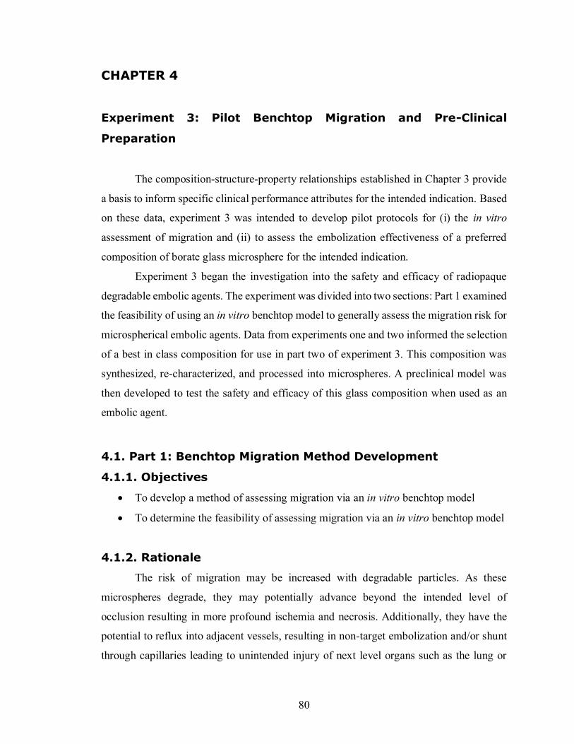
80
CHAPTER 4
Experiment 3: Pilot Benchtop Migration and Pre-Clinical
Preparation
The composition-structure-property relationships established in Chapter 3 provide
a basis to inform specific clinical performance attributes for the intended indication. Based
on these data, experiment 3 was intended to develop pilot protocols for (i) the in vitro
assessment of migration and (ii) to assess the embolization effectiveness of a preferred
composition of borate glass microsphere for the intended indication.
Experiment 3 began the investigation into the safety and efficacy of radiopaque
degradable embolic agents. The experiment was divided into two sections: Part 1 examined
the feasibility of using an in vitro benchtop model to generally assess the migration risk for
microspherical embolic agents. Data from experiments one and two informed the selection
of a best in class composition for use in part two of experiment 3. This composition was
synthesized, re-characterized, and processed into microspheres. A preclinical model was
then developed to test the safety and efficacy of this glass composition when used as an
embolic agent.
4.1. Part 1: Benchtop Migration Method Development
4.1.1. Objectives
• To develop a method of assessing migration via an in vitro benchtop model
• To determine the feasibility of assessing migration via an in vitro benchtop model
4.1.2. Rationale
The risk of migration may be increased with degradable particles. As these
microspheres degrade, they may potentially advance beyond the intended level of
occlusion resulting in more profound ischemia and necrosis. Additionally, they have the
potential to reflux into adjacent vessels, resulting in non-target embolization and/or shunt
through capillaries leading to unintended injury of next level organs such as the lung or
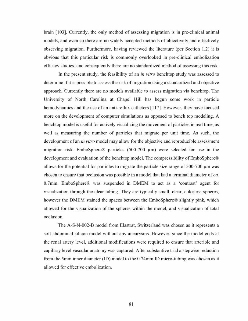
81
brain [103]. Currently, the only method of assessing migration is in pre-clinical animal
models, and even so there are no widely accepted methods of objectively and effectively
observing migration. Furthermore, having reviewed the literature (per Section 1.2) it is
obvious that this particular risk is commonly overlooked in pre-clinical embolization
efficacy studies, and consequently there are no standardized method of assessing this risk.
In the present study, the feasibility of an in vitro benchtop study was assessed to
determine if it is possible to assess the risk of migration using a standardized and objective
approach. Currently there are no models available to assess migration via benchtop. The
University of North Carolina at Chapel Hill has begun some work in particle
hemodynamics and the use of an anti-reflux catheters [117]. However, they have focused
more on the development of computer simulations as opposed to bench top modeling. A
benchtop model is useful for actively visualizing the movement of particles in real time, as
well as measuring the number of particles that migrate per unit time. As such, the
development of an in vitro model may allow for the objective and reproducible assessment
migration risk. EmboSphere® particles (500-700 µm) were selected for use in the
development and evaluation of the benchtop model. The compressibility of EmboSphere®
allows for the potential for particles to migrate the particle size range of 500-700 μm was
chosen to ensure that occlusion was possible in a model that had a terminal diameter of ca.
0.7mm. EmboSphere® was suspended in DMEM to act as a ‘contrast’ agent for
visualization through the clear tubing. They are typically small, clear, colorless spheres,
however the DMEM stained the spaces between the EmboSphere® slightly pink, which
allowed for the visualization of the spheres within the model, and visualization of total
occlusion.
The A-S-N-002-B model from Elastrat, Switzerland was chosen as it represents a
soft abdominal silicon model without any aneurysms. However, since the model ends at
the renal artery level, additional modifications were required to ensure that arteriole and
capillary level vascular anatomy was captured. After substantive trial a stepwise reduction
from the 5mm inner diameter (ID) model to the 0.74mm ID micro-tubing was chosen as it
allowed for effective embolization.
82
4.1.3. Materials & Methods
4.1.3.1. Model Set-up
An anatomically correct model (Fig 4.1) of the human abdominal aorta, renal
arteries and iliac arteries (Elastrat, Switzerland) was modified to mimic a capillary network.
The left renal artery (B) was modified by fitting a 43.4 mm x 5.7 mm connecting tube to
the model adaptor, and a reduction adaptor connected the capillary – a tube with an inner
diameter of 0.74 mm – to the connecting tube. The remaining return tubing was modified
by replacing the t-adaptor with a straight junction adapter that allowed for a continuous
flow back to the reservoir tank (A). The reservoir tank was filled with 2.5 L of tap water
and the pump was turned on to maximum strength, with a flow rate of 78.80 ± 1.96 mL/s
measured with a flowmeter (Omega, Canada).
Figure 4.1: Benchtop renal artery model. (A) represents the fluid reservoir and the pump which approximates the heart. (B) indicates the area that has been modified to incorporate the capillary bed (C) denotes the percutaneous introducer used to insert the catheter (<20Fr) into the model for
embolization.
4.1.3.2. Experimental Design
A micro-catheter (Cook cantata 2.9 4Fr) was inserted into the percutaneous
introducer and threaded into the left renal artery and down into the adapter adjacent to the
micro-tubing. 500-700 µm EmboSphere® suspended in Dulbecco’s Modified Eagles
Medium (DMEM) were injected until a bolus was formed. The embolization bolus was
marked at both the distal and proximal end using a marker directly after embolization
occurred. The distal end of the capillary was fed into a filtration system (Filtropur v50, 500
ml) to filter out any EmboSphere® that might have migrated further into the vasculature.
83
Each trial was run for two hours (n=5). At the completion of each trial the location of the
bolus was marked; the pump was turned off and the connecting tube from the renal artery
was clamped to prevent reflux of particles into the model. The adaptor and capillary tube
were then removed from the model to allow for complete drainage into the reservoir. The
entire reservoir tank was then filtered using the Nalgene vacuum filter (Filtropur v50, 500
ml).
4.1.4. Results & Discussion
A total of five runs were completed to test for the success of this model, in addition
to the preliminary troubleshooting. The final model can be seen in Figure 4.2, with a close-
up on the reduction to the micro-tubing representing the capillary.
Figure 4.2: A) Complete setup of the benchtop model, B) stepwise reduction from the ‘renal artery’ to the ‘arteriole’ to the capillary bed.
The most effective methods of analyzing migration was with a modified filtration
system. This system included a Nalgene vacuum filter (Filtropur v50, 500 ml) adapted to
allow for any filtrate to return to the reservoir tank which then can be pumped back into
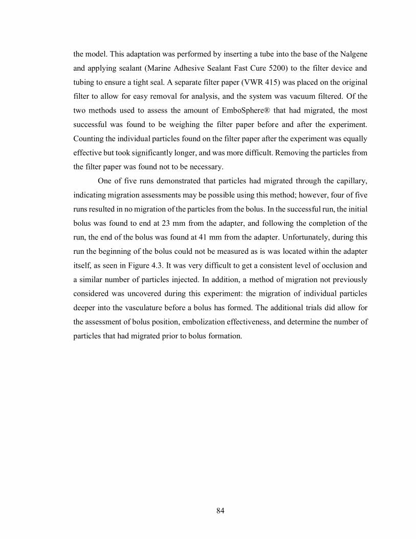
84
the model. This adaptation was performed by inserting a tube into the base of the Nalgene
and applying sealant (Marine Adhesive Sealant Fast Cure 5200) to the filter device and
tubing to ensure a tight seal. A separate filter paper (VWR 415) was placed on the original
filter to allow for easy removal for analysis, and the system was vacuum filtered. Of the
two methods used to assess the amount of EmboSphere® that had migrated, the most
successful was found to be weighing the filter paper before and after the experiment.
Counting the individual particles found on the filter paper after the experiment was equally
effective but took significantly longer, and was more difficult. Removing the particles from
the filter paper was found not to be necessary.
One of five runs demonstrated that particles had migrated through the capillary,
indicating migration assessments may be possible using this method; however, four of five
runs resulted in no migration of the particles from the bolus. In the successful run, the initial
bolus was found to end at 23 mm from the adapter, and following the completion of the
run, the end of the bolus was found at 41 mm from the adapter. Unfortunately, during this
run the beginning of the bolus could not be measured as is was located within the adapter
itself, as seen in Figure 4.3. It was very difficult to get a consistent level of occlusion and
a similar number of particles injected. In addition, a method of migration not previously
considered was uncovered during this experiment: the migration of individual particles
deeper into the vasculature before a bolus has formed. The additional trials did allow for
the assessment of bolus position, embolization effectiveness, and determine the number of
particles that had migrated prior to bolus formation.
85
Figure 4.3: Reduction adaptor and micro-tubing filled with EmboSphere® suspended in
DMEM following completion of one trial.
While the initial pilot work has indicated the potential for the development of in
vitro test set ups to assess migration, some fundamental areas require considerable attention
and development. In particular, due to the nature of the flow paths utilized in this model,
when one pathway is obstructed, the flow from the aorta is redirected to the unblocked
pathways, causing a strong decrease in pressure in the obstructed vessel. The water around
the obstruction appears to be somewhat stagnant, similar to what is seen in vivo, however
the lack of additional divisions of the renal vasculature makes it difficult to accurately
assess fluid flow dynamics. Accordingly, a much larger model with more divisions in the
renal artery would be required to accurately assess the risk of migration. The flow rates of
theses additional divisions should also be dealt with accordingly, to ensure the proper
approximation of in vivo conditions. Overall, it seems that it may be feasible to assess
migration via a benchtop model, were certain limitations addressed. The next section
briefly identifies those limitations and provides recommendations to remediate issues, and
enhance overall functionality of the model.

86
4.1.5. Limitations and Future Consideration
• There were no gradual/step-wise reductions from the renal artery to the capillary
network. The resistance going from the connector tube and capillary might be
higher than expected in an animal. It is recommended that for future work, a single
tube with a gradual reduction in size is used, rather than multiple tubes with
reduction adapters. Additionally, divisions of the renal artery should be
approximated to more similarly reproduce the fluid flow found in vivo.
• The pump used for the pilot experimentation was not pulsatile, which would be
ideal to mimic the heartbeat of a human. Further works using a pulsatile pump
would be more useful than the continuous pump used in this method. BDC
laboratories (Colarado, USA) makes a variety of pulsatile pumps that effectively
mimic cardiac cycles, with customizable stroke volumes and flow rates.
• Since a flow meter was used, it was difficult to determine the pressure exherted in
each vessle of the benchtop model. In future works, pressure guages should be fitted
at the pump, the beginning of the occluded vessel, and on the return tubing to the
resevoir tank. This will allow for consistent pressure control, and a more accurate
picture of the fluid dinamics within the model.
• Materials used in future work should mimic the compliance of in vivo vasculature
to properly assess vessel damage. Additionally, fluid used to simulate blood should
more accurately approximate dissolved ion concentrations, and potentially clotting
ability.
• The basic need for access to a vacuum for the filtering system coupled with the
nature of the size of the model itself lead to a very specific need of lab space. The
best place found in the lab space available to the authors still resulted in possible
hydrodynamic discrepancies in the set-up as some of the tubing was coiled to fit in
the space.
• The bench top model is not able to determine the final location of migrated
particles, as it is a continuous loop of the abdominal aorta and renal vasculature. It
lacks some of the finer conditions (i.e. specific venous vasculature and
anastomosis), and therefore cannot predict the severity of harm caused should
particles migrate. The severity of harm must be assessed in an animal model.
87
4.2. Part 2: Microsphere Synthesis and Embolization Efficacy
The second section of experiment three intends to assess the embolization efficacy
of a degradable embolic agent in a pre-clinical animal model. First, a best in class material
was selected, synthesized, and processed into microspheres. These microspheres were re-
characterized, sterilized, and shipped to the Centre Hospitalier de l’Université de Montréal
(CHUM) to assess their safety, efficacy, and performance in a pre-clinical porcine renal
model.
4.2.1. Objectives
• To choose the best in class material for the intended indication based on the data
collected in experiments one and two.
• To melt a new batch of best in class material and process it into microspheres.
• Re-characterize both the irregular frit and microspheres to account for any structure
property changes as a result of additional processing.
• To develop a pilot pre-clinical animal model to assess the safety, efficacy and
performance of the best in class material against the special controls criteria laid
out by the FDA guidance document [17].
4.2.2. Hypothesis
1. If thermal augmentation causes a change in the structure or properties of a glass
microsphere, then a significant change in density, glass transition temperature, and
glass stability of the frit will be observed after spherical processing.
2. The glass microspheres will be as effective at embolizing the renal arteries of pigs
as the control permanent embolic (EmboSphere®), when evaluated based on extent
of kidney ischemia and level of occlusion.
4.2.3. Rationale
Taking into consideration the ideal properties for degradable microspheres outlined in
Section 1.2.2., the best in class composition was chosen to satisfy the following criteria:
88
1. Has a degradation timeframe of 100 ± 5% in 24-48 hours – so as to adequately
address the 5-7 hour total occlusion timeline laid out by Vilos et al. [35].
2. Highest possible CT radiopacity – allow for intra- and post-procedural feedback to
the clinician
3. No visibility on MRI – so as not to confound clinical follow up scans
4. Minimized density – to eliminate as much as possible concerns related to
preferential distribution / flow under the action of gravity.
5. High glass stability –for ease of manufacturing
The BRS2 glass composition (70 B2O3 – 28 Rb2O – 2 SrO) was found to fulfill all
these criteria. To evaluate their embolization effectiveness, the objective of this phase of
the research was to produce microspherical particles of BRS2 to allow the pre-clinical
assessment of BRS2 in an accepted model. Spherical particles were chosen for use in this
study, as irregular particles tend to clump and clog catheters [12]. To convert glass frit into
spherical particles, irregular particles must be passed through a flame at temperatures
exceeding 2000ºC [118]. However, the melting temperature of these glasses was found to
be ca. 700 ºC, and while the temperature of the flame is sufficient to re-melt the glass and
under the action of surface tension form a smooth, spherical particle, this additional process
changes the thermal history of the materials. Accordingly, the elevated temperatures
necessary to convert glass frit to microspheres may have a significant effect on the structure
and properties of the resulting glass. Since glasses are a non-equilibrium system, their
properties depend not only on standard thermodynamic variables (i.e. heating and pressure
during formation, composition) but also on thermal history [119]. Therefore, additional
heat treatments may alter the structure (and thus properties) of the treated glass [120]. If
this is the case, any preliminary characterization performed on irregular glass particles may
be of limited use, as the glass microspheres may behave differently than the investigated
glass frit. If further heat treatments do in fact alter the properties of the resulting
microspheres, then preliminary in vitro investigations may be required to be conducted on
microspheres, to ensure the collected data will translate to in vivo applications. To
investigate potential structure-property changes as a result of the additional processing
step, the glass spheres created by spherical processing were characterized (XRD, PSA,
thermal analysis, sphericity, SEM, and CT radiopacity) and the data was compared to both
89
the original characterization data collected in experiments one and two (in addition, to the
newly collected data on the irregular particles).
The particle size distribution selected for this phase of the work was 100-300 μm
as this is smallest particle size range used for UAE [37] and therefore represents the worst-
case scenario in terms of potential risk. Furthermore, the selection of this particle size
represents the highest surface area to volume ratio (for the intended indication), and will
thus degrade the most quickly, potentially leading to ineffective occlusion times, or
worsening side effects (i.e. post-embolization syndrome). In addition, it is believed that
such small particles also increase the risk of migration, as they are lighter and may reflux
more easily [121]. Finally, this particle size distribution is likely the most challenging to
image using standard imaging modalities (both from a temporal and spatial distribution
standpoint [13]), and as such, should these particles prove to be imageable in vivo then the
more commonly used larger particle size distributions should also be visible under similar
conditions. The control article selected for this experiment was EmboSphere®, as it is most
commonly used for UAE, and the size range of 100-300 µm was chosen to match that of
the test article (TA).
The advantages of degradable embolic agents include limited long-term exposure
to foreign bodies, ability for repeat treatments, and peace of mind for patients who “express
worries about foreign materials remaining in the body” [14,103]. When considering uterine
artery embolization, degradable embolic agents have additional advantages. Specifically,
that allow reperfusion of blood to the uterus, potentially preserving fertility in young
women who wish to get pregnant later in life. Currently, there are a small number of
temporary embolization materials available, each exhibiting substantive limitations in the
context of UAE. Gelatin sponge particles are cut by hand prior to injection, leading to
inconsistent particle sizes, uncontrollable levels of occlusion, and unpredictable
degradation timeframes lasting from 3 weeks to 4 months [122,123,124]. Conversely,
starch and PLGA microspheres do have calibrated particle sizes; however their degradation
timeframes not suited to most temporary embolization indications. For example, starch
microspheres degrade < 40 minutes, which contraindicates their use in UAE and many
other TAE procedures [125,126]. PLGA microspheres degrade over several months, which
triggers a long-lasting inflammatory and fibrotic reactions [31]. To overcome these
90
limitations, the preceding chapter discussed the development of two series of borate-based
glass composition as potential degradable embolic agents; characterization performed in
experiments one and two have shown that these glasses may display critical performance
attributes for TAE. However, in vivo testing conditions are required to validate some
attributes and to evaluate other risks (e.g. embolization effectiveness, inflammatory effects,
etc.).
Pre-clinical evaluations are required for new medical devices to demonstrate safety
and efficacy with respect to their intended application. The degradable nature of the
microspheres developed in this work may presents new patient safety risks which have not
been previously considered with permanent embolic agents. The industry guidance
document (provided by FDA) to help ensure that all the necessary additional aspects of
safety and efficacy of embolization devices are considered is titled “Class II Special
Controls Guidance Document: Vascular and Neurovascular Embolization Devices” [17].
However, while this document is thoroughly detailed, it is important to note that the
guidance was drafted prior to any substantive work with respect to the development and
utilization of degradable embolic agents. Accordingly, many aspects of embolization
effectiveness for degradable embolic agents may be overlooked. Therefore, in addition to
consulting the required guidance documents, a Design Failure Mode and Effect Analysis
(dFMEA) was conducted to evaluate potential risks with the design of the concept and
coupled with the literature review, informed the design of an animal study protocol (drafted
with the assistance of Lauren Kiri, Dr. Kathleen O’Connell, Dr. Bob Abraham, and Dr.
Gilles Soulez).
Originally an ovine uterine model was proposed for this work, as it most closely
approximated the human uterus environment, making it an ideal model to test uterine artery
embolic agents [127]. However, the porcine renal model was implemented as a backup
when the ovine uterine model proved to be unacceptable due to the timing of the study
coinciding with lambing season. A non-atherosclerotic swine model was chosen because
the model has been used extensively for angiographic/embolization studies resulting in a
large volume of data on the vascular response properties and its correlation to human
vascular response [128,129,130]. The porcine and human anatomy share important
anatomic and physiologic characteristics [128,131]. Furthermore, the porcine kidney
91
model is accepted for use in preclinical studies by the FDA and as such has been widely
used to evaluate embolization agents [130].
The proposed study comprises a pilot investigation to evaluate the effectiveness of
biodegradable microspheres and will therefore utilize the minimum number of animals
possible to meet the study objectives. The primary outcomes for this study were determined
to be the assessment of: (i) embolization effectiveness, and (ii) test article migration.
Embolization effectiveness and initial particle distribution will be recorded and compared
to the control article. Migration will be assessed from two standpoints: movement of
particles deeper into the uterine vasculature, assessed via cone beam CT and histology of
the explanted kidneys, and ischemic damage to non-target tissues (i.e. NTE) assessed via
full conventional CT and histology (of tissues showing evidence of ischemia on CT). The
protocol was then sent for approval by both Dalhousie Animal Ethics and CHUM
Institutional Animal Care and Use Committee to ensure compliance with Canadian Council
on Animal Care (CCAC) regulations prior to the beginning of the study. For further data
on the complete protocol, please see Appendix C.
4.2.4. Materials & Methods
4.2.4.1. Glass Synthesis & Recharacterization
300 g of BRS2 was melted, ground and sieved to yield 100-300 μm particles (as
per section 3.3.1, pg. 58). Characterization including XRD, PSA, density, thermal analysis, 11B NMR, mass loss, and CT (full details in Section 3.3, pg. 58) were performed on the
newly melted glass frit.
4.2.4.2. Spherical Processing & Recharacterization
The newly synthesized irregular particles underwent spherical processing at ABK
Biomedical (Halifax, NS, Canada) to yield 100-300µm microspheres. The parameters for
the sphere processing process are as follows:
i. The collection pot was lined with Aluminum foil (Ultra-Clean, premium
aluminum foil from VWR International) and placed approximately 30 cm from
the burner
92
ii. The feeding funnel was positioned with the base directed towards the collection
pot at a 45-degree angle.
iii. The funnel was placed approximately 2.5 cm above the burner (from the center),
and 1- 2 cm in front of the burner.
Characterization including XRD, PSA, density, thermal analysis, 11B NMR, and CT
(full details in Section 3.3, pg. 58) was repeated on these microspheres. Additionally,
sphericity analysis and scanning electron microscopy (SEM) imaging of the spheres was
conducted. In particular, particle size analysis and particle shape (sphericity) were
determined using dynamic image analysis (Camsizer XT (Retsch Technology)). A
monolayer of the microsphere specimen was placed directly onto the feed chute. The feed
rate of the particles was automatically adjusted by the instrument. A total of 976,170.
particles were fed into the instrument and through the field of view of the two cameras.
Particle size (µm) distribution was reported as Dx10, Dx50, and Dx90. Particle shape was
reported as the mean sphericity. Once analysis was complete, the microsphere specimens
were stored in a desiccated environment.
For SEM, samples were mounted on SEM stubs using carbon paste, and then coated
with 20 nm of gold-palladium. Multiple sites of each sample were then inspected using a
model No. S-4700 SEM (Hitachi, Chula Vista, CA) operating at an accelerating voltage of
15 kV, a working distance of 12.3mm, and using magnifications up to 3,500.
4.2.4.3. Packaging & Sterilization
Twenty 1g (18x1g for animal study, 2x1g for 11B NMR analysis) vials of
microspheres were sterilized with a ca. 30 kGy dose of gamma radiation (Nordion) prior
to use in the pre-clinical study. The accompanying required delivery devices (ABK
Biomedical, Canada) were sterilized using ethylene oxide gas sterilization (Department of
Medicine, Dalhousie University). One control package comprising 100-300 µm
EmboSphere® and a traditional delivery stopcock where also prepared for the pilot study.
All materials, delivery devices and control microspheres were then delivered to CHUM.
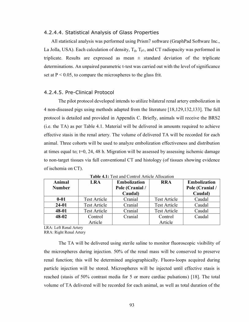
93
4.2.4.4. Statistical Analysis of Glass Properties
All statistical analysis was performed using Prism7 software (GraphPad Software Inc.,
La Jolla, USA). Each calculation of density, Tg, Tp1, and CT radiopacity was performed in
triplicate. Results are expressed as mean ± standard deviation of the triplicate
determinations. An unpaired parametric t-test was carried out with the level of significance
set at P < 0.05, to compare the microspheres to the glass frit.
4.2.4.5. Pre-Clinical Protocol
The pilot protocol developed intends to utilize bilateral renal artery embolization in
4 non-diseased pigs using methods adapted from the literature [18,129,132,133]. The full
protocol is detailed and provided in Appendix C. Briefly, animals will receive the BRS2
(i.e. the TA) as per Table 4.1. Material will be delivered in amounts required to achieve
effective stasis in the renal artery. The volume of delivered TA will be recorded for each
animal. Three cohorts will be used to analyze embolization effectiveness and distribution
at times equal to; t=0, 24, 48 h. Migration will be assessed by assessing ischemic damage
to non-target tissues via full conventional CT and histology (of tissues showing evidence
of ischemia on CT). Table 4.1: Test and Control Article Allocation
Animal Number
LRA Embolization Pole (Cranial /
Caudal)
RRA Embolization Pole (Cranial /
Caudal) 0-01 Test Article Cranial Test Article Caudal 24-01 Test Article Cranial Test Article Caudal 48-01 Test Article Cranial Test Article Caudal 48-02 Control
Article Cranial Control
Article Caudal
LRA: Left Renal Artery RRA: Right Renal Artery
The TA will be delivered using sterile saline to monitor fluoroscopic visibility of
the microspheres during injection. 50% of the renal mass will be conserved to preserve
renal function; this will be determined angiographically. Fluoro-loops acquired during
particle injection will be stored. Microspheres will be injected until effective stasis is
reached (stasis of 50% contrast media for 5 or more cardiac pulsations) [18]. The total
volume of TA delivered will be recorded for each animal, as well as total duration of the
94
procedure for each animal, and the ease of use of the material. One shot films and cone
beam CT (in the angio suite) acquisitions will be acquired to evaluate the distribution of
the radiopaque microspheres in each pig. Post-embolization angiography will be
performed to the degree of vessel occlusion and document embolization efficacy. All
embolized arteries will be qualitatively evaluated for arterial perforation or rupture and
embolization effectiveness.
Immediately prior to the scheduled sacrifice time point, angiography will be
performed in each animal to assess the embolization status of the renal vasculature) The
following grade scale will be used for qualitative evaluation of recanalization:
• 0=no angiographic visible signs of arterial occlusion
• 1=reduction of parenchymal staining of the dependent territory
• 2=reduction of the parenchymal staining and occlusion of the supplying
arcuate artery
• 3=reduction of parenchymal staining, occlusion of the supplying arcuate
artery and occlusion of the feeding artery downstream of the catheter tip
[18].
All treated animals will be subjected to necropsy, defined as gross examination of
the embolized arteries and kidneys, whole body (external surface), all orifices, thoracic and
abdominal cavities, and brain. Macroscopic pictures of all kidneys (ventral and dorsal
views) and any lesions will be taken with a ruler adjacent to the tissue and properly labeled
when possible (study number, animal number, organ). Kidneys will be evaluated
macroscopically and measured (length, width and depth) before fixation. Three regions
(cranial pole, middle region and caudal pole) will be evaluated individually on both
surfaces (ventral and dorsal surfaces) for ischemic changes. An ischemic change score will
be attributed to each region according to the following scale [31]:
• 0 = no macroscopic visible change;
• 1= discoloration of renal surface;
• 2 = Surface scars and retractions (corresponding to score 1 and 2);
• 3 = volume shrinkage of the respective area (corresponding to score 1, 2 and 3).
Each kidney with the renal artery (entire length) for each animal will be harvested
free of fat and cranial pole properly identified (with ink for example) in a consistent
95
manner. Upon histological analysis, the number of microspheres in each layer will be
recorded. The presence and extent of embolus surrounding each embolic will also be
documented, as will the presence of inflammatory reaction and the types of inflammatory
cells present (e.g. macrophages, multinucleated giant cells, lymphocytes, eosinophils).
Identified microspheres (or microsphere fragments) will be measured and their shape will
be qualitatively assessed (spherical vs. irregular). Vessel wall rupture, formation of new
endothelium within the embolized vessel, and location of particles within the vessel walls
should extravasation occur will be assesses and the degree of vascular injury will be rated
as follows [134]:
• 0 = Internal elastic lamina (IEL) intact
• 1 = Disruption of the IEL without medial disruption.
• 2 = Disruption of the IEL with disruption of the media. The external elastic lamina
is intact.
• 3 = Disruption of the IEL, media, and external elastic lamina.
Additional sections may be cut and stained with different stains to further assess
embolization efficacy at the discretion of the Study Pathologist with the approval of the
Study Director.
To assess migration, a selective angiography will be performed to evaluate the
branch division into cranial and caudal branches and segmental renal arteries. Hyper-
selective catheterization will be performed with a 2.5Fr micro catheter (Cantata, Cook Inc.,
Bloomington, IN) under fluoroscopic guidance [18,132]. All bifurcations downstream to
the cranial and caudal branches will be recorded as D1, D2, D3, etc., where D1 referrers to
the immediate and largest branches of the cranial or caudal artery, following subsequent
branches from this artery will be labelled D2, and further branches will be labelled as D3,
etc. [132], similar to what is seen in Figure 4.1. After injection, but before sacrifice, another
angiography will be performed and occlusion will again be labelled as per Figure 4.1. After
sacrifice, and necropsy, all harvested organs will be fixed in a box containing
formaldehyde. Hematoxylin-eosin-saffron staining of specimen will allow observation of
occluded vessels and the locations of identified microspheres, which will be labelled
according to the different layer of renal vasculature as per Figure 4.4 [132] for comparison
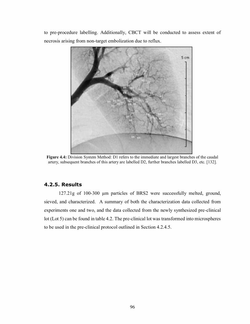
96
to pre-procedure labelling. Additionally, CBCT will be conducted to assess extent of
necrosis arising from non-target embolization due to reflux.
Figure 4.4: Division System Method: D1 refers to the immediate and largest branches of the caudal artery, subsequent branches of this artery are labelled D2, further branches labelled D3, etc. [132].
4.2.5. Results
127.21g of 100-300 μm particles of BRS2 were successfully melted, ground,
sieved, and characterized. A summary of both the characterization data collected from
experiments one and two, and the data collected from the newly synthesized pre-clinical
lot (Lot 5) can be found in table 4.2. The pre-clinical lot was transformed into microspheres
to be used in the pre-clinical protocol outlined in Section 4.2.4.5.
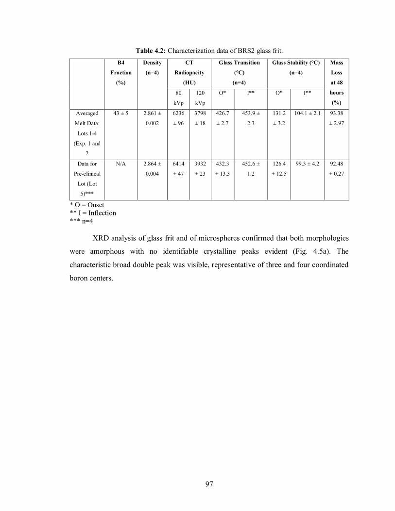
97
Table 4.2: Characterization data of BRS2 glass frit. B4
Fraction
(%)
Density
(n=4)
CT
Radiopacity
(HU)
Glass Transition
(°C)
(n=4)
Glass Stability (°C)
(n=4)
Mass
Loss
at 48
hours
(%)
80
kVp
120
kVp
O* I** O* I**
Averaged
Melt Data:
Lots 1-4
(Exp. 1 and
2
43 ± 5 2.861 ±
0.002
6236
± 96
3798
± 18
426.7
± 2.7
453.9 ±
2.3
131.2
± 3.2
104.1 ± 2.1 93.38
± 2.97
Data for
Pre-clinical
Lot (Lot
5)***
N/A 2.864 ±
0.004
6414
± 47
3932
± 23
432.3
± 13.3
452.6 ±
1.2
126.4
± 12.5
99.3 ± 4.2 92.48
± 0.27
* O = Onset ** I = Inflection *** n=4 XRD analysis of glass frit and of microspheres confirmed that both morphologies
were amorphous with no identifiable crystalline peaks evident (Fig. 4.5a). The
characteristic broad double peak was visible, representative of three and four coordinated
boron centers.
98
Figure 4.5: A) XRD data for the BRS2 glass frit and the microspheres. Two amorphous peaks
can be seen at 2Ø values of approximately 25 and 45, corresponding to 3 and 4 coordinated boron centers in the glass. B) Density and molar density data for the original BRS2 glass frit and the
microspheres. C) Glass transition temperatures and glass stabilities (both onset and inflection) for the BRS2 glass frit and the microspheres. D) CT radiopacity for the BRS2 glass frit and the
microspheres at 80 kVp and 120 kVp.
Density and molar density data (Fig. 4.5b) of the glass frit were recorded as 2.862
± 0.003 g/cm3 and 0.02775 ± 0.00003 mol/cm3 respectively. After spherical processing, the
density was recorded as 2.863 ± 0.005 g/cm3 and 0.02775 ± 0.00005 mol/cm3, which was
not significantly different from the glass frit (P=0.7674). Thermal analysis (Fig. 4.5c) data
can be found summarized in Table 4.3; no statically significant differences were observed.
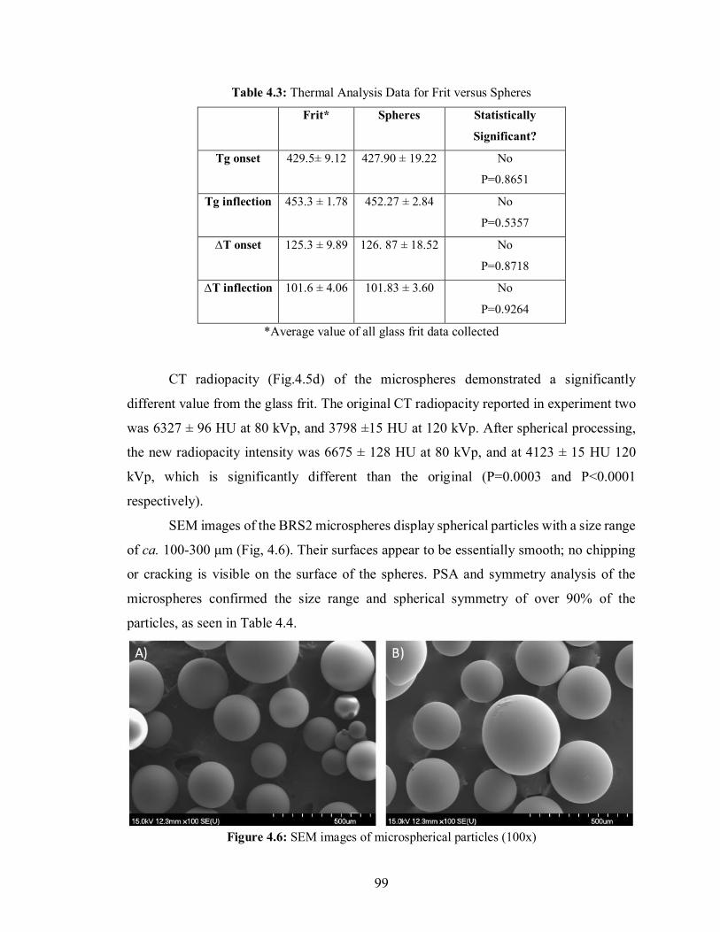
99
Table 4.3: Thermal Analysis Data for Frit versus Spheres
Frit* Spheres Statistically
Significant?
Tg onset 429.5± 9.12 427.90 ± 19.22 No
P=0.8651
Tg inflection 453.3 ± 1.78 452.27 ± 2.84 No
P=0.5357
∆T onset 125.3 ± 9.89 126. 87 ± 18.52 No
P=0.8718
∆T inflection 101.6 ± 4.06 101.83 ± 3.60 No
P=0.9264
*Average value of all glass frit data collected
CT radiopacity (Fig.4.5d) of the microspheres demonstrated a significantly
different value from the glass frit. The original CT radiopacity reported in experiment two
was 6327 ± 96 HU at 80 kVp, and 3798 ±15 HU at 120 kVp. After spherical processing,
the new radiopacity intensity was 6675 ± 128 HU at 80 kVp, and at 4123 ± 15 HU 120
kVp, which is significantly different than the original (P=0.0003 and P<0.0001
respectively).
SEM images of the BRS2 microspheres display spherical particles with a size range
of ca. 100-300 μm (Fig, 4.6). Their surfaces appear to be essentially smooth; no chipping
or cracking is visible on the surface of the spheres. PSA and symmetry analysis of the
microspheres confirmed the size range and spherical symmetry of over 90% of the
particles, as seen in Table 4.4.
Figure 4.6: SEM images of microspherical particles (100x)
100
Table 4.4: Spherical Data and Particle Sizes of BRS2 Microspheres
Symmetry 94.6%
Sphericity 92.0%
D10 103.1
D50 224.5
D90 323.2
Mean value 224.8 ± 93.2
4.2.6. Discussion
The best in class composition selected for this work was BRS2 (70 B2O3¬ - 28
Rb2O - 2 SrO). BRS2 exhibited the lowest density, second highest radiopacity, and
degraded completely in 48 hours. Additionally, BRS2 exhibited high glass stability without
any susceptibility to T1 or T2 weighted MRI scans. BRS2 was successfully melted, ground
and sieved to yield 127.21g of 100-300 μm irregular particles, and a summary of the
property data for BRS2 frit can be found in Table 4.2. No significant differences were
found between the data collected in experiments one and two, and the characterization data
collected on glass frit synthesized in experiment 3 (Table 4.2).
In order to understand the significance of the re-characterization data collected, one
must first consider the fundamentals of glass science. A glass is a solid having a structure
that displays the long-range atomic disorder typical of liquids, and as such, the heating and
cooling history of glasses can have a significant impact on their properties [71]. This can
most easily be understood by considering a volume-temperature (V-T) diagram as
displayed in Figure 4.7.
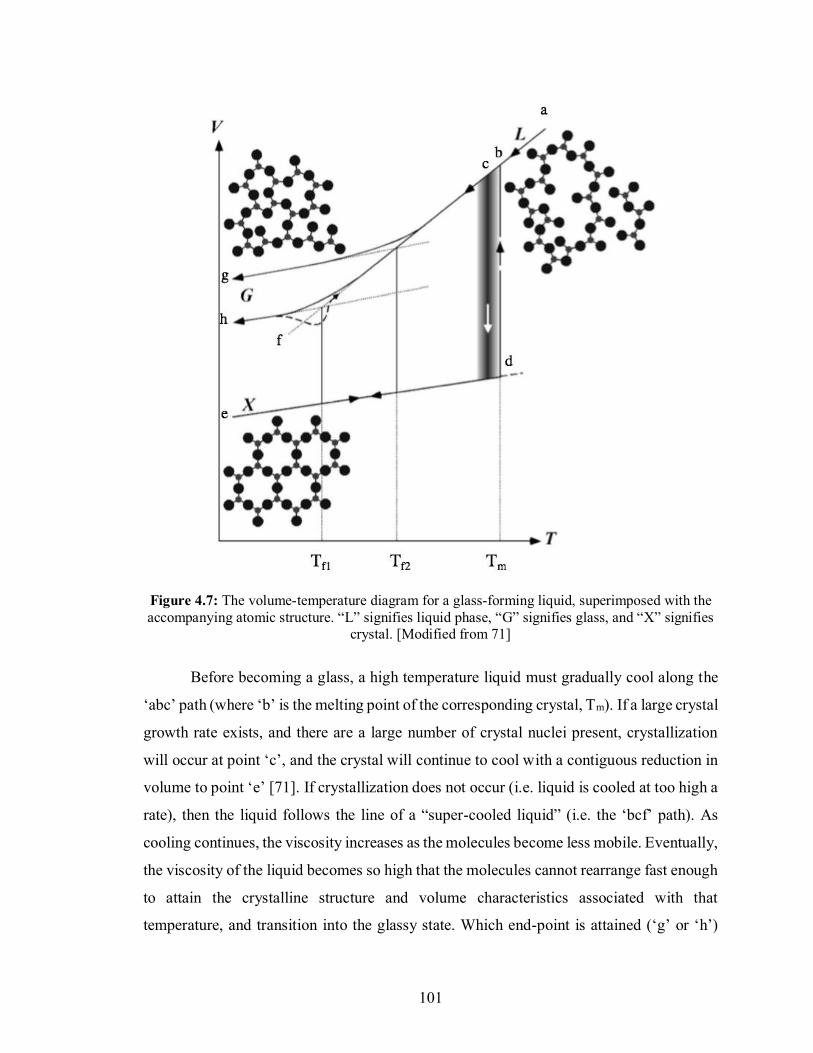
101
Figure 4.7: The volume-temperature diagram for a glass-forming liquid, superimposed with the accompanying atomic structure. “L” signifies liquid phase, “G” signifies glass, and “X” signifies
crystal. [Modified from 71]
Before becoming a glass, a high temperature liquid must gradually cool along the
‘abc’ path (where ‘b’ is the melting point of the corresponding crystal, Tm). If a large crystal
growth rate exists, and there are a large number of crystal nuclei present, crystallization
will occur at point ‘c’, and the crystal will continue to cool with a contiguous reduction in
volume to point ‘e’ [71]. If crystallization does not occur (i.e. liquid is cooled at too high a
rate), then the liquid follows the line of a “super-cooled liquid” (i.e. the ‘bcf’ path). As
cooling continues, the viscosity increases as the molecules become less mobile. Eventually,
the viscosity of the liquid becomes so high that the molecules cannot rearrange fast enough
to attain the crystalline structure and volume characteristics associated with that
temperature, and transition into the glassy state. Which end-point is attained (‘g’ or ‘h’)
102
depends on the cooling rate of the super-cooled liquid (fast cooling and slow cooling,
respectively) [71].
Since the transition to the glassy state does not occur at a single temperature, and
can follow two different paths, the thermal history of a glass has a large effect on the final
structure and resulting properties [71,118,119]. Since spherical processing is done over a
flame hot enough to melt the glass frit, there is a chance that the resulting microspheres do
not have the same structure and/or properties as the frit. Thermal augmentation after
formation may lead to a different heating or cooling rate than that undergone upon
formation, thereby changing the final state of the glass from fast cooling (‘g’) to slow
cooling (‘h’), or vice versa. It is only by confirming the structure and properties of the
thermally augmented glass that one can confirm that data collected on the original glass
frit will translate to the intended application.
The characterization data of the newly melted BRS2 irregular particles and
microspheres confirm the reproducibility of the results obtained in experiments one and
two. Both density and molar density values are statistically equivalent before and after
spherical processing. Molar density was calculated to remove the variation that molar mass
has on density. However, it was found in experiment one that molar mass has very little
effect on density for the BRS compositions, as Sr and Rb have very similar atomic weights.
The re-characterization data of the glass frit suggests that the same composition was
synthesized in this melt as in the original. The spherical data indicates that there is a good
chance that no structural or property changes have occurred in the 100-300 µm, compared
to the irregular particles.
Thermal analysis data (Fig. 4.5c) of the microspheres and the irregular particles are
not statistically different. A summary of the glass transition temperature and melt
temperature (both onset and inflection) data collected is provided in Table 4.4. These data,
combined with the consistent densities, allows for the provisional conclusion that the new
glass synthesized matches the glass characterized in experiments one and two. Since Tg is
defined as “the intersection of the glassy state line with the tangent to the steep portion of
the state curve in the transition range” [71], it can therefore give valuable information
regarding the thermal history and structure of a glass. Again, the comparable results seen
in the original, the new glass frit, and the spheres reinforces the conclusion drawn from the
103
density data; that no structural changes have occurred from the thermal augmentation
undergone during spherical processing. Therefore, there may be reason to believe that at
the 100-300 µm particle size range, spherical processing does not impact structure or
properties for BRS2. However, this does not mean that structure and/or property changes
may not occur at a smaller or larger particle size range, where different cooling kinetics
inside the microsphere may occur, or for alternative compositions
A distinct increase in radiopacity is seen from the original test frit, to the preclinical
frit, to the spheres (3798 HU, 3932 HU, and 4123 HU respectively). This increase in
radiopacity, however, is more likely due to the random packing of the irregular particles,
and the more efficient packing of the microspheres over the irregular particles, rather than
an increase in the radiopacity of the glass itself. Radiopacity is calculated by averaging the
intensity of multiple voxels within the imaged vial over five different sections. Inefficient
packing leads to a large number of air filled spaces (i.e. voxels with a value of 0 HU)
between the radiopaque particles, thereby lowering the average intensity. The more
efficient packing of the microspheres will therefore increase the radiopacity of the vial as
there is less radiolucent air present per measurement area.
SEM, PSA, and sphericity testing have proven that 100-300 μm sized particles have
been synthesized, and that over 92% of these particles are spheres. This sphericity value,
however, may be higher than reported due to the inclusion of particle agglomerations in
the test (which will not be used in the pre-clinical study) which do not display spherical
symmetry. Therefore, spherical processing was successful in creating smooth spheres from
the clear majority of the glass frit supplied, without altering the structure or properties
displayed.
Since the data collected in experiments one and two have been confirmed as
transferable to the glass spheres, these microspheres will next be tested in vivo, to assess
their embolization effectiveness. Pre-clinical animal testing is necessary as not all safety
concerns can be tested in vitro, and those which can are relying on a certain amount of
approximation. The primary benefit of the technology is enhanced patient safety coupled
with the ability to ameliorate chronic inflammation whilst permitting subsequent
procedures to be performed at the same target level of occlusion. This study is required to
demonstrate the embolization effectiveness of the technology per FDA guidance on the
104
evaluation of embolization particles [17]. All safety concerns for embolic agents laid out
by FDA have been addressed in this protocol: (i) ease of deliverability, (ii) acute
complications, (iii) local and systemic foreign body reactions, (iv) recanalization, (v)
embolization effectiveness, and (vi) device migration. This investigation was performed as
a pilot animal study, in an aim to gain better insight into the appropriateness of this design
to test the efficacy of strontium substituted rubidium-borate systems as degradable and
imageable embolic agents.
Part 2 of experiment 3 was successful in synthesizing BRS2 microspheres which
displayed the same properties as those collected on glass frit in experiments one and two.
A protocol was successfully designed and approved (by both Dalhousie and CRCHUM) to
assess the embolization efficacy of the BRS2 microspheres in a porcine renal model. No
results have been collected from the pre-clinical animal study as of yet; it is set to begin
June 11th, 2018. Upon completion of the study, a Draft Report will be provided to the
Sponsor and any revisions to the study report mutually agreed upon by the Sponsor and
Study Director and/or Pathologist will be incorporated in the Final Report. At the end of
the revision process, the Report will be signed and considered final. The study Final Report
will include a summary of the objectives and procedures of the study, description of the
TA used, the methods used for histological processing, and a discussion of any
circumstances that may have altered or affected the integrity of the data. Any corrections
or additions to the Final Report will be in the form of a report amendment.
4.3. Conclusion
Experiment 3 outlines pilot protocols to be used in both in vitro and in vivo models.
As no in vitro models exist to assess the safety issues outlined in the Class IIB Special
Controls document provided by FDA, part one attempted to determine the feasibility of
implementing an in vitro benchtop model to assess the effectiveness of degradable particles
and the risk of migration. It was found that although it is possible, much more work must
be done before meaningful results can be attained. Part two focused on developing a
protocol to assess the embolization effectiveness of a preferred composition of borate glass
microsphere in a pre-clinical animal model. It was found that spherical processing of 100-
300 µm particles does not seem to affect their structure or properties, and therefore
105
validated the data collected in experiments one and two on glass frit. The composition
BRS2 was chosen for investigation, the microspheres of this composition will be used in
the developed pre-clinical protocol found in Appendix C. Successful completion of this
study may support the deployment of a larger, pre-clinical protocol in support of full ISO
10993 tests and performance evaluations to further de-risk the technology towards clinical
use.
106
CHAPTER 5
Conclusions
Uterine leiomyomas, also referred to as uterine fibroids, are the most common
benign tumor of the female reproductive tract [1]. They are found in over 70% of women
of reproductive age and cause a variety of debilitating symptoms, such as heavy bleeding,
pain, subfertility, urinary frequency and urgency, and pelvic pressure [1,2]. Until recently,
their only effective treatment was complete or partial removal of the uterus. However, in
1995 a uterine-sparing procedure was introduced called uterine artery embolization (UAE)
[6]. UAE offered (and continues to offer) the potential to preserve fertility in young women
with uterine leiomyomas. The minimally invasive nature of UAE also provides additional
benefits, including reduced hospital stays, reduced health care costs, and shorter recovery
times [6]. Currently, the commercially available microspheres for use in UAE are
permanent implants. However, more recently there has been a push from both physicians
and from patients for degradable microspheres: products which can effectively occlude the
uterine artery, but will subsequently be eliminated safely from the body when their intended
function is complete [14].
The nascent nature of degradable products in this application requires diligent
assessment of risk, as well as evidence of efficacy, to help drive concept development and
design engineering. Evidence based medicine teaches that there are three intersecting
spheres of influence to consider when selecting treatment options for patients. These are:
(i) the best available research evidence, (ii) individual clinical expertise, and (iii) patient
values & expectations [135]. Unfortunately, due to the novelty of degradable embolic
agents, clinical evidence to support the physiological benefits of degradable microspheres
over their permanent counterparts is limited. It is believed, however, that degradable
embolic agents may have particular advantages over permanent agents in UAE, as they
allow for the complete reperfusion of blood flow to the uterus after the leiomyoma has been
treated [15,56,103]. To support this position, it is contested in the literature that while UAE
maintains uterine patency, the presence of permanent agents may impact pregnancy.
Specifically, a study completed in 2007 showed that of 164 women who desired future
107
pregnancy (at the time of their UAE procedure with permanent agents), only 21 were able
to get pregnant, resulting in 24 pregnancies. Of those 24 pregnancies, 18 live births
occurred, and 33% of those babies were born with low or very low birth weights [136]. In
addition, based on studies with sheep, this low birth weight is thought to be the result of
uterine lesion and/or a persistent blockage of uterine blood flow [137]. It is believed that
extended ischemia to the uterus could potentially inhibit the development of a large-sized
placenta, leading to spontaneous abortion or newborns of very low birth weight [137].
Additionally, the remaining permanent embolic agents may lower uterine blood flow
during gestation, as the middle uterine artery supplies the bulk of the blood supply to the
fetus. If the uterine artery is fully (or partially) occluded, then the reduction in blood flow
to the uterus will decrease fetal growth and survival [137].
Although it is believed that degradable embolic agents will provide some
physiological benefit over permanent embolic agents, the exact outcomes following
embolization with degradable particles are unknown. It is assumed that once the particles
degrade and are safely removed from the body, that the recanalized vessel will allow for
reperfusion of blood to the uterus. However, outcomes with respect to the downstream
vasculature are currently unknown, and degradable agents may present new safety risks.
The only information available on the nature of recanalized vessels after TAE with
degradable agents is provided by Owen et al. However, they do not specify if recanalization
means the reopening of the previously occluded vessel, or angiogenesis (and there appears
to be much confusion in the literature with respect to the use of this term). As such, limited
conclusions can be drawn from the literature in terms of the usefulness of recanalization,
and/or the appropriate time frames within which recanalization should occur. However,
with this said, it is important to note that evidence does exist which conclusively shows
that permanent occlusion is detrimental to the future fertility of patients [136,137].
Due to the lack of high level clinical information available regarding the
performance of degradable embolic microspheres, no specific timeframes for degradation
are provided for UAE. One can reference the surgical outcomes provided by Vilos et al.
[35] of total occlusion of 5-7 hours for effective treatment of uterine leiomyomas; however
the difference in reperfusion rates must be accounted for. In surgical evaluations,
reperfusion of blood to the uterus is instant and complete; with degradable embolic agents,
108
reperfusion may occur gradually as the particles degrade. Therefore, it is contended that
the design requirement for degradable embolic agents for UAE is that they can provide
total occlusion for 5-7 hours before degradation sufficient to allow reperfusion of blood to
the uterus. Additionally, inflammation causing additional biological occlusion may not be
favorable and may prolong ischemic time. This prolonged occlusion contradicts the
specific engineering of a particle that degrades at the appropriate timeframe to maximize
treatment outcomes while minimizing necrosis to healthy tissues. Although there is a
significant amount of evidence outlining the potential harm of permanent embolics, and
pre-clinical animal trials demonstrating the potential safety of degradable embolics,
sufficient evidence at the clinical level is missing. The current evidence is therefore not
suitable to effectively guide physicians in this section of evidence based medicine.
However, despite the lack of evidence and clinical expertise, there is a clear drive in
the evidence based medicine paradigm for degradable embolic agents for TAE, which is
that patients now want degradable products and physicians want to recommend them.
Patients commonly express concerns about “foreign materials remaining in the body” [14]
thus there is a need to continue down this path, even if the current clinical evidence
regarding degradable embolics is not satisfactory. On the basis of the published (Journal of
Functional Biomaterial) review in Chapter 1 (Section 1.2) [103], it has been identified that
the following design inputs are important when considering degradable embolic agents for
TAE. These innovative technologies must provide predictable and effective occlusion
while also providing:
1. Tailored degradation timeframes—to provide adequate infarction to the target
tissues in a variety of indications, subsequently allowing return of flow (e.g., 5–7 h for
uterine artery embolization—based on Doppler-guided transvaginal clamping) [15]
2. A variety of tightly calibrated particle size distributions—to optimize particle
delivery according to target artery anatomy [16]
3. Ease of delivery through conventional microcatheters—to facilitate adoption of the
novel technology into established embolization techniques
4. Full biological compatibility as per the relevant sections of ISO-10993—to
minimize safety concerns [17]
5. Multi-modal imageability (e.g., fluoroscopy, CT)—to allow for efficiency and
109
standardization of embolization endpoints [18].
Currently, five materials are being developed for use as degradable embolic microspheres
for TAE. None of these materials satisfy all the design inputs published in the literature.
The most common limitations are lack of variable degradation timeframes, and lack of
multi-modal imageability. Given these design input requirements, there are several
material classifications that could be considered for the development of an implantable,
degradable, and imageable embolic microsphere. Glasses are currently being investigated
for use in neuromuscular repair (fibrous constructs for muscle and nerve regeneration),
artificial cornea, orbital implants, epithelial and cardiac tissue engineering, treatment of
gastric ulcers and non-osseous cancer therapy [138]. A small variation in glass composition
can greatly modify the features of the material, thereby extending the potential applications
which glasses can fill in the medical field. Glasses present an attractive option in this
instance because not only do they allow for inherent radiopacity through the incorporation
of ions with sufficient NIST mass attenuation coefficients, they can be manufactured to
exhibit variable degradation timeframes and particle size ranges.
This thesis explored the potential of borate glasses for use in TAE (with particular
consideration toward UAE). Borate glasses are excellent glass formers for manufacturing
purposes, can accommodate several elements, and offer complete degradation in aqueous
environments, potentially allowing for multi-modal imageability, and the complete
recanalization of vessels at various degradation timeframes. This work examined two series
of glasses: (i) the BRS series with the composition 70B/30-xRb/xSr (where x=2,4,6,8,10),
and (ii) the BRG series with the composition 70B/30-xRb/xGa (where x=2,4,6,8,10).
Simple borate systems were chosen for this work so as to better understand the
composition-structure-property relationship of monovalent for divalent, or monovalent for
trivalent substituted borate networks. For borate systems, it is commonly assumed that an
increase in four-fold coordination in borate networks leads to a decrease in dissolution rates
under aqueous conditions due to an increase in glass connectivity [74,101]. Interestingly,
the opposite of this assumption was observed in the glass systems examined in this work.
The BRS series displayed a constant B4 fraction, while the percentage of B4 in the BRG
series significantly decreased with increasing substitution. Surprisingly, despite the lack of
increase in B4 fractions, an increase in hydrolytic stability and glass transition temperature
110
were seen in both glass series. Additionally, an increase in density was observed in the
BRS series with increasing strontium substitution. In contrast, a significant decrease in
density was observed for the BRG series. Essentially, these data indicate that the literature
poorly understands these glass systems, and as a result their structure-property
relationships are unpredictable. This statement is exemplified by the data collected in
experiments one and two of this thesis. Despite the commonly accepted theory that the ratio
of B3 to B4 present in glasses has the largest influence on glasses properties, this work
determined that this theory does not always accurately explain the structure-property
relationships of simple high borate glass systems. Accordingly, continued directed work
in this field is important to broaden our knowledge for such glass systems.
While the work conducted in this thesis considered rubidium and gallium
containing compositions, toxicological information on these elements exists only at the
most basic level (e.g. no ATSDR documents exist) which increases the risk profile of these
glasses as an actual medical device. These elements are likely safe in small doses, based
on toxicological studies previously published [83-88], and their clinical use to treat a
variety of diseases [93, 94]. However considerable further research into the exact toxicity
of these ions is required should they be considered in formulations for actual medical grade
microspheres. Additionally, the structural nature of the network modifiers should be
considered when discussing these glass networks. As previously mentioned, when
considering borate glasses, most of the property predictions are based on the borate
structure in the network (i.e. 3- vs 4-fold coordinate boron). However, it has been observed
in this thesis that modifier ions play an important role in predicting the properties of the
resulting glass (and not necessarily in traditionally understood ways). Despite no increase
in B4 concentration (i.e. borate glass network structure), the addition of Sr to the BRS
series increases density, Tg, and glass stability, thereby decreasing degradation rates. The
role of gallium in the BRG series is even more pronounced; 11B NMR indicates that gallium
has a significant effect on the boron structure, forming tetrahedral gallium centers, and
modulating the network to increase hydrolytic stability (as seen in Figure 3.5). When Ga is
considered a network former, rather than a network modifier, the theoretical values more
closely approximate the accepted O/B ratios for the composition. Therefore, not only do
network modifiers affect clinically important properties (i.e. radiopacity and degradation
111
timeframes), they also have a direct effect on the structure itself and should therefore be
given an equal value in the literature when discussing composition-structure-property
relationship.
Once the clinically relevant attributes of the glasses assessed in this thesis had been
determined in vitro, the next step of the work was to conduct some provisional evaluations
to examine the safety, efficacy, and performance of one of these glass compositions as a
degradable embolic agent. The BRS2 composition was chosen as it exhibited the lowest
density, second highest radiopacity, and degraded completely in 48 hours. Additionally,
BRS2 exhibited high glass stability without any susceptibility to T1 or T2 weighted MRI
scans. Experiment three began by investigating the utility of using glass frit for preliminary
evaluation of glass compositions. No significant changes were observed between the
properties of the glass frit and the processed microspheres. This discovery was useful from
a manufacturing standpoint, as it allows for reliable synthesis of consistent glass
compositions. Additionally, since spherical processing is time consuming, has a relatively
low yield, and is not readily available, the ability to use glass frit for investigative
experiments will make the research process more efficient.
Once a best in class composition was chosen for use in experiment three, the
drafting of an appropriate pilot protocol began to test the safety, efficacy, and performance
of degradable glass microspheres for TAE. FDA provides a guidance document which
outlines the criteria that requires pre-clinical investigation prior to approval of new medical
devices for transarterial embolization [17]. One of those criteria is embolization
effectiveness. This parameter can be evaluated by multiple measures, including ease of
delivery, recanalization of the vessels/durability of occlusion, local foreign body reactions,
and level of ischemia when compared to the control [17]. The durability of the occlusion
and vessel recanalization are essential when considering effectiveness of degradable
embolic agents, as the occlusion is temporary. If ischemic time is too short, full therapeutic
benefits of the treatment may not be attained [125,126]. However if the ischemic time is
too long, the potential benefits of having a degradable product may be reduced.
Additionally, local body reactions must be considered when assessing embolization
effectiveness, as biological occlusion resulting from local inflammatory effects may hinder
reperfusion of target tissue. For some applications that require longer occlusion times, this
112
body reaction may be favorable to prolong ischemia; however for uterine artery
embolization, a very short occlusion time is desired, and biological occlusion may hinder
the early reperfusion of blood to the uterus. Lingering biological occlusion defeats the
purpose of engineering the particle to degrade in 24-48 hours, and thereby may impact the
effectiveness of the embolic agent for this application.
Another important risk to consider with respect to degradable microspheres is
migration; it has been presented by FDA as a requirement to be investigated prior to
approval of new medical devices for transarterial embolization [17]. In discussions with
interventional radiologists regarding the clinical implications of the risk of migration, there
is a general consensus that migration may occur, but if it does, there is little clinical
evidence to indicate that it causes clinical complications. It is not clear from the literature
the rate of complications associated with migration, and there is limited evidence to support
that this is a major risk or a small risk. A review of the Manufacturer and User Facility
Device Experience (MAUDE) database for the last 10 years, using the search strings
“Migration” and “EmboSphere” revealed only 3 cases. These cases included examples of
an arteriovenous shunt leading to cerebral infarction, as well as a reflux of particles to non-
target areas, specifically leading to ischemic pancreatitis and postoperative cholecystitis
[139]. Although the rate of clinical complications appears to be quite low, there is a chance
that it is not being properly assessed. Since all currently investigated materials are
radiolucent, very little work has been done to assess the migration risk of degradable
embolic agents, apart from the work done by Owen et. al [31]. A secondary search of the
MAUDE database using only the search string “EmboSphere” revealed an additional 20
cases that although not directly labeled as migration, may easily have been caused by non-
target embolization. These cases included instant blindness and vision impairment,
necrosis of the gallbladder, and even death. An attempt at further understanding the risk of
migration was started in part one of experiment three. Ideally, the risk of migration will
one day be assessable via in vitro methods prior to any pre-clinical animal studies.
To assess migration, the use of radiopaque particles may be of particular benefit as
it may allow for the direct visualization of the embolic agent through CT or fluoroscopy.
Although imageability is not necessarily crucial for uterine artery embolization, it is a
convenient attribute. Imageability may be more useful when considering applications such
113
as drug eluting bead transarterial chemoembolization (DEB-TACE) for the treatment of
hepatocellular carcinoma. In this application, the spatial distribution of particles is crucial
for regulating the local concentration of chemotherapy and if the particles are
inappropriately spaced, inappropriate dosing of tumor tissue may occur [140].
Once embolization effectiveness and migration have been assessed in a preclinical
animal model, a sufficient level of clinical evidence must be amassed to give physicians
the confidence to employ degradable embolics as a mainstream option for TAE. Ideally, a
meta-analysis of several multi-centered, randomized, and appropriately powered
prospective clinical trials would occur to definitively assess the safety, efficacy, and
performance of degradable microspheres in TAE. However, this level of evidence does not
exist for most day-to-day procedures. These procedures have become trusted as safe
because the occurrence of harm associated with that procedure is low, and the severity of
that harm is also low (or if it is high, it occurs so infrequently that it’s an acceptable risk).
Therefore, since the occurrence of the highest level of clinical evidence is rare and takes a
long time to acquire, the question must be asked, ‘what is a reasonable level of evidence
for physicians to accept prior to suggesting this treatment to their patients?’.
Pre-clinical models do not register on the 6S pyramid of evidence for evidence
based medicine [141], however FDA states in the clinical testing section of the Class II
Special Controls guidance document [17] that it will rely heavily on benchtop and pre-
clinical testing when determining potential safety and efficacy approval. Specifically,
“FDA will rely upon well-designed bench and/or animal testing rather than requiring
clinical studies for new devices unless there is a specific justification for asking for clinical
information to support a determination of substantial equivalence” [17]. This may
potentially lead to the approval of materials that are not effective at their intended
application. For example, Contour SE was approved by FDA for use in uterine artery
embolization, however when compared to conventional PVA and tris-acryl gelatin
microspheres (TAGM), it was found to leave substantial portions of tumors uninfarcted
[142]. This ineffective treatment lead to the premature ending of a large clinical trial, and
called into question the “quality of the evidence we accept” before approving a novel
material for integration into practice. Therefore, there is a chance that FDA approval is not
a reasonable level of evidence for mainstream implementation of an embolic agent.
114
The intent of the pilot animal study developed as part of this thesis is to demonstrate
in a pilot model how safety, efficacy and performance could be established for degradable
agents (e.g. processing, post processing, sacrifice procedures, etc). Successful completion
of this study will support the deployment of a larger, pre-clinical protocol in support of full
ISO 10993 tests and performance evaluations to further de-risk the technology towards
clinical use. Should the larger animal study prove successful, and the product can (i) be
produced safely, non-pyrogenic, and sterile, (ii) be delivered and perform per all the
requirements laid out by FDA, and (iii) pass all biocompatibility tests, then all pre-clinical
risks have been managed as fully as possible, before moving to human trials. When
considering human trials, the implementation of a small pilot study is the logical first step.
However, the population that should be examined is less clear; should the product be tested
on women who will have a hysterectomy regardless of the outcome of the procedure, or on
women who may benefit from the UFE procedure? Increased discussion on both the
acceptable level of evidence required to adopt new embolic products into clinical practice,
and the quality of that evidence is required before degradable embolic agents become
common practice.
Despite much of the work that being conducted on the development of degradable
products, high level clinical evidence is currently missing. Despite the lack of evidence,
there is an overwhelming requirement by patients to have materials used for embolization
procedures that will be eliminated safely from their body. Therefore, in the world of
evidence based medicine, it would seem reasonable that prior to the broader deployment
of these materials, we should be considering whether or not there is a significant benefit to
degradable particles beyond the desire demonstrated by patients. Patient expectations are
only one section of evidence based medicine, and must be supported by both the
experienced physician and the highest level of acceptable clinical evidence. As this
evidence does not currently exist, this thesis has attempted to address some of the initial
uncertainties related to uterine artery embolization, while also expanding the use of glass
biomaterials outside the scope of the skeletal system.
Finally, this thesis attempted to expand the understanding of the significantly
understudied field of borate glass systems. It succeeded in contributing to the expansion of
the compositional palate for borate glasses, and endeavored to better our understanding of
115
the composition-structure-property relationship found in simple borate systems. By
assessing the effect of divalent (Sr) or trivalent (Ga) substitutions of monovalent (Rb)
network modifiers on the basic properties of the glass systems, it has been concluded that
these glasses behave unpredictably. As such, continued directed work is crucial to reach a
sufficient understanding of these unconventional glass systems. Regardless, the BRS glass
series exhibits desirable characteristics as degradable and imageable embolic agents for
TAE procedures due to its tailorable degradation timeframes and significant radiopacity.
116
REFERENCES
1. Goodwin, S. C.; Spies, J. B. Uterine fibroid embolization. N. Engl. J. Med. 2009, 361,
690–697, doi:10.1056/NEJMct0806942.
2. Gupta, J. K.; Sinha, A.; Lumsden, M. A.; Hickey, M. Uterine artery embolization for
symptomatic uterine fibroids. Cochrane Database Syst Rev 2012, CD005073,
doi:10.1002/14651858.CD005073.pub3.
3. Kashani, B. N.; Centini, G.; Morelli, S. S.; Weiss, G.; Petraglia, F. Role of Medical
Management for Uterine Leiomyomas. Best Pract Res Clin Obstet Gynaecol 2016, 34,
85–103, doi:10.1016/j.bpobgyn.2015.11.016.
4. Pelage, J.-P.; Cazejust, J.; Pluot, E.; Le Dref, O.; Laurent, A.; Spies, J. B.; Chagnon,
S.; Lacombe, P. Uterine fibroid vascularization and clinical relevance to uterine fibroid
embolization. Radiographics 2005, 25 Suppl 1, S99-117, doi:10.1148/rg.25si055510.
5. Cardozo, E. R.; Clark, A. D.; Banks, N. K.; Henne, M. B.; Stegmann, B. J.; Segars, J.
H. The Estimated Annual Cost of Uterine Leiomyomata in the United States. Am J Obstet
Gynecol 2012, 206, 211.e1-211.e9, doi:10.1016/j.ajog.2011.12.002.
6. REST Investigators. Uterine-Artery Embolization versus Surgery for Symptomatic
Uterine Fibroids. New England Journal of Medicine 2007, 356, 360–370,
doi:10.1056/NEJMoa062003.
7. Beinfeld, M. T.; Bosch, J. L.; Isaacson, K. B.; Gazelle, G. S. Cost-effectiveness of
uterine artery embolization and hysterectomy for uterine fibroids. Radiology 2004, 230,
207–213, doi:10.1148/radiol.2301021482.
8. Vaidya, S.; Tozer, K. R.; Chen, J. An Overview of Embolic Agents. Semin Intervent
Radiol 2008, 25, 204–215, doi:10.1055/s-0028-1085930.
9. Spies, J. B.; Allison, S.; Flick, P.; McCullough, M.; Sterbis, K.; Cramp, M.; Bruno, J.;
Jha, R. Polyvinyl alcohol particles and tris-acryl gelatin microspheres for uterine artery
embolization for leiomyomas: results of a randomized comparative study. J Vasc Interv
Radiol 2004, 15, 793–800, doi:10.1097/01.RVI.0000136982.42548.5D.
117
10. Abramowitz, S. D.; Israel, G. M.; McCarthy, S. M.; Pollak, J. S.; White, R. I.; Tal, M.
G. Comparison of four embolic materials at uterine artery embolization by using
postprocedural MR imaging enhancement. Radiology 2009, 250, 482–487,
doi:10.1148/radiol.2502080574.
11. Guimaraes, M.; Arrington, D.; MacFall, T.; Yamada, R.; Schonholz, C. Does
Material Matter? Particular Embolics. Endovascular Today. April 2013, pp. 70–74.
12. Worthington-Kirsch. Do Particle Size and Type Matter? Lessons learned from
experience with uterine artery embolization. Endovascular Today. June 2008, pp. 39–42.
13. Kehoe, S.; Kilcup, N.; Abraham, R.; Boyd, D. Chapter 20: Glass Materials in
Interventional Radiology and Interventional Oncology. In Bioactive Glasses; 2016; pp.
471–495.
14. Forster, R. E. J.; Thürmer, F.; Wallrapp, C.; Lloyd, A. W.; Macfarlane, W.; Phillips,
G. J.; Boutrand, J.-P.; Lewis, A. L. Characterisation of physico-mechanical properties and
degradation potential of calcium alginate beads for use in embolisation. J Mater Sci
Mater Med 2010, 21, 2243–2251, doi:10.1007/s10856-010-4080-y.
15. Verret, V.; Pelage, J. P.; Wassef, M.; Louguet, S.; Servais, E.; Bédouet, L.; Beaulieu,
T.; Moine, L.; Laurent, A. A novel resorbable embolization microsphere for transient
uterine artery occlusion: a comparative study with trisacryl-gelatin microspheres in the
sheep model. J Vasc Interv Radiol 2014, 25, 1759–1766, doi:10.1016/j.jvir.2014.06.025.
16. Transcatheter Embolization and Therapy; Kessel, D., Ray, C., Eds.; Techniques in
Interventional Radiology; Springer-Verlag: London, 2009; ISBN 978-1-84800-896-0.
17. FDA Guidance Documents: Class II Special Controls Guidance Document: Vascular
and Neurovascular Embolization Devices - Guidance for Industry and FDA Staff 2004.
18. Kehoe, S.; Amensag, S.; Looney, M.; Abraham, R. J.; Boyd, D. “Imageable” Zinc-
Silicate Glass Microspheres For Transarterial Embolization: A Renal Artery
Embolization Study. Biomedical glasses 2015, 1, doi:10.1515/bglass-2015-0007.
19. Bohner, M. Resorbable Biomaterials as Bone Graft Substitutes. Mater Today 2010,
13, 24–30, doi:10.1016/S1369-7021(10)70014-6.
118
20. Williams, D. Essential Biomaterials Science; Cambridge University Press, 2014;
ISBN 978-0-521-89908-6.
21. Gupta, B.; Revagade, N.; Hilborn, J. Poly(lactic acid) fiber: An overview. Prog
Polym Sci 2007, 32, 455–482, doi:10.1016/j.progpolymsci.2007.01.005.
22. Gentile, P.; Chiono, V.; Carmagnola, I.; Hatton, P. V. An overview of poly(lactic-co-
glycolic) acid (PLGA)-based biomaterials for bone tissue engineering. Int J Mol Sci
2014, 15, 3640–3659, doi:10.3390/ijms15033640.
23. Makadia, H. K.; Siegel, S. J. Poly Lactic-co-Glycolic Acid (PLGA) as Biodegradable
Controlled Drug Delivery Carrier. Polymers (Basel) 2011, 3, 1377–1397,
doi:10.3390/polym3031377.
24. You, Y.; Lee, S. W.; Youk, J. H.; Min, B.-M.; Lee, S. J.; Park, W. H. In vitro
degradation behaviour of non-porous ultra-fine poly(glycolic acid)/poly(l-lactic acid)
fibres and porous ultra-fine poly(glycolic acid) fibres. Polymer Degradation and Stability
2005, 90, 441–448, doi:10.1016/j.polymdegradstab.2005.04.015.
25. Houchin, M. L.; Topp, E. M. Chemical degradation of peptides and proteins in
PLGA: a review of reactions and mechanisms. J Pharm Sci 2008, 97, 2395–2404,
doi:10.1002/jps.21176.
26. Grayson, A. C. R.; Voskerician, G.; Lynn, A.; Anderson, J. M.; Cima, M. J.; Langer,
R. Differential degradation rates in vivo and in vitro of biocompatible poly(lactic acid)
and poly(glycolic acid) homo- and co-polymers for a polymeric drug-delivery microchip.
J Biomater Sci Polym Ed 2004, 15, 1281–1304.
27. Xu, Y.; Kim, C.-S.; Saylor, D. M.; Koo, D. Polymer degradation and drug delivery in
PLGA-based drug-polymer applications: A review of experiments and theories. J.
Biomed. Mater. Res. Part B Appl. Biomater. 2017, 105, 1692–1716,
doi:10.1002/jbm.b.33648.
28. Rasal, R. M.; Janorkar, A. V.; Hirt, D. E. Poly(lactic acid) modifications. Prog Polym
Sci 2010, 35, 338–356, doi:10.1016/j.progpolymsci.2009.12.003.
119
29. Boland, E. L.; Shine, R.; Kelly, N.; Sweeney, C. A.; McHugh, P. E. A Review of
Material Degradation Modelling for the Analysis and Design of Bioabsorbable Stents.
Ann Biomed Eng 2016, 44, 341–356, doi:10.1007/s10439-015-1413-5.
30. Nagarajan, S.; Reddy, B. S. R. Bio-absorbable polymers in implantation-An
overview. JSIR Vol.68(12) [December 2009] 2009.
31. Owen, R. J.; Nation, P. N.; Polakowski, R.; Biliske, J. A.; Tiege, P. B.; Griffith, I. J.
A preclinical study of the safety and efficacy of OcclusinTM 500 Artificial Embolization
Device in sheep. Cardiovasc Intervent Radiol 2012, 35, 636–644, doi:10.1007/s00270-
011-0218-7.
32. Revised 501(k) Summary for IMBiotechnologies Ltd. Occlusin 500 Artificial
Embolization Device (Per 21CFR 807.92) 2010.
33. Stuart, S.; Mayo, J. R.; Ling, A.; Schulzer, M.; Klass, D.; Power, M. A.; Roberton, B.
J.; Wan, J. M.; Liu, D. M. Retrospective study of the impact of fellowship training on two
quality and safety measures in uterine artery embolization. J Am Coll Radiol 2014, 11,
471–476, doi:10.1016/j.jacr.2013.09.020.
34. Anderson, J. M.; Rodriguez, A.; Chang, D. T. Foreign body reaction to biomaterials.
Semin. Immunol. 2008, 20, 86–100, doi:10.1016/j.smim.2007.11.004.
35. Vilos, G. A.; Vilos, E. C.; Romano, W.; Abu-Rafea, B. Temporary uterine artery
occlusion for treatment of menorrhagia and uterine fibroids using an incisionless
Doppler-guided transvaginal clamp: case report. Hum. Reprod. 2006, 21, 269–271,
doi:10.1093/humrep/dei299.
36. Food and Drug Administration Use of International Standard ISO 10993-1,
`Biological evaluation of medical devices--Part 1: Evaluation and testing within a risk
management process’’; Guidance for Industry and Food and Drug Administration Staff;
Availability Available online: https://www.regulations.gov/document?D=FDA-2013-D-
0350-0037 (accessed on Apr 4, 2018).
37. EmboSphere®® Microspheres Available online:
https://www.merit.com/interventional-oncology-
120
spine/embolotherapy/microspheres/EmboSphere®-microspheres/ (accessed on Apr 4,
2018).
38. Swielim, G. E. A. Atlas: Anatomy of Sheep; ktab INC.: Cairo, Egypt, 2006; ISBN
977-281-292-4.
39. Webster, R.; Elliott, V.; Park, B. K.; Walker, D.; Hankin, M.; Taupin, P. PEG and
PEG conjugates toxicity: towards an understanding of the toxicity of PEG and its
relevance to PEGylated biologicals. In PEGylated Protein Drugs: Basic Science and
Clinical Applications; Milestones in Drug Therapy; Birkhäuser Basel, 2009; pp. 127–146
ISBN 978-3-7643-8678-8.
40. Magnon, L.; Laurent, A.; Wassef, F.; Bedouet, L.; Louguet, S.; Verret, V.; Servais, E.
Implantable swellable bio-resorbable polymer 2012.
41. Maeda, N.; Verret, V.; Moine, L.; Bédouet, L.; Louguet, S.; Servais, E.; Osuga, K.;
Tomiyama, N.; Wassef, M.; Laurent, A. Targeting and recanalization after embolization
with calibrated resorbable microspheres versus hand-cut gelatin sponge particles in a
porcine kidney model. J Vasc Interv Radiol 2013, 24, 1391–1398,
doi:10.1016/j.jvir.2013.05.058.
42. ASTM Compass Int. Standard Specification for Woven Wire Test Sieve Cloth and
Test Sieves 2017.
43. Agrawal, C. M.; Ong, J. L.; Appleford, M. R.; Mani, G. Introduction to Biomaterials:
Basic Theory with Engineering Applications; 1 edition.; Cambridge University Press:
New York, 2013; ISBN 978-0-521-11690-9.
44. Haraldsson, B.; Nyström, J.; Deen, W. M. Properties of the glomerular barrier and
mechanisms of proteinuria. Physiol. Rev. 2008, 88, 451–487,
doi:10.1152/physrev.00055.2006.
45. NSP Nanosoft Polymers Available online:
https://www.nanosoftpolymers.com/product/plga-peg-plga/ (accessed on Apr 4, 2018).
46. Rosca, C.; Popa, M. I.; Lisa, G.; Chitanu, G. C. Interaction of chitosan with natural or
synthetic anionic polyelectrolytes. 1. The chitosan–carboxymethylcellulose complex.
Carbohydrate Polymers 2005, 62, 35–41, doi:10.1016/j.carbpol.2005.07.004.
121
47. Weng, L.; Le, H. C.; Talaie, R.; Golzarian, J. Bioresorbable hydrogel microspheres
for transcatheter embolization: preparation and in vitro evaluation. J Vasc Interv Radiol
2011, 22, 1464–1470.e2, doi:10.1016/j.jvir.2011.06.010.
48. Bajpai, A.; Shrivastava, J. In vitro enzymatic degradation kinetics of polymeric
blends of crosslinked starch and carboxymethyl cellulose. Polymer International 2005,
54, 1524–1536, doi:10.1002/pi.1878.
49. Reeves, R.; Ribeiro, A.; Lombardo, L.; Boyer, R.; Leach, J. B. Synthesis and
Characterization of Carboxymethylcellulose-Methacrylate Hydrogel Cell Scaffolds.
Polymers (Basel) 2010, 2, 252–264, doi:10.3390/polym2030252.
50. Lee, S. Y.; Bang, S.; Kim, S.; Jo, S. Y.; Kim, B.-C.; Hwang, Y.; Noh, I. Synthesis and
in vitro characterizations of porous carboxymethyl cellulose-poly(ethylene oxide)
hydrogel film. Biomaterials Research 2015, 19, 12, doi:10.1186/s40824-015-0033-3.
51. Kean, T.; Thanou, M. Biodegradation, biodistribution and toxicity of chitosan. Adv.
Drug Deliv. Rev. 2010, 62, 3–11, doi:10.1016/j.addr.2009.09.004.
52. Zhang, H.; Neau, S. H. In vitro degradation of chitosan by bacterial enzymes from rat
cecal and colonic contents. Biomaterials 2002, 23, 2761–2766.
53. Zhang, Y.; Wang, Z.; Zhang, J.; Chen, C.; Wu, Q.; Zhang, L.; Zhang, X. Quantitative
determination of chitinolytic activity of lysozyme using half-deacetylated chitosan as a
substrate. Carbohydrate polymers 2011.
54. Yang, Y. M.; Hu, W.; Wang, X. D.; Gu, X. S. The controlling biodegradation of
chitosan fibers by N-acetylation in vitro and in vivo. J Mater Sci Mater Med 2007, 18,
2117–2121, doi:10.1007/s10856-007-3013-x.
55. Weng, L.; Rusten, M.; Talaie, R.; Hairani, M.; Rosener, N. K.; Golzarian, J.
Calibrated bioresorbable microspheres: a preliminary study on the level of occlusion and
arterial distribution in a rabbit kidney model. J Vasc Interv Radiol 2013, 24, 1567–1575,
doi:10.1016/j.jvir.2013.06.009.
56. Weng, L.; Rostamzadeh, P.; Nooryshokry, N.; Le, H. C.; Golzarian, J. In vitro and in
vivo evaluation of biodegradable embolic microspheres with tunable anticancer drug
release. Acta Biomater 2013, 9, 6823–6833, doi:10.1016/j.actbio.2013.02.017.
122
57. Weng, L.; Seelig, D.; Rostamzadeh, P.; Golzarian, J. Calibrated Bioresorbable
Microspheres as an Embolic Agent: An Experimental Study in a Rabbit Renal Model. J
Vasc Interv Radiol 2015, 26, 1887–1894.e1, doi:10.1016/j.jvir.2015.01.014.
58. Pillai, C. K. S.; Paul, W.; Sharma, C. P. Chitin and chitosan polymers: Chemistry,
solubility and fiber formation. Prog Polym Sci 2009, 34, 641–678,
doi:10.1016/j.progpolymsci.2009.04.001.
59. Usman, A.; Zia, K. M.; Zuber, M.; Tabasum, S.; Rehman, S.; Zia, F. Chitin and
chitosan based polyurethanes: A review of recent advances and prospective biomedical
applications. Int. J. Biol. Macromol. 2016, 86, 630–645,
doi:10.1016/j.ijbiomac.2016.02.004.
60. Kwak, B. K.; Shim, H. J.; Han, S.-M.; Park, E. S. Chitin-based embolic materials in
the renal artery of rabbits: pathologic evaluation of an absorbable particulate agent.
Radiology 2005, 236, 151–158, doi:10.1148/radiol.2361040669.
61. Schwarz, A.; Zhang, H.; Metcalfe, A.; Salazkin, I.; Raymond, J. Transcatheter
embolization using degradable crosslinked hydrogels. Biomat 2004, 25, 5209–5215,
doi:10.1016/j.biomaterials.2003.12.022.
62. Ohta, S.; Nitta, N.; Takahashi, M.; Murata, K.; Tabata, Y. Degradable Gelatin
Microspheres as an Embolic Agent: an Experimental Study in a Rabbit Renal Model.
Korean J Radiol 2007, 8, 418–428, doi:10.3348/kjr.2007.8.5.418.
63. Jänne, J.; Alhonen, L.; Pietilä, M.; Keinänen, T. A. Genetic approaches to the cellular
functions of polyamines in mammals. Eur. J. Biochem. 2004, 271, 877–894.
64. Conn, M.; J Parker The Animal Research War; Palgrave Macmillan: New York, NY,
USA, 2008; ISBN 978-0-230-61199-3.
65. Davidson, M. K.; Lindsey, J. R.; Davis, J. K. Requirements and selection of an
animal model. Isr. J. Med. Sci. 1987, 23, 551–555.
66. Standring, PhD, DSc, S. Gray’s Anatomy: The Anatomical Basis of Clinical Practice;
41 edition.; Elsevier Canada: New York, NY, USA, 2015; ISBN 978-0-7020-5230-9.
123
67. Nagy, J. A.; Chang, S.-H.; Dvorak, A. M.; Dvorak, H. F. Why are tumour blood
vessels abnormal and why is it important to know? Br J Cancer 2009, 100, 865–869,
doi:10.1038/sj.bjc.6604929.
68. Al-Fozan, H.; Tulandi, T. Factors affecting early surgical intervention after uterine
artery embolization. Obstet Gynecol Surv 2002, 57, 810–815,
doi:10.1097/01.OGX.0000040427.23658.1D.
69. Bilhim, T.; Pisco, J. M.; Duarte, M.; Oliveira, A. G. Polyvinyl alcohol particle size
for uterine artery embolization: a prospective randomized study of initial use of 350-500
μm particles versus initial use of 500-700 μm particles. J Vasc Interv Radiol 2011, 22,
21–27, doi:10.1016/j.jvir.2010.09.018.
70. van Es, R. J.; Franssen, O.; Dullens, H. F.; Bernsen, M. R.; Bosman, F.; Hennink, W.
E.; Slootweg, P. J. The VX2 carcinoma in the rabbit auricle as an experimental model for
intra-arterial embolization of head and neck squamous cell carcinoma with dextran
microspheres. Lab. Anim. 1999, 33, 175–184, doi:10.1258/002367799780578372.
71. Varshneya, A. Fundamentals of Inorganic Glasses; Elsevier, 1994; ISBN 978-0-08-
057150-8.
72. Sun, K.-H. Fundamental Condition of Glass Formation. Journal of the American
Ceramic Society 1947, 30, 277–281, doi:10.1111/j.1151-2916.1947.tb19654.x.
73. Mauro, J. C.; Philip, C. S.; Vaughn, D. J.; Pambianchi, M. S. Glass Science in the
United States: Current Status and Future Directions. Int J Appl Glass Sci 2014, 5, 1–15,
doi:10.1111/ijag.12058.
74. Wright, A. C. My Borate Life: An Enigmatic Journey. Int J Appl Glass Sci 2015, 6,
45–63.
75. George, J. L.; Brow, R. K. In-situ characterization of borate glass dissolution kinetics
by μ-Raman spectroscopy. Journal of Non-Crystalline Solids 2015, Complete, 116–124,
doi:10.1016/j.jnoncrysol.2015.07.003.
76. Yiannopoulos, Y. D.; Chryssikos, G. D.; Kamitsos, E. I. Structure and properties of
alkaline earth borate glasses. Physics and Chemistry of Glasses 2001, 42, 164–172.
124
77. Suplee, C. X-Ray Mass Attenuation Coefficients Available online:
https://www.nist.gov/pml/x-ray-mass-attenuation-coefficients (accessed on Apr 4, 2018).
78. ATSDR - Toxicological Profile: Strontium 2015.
79. ATSDR - Toxic Substances - Boron 2011.
80. Lenk, W.; Prinz, H.; Steinmetz, A. Rubidium and Rubidium Compounds. In
Ullmann’s Encyclopedia of Industrial Chemistry; Wiley‐VCH Verlag GmbH & Co.
KGaA, Ed.; Wiley Online Library, 2010; Vol. 1 ISBN 978-3-527-30673-2.
81. Youngman, R. E.; Zwanziger, J. W. On the Formation of Tetracoordinate Boron in
Rubidium Borate Glasses. J Am Chem Soc 1995, 117, 1397–1402,
doi:10.1021/ja00109a026.
82. Kaster, T.; Mylonas, I.; Renaud, J. M.; Wells, G. A.; Beanlands, R. S. B.; deKemp, R.
A. Accuracy of low-dose rubidium-82 myocardial perfusion imaging for detection of
coronary artery disease using 3D PET and normal database interpretation. J. Nucl.
Cardiol. 2012, 19, 1135–1145, doi:10.1007/s12350-012-9621-y.
83. Tuoni, M.; Marchitiello, M.; Paternoster, G.; Gerace, S.; Palla, R.; Placidi, G. F.;
Lenzi, A.; Toschi, D.; Meltzer, H. L. Renal tolerance of rubidium chloride: short-term
clinical evaluation. J Clin Pharmacol 1987, 27, 503–507.
84. U.S EPA Provisional Peer-Reviewed Toxicity Values for Rubidium Compounds
(Rubidium Iodide). 2016.
85. Ando, A.; Ando, I.; Katayama, M.; Sanada, S.; Hiraki, T.; Mori, H.; Tonami, N.;
Hisada, K. Biodistributions of radioactive alkaline metals in tumor bearing animals:
comparison with 201Tl. Eur J Nucl Med 1988, 14, 352–357.
86. Usuda, K.; Kono, R.; Ueno, T.; Ito, Y.; Dote, T.; Yokoyama, H.; Kono, K.; Tamaki,
J. Risk Assessment Visualization of Rubidium Compounds: Comparison of Renal and
Hepatic Toxicities, In vivo. Biol. Trace Elem. Res. 2014, 159, 263–268,
doi:10.1007/s12011-014-9937-3.
125
87. Fieve, R. R.; Meltzer, H. L.; Taylor, R. M. Rubidium chloride ingestion by volunteer
subjects: Initial experience. Psychopharmacologia 1971, 20, 307–314,
doi:10.1007/BF00403562.
88. Johnson, G. T.; Lewis, T. R.; Wagner, W. D. Acute toxicity of cesium and rubidium
compounds. Toxicology and Applied Pharmacology 1975, 32, 239–245,
doi:10.1016/0041-008X(75)90216-1.
89. Hasan, M. S.; Werner-Zwanziger, U.; Boyd, D. Composition-structure-properties
relationship of strontium borate glasses for medical applications. J Biomed Mater Res A
2015, 103, 2344–2354, doi:10.1002/jbm.a.35361.
90. O’Connell, K.; Hanson, M.; O’Shea, H.; Boyd, D. Linear release of strontium ions
from high borate glasses via lanthanide/alkali substitutions. J Non-Cryst Solids 2015,
430, 1–8, doi:10.1016/j.jnoncrysol.2015.09.017.
91. Angeli, F.; Charpentier, T.; Molières, E.; Soleilhavoup, A.; Jollivet, P.; Gin, S.
Influence of lanthanum on borosilicate glass structure: A multinuclear MAS and
MQMAS NMR investigation. Journal of Non-Crystalline Solids 2013, 376, 189–198,
doi:10.1016/j.jnoncrysol.2013.05.042.
92. O’Connell, K.; Werner-Zwanziger, U.; O’Shea, H.; Boyd, D. High Borate Networks
as a Platform to Modulate Temporal Release of Therapeutic Metal Ions Gallium and
Strontium. Biomedical Glasses 2017, 3, 18–29, doi:10.1515/bglass-2017-0002.
93. Chitambar, C. R. Medical Applications and Toxicities of Gallium Compounds. Int J
Environ Res Public Health 2010, 7, 2337–2361, doi:10.3390/ijerph7052337.
94. Kretsinger, R. H.; Uversky, V. N.; Permi︠ a︡kov, E. A. Galluim Therapeutic Effects. In
Encyclopedia of metalloproteins; 2013 ISBN 978-1-4614-1533-6.
95. Place, E. S.; Evans, N. D.; Stevens, M. M. Complexity in biomaterials for tissue
engineering. Nature Materials 2009, 8, 457–470, doi:10.1038/nmat2441.
96. Gu, Y.; Huang, W.; Rahaman, M. N.; Day, D. E. Bone regeneration in rat calvarial
defects implanted with fibrous scaffolds composed of a mixture of silicate and borate
bioactive glasses. Acta Biomater 2013, 9, 9126–9136, doi:10.1016/j.actbio.2013.06.039.
126
97. Shelby, J. E. Introduction to Glass Science and Technology; 2005; ISBN 978-0-
85404-639-3.
98. Rahaman, M. N.; Day, D. E.; Bal, B. S.; Fu, Q.; Jung, S. B.; Bonewald, L. F.;
Tomsia, A. P. Bioactive glass in tissue engineering. Acta Biomater 2011, 7, 2355–2373,
doi:10.1016/j.actbio.2011.03.016.
99. O’Connell, K.; Pierlot, C.; O’Shea, H.; Beaudry, D.; Chagnon, M.; Assad, M.; Boyd,
D. Host responses to a strontium releasing high boron glass using a rabbit bilateral
femoral defect model. J. Biomed. Mater. Res. Part B Appl. Biomater. 2017, 105, 1818–
1827, doi:10.1002/jbm.b.33694.
100. Joo, C.; Werner-Zwanziger, U.; Zwanziger, J. W. The Ring Structure of Boron
Trioxide Glass. J. Non-Cryst. Solids 2000, 261, 282–286, doi:10.1016/S0022-
3093(99)00609-2.
101. Wright, A. C.; Dalba, G.; Rocca, F.; Vedishcheva, N. M. Borate versus silicate
glasses: why are they so different? Physics and Chemistry of Glasses - European Journal
of Glass Science andTechnology Part B 2010, 51, 233–265.
102. Salem, R.; Lewandowski, R.; Roberts, C.; Goin, J.; Thurston, K.; Abouljoud, M.;
Courtney, A. Use of Yttrium-90 glass microspheres (TheraSphere) for the treatment of
unresectable hepatocellular carcinoma in patients with portal vein thrombosis. J Vasc
Interv Radiol 2004, 15, 335–345.
103. Doucet, J.; Kiri, L.; O’Connell, K.; Kehoe, S.; Lewandowski, R. J.; Liu, D. M.;
Abraham, R. J.; Boyd, D. Advances in Degradable Embolic Microspheres: A State of the
Art Review. J Funct Biomater 2018, 9, doi:10.3390/jfb9010014.
104. Reynolds, A. Diagnosis and management of uterine fibroids. Radiol Technol 2007,
79, 157-178-182.
105. Kilcup, N.; Tonkopi, E.; Abraham, R. J.; Boyd, D.; Kehoe, S. Composition-property
relationships for radiopaque composite materials: pre-loaded drug-eluting beads for
transarterial chemoembolization. J Biomater Appl 2015, 30, 93–103,
doi:10.1177/0885328215572196.
127
106. Abraham, R.; Basseri, H.; Davis, C.; Tonkopi, E.; Kehoe, S.; Boyd, D.; Bowen, C.
Magnetic resonance imaging characteristics of imageable embolic microspheres. Journal
of Vascular and Interventional Radiology 2015, 26, S80, doi:10.1016/j.jvir.2014.12.222.
107. Food and Drug Administration Use of International Standard ISO 10993-12,
`Biological Evaluation of Medical Devices. Part 12: Sample Preparation and Reference
Materials’’; Guidance for Industry and Food and Drug Administration Staff Available
online: https://www.iso.org/standard/53468.html (accessed on Feb 4, 2018).
108. Kehoe, S.; Tonkopi, E.; Abraham, R. J.; Boyd, D. Predicting the thermal responses
and radiopacity of multicomponent zinc–silicate bioglasses: A focus on ZnO, La2O3,
SiO2 and TiO2 - ScienceDirect. J Non-Cryst Solids 2012, 358, 3388–3395,
doi:10.1016/j.jnoncrysol.2012.08.024.
109. Lower, N.; McRae, J. L.; Feller, H. A.; Betzen, A. R.; Kapoor, S.; Affatigato, M.;
Feller, S. A. Physical properties of alkaline-earth and alkali borate glasses prepared over
an extended range of compositions. J Non-Cryst Solids 2001, 293, 669–675,
doi:10.1016/S0022-3093(01)00768-2.
110. Bray, P.; O’Keef, J. Nuclear Magnetic Resonance Investigations of the Structure of
Alkali Borate Glasses. Physics and Chemistry of Glasses 1963, 4, 37–46.
111. Wang, S.; Ye, N.; Poeppelmeier, K. R. Flux Growth and Crystal Structure
Refinement of Calcite Type Borate GaBO3. Crystals 2015, 5, 252–260,
doi:10.3390/cryst5020252.
112. Åhman, J.; Svensson, G.; Albertsson, J. A Reinvestigation of β-Gallium Oxide. Acta
Cryst C 1996, 52, 1336–1338, doi:10.1107/S0108270195016404.
113. Zachariasen, W. H. The Atomic Arrangement In Glass. J Am Chem Soc 1932, 54,
3841–3851, doi:10.1021/ja01349a006.
114. Atomic Weights and Isotopic Compositions for All Elements Available online:
https://physics.nist.gov/cgi-bin/Compositions/stand_alone.pl (accessed on Apr 2, 2018).
115. Stanisz, G. J.; Odrobina, E. E.; Pun, J.; Escaravage, M.; Graham, S. J.; Bronskill, M.
J.; Henkelman, R. M. T1, T2 relaxation and magnetization transfer in tissue at 3T.
Magnetic Resonance in Medicine 2005, 54, 507–512, doi:10.1002/mrm.20605.
128
116. Pierre, T. G. S.; Clark, P. R.; Chua-anusorn, W.; Fleming, A. J.; Jeffrey, G. P.;
Olynyk, J. K.; Pootrakul, P.; Robins, E.; Lindeman, R. Noninvasive measurement and
imaging of liver iron concentrations using proton magnetic resonance. Blood 2005, 105,
855–861, doi:10.1182/blood-2004-01-0177.
117. Xu, Z.; Jernigan, S.; Kleinstreuer, C.; Buckner, G. D. Solid Tumor Embolotherapy
in Hepatic Arteries with an Anti-reflux Catheter System. Ann Biomed Eng 2016, 44,
1036–1046, doi:10.1007/s10439-015-1411-7.
118. Watkins, I. G.; Prado, M. Mechanical Properties of Glass Microspheres. Procedia
Materials Science 2015, 8, 1057–1065, doi:10.1016/j.mspro.2015.04.168.
119. Smedskjaer, M. M.; Bauchy, M.; Mauro, J. C.; Rzoska, S. J.; Bockowski, M. Unique
effects of thermal and pressure histories on glass hardness: Structural and topological
origin. The Journal of Chemical Physics 2015, 143, 164505, doi:10.1063/1.4934540.
120. Zheng, Q.; Mauro, J. C. Variability in the relaxation behavior of glass: Impact of
thermal history fluctuations and fragility. The Journal of Chemical Physics 2017, 146,
74504, doi:10.1063/1.4975760.
121. Kennedy, A. S.; Nutting, C.; Coldwell, D.; Gaiser, J.; Drachenberg, C. Pathologic
response and microdosimetry of (90)Y microspheres in man: review of four explanted
whole livers. Int. J. Radiat. Oncol. Biol. Phys. 2004, 60, 1552–1563,
doi:10.1016/j.ijrobp.2004.09.004.
122. Katsumori, T.; Kasahara, T. The Size of Gelatin Sponge Particles: Differences with
Preparation Method. Cardiovasc Intervent Radiol 2006, 29, 1077–1083,
doi:10.1007/s00270-006-0059-y.
123. Jander, H. P.; Russinovich, N. A. Transcatheter gelfoam embolization in abdominal,
retroperitoneal, and pelvic hemorrhage. Radiology 1980, 136, 337–344,
doi:10.1148/radiology.136.2.6967615.
124. Goldstein, H. M.; Wallace, S.; Anderson, J. H.; Bree, R. L.; Gianturco, C.
Transcatheter Occlusion of Abdominal Tumors. Radiology 1976, 120, 539–545,
doi:10.1148/120.3.539.
129
125. Yoshikawa, T.; Kokura, S.; Oyamada, H.; Iinuma, S.; Nishimura, S.; Kaneko, T.;
Naito, Y.; Kondo, M. Antitumor Effect of Ischemia-Reperfusion Injury Induced by
Transient Embolization. Cancer Research 1994, 54, 5033–5035.
126. Kennedy, A. S.; Nutting, C.; Coldwell, D.; Gaiser, J.; Drachenberg, C. Pathologic
response and microdosimetry of (90)Y microspheres in man: review of four explanted
whole livers. Int. J. Radiat. Oncol. Biol. Phys. 2004, 60, 1552–1563,
doi:10.1016/j.ijrobp.2004.09.004.
127. Laurent, A.; Wassef, M.; Namur, J.; Martal, J.; Labarre, D.; Pelage, J.-P.
Recanalization and particle exclusion after embolization of uterine arteries in sheep: a
long-term study. Fertil. Steril. 2009, 91, 884–892, doi:10.1016/j.fertnstert.2007.12.015.
128. Giraud, S.; Favreau, F.; Chatauret, N.; Thuillier, R.; Maiga, S.; Hauet, T.
Contribution of Large Pig for Renal Ischemia-Reperfusion and Transplantation Studies:
The Preclinical Model. J Biomed Biotechnol 2011, 2011, doi:10.1155/2011/532127.
129. Stampfl, S.; Stampfl, U.; Rehnitz, C.; Schnabel, P.; Satzl, S.; Christoph, P.; Henn,
C.; Thomas, F.; Kauffmann, G. W.; Richter, G. M. Experimental evaluation of early and
long-term effects of microparticle embolization in two different mini-pig models. Part I:
kidney. Cardiovasc Intervent Radiol 2007, 30, 257–267, doi:10.1007/s00270-005-0309-
4.
130. Swindle, M. M.; Makin, A.; Herron, A. J.; Clubb, F. J.; Frazier, K. S. Swine as
models in biomedical research and toxicology testing. Vet. Pathol. 2012, 49, 344–356,
doi:10.1177/0300985811402846.
131. Pereira-Sampaio, M.; Favorito, L. A.; Henry, R.; Sampaio, F. J. B. Proportional
analysis of pig kidney arterial segments: differences from the human kidney. J. Endourol.
2007, 21, 784–788, doi:10.1089/end.2006.0318.
132. Zehtabi, F.; Ispas-Szabo, P.; Djerir, D.; Sivakumaran, L.; Annabi, B.; Soulez, G.;
Mateescu, M. A.; Lerouge, S. Chitosan-doxycycline hydrogel: An MMP
inhibitor/sclerosing embolizing agent as a new approach to endoleak prevention and
treatment after endovascular aneurysm repair. Acta Biomater 2017, 64, 94–105,
doi:10.1016/j.actbio.2017.09.021.
130
133. Saeed Kilani, M.; Zehtabi, F.; Lerouge, S.; Soulez, G.; Bartoli, J. M.; Vidal, V.;
Badran, M. F. New Alcohol and Onyx Mixture for Embolization: Feasibility and Proof of
Concept in Both In Vitro and In Vivo Models. Cardiovasc Intervent Radiol 2017, 40,
735–743, doi:10.1007/s00270-016-1559-z.
134. Siskin, G. P.; Dowling, K.; Virmani, R.; Jones, R.; Todd, D. Pathologic evaluation
of a spherical polyvinyl alcohol embolic agent in a porcine renal model. J Vasc Interv
Radiol 2003, 14, 89–98.
135. Masic, I.; Miokovic, M.; Muhamedagic, B. Evidence Based Medicine – New
Approaches and Challenges. Acta Inform Med 2008, 16, 219–225,
doi:10.5455/aim.2008.16.219-225.
136. Singh, S. S.; Bordman, R.; Leyland, N. Pregnancy after uterine artery embolization
for fibroids. Can Fam Physician 2007, 53, 293–295.
137. Laurent, A.; Pelage, J.-P.; Wassef, M.; Martal, J. Fertility after bilateral uterine
artery embolization in a sheep model. Fertility and Sterility 2008, 89, 1371–1383,
doi:10.1016/j.fertnstert.2007.03.058.
138. Baino, F.; Novajra, G.; Miguez-Pacheco, V.; Boccaccini, A. R.; Vitale-Brovarone,
C. Bioactive glasses: Special applications outside the skeletal system. Journal of Non-
Crystalline Solids 2016, 432, 15–30, doi:10.1016/j.jnoncrysol.2015.02.015.
139. Food and Drug Administration. MAUDE - Manufacturer and User Facility Device
Experience. Available online:
https://www.accessdata.fda.gov/scripts/cdrh/cfdocs/cfmaude/textResults.cfm?q=RW1ib1
NwaGVyZQ==&sc=&pf=&pn=500
140. Sharma, K. V.; Dreher, M. R.; Tang, Y.; Pritchard, W.; Chiesa, O. A.; Karanian, J.;
Peregoy, J.; Orandi, B.; Woods, D.; Donahue, D.; Esparza, J.; Jones, G.; Willis, S. L.;
Lewis, A. L.; Wood, B. J. Development of “imageable” beads for transcatheter
embolotherapy. J Vasc Interv Radiol 2010, 21, 865–876, doi:10.1016/j.jvir.2010.02.031.
141. Haynes, R. B. Of studies, syntheses, synopses, and systems: the “4S” evolution of
services for finding current best evidence. BMJ Evidence-Based Medicine 2001, 6, 36–
38, doi:10.1136/ebm.6.2.36.
131
142. Spies, J. B.; Allison, S.; Flick, P.; Cramp, M.; Bruno, J.; Jha, R. C.; Ascher, S. A.
Spherical Polyvinyl Alcohol versus Tris-acryl Gelatin Microspheres for Uterine Artery
Embolization for Leiomyomas: Results of a Limited Randomized Comparative Study.
Journal of Vascular and Interventional Radiology 2005, 16, 1431–1437,
doi:10.1097/01.RVI.0000179793.69590.1A.
132
APPENDIX A
Chapter 1, Section 2 is published in the Journal of Functional Biomaterials with
permission
http://www.mdpi.com/about/openaccess
MDPI Open Access Information and Policy
All articles published by MDPI are made immediately available worldwide under an open
access license. This means:
• everyone has free and unlimited access to the full-text of all articles published in MDPI
journals, and
• everyone is free to re-use the published material if proper accreditation/citation of the
original publication is given.
• open access publication is supported by the authors' institutes or research funding
agencies by payment of a comparatively low Article Processing Charge (APC) for
accepted articles.
External Open Access Resources
MDPI is a RoMEO green publisher — RoMEO is a database of Publishers' copyright and
self-archiving policies hosted by the University of Nottingham
Those who are new to the concept of open access might find the following websites or
'Open Access 101' video informative:
Wikipedia article on 'Open Access'
Peter Suber's 'Open Access Overview'
Information Platform Open Access [in English, in German]
SHERPA's 'Authors and Open Access'
133
Meaning of Open Access
In accordance with major definitions of open access in scientific literature (namely the
Budapest, Berlin, and Bethesda declarations), MDPI defines open access by the
following conditions:
• peer-reviewed literature is freely available without subscription or price barriers,
• literature is immediately released in open access format (no embargo period), and
• published material can be re-used without obtaining permission as long as a correct
citation to the original publication is given.
Until 2008, most articles published by MDPI contained the note: "© year by MDPI
(http://www.mdpi.org). Reproduction is permitted for noncommercial purposes". During
2008, MDPI journals started to publish articles under the Creative Commons Attribution
License and are now using the latest version of the CC BY license, which grants
authors the most extensive rights. All articles published by MDPI before and during 2008
should now be considered as having been released under the post-2008 Creative
Commons Attribution License.
This means that all articles published in MDPI journals, including data, graphics, and
supplements, can be linked from external sources, scanned by search engines, re-used by
text mining applications or websites, blogs, etc. free of charge under the sole condition of
proper accreditation of the source and original publisher. MDPI believes that open access
publishing fosters the exchange of research results amongst scientists from different
disciplines, thus facilitating interdisciplinary research. Open access publishing also
provides access to research results to researchers worldwide, including those from
developing countries, and to an interested general public. Although MDPI publishes all of
its journals under the open access model, we believe that open access is an enriching part
of the scholarly communication process that can and should co-exist with other forms of
communication and publication, such as society-based publishing and conferencing
activities.
Important Note: some articles (especially Reviews) may contain figures, tables or text
taken from other publications, for which MDPI does not hold the copyright or the right to
134
re-license the published material. Please note that you should inquire with the original
copyright holder (usually the original publisher or authors), whether or not this material
can be re-used.
135
APPENDIX B Chapter 3 is submitted for publication in the Journal of Non-Crystalline Solids with
permission
Article Sharing
http://www.elsevier.com/about/company-information/policies/sharing
Authors who publish in Elsevier journals can share their research in several ways.
Researchers who have subscribed access to articles published by Elsevier can share
too. There are some simple guidelines to follow, which vary depending on the article
version you wish to share. Elsevier is a signatory to the STM Voluntary Principles for
article sharing on Scholarly Collaboration Networks and a member of the Coalition for
Responsible Sharing.
Preprint
• Authors can share their preprint anywhere at any time.
• If accepted for publication, we encourage authors to link from the preprint to their
formal publication via its Digital Object Identifier (DOI). Millions of researchers
have access to the formal publications on ScienceDirect, and so links will help
your users to find, access, cite, and use the best available version.
• Authors can update their preprints on arXiv or RePEc with their accepted
manuscript .
Please note:
• Cell Press, The Lancet, and some society-owned titles have different preprint
policies. Information on these is available on the journal homepage.
• Preprints should not be added to or enhanced in any way in order to appear more
like, or to substitute for, the final versions of articles.
136
Accepted Manuscript
Authors can share their accepted manuscript:
Immediately
• via their non-commercial personal homepage or blog
• by updating a preprint in arXiv or RePEc with the accepted manuscript
• via their research institute or institutional repository for internal institutional uses
or as part of an invitation-only research collaboration work-group
• directly by providing copies to their students or to research collaborators for their
personal use
• for private scholarly sharing as part of an invitation-only work group
on commercial sites with which Elsevier has an agreement
After the embargo period
• via non-commercial hosting platforms such as their institutional repository
• via commercial sites with which Elsevier has an agreement
In all cases accepted manuscripts should:
• link to the formal publication via its DOI
• bear a CC-BY-NC-ND license – this is easy to do, click here to find out how
• if aggregated with other manuscripts, for example in a repository or other site, be
shared in alignment with our hosting policy
• not be added to or enhanced in any way to appear more like, or to substitute for,
the published journal article
Published Journal Article
137
Policies for sharing published journal articles differ for subscription and gold open access
articles:
Subscription articles
• If you are an author, please share a link to your article rather than the full-text.
Millions of researchers have access to the formal publications on ScienceDirect,
and so links will help your users to find, access, cite, and use the best available
version
• If you are an author, you may also share your Published Journal Article privately
with known students or colleagues for their personal use
• Theses and dissertations which contain embedded PJAs as part of the formal
submission can be posted publicly by the awarding institution with DOI links
back to the formal publications on ScienceDirect
• If you are affiliated with a library that subscribes to ScienceDirect you have
additional private sharing rights for others’ research accessed under that
agreement. This includes use for classroom teaching and internal training at the
institution (including use in course packs and courseware programs), and
inclusion of the article for grant funding purposes
• Otherwise sharing is by agreement only
• The Published Journal Article cannot be shared publicly, for example on
ResearchGate or Academia.edu, to ensure the sustainability of peer-reviewed
research in journal publications.
138
Appendix C
Preclinical Research Protocol: Embolization effectiveness of degradable embolic
microspheres in a swine renal artery model.
D. Boyd and J. Doucet,
Dalhousie University.
March 2018
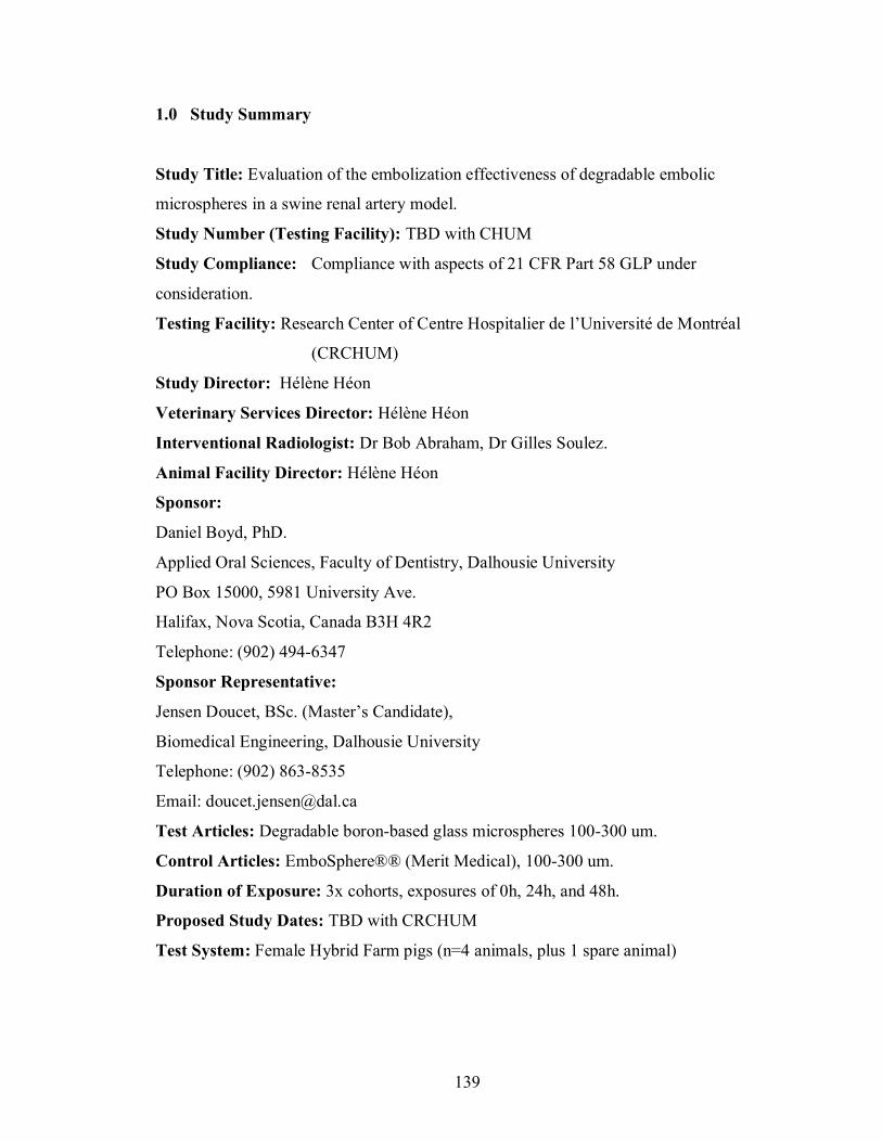
139
1.0 Study Summary
Study Title: Evaluation of the embolization effectiveness of degradable embolic
microspheres in a swine renal artery model.
Study Number (Testing Facility): TBD with CHUM
Study Compliance: Compliance with aspects of 21 CFR Part 58 GLP under
consideration.
Testing Facility: Research Center of Centre Hospitalier de l’Université de Montréal
(CRCHUM)
Study Director: Hélène Héon
Veterinary Services Director: Hélène Héon
Interventional Radiologist: Dr Bob Abraham, Dr Gilles Soulez.
Animal Facility Director: Hélène Héon
Sponsor:
Daniel Boyd, PhD.
Applied Oral Sciences, Faculty of Dentistry, Dalhousie University
PO Box 15000, 5981 University Ave.
Halifax, Nova Scotia, Canada B3H 4R2
Telephone: (902) 494-6347
Sponsor Representative:
Jensen Doucet, BSc. (Master’s Candidate),
Biomedical Engineering, Dalhousie University
Telephone: (902) 863-8535
Email: [email protected]
Test Articles: Degradable boron-based glass microspheres 100-300 um.
Control Articles: EmboSphere®® (Merit Medical), 100-300 um.
Duration of Exposure: 3x cohorts, exposures of 0h, 24h, and 48h.
Proposed Study Dates: TBD with CRCHUM
Test System: Female Hybrid Farm pigs (n=4 animals, plus 1 spare animal)
140
2.0 Signatures
Study Director
Date
Testing Facility Management
Date
Sponsor Representative
Date
141
3.0 Introduction
Dalhousie University researchers have developed a degradable embolic microsphere
indicated for the transarterial embolization of hypervascular tumors including uterine
leiomyoma. The product is targeted for commercialization as a Class IIb special control
medical device [1] and is subject to design control regulations pertaining to CFR 820.30
[2]. The primary benefit of the technology is enhanced patient safety coupled with the
ability to ameliorate chronic inflammation whilst permitting subsequent procedures to be
performed at the same target level of occlusion. This study is required to demonstrate the
embolization effectiveness of the technology, while also monitoring for particle migration,
per FDA guidance on the evaluation of microspherical embolization particles [1].
4.0 Definitions
a. Recanalization: reopening of a previously embolized vessel
b. Effective stasis: stasis of 50% contrast media for 5 or more cardiac pulsations
5.0 Purpose and Objectives
The primary objective of this study is to evaluate the embolization effectiveness of
the test article in a swine bilateral partial renal artery embolization model. This study will
use microspheres with a particle size distribution of 100-300 μm. The primary outcomes
for this study will be the assessment of; (i) embolization effectiveness, and (ii) test article
migration.
6.0 Test Materials
6.1 Description of the Test Article and Control Articles
The test article is a proprietary degradable glass microsphere developed by
Dalhousie researchers, and is indicated for the embolization of hypervascular tumors and
arteriovenous malformations including uterine leiomyoma. The test article will be supplied
in sterile individual packages (gamma sterilization, Nordion), having a particle size
distribution of 100-300 μm. The test article is designed to undergo hydrolytic degradation
in ca. 48h under physiological conditions. It is required that such pre-clinical evaluations
142
represent an elevated risk clinical scenario; accordingly, the particle size selected
represents the highest risk product for human use, with respect to particle migration and
non-target tissue embolization. The control article will be EmboSphere®® (100-300um).
6.2 Test Article Preparation The Sponsor will supply the Test Article (TA) in a sterile and ready to deliver form. The
TA will be delivered using an ABK delivery device, which will also be provided in a
sterile, ready to use state.
6.3 Test Article Accountability
While at the Testing Facility, accountability of the TA and delivery device equipment will
be maintained using the Facility’s standard operating procedures (SOP).
6.4 Test Article Characterization The Sponsor assumes responsibility for characterization of the TA.
6.5 Reserve Samples
No reserve samples of the TA will be retained by the Testing Facility.
6.6 Test Article Disposition Unused TA and delivery device equipment will be returned to the Sponsor at the end of
the study.
6.7 Storage Conditions The TA will be stored at room temperature (below 25°C), desiccated, and in a secure
location.
143
7.0 Animals
7.1 Model Description Species: Sus scrofa domesticus
Strain: Hybrid farm pigs
Condition: Non-diseased
Source:
Ferme Triporc
3250, rang Haut-de-la-Rivière
Sainte-Élizabeth, Qc
Age at implant: Age commensurate with weight.
Weight at implant: ca. 75-85kg
Sex: Female
Number: 4 animals (3 animals to receive TA, 1 animal to receive EmboSphere® control)
7.2 Justification 7.2.1 Selection of Animal Species and Model
A non-atherosclerotic swine model was chosen because the model has been used
extensively for angiographic/embolization studies resulting in a large volume of data on
the vascular response properties and its correlation to human vascular response [3,4,5]. It
is widely accepted that this type of study should be conducted in vivo, since no in vitro
model currently exists that can mimic the complex biological responses to embolization.
The porcine and human anatomy share important anatomic and physiologic characteristics
[4,6]. Furthermore, the porcine kidney model, recommended for use in preclinical studies
by the FDA has been widely used to evaluate embolization agents [7].
7.2.2 Number of Animals
This study comprises a pilot investigation to evaluate embolization effectiveness and
migration risk associated with biodegradable microspheres and will therefore utilize the
minimum number of animals possible to meet the study objectives. Three different time
cohorts will be analyzed: t=0, 24, and 48 h, to capture (i) embolization effectiveness and
initial particle distribution, (ii) particle distribution after partial degradation, and (iii)
144
recanalization and residual material at the degradation time of the material (t=48 h),
respectively. 1 animal, treated bilaterally will be included in each time cohort for the TA.
1 animal, treated bilaterally with the control article will be sacrificed at the 48h time point.
7.3 Animal Welfare and Institutional Animal Care and Use
Committee Review
The protocol will be reviewed and approved by the Testing Facility’s Institutional Animal
Care and Use Committee. The review will insure compliance with Canadian Council on
Animal Care (CCAC) regulations. If an animal entered on the study is found to have
contracted a condition or disease that might interfere with the purpose of the study, that
animal may be isolated and treated as prescribed by the Facility Veterinarian in
consultation with the Study Director.
7.4 Animal Husbandry 7.4.1 Environmental Acclimation
Prior to the interventional procedure, animals will have had environmental acclimation
for at least 7 days.
7.4.2 Testing Facility Housing
In the Testing Facility, animals will be housed in a room with suspended floors. Pens and
the room are washed daily with water and Virex ® (quaternary-based disinfectant cleaner).
All housing conditions meet or exceed the standards set forth in the Guide for the Care and
Use of Laboratory Animals [8].
7.4.3 Food
Standard dehydrated food pellets will be given to the animal. Fresh vegetables and certified
treats may be given at the facilities discretion.
145
7.4.4 Water
The animals receive reverse osmosis water. A bacterial load test (aerobic and anaerobic
heterotrophic bacterial count) is conducted every 4-6 weeks on the reservoir water. Twice
a year, a microbial burden tests (coliform, aerobic bacteria, total count) is conducted on
production water and end of distribution line. Once a year, a specific search for
pseudomonas and fungi is conducted as well as a determination of inorganic contaminants
such as heavy metals. Results of these analyses are on file at the Testing Facility and are
available to the Sponsor on request.
7.4.5 Contaminants
Certified diet will be fed daily to the animals. The food manufacturer will routinely analyze
the diet for nutritional components and environmental contaminants. Results of the
manufacturer’s analyses are retained on file at the Testing Facility. Details of nutritional
components and other manufacturing information will be made available to the Sponsor on
request.
7.4.6 Testing Facility Environment
Temperature and humidity will be monitored. Light cycle will be controlled by an
automated system.
7.5 Animal Identification The animals will be identified using unique Testing Facility number, which will be
indicated on its back with a marker and posted on the animals’ enclosures with cage cards.
At implantation the animals will be attributed a study number composed of the scheduled
time point and sequential number (e.g. 24-01 for 24 h animal #1). Ear tag number will be
recorded at implantation. If no tag is present, an ear tag with the same number as listed
above will be installed.
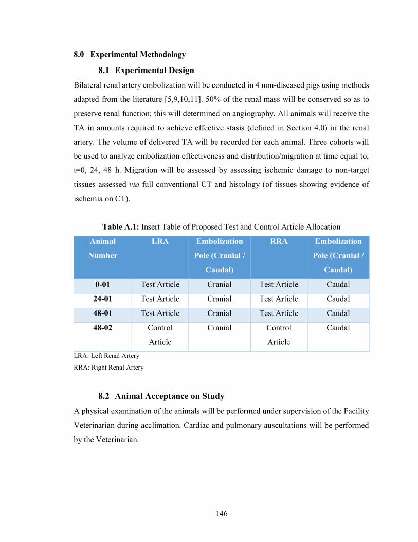
146
8.0 Experimental Methodology
8.1 Experimental Design Bilateral renal artery embolization will be conducted in 4 non-diseased pigs using methods
adapted from the literature [5,9,10,11]. 50% of the renal mass will be conserved so as to
preserve renal function; this will determined on angiography. All animals will receive the
TA in amounts required to achieve effective stasis (defined in Section 4.0) in the renal
artery. The volume of delivered TA will be recorded for each animal. Three cohorts will
be used to analyze embolization effectiveness and distribution/migration at time equal to;
t=0, 24, 48 h. Migration will be assessed by assessing ischemic damage to non-target
tissues assessed via full conventional CT and histology (of tissues showing evidence of
ischemia on CT).
Table A.1: Insert Table of Proposed Test and Control Article Allocation
Animal
Number
LRA Embolization
Pole (Cranial /
Caudal)
RRA Embolization
Pole (Cranial /
Caudal)
0-01 Test Article Cranial Test Article Caudal
24-01 Test Article Cranial Test Article Caudal
48-01 Test Article Cranial Test Article Caudal
48-02 Control
Article
Cranial Control
Article
Caudal
LRA: Left Renal Artery
RRA: Right Renal Artery
8.2 Animal Acceptance on Study
A physical examination of the animals will be performed under supervision of the Facility
Veterinarian during acclimation. Cardiac and pulmonary auscultations will be performed
by the Veterinarian.
147
8.3 Procedures Following Acceptance on the Study 8.3.1 Animal Monitoring and Examination
Upon assignment on the study and until sacrifice, animals will be monitored and observed
at least twice a day by a trained employee (cage side observation).
8.4 Pre-Interventional Procedures
8.4.1 Fasting
Fasting (food, including any dietary supplements) will be conducted prior to interventional
procedures and scheduled sacrifice. Water will not be withheld. After anesthesia and
appropriate recovery, animals may have access to food and water.
8.4.2 General Anesthesia and Fluid Therapy
Animals will be anesthetized with 25 mg/kg ketamine and 2 mg/kg xylazine administered
intramuscularly (IM). Animal weight will be recorded. Anesthesia induction for tracheal
intubation will be achieved with 1.66 mg/kg propofol injected intravenously (IV) via a
catheter in a vessel of the left or right ear or another appropriate vessel. Upon induction of
anesthesia, the subject animal will be intubated and supported with mechanical ventilation.
Isoflurane in oxygen will be administered to maintain a surgical plane of anesthesia. Blood
samples will be collected for clinical pathology (blood hematology and serum biochemistry
analysis described in the Clinical Pathology section). Pre-implant and pre-euthanasia
samples will be harvested during anesthesia for implant and terminal procedures. Blood
will be sampled prior to heparin administration and prior to initiation of intravenous fluid
therapy.
Clinical Pathology Samples: Harvested from the arterial sheath
An appropriate volume of blood will be collected as follows:
• Whole blood samples for hematology will be collected in EDTA tubes.
• Blood samples for biochemistry will be collected in serum clotting activator tubes,
processed, and the serum extracted.
All clinical pathology samples will be analyzed on collection day. IV fluid therapy will be

148
initiated and maintained throughout the procedure as per Operator’s judgment.
8.4.3 Antibiotic Therapy Preemptive Analgesia
Animals will be injected (IV) with one dose of antibiotic Cefazolin® (22mg/kg) at the
beginning of the procedure, and every 2 hours during the procedure to prevent
postoperative infections (excluding the t=0 cohort). Additional doses may be administered
at the discretion of the Facility Veterinarian in consultation with the Study Director but
will be reported to the Sponsor in the final report. To prevent pain sensitization and
minimize postoperative pain, a starting IV bolus of fentanyl (0.005 mg/kg), ketamine (0.2
mg/kg), and lidocaine (1 mg/kg) will be administered. A bag of 500 mL saline will then be
prepared with 0.5 mg fentanyl, 30 mg ketamine, and 300 mg lidocaine. This bag will be
administered during the entire surgical procedure at a flow rate of 5 mL/kg/h,
corresponding to a dose of 0.005 mg/kg/h fentanyl, 0.3 mg/kg/h ketamine, and 3 mg/kg/h
lidocaine.
8.4.4 Animal Positioning, Monitoring, and Surgical Site Preparation
The animal will be placed in dorsal recumbence, and the hair will be removed from the
access area (inguinal area). A rectal temperature probe will be inserted, and the temperature
will be monitored regularly. Electrocardiography limb-leads will be placed and
electrocardiography will be established. The access site will be prepared with topical
application of chlorhexidine, 70% isopropyl alcohol. The area will then be appropriately
draped to maintain a sterile field. A urinary catheter may be installed.
8.5 Interventional Procedures 8.5.1 Local Anesthesia, Vascular Access, Monitoring and
Anticoagulant Therapy and Surgical Approach
Parameters including blood oxygen saturation, heart rate, blood pressure (at least before
and after embolization), and temperature will be regularly monitored and manually
recorded in the raw data for each animal. Electrocardiogram will be monitored but not
recorded or retained. After induction of anesthesia, the left or right femoral artery will be
accessed under ultrasound guidance (echography, non-GLP). A 5 French introducer sheath
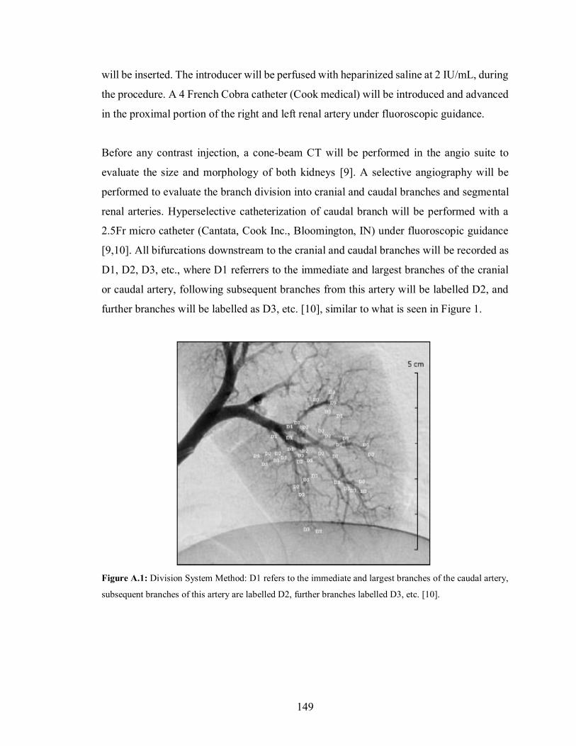
149
will be inserted. The introducer will be perfused with heparinized saline at 2 IU/mL, during
the procedure. A 4 French Cobra catheter (Cook medical) will be introduced and advanced
in the proximal portion of the right and left renal artery under fluoroscopic guidance.
Before any contrast injection, a cone-beam CT will be performed in the angio suite to
evaluate the size and morphology of both kidneys [9]. A selective angiography will be
performed to evaluate the branch division into cranial and caudal branches and segmental
renal arteries. Hyperselective catheterization of caudal branch will be performed with a
2.5Fr micro catheter (Cantata, Cook Inc., Bloomington, IN) under fluoroscopic guidance
[9,10]. All bifurcations downstream to the cranial and caudal branches will be recorded as
D1, D2, D3, etc., where D1 referrers to the immediate and largest branches of the cranial
or caudal artery, following subsequent branches from this artery will be labelled D2, and
further branches will be labelled as D3, etc. [10], similar to what is seen in Figure 1.
Figure A.1: Division System Method: D1 refers to the immediate and largest branches of the caudal artery,
subsequent branches of this artery are labelled D2, further branches labelled D3, etc. [10].
150
8.5.2 Procedure for Test Article Injection and Medical Imaging
The TA will be delivered using sterile saline to monitor fluoroscopic visibility of the
microspheres during injection. Fluoro-loops acquired during particle injection will be
stored. Microspheres will be injected until effective stasis is reached (defined in Section
4.0) [9]. The total volume of TA delivered will be recorded for each animal, as well as total
duration of the procedure for each animal, and the ease of use of the material. One shot
films and cone beam CT (in the angio suite) acquisitions will be acquired to evaluate the
distribution of the radiopaque microspheres in each pig. Post-embolization angiography
will be performed to the degree of vessel occlusion and document embolization efficacy.
All embolized arteries will be qualitatively evaluated for arterial perforation or rupture and
embolization effectiveness. Semi-quantitative analysis of the procedure will be done by
recording the level of occlusion of distal branches (both beginning and end of the bolus),
as per Section 8.5.1 [modified from 10,11].
Lidocaine may be administered to treat arrhythmia. Nitroglycerin may be administered to
treat
arterial vasospasm. Atropine sulfate may be administered to treat bradycardia. Additional
drugs may be administered as needed per the Operator’s orders.
8.5.3 Complications
Vessels that received an unsuccessful embolization (e.g., Operator’s error) unrelated to
either the Test or Control Articles may be replaced. Replacement may be achieved by
embolization in an alternative location or in an additional animal. An animal that receives
an unsuccessful embolization that may be life-threatening will be euthanized; no necropsy
will be performed.
8.5.4 Closure
All catheters and sheaths will be removed after embolization and completion of
angiography. Manual pressure will be applied to percutaneous puncture access until
hemostasis is achieved, followed by a purse string suture if deemed necessary by the
technician (glue may also be used). If cutdown access is performed, the femoral artery will
151
be ligated and inguinal incision layers will be sutured. Antibiotic ointment will be applied
to the wound.
8.6 Post-Interventional Procedures 8.6.1 Animal Monitoring
The anesthetic will be discontinued and the animals ventilated until the return of normal
respiration [9].
8.6.2 Postoperative Analgesia and Antibiotic Therapy
Buprenorphine (0.005mg/kg SC or IV) will be injected during recovery from anesthesia.
Meloxicam (0.3 mg/kg OS) will be given as post-operative analgesia, and buprenorphine
(0.005mg/kg SC) if pain persists regardless of the administration of meloxicam.
8.6.3 Veterinary Intervention and Care
At the discretion of the facility veterinary team, the drugs listed in Table 2 will be available.
If any of the following symptoms are observed, the animal will be euthanized as per Section
9.1:
During the procedure:
• Hemorrhage causing hypovolemia and risk of shock.
• Severe hypotension
• Unsuccessful embolization that may be life-threatening
During recovery & post-op:
• Respiratory distress non-responsive to supportive care
• Fever and prostration non-responsive to treatment
• Uncontrollable pain
• Uncontrollable bleeding
152
Table A.2: List of Drugs used in the Swine Renal Model Drug Drug type Purpose Administration Manufacturer
Anesthesia
Ketamine Dissociative
general
anesthetic and
NMDA
receptor
antagonist
Pre anesthesia IM Ketalean® Bimeda-MTC
Animal Health Inc. /Santé
animale INc. Cambrige , ON
N3C 2W4
Xylazine Alpha2-
adrenergic
agonist
Pre anesthesia IM Rompun®, Bayer Inc,
Mississauga, On, Canada
Propofol Anesthetic Anesthesia induction IV Diprivan 1%, AstraZeneca,
Mississauga, On, Canada
Isoflurane General
anesthetic,
inhalant
Anesthesia inhalation Forane, Baxter, Mississauga,
On, Canada)
Analgesia
Meloxicam Nonsteroidal
anti-
inflammatory
drug
Pain prevention SC or per os Metacam, Boehringer
Ingelheim, Ltd, Burlington,
On, Canada
Bupivacaine Local
anesthetic
Pain prevention Infusion
surgical site
Marcaine TM 0,50 %
hydrochloride injection USP
Hospira, Montreal, Qc,
Canada).
Ketamine Dissociative
general
anesthetic and
NMDA
receptor
antagonist
Pain prevention IV, bolus and
constant rate
infusion
Fentanyl citrate injection USP
Sandoz Canada Inc.
Lidocaine Antiarrhythmic/
Local
anesthetic
Pain prevention
Arrhythmia
prevention
IV, bolus and
constant rate
infusion
Lidocaine hydrochloride 2 %
(1.5 mg/kg/h, Hospira,
Montreal, Qc, Canada).
153
Drug Drug type Purpose Administration Manufacturer
Fentanyl Opioid Pain prevention IV, bolus and
constant rate
infusion
Fentanyl citrate injection USP
Sandoz Canada Inc.
Antibiotherapy
Cefazolin 1st generation
cepholosporin
Surgical prophylaxia IV Cefazolin for injection USP
Hospira Health Care
corporation St-Laurent , QC
Distributed by Apotex, Inc
Toronto Canada
Drugs that may be used during the procedure
Atropine Anticholinergic Treatment of
bradycardia
IV Atropine Sulfate injection USP
0.5mg/ml, Rafter 8 Products,
87 Skyline Crescent N.E.,
Calgary, Alberta, T2K 5X2
Rocuronium Non—
depolarizing
neuromuscular
blocker
To control breathing
during positive
ventilation
IV Bromide Injection, Sandoz
Canada Inc, Québec, Canada,
J4B 7K8
Phenylephrine Alpha-
adrenergic
agonist
Treatment of
hypotension
IV Bolus and
infusion
Phenylephrine Hcl Sandoz
10mg/ml (to treat hypotension)
Heparin Anticoagulant Thromboprophylaxis IV Heparin sodium injection USP
, 10 000 U/ml Sandoz ,
Canada Inc,
Buprenorphine Opioid Treatment of post-op
pain
SC or IV Vetergesic, Healthcare Ltd,
Hull, UK,
imported/distributed by GMD
Distribution Inc., Oakville,
ON, L6M 2W2 for: Champion
Alstoe Animal Health Inc.,
Whitby, Ontario, L1N 4V1
**Please note that the manufacturer may not be the same at the time of the study. The table will be amended
accordingly.
154
8.6.4 Additional Health Examinations
All animals will be monitored postoperatively at 2, 4, 12, 24, and 36 h post-op by a
trained employee (Testing Facility Veterinarian) and the following physical examinations
will be conducted:
i. Monitoring for signs of PES by taking temperature and inspecting for
malaise and pain
ii. Monitoring for signs of respiratory distress (O2 sats, hemoptysis,
respiration rate)
iii. Neurological examination of the lower limbs by conducting motor and
sensory exams
iv. Monitoring for signs of stroke (gait)
v. Monitoring for bleeding from the incision site
Clinical observations and findings during monitoring and examination will be included in
the final report.
8.6.5 Pre-Sacrifice Angiography for Assessment of Recanalization
Immediately prior to the scheduled sacrifice time point, angiography will be performed in
each animal using the methods outlined in Section 8.5.1 to assess the embolization status
of the renal vasculature, and position of embolic agent (both beginning and end of the
bolus). The following grade scale will be used for qualitative evaluation of recanalization:
0=no angiographic visible signs of arterial occlusion; 1=reduction of parenchymal staining
of the dependent territory; 2=reduction of the parenchymal staining and occlusion of the
supplying arcuate artery; 3=reduction of parenchymal staining, occlusion of the supplying
arcuate artery and occlusion of the feeding artery downstream of the catheter tip [9]. On
the baseline and follow-up cone beam CT the distribution of particles will be compared
and distal migration will be evaluated by measurement the distance with the renal capsule
[11].
155
9.0 Scheduled and Unscheduled Sacrifice
9.1 Euthanasia After final angiography, animals (in deep anesthesia) will be euthanized by a lethal
injection of sodium pentobarbital (euthanyl, rapid IV bolus, 108mg/kg). Death will be
confirmed by observation of asystole or ventricular fibrillation on the ECG.
9.2 Necropsy and Tissue Harvest
All treated animals will be subjected to necropsy, defined as gross examination of the
embolized arteries and kidneys, whole body (external surface), all orifices, thoracic and
abdominal cavities, and brain. Macroscopic pictures of all kidneys (ventral and dorsal
views) and any lesions will be taken with a ruler adjacent to the tissue and properly labeled
when possible (study number, animal number, organ). Kidneys will be evaluated
macroscopically and measured (length, width and depth) before fixation. Three regions
(cranial pole, middle region and caudal pole) will be evaluated individually on both
surfaces (ventral and dorsal surfaces) for ischemic changes. An ischemic change score will
be attributed to each region according to the following scale [8]:
• 0 = no macroscopic visible change;
• 1= discoloration of renal surface;
• 2 = Surface scars and retractions (corresponding to score 1 and 2);
• 3 = volume shrinkage of the respective area (corresponding to score 1, 2 and 3).
Each kidney with the renal artery (entire length) for each animal will be harvested free of
fat and cranial pole properly identified (with ink for example) in a consistent manner. To
allow good fixation, the kidney will be cut longitudinally in two equal sections (but not
completely to avoid damage of renal arteries before fixation) from the lateral aspect to the
hilus. Both kidneys from an animal will be placed in neutral-buffered formalin (NBF)
together and the right kidney will be identified with a suture on the hilus. Selected organs
[adrenal glands, lungs (samples of right middle lobe and left caudal lobe) will then be
placed into an appropriately labeled container filled with NBF. Lesions will be documented
and collected when feasible, immersion-fixed in NBF and processed for histology.
156
9.3 Unscheduled Sacrifice If an animal dies after recovery from embolization but prior to scheduled termination, the
Study Director will be notified immediately. After the discovery of the death, the following
procedures will be completed as soon as possible and same as per the Necropsy and Tissue
Harvest section above (9.2)
The animal may be replaced at the discretion of the Study Director and documentation of
the necropsy results may be included in the final report.
Moribund animals will be treated as described above (9.2), except that if possible a final
angiogram and blood samples will be taken for clinical pathology
10.0 Sample Processing and Analyses
10.1 Cone Beam CT
After necropsy, all harvested organs was be fixed in a box containing formaldehyde. A
cone-beam CT will be repeated on the explanted structures (Kidneys and accompanying
renal vasculature, lungs and any structures showing signs of ischemia or necrosis on
necropsy) to detect the best zones for samples.
10.2 Sample Preparation Following ex vivo imaging, the kidneys and renal vasculature will be sectioned, and
specimens will be embedded in paraffin and placed in an individual cassette. Six blocks of
one cm each will be taken in each kidney (four in infarcted areas upper pole and two
controls in the lower pole = 48 blocs). This will also be completed for any tissues where
possible infarction was located on necropsy or where microspheres were identified on cone
beam CT.
10.3 Micro-CT and Sectioning The blocks will be analyzed by micro-CT in order to choice the three best samples in each
kidney for histology analyses. The blocks will be sent to the histology platform for
157
processing and embedding. Ten sections will be cut 4-micrometer-thick tissue every mm
and will be mounted on a glass microscope slide.
10.4 Histological Staining and Tissue Processing Specimens will be stained with hematoxylin-eosin-saffron. Hematoxylin-eosin-saffron will
allow observation of occluded vessels and the locations of identified microspheres, which
will be labelled according to the different layer of renal vasculature as per Section 8.5.1
[10]
The number of microspheres in each layer will be recorded. The presence and extent of
embolus surrounding each embolic will also be documented, as will the presence of
inflammatory reaction and the types of inflammatory cells present (e.g. macrophages,
multinucleated giant cells, lymphocytes, eosinophils). Identified microspheres (or
microsphere fragments) will be measured (Section 10.4) and their shape will be
qualitatively assessed (spherical vs. irregular). Vessel wall rupture, formation of new
endothelium within the embolized vessel, and location of particles within the vessel walls
should extravasation occur will be assesses and the degree of vascular injury will be rated
as follows [12]:
0 = Internal elastic lamina (IEL) intact
1 = Disruption of the IEL without medial disruption.
2 = Disruption of the IEL with disruption of the media. The external elastic
lamina is intact.
3 = Disruption of the IEL, media, and external elastic lamina.
Additional sections may be cut and stained with different stains at the discretion of the
Study Pathologist with the approval of the Study Director.
158
Table A.3: Summary of sample preparation, histological staining and tissue
processing
4 swines
8 kidneys
0-01
Test Article
024-01
Test Article
48-01
Test Article
48-02
Control Article
Six blocks of 1 cm each will be taken in each kidney (4 in infarcted areas upper pole
and 2 control in the lower pole). 6 x 8 = 48
The blocks will be processing and embedded in paraffin. A micro-CT will be made to
choice the 3 relevant blocks.
10 sections of about 4 μm will be sliced on each block every mm and put on slides
Specimens will be stained with hematoxylin-eosin-saffron. 4 blocs x 10 = 240 slides
The slides will be scanned at 40x
1 cm block will be taken for all tissues where the infarction has been located during
necropsy or when microspheres have been identified on imaging.
10.5 Digital Imaging All slides will be scanned for Software analysis. A low and a high-magnification image of
each produced histology slide will be digitally captured and included in the Final Report.
The magnification of each image will be obtained with a scale bar. Additional images will
be captured and included in the Pathology Report to illustrate histopathology findings as
per Study Pathologist, Study Director and/or Sponsor request following issuance of an
Amendment.
10.6 Histological Measurements Measurements of the scanned slides will be performed using a Software Program. All
identified microspheres will be measured to approximate vessel diameter and indicate the
extent of degradation. The number and size of all microspheres in the analyzed specimens
will be documented, along with their qualitative distribution (aggregates vs. individual)
and position (renal artery division).
159
11.0 Reporting
A Draft Report will be provided to the Sponsor and any revisions to the study report
mutually agreed upon by the Sponsor and Study Director and/or Pathologist will be
incorporated in the Final Report. At the end of the revision process, the Report will be
signed and considered final. The study Final Report will include a summary of the
objectives and procedures of the study, description of the TA used, the methods used for
histological processing, and a discussion of any circumstances that may have altered or
affected the integrity of the data. Any corrections or additions to the Final Report will be
in the form of a report amendment.
160
12.0 References
[1] Food and Drug Administration. Class II Special Controls Guidance Document:
Vascular
and Neurovascular Embolization Devices. Dec 2004.
[2] Food and Drug Administration, Department of Health and Human Services. 21 C.F.R.
§ 820.30 2012.
[3] Targeting and Recanalization after Embolization with Calibrated Resorbable
Microspheres versus Hand-cut Gelatin Sponge Particles in a Porcine Kidney Model.
Journal of vascular and interventional radiology. 24(9), pp. 1391 -1398. 2013.
[4] Giraud S., Favreau F., Chatauret N., Thuillier R., Maiga S., Hauet T. Contribution of
Large Pig for Renal Ischemia-Reperfusion and Transplantation Studies: The Preclinical
Model. Journal of Biomedicine and Biotechnology. 2011;2011:532127.
[5] Stampfl, S. et al. Experimental Evaluation of Early and Long-Term Effects of
Microparticle Embolization in Two Different Mini-Pig Models. Part I: Kidney.
CardioVascular and Interventional Radiology . 30, pp. 257–267. 2007.
[6] Pereira-Sampaio M., Favorito L.A., Henry R, Sampaio F.J. Proportional analysis of pig
kidney arterial segments: differences from the human kidney. J Endourol. 21(7), pp. 784-
788. 2007.
[7] Owen, R.J., Nation, P.N., Polakowski, R., Biliske, J.A., Tiege, P.B., Griffith, I.J. A
Preclinical Study of the Safety and Efficacy of Occlusion 500 Artificial Embolization
Device in Sheep. Cardiovascular Interventional Radiology. 25, pp. 636-644. 2012.
[8] Guide for the Care and Use of Laboratory Animals. National Research Council.
National Academy Press. 1996.
161
[9] Kehoe, S., Amensag, S., Looney, M., Abraham, R.J., Boyd, D. “Imageable” Zinc-
Silicate Glass Microspheres for Transarterial Embolization: A Renal Artery Embolization
Study. Biomedical Glasses. 1, pp. 70-79. 2015.
[10] Zehtabi F., Ispas-Szabo P., Djerir D., Sivakumaran L., Annabi B., Soulez G.,
Mateescu M.A., Lerouge S. Chitosan-doxycycline hydrogel: An MMP
inhibitor/sclerosing embolizing agent as a new approach to endoleak prevention and
treatment after endovascular aneurysm repair. Acta Biomaterialia. 64, pp. 94–105. 2017.
[11] Kilani M.S., Zehtabi F., Lerouge S., Soulez G., Bartoli J.M., Vidal V., Badran M.F.
New Alcohol and Onyx Mixture for Embolization: Feasibility and Proof of Concept in
Both In Vitro and In Vivo Models. Cardiovasc Intervent Radiol. 40, pp. 735–743. 2017.
Related Documents